Differential diagnosis of hematolymphoid tumors composed of medium-sized cells. Brian Skinnider B.C. Cancer Agency, Vancouver General Hospital
|
|
|
- Dwain Chad Leonard
- 5 years ago
- Views:
Transcription
1 Differential diagnosis of hematolymphoid tumors composed of medium-sized cells Brian Skinnider B.C. Cancer Agency, Vancouver General Hospital

2
3 Lymphoma classification Lymphoma diagnosis starts with morphologic differential diagnosis based on cell size and architecture Small cells, diffuse or follicular Medium-sized cells Large cells Large cells in a reactive background
4 Hematolymphoid tumors composed of Burkitt lymphoma medium-sized cells B cell lymphoma, unclassifiable, with features intermediate between DLBCL and BL Lymphoblastic lymphoma Myeloid sarcoma Blastoid mantle cell lymphoma SLL/CLL with prominent proliferation centers T/NK cell lymphomas
5 Tumors with medium-sized cells These are uncommon diagnosis and may not be readily identified Diseases requiring immediate diagnosis and treatment: Burkitt lymphoma Double-hit lymphoma Lymphoblastic lymphoma Myeloid sarcoma
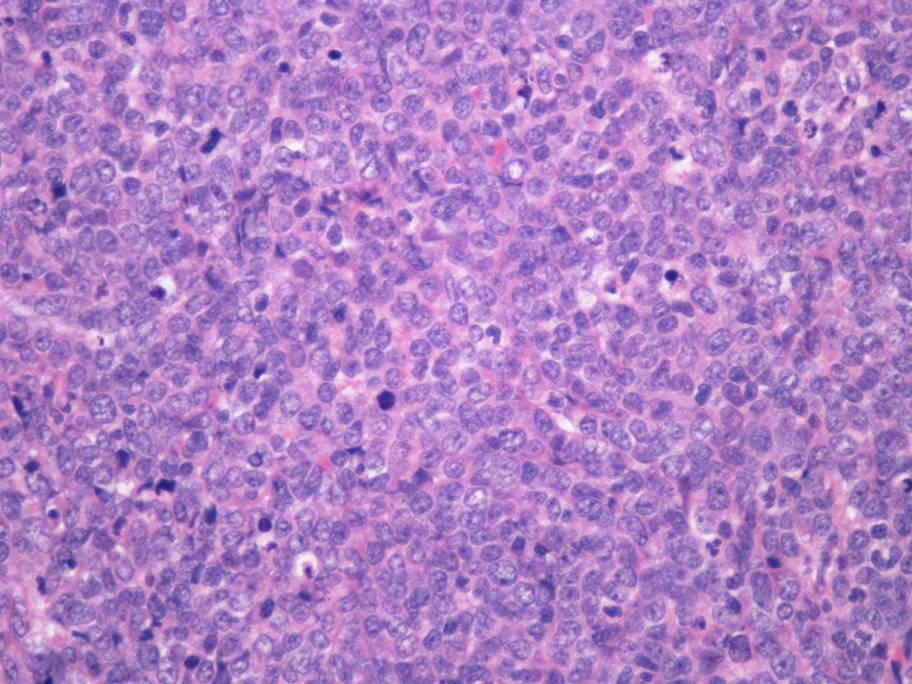
6 Cell size How do you decide cell size? WHO 2008 classification: DLBCL: nuclear size equal to or exceeding normal macrophage nuclei Burkitt lymphoma: medium-size cells (nuclei similar or smaller to those of histiocytes)

7 Nuclei of medium-sized cells have same size as histiocyte nuclei histiocyte
8 Burkitt Lymphoma
9 Burkitt lymphoma Three clinical variants: Endemic Sporadic Immunodeficiency-associated Sporadic Burkitt lymphoma Predominantly in children/young adults Rapidly growing bulky disease Often extranodal (GI, ovaries, kidney, breast) Lymph node involvement more common in adults
10 Burkitt lymphoma: Diagnosis There is no specific diagnostic marker for Burkitt lymphoma Diagnosis relies on combination of: Morphology Immunophenotype Cytogenetics

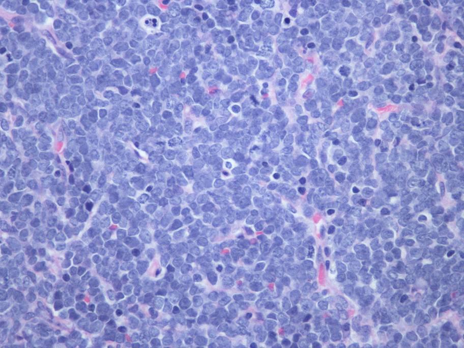
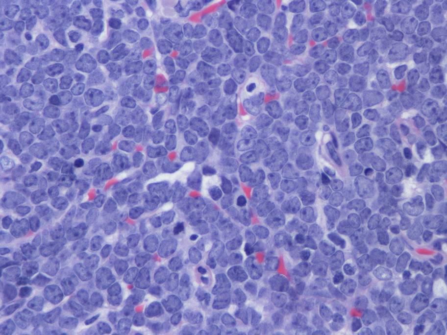
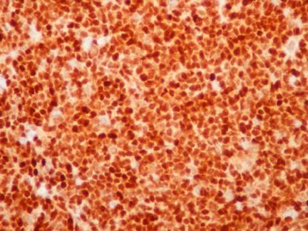
11 Burkitt lymphoma

12 Burkitt lymphoma
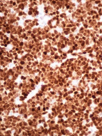
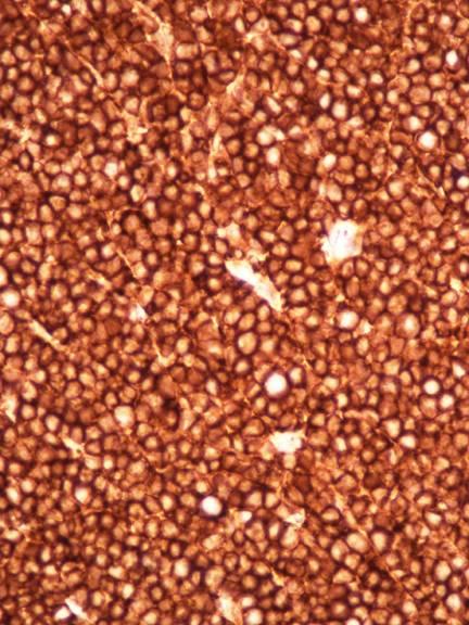
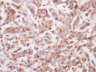
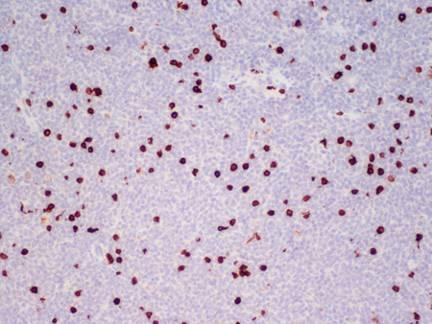
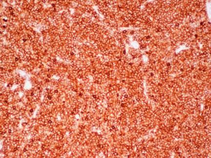
13 Burkitt lymphoma: phenotype Mature B cell (CD20 with clonal surface light chain) Tdt negative CD10, Bcl-6 positive Bcl-2 negative or very weakly positive Mib-1 proliferation rate close to 100% EBV+ in ~40% of sporadic cases


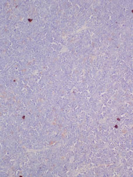
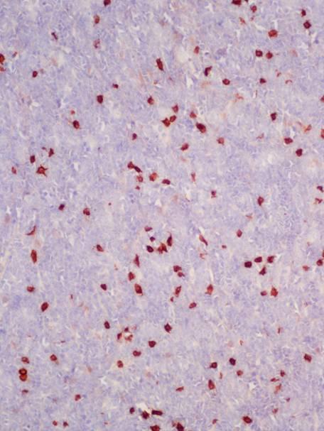
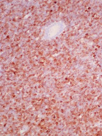
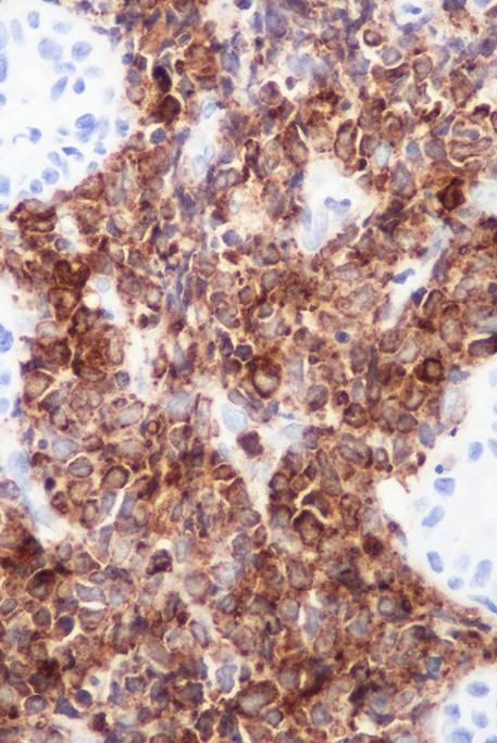
14 Burkitt lymphoma: IHC CD10 CD20 Bcl6


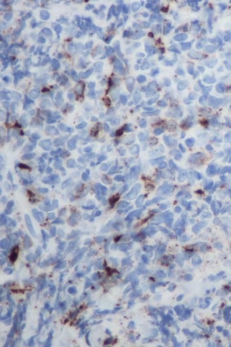
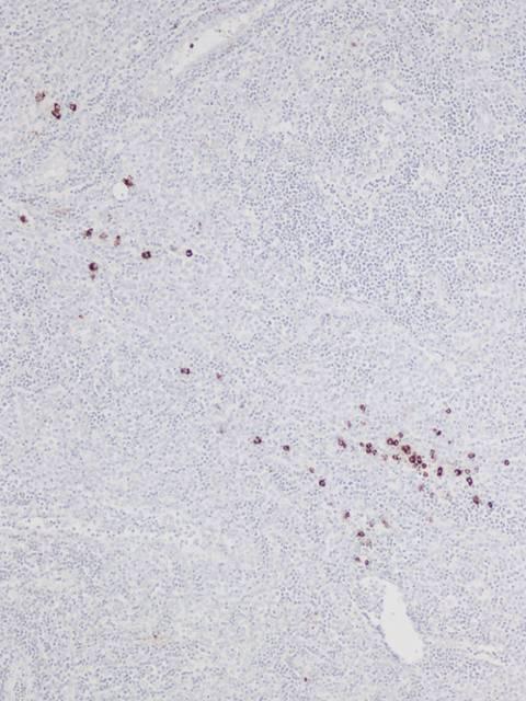
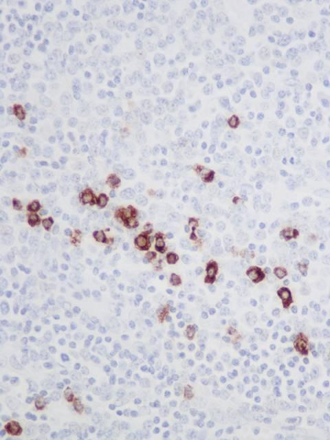
15 Burkitt lymphoma: IHC Bcl2 Mib1 Tdt

16 Burkitt lymphoma: Cytogenetics Characterized by translocations involving MYC gene on chromosome 8q24 Typically involves IG genes Can be detected by FISH MYC rearrangement is not specific for Burkitt lymphoma translocation Normal MYC MYC breakapart probes
17 Burkitt lymphoma may be negative for MYC rearrangement FISH probes may not cover all possible breakpoints May represent a true absence of MYC rearrangement Recurrent 11q abnormalities associated with lymphomas with gene expression pattern of Burkitt lymphoma and absence of MYC rearrangement Blood 2014;123:1187 Diagnosis of BL can be made without MYC rearrangement, but classical morphology and immunophenotype must be present
18 Burkitt lymphoma in small biopsies Core biopsies, endoscopic GI biopsies May not be easily recognized: Starry sky appearance not prominent Biopsies often crushed Often poor fixation Always consider Burkitt lymphoma in small biopsies that show an aggressive B cell lymphoma Use Bcl-2 and Mib-1 to help


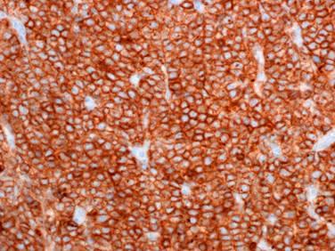
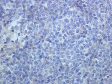
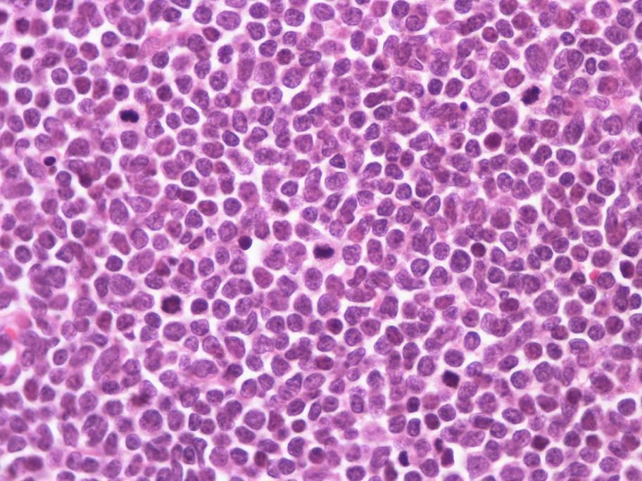
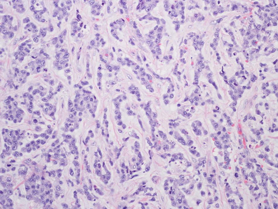
19 Gastric mass, 21 yo male

20 Gastric mass, 21 yo male


21 Gastric mass, 21 yo male CD20 CD10 Bcl2 MIB1 FISH: positive for MYC gene rearrangement
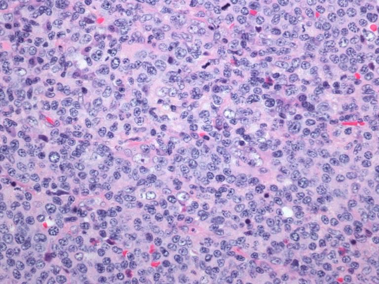
22 Burkitt lymphoma CLINICAL - young age - rapidly growing mass MORPHOLOGY - uniform medium sized cells - high mitotic rate - starry sky appearance PHENOTYPE - CD20 pos, CD10/Bcl6 pos - Mib-1 ~100% - Bcl-2 neg GENETICS - MYC translocation
23 B cell lymphoma, unclassifiable, with features between DLBCL and BL Some lymphomas can show transitional features between Burkitt lymphoma and diffuse large B cell lymphoma Previously known as Burkitt-like lymphoma Heterogenous entity, not uniformly treated Clinically important to recognize double hit lymphomas (MYC rearrangement with BCL2 or BCL6 rearrangement) Proposed classification in upcoming WHO classification: High grade B cell lymphoma: 1) High grade B cell lymphoma with MYC and BCL2 or BCL6 rearrangements (double hit lymphoma) 2) High grade B cell lymphoma, NOS
24 B cell lymphoma, unclassifiable, with features between DLBCL and BL Not a distinct morphologic or immunophenotypic entity Diagnosis should be made in: lymphomas that have the immunophenotype of BL, but are too morphologically pleomorphic lymphomas that have the morphology of BL, but one or more inconsistent findings such as: strong BCL2 expression proliferation rate significantly less than 100% BCL2 or BCL6 gene rearrangements 30-50% will be double hit lymphoma
25 Double hit lymphomas 1999: Cytogenetics in Burkitt-like lymphomas identified subset of cases with MYC rearrangement and t(14;18) Very poor prognosis Overall survival other Double hit c-myc JCO 1999;17:1558
26 Double hit lymphoma All B cell lymphomas, unclassifiable between DLBCL and BL should be examined for MYC, BCL2, BCL6 rearrangements by FISH Double hit lymphoma can also be seen in ~5% of diffuse large B cell lymphomas
")
27 B cell lymphoma, unclassified, DLBCL/BL (Double hit lymphoma)


28 Double hit lymphoma, confirmed by FISH analysis Bcl2 Mib1

29 Burkitt lymphoma Intermediate between BL & DLBCL
30 Lymphoblastic lymphoma Lymphoblastic neoplasm presenting in tissue with less than 25% blasts in bone marrow Most are T cell Rare cases are B cell (typically present as lymphoblastic leukemia)
31 T-lymphoblastic lymphoma Highly aggressive disease Children, adolescents; M>F Mediastinal involvement common Any other nodal or extranodal site (skin, tonsil, CNS, testis) Morphology: Lymphoblasts with high mitotic rate +/- starry sky appearance May have a more mature appearance if not wellpreserved
32
33
34
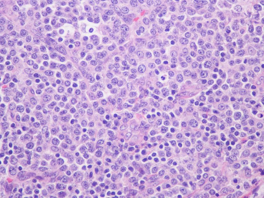
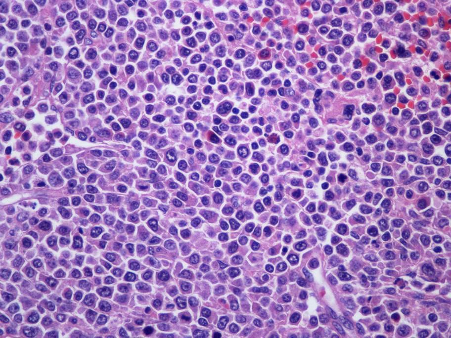
 Other markers of immature cells (CD99, CD34, CD1a) Usually lack B-cell and myeloid")
35 Immunophenotype T cell markers: CD3 (usually cytoplasmic) Polyclonal CD3 antibody is not T cell specific (detects zeta chain) CD4, CD8 frequently coexpressed Lymphoblastic markers: Usually Tdt positive (~95% of cases) Other markers of immature cells (CD99, CD34, CD1a) Usually lack B-cell and myeloid markers CD3
36 Tdt CD99
37 B cell lymphoblastic lymphoma Morphology Lymphoblasts Immunophenotype Tdt, CD10, CD19, CD22, CD79a, PAX5 PAX5 most sensitive and specific IHC marker for B cell lineage May be negative for CD20 Cytogenetics Further classified based on cytogenetic abnormalities Flow cytometry, cytogenetics recommended FISH on paraffin tissue may be performed
38 2008 WHO Classification

39 Small bowel mass, 32 yo male
40 CD20 CD3

41 CD79a PAX5

42 Tdt Bone marrow negative No other tumor present following resection No material for cytogenetics Bone marrow involved at recurrence several months later Confirmed diagnosis of B cell lymphoblastic leukemia/lymphoma, NOS
43 Myeloid sarcoma Myeloid sarcoma (2008 WHO classification): tumor mass consisting of myeloid blasts with or without maturation, occurring outside the bone marrow Also known as extramedullary myeloid tumor, granulocytic sarcoma, chloroma Sarcoma designation may confuse some clinicians Represents tissue involvement by acute myeloid leukemia
44 Myeloid sarcoma Several clinical scenarios: Concurrent diagnosis with AML involving bone marrow Isolated primary diagnosis, without bone marrow involvement Recurrent disease Previous history of myeloproliferative or myelodysplastic disorder
45 Myeloid sarcoma Any site can be involved (skin, lymph node, GI tract, bone, testis) May rarely involve the female genital tract: Am J Clin Pathol 2006;125:783 Report of 11 cases of myeloid sarcoma involving the gynecologic tract 5 cases presented as isolated mass Uterus was the most common site (5 in corpus; 3 in cervix) Cytology ranged from immature to differentiated
46 Myeloid sarcoma Wide morphologic spectrum More likely to have myelomonocytic or monoblastic differentiation Can resemble lymphoblastic lymphoma Can mimic mature lymphomas Clues to the diagnosis: Looks lymphoid, but: Streaming Amphophilic cytoplasm Eosinophilic precursors CD20, CD3 negative
47
48
49

50 AML, skin biopsy

51
52
53 Myeloid sarcoma: IHC Immunohistochemical markers in myeloid sarcoma, in decreasing frequency: CD68 (KP1) Myeloperoxidase CD117 CD99 CD68 (PG-M1) Lysozyme CD34 Tdt
54 Myeloid sarcoma: IHC CD68 staining depends on antibody used: KP1 identifies virtually all myeloid sarcomas and other malignancies PG-M1 correlates with monocytic differentiation Myeloperoxidase is not positive in every case Several markers are nonspecific: CD117, CD99, Tdt CD34 is positive in approximately 50% of cases
55 Myeloid sarcoma: IHC AML is not classified based on tissue biopsy AML best characterized by flow cytometry and cytogenetics/fish Bone marrow aspirate and biopsy performed for definitive classification In cases without bone marrow involvement, consider FNA to obtain material for flow cytometry and cytogenetics
56 2008 WHO Classification

57 myeloperoxidase

58 CD68, KP1 CD68, PGM1
59 myeloperoxidase


60 lysozyme CD68, KP1 CD117 CD34 Bone marrow: Acute myeloid leukemia with t(9;11)(p22;q23)
61


62 CD45 MPO lysozyme CD68, KP1 Bone marrow: AML with myelodysplasia-related changes
63 Immunophenotyping in lymphoblastic and myeloid tumors Flow cytometry is best method Bone marrow sample or FNA of extramedullary tumor IHC provides a more limited panel Tumors may have some overlap: PAX5 in AML with t(8;21) Single myeloid marker in B lymphoblastic leukemia Do wide IHC panel: Myeloid (MPO, CD117), monocytic (lysozyme, CD68), B cell (PAX5), T cell (CD3), Tdt Acute Leukemia Immunohistochemistry, Arch Pathol Lab Med 2008;132:462
64 Lymphomas with medium-sized cells Burkitt lymphoma B cell lymphoma, unclassified (BL/DLBCL) Lymphoblastic lymphoma Myeloid sarcoma Blastoid mantle cell lymphoma SLL/CLL with prominent proliferation centers T/NK cell lymphomas
65 Mantle cell lymphoma Typical mantle cell lymphoma is composed of small irregular lymphocytes May transform into blastoid mantle cell lymphoma, morphologically resembling lymphoblastic lymphoma Can occur in patients with a past history of mantle cell lymphoma, or occur as a de novo presentation CD20, CD5, cyclin D1, SOX11 positive High proliferation rate Associated with more aggressive disease

66 Typical mantle cell lymphoma

67 CD20 CD3 CD5 Cyclin D1

68 MCL, blastoid variant
69 T-lymphoblastic lymphoma vs. Mantle cell lymphoma T-lymphoblastic lymphoma Mantle cell lymphoma Age Young Older CD3 + - CD CD5 + + Tdt + - Cyclin D1, SOX11 - +
70 SLL/CLL Diffuse infiltrate of small mature lymphocytes Most cases contain proliferation centers containing prolymphocytes and paraimmunoblasts, medium sized cells with single prominent nucleoli Proliferation centers may sheet out Should not be confused with transformation to diffuse large B cell lymphoma (Richter transformation)
71

72 SLL/CLL: Proliferation center prolymphocytes and paraimmunoblasts

73 SLL/CLL with prominent proliferation centers

74 SLL/CLL with prominent proliferation centers
75 Survival from biopsy according to the histological patterns of non-accelerated CLL, accelerated CLL and DLBCL transformation: median survival 76 months, 34 months and 4.3 months, respectively (P<0.001). Accelerated CLL: Proliferation centers broader than a 20x field OR High proliferation rate: >2.4 mitoses/ proliferation center OR Ki-67 >40% in proliferation center Eva Giné et al. Haematologica 2010;95: by Ferrata Storti Foundation
76 T/NK lymphomas T/NK cell lymphomas can have a wide morphologic range, including a predominance of medium-sized cells Diagnosis based on expression of T/NK cell markers Exclude lymphoblastic lymphoma in monomorphous T cell lymphomas with high proliferation rate (may have a more mature morphology)
77 PTCL, NOS

78 Extranodal NK/T cell lymphoma, nasal type
79 Summary Be aware of the differential diagnosis of medium-sized cells, especially diagnoses that require immediate treatment It is important to differentiate Burkitt lymphoma from B cell lymphoma, unclassified (BL/DLBCL), by immunophenotypic and FISH studies Consider myeloid sarcoma in tumors that have lymphoid morphology and are negative for CD20 and CD3
80 Summary Perform a broad IHC panel for myeloid and lymphoblastic tumors and be aware of exceptions, such as: Some T-LBLs may be Tdt negative Some myeloid sarcomas may be MPO negative Be aware of unusual morphologic variants of mantle cell lymphoma and SLL/CLL
Aggressive B-Cell Lymphomas
 Aggressive B-cell Lymphomas Aggressive B-Cell Lymphomas Stephen Hamilton Dutoit Institute of Pathology Aarhus Kommunehospital B-lymphoblastic lymphoma Diffuse large cell lymphoma, NOS T-cell / histiocyte-rich;
7 Omar Abu Reesh. Dr. Ahmad Mansour Dr. Ahmad Mansour
 7 Omar Abu Reesh Dr. Ahmad Mansour Dr. Ahmad Mansour -Leukemia: neoplastic leukocytes circulating in the peripheral bloodstream. -Lymphoma: a neoplastic process in the lymph nodes, spleen or other lymphatic
DETERMINATION OF A LYMPHOID PROCESS
 Chapter 2 Applications of Touch Preparation Cytology to Intraoperative Consultations: Lymph Nodes and Extranodal Tissues for Evaluation of Hematolymphoid Disorders INTRODUCTION As discussed in Chap. 1,
Immunopathology of Lymphoma
 Immunopathology of Lymphoma Noraidah Masir MBBCh, M.Med (Pathology), D.Phil. Department of Pathology Faculty of Medicine Universiti Kebangsaan Malaysia Lymphoma classification has been challenging to pathologists.
Gray Zones and Double Hits Distinguishing True Burkitt Lymphoma from Other High-Grade B-NHLs Burkitt Lymphoma Burkitt-Like Lymphoma DLBCL Patrick Tres
 Gray Zones and Double Hits Distinguishing True Burkitt Lymphoma from Other High-Grade B-NHLs Burkitt Lymphoma Burkitt-Like Lymphoma DLBCL Patrick Treseler, MD, PhD University of California San Francisco
Burkitt lymphoma. Sporadic Endemic in Africa associated with EBV Translocations involving MYC gene on chromosome 8
 Heme 8 Burkitt lymphoma Sporadic Endemic in Africa associated with EBV Translocations involving MYC gene on chromosome 8 Most common is t(8;14) Believed to be the fastest growing tumor in humans!!!! Morphology
From Morphology to Molecular Pathology: A Practical Approach for Cytopathologists Part 1-Cytomorphology. Songlin Zhang, MD, PhD LSUHSC-Shreveport
 From Morphology to Molecular Pathology: A Practical Approach for Cytopathologists Part 1-Cytomorphology Songlin Zhang, MD, PhD LSUHSC-Shreveport I have no Conflict of Interest. FNA on Lymphoproliferative
HIGH GRADE B-CELL LYMPHOMA DAVID NOLTE, MD (PGY-2) HUSSAM AL-KATEB, PHD, FACMG DEBORAH FUCHS, MD
 HUSSAM AL-KATEB, PHD, FACMG DEBORAH FUCHS, MD") HIGH GRADE B-CELL LYMPHOMA DAVID NOLTE, MD (PGY-2) HUSSAM AL-KATEB, PHD, FACMG DEBORAH FUCHS, MD OUTLINE High grade B-cell lymphoma with MYC and BCL2 and/or BCL6 rearrangements Patient presentation 2008/2016
HIGH GRADE B-CELL LYMPHOMA DAVID NOLTE, MD (PGY-2) HUSSAM AL-KATEB, PHD, FACMG DEBORAH FUCHS, MD OUTLINE High grade B-cell lymphoma with MYC and BCL2 and/or BCL6 rearrangements Patient presentation 2008/2016
Non-Hodgkin lymphomas (NHLs) Hodgkin lymphoma )HL)
 Hodgkin lymphoma )HL)") Non-Hodgkin lymphomas (NHLs) Hodgkin lymphoma )HL) Lymphoid Neoplasms: 1- non-hodgkin lymphomas (NHLs) 2- Hodgkin lymphoma 3- plasma cell neoplasms Non-Hodgkin lymphomas (NHLs) Acute Lymphoblastic Leukemia/Lymphoma
Non-Hodgkin lymphomas (NHLs) Hodgkin lymphoma )HL) Lymphoid Neoplasms: 1- non-hodgkin lymphomas (NHLs) 2- Hodgkin lymphoma 3- plasma cell neoplasms Non-Hodgkin lymphomas (NHLs) Acute Lymphoblastic Leukemia/Lymphoma
EQA SCHEME CIRCULATION 33 EDUCATIONAL SLIDES DR GRAEME SMITH MONKLANDS DGH
 EQA SCHEME CIRCULATION 33 EDUCATIONAL SLIDES DR GRAEME SMITH MONKLANDS DGH CASE E1 M: 68 yrs Left destructive sinonasal lesion.?lymphoma?adenocarcinoma CD20 CD10 BCL6 MIB1 Answers Diffuse large B cell
Hepatic Lymphoma Diagnosis An Algorithmic Approach
 Hepatic Lymphoma Diagnosis An Algorithmic Approach Ryan M. Gill, M.D., Ph.D. University of California, San Francisco PLEASE TURN OFF YOUR CELL PHONES Disclosure of Relevant Financial Relationships USCAP
Aggressive B-cell Lymphoma 2013
 Aggressive B-cell Lymphoma 2013 Diffuse Large B-Cell Lymphoma Burkitt Lymphoblastic lymphoma Gray zone Intermediate DLBCL/HL Intermediate BL/DLBCL Diffuse Large B-cell lymphoma Common morphology: diffuse
Many of the hematolymphoid disorders are derived
 REVIEW ARTICLE Practical Immunohistochemistry in Hematopathology: A Review of Useful Antibodies for Diagnosis Ji Lu, MD and Karen L. Chang, MD Abstract: This review article offers some useful panels of
Pearls and pitfalls in interpretation of lymphoid lesions in needle biopsies
 Pearls and pitfalls in interpretation of lymphoid lesions in needle biopsies Megan S. Lim MD PhD University of Pennsylvania October 8, 2018 Objectives To understand how the trend toward less invasive lymph
Aggressive B-cell Lymphomas
 Neoplastic Hematopathology Update 2018 Aggressive B-cell Lymphomas Raju K. Pillai City of Hope National Medical Center I do not have any disclosures Disclosures Outline New entities and changes in WHO
FOLLICULARITY in LYMPHOMA
 FOLLICULARITY in LYMPHOMA Reactive Follicular Hyperplasia Follicular Hyperplasia irregular follicles Follicular Hyperplasia dark and light zones Light Zone Dark Zone Follicular hyperplasia MIB1 Follicular
Small B-cell (Histologically Low Grade) Lymphoma
 Lymphoma") Frequency of Lymphoid Neoplasms Small B-cell (Histologically Low Grade) Lymphoma Stephen Hamilton-Dutoit Institute of Pathology Aarhus University Hospital B-cell neoplasms 88% Diffuse large B-cell lymphoma
Frequency of Lymphoid Neoplasms Small B-cell (Histologically Low Grade) Lymphoma Stephen Hamilton-Dutoit Institute of Pathology Aarhus University Hospital B-cell neoplasms 88% Diffuse large B-cell lymphoma
Lymphoma: What You Need to Know. Richard van der Jagt MD, FRCPC
 Lymphoma: What You Need to Know Richard van der Jagt MD, FRCPC Overview Concepts, classification, biology Epidemiology Clinical presentation Diagnosis Staging Three important types of lymphoma Conceptualizing
Contents. vii. Preface... Acknowledgments... v xiii
 Contents Preface... Acknowledgments... v xiii SECTION I 1. Introduction... 3 Knowledge-Based Diagnosis... 4 Systematic Examination of the Lymph Node... 7 Cell Type Identification... 9 Cell Size and Cellularity...
Lymphoma Update: Lymphoma Update: What s Likely to be New in the New WHO. Patrick Treseler, MD, PhD University of California San Francisco
 Lymphoma Update: What s Likely to be New in the New WHO Blood 127:2375; 2016 Patrick Treseler, MD, PhD University of California San Francisco Lymphoma Update: What IS New in the New WHO! Patrick Treseler,
Case 3. Ann T. Moriarty,MD
 Case 3 Ann T. Moriarty,MD Case 3 59 year old male with asymptomatic cervical lymphadenopathy. These images are from a fine needle biopsy of a left cervical lymph node. Image 1 Papanicolaou Stained smear,100x.
LYMPHOMAS an overview of some subtypes of NHLs
 One of the confusing aspects of the lymphoid neoplasms concerns the use of the descriptive terms "leukemia" and "lymphoma." LYMPHOMAS an overview of some subtypes of NHLs Leukemia is used for lymphoid
Mixed Phenotype Acute Leukemias
 Mixed Phenotype Acute Leukemias CHEN GAO; AMY M. SANDS; JIANLAN SUN NORTH AMERICAN JOURNAL OF MEDICINE AND SCIENCE APR 2012 VOL 5 NO.2 INTRODUCTION Most cases of acute leukemia can be classified based
Aggressive B cell Lymphomas
 Aggressive B cell Lymphomas I have nothing to disclose. Disclosures Raju K. Pillai City of Hope National Medical Center Outline WHO 2016 Classification Large B cell Lymphomas New entities and changes in
3/24/2017 DENDRITIC CELL NEOPLASMS: HISTOLOGY, IMMUNOHISTOCHEMISTRY, AND MOLECULAR GENETICS. Disclosure of Relevant Financial Relationships
 DENDRITIC CELL NEOPLASMS: HISTOLOGY, IMMUNOHISTOCHEMISTRY, AND MOLECULAR GENETICS Jason L. Hornick, M.D., Ph.D. Director of Surgical Pathology and Immunohistochemistry Brigham and Women s Hospital Professor
Classification of Hematologic Malignancies. Patricia Aoun MD MPH
 Classification of Hematologic Malignancies Patricia Aoun MD MPH Objectives Know the basic principles of the current classification system for hematopoietic and lymphoid malignancies Understand the differences
Methods used to diagnose lymphomas
 Institut für Pathologie Institut für Pathologie Methods used to diagnose lymphomas Prof. Dr.Med. Leticia Quintanilla-Fend Molecular techniques NGS histology Cytology AS-PCR Sanger seq. MYC Immunohistochemistry
High grade B-cell lymphomas (HGBL): Altered terminology in the 2016 WHO Classification (Update of the 4 th Edition) and practical issues Xiao-Qiu Li,
: Altered terminology in the 2016 WHO Classification (Update of the 4 th Edition) and practical issues Xiao-Qiu Li,") High grade B-cell lymphomas (HGBL): Altered terminology in the 2016 WHO Classification (Update of the 4 th Edition) and practical issues Xiao-Qiu Li, M.D., Ph.D. Fudan University Shanghai Cancer Center
High grade B-cell lymphomas (HGBL): Altered terminology in the 2016 WHO Classification (Update of the 4 th Edition) and practical issues Xiao-Qiu Li, M.D., Ph.D. Fudan University Shanghai Cancer Center
Hematology Unit Lab 2 Review Material
 Objectives Hematology Unit Lab 2 Review Material - 2018 Laboratory Instructors: 1. Assist students during lab session Students: 1. Review the introductory material 2. Study the case histories provided
HENATOLYMPHOID SYSTEM THIRD YEAR MEDICAL STUDENTS- UNIVERSITY OF JORDAN AHMAD T. MANSOUR, MD. Parts 2 and 3
 HENATOLYMPHOID SYSTEM THIRD YEAR MEDICAL STUDENTS- UNIVERSITY OF JORDAN AHMAD T. MANSOUR, MD Parts 2 and 3 NEOPLASTIC LYMPHOID DISEASES Introduction o The bone marrow is the source of all cells in the
Integrated Hematopathology. Morphology and FCI with IHC
 Integrated Hematopathology Morphology and FCI with IHC FrontMatter.indd i 9/6/2009 9:30:12 PM FrontMatter.indd ii 9/6/2009 9:30:18 PM Integrated Hematopathology Morphology and FCI with IHC Cherie H Dunphy,
Aggressive B-cell Lymphomas Updated WHO classification Elias Campo
 Aggressive B-cell Lymphomas Updated WHO classification Elias Campo Hospital Clinic, University of Barcelona Diffuse Large B-cell Lymphoma A Heterogeneous Category Subtypes with differing: Histology and
Aggressive B cell Lymphomas
 Aggressive B cell Lymphomas Raju K. Pillai City of Hope National Medical Center I have no disclosures Outline What is new in the WHO 2016 classification Insights from genomic studies Double Hit Lymphoma
Immunohistochemical classification of haematolymphoid tumours. Stephen Hamilton-Dutoit Institute of Pathology Aarhus University Hospital
 Immunohistochemical classification of haematolymphoid tumours Stephen Hamilton-Dutoit Institute of Pathology Aarhus University Hospital Malignant lymphoproliferative diseases What are they? Haematolymphoid
A Practical Guide To Diagnose B-Cell Lymphomas on FNAs. Nancy P. Caraway, M.D.
 A Practical Guide To Diagnose B-Cell Lymphomas on FNAs Nancy P. Caraway, M.D. Major Factors Impacting Dx Lymphomas on Small Bxs Classification systems Immunophenotyping by multiprobe flow cytometry and
Bone Marrow. Procedures Blood Film Aspirate, Cell Block Trephine Biopsy, Touch Imprint
 Bone Marrow Protocol applies to acute leukemias, myelodysplastic syndromes, myeloproliferative disorders, chronic lymphoproliferative disorders, malignant lymphomas, plasma cell dyscrasias, histiocytic
Defined lymphoma entities in the current WHO classification
 Defined lymphoma entities in the current WHO classification Luca Mazzucchelli Istituto cantonale di patologia, Locarno Bellinzona, January 29-31, 2016 Evolution of lymphoma classification Rappaport Lukes
Case year old male with abdominal lymphadenopathy Treated with 8 cycles of R-CHOP One year later B-symptoms and progressive disease
 Codirectors Tsieh Sun, M.D., FASCP Francisco Vega, M.D., Ph.D. Department of Hematopathology UT MD Anderson Cancer Center Houston Texas There is no conflict of interest involved in the content and presentation
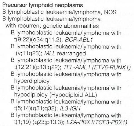
PRECURSOR LYMHPOID NEOPLASMS. B lymphoblastic leukaemia/lymphoma T lymphoblastic leukaemia/lymphoma
 PRECURSOR LYMHPOID NEOPLASMS B lymphoblastic leukaemia/lymphoma T lymphoblastic leukaemia/lymphoma B lymphoblastic leukaemia/lymphoma Definition: B lymphoblastic leukaemia/lymphoma is a neoplasm of precursor
Non-Hodgkin s Lymphomas Version
 NCCN Clinical Practice Guidelines in Oncology (NCCN Guidelines ) Non-Hodgkin s Lymphomas Version 2.2015 NCCN.org Continue Use of Immunophenotyping/ Genetic Testing in Differential Diagnosis of Mature B-Cell
3/23/2017. Disclosure of Relevant Financial Relationships. Pitfalls in Immunohistochemistry in Hematopathology: CD20 and CD3 Can Let Me Down?!
 Pitfalls in Immunohistochemistry in Hematopathology: CD20 and CD3 Can Let Me Down?! Judith A. Ferry Massachusetts General Hospital Disclosure of Relevant Financial Relationships USCAP requires that all
Anaplastic Large Cell Lymphoma (of T cell lineage)
") Anaplastic Large Cell Lymphoma (of T cell lineage) Definition T-cell lymphoma comprised of large cells with abundant cytoplasm and pleomorphic, often horseshoe-shaped nuclei CD30+ Most express cytotoxic
Anaplastic Large Cell Lymphoma (of T cell lineage) Definition T-cell lymphoma comprised of large cells with abundant cytoplasm and pleomorphic, often horseshoe-shaped nuclei CD30+ Most express cytotoxic
2010 Hematopoietic and Lymphoid ICD-O Codes - Alphabetical List THIS TABLE REPLACES ALL ICD-O-3 Codes
 Acute basophilic leukemia 9870/3 Acute biphenotypic leukemia [OBS] 9805/3 Acute erythroid leukemia 9840/3 Acute megakaryoblastic leukemia 9910/3 Acute monoblastic and monocytic leukemia 9891/3 Acute myeloid
2012 Hematopoietic and Lymphoid ICD-O Codes - Numerical List THIS TABLE REPLACES ALL ICD-O-3 Codes
 Malignant lymphoma, NOS 9590/3 Non-Hodgkin lymphoma, NOS 9591/3 B-cell lymphoma, unclassifiable, with features intermediate between diffuse large B-cell lymphoma and classical Hodgkin lymphoma 9596/3 Primary
Group of malignant disorders of the hematopoietic tissues characteristically associated with increased numbers of white cells in the bone marrow and
 Group of malignant disorders of the hematopoietic tissues characteristically associated with increased numbers of white cells in the bone marrow and / or peripheral blood Classified based on cell type
WBCs Disorders 1. Dr. Nabila Hamdi MD, PhD
 WBCs Disorders 1 Dr. Nabila Hamdi MD, PhD ILOs Compare and contrast ALL, AML, CLL, CML in terms of age distribution, cytogenetics, morphology, immunophenotyping, laboratory diagnosis clinical features
Combinations of morphology codes of haematological malignancies (HM) referring to the same tumour or to a potential transformation
 referring to the same tumour or to a potential transformation") Major subgroups according to the World Health Organisation (WHO) Classification Myeloproliferative neoplasms (MPN) Myeloid and lymphoid neoplasms with eosinophilia and abnormalities of PDGFRA, PDGFRB or
Major subgroups according to the World Health Organisation (WHO) Classification Myeloproliferative neoplasms (MPN) Myeloid and lymphoid neoplasms with eosinophilia and abnormalities of PDGFRA, PDGFRB or
Citation International Journal of Hematology, 2013, v. 98 n. 4, p The original publication is available at
 Title Diagnostic challenges in a case of B cell lymphoma unclassifiable with features intermediate between diffuse large B-cell lymphoma and Burkitt lymphoma Author(s) So, JCC; Yung, KH; Chu, ML; Wan,
LINFOMA B (INCLASIFICABLE) CON RASGOS INTERMEDIOS ENTRE LINFOMA DE BURKITT Y LINFOMA B DIFUSO DE CÉLULAS GRANDES.
 CON RASGOS INTERMEDIOS ENTRE LINFOMA DE BURKITT Y LINFOMA B DIFUSO DE CÉLULAS GRANDES.") Congreso Nacional SEAP 2013. LINFOMA B (INCLASIFICABLE) CON RASGOS INTERMEDIOS ENTRE LINFOMA DE BURKITT Y LINFOMA B DIFUSO DE CÉLULAS GRANDES. Santiago Montes Moreno Servicio de Anatomía Patológica, HUMV
Congreso Nacional SEAP 2013. LINFOMA B (INCLASIFICABLE) CON RASGOS INTERMEDIOS ENTRE LINFOMA DE BURKITT Y LINFOMA B DIFUSO DE CÉLULAS GRANDES. Santiago Montes Moreno Servicio de Anatomía Patológica, HUMV
Low-grade B-cell lymphoma
 Low-grade B-cell lymphoma Patho-Basic 11. September 2018 Stephan Dirnhofer Pathology Outline Definition LPL, MBL/CLL/SLL, MCL FL Subtypes & variants Diagnosis including Grading Transformation Summary Be
Successful flow cytometric immunophenotyping of body fluid specimens
 Successful flow cytometric immunophenotyping of body fluid specimens Fiona E. Craig, MD Division of Hematopathology Mayo Clinic Arizona 2017 MFMER slide-1 Financial disclosure No conflicts 2017 MFMER slide-2
Mimics of Lymphoma in Routine Biopsies. I have nothing to disclose regarding the information to be reported in this talk.
 Mimics of Lymphoma in Routine Biopsies Patrick Treseler, MD, PhD Professor of Pathology University of California San Francisco I have nothing to disclose regarding the information to be reported in this
Extramedullary precursor T-lymphoblastic transformation of CML at presentation
 Extramedullary precursor T-lymphoblastic transformation of CML at presentation Neerja Vajpayee, Constance Stein, Bernard Poeisz & Robert E. Hutchison Clinical History 30 year old man presented to the emergency
WBCs Disorders. Dr. Nabila Hamdi MD, PhD
 WBCs Disorders Dr. Nabila Hamdi MD, PhD ILOs Compare and contrast ALL, AML, CLL, CML in terms of age distribution, cytogenetics, morphology, immunophenotyping, laboratory diagnosis clinical features and
Leukemia (2007) 21, Cytoplasmic mutated nucleophosmin (NPM) defines the molecular status of a significant fraction of myeloid sarcomas
 21, Cytoplasmic mutated nucleophosmin (NPM) defines the molecular status of a significant fraction of myeloid sarcomas") Leukemia (2007) 21, 1566-1570 Cytoplasmic mutated nucleophosmin (NPM) defines the molecular status of a significant fraction of myeloid sarcomas Clinical presentation Mean age: 55.8 years (range: 16-87).
Leukemia (2007) 21, 1566-1570 Cytoplasmic mutated nucleophosmin (NPM) defines the molecular status of a significant fraction of myeloid sarcomas Clinical presentation Mean age: 55.8 years (range: 16-87).
3/2/2010. Case 1. Clinical History. Infectious Disease Pathology Specialty Conference Case 1
 Case 1. Clinical History Infectious Disease Pathology Specialty Conference Case 1 A 6 year-old girl with a large left neck mass. Past medical history: Biliary cirrhosis secondary to extra hepatic biliary
HEMATOPATHOLOGY (SHANDS HOSPITAL AT THE UNIVERSITY OF FLORIDA): Rotation Director: Ying Li, M.D., Ph.D., Assistant Professor
: Rotation Director: Ying Li, M.D., Ph.D., Assistant Professor") HEMATOPATHOLOGY (SHANDS HOSPITAL AT THE UNIVERSITY OF FLORIDA): Rotation Director: Ying Li, M.D., Ph.D., Assistant Professor I. Description of the rotation: During this rotation, the resident will gain
HEMATOPATHOLOGY (SHANDS HOSPITAL AT THE UNIVERSITY OF FLORIDA): Rotation Director: Ying Li, M.D., Ph.D., Assistant Professor I. Description of the rotation: During this rotation, the resident will gain
Acute Myeloid Leukemia with Recurrent Cytogenetic Abnormalities
 Acute Myeloid Leukemia with Recurrent Cytogenetic Abnormalities Acute Myeloid Leukemia with recurrent cytogenetic Abnormalities -t(8;21)(q22;q22)(aml/eto) -inv(16) or t(16;16) -t(15;17) -11q23 Acute Myeloid
Mimics of Lymphoma in Routine Biopsies. Mixed follicular and paracortical hyperplasia. Types of Lymphoid Hyperplasia
 Mimics of Lymphoma in Routine Biopsies Patrick Treseler, MD, PhD Professor of Pathology University of California San Francisco Types of Lymphoid Hyperplasia Follicular hyperplasia (B-cells) Paracortical
Presentation material is for education purposes only. All rights reserved URMC Radiology Page 1 of 98
 Presentation material is for education purposes only. All rights reserved. 2011 URMC Radiology Page 1 of 98 Radiology / Pathology Conference February 2011 Brooke Koltz, Cytopathology Resident Presentation
Infectious Disease Pathology Specialty Conference Case 1
 Infectious Disease Pathology Specialty Conference Case 1 Miguel Reyes Múgica, M.D. Department of Pathology Children s Hospital of Pittsburgh University of Pittsburgh Medical Center USCAP Annual Meeting.
The spectrum of flow cytometry of the bone marrow
 The spectrum of flow cytometry of the bone marrow Anna Porwit Lund University Faculty of Medicine Dept. of Clinical Sciences Div. Oncology and Pathology anna.porwit@med.lu.se Disclosure of speaker s interests
Case Workshop of Society for Hematopathology and European Association for Haematopathology
 Case 24 2007 Workshop of Society for Hematopathology and European Association for Haematopathology Aliyah Rahemtullah 1, Martin K Selig 1, Paola Dal Cin 2 and Robert P Hasserjian 1 Departments of Pathology,
SH/EAHP WORKSHOP 2017 CASE 210 PRESENTATION
 SH/EAHP WORKSHOP 2017 CASE 210 PRESENTATION Jonathon H Gralewski DO, MS, Ginell R Post MD, PhD, Youzhong Yuan MD September 9, 2017 Clinical History 60 year old male with history of c-maf high-risk IgG
Lymphoma/CLL 101: Know your Subtype. Dr. David Macdonald Hematologist, The Ottawa Hospital
 Lymphoma/CLL 101: Know your Subtype Dr. David Macdonald Hematologist, The Ottawa Hospital Function of the Lymph System Lymph Node Lymphocytes B-cells develop in the bone marrow and influence the immune
Clinicopathologic features of 112 cases with mantle cell lymphoma
 Cancer Biol Med 2015;12:46-52. doi: 10.7497/j.issn.2095-3941.2015.0007 ORIGINAL ARTICLE Clinicopathologic features of 112 cases with mantle cell lymphoma Dong-Mei Zhou, Gang Chen, Xiong-Wei Zheng, Wei-Feng
NEW ENTITIES IN AGGRESSIVE B CELL LYMPHOMA. Joon Seong Park, M.D. Dept. of Hematology-Oncology Ajou University School of Medicine
 NEW ENTITIES IN AGGRESSIVE B CELL LYMPHOMA Joon Seong Park, M.D. Dept. of Hematology-Oncology Ajou University School of Medicine Historical background of Lymphoma classification Rappaport classification
Classifications of lymphomas
 Classifications of lymphomas Lukes and Collins Kiel classification Working formulation REAL classification (1994) WHO classification (2000) WHO CLASSIFICATIONF OF NEOPLASMS HAEMATOPETIC AND LYMPHOID TISSUES
Methotrexate-associated Lymphoproliferative Disorders
 Methotrexate-associated Lymphoproliferative Disorders Definition A lymphoid proliferation or lymphoma in a patient immunosuppressed with methotrexate, typically for treatment of autoimmune disease (rheumatoid
B Cell Lymphoma: Aggressive
 B Cell Lymphoma: Aggressive UpToDate: Introduction: Risk Factors: Biology: Symptoms: Diagnosis: Ibrutinib approved for mantle cell lymphoma as 2nd line therapy. - Aggressive lymphomas are a group of malignant
V. Acute leukemia. Flow cytometry in evaluation of hematopoietic neoplasms: A case-based approach
 V. Acute leukemia Evaluating a sample for an acute leukemia Acute leukemia is a neoplasm of immature myeloid or lymphoid cells characterized by a block in maturation, usually at the stage of an early progenitor
Large cell immunoblastic Diffuse histiocytic (DHL) Lymphoblastic lymphoma Diffuse lymphoblastic Small non cleaved cell Burkitt s Non- Burkitt s
 Lymphoblastic lymphoma Diffuse lymphoblastic Small non cleaved cell Burkitt s Non- Burkitt s") Non Hodgkin s Lymphoma Introduction 6th most common cause of cancer death in United States. Increasing in incidence and mortality. Since 1970, the incidence of has almost doubled. Overview The types of
Non Hodgkin s Lymphoma Introduction 6th most common cause of cancer death in United States. Increasing in incidence and mortality. Since 1970, the incidence of has almost doubled. Overview The types of
Approach to Core Biopsy Specimens
 BDIAP 108th Symposium on Haematopathology Joint Meeting of the BDIAP and BLPG at-bristol, Anchor Road, Harbourside, Bristol BS1 5DB 15th - 17th May 2014 Approach to Core Biopsy Specimens Dr Stefan Dojcinov
Classification! Immunohistochemical classification of haematolymphoid tumours. Malignant lymphoproliferative diseases
 Immunohistochemical classification of haematolymphoid tumours Haematolymphoid Neoplasias: Leukaemia vs Lymphoma C L O N A L M A L I G N A N C I E S Stephen Hamilton-Dutoit Institute of Pathology Aarhus
Case Report A case of EBV positive diffuse large B-cell lymphoma of the adolescent
 Int J Clin Exp Med 2014;7(1):307-311 www.ijcem.com /ISSN:1940-5901/IJCEM1311029 Case Report A case of EBV positive diffuse large B-cell lymphoma of the adolescent Qilin Ao 2, Ying Wang 1, Sanpeng Xu 2,
Done By : WESSEN ADNAN BUTHAINAH AL-MASAEED
 Done By : WESSEN ADNAN BUTHAINAH AL-MASAEED Acute Myeloid Leukemia Firstly we ll start with this introduction then enter the title of the lecture, so be ready and let s begin by the name of Allah : We
Lymph node cytopathology : A practical approach to lymphoproliferative disorders
 Lymph node cytopathology : A practical approach to lymphoproliferative disorders Koray Ceyhan, M.D Department of Pathology Faculty of Medicine Ankara University Ankara, Turkey Diagnostic use of FNA in
Chronic Lymphocytic Leukemia Mantle Cell Lymphoma Elias Campo
 Chronic Lymphocytic Leukemia Mantle Cell Lymphoma Elias Campo Hospital Clinic, University of Barcelona Small B-cell lymphomas NAIVE -B LYMPHOCYTE MEMORY CELL CLL MCL FL MZL Small cell size Low proliferation
Low grade High grade , immune suppression chronic persistent inflammation viruses B-symptoms
 We've one category for lymphoid neoplasm which is the lymphoma in contrast to that of myeloid which has three categories; acute myeloid leukemias, myeloproliferative & myelodysplastic disorders. Lymphoma
Update on the Classification of Aggressive B-cell Lymphomas and Hodgkin Lymphoma
 Update on the Classification of Aggressive B-cell Lymphomas and Hodgkin Lymphoma Nancy Lee Harris, M. D. Massachusetts General Hospital Harvard Medical School Aggressive B-cell Lymphomas WHO 4 th Edition
PhenoPath. Diagnoses you can count on B CELL NON-HODGKIN LYMPHOMA
 PhenoPath Diagnoses you can count on B CELL NON-HODGKIN LYMPHOMA C urrent diagnosis of B cell non-hodgkin lymphoma (B-NHL) is based on the 2008 WHO Classification of Tumours of Haematopoietic and Lymphoid
The patient had a mild splenomegaly but no obvious lymph node enlargement. The consensus phenotype obtained from part one of the exercise was:
 Case History An 86 year old male was admitted to hospital with chest infection. Haematological examination subsequently revealed the following: Hb- 11.0 g/dl; WBC- 67.1 x 10^9/l; PLT- 99 x10^9/l; RBC-
HODGKIN LYMPHOMA DR. ALEJANDRA ZARATE OSORNO HOSPITAL ESPAÑOL DE MEXICO
 HODGKIN LYMPHOMA DR. ALEJANDRA ZARATE OSORNO HOSPITAL ESPAÑOL DE MEXICO HODGKIN LYMPHOMA CLASSIFICATION Lukes & Butler Rye WHO-2016 Linphocytic and/or histiocytic Nodular & diffuse Nodular Sclerosis Lymphocyte
5000 International Clinical Cytometry Society: Practical Flow Cytometry in Hematopathology A Case-Based Approach
 5000 International Clinical Cytometry Society: Practical Flow Cytometry in Hematopathology A Case-Based Approach Joseph A DiGiuseppe, MD, PhD Hartford Hospital Disclosures In the past 12 months, I have
Plasma cell myeloma (multiple myeloma)
") Plasma cell myeloma (multiple myeloma) Common lymphoid neoplasm, present at old age (70 years average) Remember: plasma cells are terminally differentiated B-lymphocytes that produces antibodies. B-cells
Plasma cell myeloma (multiple myeloma) Common lymphoid neoplasm, present at old age (70 years average) Remember: plasma cells are terminally differentiated B-lymphocytes that produces antibodies. B-cells
Diagnosis of lymphoid neoplasms has been
 Iranian Journal of Pathology (2007)2 (1), 1-61 Review Article Mehdi Nassiri Dep. of Pathology, University of Miami Miller School of Medicine, Miami, USA Abstract Correct diagnosis and classification of
Hematopathology Lab. Third year medical students
 Hematopathology Lab Third year medical students Objectives Identify the lesion Know the specific name of the lesion Know associated disease Know relevant pathologic background Spherocytes: appear small,
Pathology of the indolent B-cell lymphomas Elias Campo
 Pathology of the indolent B-cell lymphomas Elias Campo Hospital Clinic, University of Barcelona Small B-cell lymphomas Antigen selection NAIVE -B LYMPHOCYTE MEMORY B-CELL MCL FL LPL MZL CLL Small cell
a resource for physicians Recommended Referral Timing for Stem Cell Transplant Evaluation
 a resource for physicians Recommended Referral Timing for Stem Cell Transplant Evaluation This resource has been developed to help guide you regarding the appropriate timing and conditions for a referral
Follicular Lymphoma: the WHO
 Follicular Lymphoma: the WHO and the WHERE? Yuri Fedoriw, MD Associate Professor of Pathology and Laboratory Medicine Director of Hematopathology University of North Carolina Chapel Hill, NC Disclosure
Nodular lymphocyte predominant Hodgkin lymphoma. Lymphoma Tumor Board. January 5, 2018
 Nodular lymphocyte predominant Hodgkin lymphoma Lymphoma Tumor Board January 5, 2018 Etiology Subtypes of Classical Hodgkin Lymphoma (chl)* Nodular sclerosing HL Most common subtype Composed of large tumor
Does the proliferation fraction help identify mature B cell lymphomas with double- and triple-hit translocations?
 Histopathology 2012, 61, 1214 1218. DOI: 10.1111/j.1365-2559.2012.04351.x SHORT REPORT Does the proliferation fraction help identify mature B cell lymphomas with double- and triple-hit translocations?
GENETIC MARKERS IN LYMPHOMA a practical overview. P. Heimann Dpt of Medical Genetics Erasme Hospital - Bordet Institute
 GENETIC MARKERS IN LYMPHOMA a practical overview P. Heimann Dpt of Medical Genetics Erasme Hospital - Bordet Institute B and T cell monoclonalities Rearrangement of immunoglobin and TCR genes may help
Pathology. #11 Acute Leukemias. Farah Banyhany. Dr. Sohaib Al- Khatib 23/2/16
 35 Pathology #11 Acute Leukemias Farah Banyhany Dr. Sohaib Al- Khatib 23/2/16 1 Salam First of all, this tafreegh is NOT as long as you may think. If you just focus while studying this, everything will
Pathology of aggressive lymphomas
 Institute of Pathology Pathology of aggressive lymphomas Leticia Quintanilla-Martinez Changes in the new 2016 WHO Aggressive B-cell lymphoid neoplasms Major changes that impact how cases should be evaludated
The next lymphoma classification Luca Mazzucchelli Istituto cantonale di patologia, Locarno
 Evolution of classification The next classification Luca Mazzucchelli Istituto cantonale di patologia, Locarno The Lymphoma Forum of Excellence, Bellinzona, January 2011 Rappaport Lukes and Collins (immunophenotype)
Pathology of aggressive lymphomas
 Institute of Pathology Pathology of aggressive lymphomas Leticia Quintanilla-Martinez Changes in the new 2016 WHO Aggressive B-cell lymphoid neoplasms Major changes that impact how cases should be evaludated