Presentation material is for education purposes only. All rights reserved URMC Radiology Page 1 of 98
|
|
|
- Lee Chambers
- 5 years ago
- Views:
Transcription


1 Presentation material is for education purposes only. All rights reserved URMC Radiology Page 1 of 98

2 Radiology / Pathology Conference February 2011 Brooke Koltz, Cytopathology Resident Presentation material is for education purposes only. All rights reserved URMC Radiology Page 2 of 98

3 Presentation material is for education purposes only. All rights reserved URMC Radiology Page 3 of 98

4 Presentation material is for education purposes only. All rights reserved URMC Radiology Page 4 of 98
5 Presentation material is for education purposes only. All rights reserved URMC Radiology Page 5 of 98


6 Presentation material is for education purposes only. All rights reserved URMC Radiology Page 6 of 98


7 Presentation material is for education purposes only. All rights reserved URMC Radiology Page 7 of 98


8 Presentation material is for education purposes only. All rights reserved URMC Radiology Page 8 of 98


9 Presentation material is for education purposes only. All rights reserved URMC Radiology Page 9 of 98

10 Presentation material is for education purposes only. All rights reserved URMC Radiology Page 10 of 98


11 Presentation material is for education purposes only. All rights reserved URMC Radiology Page 11 of 98

12 Presentation material is for education purposes only. All rights reserved URMC Radiology Page 12 of 98

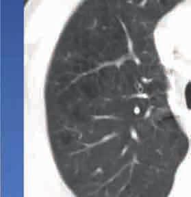
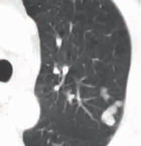
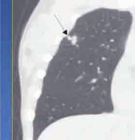
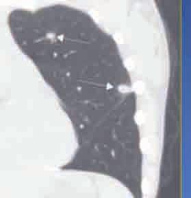
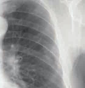
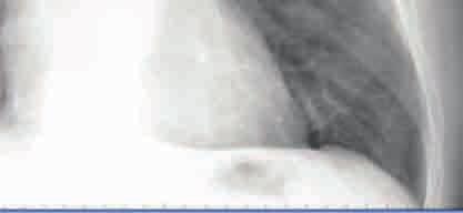
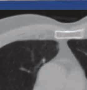
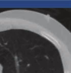
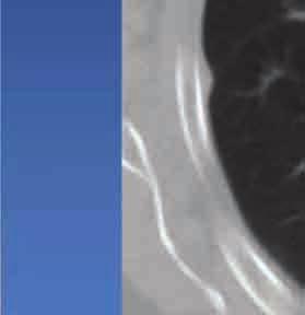
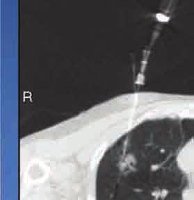
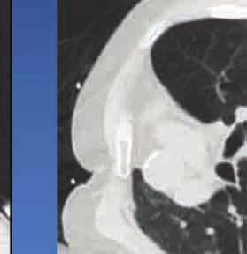
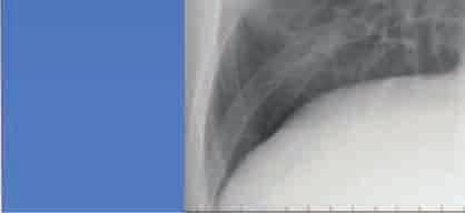
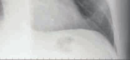
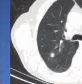
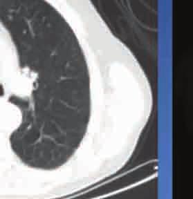
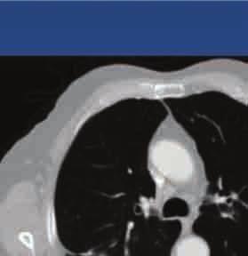
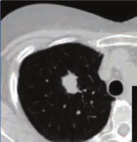
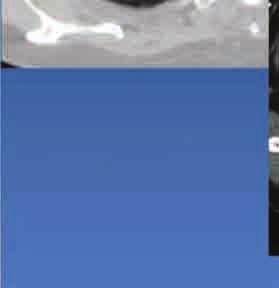
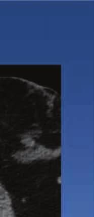
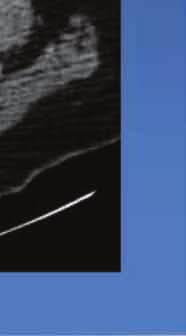
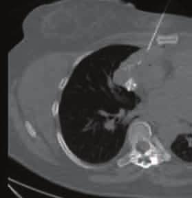
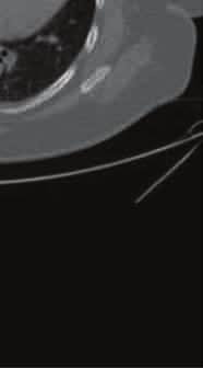
13 Case 1 Lung, RUL, CT-guided FNA: Diff-Quik Presentation material is stain for education purposes only. All rights reserved URMC Radiology Page 13 of 98

14 Lung, RUL, CT-guided FNA: Diff-Quik stain Presentation material is for education purposes only. All rights reserved URMC Radiology Page 14 of 98

15 Lung, RUL, CT-guided FNA: Papanicolaou stain Presentation material is for education purposes only. All rights reserved URMC Radiology Page 15 of 98

16 Lung, RUL, CT-guided FNA: Papanicolaou stain Presentation material is for education purposes only. All rights reserved URMC Radiology Page 16 of 98

17 Lung, RUL, CT-guided FNA: Cell Block, hematoxylin & eosin stain Presentation material is for education purposes only. All rights reserved URMC Radiology Page 17 of 98


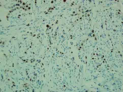
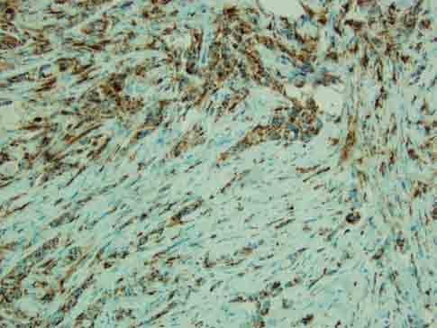
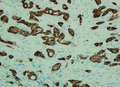
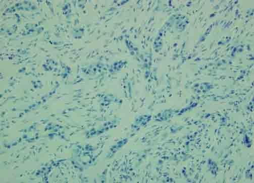
18 Lung, RUL, CT-guided FNA, Cell block: Immunohistochemical stains Cytokeratin 20 CDX-2 Presentation material is for education purposes only. All rights reserved URMC Radiology Page 18 of 98
19 Lung, RUL, CT-guided fine needle aspiration: Malignant tumor cells present derived from adenocarcinoma, favor colonic primary. Cell block and cytologic preparations examined. The tumor cells are positive for CDX-2 and focal positive for CK 20, negative for CK 7 and TTF-1. The results support the diagnosis of metastatic adenocarcinoma, favor colonic primary. Presentation material is for education purposes only. All rights reserved URMC Radiology Page 19 of 98

20 Lung, right upper lobe, wedge resection: H & E stain Presentation material is for education purposes only. All rights reserved URMC Radiology Page 20 of 98

21 Lung, right upper lobe, wedge resection: H & E stain Presentation material is for education purposes only. All rights reserved URMC Radiology Page 21 of 98
22 Lung, right upper lobe, wedge resection: Adenocarcinoma, moderately differentiated, morphologically consistent with a metastatic carcinoma of known history of rectal carcinoma. Lymph node, level 4 and level 10, biopsy: Chronic lymphocytic leukemia. Presentation material is for education purposes only. All rights reserved URMC Radiology Page 22 of 98

23 Rectum, low anterior resection: H & E Presentation material is for education purposes only. All rights reserved URMC Radiology stain Page 23 of 98

24 Rectum, low anterior resection: H & E Presentation material is for education purposes only. All rights reserved URMC Radiology stain Page 24 of 98
25 Rectum, low anterior resection: Adenocarcinoma, status post radiation therapy. K-RAS Gene Rearrangement: K-RAS Gene: Normal, no mutations in codon 12 and 13 were seen. Lymph node, pericolic: Metastatic adenocarcinoma identified in two of fifty one lymph nodes (2/51). Effacement of multiple lymph nodes by monomorphic lymphoid population consistent with previous known diagnosis of chronic lymphocytic leukemia. Presentation material is for education purposes only. All rights reserved URMC Radiology Page 25 of 98
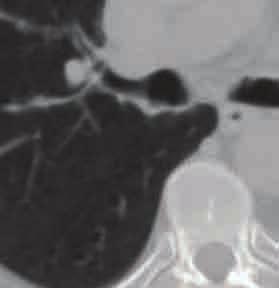
26 Metastatic Adenocarcinoma Colonic Primary! Metastatic carcinoma to the lung more common than primary lung CA! Most common metastatic neoplasms are adenocarcinomas from breast, kidney and colon! Cytomorphology of columnar cells and elongated nuclei forming a palisade-like arrangement or clusters of signet ring- type cells! Immunohistochemical stains:! CDX-2, CK 20 positive! CK 7 negative! Histochemical mucin stain + Presentation material is for education purposes only. All rights reserved URMC Radiology Page 26 of 98


27 Presentation material is for education purposes only. All rights reserved URMC Radiology Page 27 of 98


28 Presentation material is for education purposes only. All rights reserved URMC Radiology Page 28 of 98


29 Presentation material is for education purposes only. All rights reserved URMC Radiology Page 29 of 98

30 Presentation material is for education purposes only. All rights reserved URMC Radiology Page 30 of 98


31 Presentation material is for education purposes only. All rights reserved URMC Radiology Page 31 of 98
32 Presentation material is for education purposes only. All rights reserved URMC Radiology Page 32 of 98

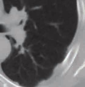
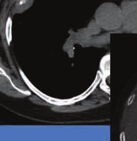
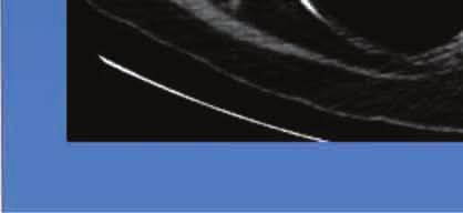
33 Lung, RUL, CT-guided FNA: Diff-Quik stain Case 2 Presentation material is for education purposes only. All rights reserved URMC Radiology Page 33 of 98

34 Lung, RUL, CT-guided FNA: Diff-Quik stain Presentation material is for education purposes only. All rights reserved URMC Radiology Page 34 of 98

35 Lung, RUL, CT-guided FNA: Papanicolaou stain Presentation material is for education purposes only. All rights reserved URMC Radiology Page 35 of 98

36 Lung, RUL, CT-guided FNA: Papanicolaou stain Presentation material is for education purposes only. All rights reserved URMC Radiology Page 36 of 98
37 Lung, right upper lobe, CT-guided fine needle aspiration: Malignant tumor cells present derived from adenocarcinoma. Cell block and cytologic preparations examined. Presentation material is for education purposes only. All rights reserved URMC Radiology Page 37 of 98

38 Lung, right upper lobe, biopsy: H & E stain Presentation material is for education purposes only. All rights reserved URMC Radiology Page 38 of 98

39 Lung, right upper lobe, biopsy: H & E stain Presentation material is for education purposes only. All rights reserved URMC Radiology Page 39 of 98
40 Lung, right upper lobe, biopsy: Adenocarcinoma. Papillary features are identified. Presentation material is for education purposes only. All rights reserved URMC Radiology Page 40 of 98
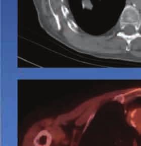
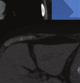
41 Adenocarcinoma of Lung! Adenocarcinoma: most common lung carcinoma in females and non-smokers! Positive immunostaining for CK-7, TTF-1, Napsin A, and negative for CK-20! Positive intracellular histochemical staining for mucin! Broad classification is non-small cell carcinoma (80%) versus small cell carcinoma (20%) Presentation material is for education purposes only. All rights reserved URMC Radiology Page 41 of 98

42 Presentation material is for education purposes only. All rights reserved URMC Radiology Page 42 of 98


43 Presentation material is for education purposes only. All rights reserved URMC Radiology Page 43 of 98

44 Presentation material is for education purposes only. All rights reserved URMC Radiology Page 44 of 98
45 Presentation material is for education purposes only. All rights reserved URMC Radiology Page 45 of 98


46 Presentation material is for education purposes only. All rights reserved URMC Radiology Page 46 of 98
47 Presentation material is for education purposes only. All rights reserved URMC Radiology Page 47 of 98

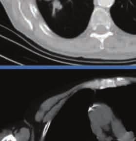
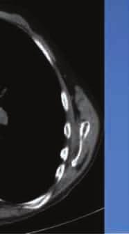
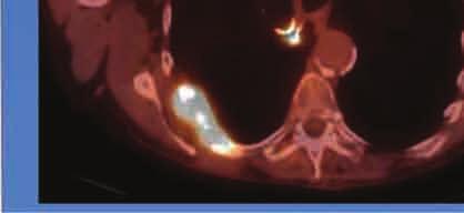
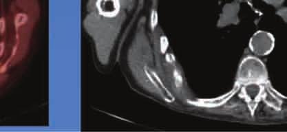
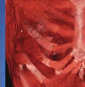
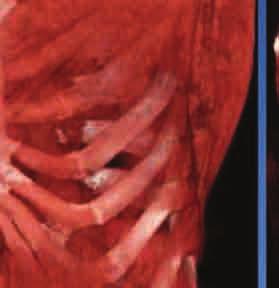
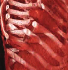
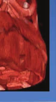
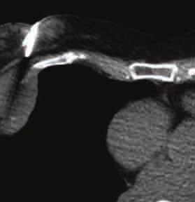
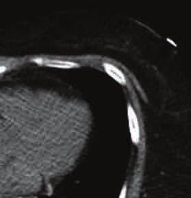
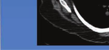
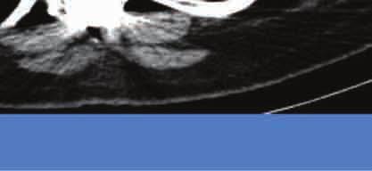
48 Case 3 Rib, right posterior, CT-guided FNA: Diff-Quik Presentation material is stain for education purposes only. All rights reserved URMC Radiology Page 48 of 98

49 Rib, right posterior, CT-guided FNA: Diff-Quik stain Presentation material is for education purposes only. All rights reserved URMC Radiology Page 49 of 98

50 Rib, right posterior, CT-guided FNA: Papanicolaou Presentation material is for education stain purposes only. All rights reserved URMC Radiology Page 50 of 98

51 Rib, right posterior, CT-guided FNA: Cell Block, hematoxylin & eosin stain Presentation material is for education purposes only. All rights reserved URMC Radiology Page 51 of 98
52 Rib, right posterior, CT-guided fine needle aspiration: Malignant tumor cells with focal spindle cells present derived from squamous cell carcinoma. Cell block and cytologic preparations examined. Presentation material is for education purposes only. All rights reserved URMC Radiology Page 52 of 98

53 Lung, right upper lobe, lobectomy: H & E stain Presentation material is for education purposes only. All rights reserved URMC Radiology Page 53 of 98

54 Lung, right upper lobe, lobectomy: H & E stain Presentation material is for education purposes only. All rights reserved URMC Radiology Page 54 of 98
55 Lung, right upper lobe, lobectomy: Pleomorphic carcinoma with extensive necrosis. The tumor consists of typical areas of squamous cell carcinoma and adenocarcinoma along with prominent spindle cell areas, consistent with a diagnosis of pleomorphic carcinoma. No metastatic carcinoma identified - 0/33 lymph nodes. Presentation material is for education purposes only. All rights reserved URMC Radiology Page 55 of 98
56 Pleomorphic Carcinoma of Lung! Rare, <1% of all lung carcinomas! Male predominance! Smokers! Subtype of sarcomatoid CA of lung! Dual malignant epithelial cell component and sarcomatoid spindle cell elements! Prognosis is poor! Early relapse of disease common Presentation material is for education purposes only. All rights reserved URMC Radiology Page 56 of 98

57 Presentation material is for education purposes only. All rights reserved URMC Radiology Page 57 of 98


58 Presentation material is for education purposes only. All rights reserved URMC Radiology Page 58 of 98


59 Presentation material is for education purposes only. All rights reserved URMC Radiology Page 59 of 98
60 Presentation material is for education purposes only. All rights reserved URMC Radiology Page 60 of 98
61 Presentation material is for education purposes only. All rights reserved URMC Radiology Page 61 of 98


62 Presentation material is for education purposes only. All rights reserved URMC Radiology Page 62 of 98

63 Presentation material is for education purposes only. All rights reserved URMC Radiology Page 63 of 98


64 Presentation material is for education purposes only. All rights reserved URMC Radiology Page 64 of 98
65 Presentation material is for education purposes only. All rights reserved URMC Radiology Page 65 of 98


66 Presentation material is for education purposes only. All rights reserved URMC Radiology Page 66 of 98

67 Presentation material is for education purposes only. All rights reserved URMC Radiology Page 67 of 98


68 Presentation material is for education purposes only. All rights reserved URMC Radiology Page 68 of 98
69 Presentation material is for education purposes only. All rights reserved URMC Radiology Page 69 of 98
70 Presentation material is for education purposes only. All rights reserved URMC Radiology Page 70 of 98


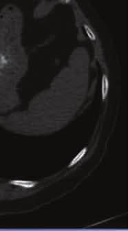
71 Presentation material is for education purposes only. All rights reserved URMC Radiology Page 71 of 98

72 Presentation material is for education purposes only. All rights reserved URMC Radiology Page 72 of 98
73 Presentation material is for education purposes only. All rights reserved URMC Radiology Page 73 of 98
74 Presentation material is for education purposes only. All rights reserved URMC Radiology Page 74 of 98
75 Presentation material is for education purposes only. All rights reserved URMC Radiology Page 75 of 98
76 Presentation material is for education purposes only. All rights reserved URMC Radiology Page 76 of 98


77 Presentation material is for education purposes only. All rights reserved URMC Radiology Page 77 of 98

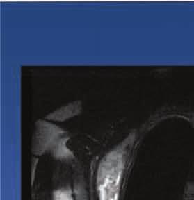
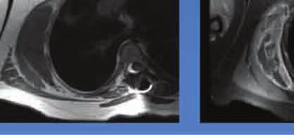
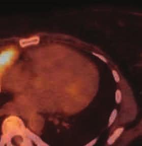
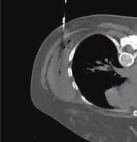
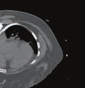
78 Case 4 Pleural based mass, CT-guided FNA: Diff-Quik Presentation material is stain for education purposes only. All rights reserved URMC Radiology Page 78 of 98

79 Pleural based mass, CT-guided FNA: Diff-Quik stain Presentation material is for education purposes only. All rights reserved URMC Radiology Page 79 of 98

80 Pleural based mass, CT-guided FNA: Papanicolaou stain Presentation material is for education purposes only. All rights reserved URMC Radiology Page 80 of 98

81 Pleural based mass, CT-guided FNA: Papanicolaou stain Presentation material is for education purposes only. All rights reserved URMC Radiology Page 81 of 98
82 Pleural based mass, CT-guided fine needle aspiration: Malignant tumor cells present derived from adenocarcinoma. Cell block and cytologic preparations examined. Presentation material is for education purposes only. All rights reserved URMC Radiology Page 82 of 98

83 Pleura, base mass, biopsy: H & E stain Presentation material is for education purposes only. All rights reserved URMC Radiology Page 83 of 98

84 Pleura, base mass, biopsy: H & E stain Presentation material is for education purposes only. All rights reserved URMC Radiology Page 84 of 98

85 Pleura, base mass, biopsy: Immunohistochemical stains TTF-1 CK 20 Napsin A CK 7 Presentation material is for education purposes only. All rights reserved URMC Radiology Page 85 of 98
86 Pleura, base mass, biopsy: Adenocarcinoma, most consistent with origin from lung. Comment: Immunohistochemical stain for Napsin A is strongly +.. TTF-1 and CK 7 are positive. CK 20, ER and PR, Her2, BRST2, mammoglobin and CDX-2 are negative. The staining pattern, combined with the morphology and in comparison with the patientõs s prior breast carcinoma, is most consistent with an origin from lung. Presentation material is for education purposes only. All rights reserved URMC Radiology Page 86 of 98
87 Adenocarcinoma of Lung! More common in women then men.! Tend to be peripheral in location.! Metastatic carcinoma to the lung must be ruled out.! Immunohistochemical stains TTF-1 and Napsin A, Cytokeratin 7 + Cytokeratin 20 -! Clinical history Presentation material is for education purposes only. All rights reserved URMC Radiology Page 87 of 98

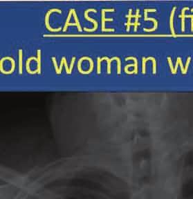
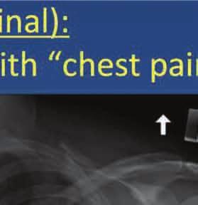
88 Case 5 Mediastinum, anterior, CT-guided FNA: Diff-Quik stain Presentation material is for education purposes only. All rights reserved URMC Radiology Page 88 of 98

89 Mediastinum, anterior, CT-guided FNA: Presentation material is for education purposes only. All rights reserved URMC Radiology Diff-Quik stain Page 89 of 98

90 Mediastinum, anterior, CT-guided FNA: Presentation material is for education purposes only. All rights reserved URMC Radiology Papanicolaou stain Page 90 of 98

91 Mediastinum, anterior, CT-guided FNA: Presentation material is for education purposes only. All rights reserved URMC Radiology Papanicolaou stain Page 91 of 98
92 Mediastinum, anterior, CT-guided fine needle aspiration: Atypical lymphocytes present. Please see separate flow cytometry and concurrent core biopsy report. Presentation material is for education purposes only. All rights reserved URMC Radiology Page 92 of 98

93 Soft tissue, anterior mediastinum, Presentation material is for education purposes only. All rights reserved URMC Radiology Core needle biopsy: H & E Page stain 93 of 98


94 Soft tissue, anterior mediastinum: Immunohistochemical stains Pancytokeratin CD8 TdT Presentation material is for education purposes only. All rights reserved URMC Radiology Page 94 of 98
95 Soft tissue, anterior mediastinum, core needle biopsy: Thymoma. This diagnosis was rendered in conjunction with the flow cytometry specimen. Sections show core needle biopsies of soft tissue with diffuse sheets of small round blue cells with scant cytoplasm that are + for TdT, CD3, CD4 and CD8. Pancytokeratin stain is strongly +. Presentation material is for education purposes only. All rights reserved URMC Radiology Page 95 of 98
96 Soft tissue, anterior mediastinum, fine needle aspiration (flow cytometry): Flow cytometry findings most consistent with thymoma. Cytospin shows numerous small lymphocytes as well as larger forms with high N/C ratios and immature chromatin. No monoclonal B-cell population is detected. Events in the small lymphocyte gate co-express CD4 and CD9, consistent with thymic origin, with maturational loss of CD4 and CD8 in two separate populations. By side scatter versus CD45, half of events are + for cytoplasmic TdT and CD3 and negative for CD34. Presentation material is for education purposes only. All rights reserved URMC Radiology Page 96 of 98
97 Thymoma! Rarely encountered neoplasm located in anterior mediastinum.! Tumor stage is most significant prognostic factor.! Occurs in all age groups peaks in 6 th decade.! No sex predilection.! 10-80% of patients have symptoms of myasthenia gravis.! Cytologically dual populations of epithelial cells and lymphocytes in various proportions.! Differential diagnosis includes lymphoepithelioma-like CA, carcinoid tumor, precursor T-lymphoblastic lymphoma, HodgkinÕs s lymphoma, thymic CA and reactive lymphoid hyperplasia.! Immunohistochemical Stains:! Epithelial cells Ð Cytokeratin +! CD8, CD3 and CD4 +! Predominance of cortical component: TdT + Presentation material is for education purposes only. All rights reserved URMC Radiology Page 97 of 98


98 Presentation material is for education purposes only. All rights reserved URMC Radiology Page 98 of 98
Radiology Pathology Conference
 Radiology Pathology Conference Sharlin Johnykutty,, MD, Cytopathology Fellow Sara Majewski, MD, Radiology Resident Friday, August 28, 2009 Presentation material is for education purposes only. All rights
Radiology Pathology Conference
 Radiology Pathology Conference Nadia F. Yusaf, M.D. PGY-3 1/29/2010 Presentation material is for education purposes only. All rights reserved. 2010 URMC Radiology Page 1 of 90 Case 1 60 year- old man presents
Impact of immunostaining of pulmonary and mediastinal cytology
 Impact of immunostaining of pulmonary and mediastinal cytology Harman Sekhon MD, PhD Director of Cytopathology Head of Ottawa-site Ontario Tumour Bank June 20, 2014 Disclaimer Pfizer: Honorarium-Advisory
Respiratory Tract Cytology
 Respiratory Tract Cytology 40 th European Congress of Cytology Liverpool, UK Momin T. Siddiqui M.D. Professor of Pathology and Laboratory Medicine Director of Cytopathology Emory University Hospital, Atlanta,
Radiology-Pathology Conference
 July 31, 2009 Radiology-Pathology Conference Daniel T Ginat, M.D., M.S. Sharlin Johnykutty,, M.D. Presentation material is for education purposes only. All rights reserved. 2009 URMC Radiology Page 1 of
Lung Cytology: Lessons Learned from Errors in Practice
 Lung Cytology: Lessons Learned from Errors in Practice Stephen S. Raab, M.D. Department of Laboratory Medicine Eastern Health and Memorial University of Newfoundland, St. John s, NL and University of Washington,
A 53 year-old woman with a lung mass, right hilar mass and mediastinal adenopathy.
 November 2015 Case of the Month A 53 year-old woman with a lung mass, right hilar mass and mediastinal adenopathy. Contributed by: Rasha Salama, M.D., IU Department of Pathology and Laboratory Medicine
SELECTED DILEMMAS IN RESPIRATORY CYTOPATHOLOGY (2 CASES)
") SELECTED DILEMMAS IN RESPIRATORY CYTOPATHOLOGY (2 CASES) Dr. Mariamma Joseph Professor of Pathology Division Head Cytopathology Department of Pathology and Laboratory Medicine LHSC and Western University
SELECTED DILEMMAS IN RESPIRATORY CYTOPATHOLOGY (2 CASES) Dr. Mariamma Joseph Professor of Pathology Division Head Cytopathology Department of Pathology and Laboratory Medicine LHSC and Western University
Update on 2015 WHO Classification of Lung Adenocarcinoma 1/3/ Mayo Foundation for Medical Education and Research. All rights reserved.
 1 Our speaker for this program is Dr. Anja Roden, an associate professor of Laboratory Medicine and Pathology at Mayo Clinic as well as consultant in the Anatomic Pathology Laboratory and co-director of
Cytological Sub-classification of Lung Cancer: Morphologic and Molecular Characteristics. Mercè Jordà, University of Miami
 Cytological Sub-classification of Lung Cancer: Morphologic and Molecular Characteristics Mercè Jordà, University of Miami Mortality Lung cancer is the most frequent cause of cancer incidence and mortality
The Role of Fine Needle Aspiration Cytology in the Diagnosis and Management of Thymic Neoplasia
 MALIGNANCIES OF THE THYMUS The Role of Fine Needle Aspiration Cytology in the Diagnosis and Management of Thymic Neoplasia Maureen F. Zakowski, MD, James Huang, MD, and Matthew P. Bramlage, MD Background:
The role of Electron Microscopy in the study of cytologic specimens. Elba A. Turbat-Herrera, MD
 The role of Electron Microscopy in the study of cytologic specimens. Elba A. Turbat-Herrera, MD Louisiana State University Health Sciences Center Shreveport, LA, USA Introduction The field of Cytology
FNA of Thyroid. Toward a Uniform Terminology With Management Guidelines. NCI NCI Thyroid FNA State of the Science Conference
 FNA of Thyroid NCI NCI Thyroid FNA State of the Science Conference Toward a Uniform Terminology With Management Guidelines Thyroid Thyroid FNA Cytomorphology NCI Thyroid FNA State of the Science Conference
Oncocytic-Appearing Salivary Gland Tumors. Oncocytic, Cystic, Mucinous, and High Grade Salivary Gland Tumors SALIVARY GLAND FNA: PART II
 William C. Faquin, MD, PhD Professor of Pathology Harvard Medical School Director of Head and Neck Pathology Massachusetts Eye and Ear Massachusetts General Hospital SALIVARY GLAND FNA: PART II Oncocytic,
ACCME/Disclosures. Diagnosing Mesothelioma in Limited Tissue Samples. Papanicolaou Society of Cytopathology Companion Meeting March 12 th, 2016
 Diagnosing Mesothelioma in Limited Tissue Samples Papanicolaou Society of Cytopathology Companion Meeting March 12 th, 2016 Sanja Dacic, MD, PhD University of Pittsburgh ACCME/Disclosures GENERAL RULES
Non Small Cell Lung Cancer Histopathology ד"ר יהודית זנדבנק
 Non Small Cell Lung Cancer Histopathology ד"ר יהודית זנדבנק 26.06.09 Lecture outlines WHO histological classification Macro/Micro assessment Early diagnosis Minimal pathology Main subtypes SCC, AdCa, LCLC
Primary enteric adenocarcinoma with predominantly signet ring features of the lung: A case report with clinicopathological and molecular findings
 CASE REPORT Primary enteric adenocarcinoma with predominantly signet ring features of the lung: A case report with clinicopathological and molecular findings Makoto Nagashima 1, Ayako Moriyama 1, Yasuo
DETERMINATION OF A LYMPHOID PROCESS
 Chapter 2 Applications of Touch Preparation Cytology to Intraoperative Consultations: Lymph Nodes and Extranodal Tissues for Evaluation of Hematolymphoid Disorders INTRODUCTION As discussed in Chap. 1,
DIAGNOSTIC DILEMMA. Case Reports Clinical history. Materials and Methods
 DIAGNOSTIC DILEMMA A Metastatic Renal Carcinoid Tumor Presenting as Breast Mass: A Diagnostic Dilemma Farnaz Hasteh, M.D., 1 Robert Pu, M.D., Ph.D., 2 and Claire W. Michael, M.D. 2 * We present clinicopathological
From Morphology to Molecular Pathology: A Practical Approach for Cytopathologists Part 1-Cytomorphology. Songlin Zhang, MD, PhD LSUHSC-Shreveport
 From Morphology to Molecular Pathology: A Practical Approach for Cytopathologists Part 1-Cytomorphology Songlin Zhang, MD, PhD LSUHSC-Shreveport I have no Conflict of Interest. FNA on Lymphoproliferative
Ascitic Fluid and Use of Immunocytochemistry. Mercè Jordà, University of Miami
 Ascitic Fluid and Use of Immunocytochemistry Mercè Jordà, University of Miami Is It Malignant? Yes? No Ascitic Fluid Cytomorphologic Useful Findings Tight clusters with smooth borders Cellular and nuclear
INTRODUCTION TO PATHOLOGICAL TECHNIQUES. 1. Types of routine biopsy procedures 2. Special exams (IHC, FISH)
") INTRODUCTION TO PATHOLOGICAL TECHNIQUES 1. Types of routine biopsy procedures 2. Special exams (IHC, FISH) Biopsy-Indications Diffuse/multifocal lesions (neoplastic, inflammatory, etc) Etiology of the
INTRODUCTION TO PATHOLOGICAL TECHNIQUES 1. Types of routine biopsy procedures 2. Special exams (IHC, FISH) Biopsy-Indications Diffuse/multifocal lesions (neoplastic, inflammatory, etc) Etiology of the
Objectives. Salivary Gland FNA: The Milan System. Role of Salivary Gland FNA 04/26/2018
 Salivary Gland FNA: The Milan System Dr. Jennifer Brainard Section Head Cytopathology Cleveland Clinic Objectives Introduce the Milan System for reporting salivary gland cytopathology Define cytologic
Medullary Thyroid Carcinoma. This case was provided by Treant Hospital, Bethesda, Hoogeveen, The Netherlands
 Medullary Thyroid Carcinoma This case was provided by Treant Hospital, Bethesda, Hoogeveen, The Netherlands ADS-01504 Rev. 001 2016 Hologic, Inc. All rights reserved. Overview Medullary Thyroid Carcinoma
Basement membrane in lobule.
 Bahram Memar, MD Basement membrane in lobule. Normal lobule-luteal phase Normal lobule-follicular phase Lactating breast Greater than 95% are adenocarcinomas in situ carcinomas and invasive carcinomas.
Neuroendocrine Lung Tumors Myers
 Diagnosis and Classification of Neuroendocrine Lung Tumors Jeffrey L. Myers, M.D. A. James French Professor Director, Anatomic Pathology & MLabs University of Michigan, Ann Arbor, MI myerjeff@umich.edu
Case Report Tumor-to-Tumor Metastasis: Lung Carcinoma Metastasizing to Thyroid Neoplasms
 Hindawi Publishing Corporation Volume 2015, Article ID 153932, 5 pages http://dx.doi.org/10.1155/2015/153932 Case Report Tumor-to-Tumor Metastasis: Lung Carcinoma Metastasizing to Thyroid Neoplasms Shiuan-Li
Almost any suspected tumor can be aspirated easily and safely. Some masses are more risky to aspirate including:
 DOES THIS PATIENT HAVE CANCER? USING IN-HOUSE CYTOLOGY TO HELP YOU MAKE THIS DIAGNOSIS. Joyce Obradovich, DVM, Diplomate, ACVIM (Oncology) Animal Cancer & Imaging Center, Canton, Michigan Almost every
Radiology/Pathology Conference
 Radiology/Pathology Conference NARASIMHACHAR G. PRATIVADI (RAD) & KIRSTEN WOOLF (PATH) Presentation material is for education purposes only. All rights reserved. 2010 URMC Radiology Page 1 of 132 Case
Case Report Tumor-to-Tumor Metastasis: Lung Carcinoma Metastasizing to Thyroid Neoplasms
 Case Reports in Pathology Volume 2015, Article ID 153932, 5 pages http://dx.doi.org/10.1155/2015/153932 Case Report Tumor-to-Tumor Metastasis: Lung Carcinoma Metastasizing to Thyroid Neoplasms Shiuan-Li
Cytology for the Endocrinologist. Nicole Massoll M.D
 Cytology for the Endocrinologist Nicole Massoll M.D Objectives Discuss slide preperation Definitions of adequacy ROSE (Rapid On-Site Evaluation) Thyroid Cytology Adequacy Nicole Massoll M.D. University
Case #1 FNA of nodule in left lobe of thyroid in 67 y.o. woman
 Challenging Cases Manon Auger M.D., F.R.C.P. (C) Professor, Department of Pathology McGill University Director, Cytopathology Laboratory McGill University it Health Center Case #1 FNA of nodule in left
Lesions Mimicking Adenoid Cystic Carcinoma. Diagnostic Problems in Salivary Gland Pathology An Update 5/29/2009
 Diagnostic Problems in Salivary Gland Pathology An Update Lesions Mimicking Adenoid Cystic Carcinoma Stacey E. Mills, M.D. W.S. Royster Professor of Pathology Director of Surgical and Cytopathology University
Respiratory Interactive Session. Elaine Borg
 Respiratory Interactive Session Elaine Borg Case 1 Respiratory Cytology 55 year old gentleman Anterior mediastinal mass EBUS FNA Case 1 Respiratory Cytology 55 year old gentleman with anterior mediastinal
Cytyc Corporation - Case Presentation Archive - March 2002
 FirstCyte Ductal Lavage History: 68 Year Old Female Gail Index: Unknown Clinical History: Negative Mammogram in 1995 6 yrs. later presents with bloody nipple discharge Subsequent suspicious mammogram Suspicious
THYMIC CARCINOMAS AN UPDATE
 THYMIC CARCINOMAS AN UPDATE Mark R. Wick, M.D. University of Virginia Medical Center Charlottesville, VA CARCINOMA OF THE THYMUS General Clinical Features No apparent gender predilection Age range of 35-75
3/27/2017. Pulmonary Pathology Specialty Conference. Disclosure of Relevant Financial Relationships. Clinical History:
 Pulmonary Pathology Specialty Conference Saul Suster, M.D. Medical College of Wisconsin Disclosure of Relevant Financial Relationships USCAP requires that all planners (Education Committee) in a position
7 Omar Abu Reesh. Dr. Ahmad Mansour Dr. Ahmad Mansour
 7 Omar Abu Reesh Dr. Ahmad Mansour Dr. Ahmad Mansour -Leukemia: neoplastic leukocytes circulating in the peripheral bloodstream. -Lymphoma: a neoplastic process in the lymph nodes, spleen or other lymphatic
performed to help sway the clinician in what the appropriate diagnosis is, which can substantially alter the treatment of management.
 Hello, I am Maura Polansky at the University of Texas MD Anderson Cancer Center. I am a Physician Assistant in the Department of Gastrointestinal Medical Oncology and the Program Director for Physician
How to Recognize Gynecologic Cancer Cells from Pelvic Washing and Ascetic Specimens
 How to Recognize Gynecologic Cancer Cells from Pelvic Washing and Ascetic Specimens Wenxin Zheng, M.D. Professor of Pathology and Gynecology University of Arizona zhengw@email.arizona.edu http://www.zheng.gynpath.medicine.arizona.edu/index.html
Difficult Diagnoses and Controversial Entities in Neoplastic Lung
 Difficult Diagnoses and Controversial Entities in Neoplastic Lung Lynette M. Sholl, M.D. Associate Pathologist, Brigham and Women s Hospital Chief, Pulmonary Pathology Service Associate Professor, Harvard
EBUS-FNAB: HOW TO OPTIMIZE YOUR CYTOLOGY SAMPLES, LHSC EXPERIENCE. Dr. Mariamma Joseph Division Head of Cytopathology LHSC and Western University
 EBUS-FNAB: HOW TO OPTIMIZE YOUR CYTOLOGY SAMPLES, LHSC EXPERIENCE Dr. Mariamma Joseph Division Head of Cytopathology LHSC and Western University Objectives Brief overview of EBUS-FNA Strategies to optimize
Pancreatic Cytopathology: The Solid Neoplasms
 Pancreatic Cytopathology: The Solid Neoplasms Syed Z. Ali, M.D. Professor of Pathology and Radiology Director of Cytopathology The Johns Hopkins Hospital Baltimore, Maryland Pancreatic Cytopathology: Past,
Lung Tumor Cases: Common Problems and Helpful Hints
 Lung Tumor Cases: Common Problems and Helpful Hints Brandon T. Larsen, MD, PhD Senior Associate Consultant Department of Laboratory Medicine and Pathology Mayo Clinic Arizona Arizona Society of Pathologists
Case year old female presented with asymmetric enlargement of the left lobe of the thyroid
 Case 4 22 year old female presented with asymmetric enlargement of the left lobe of the thyroid gland. No information available relative to a prior fine needle aspiration biopsy. A left lobectomy was performed.
Case 4 Diagnosis 2/21/2011 TGB
 Case 4 22 year old female presented with asymmetric enlargement of the left lobe of the thyroid gland. No information available relative to a prior fine needle aspiration biopsy. A left lobectomy was performed.
Case Report Diagnosis of Thymic Clear Cell Carcinoma by Cytology
 Case Reports in Pathology Volume 2013, Article ID 617810, 4 pages http://dx.doi.org/10.1155/2013/617810 Case Report Diagnosis of Thymic Clear Cell Carcinoma by Cytology Seema A. Lale, 1 Patricia G. Tiscornia-Wasserman,
PRELIMINARY CYTOLOGIC DIAGNOSIS: Suspicious for Acinic Cell Carcinoma. Cell Block: Immunohistochemical Studies CYTOLOGIC DIAGNOSIS:
 1 PRELIMINARY CYTOLOGIC DIAGNOSIS: Suspicious for Acinic Cell Carcinoma. Cell Block: Immunohistochemical Studies GCDFP-15 S-100 CYTOLOGIC DIAGNOSIS: Consistent with mammary analogue secretory carcinoma.
Urinary Bladder: WHO Classification and AJCC Staging Update 2017
 Urinary Bladder: WHO Classification and AJCC Staging Update 2017 Houston Society of Clinical Pathologists 58 th Annual Spring Symposium Houston, TX April 8, 2017 Jesse K. McKenney, MD Classification
Differential diagnosis of hematolymphoid tumors composed of medium-sized cells. Brian Skinnider B.C. Cancer Agency, Vancouver General Hospital
 Differential diagnosis of hematolymphoid tumors composed of medium-sized cells Brian Skinnider B.C. Cancer Agency, Vancouver General Hospital Lymphoma classification Lymphoma diagnosis starts with morphologic
Salivary Gland Cytology
 Salivary Gland Cytology Diagnostic challenges and potential pitfalls Tarik M. Elsheikh, MD Professor and Medical Director Anatomic Pathology Cleveland Clinic FNA Salivary Gland Lesions Indications Distinguish
Case 3 - GYN. History: 66 year old, routine Pap test. Dr. Stelow
 Case 3 - GYN History: 66 year old, routine Pap test Dr. Stelow Case 3 66 year year old woman Routine Pap Test Cytologic Features 3 dimensional clusters of cells with small to moderate amount of
Kidney Case 1 SURGICAL PATHOLOGY REPORT
 Kidney Case 1 Surgical Pathology Report February 9, 2007 Clinical History: This 45 year old woman was found to have a left renal mass. CT urography with reconstruction revealed a 2 cm medial mass which
Update on Thyroid FNA The Bethesda System. Shikha Bose M.D. Associate Professor Cedars Sinai Medical Center
 Update on Thyroid FNA The Bethesda System Shikha Bose M.D. Associate Professor Cedars Sinai Medical Center Thyroid Nodules Frequent occurrence Palpable: 4-7% of adults Ultrasound: 10-31% Majority benign
FINALIZED SEER SINQ S NOVEMBER 2011
 : 20110133 Multiple primaries/heme & Lymphoid Neoplasms: A patient was diagnosed 7/31/08 with DLBCL (9680/3) (biopsy left supraclav. node), stage IIIB. Treated with chemo. 10/14/10 biopsy right supraclav.
FNA OF SALIVARY GLANDS: A PRACTICAL APPROACH
 FNA OF SALIVARY GLANDS: A PRACTICAL APPROACH FNA of Salivary Glands: Challenges Wide range of neoplastic and non-neoplastic lesions Cytological overlap between the different benign and malignant tumors
Role of fine needle aspiration cytology and cytohistopathological co-relation in thyroid lesions: experience at a tertiary care centre of North India
 International Journal of Research in Medical Sciences Chandra S et al. Int J Res Med Sci. 2016 Oct;4(10):4552-4556 www.msjonline.org pissn 2320-6071 eissn 2320-6012 Research Article DOI: http://dx.doi.org/10.18203/2320-6012.ijrms20163328
Case Scenario 1. The patient agreed to a CT guided biopsy of the left upper lobe mass. This was performed and confirmed non-small cell carcinoma.
 Case Scenario 1 An 89 year old male patient presented with a progressive cough for approximately six weeks for which he received approximately three rounds of antibiotic therapy without response. A chest
Kimberly Rohan ANP-BC, AOCN Nurse Practitioner Edward Cancer Center
 Kimberly Rohan ANP-BC, AOCN Nurse Practitioner Edward Cancer Center Objective The nurse will be able to explain biomarkers and their implications to patient s prescribed treatment plan Biomarkers Use in
Diagnostic Cytology of Cancer Cases
 Diagnostic Cytology of Cancer Cases Somporn Techangamsuwan Companion Animal Cancer Research Unit (CAC-RU) Department of Pathology, Faculty of Veterinary Science, Chulalongkorn University 1 Tumor or Non-tumor
Diplomate of the American Board of Pathology in Anatomic and Clinical Pathology
 A 33-year-old male with a left lower leg mass. Contributed by Shaoxiong Chen, MD, PhD Assistant Professor Indiana University School of Medicine/ IU Health Partners Department of Pathology and Laboratory
Problem 1: Differential of Neuroendocrine Carcinoma 3/23/2017. Disclosure of Relevant Financial Relationships
 Differential of Neuroendocrine Carcinoma Alain C. Borczuk,MD Weill Cornell Medicine Disclosure of Relevant Financial Relationships USCAP requires that all faculty in a position to influence or control
Effusion Cytology: Diagnostic Challenges
 Effusion Cytology: Diagnostic Challenges Tarik M. Elsheikh, MD Professor and Medical Director, Anatomic Pathology Cleveland Clinic Outside Consult Case 45 year old woman, presented with nausea, dyspnea,
Anaplastic Large Cell Lymphoma (of T cell lineage)
") Anaplastic Large Cell Lymphoma (of T cell lineage) Definition T-cell lymphoma comprised of large cells with abundant cytoplasm and pleomorphic, often horseshoe-shaped nuclei CD30+ Most express cytotoxic
Anaplastic Large Cell Lymphoma (of T cell lineage) Definition T-cell lymphoma comprised of large cells with abundant cytoplasm and pleomorphic, often horseshoe-shaped nuclei CD30+ Most express cytotoxic
Objectives. Atypical Glandular Cells. Atypical Endocervical Cells. Reactive Endocervical Cells
 2013 California Society of Pathologists 66 th Annual Meeting San Francisco, CA Atypical Glandular Cells to Early Invasive Adenocarcinoma: Cervical Cytology and Histology Christina S. Kong, MD Associate
Immunohistochemical Profile of Lung Tumors in Image Guided Biopsies
 Original Article DOI: 10.21276/APALM.1342 Immunohistochemical Profile of Lung Tumors in Image Guided Biopsies T. Pavithra 1 *, A. Dhanalakshmi 1, C. Lalitha 1, K.B. Lavanya 1 and S. Shifa 2 Department
Disclosure of Relevant Financial Relationships
 Squamous entities of the thyroid: Reactive to Neoplastic Michelle D. Williams Associate Professor Dept of Pathology, Head & Neck Section University of Texas MD Anderson Cancer Center Disclosure of Relevant
6/3/2010. Outline of Talk. Lobular Breast Cancer: Definition of lobular differentiation. Common Problems in Diagnosing LCIS in Core Biopsies
 Outline of Talk Lobular Breast Cancer: Common Problems in Diagnosing LCIS in Core Biopsies Definition of lobular differentiation Variants of LCIS that: carry risk for unsampled invasive cancer mimic DCIS
Pancreatitis: A Potential Pitfall in Endoscopic Ultrasound Guided Pancreatic FNA
 Pancreatitis: A Potential Pitfall in Endoscopic Ultrasound Guided Pancreatic FNA Jack Yang, MD Department of Pathology, Medical University of South Carolina Objectives Understand the indication of EUS
Hyperchromatic Crowded Groups: What is Your Diagnosis? Session 3000
 Hyperchromatic Crowded Groups: What is Your Diagnosis? Session 3000 Thomas A. Bonfiglio, M.D. Professor Emeritus, Pathology and Laboratory Medicine University of Rochester Disclosures In the past 12 months,
Cancers of unknown primary : Knowing the unknown. Prof. Ahmed Hossain Professor of Medicine SSMC
 Cancers of unknown primary : Knowing the unknown Prof. Ahmed Hossain Professor of Medicine SSMC Definition Cancers of unknown primary site (CUPs) Represent a heterogeneous group of metastatic tumours,
Case Scenario 1: Thyroid
 Case Scenario 1: Thyroid History and Physical Patient is an otherwise healthy 80 year old female with the complaint of a neck mass first noticed two weeks ago. The mass has increased in size and is palpable.
YOUR LUNG CANCER PATHOLOGY REPORT
 UNDERSTANDING SERIES YOUR LUNG CANCER PATHOLOGY REPORT 1-800-298-2436 LungCancerAlliance.org A GUIDE FOR THE PATIENT 1 CONTENTS What is a Pathology Report?...2 The Basics...3 Sections of a Pathology Report...6
Immunohistochemical Evaluation of Necrotic Malignant Melanomas
 Anatomic Pathology / EVALUATION OF NECROTIC MALIGNANT MELANOMAS Immunohistochemical Evaluation of Necrotic Malignant Melanomas Daisuke Nonaka, MD, Jordan Laser, MD, Rachel Tucker, HTL(ASCP), and Jonathan
Salivary Glands 3/7/2017
 Salivary Glands 3/7/2017 Goals and objectives Focus on the entities unique to H&N Common board type facts Information for your future practice Salivary Glands Salivary Glands Major gland. Paratid. Submandibular.
Case 3. Ann T. Moriarty,MD
 Case 3 Ann T. Moriarty,MD Case 3 59 year old male with asymptomatic cervical lymphadenopathy. These images are from a fine needle biopsy of a left cervical lymph node. Image 1 Papanicolaou Stained smear,100x.
Thyroid master class. Thyroid Fine needle aspiration cytology and liquid-based techniques: Hologic and Becton Dickinson
 Thyroid master class Thyroid Fine needle aspiration cytology and liquid-based techniques: Hologic and Becton Dickinson Principle of LBC Collection of cells in liquid medium Immediate fixation Processor-prepared
Case 1. Slide 1 History: 65 year old male presents with bilateral pleural effusions, a 40 pack year smoking history and peripheral and hilar lung
 Case 1. Slide 1 History: 65 year old male presents with bilateral pleural effusions, a 40 pack year smoking history and peripheral and hilar lung masses. Specimen shown is from a tap of the pleural effusion.
Key Words: napsin-a; TTF-1; p63; cytology; lung cancer. Ann Arbor, Michigan. School, Ann Arbor, Michigan
 The Application and Diagnostic Utility of Immunocytochemistry on Direct Smears in the Diagnosis of Pulmonary Adenocarcinoma and Squamous Cell Carcinoma Michael H. Roh, M.D., Ph.D., 1{ Lindsay Schmidt,
CN 925/15 History. Microscopic Findings
 CN 925/15 History 78 year old female. FNA indeterminate lesion right thyroid lobe. Previous THY1C (UK) Bethesda category 1 cyst fluid. Ultrasound showed part solid/cystic changes, indeterminate in nature
and management of lung cancer Maureen F. Zakowski, M.D. Memorial Sloan-Kettering Cancer Center
 The new role of cytology in the diagnosis and management of lung cancer Maureen F. Zakowski, M.D. Memorial Sloan-Kettering Cancer Center Outline Role of cytology in the diagnosis of lung cancer Non-small
The Frozen Section: Diagnostic Challenges and Pitfalls
 The Frozen Section: Diagnostic Challenges and Pitfalls William C. Faquin, M.D., Ph.D. Director, Head and Neck Pathology Massachusetts General Hospital & Massachusetts Eye and Ear Infirmary Harvard Medical
Neoplasia 2018 lecture 11. Dr H Awad FRCPath
 Neoplasia 2018 lecture 11 Dr H Awad FRCPath Clinical aspects of neoplasia Tumors affect patients by: 1. their location 2. hormonal secretions 3. paraneoplastic syndromes 4. cachexia Tumor location Even
Disclosures. Parathyroid Pathology. Objectives. The normal parathyroid 11/10/2012
 Disclosures Parathyroid Pathology I have nothing to disclose Annemieke van Zante MD/PhD Assistant Professor of Clinical Pathology Associate Chief of Cytopathology Objectives 1. Review the pathologic features
MECHANISMS OF HUMAN DISEASE: LABORATORY SESSION CYTOPATHOLOGY Monday, April 26, 2013 FACULTY COPY
 GOAL: MECHANISMS OF HUMAN DISEASE: LABORATORY SESSION CYTOPATHOLOGY Monday, April 26, 2013 FACULTY COPY 1. Understated the role of cytopathology in the clinical management of the patient and recognize
57th Annual HSCP Spring Symposium 4/16/2016
 An Unusual Malignant Spindle Cell Lesion to Involve the Breast Erinn Downs-Kelly, D.O. Associate Professor of Pathology University of Utah & ARUP Laboratories No disclosures Case 39 y/o female with no
Case 2. Dr. Sathima Natarajan M.D. Kaiser Permanente Medical Center Sunset
 Case 2 Dr. Sathima Natarajan M.D. Kaiser Permanente Medical Center Sunset History 24 year old male presented with a 3 day history of right flank pain, sharp in nature Denies fever, chills, hematuria or
Case Presentation. Maha Akkawi, MD, Fatima Obeidat, MD, Tariq Aladily, MD. Department of Pathology Jordan University Hospital Amman, Jordan
 Case Presentation Maha Akkawi, MD, Fatima Obeidat, MD, Tariq Aladily, MD Department of Pathology Jordan University Hospital Amman, Jordan The 25th Annual Congress of the ADIAP The 8/11/2013 1 5th International
Contents. vii. Preface... Acknowledgments... v xiii
 Contents Preface... Acknowledgments... v xiii SECTION I 1. Introduction... 3 Knowledge-Based Diagnosis... 4 Systematic Examination of the Lymph Node... 7 Cell Type Identification... 9 Cell Size and Cellularity...
TEST MENU BY SPECIALTY
 1 TEST MENU BY SPECIALTY Breast Pathology Surgical excisions, needle core biopsies, and plastic surgery accepted from all sites Assessment of margins Axillary lymph node dissections Sentinel lymph nodes
International Journal of Health Sciences and Research ISSN:
 International Journal of Health Sciences and Research www.ijhsr.org ISSN: 2249-9571 Original Research Article Utility of Modified Cell Block Technique in Cases of Pleural Effusion Suspected of Malignancy
EBUS-TBNA Diagnosis and Staging of Lung Cancer
 EBUS-TBNA Diagnosis and Staging of Lung Cancer Nirag Jhala MD, MIAC Professor of Pathology and Lab Med. Director of Anatomic Pathology and Cytopathology Lewis Katz School of Medicine@ Temple University
Assessing the lung and mediastinum in cancer-is tissue the issue? George Santis
 1 Assessing the lung and mediastinum in cancer-is tissue the issue? George Santis Optimal management of Cancer Histological diagnosis & accurate staging at presentation Molecular analysis of primary tumour
Unknown Case 6. Ann T. Moriarty, MD
 Unknown Case 6 Ann T. Moriarty, MD Unknown Case 6 61 year old male with an enlarged cervical lymph node. He has a history of lung carcinoma, renal cell carcinoma and lymphoma. Case 6 Image 1: Fine needle
4/12/2018. MUSC Pathology Symposium Kiawah Island April 18, Jesse K. McKenney, MD
 MUSC Pathology Symposium Kiawah Island April 18, 2018 Jesse K. McKenney, MD 1 Urothelial Carcinoma with Alternative Differentiation 2 Urothelial Carcinoma with Alternative Differentiation Recognition as
Benign, Reactive and Inflammatory Lesions of the Breast
 Benign, Reactive and Inflammatory Lesions of the Breast Marilin Rosa, MD Associate Member Section Head of Breast Pathology Department of Anatomic Pathology Program Director, Breast Pathology Fellowship
POORLY DIFFERENTIATED, HIGH GRADE AND ANAPLASTIC CARCINOMAS: WHAT IS EVERYONE TALKING ABOUT?
 POORLY DIFFERENTIATED, HIGH GRADE AND ANAPLASTIC CARCINOMAS: WHAT IS EVERYONE TALKING ABOUT? AGGRESSIVE THYROID CANCERS PAPILLARY CARCINOMA CERTAIN SUBTYPES POORLY DIFFERENTIATED CARCINOMA HIGH GRADE DIFFERENTIATED
Outline 11/2/2017. Pancreatic EUS-FNA general aspects. Cytomorphologic features of solid neoplasms/lesions of the pancreas
 ENDOSCOPIC ULTRASOUND GUIDED-FINE NEEDLE ASPIRATION CYTOLOGY OF PANCREAS Khalid Amin M.D. Assistant Professor Department of Laboratory Medicine and Pathology University of Minnesota Outline Pancreatic
04/10/2018. Intraductal Papillary Neoplasms Of Breast INTRADUCTAL PAPILLOMA
 Intraductal Papillary Neoplasms Of Breast Savitri Krishnamurthy MD Professor of Pathology Deputy Division Head The University of Texas MD Anderson Cancer Center 25 th Annual Seminar in Pathology Pittsburgh,
Applications of IHC. Determination of the primary site in metastatic tumors of unknown origin
 Applications of IHC Determination of the primary site in metastatic tumors of unknown origin Classification of tumors that appear 'undifferentiated' by standard light microscopy Precise classification