Neural Communications. Lecture 3
|
|
|
- Theodore Whitehead
- 5 years ago
- Views:
Transcription
1 Neural Communications Lecture 3

2 Neuronal Recording Techniques Electrode can be affine wire; or a salt filled capillary electrode. Electrode is a fine glass capillary of 0.1 micro-m diameter with salt solution. Extracellular Recordings Intracellular Recordings

3 Neuronal Recording Techniques

4 Neuronal Recording Techniques
5 Types of Electrical Signals 1. Localized potentials: They are passive and subject to attenuation 2. Action Potentials: They involve active processing and move fast as impulse and never attanuate
6 Localized Pot. 1. They are graded. The amplitude of the voltage changes with distance. Increase injected current increase the voltage. 2. Duration of potential varies with the duration of the current pulse, it rise and slowly due to capacitive effect of the membrane. (Recording from V1) 3. The changes are almost negligible at far distances, so localized potentials are useless for long-range signaling. (recording from V2)
7 Action Potentials A modest amount of electric current produce Localized Potentials. However, these localized potentials can be converted into action potentials once the membrane is depolarized to a certain level called Threshold. 1. It is all-or-nothing event. 2. Fixed amplitude and duration: Large current don t gives large action potentials and current of longer duration does not increase the duration of action potential. 3. Every action potentials is followed by a refractory period during which a second pulse cant be initiated. The frequency of repeated action potentials is limited by the refractory period. 4. The amplitude does not decline with distance.
8 Action Potentials 1. Once initiated: APs are independent on initiating event (larger current do not give larger action potentials, and currents of prolonged duration does not prolong the action potential) 2. Entire action potential sequence must be completed before starting a new one. The sequence also includes the refractory period. 3. Action potential don t decline with the distance.
9 Example
10 Neuron processes (armlike; extend from the cell body) Nerve fibers = axons Nerve impulse generators & transmitters One per neuron, although can branch into collaterals At terminal end branch a lot (e.g. 10,000/terminus) Receptive regions called dendrites Have receptors for neurotransmitters (chemicals released by other neurons)
11 Synapse Junctions between neurons Information is passed (usually chemically or electrically) Unidirectional Presynaptic (toward synapse) vs postsynaptic (away from synapse): most neurons function as both Synaptic cleft (tiny gap)
12 Synapse When the neuron is stimulated, the electrical signal (action potential) travels down the axon to the axon terminals. When the electrical signal reaches the end of the axon, it triggers a series of chemical changes in the axon terminal. Calcium ions (Ca++) flow into the axon terminal, which then initiates the release of neurotransmitters. A neurotransmitter is a molecule that is released from a neuron to relay information to another cell. Neurotransmitter molecules are stored in membranous sacs called vesicles in the axon terminal. Each vesicle contains thousands of molecules of a given neurotransmitter. For neurons to release their neurotransmitter, the vesicles fuse with the neuronal membrane and then release their contents, the neurotransmitter, via exocytosis. The neurotransmitter molecules are released into the synaptic space and diffuse across the synaptic space to the postsynaptic neuron. A neurotransmitter molecule can then bind to a special receptor on the membrane of the postsynaptic neuron. Receptors are membrane proteins that are able to bind a specific chemical substance, such as a neurotransmitter. For example, the dopamine receptor binds the neurotransmitter dopamine but does not bind other neurotransmitters such as serotonin. The chemical binding of neurotransmitter and receptor initiates changes in the postsynaptic neuron that may facilitate or inhibit an action potential in the postsynaptic neuron. If it does trigger an action potential, the communication process continues.
13 The story so far We have discussed the basic structure of neurons and the nature of the action potential. We have also discussed the firing of single neuron. It is time to describe the ways in which neurons communicate with each other. These communications make it possible for circuits of neurons to gather sensory information, make plans, and initiate behaviors.
14 Neurons convey information by transmitting messages to other neurons or other types of cells, such as muscles. How one neuron communicates with another neuron? The end of the axon branches off into several terminals. Each axon terminal is highly specialized to pass along action potentials to adjacent neurons, or target tissue, in the neural pathway. There are two ways of communication 1. Some cells communicate this information via electrical synapses. In such cases, the action potential simply travels from one cell to the next through specialized channels, called gap junctions, which connect the two cells.
15 2. Most cells, however, communicate via chemical synapses. Such cells are separated by a space called a synaptic cleft and thus cannot transmit action potentials directly. Instead, chemicals called neurotransmitters are used to communicate the signal from one cell to the next. Some neurotransmitters are excitatory and depolarize the next cell, increasing the probability that an action potential will be fired. Others are inhibitory, causing the membrane of the next cell to hyperpolarize, thus decreasing the probability of that the next neuron will fire an action potential.
16 Acetylcholine Dopamine GABA (gammaaminobutyric acid) Glutamate Glycine Norepinephrine Serotonin Used by spinal cord motor neurons to cause muscle contraction and by many neurons in the brain to regulate memory. In most instances, acetylcholine is excitatory. Produces feelings of pleasure when released by the brain reward system. Dopamine has multiple functions depending on where in the brain it acts. It is usually inhibitory. The major inhibitory neurotransmitter in the brain. It is important in producing sleep, reducing anxiety, and forming memories. The most common excitatory neurotransmitter in the brain. It is important in learning and memory. Used mainly by neurons in the spinal cord. It probably always acts as an inhibitory neurotransmitter. Acts as a neurotransmitter and a hormone. In the peripheral nervous system, it is part of the fight-or-flight response. In the brain, it acts as a neurotransmitter regulating blood pressure and calmness. Norepinephrine is usually excitatory, but it is inhibitory in a few brain areas. Involved in many functions including mood, appetite, and sensory perception. In the spinal cord, serotonin is inhibitory in pain pathways.
17 Neurotransmitters (NT) Neurotransmitters exert their effects on cells by attaching to a particular region of a receptor molecule called the binding site. Binding Site The location on a receptor protein to which a ligand binds. A molecule of the chemical fits into the binding site the way a key fits into a lock; the shape of the binding site and the shape of the molecule of the neurotransmitter are complementary.
18 Neurotransmitters (NT) Neurotransmitters produce postsynaptic potentials brief depolarizations or hyperpolarizations that increase or decrease the rate of firing of the axon of the postsynaptic neuron. Postsynaptic Potential Alterations in the membrane potential of a postsynaptic neuron; produced by liberation of neurotransmitter at the synapse
19
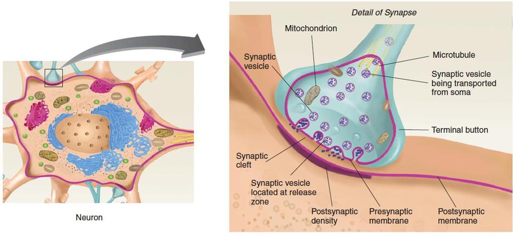
20 Neurotransmitters (NT) The presynaptic membrane, located at the end of the terminal button, faces the postsynaptic membrane, located on the neuron that receives the message (the postsynaptic neuron). Presynaptic Membrane The membrane of a terminal button that lies adjacent to the postsynaptic membrane and through which the neurotransmitter is released Postsynaptic Membrane The cell membrane opposite the terminal button in a synapse; the membrane of the cell that receives the message
21 Release of NT These two membranes face each other across the synaptic cleft, a gap that varies in size from synapse to synapse but is usually around 20 nm wide. (A nanometer (nm) is one billionth of a meter.) Synaptic Cleft The space between the presynaptic membrane and the postsynaptic membrane. The synaptic cleft contains extracellular fluid, through which the neurotransmitter diffuses. A meshwork of filaments crosses the synaptic cleft and keeps the presynaptic and postsynaptic membranes in alignment.
22 Release of NT Synaptic vesicles are found in greatest numbers around the part of the presynaptic membrane that faces the synaptic cleft near the release zone, the region from which the neurotransmitter is released. When action potentials are conducted down an axon (and down all of its branches), something happens inside all of the terminal buttons: A number of small synaptic vesicles located just inside the presynaptic membrane fuse with the membrane and then break open, spilling their contents into the synaptic cleft. Release Zone A region of the interior of the presynaptic membrane of a synapse to which synaptic vesicles attach and release their neurotransmitter into the synaptic cleft

23 1. The process begins when a population of synaptic vesicles become docked against the presynaptic membrane, ready to release their neurotransmitter into the synaptic cleft. 4. Some of the calcium ions that enter the terminal button bind with the clusters of protein molecules. This event makes the segments of the clusters of protein molecules move apart, producing a fusion pore a hole through both membranes that enables them to fuse together. The process of fusion takes approximately 0.1 msec 2. Docking is accomplished when clusters of protein molecules attach to other protein molecules located in the presynaptic membrane 3. The release zone of the presynaptic membrane contains voltage-dependent calcium channels. When the membrane of the terminal button is depolarized by an arriving action potential, the calcium channels open. Thus, when the voltagedependent calcium channels open, Ca2+ flows into the cell.

24 Recycling of the membrane Release of NT Kiss and run These synaptic vesicles release most or all of their neurotransmitter, the fusion pore closes, and the vesicles break away from the presynaptic membrane and get filled with neurotransmitter again. Merge and recycle Other vesicles merge and recycle and consequently lose their identity. The membranes of these vesicles merge with the presynaptic membrane. Little buds of membrane then pinch off into the cytoplasm and become synaptic vesicles. The appropriate proteins are inserted into the membrane of these vesicles, and the vesicles are filled with molecules of the neurotransmitter. Bulk endocytosis Large pieces of the membrane of the terminal button fold inward, break off, and enter the cytoplasm. New vesicles are formed from small buds that break off of these pieces of membrane.
25 Activation of NT Neurotransmitters then diffusing across the synaptic cleft and attaching to the binding sites of special protein molecules located in the postsynaptic membrane, called postsynaptic receptors. Once binding occurs, the postsynaptic receptors open neurotransmitterdependent ion channels, which permit the passage of specific ions into or out of the cell. Neurotransmitters open ion channels by at least two different methods: direct and indirect. We will discuss only direct method.

26 When a molecule of the appropriate neurotransmitter attaches to it, the ion channel opens. That receptor/ion channel is an ionotropic receptor.

27 Postsynaptic potentials can be either depolarizing (excitatory) or hyperpolarizing (inhibitory). What determines the nature of the postsynaptic potential at a particular synapse is determined by the characteristics of the postsynaptic receptors in particular, by the particular type of ion channel they open. Four major types of neurotransmitter-dependent ion channels are found in the postsynaptic membrane: sodium (Na+), potassium (K+), chloride (Cl ), and calcium (Ca2+)
.")
28 Postsynaptic potentials The neurotransmitter-dependent sodium channel is the most important source of excitatory postsynaptic potentials. When sodium channels are opened, the result is a depolarization an excitatory postsynaptic potential (EPSP). Excitatory Postsynaptic Potential (EPSP) An excitatory depolarization of the postsynaptic membrane of a synapse caused by the liberation of a neurotransmitter by the terminal button
 An inhibitory hyperpolarization of the postsynaptic membrane of a synapse caused by the liberation of a neurotransmitter by")
29 Postsynaptic potentials When potassium channels are open, they hyperpolarize the membrane, producing an inhibitory postsynaptic potential (IPSP). Inhibitory Postsynaptic Potential (IPSP) An inhibitory hyperpolarization of the postsynaptic membrane of a synapse caused by the liberation of a neurotransmitter by the terminal button
30 Postsynaptic potentials If the membrane is at the resting potential, nothing happens, because (as we saw earlier) the forces of diffusion and electrostatic pressure balance perfectly for the chloride ion. However, if the membrane potential has already been depolarized by the activity of excitatory synapses located nearby, then the opening of chloride channels will permit Cl to enter the cell. The influx of anions will bring the membrane potential back to its normal resting condition. Thus, the opening of chloride channels serves to neutralize EPSPs.
31 Postsynaptic potentials The fourth type of neurotransmitter-dependent ion channel is the calcium channel. Calcium ions (Ca2+), being positively charged and being located in highest concentration outside the cell, act like sodium ions; that is, the opening of calcium channels depolarizes the membrane, producing EPSPs. But calcium does even more. The entry of calcium into the terminal button triggers the migration of synaptic vesicles and the release of the neurotransmitter. In the dendrites of the postsynaptic cell, calcium binds with and activates special enzymes. These enzymes have a variety of effects, including the production of biochemical and structural changes in the postsynaptic neuron.

32 The more synapses a neuron has, the greater its information-processing capability. Each pyramidal cell in cerebral cortex has about 40,000 synaptic contacts with other neurons. The cerebral cortex alone (the main information-processing tissue of your brain) is estimated to have 100 trillion (10 14 ) synapses. So how a synaptic knob decide to fire?
33 Neural Integration Effects of Postsynaptic Potentials: Neural Integration Neurons are interconnected by means of synapses, and action potentials trigger the release of neurotransmitters, these chemicals initiate excitatory or inhibitory postsynaptic potentials. Excitatory postsynaptic potentials increase the likelihood that the postsynaptic neuron will fire; inhibitory postsynaptic potentials decrease this likelihood. (Remember, firing refers to the occurrence of an action potential.) Thus, the rate at which an axon fires is determined by the relative activity of the excitatory and inhibitory synapses on the soma and dendrites of that cell. If there are no active excitatory synapses, or if the activity of inhibitory synapses is particularly high, that rate could be close to zero.
 are then transmitted down the dendrites, across the soma, to the axon hillock located at the base of the axon.")
34 The release of the neurotransmitter produces depolarizing EPSPs in the dendrites of the neuron. These EPSPs (represented in red) are then transmitted down the dendrites, across the soma, to the axon hillock located at the base of the axon. If the depolarization is still strong enough when it reaches this point, the axon will fire. Now let s consider what would happen if, at the same time, inhibitory synapses also become active. Inhibitory postsynaptic potentials are hyperpolarizing they bring the membrane potential away from the threshold of excitation. Thus, they tend to cancel the effects of excitatory postsynaptic potentials.
35 Neural Integration The rate at which a neuron fires is controlled by the relative activity of the excitatory and inhibitory synapses on its dendrites and soma. If the activity of excitatory synapses goes up, the rate of firing will go up. If the activity of inhibitory synapses goes up, the rate of firing will go down. Note that neural inhibition (that is, an inhibitory postsynaptic potential) does not always produce behavioral inhibition. For example, suppose a group of neurons inhibits a particular movement. If these neurons are inhibited, they will no longer suppress the behavior.
36 Thus, inhibition of the inhibitory neurons makes the behavior more likely to occur. Of course, the same is true for neural excitation. Excitation of neurons that inhibit a behavior suppresses that behavior. For example, when we are dreaming, a particular set of inhibitory neurons in the brain becomes active and prevents us from getting up and acting out our dreams. Neurons are elements in complex circuits; without knowing the details of these circuits, one cannot predict the effects of the excitation or inhibition of one set of neurons on an organism s behavior.
37 Electrical communication Some synapses are electrical; the membranes meet and almost touch, forming a gap junction. Gap Junction is a special junction between cells that permits direct communication by means of electrical coupling. The membranes on both sides of a gap junction contain channels that permit ions to diffuse from one cell to another. Thus, changes in the membrane potential of one neuron induce changes in the membrane of the other.
38 A single action potential in a synaptic knob does not produce enough activity to make a postsynaptic cell fire. An EPSP may be produced, but it fades before reaching threshold. A typical EPSP is a voltage change of only 0.5 mv and lasts only 15 to 20 ms. If a neuron has an MP of 70 mv and a threshold of 55 mv, it needs at least 30 EPSPs to reach threshold and fire. There are two ways in which EPSPs can add up to do this, and both may occur simultaneously.

39 Spatial summation. This occurs when EPSPs from several synapses add up to threshold at the axon hillock. Any one synapse may admit only a moderate amount of Na+ into the cell, but several synapses acting together admit enough Na+ to reach threshold. The presynaptic neurons cooperate to induce the postsynaptic neuron to fire. Temporal summation. This occurs when a single synapse generates EPSPs so quickly that each is generated before the previous one fades. This allows the EPSPs to add up over time to a threshold voltage that triggers an action potential. Temporal summation can occur if even one presynaptic neuron stimulates the postsynaptic neuron intensely enough.
40 Neural coding The nervous system must interpret and pass along both quantitative and qualitative information about its environment whether a light is dim or bright, red or green; whether a taste is mild or intense, salty or sour; whether a sound is loud or soft, high-pitched or low. Considering the complexity of information to be communicated about conditions in and around the body, it seems a marvel that it can be done in the form of something as simple as action potentials particularly since all the action potentials of a given neuron are identical. The way in which the nervous system converts information to a meaningful pattern of action potentials is called neural coding (or sensory coding when it occurs in the sense organs).
41 Neural coding Qualitative Information coding: The most important mechanism for transmitting qualitative information is the labeled line code. This code is based on the fact that each nerve fiber to the brain leads from a receptor that specifically recognizes a particular stimulus type. Nerve fibers in the optic nerve, for example, carry signals only from light receptors in the eye; these fibers will never carry information about taste or sound. The brain therefore interprets any signals in those fibers in terms of light even if the signals result from artificial stimulation of the nerve.

42 Neural coding Quantitative information information about the intensity of a stimulus strength depends on the fact that the more strongly a neuron is stimulated, the more frequently it fires. A weak stimulus may cause a neuron to generate 6 action potentials per second, and a strong stimulus, 600 per second. Thus, the central nervous system can judge stimulus strength from the firing frequency of afferent neurons This figure is based on recordings made from a sensory fiber of the frog sciatic nerve as the gastrocnemius muscle was stretched by suspending weights from it. As the stimulus strength (weight) and stretch increase, the firing frequency of the neuron increases. Firing frequency is a coded message that informs the CNS of stimulus intensity.
43 Neural Circuits

44 Neural Circuits
45 Neural Circuits Reverberating (oscillatory) circuit: Some circuits are constructed so that once the presynaptic cell is stimulated, it will cause the postsynaptic cell to transmit a series of nerve impulses. One such circuit is called a reverberating (oscillatory) circuit. In this pattern, the incoming impulse stimulates the first neuron, which stimulates the second, which stimulates the third, and so on. Branches from later neurons synapse with earlier ones, however, sending impulses back through the circuit again and again. The output signal may last from a few seconds to many hours, depending on the number of synapses and arrangement of neurons in the circuit. Inhibitory neurons may turn off a reverberating circuit after a period of time. Among the body responses thought to be the result of output signals from reverberating circuits are breathing (A reverberating circuit sends repetitious signals to your diaphragm and intercostal muscles, for example, to make you inhale. When the circuit stops firing, you exhale; the next time it fires, you inhale again. ), coordinated muscular activities etc.
46 Neural Circuits A fourth type of circuit is the parallel after-discharge circuit. In this circuit, a single presynaptic cell stimulates a group of neurons, each of which synapses with a common postsynaptic cell. A differing number of synapses between the first and last neurons imposes varying synaptic delays so that the last neuron exhibits multiple EPSPs or IPSPs. If the input is excitatory, the postsynaptic neuron then can send out a stream of impulses in quick succession. It is thought that parallel afterdischarge circuits may be employed for precise activities such as mathematical calculations.
47 The story so far. The excitation as a result of integration could result in neuronal firing (spikes) Spikes are important since other neurons receive them (as a signals) Neurons communicate with spikes Information is coded by spikes So if we can manage to measure the spiking time, we decipher how the brain works.
48 Example: Neural integration Neural integration of bilateral olfactory inputs enhances signal-to-noise ratio. (a) Schematic diagram of the bilateral olfactory input pathways and a hypothetical central neuron (grey circle) receiving those inputs. Information is transmitted as spiking activity. Typically, in the absence of any olfactory stimulus, the receptor neurons tend to show a baseline spiking response that contributes to the 'noise' in the system. (b) Neural integration can reduce uncorrelated noise. The plots on the left represent the firing rate of two receptor neurons over time. The baseline fluctuations observed in the two independent channels (left) are reduced after integrating them (right), thus improving signal-tonoise ratio. This improvement may be the chief contribution of dual olfactory inputs to chemotaxis. The green box indicates the release of a puff of odor.
49 Neural integration Single spiking time is meaningless and to extract useful information, we have to average 1. a group of neurons in a local circuit where neuro codes the same information 2. over a time window to obtain the firing rate r r = = Local circuit = 6 Hz Time window = 1 sec
50 Hence we have firing rate of a group of neurons Neural integration r 1 So we can have a network of these local groups w 1: synaptic strength R = f w r j ) ( j w n r n
51 r i is the firing rate of input local circuit The neurons at output local circuits receives signals in the form N i= 1 w i r i The output firing rate of the output local circuit is then given by R R = f ( N i= 1 w i r i where f is the activation function, generally a Sigmoidal function of some sort ) w i weight, (synaptic strength) measuring the strength of the interaction between neurons.

52 EEG and multi cell activity
53 Learning in the Brain Brains learn Altering strength between neurons Creating/deleting connections Hebb s Postulate (Hebbian Learning) When an axon of cell A is near enough to excite a cell B and repeatedly or persistently takes part in firing it, some growth process or metabolic change takes place in one or both cells such that A's efficiency, as one of the cells firing B, is increased. Long Term Potentiation (LTP) Cellular basis for learning and memory LTP is the long-lasting strengthening of the connection between two nerve cells in response to stimulation Discovered in many regions of the cortex
54 The EEG measures not action potentials but summation of graded Post Synaptic Potentials (PSPs) What is summation of graded Post Synaptic Potentials (PSPs)? Let s revisit
55 Types of Synapses CNS Synapses Axodendritic: Axon to dendrite Axosomatic: Axon to cell body Axoaxonic: Axon to axon Dendrodendritic: Dendrite to dendrite

56 Principles of Synaptic Integration

57 Principles of Synaptic Integration EPSP Summation Allows for neurons to perform sophisticated computations Integration: EPSPs added together to produce significant postsynaptic depolarization Spatial summation : adding together of EPSPs generated simultaneously at different synapses Temporal summation : adding together of EPSPs generated at the same synapse in rapid succession (within 1-15 msec of one another)

58 The Contribution of Dendritic Properties to Synaptic Integration Dendrite as a straight cable : EPSPs have to travel down to spike-initiation zone to generate action potential Membrane depolarization falls off exponentially with increasing distance V x = V o /e x/ Vo : depolarization at the origin : Dendritic length constant Distance where the depolarization is 37% of origin (V = 0.37 V o ) In reality, dendrites have branches, changing diameter..
59 Postsynaptic Potentials Signaling in Dendrites & Soma V m changes dendrites & soma Excitatory: + Inhibitory: -
60 Postsynaptic Potentials - PSPs Chemically-gated ion channels Graded Summation Duration milliseconds Decremental Weaken over time & space
61 Postsynaptic Potentials Soma & Dendrites Chemically-gated channels Passive current Graded Summation EPSP Excitatory Depolarization Na+ influx AP more likely IPSP Inhibitory Hyperpolarization K+ efflux AP less likely
62 EPSPs Excitatory Postsynaptic Potential Depolarization (+) V m becomes more positive Na+ influx
63 EPSP Record here + + Depolarization more likely to Action Potential V m -55 mv - 65 mv AT REST - Time
64 Temporal Summation + V m -55 mv + 2 pulses same synapse ~ - 65 mv AT REST - Time
65 Temporal Summation + + Repeated stimulation V m -55 mv - 65 mv AT REST - Time
66 Spatial Summation V m -55 mv Multiple synapses - 65 mv AT REST - Time
67 IPSPs Inhibitory Postsynaptic Potential similar to EPSPs but opposite hyperpolarization (-) V m becomes more negative K+ efflux
68 IPSP - + V m -55 mv - 65 mv Hyperpolarization less likely to fire also summate (max) AT REST - Time
69 EPSPs & IPSPs summate CANCEL EACH OTHER Net stimulation EPSPs + IPSPs = net effects Or we Integrate over time
70 + EPSP - IPSP - 65 mv
71 Dipole

72 Summation of post synaptic potentials If an excitatory neurotransmitter is released at the apical dendrites of a cortical pyramidal cell, current will flow from the extracellular space into the cell, yielding a net negativity on the outside of the cell in the region of the apical dendrites. To complete the circuit, current will also flow out of the cell body and basal dendrites, yielding a net positivity in this area. Together, the negativity at the apical dendrites and the positivity at the cell body create a tiny dipole (a dipole is simply a pair of positive and negative electrical charges separated by a small distance)

73 Aligned neurons and synchronous activity Neurons which are radially symmetric, randomly oriented or asynchronously activated do not produce externally observable electric or magnetic fields. Neurons which are non radially symmetric, spatially aligned and synchronously activated add up to produce externally observable electric or magnetic fields
74 The summation of the individual dipoles is complicated by the fact that the cortex is not flat, but instead has many folds. But the summation of many dipoles is essentially equivalent to a single dipole formed by averaging the orientations of the individual dipoles. This averaged dipole is called an equivalent current dipole (ECD). It is important to note, however, that whenever the individual dipoles are more than 90 degrees from each other, they will cancel each other to some extent, with complete cancellation at 180 degrees. Why its hard to record cerebellar activity from the scalp For example, the Purkinje cells in the cerebellar cortex are beautifully aligned with each other and oriented perpendicular to the cortical surface, but the cortical surface is so highly folded that the dipoles in one small patch of cerebellar cortex will almost always be cancelled by dipoles in a nearby but oppositely oriented patch, making it difficult or impossible to record cerebellar activity from the scalp.
75 Volume conduction Alignment of the dipoles is primarily responsible for over the skull recordings. But it cause a problem as well. When a dipole is present in a conductive medium such as the brain, current is conducted throughout that medium until it reaches the surface. This is called volume conduction. The voltage that will be present at any given point on the surface of the scalp will depend on the position and orientation of the generator dipole. Electricity does not just run directly between the two poles of a dipole in a conductive medium, but instead spreads out through the conductor. Consequently, potentials spread out as they travel through the brain. In addition, because electricity tends to follow the path of least resistance, potentials tend to spread laterally when they encounter the high resistance of the skull. Volume conduction eliminating algorithms will briefly be covered later.
76 Volume conduction The voltage that will be present at any given point on the surface of the scalp will depend on the position and orientation of the generator dipole. Consider a radial oriented dipole. The resultant electric potential on the scalp is as in figure.
77 Magnetic Field of Brain Blurring of the electrical field can be addressed by an other way. An electrical dipole is always surrounded by a magnetic field, and these fields summate in the same manner as voltages. Thus, whenever potential is generated, a magnetic field is also generated, running around the dipole. This is called magnetoencephalogram (MEG). However, magnetic recordings are very expensive because supercooling is expensive and because an expensive magnetically shielded recording chamber is necessary to attenuate the Earth s relatively large magnetic field.
Communication within a Neuron
 Neuronal Communication, Ph.D. Communication within a Neuron Measuring Electrical Potentials of Axons The Membrane Potential The Action Potential Conduction of the Action Potential 1 The withdrawal reflex
Chapter 11 Introduction to the Nervous System and Nervous Tissue Chapter Outline
 Chapter 11 Introduction to the Nervous System and Nervous Tissue Chapter Outline Module 11.1 Overview of the Nervous System (Figures 11.1-11.3) A. The nervous system controls our perception and experience
QUIZ YOURSELF COLOSSAL NEURON ACTIVITY
 QUIZ YOURSELF What are the factors that produce the resting potential? How is an action potential initiated and what is the subsequent flow of ions during the action potential? 1 COLOSSAL NEURON ACTIVITY
Na + K + pump. The beauty of the Na + K + pump. Cotransport. The setup Cotransport the result. Found along the plasma membrane of all cells.
 The beauty of the Na + K + pump Na + K + pump Found along the plasma membrane of all cells. Establishes gradients, controls osmotic effects, allows for cotransport Nerve cells have a Na + K + pump and
Portions from Chapter 6 CHAPTER 7. The Nervous System: Neurons and Synapses. Chapter 7 Outline. and Supporting Cells
 CHAPTER 7 The Nervous System: Neurons and Synapses Chapter 7 Outline Neurons and Supporting Cells Activity in Axons The Synapse Acetylcholine as a Neurotransmitter Monoamines as Neurotransmitters Other
The Nervous System. Nervous System Functions 1. gather sensory input 2. integration- process and interpret sensory input 3. cause motor output
 The Nervous System Nervous System Functions 1. gather sensory input 2. integration- process and interpret sensory input 3. cause motor output The Nervous System 2 Parts of the Nervous System 1. central
Physiology of synapses and receptors
 Physiology of synapses and receptors Dr Syed Shahid Habib Professor & Consultant Clinical Neurophysiology Dept. of Physiology College of Medicine & KKUH King Saud University REMEMBER These handouts will
Omar Sami. Muhammad Abid. Muhammad khatatbeh
 10 Omar Sami Muhammad Abid Muhammad khatatbeh Let s shock the world In this lecture we are going to cover topics said in previous lectures and then start with the nerve cells (neurons) and the synapses
Neurons. Pyramidal neurons in mouse cerebral cortex expressing green fluorescent protein. The red staining indicates GABAergic interneurons.
 Neurons Pyramidal neurons in mouse cerebral cortex expressing green fluorescent protein. The red staining indicates GABAergic interneurons. MBL, Woods Hole R Cheung MSc Bioelectronics: PGEE11106 1 Neuron
What is Anatomy and Physiology?
 Introduction BI 212 BI 213 BI 211 Ecosystems Organs / organ systems Cells Organelles Communities Tissues Molecules Populations Organisms Campbell et al. Figure 1.4 Introduction What is Anatomy and Physiology?
Ameen Alsaras. Ameen Alsaras. Mohd.Khatatbeh
 9 Ameen Alsaras Ameen Alsaras Mohd.Khatatbeh Nerve Cells (Neurons) *Remember: The neural cell consists of: 1-Cell body 2-Dendrites 3-Axon which ends as axon terminals. The conduction of impulse through
3) Most of the organelles in a neuron are located in the A) dendritic region. B) axon hillock. C) axon. D) cell body. E) axon terminals.
 Most of the organelles in a neuron are located in the A) dendritic region. B) axon hillock. C) axon. D) cell body. E) axon terminals.") Chapter 48 Neurons, Synapses, and Signaling Multiple-Choice Questions 1) A simple nervous system A) must include chemical senses, mechanoreception, and vision. B) includes a minimum of 12 ganglia. C) has
Chapter 48 Neurons, Synapses, and Signaling Multiple-Choice Questions 1) A simple nervous system A) must include chemical senses, mechanoreception, and vision. B) includes a minimum of 12 ganglia. C) has
NEURONS COMMUNICATE WITH OTHER CELLS AT SYNAPSES 34.3
 NEURONS COMMUNICATE WITH OTHER CELLS AT SYNAPSES 34.3 NEURONS COMMUNICATE WITH OTHER CELLS AT SYNAPSES Neurons communicate with other neurons or target cells at synapses. Chemical synapse: a very narrow
Introduction to Neurobiology
 Biology 240 General Zoology Introduction to Neurobiology Nervous System functions: communication of information via nerve signals integration and processing of information control of physiological and
BIOL Week 6. Nervous System. Transmission at Synapses
 Collin County Community College BIOL 2401 Week 6 Nervous System 1 Transmission at Synapses Synapses are the site of communication between 2 or more neurons. It mediates the transfer of information and
ANATOMY AND PHYSIOLOGY OF NEURONS. AP Biology Chapter 48
 ANATOMY AND PHYSIOLOGY OF NEURONS AP Biology Chapter 48 Objectives Describe the different types of neurons Describe the structure and function of dendrites, axons, a synapse, types of ion channels, and
BIOLOGY 2050 LECTURE NOTES ANATOMY & PHYSIOLOGY I (A. IMHOLTZ) FUNDAMENTALS OF THE NERVOUS SYSTEM AND NERVOUS TISSUE P1 OF 5
 FUNDAMENTALS OF THE NERVOUS SYSTEM AND NERVOUS TISSUE P1 OF 5") P1 OF 5 The nervous system controls/coordinates the activities of cells, tissues, & organs. The endocrine system also plays a role in control/coordination. The nervous system is more dominant. Its mechanisms
P1 OF 5 The nervous system controls/coordinates the activities of cells, tissues, & organs. The endocrine system also plays a role in control/coordination. The nervous system is more dominant. Its mechanisms
Cell communication. Gated ion channels. Allow specific ions to pass only when gates are open
 increase decrease Cell communication Gated ion channels Allow specific ions to pass only when gates are open Triggered by: potential change, chemical binding, temperature change, stretching 1 Voltage-Gated
Cell communication. Gated ion channels. Voltage-Gated Na + Channel. Allow specific ions to pass only when gates are open
 increase decrease Cell communication Gated ion channels Allow specific ions to pass only when gates are open Voltage-Gated Na + Channel Activation gate ECF Triggered by: change, chemical binding, temperature
Ch. 45 Continues (Have You Read Ch. 45 yet?) u Central Nervous System Synapses - Synaptic functions of neurons - Information transmission via nerve
 u Central Nervous System Synapses - Synaptic functions of neurons - Information transmission via nerve") Ch. 45 Continues (Have You Read Ch. 45 yet?) u Central Nervous System Synapses - Synaptic functions of neurons - Information transmission via nerve impulses - Impulse may be blocked in its transmission
Ch. 45 Continues (Have You Read Ch. 45 yet?) u Central Nervous System Synapses - Synaptic functions of neurons - Information transmission via nerve impulses - Impulse may be blocked in its transmission
5-Nervous system II: Physiology of Neurons
 5-Nervous system II: Physiology of Neurons AXON ION GRADIENTS ACTION POTENTIAL (axon conduction) GRADED POTENTIAL (cell-cell communication at synapse) SYNAPSE STRUCTURE & FUNCTION NEURAL INTEGRATION CNS
LESSON 3.2 WORKBOOK How do our neurons communicate with each other?
 LESSON 3.2 WORKBOOK How do our neurons communicate with each other? This lesson introduces you to how one neuron communicates with another neuron during the process of synaptic transmission. In this lesson
Applied Neuroscience. Conclusion of Science Honors Program Spring 2017
 Applied Neuroscience Conclusion of Science Honors Program Spring 2017 Review Circle whichever is greater, A or B. If A = B, circle both: I. A. permeability of a neuronal membrane to Na + during the rise
9/28/2016. Neuron. Multipolar Neuron. Astrocytes Exchange Materials With Neurons. Glia or Glial Cells ( supporting cells of the nervous system)
") Neuron Multipolar Neuron https://www.youtube.com/watch?v=lw-psbnu5xago to :38 Glia or Glial Cells ( supporting cells of the nervous system) 10X more numerous than neurons but one-tenth the size make up
Neuron Multipolar Neuron https://www.youtube.com/watch?v=lw-psbnu5xago to :38 Glia or Glial Cells ( supporting cells of the nervous system) 10X more numerous than neurons but one-tenth the size make up
LESSON 3.3 WORKBOOK. Why does applying pressure relieve pain?
 Postsynaptic potentials small changes in voltage (membrane potential) due to the binding of neurotransmitter. Receptor-gated ion channels ion channels that open or close in response to the binding of a
Lecture 22: A little Neurobiology
 BIO 5099: Molecular Biology for Computer Scientists (et al) Lecture 22: A little Neurobiology http://compbio.uchsc.edu/hunter/bio5099 Larry.Hunter@uchsc.edu Nervous system development Part of the ectoderm
Neurons: Structure and communication
 Neurons: Structure and communication http://faculty.washington.edu/chudler/gall1.html Common Components of a Neuron Dendrites Input, receives neurotransmitters Soma Processing, decision Axon Transmits
The Nervous System. B. The Components: 1) Nerve Cells Neurons are the cells of the body and are specialized to carry messages through an process.
 Nerve Cells Neurons are the cells of the body and are specialized to carry messages through an process.") The Nervous System A. The Divisions: 1) The Central Nervous System includes the and. The brain contains billions of nerve cells called, and trillions of support cells called. 2) The Peripheral Nervous
The Nervous System A. The Divisions: 1) The Central Nervous System includes the and. The brain contains billions of nerve cells called, and trillions of support cells called. 2) The Peripheral Nervous
Neurons, Synapses and Signaling. Chapter 48
 Neurons, Synapses and Signaling Chapter 48 Warm Up Exercise What types of cells can receive a nerve signal? Nervous Organization Neurons- nerve cells. Brain- organized into clusters of neurons, called
Nervous System. Master controlling and communicating system of the body. Secrete chemicals called neurotransmitters
 Nervous System Master controlling and communicating system of the body Interacts with the endocrine system to control and coordinate the body s responses to changes in its environment, as well as growth,
LESSON 3.3 WORKBOOK. Why does applying pressure relieve pain? Workbook. Postsynaptic potentials
 Depolarize to decrease the resting membrane potential. Decreasing membrane potential means that the membrane potential is becoming more positive. Excitatory postsynaptic potentials (EPSP) graded postsynaptic
Chapter 4 Neuronal Physiology
 Chapter 4 Neuronal Physiology V edit. Pg. 99-131 VI edit. Pg. 85-113 VII edit. Pg. 87-113 Input Zone Dendrites and Cell body Nucleus Trigger Zone Axon hillock Conducting Zone Axon (may be from 1mm to more
Neurons Chapter 7 2/19/2016. Learning Objectives. Cells of the Nervous System. Cells of the Nervous System. Cells of the Nervous System
 Learning Objectives Neurons Chapter 7 Identify and describe the functions of the two main divisions of the nervous system. Differentiate between a neuron and neuroglial cells in terms of structure and
EE 791 Lecture 2 Jan 19, 2015
 EE 791 Lecture 2 Jan 19, 2015 Action Potential Conduction And Neural Organization EE 791-Lecture 2 1 Core-conductor model: In the core-conductor model we approximate an axon or a segment of a dendrite
Chapter 45: Synapses Transmission of Nerve Impulses Between Neurons. Chad Smurthwaite & Jordan Shellmire
 Chapter 45: Synapses Transmission of Nerve Impulses Between Neurons Chad Smurthwaite & Jordan Shellmire The Chemical Synapse The most common type of synapse used for signal transmission in the central
Action potential. Definition: an all-or-none change in voltage that propagates itself down the axon
 Action potential Definition: an all-or-none change in voltage that propagates itself down the axon Action potential Definition: an all-or-none change in voltage that propagates itself down the axon Naturally
Chapter 11: Nervous System and Nervous Tissue
 Chapter 11: Nervous System and Nervous Tissue I. Functions and divisions of the nervous system A. Sensory input: monitor changes in internal and external environment B. Integrations: make decisions about
Anatomy Review. Graphics are used with permission of: Pearson Education Inc., publishing as Benjamin Cummings (
 Anatomy Review Graphics are used with permission of: Pearson Education Inc., publishing as Benjamin Cummings (http://www.aw-bc.com) Page 1. Introduction Neurons communicate with other cells at junctions
Chapter 6 subtitles postsynaptic integration
 CELLULAR NEUROPHYSIOLOGY CONSTANCE HAMMOND Chapter 6 subtitles postsynaptic integration INTRODUCTION (1:56) This sixth and final chapter deals with the summation of presynaptic currents. Glutamate and
PSY 215 Lecture 3 (1/19/2011) (Synapses & Neurotransmitters) Dr. Achtman PSY 215
 (Synapses & Neurotransmitters) Dr. Achtman PSY 215") Corrections: None needed. PSY 215 Lecture 3 Topic: Synapses & Neurotransmitters Chapters 2 & 3, pages 40-57 Lecture Notes: SYNAPSES & NEUROTRANSMITTERS, CHAPTER 3 Action Potential (above diagram found
Corrections: None needed. PSY 215 Lecture 3 Topic: Synapses & Neurotransmitters Chapters 2 & 3, pages 40-57 Lecture Notes: SYNAPSES & NEUROTRANSMITTERS, CHAPTER 3 Action Potential (above diagram found
Chapter 11: Functional Organization of Nervous Tissue
 Chapter 11: Functional Organization of Nervous Tissue I. Functions of the Nervous System A. List and describe the five major nervous system functions: 1. 2. 3. 4. 5. II. Divisions of the Nervous System
NERVOUS SYSTEM 1 CHAPTER 10 BIO 211: ANATOMY & PHYSIOLOGY I
 BIO 211: ANATOMY & PHYSIOLOGY I 1 Ch 10 A Ch 10 B This set CHAPTER 10 NERVOUS SYSTEM 1 BASIC STRUCTURE and FUNCTION Dr. Lawrence G. Altman www.lawrencegaltman.com Some illustrations are courtesy of McGraw-Hill.
Chapter 7. Objectives
 Chapter 7 The Nervous System: Structure and Control of Movement Objectives Discuss the general organization of the nervous system Describe the structure & function of a nerve Draw and label the pathways
2/27/2019. Functions of the Nervous System. Nervous Tissue and Neuron Function. Fundamentals Of The Nervous System And Nervous Tissue
 Nervous Tissue and Neuron Function Fundamentals Of The Nervous System And Nervous Tissue Learn and Understand 1. Like muscle cells, neurons use membrane polarity upset (AP) as a signal therefore keeping
NEURAL TISSUE (NEUROPHYSIOLOGY) PART I (A): NEURONS & NEUROGLIA
 PART I (A): NEURONS & NEUROGLIA") PART I (A): NEURONS & NEUROGLIA Neural Tissue Contains 2 kinds of cells: neurons: cells that send and receive signals neuroglia (glial cells): cells that support and protect neurons Neuron Types Sensory
PART I (A): NEURONS & NEUROGLIA Neural Tissue Contains 2 kinds of cells: neurons: cells that send and receive signals neuroglia (glial cells): cells that support and protect neurons Neuron Types Sensory
Bio11: The Nervous System. Body control systems. The human brain. The human brain. The Cerebrum. What parts of your brain are you using right now?
 Bio11: The Nervous System Body control systems Nervous system Quick Sends message directly to target organ Endocrine system Sends a hormone as a messenger to the target organ Can target several organs
Lesson 14. The Nervous System. Introduction to Life Processes - SCI 102 1
 Lesson 14 The Nervous System Introduction to Life Processes - SCI 102 1 Structures and Functions of Nerve Cells The nervous system has two principal cell types: Neurons (nerve cells) Glia The functions
1. What are the two basic types of cells in the nervous system? Neurons and Glial Cells
 Biological Psychology Basic Structure of a Neuron 1. What are the two basic types of cells in the nervous system? Neurons and Glial Cells a. Cells that process incoming signals and respond by sending out
Chapter 7. The Nervous System: Structure and Control of Movement
 Chapter 7 The Nervous System: Structure and Control of Movement Objectives Discuss the general organization of the nervous system Describe the structure & function of a nerve Draw and label the pathways
Neurons, Synapses, and Signaling
 Neurons, Synapses, and Signaling The Neuron is the functional unit of the nervous system. Neurons are composed of a cell body, which contains the nucleus and organelles; Dendrites which are extensions
Branches of the Nervous System
 The Nervous System Branches of the Nervous System There are 2 main branches of the nervous system Central Nervous System Brain Spinal Cord Peripheral Nervous System All nerves leading to rest of body Anatomy
Synaptic Communication. Steven McLoon Department of Neuroscience University of Minnesota
 Synaptic Communication Steven McLoon Department of Neuroscience University of Minnesota 1 Course News The first exam is next week on Friday! Be sure to checkout the sample exam on the course website. 2
Unit III. Biological Bases of Behavior
 Unit III Biological Bases of Behavior Module 9: Biological Psychology and Neurotransmission Module 10: The Nervous and Endocrine Systems Module 11: Studying the Brain, and Other Structures Module 12: The
BIOLOGICAL PROCESSES
 BIOLOGICAL PROCESSES CHAPTER 3 1 LEARNING GOALS Discuss how the nervous system communicates internally. Describe the structure and function of neurons Describe how the neuron transmits information Describe
Outline. Neuron Structure. Week 4 - Nervous System. The Nervous System: Neurons and Synapses
 Outline Week 4 - The Nervous System: Neurons and Synapses Neurons Neuron structures Types of neurons Electrical activity of neurons Depolarization, repolarization, hyperpolarization Synapses Release of
Chapter 2: Cellular Mechanisms and Cognition
 Chapter 2: Cellular Mechanisms and Cognition MULTIPLE CHOICE 1. Two principles about neurons were defined by Ramón y Cajal. The principle of connectional specificity states that, whereas the principle
Chapter 2. The Cellular and Molecular Basis of Cognition Cognitive Neuroscience: The Biology of the Mind, 2 nd Ed.,
 Chapter 2. The Cellular and Molecular Basis of Cognition Cognitive Neuroscience: The Biology of the Mind, 2 nd Ed., M. S. Gazzaniga, R. B. Ivry, and G. R. Mangun, Norton, 2002. Summarized by B.-W. Ku,
SYNAPTIC COMMUNICATION
 BASICS OF NEUROBIOLOGY SYNAPTIC COMMUNICATION ZSOLT LIPOSITS 1 NERVE ENDINGS II. Interneuronal communication 2 INTERNEURONAL COMMUNICATION I. ELECTRONIC SYNAPSE GAP JUNCTION II. CHEMICAL SYNAPSE SYNAPSES
Division Ave. High School AP Biology. cell body. signal direction
 signal direction Nervous system cells Neuron a nerve cell dendrites myelin sheath axon cell body dendrite cell body axon Structure fits function many entry points for signal one path out transmits signal
Human Brain and Senses
 Human Brain and Senses Outline for today Levels of analysis Basic structure of neurons How neurons communicate Basic structure of the nervous system Levels of analysis Organism Brain Cell Synapses Membrane
STRUCTURAL ELEMENTS OF THE NERVOUS SYSTEM
 STRUCTURAL ELEMENTS OF THE NERVOUS SYSTEM STRUCTURE AND MAINTENANCE OF NEURONS (a) (b) Dendrites Cell body Initial segment collateral terminals (a) Diagrammatic representation of a neuron. The break in
Thursday, January 22, Nerve impulse
 Nerve impulse Transmembrane Potential caused by ions moving through cell membrane at different rates Two main ions of concern Na + - Sodium K + - potassium Cell membrane not freely permeable therefore
Cellular Bioelectricity
 ELEC ENG 3BB3: Cellular Bioelectricity Notes for Lecture 24 Thursday, March 6, 2014 8. NEURAL ELECTROPHYSIOLOGY We will look at: Structure of the nervous system Sensory transducers and neurons Neural coding
Part 11: Mechanisms of Learning
 Neurophysiology and Information: Theory of Brain Function Christopher Fiorillo BiS 527, Spring 2012 042 350 4326, fiorillo@kaist.ac.kr Part 11: Mechanisms of Learning Reading: Bear, Connors, and Paradiso,
Introduction to Physiological Psychology
 Introduction to Physiological Psychology Review Kim Sweeney ksweeney@cogsci.ucsd.edu www.cogsci.ucsd.edu/~ksweeney/psy260.html Today n Discuss Final Paper Proposal (due 3/10) n General Review 1 The article
Synaptic Integration
 Synaptic Integration 3 rd January, 2017 Touqeer Ahmed PhD Atta-ur-Rahman School of Applied Biosciences National University of Sciences and Technology Excitatory Synaptic Actions Excitatory Synaptic Action
Nervous System. Nervous system cells. Transmission of a signal 2/27/2015. Neuron
 Nervous System 2007-2008 signal direction Neuron a nerve cell Nervous system cells dendrites axon cell body Structure fits function many entry points for signal one path out transmits signal signal direction
The Nervous System AP Biology
 The Nervous System 2005-2006 Neuron (nerve cell) signal direction dendrites cell body Structure fits function, it have many entry points for signal one path out transmits signal Nodes of Ranvier axon signal
AP Biology Unit 6. The Nervous System
 AP Biology Unit 6 The Nervous System Branches of the Nervous System There are 2 main branches of the nervous system Central Nervous System Brain Spinal Cord Peripheral Nervous System All nerves leading
Chapter 2 The Brain or Bio Psychology
 Chapter 2 The Brain or Bio Psychology 1 2 3 1 Glial Cells Surround neurons and hold them in place Make Myelin (covering for neurons) Manufacture nutrient chemicals neurons need Absorb toxins and waste
BIPN100 F15 Human Physiology 1 Lecture 3. Synaptic Transmission p. 1
 BIPN100 F15 Human Physiology 1 Lecture 3. Synaptic Transmission p. 1 Terms you should know: synapse, neuromuscular junction (NMJ), pre-synaptic, post-synaptic, synaptic cleft, acetylcholine (ACh), acetylcholine
Function of the Nervous System
 Nervous System Function of the Nervous System Receive sensory information, interpret it, and send out appropriate commands to form a response Composed of neurons (functional unit of the nervous system)
Structure of a Neuron:
 Structure of a Neuron: At the dendrite the incoming signals arrive (incoming currents) At the soma current are finally integrated. At the axon hillock action potential are generated if the potential crosses
Neural Communication. Central Nervous System Peripheral Nervous System. Communication in the Nervous System. 4 Common Components of a Neuron
 Neural Communication Overview of CNS / PNS Electrical Signaling Chemical Signaling Central Nervous System Peripheral Nervous System Somatic = sensory & motor Autonomic = arousal state Parasympathetic =
Action Potentials and Synaptic Transmission. BIO 219 Napa Valley College Dr. Adam Ross
 Action Potentials and Synaptic Transmission BIO 219 Napa Valley College Dr. Adam Ross Review of action potentials Nodes of Ranvier Nucleus Dendrites Cell body In saltatory conduction, the nerve impulses
浙江大学医学院基础医学整合课程 各论 III. The Nervous System. Dr. ZHANG Xiong Dept. of Physiology ZJU School of Medicine
 The Nervous System Dr. ZHANG Xiong Dept. of Physiology ZJU School of Medicine xiongzhang@zju.edu.cn http://10.202.77.12/ 1 Part 1. Summary of the nervous system 2 The Nervous System Central Nervous System
Chapter 2. The Cellular and Molecular Basis of Cognition
 Chapter 2. The Cellular and Molecular Basis of Cognition Cognitive Neuroscience: The Biology of the Mind, 2 nd Ed., M. S. Gazzaniga,, R. B. Ivry,, and G. R. Mangun,, Norton, 2002. Summarized by B.-W. Ku,
The Nervous System. Dr. ZHANG Xiong Dept. of Physiology ZJU School of Medicine.
 The Nervous System Dr. ZHANG Xiong Dept. of Physiology ZJU School of Medicine Http://10.10.10.151 Part 1. Summary of the nervous system The Nervous System Central Nervous System Brain + Spinal Cord Peripheral
The action potential travels down both branches because each branch is a typical axon with voltage dependent Na + and K+ channels.
 BIO 360 - MIDTERM FALL 2018 This is an open book, open notes exam. PLEASE WRITE YOUR NAME ON EACH SHEET. Read each question carefully and answer as well as you can. Point values are shown at the beginning
Electrophysiology. General Neurophysiology. Action Potentials
 5 Electrophysiology Cochlear implants should aim to reproduce the coding of sound in the auditory system as closely as possible, for best sound perception. The cochlear implant is in part the result of
35-2 The Nervous System Slide 1 of 38
 1 of 38 35-2 The Nervous System The nervous system controls and coordinates functions throughout the body and responds to internal and external stimuli. 2 of 38 Neurons Neurons The messages carried by
Chapter 3 subtitles Action potentials
 CELLULAR NEUROPHYSIOLOGY CONSTANCE HAMMOND Chapter 3 subtitles Action potentials Introduction (3:15) This third chapter explains the calcium current triggered by the arrival of the action potential in
Department of medical physiology 1 st week
 Department of medical physiology 1 st week Semester: summer Study program: Dental medicine Lecture: RNDr. Soňa Grešová, PhD. Department of medical physiology 1 st week 1. General neurophysiology 2. Central
THE HISTORY OF NEUROSCIENCE
 THE HISTORY OF NEUROSCIENCE BIOLOGICAL ASPECTS OF BEHAVIOR: THE NEURON & NEURAL COMMUNICATION NERVOUS SYSTEM Combined activity of the brain, spinal cord & other nerve fibers Acts as an information processing
Neural Basis of Motor Control
 Neural Basis of Motor Control Central Nervous System Skeletal muscles are controlled by the CNS which consists of the brain and spinal cord. Determines which muscles will contract When How fast To what
THE HISTORY OF NEUROSCIENCE
 1. Historically, how have neuroscientists determined the function of various brain regions? 2. Describe the impact of the Phineas Gage case on the field of neuroscience. 3. Explain neuron theory. THE HISTORY
MOLECULAR AND CELLULAR NEUROSCIENCE
 MOLECULAR AND CELLULAR NEUROSCIENCE BMP-218 November 4, 2014 DIVISIONS OF THE NERVOUS SYSTEM The nervous system is composed of two primary divisions: 1. CNS - Central Nervous System (Brain + Spinal Cord)
10.1: Introduction. Cell types in neural tissue: Neurons Neuroglial cells (also known as neuroglia, glia, and glial cells) Dendrites.
 Dendrites.") 10.1: Introduction Copyright The McGraw-Hill Companies, Inc. Permission required for reproduction or display. Cell types in neural tissue: Neurons Neuroglial cells (also known as neuroglia, glia, and glial
10.1: Introduction Copyright The McGraw-Hill Companies, Inc. Permission required for reproduction or display. Cell types in neural tissue: Neurons Neuroglial cells (also known as neuroglia, glia, and glial
Summarized by B.-W. Ku, E. S. Lee, and B.-T. Zhang Biointelligence Laboratory, Seoul National University.
 Chapter 2. The Cellular l and Molecular Basis of Cognition Cognitive Neuroscience: The Biology of the Mind, 3 rd Ed., M. S. Gazzaniga, R. B. Ivry, and G. R. Mangun, Norton, 2008. Summarized by B.-W. Ku,
NERVOUS SYSTEM. Somatic (SNS) Fibers - transmit impulses from CNS to control voluntary action of skeletal muscle
 Fibers - transmit impulses from CNS to control voluntary action of skeletal muscle") NERVOUS SYSTEM The master controlling and communicating system of the body --- cells communicate via electrical and chemical signals. Signals are rapid, specific and cause almost immediate responses. Functions
NERVOUS SYSTEM The master controlling and communicating system of the body --- cells communicate via electrical and chemical signals. Signals are rapid, specific and cause almost immediate responses. Functions
3.E.2 Continued. This is the essential knowledge statement from the curriculum framework. Detect---process--- response
 Nervous System: Part III What Happens at a Synapse? 3.E. Continued Animals have nervous systems that detect external and internal signals, transmit and integrate information, and produce responses. This
Physiological and Physical Basis of Functional Brain Imaging 6. EEG/MEG. Kâmil Uludağ, 20. November 2007
 Physiological and Physical Basis of Functional Brain Imaging 6. EEG/MEG Kâmil Uludağ, 20. November 2007 Course schedule 1. Overview 2. fmri (Spin dynamics, Image formation) 3. fmri (physiology) 4. fmri
What effect would an AChE inhibitor have at the neuromuscular junction?
 CASE 4 A 32-year-old woman presents to her primary care physician s office with difficulty chewing food. She states that when she eats certain foods that require a significant amount of chewing (meat),
Endocrine System Nervous System
 Cells Endocrine System Nervous System Tissues Controls Organs Nervous System vs Endocrine System Electrical signals (graded potentials and action potentials) and chemical signals (neurotransmitters) Fast
PARTS central nervous system brain and spinal cord nerve bundle of neurons wrapped in connective tissue
 NEUROPHYSIOLOGY Electrical Properties of Nerve cells (neurons) Electro physiology of neurons lie in Membrane Physiology Model organisms is Squid Giant Axon (SGA) diversity of Nervous systems NERVOUS SYSTEM
Nervous Tissue and Neurophysiology
 Nervous Tissue and Neurophysiology Objectives Describe the two major divisions of the nervous system and their characteristics. Identify the structures/functions of a typical neuron. Describe the location
Neurophysiology scripts. Slide 2
 Neurophysiology scripts Slide 2 Nervous system and Endocrine system both maintain homeostasis in the body. Nervous system by nerve impulse and Endocrine system by hormones. Since the nerve impulse is an
Organization of the nervous system. [See Fig. 48.1]
![Organization of the nervous system. [See Fig. 48.1]](/thumbs/90/103926552.jpg "Organization of the nervous system. [See Fig. 48.1]") Nervous System [Note: This is the text version of this lecture file. To make the lecture notes downloadable over a slow connection (e.g. modem) the figures have been replaced with figure numbers as found
Nervous System [Note: This is the text version of this lecture file. To make the lecture notes downloadable over a slow connection (e.g. modem) the figures have been replaced with figure numbers as found
Version A. AP* Biology: Nervous System. Questions 1 and 2. Name: Period
 Name: Period Version A AP* Biology: Nervous System Directions: Each of the questions or incomplete statements below is followed by four suggested answers or completions. Select the one that is best in
Chapter 45 Neurons and Nervous Systems
 Nervous System Cells Neuron a cell Chapter 45 Neurons and Nervous Systems signal direction dendrites cell body Structure fits function many entry points for signal one path out axon signal direction transmits