Kaan Yücel M.D., Ph.D. 14.January.2014 Tuesday
|
|
|
- Anabel Walton
- 6 years ago
- Views:
Transcription

1 Kaan Yücel M.D., Ph.D. 14.January.2014 Tuesday
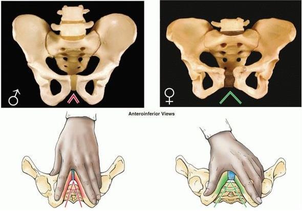
2 Sexual differences are related mainly 1. Heavier build and larger muscles of most men 2. Adaptation of the pelvis (particularly the lesser pelvis) in women for parturition (childbearing). The difference between the male and female pelvis Difference Between Male & Female Pelvis

3 male or funnel-shaped pelvis with a contracted outlet long, narrow, and oval shaped 41% of women wide pelvis 2% of women
, identification of human skeletal remains usually involves the")

4 In forensic medicine (the application of medical and anatomical knowledge for the purposes of law), identification of human skeletal remains usually involves the diagnosis of sex. A prime focus of attention is the pelvic girdle because sexual differences usually are clearly visible. Even fragments of the pelvic girdle are useful in determining sex.
5 eature Male pelvis Female pelvis eneral tructure Thick & Heavy Thin & Light reater elvis esser elvis Deep Narrow and deep, tapering Shallow Wide and shallow, cylindirical elvic inlet Heart-shaped, narrow Oval and rounded, wide elvic outlet Comparatively small Comparatively large schial pines Project further medially into the pelvic cavity Do not project as far medially into the pelvic cavity & smooth
6 Feature Male pelvis Female pelvis Obturator foramen Round Oval Acetabulum Large Small Greater schiatic notch Subpubic angle Sacral promontory Narrow, inverted V (approximately 70 degrees) Smaller (50-60 degrees) Prominent Almost 90 degrees Larger (80-85 degrees) Not prominent

7


8


9 PELVIC DIAMETERS (CONJUGATES) Size of the lesser pelvis important in obstetrics Because it is the bony canal through which the fetus passes during a vaginal birth. To determine the capacity of the female pelvis for childbearing, diameters of the lesser pelvis are noted radiographically or manually during a pelvic examination.


10 PELVIC DIAMETERS (CONJUGATES)


11 Diameters of pelvic outlet Antero - posterior diameters Anatomical antero-posterior diameter 11cm from tip of the coccyx to lower border of symphysis pubis Obstetric antero-posterior diameter 13 cm from tip of the sacrum to lower border of symphysis pubis as the coccyx moves backwards during the second stage of labour.


12 Diameters of pelvic outlet Transverse diameters Bituberous diameter 11 cm between inner aspects of ischial tuberosities Bispinous diameter 10.5 cm between tips of ischial spines
13 Diameters of pelvic inlet Antero - posterior diameters Anatomical antero-posterior diameter True conjugate 11cm from tip of sacral promontory to upper border of symphysis pubis

14 Diameters of pelvic inlet Antero - posterior diameters Obstetric conjugate 10.5 cm from tip of sacral promontory to the most bulging point on back of symphysis pubis,about 1 cm below its upper border. shortest antero-posterior diameter

15 Diameters of pelvic inlet Antero - posterior diameters Diagonal conjugate 12.5 cm 1.5 cm longer than the true conjugate From tip of sacral promontory to lower border of symphysis pubis
16 Minimum anteroposterior (AP) diameter of the lesser pelvis True (obstetrical) conjugate Narrowest distance through which the baby's head must pass in a vaginal delivery. This distance, however, cannot be measured directly during a pelvic examination because of the presence of the bladder.
 Measured by palpating sacral promontory with the tip of the middle finger, using the other hand to mark the")

17 Diagonal conjugate (from inferior pubic lig. to promontory) Measured by palpating sacral promontory with the tip of the middle finger, using the other hand to mark the level of the inferior margin of the pubic symphysis on the examining hand. After the examining hand is withdrawn, the distance between the tip of the index finger (1.5 cm shorter than the middle finger) and the marked level of the pubic symphysis is measured to estimate the true conjugate, which should be 11.0 cm or greater.

18

19

20 Transverse diameter is the greatest distance between the linea terminalis on either side of the pelvis.
21 Anteroposterior compression of the pelvis occurs during crush accidents (as when a heavy object falls on the pelvis). This type of trauma commonly produces fractures of the pubic rami. When the pelvis is compressed laterally, the acetabula and ilia are squeezed toward each other and may be broken.

22 Fractures of the bony pelvic ring are almost always multiple fractures or a fracture combined with a joint dislocation. Pelvic fractures can result from direct trauma to the pelvic bones, such as occurs during an automobile accident, or be caused by forces transmitted to these bones from the lower limbs during falls on the feet.

23 Weak areas of the pelvis, where fractures often occur: Pubic rami Acetabula Region of the sacroiliac joints Alae of the ilium 25 Year Old Male with displaced fracture of the sacrum and symphysis pubis. The most severe pelvic fractures separate the two sides of the pelvis from each other.


24 Pelvic fractures may cause injury to pelvic soft tissues, blood vessels, nerves, and organs. Fractures in the pubo-obturator area are relatively common and are often complicated because of their relationship to the urinary bladder and urethra, which may be ruptured or torn.


25 Sacroiliac joint dysfunction Degenerative arthritis (osteoarthritis) Pregnancy Gout Rheumatoid arthritis Psoriasis Ankylosing spondylitis X-ray of the sacroiliac joints showing joint space narrowing, erosive change and indistinct margins, due to sacroiliitis
PELVIS & SACRUM Dr. Jamila El-Medany Dr. Essam Eldin Salama
 PELVIS & SACRUM Dr. Jamila El-Medany Dr. Essam Eldin Salama Learning Objectives At the end of the lecture, the students should be able to : Describe the bony structures of the pelvis. Describe in detail
Skeletal System Module 13: The Pelvic Girdle and Pelvis
 OpenStax-CNX module: m47993 1 Skeletal System Module 13: The Pelvic Girdle and Pelvis Donna Browne Based on The Pelvic Girdle and Pelvis by OpenStax College This work is produced by OpenStax-CNX and licensed
C. Bones of the Pelvic Girdle
 C. Bones of the Pelvic Girdle 1. 2 coxal bones (a.k.a hip bones): -bony pelvis is made up of hip bones, sacrum, & coccyx -pelvic bones are large & heavy & attach to the axial skeleton via sacrum/coccyx
Copyright 2003 Pearson Education, Inc. publishing as Benjamin Cummings. Dr. Nabil Khouri MD, MSc, Ph.D
 Dr. Nabil Khouri MD, MSc, Ph.D Pelvic Girdle (Hip) Organization of the Lower Limb It is divided into: The Gluteal region The thigh The knee The leg The ankle The foot The thigh and the leg have compartments
Anatomy & Physiology Pelvic Girdles 10.1 General Information
 Anatomy & Physiology Pelvic Girdles 10.1 General Information ICan2Ed, Inc. In human anatomy, the pelvis (plural pelves or pelvises) is the lower part of. The area of the body that is between the abdomen
The os coxae or hip bone consists of three flat bones, ilium, ischium and pubis, which fuse together to form the acetabulum.
 The os coxae The os coxae or hip bone consists of three flat bones, ilium, ischium and pubis, which fuse together to form the acetabulum. The ilium extends from the acetabulum upwards forming the lateral
Copyright 2003 Pearson Education, Inc. publishing as Benjamin Cummings. Dr. Nabil khouri
 Dr. Nabil khouri Appendicular Skeleton The appendicular skeleton is made up of the bones of the upper and lower limbs and their girdles Two girdles: Pectoral girdles attach the upper limbs to the body
First practical session. Bones of the gluteal region
 First practical session 2017 Bones of the gluteal region The Hip bone The hip bone is made of: 1 The ilium: superior in position 2 The ischium:postero-inferior in position 3 The pubis: antero-inferior
Figure 7: Bones of the lower limb
 BONES OF THE APPENDICULAR SKELETON The appendicular skeleton is composed of the 126 bones of the appendages and the pectoral and pelvic girdles, which attach the limbs to the axial skeleton. Although the
Introduction to Anatomy. Dr. Maher Hadidi. Tala Ar ar. Mar/10th/2013
 Sheet Introduction to Anatomy Dr. Maher Hadidi Tala Ar ar 15 Mar/10th/2013 Lower limb The skeleton of the lower limb is the lower appendicular skeleton which consists of 2 parts: 1- Pelvic girdle. 2- Bones
Slide Read the tables it is about the difference between male & female pelvis.
 I didn t include the slides, this is only what the doctor read or said because he skipped a lot of things because we took it previously, very important to go back to the slides (*there is an edited version)
Inferior Pelvic Border
 Pelvis + Perineum Pelvic Cavity Enclosed by bony, ligamentous and muscular wall Contains the urinary bladder, ureters, pelvic genital organs, rectum, blood vessels, lymphatics and nerves Pelvic inlet (superior
LAB Notes#1. Ahmad Ar'ar. Eslam
 LAB Notes#1 Ahmad Ar'ar Eslam 1 P a g e Anatomy lab Notes Lower limb bones :- Pelvic girdle: It's the connection between the axial skeleton and the lower limb; it's made up of one bone called the HIP BONE
NOTES FROM GUTMAN LECTURE 10/26 Use this outline to study from. As you go through Gutman s lecture, fill in the topics.
 NOTES FROM GUTMAN LECTURE 10/26 Use this outline to study from. As you go through Gutman s lecture, fill in the topics. Anatomy above the arcuate line Skin Camper s fascia Scarpa s fascia External oblique
Chapter 8. The Appendicular Skeleton. Lecture Presentation by Lee Ann Frederick University of Texas at Arlington Pearson Education, Inc.
 Chapter 8 The Appendicular Skeleton Lecture Presentation by Lee Ann Frederick University of Texas at Arlington An Introduction to the Appendicular Skeleton The Appendicular Skeleton 126 bones Allows us
Biology 218 Human Anatomy. Adapted from Martini Human Anatomy 7th ed. Chapter 7 The Skeletal System Appendicular Division
 Adapted from Martini Human Anatomy 7th ed. Chapter 7 The Skeletal System Appendicular Division Introduction The appendicular skeleton includes: Pectoral girdle Shoulder bones Upper limbs Pelvic girdle
The Lower Limb. Anatomy RHS 241 Lecture 2 Dr. Einas Al-Eisa
 The Lower Limb Anatomy RHS 241 Lecture 2 Dr. Einas Al-Eisa The bony pelvis Protective osseofibrous ring for the pelvic viscera Transfer of forces to: acetabulum & head of femur (when standing) ischial
Pectoral (Shoulder) Girdle
 Girdle") Chapter 8 Skeletal System: Appendicular Skeleton Pectoral girdle Pelvic girdle Upper limbs Lower limbs 8-1 Pectoral (Shoulder) Girdle Consists of scapula and clavicle Clavicle articulates with sternum
Chapter 8 Skeletal System: Appendicular Skeleton Pectoral girdle Pelvic girdle Upper limbs Lower limbs 8-1 Pectoral (Shoulder) Girdle Consists of scapula and clavicle Clavicle articulates with sternum
Table 2. First Generated List of Expert Responses. Likert-Type Scale. Category or Criterion. Rationale or Comments (1) (2) (3) (4)
 (2) (3) (4)") Table 2. First Generated List of Expert Responses. Likert-Type Scale Category or Criterion Anatomical Structures and Features Skeletal Structures and Features (1) (2) (3) (4) Rationale or Comments 1. Bones
Table 2. First Generated List of Expert Responses. Likert-Type Scale Category or Criterion Anatomical Structures and Features Skeletal Structures and Features (1) (2) (3) (4) Rationale or Comments 1. Bones
Overview of the Skeleton: Bone Markings
 Name Overview of the Skeleton: Bone Markings Match the terms in column B with the appropriate description in column A. Column A 1. sharp, slender process* 2. small rounded projection* 3. narrow ridge of
SKELETAL SYSTEM 206. AXIAL SKELETON 80 APPENDICULAR SKELETON 126 (see Figure 6.1) Clavicle. Clavicle. Pectoral girdles. Scapula. Scapula.
 Clavicle. Clavicle. Pectoral girdles. Scapula. Scapula.") SKELETAL SYSTEM 206 AXIAL SKELETON 80 APPENDICULAR SKELETON 126 (see Figure 6.1) Pectoral girdles 4 Clavicle Scapula 2 2 Clavicle Scapula Humerus 2 Humerus Upper limbs 60 Radius 2 Ulna Carpal bones Metacarpal
SKELETAL SYSTEM 206 AXIAL SKELETON 80 APPENDICULAR SKELETON 126 (see Figure 6.1) Pectoral girdles 4 Clavicle Scapula 2 2 Clavicle Scapula Humerus 2 Humerus Upper limbs 60 Radius 2 Ulna Carpal bones Metacarpal
The Appendicular Skeleton
 8 The Appendicular Skeleton PowerPoint Lecture Presentations prepared by Jason LaPres Lone Star College North Harris 8-1 The Pectoral Girdle The Pectoral Girdle Also called shoulder girdle Connects the
It is formed by fusion of 3 bones: I. Ilium (superior bone). II. Pubis (antero-inferior bone). III. Ischium (postero-inferior bone).
. II. Pubis (antero-inferior bone). III. Ischium (postero-inferior bone).") It is formed by fusion of 3 bones: I. Ilium (superior bone). II. Pubis (antero-inferior bone). III. Ischium (postero-inferior bone). Pubis Acetabulum Ana (242 ) The three constituent of bones of the hip
It is formed by fusion of 3 bones: I. Ilium (superior bone). II. Pubis (antero-inferior bone). III. Ischium (postero-inferior bone). Pubis Acetabulum Ana (242 ) The three constituent of bones of the hip
Biology 218 Human Anatomy
 Chapter 8 Adapted from Tortora 10 th ed. LECTURE OUTLINE A. Introduction (p. 203) 1. The appendicular skeleton contains 126 bones that form: i. two pectoral (shoulder) girdles two upper limbs i one pelvic
Bones of Lower Limb. Dr. Heba Kalbouneh Associate Professor of Anatomy and Histology
 Bones of Lower Limb Dr. Heba Kalbouneh Associate Professor of Anatomy and Histology Bones of the lower limb Hip Bone Made up of 3 bones: 1) Ilium (flat), superior in position 2) Ischium (L), postero-inferior
Chapter 8B. The Skeletal System: Appendicular Skeleton. The Appendicular Skeleton. Clavicle. Pectoral (Shoulder) Girdle
 Girdle") The Appendicular Skeleton Chapter 8B The Skeletal System: Appendicular Skeleton 126 bones Pectoral (shoulder) girdle Pelvic (hip) girdle Upper limbs Lower limbs Functions primarily to facilitate movement
The Appendicular Skeleton Chapter 8B The Skeletal System: Appendicular Skeleton 126 bones Pectoral (shoulder) girdle Pelvic (hip) girdle Upper limbs Lower limbs Functions primarily to facilitate movement
Axial skeleton bones and markings
 Axial skeleton bones and markings Skull Cranial bones Frontal x 1 Supraorbital foramen Occipital x 1 Foramen magnum Occipital condyles Superior nuchal line Inferior nuchal line Anterior cranial fossa External
PRE-LAB EXERCISES. Before we get started, look up the definitions of these common bone marking terms: Canal: Condyle: Facet: Fissure:
 1 PRE-LAB EXERCISES When studying the skeletal system, the bones are often sorted into two broad categories: the axial skeleton and the appendicular skeleton. This lab focuses on the appendicular skeleton,
Chapter 8 Outline. Pectoral Girdle Upper Limb Pelvic Girdle Lower Limb Aging of the Appendicular Skeleton Development of the Appendicular Skeleton
 Chapter 8 Outline Pectoral Girdle Upper Limb Pelvic Girdle Lower Limb Aging of the Appendicular Skeleton Development of the Appendicular Skeleton Figure 8.1 Appendicular Skeleton Pectoral Girdle Clavicle
The Axial Skeleton Hyoid Bone. Lecture Overview. Marieb s Human Anatomy and Physiology. Chapter 7 The Axial and Appendicular Skeleton Lecture 14
 Marieb s Human Anatomy and Physiology Marieb Hoehn Chapter 7 The Axial and Appendicular Skeleton Lecture 14 1 Axial Skeleton Hyoid bone Bones of the orbit Paranasal sinuses Infantile skull Vertebral column
Pelvic Girdle
 ARTICULATIONS OF LOWER EXTREMITY Pages 429-437 Pelvic Girdle formed by connection of the hip bones and the sacrum Sacroiliac Joints compound joints synovial joint - anterior, between the auricular surfaces
B) cervix of uterus C) vagina D) rectum. 1. What number illustrates the adnexal area? (Fig. 4-64) A) 4 B) 5 C) 8 D) 9
 cervix of uterus C) vagina D) rectum. 1. What number illustrates the adnexal area? (Fig. 4-64) A) 4 B) 5 C) 8 D) 9") Pelvis Practice Problems 1. What number illustrates the adnexal area? (Fig. 4-64) A) 4 B) 5 C) 8 D) 9 2. What number illustrates the cervix? (Fig. 4-64) A) 4 B) 8 C) 5 D) 6 3. Which of the following is
Pelvis Practice Problems 1. What number illustrates the adnexal area? (Fig. 4-64) A) 4 B) 5 C) 8 D) 9 2. What number illustrates the cervix? (Fig. 4-64) A) 4 B) 8 C) 5 D) 6 3. Which of the following is
ABC of Emergency Radiology
 ABC of Emergency Radiology It is possible for the non-specialist to interpret pelvic radiographs accurately Important anatomical considerations THE PELVIS P A Driscoll, R Ross, D A Nicholson FIG i-line
Figure ) The area that causes the lengthwise growth of a long bone is indicated by letter. Diff: 2 Page Ref:
 The area that causes the lengthwise growth of a long bone is indicated by letter. Diff: 2 Page Ref:") Essentials of Anatomy and Physiology, 9e (Marieb) Chapter 5 The Skeletal System Short Answer Figure 5.1 Using Figure 5.1, identify the following: 1) Spongy bone is indicated by letter. Diff: 1 Page Ref:
Essentials of Anatomy and Physiology, 9e (Marieb) Chapter 5 The Skeletal System Short Answer Figure 5.1 Using Figure 5.1, identify the following: 1) Spongy bone is indicated by letter. Diff: 1 Page Ref:
Chapter 7: Skeletal System: Gross Anatomy
 Chapter 7: Skeletal System: Gross Anatomy I. General Considerations A. How many bones in an average adult skeleton? B. Anatomic features of bones are based on II. Axial Skeleton A. Skull 1. Functionally
UROGENITAL SYSTEM By Dr.Ahmed Salman
 The University Of Jordan Faculty Of Medicine Anatomy Department UROGENITAL SYSTEM By Dr.Ahmed Salman Assistance Professor of Anatomy &embryology PELVIS Learning Objectives 1. Bony pelvis, its joints and
Skeletal System. It s all about the bones!!!
 Skeletal System It s all about the bones!!! The Skeletal System in Action!! The Skeletal System in Action! https://www.youtube.com/watch?v=icwllrqkv cg&list=plzile25upgebvru0jneppcabh0fhktgt Q 1. FYI 5
Human Female, Blunt Force Trauma
 Human Female, Blunt Force Trauma Product Number: FM-540-SET Known Information: These postcranial bones are from a Native American female who died when hit by an 18- wheel truck. This information was documented
The National Ribat University
 بسم هللا الرحمن الرحيم The National Ribat University Faculty of Graduate Studies and Scientific Research Measurements of Acetabular Angle among Sudanese Population in Omdurman Teaching Hospital (march
The Hip (Iliofemoral) Joint. Presented by: Rob, Rachel, Alina and Lisa
 Joint. Presented by: Rob, Rachel, Alina and Lisa") The Hip (Iliofemoral) Joint Presented by: Rob, Rachel, Alina and Lisa Surface Anatomy: Posterior Surface Anatomy: Anterior Bones: Os Coxae Consists of 3 Portions: Ilium Ischium Pubis Bones: Pubis Portion
The Hip (Iliofemoral) Joint Presented by: Rob, Rachel, Alina and Lisa Surface Anatomy: Posterior Surface Anatomy: Anterior Bones: Os Coxae Consists of 3 Portions: Ilium Ischium Pubis Bones: Pubis Portion
10/12/2010. Upper Extremity. Pectoral (Shoulder) Girdle. Clavicle (collarbone) Skeletal System: Appendicular Skeleton
 Girdle. Clavicle (collarbone) Skeletal System: Appendicular Skeleton") Skeletal System: Appendicular Skeleton Pectoral girdle Pelvic girdle Upper limbs Lower limbs 8-1 Pectoral (Shoulder) Girdle Consists of scapula and clavicle Clavicle articulates with sternum (Sternoclavicular
Skeletal System: Appendicular Skeleton Pectoral girdle Pelvic girdle Upper limbs Lower limbs 8-1 Pectoral (Shoulder) Girdle Consists of scapula and clavicle Clavicle articulates with sternum (Sternoclavicular
Axial Skeleton: Vertebrae and Thorax
 Axial Skeleton: Vertebrae and Thorax Function of the vertebral column (spine or backbone): 1) 2) 3) Composition of Vertebral column The vertebral column is formed by 33 individual vertebrae (some of which
Main Menu. Joint and Pelvic Girdle click here. The Power is in Your Hands
 1 Hip Joint and Pelvic Girdle click here Main Menu K.6 http://www.handsonlineeducation.com/classes//k6entry.htm[3/23/18, 2:01:12 PM] Hip Joint (acetabular femoral) Relatively stable due to : Bony architecture
BIOLOGY 113 LABORATORY Skeletal System
 BIOLOGY 113 LABORATORY Skeletal System Objectives Distinguish between the axial and appendicular skeleton. Distinguish between the cranium and facial skeleton. Locate and name the bones of the skull and
Lesson 24. A & P Hip
 Lesson 24 A & P Hip 1 Aims of the Session This session will allow candidates to have an understanding of the bony prominences and soft tissues of the hip 2 Learning Outcomes By the end of the lesson the
Making No Bones About It! A Lesson on Skeletal Evidence
 Making No Bones About It! A Lesson on Skeletal Evidence Introduction Archaeologists can tell a lot about a dead person s life by looking at all the parts of the skeleton. This is why archaeologists are
Copyright 2010 Pearson Education, Inc.
 E. VERTEBRAL COLUMN 1. The vertebral column extends from the skull to the pelvis and forms the vertical axis of the skeleton. 2. The vertebral column is composed of vertebrae that are separated by intervertebral
Pelvic fractures. Dr Raymond Yean, MBBS Surgical SRMO
 Pelvic fractures Dr Raymond Yean, MBBS Surgical SRMO PELVIC FRACTURES Pelvic fracture account for 2-8% all skeletal injuries Associated with High energy trauma Soft tissue injuries and blood loss. Shock,
ORIENTING TO BISECTED SPECIMENS ON THE PELVIS PRACTICAL
 ORIENTING TO BISECTED SPECIMENS ON THE PELVIS PRACTICAL The Pelvis is just about as complicated as head and neck and considerably more mysterious. You have to be able to visualize (imagine) the underlying
Figure 1 - Hip and Pelvis
 Hip Figure 1 - Hip and Pelvis The terms hip and pelvis are frequently used interchangeably, but strictly speaking, the pelvis is a girdle of bones and the hip is a joint. The pelvis consists of The sacrum
Human, Male, surgically altered radius, ulna and innominate
 Human, Male, surgically altered radius, ulna and innominate PRODUCT NUMBER: FO-102 SPECIMEN EVALUATED: Bone Clones replica SKELETAL INVENTORY: Left radius Left ulna Right innominate GENERAL OBSERVATIONS:
The Appendicular Skeleton
 8 The Appendicular Skeleton PowerPoint Lecture Presentations prepared by Jason LaPres Lone Star College North Harris An Introduction to the Appendicular Skeleton Learning Outcomes 8-1 Identify the bones
The Skeletal System THE APPENDICULAR SKELETON
 The Skeletal System THE APPENDICULAR SKELETON The appendicular skeleton consists of the girdles and the skeleton of the limbs. The upper (anterior) limbs are attached to the pectoral (shoulder) girdle
Spring Written By: J. E. Sutton. Contents: I. Overview of the Skeleton: II. Appendicular Skeleton III. Axial Skeleton IV.
 Spring 2012 Written By: J. E. Sutton Contents: I. Overview of the Skeleton: II. Appendicular Skeleton III. Axial Skeleton IV. Articulations Overview of the Skeleton: I. Orientation to Human Skeleton: a.
RADIOLOGY OF THE NORMAL ACETABULUM. X-ray X-ray X-ray. Figure. Figure ILIAC OBLIQUE VIEW OBTURATOR OBLIQUE VIEW AP VIEW
 RADIOLOGY OF THE NORMAL ACETABULUM Six radiological landmarks should be recognized on the Anterior Posterior radiograph: 1. Posterior wall of the acetabulum 2. Anterior wall of the acetabulum 3. Roof /
THE HIP. Cooler than cool, the pinnacle of what is "it". Beyond all trends and conventional coolness.
 THE HIP Cooler than cool, the pinnacle of what is "it". Beyond all trends and conventional coolness. Objectives Hip anatomy Causes of hip pain Hip exam Anatomy Bones Ilium Anterior Superior Iliac Spine
The thigh. Prof. Oluwadiya KS
 The thigh Prof. Oluwadiya KS www.oluwadiya.com The Thigh: Boundaries The thigh is the region of the lower limb that is approximately between the hip and knee joints Anteriorly, it is separated from the
Perineum. done by : zaid al-ghnaneem
 Perineum done by : zaid al-ghnaneem Hello everyone, this sheet will talk about 2 nd Lecture which is Perineum but there are some slides and info from 1 st Lecture. Everything included Slides + Pics Let
ANATOMY (7). INTRODUCTION TO THE PELVIS. MOHAMMAD ALLOUH. BNB. 1 P a g e
. INTRODUCTION TO THE PELVIS. MOHAMMAD ALLOUH. BNB. 1 P a g e") ANATOMY (7). INTRODUCTION TO THE PELVIS. MOHAMMAD ALLOUH. BNB 1 P a g e Introduction to the Pelvis: Pelvic Wall & Pelvic Cavity pelvis (L, basin, sink ), is The region of the trunk that lies inferoposterior
ANATOMY (7). INTRODUCTION TO THE PELVIS. MOHAMMAD ALLOUH. BNB 1 P a g e Introduction to the Pelvis: Pelvic Wall & Pelvic Cavity pelvis (L, basin, sink ), is The region of the trunk that lies inferoposterior
Appendicular Skeleton. Dr. Carmen E. Rexach Anatomy 35 Mt. San Antonio College
 Appendicular Skeleton Dr. Carmen E. Rexach Anatomy 35 Mt. San Antonio College Pectoral girdle clavicle scapula Upper limb brachium antebrachium carpus manus Pelvic girdle oscoxae Lower limb femoral region
1TRUNK: BODY WALL AND SPINE
 TRUNK: BODY WALL AND SPINE SURFACE ANATOMY SKELETON JOINTS & LIGAMENTS MUSCLES VASCULATURE NERVES SPINAL CORD & VERTEBRAL CANAL ANTERIOR BODY WALL & MAMMARY GLAND LATERAL BODY WALL INGUINAL REGION SUPERFICIAL
Dana Alrafaiah. - Amani Nofal. - Ahmad Alsalman. 1 P a g e
 - 2 - Dana Alrafaiah - Amani Nofal - Ahmad Alsalman 1 P a g e This lecture will discuss five topics as follows: 1- Arrangement of pelvic viscera. 2- Muscles of Pelvis. 3- Blood Supply of pelvis. 4- Nerve
Lecture 10 Arteries and veins of the upper limb
 Lecture 10 Arteries and veins of the upper limb 1. Identify the Subclavian, axillary, brachial (deep and superficial), radial and ulnar arteries and superficial/deep palmar arches 2. Describe the major
Chapter 8 The Skeletal System: The Appendicular Skeleton. Copyright 2009 John Wiley & Sons, Inc.
 Chapter 8 The Skeletal System: The Appendicular Skeleton Appendicular Skeleton The primary function is movement It includes bones of the upper and lower limbs Girdles attach the limbs to the axial skeleton
8 THE APPENDICULAR SKELETON
 CHAPTER 8 THE APPENDICULAR SKELETON 293 8 THE APPENDICULAR SKELETON Figure 8.1 Dancer The appendicular skeleton consists of the upper and lower limb bones, the bones of the hands and feet, and the bones
Forensic Anthropology. What can it tell us?
 Forensic Anthropology What can it tell us? History 1800s scientists began using skull measurements to differentiate human bodies 1897 Luetgert murder case; man killed his wife and boiled down her remains
bio4165 lab quiz 1 Posterior View Anterior View Lateral View Anterior View bio fall.quarter lab.quiz.1...page.1 of 6
 B A Posterior View D C E Lateral View bio.4165...fall.quarter.2005...lab.quiz.1...page.1 of 6 F I G 35 Posterior View H bio.4165...fall.quarter.2005...lab.quiz.1...page.2 of 6 J Posterior View L K Inferior
The Skeletal System. Mosby items and derived items 2010, 2006, 2002, 1997, 1992 by Mosby, Inc., an affiliate of Elsevier Inc.
 The Skeletal System Functions of Skeletal System Provides internal framework that supports the body Protects internal organs Helps fight disease by producing white blood cells 2 Functions of Skeletal System
Lab Activity 9. Appendicular Skeleton Martini Chapter 8. Portland Community College BI 231
 Lab Activity 9 Appendicular Skeleton Martini Chapter 8 Portland Community College BI 231 Appendicular Skeleton Upper & Lower extremities Shoulder Girdle Pelvic Girdle 2 Humerus 3 Humerus: Proximal End
Chapter 7 Part C The Skeleton
 Chapter 7 Part C The Skeleton Part 2 The Appendicular Skeleton Consists of bones of the limbs and their girdles Pectoral girdle Attaches upper limbs to body trunk Pelvic girdle Attaches lower limbs to
Pelvic Trauma. Ø Pelvis: The most important area of the body. Ø Pelvic injuries often represent multisystem
 Pelvic Trauma Jim Holliman, M.D. FACEP Associate Professor of Surgery / Emergency Medicine Director, Center for International Emergency Medicine M.S. Hershey Medical Center Penn State University Hershey,
Carpals Tarsals Classification of Bones Flat bones Thin, flattened, and usually curved
 The Skeletal System The Skeletal System Parts of the skeletal system Bones (skeleton) Joints Cartilages Ligaments Two subdivisions of the skeleton Axial skeleton Appendicular skeleton Functions of Bones
Gluteal region DR. GITANJALI KHORWAL
 Gluteal region DR. GITANJALI KHORWAL Gluteal region The transitional area between the trunk and the lower extremity. The gluteal region includes the rounded, posterior buttocks and the laterally placed
Riverside Community College Anatomy & Physiology 2B SPRING 2012 EXAM #1-ABC (Nervous System)
") Riverside Community College Anatomy & Physiology 2B SPRING 2012 EXAM #1-ABC (Nervous System) Name: 1) This vertebra is an example of a(n). 1) A) thoracic B) axis C) atlas D) lumbar E) sacral 1 2) W hich
Riverside Community College Anatomy & Physiology 2B SPRING 2012 EXAM #1-ABC (Nervous System) Name: 1) This vertebra is an example of a(n). 1) A) thoracic B) axis C) atlas D) lumbar E) sacral 1 2) W hich
Biology 2401 The Skeletal System
 Biology 2401 The Skeletal System Purpose: The lab will describe the microscopic and gross anatomy of bone, identify bones of the body, and identify important bone markings. I. Overview of the Skeleton
The sacrum is a complex anatomical structure.
 A Review Paper Rongming Xu, MD, Nabil A. Ebraheim, MD, and Nicholas K. Gove, MD Abstract Treatment in spinal disorders, sacroiliac joint disruption, and sacral fractures may involve instrumentation of
Forensic Anthropology. What can it tell us?
 Forensic Anthropology What can it tell us? History 1800s scientists began using skull measurements to differentiate human bodies 1897 Luetgert murder case; man killed his wife and boiled down her remains
AXIAL SKELETON FORM THE VERTICAL AXIS OF THE BODY CONSISTS OF 80 BONES INCLUDES BONES OF HEAD, VERTEBRAL COLUMN, RIBS,STERNUM
 AXIAL SKELETON FORM THE VERTICAL AXIS OF THE BODY CONSISTS OF 80 BONES INCLUDES BONES OF HEAD, VERTEBRAL COLUMN, RIBS,STERNUM APPENDICULAR SKELETON BONES OF THE FREE APPENDAGES & THEIR POINTS OF ATTACHMENTS
Bony ypelvis. Composition: formed by coccyx, and their articulations Two portions
 Pelvis Bony ypelvis Composition: formed by paired hip bones, sacrum, coccyx, and their articulations Two portions Greater pelvis Lesser pelvis Terminal line ( pelvic inlet): formed by promontory of sacrum,
Information within the handout. Brief Introduction Anatomy & Biomechanics Assessment & Diagnosis Treatment through Muscle Energy
 Manual Medicine Diagnosis and Treatment for Somatic Dysfunction of the Pelvis Through Muscle Energy Greenman s Priciples of Manual Medicine (5 th Ed.)- Lisa DeStefano,DO Speaker disclosure I declare I
What is the primary job of a forensic anthropologist? What are the three main things that can determined from a skeleton?
 What is the primary job of a forensic anthropologist? What are the three main things that can determined from a skeleton? What three areas of a skeleton can be used to determine sex? Physical anthropologists
TEST YOURSELF- Chapter 7
 TEST YOURSELF- Chapter 7 Cranial Bones 1. Give the name of the bone for each of the following markings. Some of the markings are found on more than one bone. List all that apply. Cranium a. Frontal squama:
Lectures of Human Anatomy
 Lectures of Human Anatomy Lower Limb Gluteal Region and Hip Joint By DR. ABDEL-MONEM AWAD HEGAZY M.B. with honor 1983, Dipl."Gynecology and Obstetrics "1989, Master "Anatomy and Embryology" 1994, M.D.
The Lower Limb. Sevda LAFCI FAHRİOĞLU, MD.PhD.
 The Lower Limb Sevda LAFCI FAHRİOĞLU, MD.PhD. The Lower Limb The bones of the lower limb form the inferior part of the appendicular skeleton the organ of locomotion for bearing the weight of body stronger
REPRODUCTIVE SYSTEM By Dr.Ahmed Salman
 The University Of Jordan Faculty Of Medicine Anatomy Department REPRODUCTIVE SYSTEM By Dr.Ahmed Salman Assistant Professor of Anatomy &embryology Perineum It is the diamond-shaped lower end of the trunk
Bones of the Lower Limb Bone Structure Description Notes. border of the superior ramus. inferolaterally from the pubic symphysis
 Bones of the Lower Limb Bone Structure Description Notes pubis an angulated bone the forms the anterior part of the pelvis one of three bones that form the os coxae: ilium, ischium, pubis; its forms 1/5
ACTIVITY 1: BONY LANDMARK ANALYSIS
 Group members name and responsibilities (specifically): Must have all handwriting samples on project! ACTIVITY 1: BONY LANDMARK ANALYSIS 1. Help Tom McCune with this case by finding the bones that have
Located more distal and anterior together with Trapezoid, anterior to scaphoid Trapezium rarely to be fractured.
 The hand The hand consists of the 3 groups: Proximal part: carpals bones (8) Middle part: metacarpal bones (5) Distal part: fingers or phalanges bones (3 for each finger except for the thumb just 2 bones).
Classification of Pelvis and Aetabulum Injuries
 Introduction The earliest attempt at classifying pelvic ring injuries was made by Bucholz where he described three groups essentially defining anteroposterior injuries of later classification systems.[1]
The University Of Jordan Faculty Of Medicine THE LOWER LIMB. Dr.Ahmed Salman Assistant Prof. of Anatomy. The University Of Jordan
 The University Of Jordan Faculty Of Medicine THE LOWER LIMB Dr.Ahmed Salman Assistant Prof. of Anatomy. The University Of Jordan Gluteal Region Cutaneous nerve supply of (Gluteal region) 1. Lateral cutaneous
Bone List Anatomy
 1 Frontal Bone Skull 2 Parietal Bone Skull 3 Occipital Bone Skull 4 Temporal Bone Skull 5 Coronal Suture Skull 6 Sagittal Suture Skull 7 Squamous suture Skull 8 Lambdoid Suture Skull 9 Surpaorbital Ridge
Chapter 5- The Skeletal System
 Chapter 5- The Skeletal System I. The skeletal system A. Parts of the skeletal system 1. Bones (skeleton) 2. Joints 3. Ligaments 4. Cartilage B. Two subdivisions of the skeleton 1. Axial skeleton 2. Appendicular
symphysis in rheumatic disorders
 Annals of Rheumatic Diseases, 1979, 38, 529-534 A comparative radiological study of the pubic symphysis in rheumatic disorders D. L. SCOTT, C. J. EASTMOND, AND V. WRIGHT From the Rheumatism Research Unit,
Pathogenesis of Chronic Pelvic Pain
 Pathogenesis of Chronic Pelvic Pain Yong-Chul Kim Department of anesthesia and pain medicine, Seoul National University College of Medicine 1 Overview Anatomy Nerve innervation CPP by pathology CPP by
Bone Flashcards for 10a
 Bone Flashcards for 0a CLAVICLE (collar bone). Sternal extremity (end) flat end. Acromial extremity (end) rounded end. SCAPULA (shoulder blade). Right or left scapula?. Superior border (superior margin).
The Skeletal System. Yong Jeong, MD, PhD Department of Bio and Brain Engineering
 5 The Skeletal System Yong Jeong, MD, PhD Department of Bio and Brain Engineering The Skeletal System Parts of the skeletal system Bones (skeleton) Joints Cartilages Ligaments Two subdivisions of the skeleton
Identify the muscles associated with the medial compartment of the thigh. Identify the attachment points of the medial thigh muscles.
 L 8 A B O R A T O R Y Thigh MEDIAL THIGH Identify the muscles associated with the medial compartment of the thigh. Identify the attachment points of the medial thigh muscles. Identify the actions of these
Bone Clones Osteological Evaluation Report
 Human Fracture Set Product Number: FM-501-SET Known Information: These remains are from a 62-year-old European American male who died due to alcoholism. This information was documented at the time of the
External Acoustic Meatus. Mastoid Process. Zygomatic Process. Temporal Bone
 Bone lab review 1. Frontal Bone 2. Supra-Orbital Foramen 3. Orbit (Orbital Cavity) 4. Superior Orbital Fissure 5. Inferior Orbital Fissure 6. Zygomatic Bone 7. Infra-Orbital Foramen 8. Maxilla 9. Mandible
Bony Anatomy. Femur. Femoral Head Femoral Neck Greater Trochanter Lesser Trochanter Intertrochanteric Crest Intertrochanteric Line Gluteal Tuberosity
 Hip Anatomy Bony Anatomy Femur Femoral Head Femoral Neck Greater Trochanter Lesser Trochanter Intertrochanteric Crest Intertrochanteric Line Gluteal Tuberosity Bony Anatomy Pelvic Girdle Acetabulum 3 bones