The Hip (Iliofemoral) Joint. Presented by: Rob, Rachel, Alina and Lisa
|
|
|
- Stanley Fleming
- 5 years ago
- Views:
Transcription

1 The Hip (Iliofemoral) Joint Presented by: Rob, Rachel, Alina and Lisa

2 Surface Anatomy: Posterior

3 Surface Anatomy: Anterior

4 Bones: Os Coxae Consists of 3 Portions: Ilium Ischium Pubis


5 Bones: Pubis Portion Pubic Crest Pubic Symphysis Superior Pubic Ramus Acetabulum Inferior Pubic Ramus Obturator Foramen

6 Bones: Ischium Portion Ischial Spine Lesser Sciatic Notch Ischial Tuberosity

7 Bones: Ilium Portion Iliac crest Anterior Superior Iliac Spine Anterior Inferior Iliac Spine Posterior Superior Iliac Spine Posterior Inferior Iliac Spine Greater Sciatic Notch Iliac Fossa

8 Bones: Femur Head Greater trochanter Linea aspera Adductor tubercle Neck Lesser trochanter Pectineal line
 To: Femur (intertrochanteric line) Prevents hyperextension of")
9 Ligaments Iliofemoral Ligament Thickened portion of articular capsule Y shaped From: Ilium (anterior inferior iliac spine) To: Femur (intertrochanteric line) Prevents hyperextension of the hip

10 Ligaments Pubofemoral Ligament Thickened portion of articular capsule From: Pubic part of the rim of the acetabulum To: Neck of the femur Prevents hyper-abduction of hip joint

11 Ligaments Ischiofemoral Ligament Thickened portion of the articular capsule From: Ischial wall of acetabulum To: Neck of the femur Prevents hyperextension of hip joint

12 Ligaments Ligament of the Head of the Femur Also called round ligament Flat, triangular band From: Fossa of the acetabulum To: Fovea capitis Weak, little importance of strengthening the hip joint; conducts artery to head of femur Prevents dislocation of head of femur

13 Ligaments Zona Orbicularis Also referred as annular ligament Ligament on the neck of the femur Formed by the circular fibers of the articular capsule of the hip joint Key structure for hip stability in distraction

14 Ligaments Transverse Ligament of the Acetabulum Joins the inferior ends of the labrum, crosses acetabular notch Completes the acetabular ring Provides support for head of femur Forms a tunnel which blood vessels pass through

15 Ligaments Sacrotuberous Ligament From: Sacrum To: Tuberosity of ischium Contains coccygeal branch of inferior gluteal artery Provides support for sacroiliac joint

16 Ligaments Inguinal Ligament From: Pubic tubercle To: Anterior superior iliac spine Important for operating on hernia patients

17 Bursae Trochanteric Bursa Deep to Tensor Fasciae Latae Reduce friction with IT band/tfl and Greater trochanter

18 Bursae Ischial Bursa Reduces friction between gluteus maximus and ischial tuberosity

19 Bursae Gluteofemoral Bursa Separates gluteus maximus from superior part of vastus lateralis Also reduces friction between the Gluteus maximus and femur

20 Cartilage Acetabular Labrum Ring of cartilage that surrounds the acetabulum of the hip Articular Cartilage of Head of Femur Articular cartilage is very smooth and it can withstand high compression forces for its weightbearing function

21 Articular Capsule Fibrous Layer Attaches to rim of the acetabulum to neck of the femur Synovial Membrane Lines the fibrous layer along with bony surfaces not lined with articular cartilage


22 Muscles: Those Innervated by the Gluteal Nerves Gluteus maximus - Inferior gluteal nerve Gluteus medius, gluteus minimus, TFL - Superior gluteal nerve
 of femur A: Extends thigh at hip & laterally rotates thigh; upper fibers abduct N:")

23 Muscles: Gluteus Maximus O: Iliac crest, sacrum,coccyx and aponeurosis of sacrospinalis I: Iliotibial tract of fascia latae & lateral part of linea aspera under greater trochanter (gluteal tuberosity) of femur A: Extends thigh at hip & laterally rotates thigh; upper fibers abduct N: Inferior gluteal nerve R: L5, S1, S2 S: Flex: rectus femoris, TFL, iliopsoas, pectineus, sartorius; Med Rot: gluteus (min & med), TFL, pectineus, gracilis, adductors A: Ext: semimembranosus, semitendinosus, biceps femoris, adductor magnus; Lat Rot: piriformis, iliopsoas, sartorius
, TFL, sartorius; Med Rot: gluteus min, TFL, adductors, pectineus,")
24 Muscles: Gluteus Medius O: Dorsal of ilium inferior to iliac crest I: Greater trochanter of femur A: Abducts & medially rotates hip N: Superior gluteal nerve R: L4, L5, S1 S: Add: adductors, gracilis, pectineus; Lat Rot: gluteus max, piriformis, iliopsoas, sartorius A: Abd: gluteus (max & min), TFL, sartorius; Med Rot: gluteus min, TFL, adductors, pectineus, gracilis
, TFL, sartorius; Med Rot: adductors, gluteus med, TFL, pectineus,")
25 Muscles: Gluteus Minimus O: Dorsal of ilium between inferior & anterior gluteal lines I: Greater trochanter of femur A: Abducts & medially rotates hip N: Superior gluteal nerve R: L4, L5, S1 S: Add: adductors, gracilis, pectineus; Lat Rot: gluteus max, piriformis, iliopsoas, sartorius A: Abd: gluteus (max & med), TFL, sartorius; Med Rot: adductors, gluteus med, TFL, pectineus, gracilis

26 Muscles: Tensor Fasciae Latae O: Anterior superior iliac spine & anterior iliac crest I: Tibia by way of iliotibial tract A: Flexes & abducts thigh at hip joint N: Superior gluteal nerve R: L4, L5, S1 S: Flex: rectus femoris, iliopsoas, pectineus, sartorius; Abd: glutes, sartorius A: Ext: gluteus max, semimembranosus, semitendinosus, bicep femoris; Add: gracilis, pectineus, adductors


27 Muscles: Those Innervated by the Obturator & Plexi Gracilis & adductors (longus & brevis) - Obturator nerve Piriformis - Sacral plexus Psoas major - Lumbar plexus

28 Muscles Continued: Overview
, adductors, pectineus, TFL; Knee Flex: semimembranosus, semitendinosus, biceps femoris, gracilis, gastrocnemius, popliteus, sartorius A: Abd: glutes, TFL, sartorius; Lat")
29 Muscles: Gracilis O: Body and inferior ramus of pubis I: Anterior medial proximal tibia A: Adducts & medially hip; flexes knee N: Obturator nerve R: L2 & L3 S: Add: adductors, gracilis, pectineus; Med Rot: gluteus (min & med), adductors, pectineus, TFL; Knee Flex: semimembranosus, semitendinosus, biceps femoris, gracilis, gastrocnemius, popliteus, sartorius A: Abd: glutes, TFL, sartorius; Lat Rot: gluteus max, sartorius, piriformis, iliopsoas; Knee Ext: rectus femoris, vastus muscles
, TFL, adductors, gracilis, pectineus A: Abd: gluteus")
30 Muscles: Piriformis O: Anterior surface of lateral process of sacrum and gluteal surface of ilium at the margin of the greater sciatic notch I: Superior border of greater trochanter A: Laterally rotates & abducts thigh when hip is flexed N: Piriformis nerve R: L5, S1, S2 S: Add: adductors, gracilis, pectineus; Lat Rot: gluteus (min & med), TFL, adductors, gracilis, pectineus A: Abd: gluteus (med & min), TFL, sartorius; Med Rot: gluteus max, iliopsoas, sartorius
 A: Hip ext: gluteus max, semimembranosus, semitendinosus, biceps femoris (LH), adductor")
31 Muscles: Psoas Major O: Transverse processes of L1-L5 I: Lesser trochanter A: Hip/thigh flexion N: Lumbar nerves (L1-L3) R: L1-L3 S: Hip flex: rectus femoris, pectineus, TFL, sartorius, adductors (longus & brevis) A: Hip ext: gluteus max, semimembranosus, semitendinosus, biceps femoris (LH), adductor magnus
, gracilis, pectineus A: Abd: glutes, TFL,")
32 Muscles: Adductor Longus O: Body of pubis inferior to pubic crest & lateral pubic symphysis I: Middle ⅓ of linea aspera of femur A: Adducts, flexes, and helps medially rotate hip N: Obturator nerve R: L2-L4 S: Add: adductors (magnus & longus), gracilis, pectineus A: Abd: glutes, TFL, sartorius
, gracilis, pectineus A: Abd: glutes, TFL,")
33 Muscles: Adductor Brevis O: Anterior surface of the inferior ramus of the pubis I: Pectineal line, medial lip of linea aspera of femur A: Adducts, flexes, and helps medially rotate hip N: Obturator nerve R: L2-L4 S: Add: adductors (longus & magnus), gracilis, pectineus A: Abd: glutes, TFL, sartorius


34 Muscles: Those Innervated by the Femoral Nerve
 A: Hip Ext: gluteus max, semimembranosus, semitendinosus, biceps femoris (LH), adductor")
35 Muscles: Iliacus O: Iliac crest, fossa, anterior sacroiliac ligament I: Lesser trochanter A: Hip & thigh flexion N: Femoral R: L2-L3 S: Hip Flex: rectus femoris, pectineus, TFL, sartorius, adductors (longus & brevis) A: Hip Ext: gluteus max, semimembranosus, semitendinosus, biceps femoris (LH), adductor magnus
; Knee Flex: semimembranosus, semitendinosus, biceps femoris, gracilis, gastrocnemius, popliteus, sartorius; Lat Rot: gluteus max,")
36 Muscles: Sartorius O: Anterior superior iliac spine I: Upper medial surface of body of tibia A: Flexes knee; flexes, abducts, & laterally rotates hip N: Femoral nerve R: L2-L3 S: Hip Flex: rectus femoris, iliopsoas, pectineus, TFL; Hip Abd: gluteus (min & med); Knee Flex: semimembranosus, semitendinosus, biceps femoris, gracilis, gastrocnemius, popliteus, sartorius; Lat Rot: gluteus max, piriformis A: Hip Ext: gluteus max, semimembranosus, semitendinosus, biceps femoris, adductor magnus; Hip Add: adductors, pectineus, gracilis; Knee Ext: quadriceps; Med Rot: gluteus (med & min), adductors (longus & brevis), gracilis, pectineus
, adductors (longus & brevis), gracilis A: Ext: gluteus max, semimembranosus,")
37 Muscles: Pectineus O: Superior ramus of pubis I: Pectineal line interior to lesser trochanter of femur A: Flexes, adducts & assists in medial rotation N: Femoral & obturator R: L2-L4 S: Hip Flex: rectus femoris, iliopsoas, pectineus, TFL; Hip Add: adductors, gracilis, pectineus; Med Rot: gluteus (med & min), adductors (longus & brevis), gracilis A: Ext: gluteus max, semimembranosus, semitendinosus, bicep femoris, adductor magnus; Abd: gluteus (min & med), TFL, sartorius; Lat Rot: gluteus max, piriformis, sartorius
; Knee Ext: vastus muscles A: Hip Ext: gluteus max, semimembranosus, semitendinosus, biceps femoris; Knee Flex:")
38 Muscles: Rectus Femoris O: Anterior inferior iliac spine I: Patella via quadriceps tendon & tibial tuberosity via patellar ligament A: Extension at the knee and flexion at the hip N: Femoral nerve R: L2-L4 S: Hip Flex: iliopsoas, pectineus, TFL, sartorius, adductors (longus & brevis); Knee Ext: vastus muscles A: Hip Ext: gluteus max, semimembranosus, semitendinosus, biceps femoris; Knee Flex: semimembranosus, semitendinosus, biceps femoris, gracilis, gastrocnemius, popliteus, sartorius

 innervate the hamstrings")
39 Muscles: Those Innervated by the Sciatic Nerve The sciatic nerve branches (tibial and fibular or peroneal) innervate the hamstrings and adductor magnus
40 Muscles: Biceps Femoris O: Ischial tuberosity (LH) & linea aspera of femur (SH) I: Head of fibula & lateral condyle of tibia A: extends hip and flexes knee (LH) & flexes knee, laterally rotates hip and flexed knee (SH) N: Tibial (LH) & Fibular (SH) R: L5, S1, S2 S: Hip Ext: gluteus max, semitendinosus, semimembranosus, adductors; Lat Rot: gluteus max, piriformis, iliopsoas, sartorius; Knee Flex: semitendinosus, semimembranosus, gracilis, sartorius, gastrocnemius A: Hip Flex: rectus femoris, iliopsoas, pectineus, TFL, sartorius; Med Rot: gluteus med & min, TFL, adductors, pectineus, gracilis; Knee Ext: vastus, rectus femoris
41 Muscles: Semitendinosus O: Ischial tuberosity I: Proximal medial shaft of tibia at Pes Anserinus tendon A: Flexes knee, extends hip, medially rotates flexed knee N: Tibial R: L5, S1, S2 S: Knee Flex: biceps femoris, semimembranosus, gracilis, sartorius, gastrocnemius, popliteus; Hip Ext: gluteus max, biceps femoris, semimembranosus; Med Rot: semimembranosus, gracilis, sartorius, popliteus A: Knee Ext: rectus femoris, vastus; Hip Flex: rectus femoris, iliopsoas, pectineus, sartorius; Lat Rot: biceps femoris
42 Muscles: Semimembranosus O: [same as semitendinosus] I: Posterior medial condyle of tibia A: [same as semitendinosus] N: [same as semitendinosus] R: [same as semitendinosus] S: [same as semitendinosus] A: [same as semitendinosus]
, gracilis, pectineus A: Abd: glutes, TFL,")
43 Muscles: Adductor Magnus O: Inferior ramus of pubis and ischium to ischial tuberosity I: Linea aspera of femur, medial supracondylar line & adductor tubercle A: Adducts thigh, superior horizontal fibers flexes thigh; posterior portion extends thigh at hip joint N: Obturator & tibial nerves R: L2-L4 S: Add: adductors (longus & brevis), gracilis, pectineus A: Abd: glutes, TFL, pectineus
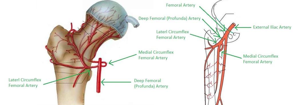
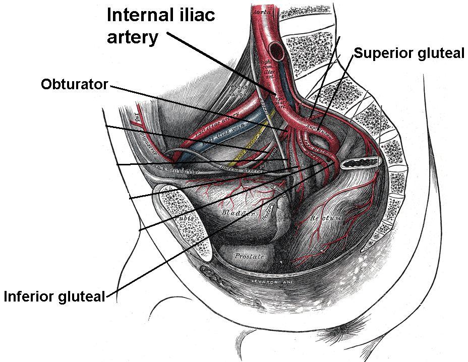
44 Blood Vessels: Muscles they Supply Superficial Femoral The entire lower limb (MAIN ARTERY OF LE) Deep (Profunda) Femoral Femur & deep muscles of the thigh Superior Gluteal Glutes, TFL, & piriformis Inferior Gluteal Piriformis, gluteus max, & superior hamstrings Lateral Circumflex Femoral Femoral neck Medial Circumflex Femoral Femoral neck & adductor magnus Femoral Vein Those supplied by the femoral artery (MAIN VEIN OF LE) Great Saphenous Vein Superficial muscles as part of the superficial venous system of the lower limb Obturator Deep Femoral Vein Iliacus & the adductors Those supplied by the deep (profunda) femoral artery
45 Arteries

46 Arteries Continued
47 Veins

48 Clinical Concerns: Piriformis Syndrome
49 Clinical Concerns: Total Hip Replacement

50 Conclusion PROOF YOU CAN LITERALLY FIND ANYTHING ON THE INTERNET.
The thigh. Prof. Oluwadiya KS
 The thigh Prof. Oluwadiya KS www.oluwadiya.com The Thigh: Boundaries The thigh is the region of the lower limb that is approximately between the hip and knee joints Anteriorly, it is separated from the
Identify the muscles associated with the medial compartment of the thigh. Identify the attachment points of the medial thigh muscles.
 L 8 A B O R A T O R Y Thigh MEDIAL THIGH Identify the muscles associated with the medial compartment of the thigh. Identify the attachment points of the medial thigh muscles. Identify the actions of these
Lesson 24. A & P Hip
 Lesson 24 A & P Hip 1 Aims of the Session This session will allow candidates to have an understanding of the bony prominences and soft tissues of the hip 2 Learning Outcomes By the end of the lesson the
Human Anatomy Biology 351
 Human Anatomy Biology 351 Lower Limb Please place your name on the back of the last page of this exam. You must answer all questions on this exam. Because statistics demonstrate that, on average, between
Muscles of Lesson Five. Muscular Nomenclature and Kinesiology - Two. Muscles of Lesson Five, cont. Chapter 16
 Chapter 16 Muscular Nomenclature and Kinesiology - Two Lessons 5-6 Muscles of Lesson Five Iliopsoas (psoas major, iliacus) Hip outward rotators (piriformis, gemellus superior, gemellus inferior, obturator
Muscles of the lower extremities. Dr. Nabil khouri MD, MSc, Ph.D
 Muscles of the lower extremities Dr. Nabil khouri MD, MSc, Ph.D Posterior leg Popliteal fossa Boundaries Biceps femoris (superior-lateral) Semitendinosis and semimembranosis (superior-medial) Gastrocnemius
Lectures of Human Anatomy
 Lectures of Human Anatomy Lower Limb Gluteal Region and Hip Joint By DR. ABDEL-MONEM AWAD HEGAZY M.B. with honor 1983, Dipl."Gynecology and Obstetrics "1989, Master "Anatomy and Embryology" 1994, M.D.
Lower limb summary. Anterior compartment of the thigh. Done By: Laith Qashou. Doctor_2016
 Lower limb summary Done By: Laith Qashou Doctor_2016 Anterior compartment of the thigh Sartorius Anterior superior iliac spine Upper medial surface of shaft of tibia 1. Flexes, abducts, laterally rotates
Hip joint and pelvic girdle. Lower Extremity. Pelvic Girdle 6/5/2017
 Hip joint and pelvic girdle Lower Extremity The relationship between the pelvic girdle and hip is similar to that between the shoulder girdle and shoulder joint. The lower limbs are attached to the axial
lesser trochanter of femur lesser trochanter of femur iliotibial tract (connective tissue) medial surface of proximal tibia
 medial surface of proximal tibia") LOWER LIMB MUSCLES OF THE APPENDICULAR SKELETON The muscles that act on the lower limb fall into three groups: those that move the thigh, those that move the lower leg, and those that move the ankle, foot,
LOWER LIMB MUSCLES OF THE APPENDICULAR SKELETON The muscles that act on the lower limb fall into three groups: those that move the thigh, those that move the lower leg, and those that move the ankle, foot,
Main Menu. Joint and Pelvic Girdle click here. The Power is in Your Hands
 1 Hip Joint and Pelvic Girdle click here Main Menu K.6 http://www.handsonlineeducation.com/classes//k6entry.htm[3/23/18, 2:01:12 PM] Hip Joint (acetabular femoral) Relatively stable due to : Bony architecture
Gluteal region DR. GITANJALI KHORWAL
 Gluteal region DR. GITANJALI KHORWAL Gluteal region The transitional area between the trunk and the lower extremity. The gluteal region includes the rounded, posterior buttocks and the laterally placed
Human Anatomy Biology 351
 Human Anatomy Biology 351 Lower Limb Please place your name on the back of the last page of this exam. You must answer all questions on this exam. Because statistics demonstrate that, on average, between
Bony Anatomy. Femur. Femoral Head Femoral Neck Greater Trochanter Lesser Trochanter Intertrochanteric Crest Intertrochanteric Line Gluteal Tuberosity
 Hip Anatomy Bony Anatomy Femur Femoral Head Femoral Neck Greater Trochanter Lesser Trochanter Intertrochanteric Crest Intertrochanteric Line Gluteal Tuberosity Bony Anatomy Pelvic Girdle Acetabulum 3 bones
The Hip Joint. Shenequia Howard David Rivera
 The Hip Joint Shenequia Howard David Rivera Topics Of Discussion Movement Bony Anatomy Ligamentous Anatomy Muscular Anatomy Origin/Insertion/Action/Innervation Common Injuries MOVEMENT Flexion Extension
rotation of the hip Flexion of the knee Iliac fossa of iliac Lesser trochanter Femoral nerve Flexion of the thigh at the hip shaft of tibia
 Anatomy of the lower limb Anterior & medial compartments of the thigh Dr. Hayder The fascia lata encloses the entire thigh like a sleeve/stocking. Three intramuscular fascial septa (lateral, medial, and
The University Of Jordan Faculty Of Medicine THE LOWER LIMB. Dr.Ahmed Salman Assistant Prof. of Anatomy. The University Of Jordan
 The University Of Jordan Faculty Of Medicine THE LOWER LIMB Dr.Ahmed Salman Assistant Prof. of Anatomy. The University Of Jordan Gluteal Region Cutaneous nerve supply of (Gluteal region) 1. Lateral cutaneous
Anatomage Table Instructors Guide- Lower Limb
 The Lower Limb Anatomage Table Instructors Guide- Lower Limb Table of Contents Lower Limb 1- The Skeletal System...3 1: Hip Bone...3 2: Hip Joint and Femur...4 3: Patella and Knee Joint...7 4: Tibia, Fibula,
Baraa Ayed حسام أبو عوض. Ahmad Salman. 1 P a g e
 4 Baraa Ayed حسام أبو عوض Ahmad Salman 1 P a g e Today we are going to cover these concepts: Iliotibial tract Anterior compartment of the thigh and the hip Medial compartment of the thigh Femoral triangle
The psoas minor is medial to the psoas major. The iliacus is a fan-shaped muscle that when contracted helps bring the swinging leg forward in walking
 1 p.177 2 3 The psoas minor is medial to the psoas major. The iliacus is a fan-shaped muscle that when contracted helps bring the swinging leg forward in walking and running. The iliopsoas and adductor
Muscles of the Hip 1. Tensor Fasciae Latae O: iliac crest I: lateral femoral condyle Action: abducts the thigh Nerve: gluteal nerve
 Muscles of the Hip 1. Tensor Fasciae Latae O: iliac crest I: lateral femoral condyle Action: abducts the thigh Nerve: gluteal nerve 2. Gluteus Maximus O: ilium I: femur Action: abduct the thigh Nerve:
Muscles of the Thigh. 6.1 Identify, describe the attachments of and deduce the actions of the muscles of the thigh: Anterior group
 Muscles of the Thigh 6.1 Identify, describe the attachments of and deduce the actions of the muscles of the thigh: Anterior group Sartorius: This is a long strap like muscle with flattened tendons at each
First practical session. Bones of the gluteal region
 First practical session 2017 Bones of the gluteal region The Hip bone The hip bone is made of: 1 The ilium: superior in position 2 The ischium:postero-inferior in position 3 The pubis: antero-inferior
Mohammad Ashraf. Abdulrahman Al-Hanbali. Ahmad Salman. 1 P a g e
 - 7 Mohammad Ashraf Abdulrahman Al-Hanbali Ahmad Salman 1 P a g e Structures under the cover of Gluteus Maximus: 1-Bones: Ileum, Femur (Head, greater trochanter and gluteal tuberosity), Ischium (ischial
Joints of the lower limb
 Joints of the lower limb 1-Type: Hip joint Synovial ball-and-socket joint 2-Articular surfaces: a- head of femur b- lunate surface of acetabulum Which is deepened by the fibrocartilaginous labrum acetabulare
Anterior and Medial compartments of the thigh. Dr. Heba Kalbouneh Associate Professor of Anatomy and Histology
 Anterior and Medial compartments of the thigh Dr. Heba Kalbouneh Associate Professor of Anatomy and Histology Terms Related to Movements Movement Flexion Extension Abduction Adduction Medial (internal)
ANATYOMY OF The thigh
 ANATYOMY OF The thigh 1- Lateral cutaneous nerve of the thigh Ι) Skin of the thigh Anterior view 2- Femoral branch of the genitofemoral nerve 5- Intermediate cutaneous nerve of the thigh 1, 2 and 3 are
Muscles of Gluteal Region
 1 The Gluteal Region In the gluteal region the skin is tough with many layers underneath. Directly under it is the superficial fascia followed by the deep fascia then the muscles and the bones of the thigh.
Acland's DVD Atlas of Human Anatomy. Transcript for Volume Robert D Acland
 Acland's DVD Atlas of Human Anatomy Transcript for Volume 2 2007 Robert D Acland This free downloadable pdf file is to be used for individual study only. It is not to be reproduced in any form without
ANATOMY TEAM GLUTEAL REGION & BACK OF THIGH
 ANATOMY TEAM GLUTEAL REGION & BACK OF THIGH OBJECTIVES By the end of this lecture, the student should be able to identify and discuss: Contents of gluteal region: Groups of Glutei muscles and small muscles
Applied anatomy of the hip and buttock
 CHAPTER CONTENTS The hip joint e9 Capsule and ligaments e9 s e0 Flexor muscles................... e0 Extensor muscles.................. e Abductor muscles.................. e Adductor muscles..................
Anatomy & Physiology. Muscles of the Lower Limbs.
 Anatomy & Physiology Muscles of the Lower Limbs http://www.ishapeup.com/musclecharts.html Muscles of the Lower Limbs Among the strongest muscles in the body. Because pelvic girdle is composed of heavy,
The Muscular System. Chapter 10 Part D. PowerPoint Lecture Slides prepared by Karen Dunbar Kareiva Ivy Tech Community College
 Chapter 10 Part D The Muscular System Annie Leibovitz/Contact Press Images PowerPoint Lecture Slides prepared by Karen Dunbar Kareiva Ivy Tech Community College Table 10.14: Muscles Crossing the Hip and
Human Anatomy Biology 255
 Human Anatomy Biology 255 Exam #4 Please place your name and I.D. number on the back of the last page of this exam. You must answer all questions on this exam. Because statistics demonstrate that, on average,
DISSECTION SCHEDULE. Session I - Hip (Front) & Thigh (Superficial)
 & Thigh (Superficial)") DISSECTION SCHEDULE Session I - Hip (Front) & Thigh (Superficial) Surface anatomy Inguinal region Gluteal region Thigh Leg Foot bones Hip bone Femur Superficial fascia Great saphenous vein Superficial
DISSECTION SCHEDULE Session I - Hip (Front) & Thigh (Superficial) Surface anatomy Inguinal region Gluteal region Thigh Leg Foot bones Hip bone Femur Superficial fascia Great saphenous vein Superficial
Copyright 2003 Pearson Education, Inc. publishing as Benjamin Cummings. Dr. Nabil Khouri MD, MSc, Ph.D
 Dr. Nabil Khouri MD, MSc, Ph.D Pelvic Girdle (Hip) Organization of the Lower Limb It is divided into: The Gluteal region The thigh The knee The leg The ankle The foot The thigh and the leg have compartments
The Lower Limb. Anatomy RHS 241 Lecture 2 Dr. Einas Al-Eisa
 The Lower Limb Anatomy RHS 241 Lecture 2 Dr. Einas Al-Eisa The bony pelvis Protective osseofibrous ring for the pelvic viscera Transfer of forces to: acetabulum & head of femur (when standing) ischial
Lecture 08 THIGH MUSCLES ANTERIOR COMPARTMENT. Dr Farooq Khan Aurakzai. Dated:
 Lecture 08 THIGH MUSCLES ANTERIOR COMPARTMENT BY Dr Farooq Khan Aurakzai Dated: 11.02.2017 INTRODUCTION to the thigh Muscles. The musculature of the thigh can be split into three sections by intermuscular
LOWER LIMB. As we know the bony part of the body is divided into Axial and Appendicular (upper and lower Limbs)
") LOWER LIMB As we know the bony part of the body is divided into Axial and Appendicular (upper and lower Limbs) Bones of the Lower limb: 1-Pelvic Girdle: composed of: 1. Right hip bone : is formed by 3
LOWER LIMB As we know the bony part of the body is divided into Axial and Appendicular (upper and lower Limbs) Bones of the Lower limb: 1-Pelvic Girdle: composed of: 1. Right hip bone : is formed by 3
Topic 7: Hip and pelvis. Parts of the hip. Parts of the femur
 Topic 7: Hip and pelvis Parts of the hip Parts of the femur Classifying the hip joint Ball and socket Synovial Multiaxial Movements of the hip: Abduction/adduction Flexion/extension Medial/lateral rotation
ANATYOMY OF The thigh
 ANATYOMY OF The thigh 1- Lateral cutaneous nerve of the thigh Ι) Skin of the thigh Anterior view 2- Femoral branch of the genitofemoral nerve 5- Intermediate cutaneous nerve of the thigh 1, 2 and 3 are
Sports Medicine 15. Unit I: Anatomy. The knee, Thigh, Hip and Groin. Part 4 Anatomies of the Lower Limbs
 Sports Medicine 15 Unit I: Anatomy Part 4 Anatomies of the Lower Limbs The knee, Thigh, Hip and Groin Anatomy of the lower limbs In Part 3 of this section we focused upon 11 of the 12 extrinsic muscles
Figure 1 - Hip and Pelvis
 Hip Figure 1 - Hip and Pelvis The terms hip and pelvis are frequently used interchangeably, but strictly speaking, the pelvis is a girdle of bones and the hip is a joint. The pelvis consists of The sacrum
Anatomy of the lower limb
 Anatomy of the lower limb 1. Bones of the lower limb Pelvis Hip bone/coxal bone Acetabulum o Acetabular margin o Acetabular fossa o Acetabular notch o Lunate surface Ischiopubic ramus Obturator foramen
LAB Notes#1. Ahmad Ar'ar. Eslam
 LAB Notes#1 Ahmad Ar'ar Eslam 1 P a g e Anatomy lab Notes Lower limb bones :- Pelvic girdle: It's the connection between the axial skeleton and the lower limb; it's made up of one bone called the HIP BONE
Posterior compartment of the thigh. Dr. Heba Kalbouneh Associate Professor of Anatomy and Histology
 Posterior compartment of the thigh Dr. Heba Kalbouneh Associate Professor of Anatomy and Histology Posterior compartment of the thigh 1-Muscles: Biceps femoris Semitendinosus Semimembranosus Adductor magnus
MUSCULOSKELETAL LOWER LIMB
 MUSCULOSKELETAL LOWER LIMB Spinal Cord Lumbar and Sacral Regions Spinal cord Dorsal root ganglion Conus medullaris Cauda equina Dorsal root ganglion of the fifth lumbar nerve End of subarachnoid space
Bones of the Lower Limb Bone Structure Description Notes. border of the superior ramus. inferolaterally from the pubic symphysis
 Bones of the Lower Limb Bone Structure Description Notes pubis an angulated bone the forms the anterior part of the pelvis one of three bones that form the os coxae: ilium, ischium, pubis; its forms 1/5
lower limb Anterior Compartment: lecture 3 The deep fascia ( fascia lata) divides the thigh into 3 compartments:
 divides the thigh into 3 compartments:") lower limb lecture 3 The deep fascia ( fascia lata) divides the thigh into 3 compartments: 1. Anterior Extensor compartment 2. Medial Adductor compartment 3. Posterior Flexor compartment Anterior Compartment:
lower limb lecture 3 The deep fascia ( fascia lata) divides the thigh into 3 compartments: 1. Anterior Extensor compartment 2. Medial Adductor compartment 3. Posterior Flexor compartment Anterior Compartment:
Myology of the Knee. PTA 105 Kinesiology
 Myology of the Knee PTA 105 Kinesiology Objectives Describe the planes of motion and axes of rotation of the knee joint Visualize the origins and insertions of the muscles about the knee List the innervations
this makes sense, however this is lower order thinking and does not solve the lower leg
 Functional Knee Valgus in a Barbell Squat 1 One of the most common lower leg dysfunction we see in athletes, particularly general population is functional knee valgus, or better referred to as the knees
ANATYOMY OF The thigh
 ANATYOMY OF The thigh 1- Lateral cutaneous nerve of the thigh Ι) Skin of the thigh Anterior view 2- Femoral branch of the genitofemoral nerve 1, 2 and 3 are From the lumber plexus 5- Intermediate cutaneous
The Lower Limb II. Anatomy RHS 241 Lecture 3 Dr. Einas Al-Eisa
 The Lower Limb II Anatomy RHS 241 Lecture 3 Dr. Einas Al-Eisa Tibia The larger & medial bone of the leg Functions: Attachment of muscles Transfer of weight from femur to skeleton of the foot Articulations
Bones of Lower Limb. Dr. Heba Kalbouneh Associate Professor of Anatomy and Histology
 Bones of Lower Limb Dr. Heba Kalbouneh Associate Professor of Anatomy and Histology Bones of the lower limb Hip Bone Made up of 3 bones: 1) Ilium (flat), superior in position 2) Ischium (L), postero-inferior
1-Muscles: 2-Blood supply: Branches of the profunda femoris artery. 3-Nerve supply: Sciatic nerve
 1-Muscles: B i c e p s f e m o r i s S e m i t e n d i n o s u s S e m i m e m b r a n o s u s a small part of the adductor magnus (h a m s t r i n g p a r t o r i s c h i a l p a r t ) 2-Blood supply:
The Knee. Clarification of Terms. Osteology of the Knee 7/28/2013. The knee consists of: The tibiofemoral joint Patellofemoral joint
 The Knee Clarification of Terms The knee consists of: The tibiofemoral joint Patellofemoral joint Mansfield, p273 Osteology of the Knee Distal Femur Proximal tibia and fibula Patella 1 Osteology of the
MUSCLES OF THE LOWER LIMBS
 MUSCLES OF THE LOWER LIMBS Naming, location and general function Dr. Nabil khouri ROLES THAT SHOULD NOT BE FORGOTTEN Most anterior compartment muscles of the hip and thigh Flexor of the femur at the hip
In-Depth Foundations: Anatomy Terms to Know
 Be familiar with / able to identify and define all the following parts. The Spine Cranium Vertebrae Cervical, Thoracic, Lumbar Sacrum Coccyx Bones of Upper Body Cranium Mastoid process; Occipital condyle,
Contents of the Posterior Fascial Compartment of the Thigh
 Contents of the Posterior Fascial Compartment of the Thigh 1-Muscles: B i c e p s f e m o r i s S e m i t e n d i n o s u s S e m i m e m b r a n o s u s a small part of the adductor magnus (h a m s t
5 Testing the Muscles of the Lower Extremity
 C H A P T E R 5 Testing the Muscles of the Lower Extremity Hip Flexion Hip Flexion, Abduction, and External Rotation with Knee Flexion Hip Extension Hip Abduction Hip Abduction from Flexed Position Hip
BONES JOINTS MUSCLES OF THE LOWER LIMB
 BONES JOINTS MUSCLES OF THE LOWER LIMB LOWER LIMB: BONES LOWER LIMB GLUTEAL REGION consists of 6 major segments: FEMORAL REGION (THIGH) KNEE REGION LEG REGION TALOCRURAL REGION (ANKLE) FOOT REGION LOWER
Scapula Spine Lateral edge of clavicle. Medial border Scapula. Medial border of Scapula, between superior angle and root of spine. Scapula.
 Muscle attachments and actions answer sheet Muscle Origins insertions Movements Joints crossed Trapezius Base of skull Spinous process of C7 Thoracic Spine Lateral edge of clavicle Elevation Retraction
ANATOMY. Lecturer : Maher Hadidi Done by: ,,Subject : lecture#: ~ Date:
 ANATOMY,,Subject : Lecturer : Maher Hadidi Done by: ~~ lecture#: ~ Date: GftJteal Reaion ' ' v Highest point of iliac cres Safe site for (/M) injection Horizantal line Sciatic nerve Ischial tubrosity
It is formed by fusion of 3 bones: I. Ilium (superior bone). II. Pubis (antero-inferior bone). III. Ischium (postero-inferior bone).
. II. Pubis (antero-inferior bone). III. Ischium (postero-inferior bone).") It is formed by fusion of 3 bones: I. Ilium (superior bone). II. Pubis (antero-inferior bone). III. Ischium (postero-inferior bone). Pubis Acetabulum Ana (242 ) The three constituent of bones of the hip
It is formed by fusion of 3 bones: I. Ilium (superior bone). II. Pubis (antero-inferior bone). III. Ischium (postero-inferior bone). Pubis Acetabulum Ana (242 ) The three constituent of bones of the hip
Lower Limb Dr. Robin Paudel
 Lower Limb n What is a limb? n Skeleton n Joints n Pelvis or limb girdle n Hip/Hip Muscles n Lumber and sacral plexus getting spinal nerves out onto limb n Muscles anterior and posterior compartments n
The os coxae or hip bone consists of three flat bones, ilium, ischium and pubis, which fuse together to form the acetabulum.
 The os coxae The os coxae or hip bone consists of three flat bones, ilium, ischium and pubis, which fuse together to form the acetabulum. The ilium extends from the acetabulum upwards forming the lateral
Amy Warenda Czura, Ph.D. 1 SCCC BIO130 Lab 7 Appendicular Skeleton & Articulations
 The Skeletal System II: Appendicular Skeleton and Articulations Exercises 11, 13 (begins: page 145 in 9 th and 10 th editions) Exercises 10, 11 (begins: page 147 in 11 th edition, page 149 in 12 th edition)
HUMAN BODY COURSE LOWER LIMB NERVES AND VESSELS
 HUMAN BODY COURSE LOWER LIMB NERVES AND VESSELS October 22, 2010 D. LOWER LIMB MUSCLES 2. Lower limb compartments ANTERIOR THIGH COMPARTMENT General lfunction: Hip flexion, knee extension, other motions
CHAPTER 8: THE BIOMECHANICS OF THE HUMAN LOWER EXTREMITY
 CHAPTER 8: THE BIOMECHANICS OF THE HUMAN LOWER EXTREMITY _ 1. The hip joint is the articulation between the and the. A. femur, acetabulum B. femur, spine C. femur, tibia _ 2. Which of the following is
Thigh, Hip, & Low Back Evaluation.
 Thigh, Hip, & Low Back Evaluation www.fisiokinesiterapia.biz Thigh Injuries Quad contusions - Myositis Ossificans Trochanteric bursitis - snapping hip Ischial bursitis - bench-warmer s bursitis Strains
5.1 Identify, describe the attachments of and deduce the actions of the muscles of the thigh:
 5.1 Identify, describe the attachments of and deduce the actions of the muscles of the thigh: Anterior group Proximal attachment Distal attachment Sartorius ASIS» Upper part of shaft tibia (middle surface)»
Bio 113 Anatomy and Physiology The Muscles. Muscles of the Head and Neck. Masseter. Orbicularis occuli. Orbicularis oris. Sternocleidomastoid
 Bio 113 Anatomy and Physiology The Muscles Muscles of the Head and Neck Masseter Orbicularis occuli Orbicularis oris Sternocleidomastoid Temporalis BIO 113 Fall 2011 Muscles Page 1 of 5 Muscles of the
Lumbar Plexus. Ventral rami L1 L4 Supplies: Major nerves.. Abdominal wall External genitalia Anteromedial thigh
 Lower Limb Nerves Lectures Objectives Describe the structure and relationships of the plexuses of the lower limb. Describe the course, relationships and structures supplied for the major nerves of the
Gluteal Region and Back of Thigh
 Gluteal Region and Back of Thigh Musculoskeletal block- Anatomy-lecture 14 Editing file Objectives Know contents of gluteal region: 1. Groups of Glutei muscles and small muscles (Lateral Rotators). 2.
The Hay is in the Barn
 Anatomy 1 Practical 1 Review Made by Forrest Allen (nerd) Edited by TJ Williamson (not nerd) The Hay is in the Barn 2019 Thunderbringers Too much to handle https://www.youtube.com/watch?v=glii-kaza d8
Muscles of the Gluteal Region
 Muscles of the Gluteal Region 1 Some of the most powerful in the body Extend the thigh during forceful extension Stabilize the iliotibial band and thoracolumbar fascia Related to shoulders and arms because
SURGICAL AND APPLIED ANATOMY
 Página 1 de 6 Copyright 2001 Lippincott Williams & Wilkins Bucholz, Robert W., Heckman, James D. Rockwood & Green's Fractures in Adults, 5th Edition SURGICAL AND APPLIED ANATOMY Part of "37 - HIP DISLOCATIONS
TABLE OF MUSCLES OF LOWER EXTREMITY 2018zillmusom ANTERIOR THIGH
 TABLE OF MUSCLES OF LOWER EXTREMITY 2018zillmusom ANTERIOR THIGH MUSCLE ORIGIN INSERTION ACTION NERVE Iliopsoas Ilium, vertebra Femur Flex hip joint Femoral nerve (T12-L5) Pectineus Pubis Femur Flex hip
Muscle Testing of Knee Extensors. Yasser Moh. Aneis, PhD, MSc., PT. Lecturer of Physical Therapy Basic Sciences Department
 Muscle Testing of Knee Extensors Yasser Moh. Aneis, PhD, MSc., PT. Lecturer of Physical Therapy Basic Sciences Department Muscle Testing of Knee Extensors othe Primary muscle Quadriceps Femoris -Rectus
Lower Limb Nerves. Clinical Anatomy
 Lower Limb Nerves Clinical Anatomy Lumbar Plexus Ventral rami L1 L4 Supplies: Abdominal wall External genitalia Anteromedial thigh Major nerves.. Lumbar Plexus Nerves relation to psoas m. : Obturator n.
Where should you palpate the pulse of different arteries in the lower limb?
 Where should you palpate the pulse of different arteries in the lower limb? The femoral artery In the femoral triangle, its pulse is easily felt just inferior to the inguinal ligament midway between the
Anatomy images for MSS practical exam- 2019
 Anatomy images for MSS practical exam- 2019 Ilium Ischium Pubis Acetabulaum Iliac crest Iliac tubercle ASIS (muscle and ligament attached) AIIS (muscle attached) PSIS PIIS Ischial spine Ischial tuberosity
Practical 1 Worksheet
 Practical 1 Worksheet ANATOMICAL TERMS 1. Use the word bank to fill in the missing words. reference side stand body arms palms anatomical forward All anatomical terms have a(n) point which is called the
Lowe w r e r B ody Resistance Training
 Lower Body Resistance Training Tibialis Anterior Extensor Hallucis Longus Extensor Digitorum Longus Proneus Tertius AROM 25-40 degrees Extensor active-insufficiency Flexion & Eversion (Pronation) Flexion
Lab Activity 9. Appendicular Skeleton Martini Chapter 8. Portland Community College BI 231
 Lab Activity 9 Appendicular Skeleton Martini Chapter 8 Portland Community College BI 231 Appendicular Skeleton Upper & Lower extremities Shoulder Girdle Pelvic Girdle 2 Humerus 3 Humerus: Proximal End
SURGICAL EXPOSURES SURGERY OF THE HIP
 1 of 24 11/19/03 1:11 PM SURGICAL EXPOSURES SURGERY OF THE HIP by R. CALANDRUCCIO In: Atlas of Orthopaedic Surgery Volume 3 Lower Extremity; Editors: Laurin, CA, Riley Jr. LH, Roy-Camille R Reprinted with
Regional Anaesthesia
 Regional Anaesthesia Lower limb anatomy and blocks Hip and Knee Joint Hip Joint: Nerve supply Lumbar plexus Femoral nerve through the nerve to the Rectus Femoris Ant division of the Obturator nerve The
Due in Lab weeks because of Thanksgiving Prelab #10. Homework #8. Both sides! Both sides!
 Lab 8 MUSCLES Due in Lab 10 2 weeks because of Thanksgiving Prelab #10 Both sides! Homework #8 Both sides! Refer to Muscles 22-23 Naming of muscles Origin Site of muscle attachment that doesn t move during
musculoskeletal system anatomy nerves of the lower limb 2 done by: Dina sawadha & mohammad abukabeer
 musculoskeletal system anatomy nerves of the lower limb 2 done by: Dina sawadha & mohammad abukabeer #Sacral plexus : emerges from the ventral rami of the spinal segments L4 - S4 and provides motor and
11/15/2018. Temporalis Elevates & retracts mandible. Masseter = Prime mover of jaw closure. Levator scapulae Supraspinatus Clavicle.
 Due in Lab 10 Lab 8 MUSCLES 2 weeks because of Thanksgiving Prelab #10 Both sides! Homework #8 Both sides! Refer to Muscles 22-23 Examples of Origin & Insertion Naming of muscles Origin Site of muscle
The Appendicular Skeleton
 8 The Appendicular Skeleton PowerPoint Lecture Presentations prepared by Jason LaPres Lone Star College North Harris 8-1 The Pectoral Girdle The Pectoral Girdle Also called shoulder girdle Connects the
Figure 7: Bones of the lower limb
 BONES OF THE APPENDICULAR SKELETON The appendicular skeleton is composed of the 126 bones of the appendages and the pectoral and pelvic girdles, which attach the limbs to the axial skeleton. Although the
Organization of the Lower Limb
 Organization of the Lower Limb Most illustrations from: Thieme Atlas of Anatomy: Musculoskeletal System. M Schuenke, et al, 2006. Anatomy: A Regional Atlas of the Human Body. Carmine Clemente, 4th edition.
MR Imaging in Athlete s Hip/Pelvis
 MR Imaging in Athlete s Hip/Pelvis Tara Lawrimore, MD FRCPC Department of Radiology Musculoskeletal Division Massachusetts General Hospital Harvard Medical School No disclosures MR and Hip Pain in the
Muscles to know. Lab 21. Muscles of the Pelvis and Lower Limbs. Muscles that Position the Lower Limbs. Generally. Muscles that Move the Thigh
 Muscles to know Lab 21 Muscles of the Pelvis, Leg and Foot psoas major iliacus gluteus maximus gluteus medius sartorius quadriceps femoris (4) gracilus adductor longus biceps femoris semitendinosis semimembranosus
To classify the joints relative to structure & shape
 To classify the joints relative to structure & shape To describe the anatomy of the hip joint To describe the ankle joint To memorize their blood & nerve supply JOINTS: Joints are sites where skeletal
Organization of the Lower Limb Audrone Biknevicius, Ph.D. Dept. Biomedical Sciences, OU HCOM at Dublin Clinical Anatomy Immersion 2014
 Organization of the Lower Limb Audrone Biknevicius, Ph.D. Dept. Biomedical Sciences, OU HCOM at Dublin Clinical Anatomy Immersion 2014 www.thestudio1.co.za LIMB FUNCTION choco-locate.com blog.coolibar.com
Overview. Overview. Introduction. Introduction Anatomy History Examination Common Disorders. Introduction Anatomy History Examination Common Disorders
 Common Hip Disorders in Figure Skaters 14 th Annual Meeting of Sports Medicine and Science in Figure Skating January 25, 2009 8:15-8:45am Robert J. Dimeff, MD Medical Director of Sports Medicine Overview
musculoskeletal system anatomy nerves of the lower limb 1 done by: dina sawadha & mohammad abukabeer
 musculoskeletal system anatomy nerves of the lower limb 1 done by: dina sawadha & mohammad abukabeer What is the importance of plexuses? plexuses provides us the advantage of a phenomenon called convergence
Organization of the Lower Limb
 Organization of the Lower Limb Limb Development Lower limb develops in an aterolateral position at the level of the L2 to S3 trunk segments Great toe positioned cephalic direction with the soles of the