Introduction to Chest Radiography
|
|
|
- Scott Sherman
- 5 years ago
- Views:
Transcription

1 Introduction to Chest Radiography RSTH 366: DIAGNOSTIC TECHNIQUES Alan Alipoon BS, RCP, RRT Instructor Department of Cardiopulmonary Sciences 1
2
3 Introduction Discovered in 1895 by Wilhelm Roentgen Terminology 1) X-Rays Roentgenograms Radiograph 3
4 Radiation Physics Electromagnetic waves 4
5 Radiation Physics (page188 text) Cathode produces electrons which impact tungstenanode to produce x-rays 5
6 Radiation Physics
7 Radiation Physics High energy waves have penetrating power Degree of penetration recorded as shadows on image receptor (X-ray film) 7
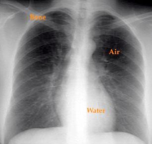
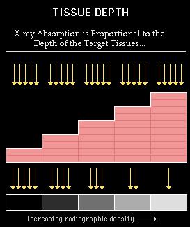
8 Radiation Physics X-rays are not reflected back like light rays but penetrate matter Their ability to penetrate is dependent of the density of matter Dense objects such bone absorb more x- rays (allow less penetration) Low-density objects such as air filled objects allow x-rays to pass through easily Radiographic densities 8
 *decrease density = darker film shadow (radiolucent)")
9 Radiographic Densities Radiation Physics *increase density = lighter (white) film shadow (radiopaque) *decrease density = darker film shadow (radiolucent) 9
10 Radiation Physics Four classifications of radiographic densities 1) Bone, very dense 2) Water, less dense 3) Fat, mildly radiolucent 4) Air, very radiolucent 10
11
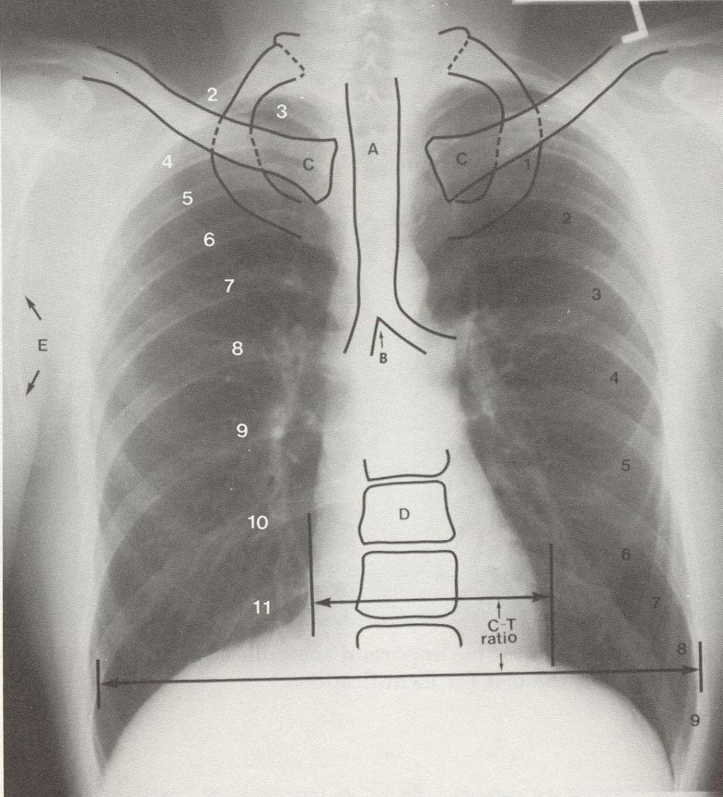
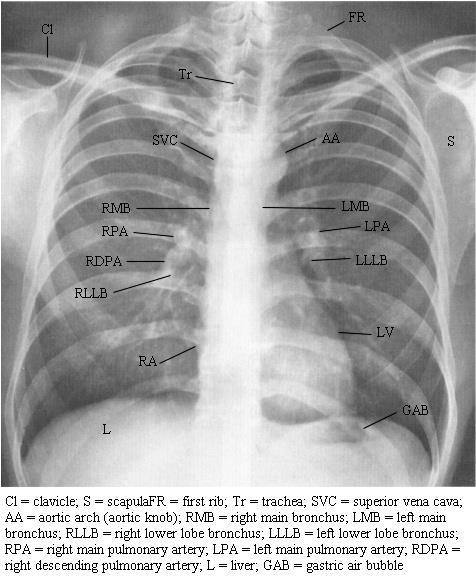
12 12
13 Pulmonary Edema- Pre and Post Diuresis
14 14

15 Densities 15
16 Radiation Physics Distance between the source of x-ray and object The closer the object is to the source the greater the magnification and distortion of the object The further the object is to the source the smaller and sharper the object For a PA chest x-ray the patient and film are positioned approximately 6 feet from the x-ray source A portable chest x-ray the patient and film are positioned approximately 4 feet from the x-ray source 16
17 Radiation Physics
18 Value of the Chest X-ray Relevancy to Respiratory Therapy Diagnosis of lung disease Help determine appropriate and effective therapy (pneumothorax) Evaluate the effectiveness of treatment Determine position of endotracheal tubes Provide dynamic portrait of the progression of the disease process (serial films) Identify complications 18
19
20
21
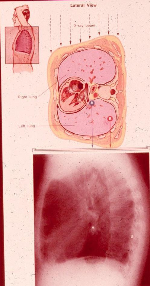
22 22
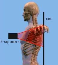
23
24 Chest X-ray Usage Stable patient with undiagnosed respiratory problems Routine chest x-ray important for patients over the age of 40, especially in smokers Evaluate changes in lung pathology a. effects of treatment (ex. IPPB with atelectasis, lung recruitment maneuvers) b. acute problems may require frequent chest x-rays while chronic changes require less frequent films 24
25 Clinical Indications For Chest Radiograph See Box 10-1 page
26 Chest X-ray Usage, cont. Do not withhold oxygen or mechanical ventilation to get a chest X-ray in acutely ill 26
27 Limitations 1.May be normal with significant lung disease (ex. asthma, chronic bronchitis) 2.Abnormalities in "blind" areas may not be detected 3.X-ray may be clinically behind pathology (ex. pneumonia and ARDS) 4.Chest X-ray may be normal in patients with respiratory insufficiency from extrapulmonary abnormalities 27
28 Views (Positions) 1.Magnification and sharpness depend on distance between X-ray source and chest - closer source is to chest, greater magnification and less focused (and vice versa) 28
29 Views (Positions) 2. Exposure regulated by technician 3. Standard chest radiographs at full inspiration. 29
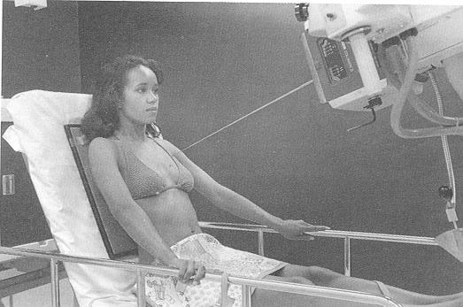
30 Views (Positions) Posteroanterior (PA) view Anteroposterior (AP) view Left lateral view Lateral decubitus view Lordotic view Oblique view End expiratory film
31 Standard view Posteroanterior (PA) view Film against anterior chest. X-ray travels through posterior chest to anterior chest to expose film 31
32 Posteroanterior (PA) view
33 Posteroanterior (PA) view 33
34 Posteroanterior (PA) view 34

35 Heart chambers PA view
36 Posteroanterior (PA) view Advantages a) decreased magnification and increased sharpness b) rotate scapulae out of way c) easily identified air/fluid levels d) anatomical reality 36
37 Posteroanterior (PA) view Disadvantages?
38 Anteroposterior (AP) View 1. Portable CXR 2. Film against posterior chest X-rays pass through anterior to posterior to expose film 38
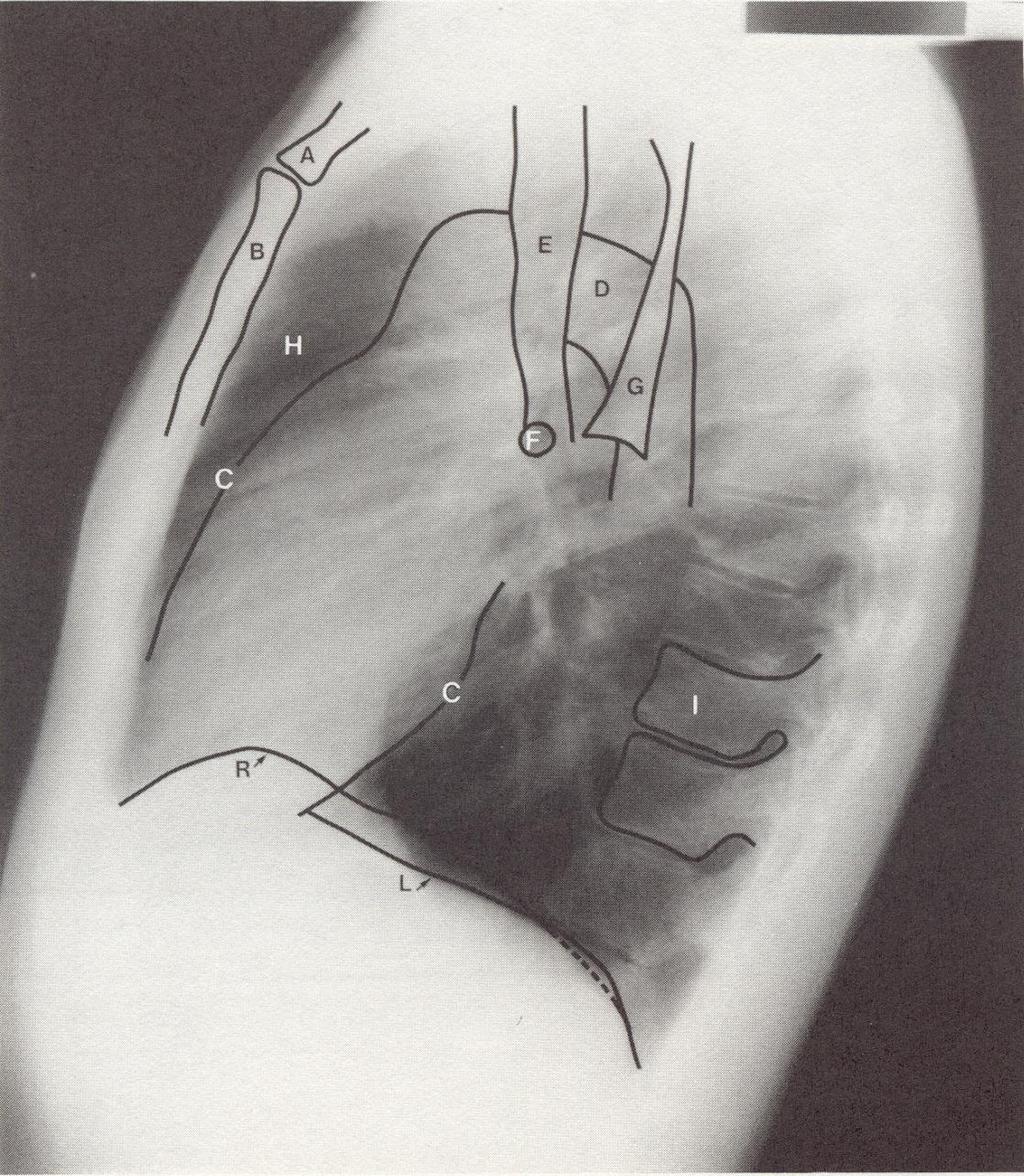
39 Anteroposterior (AP) View
40 Anteroposterior (AP) View 40
41 AP PA 41
42 Anteroposterior (AP) View Advantages a) portability b) viewing posterior chest abnormalities 42
 decreased sharpness d) poor inspiratory effort e) artifactual shadows")
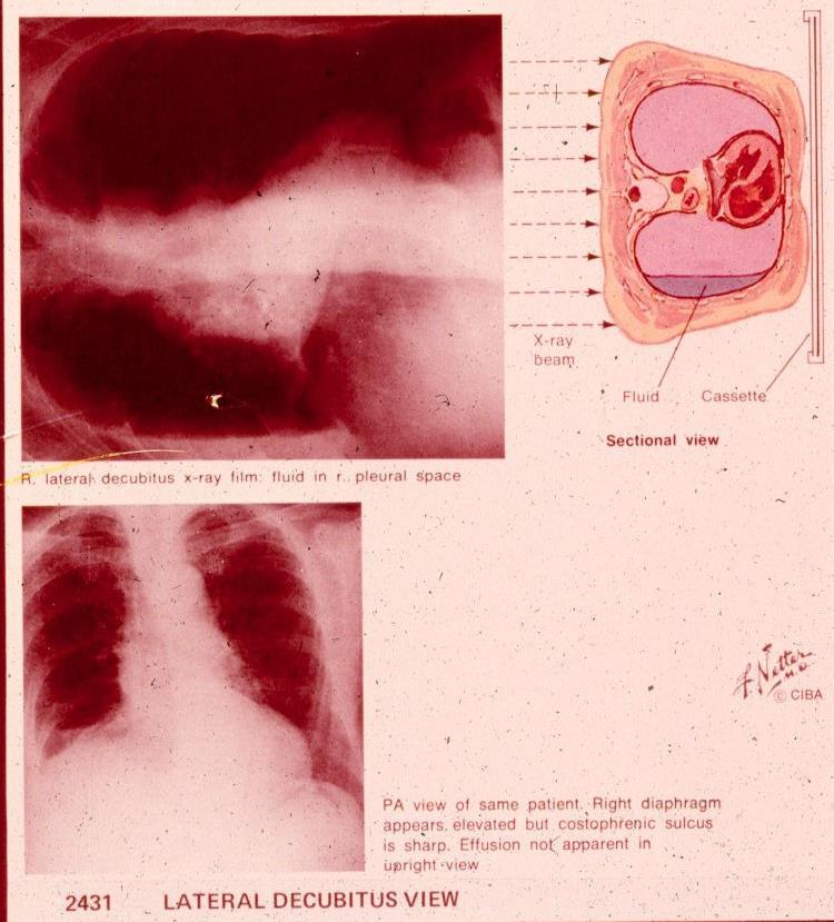
43 Anteroposterior (AP) View Disadvantages a) heart shadow magnified b) asymmetric radiograph c) decreased sharpness d) poor inspiratory effort e) artifactual shadows present 43
44 44
45 45

46 AP views PA 46
47 Left Lateral View Left lateral most common Film against lateral chest, X-rays pass through right and then left side of chest to expose film 47
48 48

49 Left Lateral View 49

50 Heart chambers- Lateral view
51 Left Lateral View Advantages a) heart magnification decreased b) can see behind heart and diaphragms c) 3D view d) can view left lung better than with PA view 51
52 Lateral Decubitus View Left or right 2. Patient on affected side 3. Film against posterior chest. Patient lies on right/left side with X-rays passing through chest in AP manner to expose film 4. Advantage - show free fluid (pleural or parenchymal) 52

53 Lateral Decubitus View 53
54 PA/Lateral view of a patient with a large right pleural effusion 54

55 Right lateral decubitus (right side down) view of the same patient, showing layering of the large effusion along the right chest wall. 55
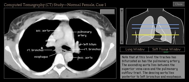
56 Lordotic View Film against posterior chest. X-rays pass upward at approximately 45 o angle A to P from a lower position to expose film 56
57 Lordotic View Advantages a) clearer (bone-free) view of upper lobes b) separation of superimposed densities 57

58 Lordotic 58

59 Lordotic 59
60 Oblique View Right or left Film against anterior or posterior chest. Patient is rotated right (45 0 ) or left and X-rays pass P to A or A to P to expose film 60
61 Oblique View Advantage - reduces superimposed images 61
62 End-expiratory film Taken at end-exhalation Advantages a) evaluate diaphragm excursion b) aid pneumothorax detection 62
63 Inspiration:Expiration CXR 63
64 Postprocedural Chest X-Ray Evaluation Tracheal Intubations Central Venous Pressure Line Pulmonary Artery Catheter Placement Nasogastric Feeding Tubes Chest Tubes
65 Tomography 1. Simultaneous movement of film and X-ray source around a specific axis. 2. Allows selective visualization of specific area. 3. Used to evaluate mediastinal or parenchymal areas *C.A.T. (Computerized Axial Tomography) ("Cat Scan") - computer processing of X-ray which gives cross-sectional image 65


66 Tomography is the process of generating a two-dimensional image of a slice or section through a 3- dementional object (a tomogram) 66
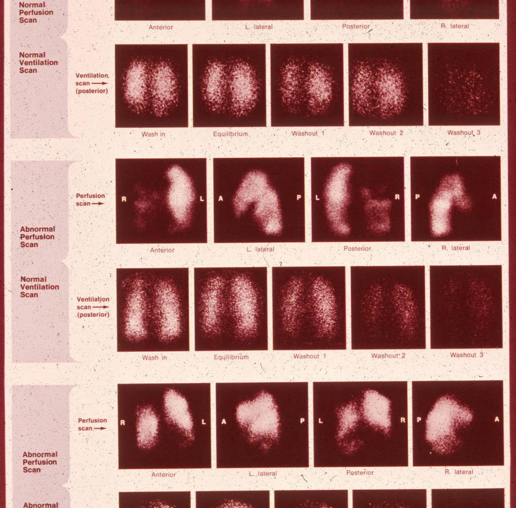
67 Tomography 67
68 68

69 CT of the lungs, window level set to demonstrate the vessels and air ways - not intended to demonstrate the heart, spine muscles etc. This is used to look for things like pneumonia or lung cancer
70 Chest Tomography 70
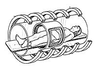
71 Computed Tomography CT scanning Computed enhancement of x-ray shadows Results in an amazing clear look at internal anatomy Clarity of image is indispensable Cost 71
72 Computed Tomography CT scanning is extremely useful in the following areas (page 194 text) Lung tumors Chronic interstitial lung disease Acquired Immune Deficiency Syndrome (AIDS) Occupational lung disease Pneumonia Bronchiectasis Chronic Obstructive Pulmonary Disease 72
73 Bronchography 1. Instillation of radiopaque material (fluid) into tracheobronchial tree with subsequent radiographic analysis (Bronchogram) 2. Used to Diagnose - Carcinoma (Bronchogenic) - Tracheobronchial lesions - Congenital abnormalities - Airway deterioration 73
74 Bronchography 74
75 75
76 Ventilation/Perfusion scans (V/Q scans) 1. Inhalation and/or injection of radioactive tracer into airways and/or pulmonary vasculature 2. Chest scanned with defects in ventilation or perfusion assessed 76
77 77
78 Ventilation/Perfusion scans (V/Q scans) Abnormalities Decreased ventilation such as that caused by: - airway occlusion - bronchospasm - loss of elasticity - alveolar consolidation - airway compression 78
79 Ventilation/Perfusion scans (V/Q scans) Abnormalities decreased perfusion such as that caused by: - pulmonary emboli - loss of vascular bed - vascular compression 79
80 Magnetic Resonance Imaging (MRI) 1. Principle - measurement of resonant protons present in tissue 2. Related to water and lipid content 3. Signal intensity proportional to fluid content, therefore, fluid appears white and air and cortical bone black 80
81 Magnetic Resonance Imaging (MRI) Advantages a) contrast resolution greater than CT b) can be used to measure fluid flow c) can be obtained in any plane d) tissue-specific diagnosis possible 81
82 82
 8.")
83 Lateral MRI 1.Left ventricle 2.Aorta 3.Pulmonary trunk 4.Left atrium 5.Breast 6.Spinal column 7.Serous pericardium (light green) 8.Fibrous pericardium (dark green) 83

 84")
84 Magnetic Resonance Imaging (MRI) 84
85 Magnetic Resonance Imaging (MRI) Disadvantages a) cost b) size c) motion artifact d) magnetic field 85
4/16/2017. Learning Objectives. Interpretation of the Chest Radiograph. Components. Production of the Radiograph. Density & Appearance
 Interpretation of the Arthur Jones, EdD, RRT Learning Objectives Identify technical defects in chest radiographs Identify common radiographic abnormalities This Presentation is Approved for 1 CRCE Credit
Disclosure. Clinical Chest Radiography Interpretation Part I
 Clinical Chest Radiography Interpretation Part I Anthony M. Angelow, PhD(c), MSN, ACNPC, AGACNP-BC, CEN Associate Lecturer, Fitzgerald Health Education Associates Clinical practice Division of Trauma Surgery
PATIENT DATA EVALUATION AND RECOMMENDATION: IMAGING STUDIES
 PATIENT DATA EVALUATION AND RECOMMENDATION: IMAGING STUDIES Robert Harwood, MSA, RRT-NPS Objectives At the end of this presentation the student should be able to: Describe the indications of a chest radiograph.
Shedding Light on Neonatal X-rays. Objectives. Indications for X-Rays 5/14/2018
 Shedding Light on Neonatal X-rays Barbara C. Mordue, MSN, NNP-BC Neonatal Nurse Practitioner LLUH Children s Hospital, NICU Objectives Utilize a systematic approach to neonatal x-ray interpretation Identify
10/17/2016. Nuts and Bolts of Thoracic Radiology. Objectives. Techniques
 Nuts and Bolts of Thoracic Radiology October 20, 2016 Carleen Risaliti Objectives Understand the basics of chest radiograph Develop a system for interpreting chest radiographs Correctly identify thoracic
Interpreting thoracic x-ray of the supine immobile patient: Syllabus
 Interpreting thoracic x-ray of the supine immobile patient: Syllabus Johannes Godt Dep. of Radiology and Nuclear Medicine Oslo University Hospital Ullevål NORDTER 2017, Helsinki Content - Why bedside chest
Tests Your Pulmonologist Might Order. Center For Cardiac Fitness Pulmonary Rehab Program The Miriam Hospital
 Tests Your Pulmonologist Might Order Center For Cardiac Fitness Pulmonary Rehab Program The Miriam Hospital BASIC ANATOMY OF THE LUNGS Lobes of Lung 3 lobes on the Right lung 2 lobes on the Left Blood
TB Radiology for Nurses Garold O. Minns, MD
 TB Nurse Case Management Salina, Kansas March 31-April 1, 2010 TB Radiology for Nurses Garold O. Minns, MD April 1, 2010 TB Radiology for Nurses Highway Patrol Training Center Salina, KS April 1, 2010
Radiological Anatomy of Thorax. Dr. Jamila Elmedany & Prof. Saeed Abuel Makarem
 Radiological Anatomy of Thorax Dr. Jamila Elmedany & Prof. Saeed Abuel Makarem Indications for Chest x - A chest x-ray may be used to diagnose and plan treatment for various conditions, including: Diseases/Fractures
An Introduction to Radiology for TB Nurses
 An Introduction to Radiology for TB Nurses Garold O. Minns, MD September 14, 2017 TB Nurse Case Management September 12 14, 2017 EXCELLENCE EXPERTISE INNOVATION Garold O. Minns, MD has the following disclosures
Chest X-ray Interpretation
 Chest X-ray Interpretation Introduction Routinely obtained Pulmonary specialist consultation Inherent physical exam limitations Chest x-ray limitations Physical exam and chest x-ray provide compliment
Learning Radiology: Recognizing the Basics. Text with Student Consult Online Access Code
 Learning Radiology: Recognizing the Basics. Text with Student Consult Online Access Code Herring, W ISBN-13: 9780323074445 Table of Contents 1. Recognizing Anything The "colorful" world of radiology A
Lines and tubes. 1 Nasogastric tubes Endotracheal tubes Central lines Permanent pacemakers Chest drains...
 Lines and tubes 1 Nasogastric tubes... 15 2 Endotracheal tubes.... 19 3 Central lines... 21 4 Permanent pacemakers.... 25 5 Chest drains... 30 This page intentionally left blank 1 Nasogastric tubes Background
Manage TB Dr. A. Chitrakumar Madras Medical College and RGGGH Institute of Thoracic Medicine, Chennai
 Manage TB Dr. A. Chitrakumar Madras Medical College and RGGGH Institute of Thoracic Medicine, Chennai Lecture 16 Radiology in diagnosis of Tuberculosis Session 01 So, welcome to the session Radiology in
UERMMMC Department of Radiology. Basic Chest Radiology
 UERMMMC Department of Radiology Basic Chest Radiology PHYSICS DENSITIES BONE SOFT TISSUES WATER FAT AIR TELEROENTGENOGRAM Criteria for an Ideal Chest Radiograph 1. Upright 2. Posteroanterior View 3. Full
B-I-2 CARDIAC AND VASCULAR RADIOLOGY
 (YEARS 1 3) CURRICULUM FOR RADIOLOGY 13 B-I-2 CARDIAC AND VASCULAR RADIOLOGY KNOWLEDGE To describe the normal anatomy of the heart and vessels including the lymphatic system as demonstrated by radiographs,
Shades of Gray Interpretation of Perioperative Imaging
 Stanford Hospital and Clinics DEPARTMENT OF CARDIOTHORACIC SURGERY-THORACIC AORTIC SURGERY UNIT FALK CARDIOVASCULAR RESEARCH CENTER STANFORD, CALIFORNIA 94305-5407 MICHAEL SHEEHAN, MSN, RNFA, NPC TELEPHONE
Chest Radiology Interpretation: Findings of Tuberculosis
 Chest Radiology Interpretation: Findings of Tuberculosis Get out your laptops, smart phones or other devices pollev.com/chestradiology Case #1 1 Plombage Pneumonia Cancer 2 Reading the TB CXR Be systematic!
Objectives. What is a Chest X Ray? CXR Workshop. Definition (diagnostic tool/internal PE) Types. Cost
 Types. Cost") Objectives CAPA 2011 Christy Wilson, PA C Georgia Lung Associates Identify the radiographic landmarks on a chest radiograph Recognize identifiers of poor quality on the chest radiograph Outline an approach
Objectives CAPA 2011 Christy Wilson, PA C Georgia Lung Associates Identify the radiographic landmarks on a chest radiograph Recognize identifiers of poor quality on the chest radiograph Outline an approach
Alexander A Schult, M.D., FCCP. October 21, 2017 Revised 1/10/18
 Alexander A Schult, M.D., FCCP October 21, 2017 Revised 1/10/18 Identifying normal anatomy Identifying various pathologic states Identifying placement of hardware Identifying limitations of portable CXR
Pulmonary Embolism. Thoracic radiologist Helena Lauri
 Pulmonary Embolism Thoracic radiologist Helena Lauri 8.5.2017 Statistics 1-2 out of 1000 adults annually are diagnosed with deep vein thrombosis (DVT) and/or pulmonary embolism (PE) About half of patients
Children are not small adults Children are Not Small Adults Anatomic considerations Pliable bony & cartilagenous structures - Significant thoracic inj
 PEDIATRIC CHEST TRAUMA Children are not small adults Role of imaging Spectrum of injury Children are not small adults Children are Not Small Adults Anatomic considerations Pliable bony & cartilagenous
Undergraduate Teaching
 Prof. James F Meaney Undergraduate Teaching Chest X-Ray Understanding the normal anatomical by reference to cross sectional imaging Radiology? It s FUN! Cryptic puzzle Sudoku (Minecraft?) It s completely
Approach to CXR. Terminology. 1.Identification. Greg Blecher SCH Respir Fellow. Correct patient Correct date and time Correct examination
 Approach to CXR Greg Blecher SCH Respir Fellow From Rob Posteraro http://home.earthlink.net/~rhpos/cxr_interpret.txt.html ; http://home.earthlink.net/~rhpos/cxr_main.txt.html) Approach to viewing Chest
Chapter 16 Worksheet Code It
 Name: Class: Date: ID: A Chapter 16 Worksheet 3 2 1 Code It True/False Indicate whether the statement is true or false. 1. CT scans generate three-dimensional images. 2. An ultrasound produces images of
Interactive Lecture. Lecture 7 - Interactive. Radiology of cardiorespiratory disease. Editing File. Done By. Color Coding Important Notes Extra
 Lecture 7 - Interactive 436 Teams Interactive Lecture Radiology of cardiorespiratory disease Done By Team Leaders: Khalid Alshehri Hanin Bashaikh Team Members: Ghaida Alsaeed Maha Alissa Nawwaf AlHarbi
Interpretation of the chest radiograph Elizabeth Puddy MB ChB FCARCSI Catherine Hill MB ChB MRCP FRCR
 Interpretation of the chest radiograph Elizabeth Puddy MB ChB FCARCSI Catherine Hill MB ChB MRCP FRCR The traditional technique used in the acquisition and development of a chest radiograph uses methods
Chest and cardiovascular
 Module 1 Chest and cardiovascular A. Doss and M. J. Bull 1. Regarding the imaging modalities of the chest: High resolution computed tomography (HRCT) uses a slice thickness of 4 6 mm to identify mass lesions
Chest X-ray (CXR) Interpretation Brent Burbridge, MD, FRCPC
 Interpretation Brent Burbridge, MD, FRCPC") Chest X-ray (CXR) Interpretation Brent Burbridge, MD, FRCPC An approach to reviewing a chest x-ray will create a foundation that will facilitate the detection of abnormalities. You should create your own
Chest X-ray (CXR) Interpretation Brent Burbridge, MD, FRCPC An approach to reviewing a chest x-ray will create a foundation that will facilitate the detection of abnormalities. You should create your own
Radiology of the respiratory disease
 Radiology of the respiratory disease [ Color index: Important Notes Extra ] [ Editing file Feedback Share your notes Shared notes ] Resources: - 435 Slides - 434 Team - 435 Notes Done by: - Mai Alageel
Chest X rays and Case Studies. No disclosures. Outline 5/31/2018. Carlo Manalo, M.D. Department of Radiology Loma Linda University Children s Hospital
 Chest X rays and Case Studies Carlo Manalo, M.D. Department of Radiology Loma Linda University Children s Hospital No disclosures. Outline Importance of history Densities delineated on radiography An approach
Introduction to Chest CT Interpretation. Objectives 8/28/2017
 Introduction to Chest CT Interpretation Deborah Stein ACNP BC, CCRN NP Education Specialist Department of Anesthesia and Critical Care Medicine August 28, 2017 Objectives Basic Principles Thoracic Anatomy
Cardiac Radiography. Jared D. Christensen, M.D.
 Cardiac Radiography Jared D. Christensen, M.D. Cardiac radiography Jared D. Christensen, M.D. Overview Basic Concepts Technique Normal anatomy Cases Technique 3 Standard Views Posterior-Anterior (PA) Anterior-Posterior
Evaluation of the chest
 Evaluation of the chest part 1 Nagy Endre SZEGEDI TUDOMÁNYEGYETEM ÁOK, RADIOLÓGIAI KLINIKA, SZEGED Indication In case of complaints or symptoms: In suspicion of lesions, diseases or injuries of the chest
Imaging of Respiratory Disorders: M2 Pathology correlated with Radiology
 Imaging of Respiratory Disorders: M2 Pathology correlated with Radiology by (c) Dr Goh Poh Sun MBBS(Melb), FRCR(UK), FAMS(Singapore), MHPE(Maastricht) Senior Consultant Radiologist and Associate Professor
Case 1. A 35-year-old male presented with fever, cough, and purulent sputum for one week. This was his CXR (Fig. 1.1). What is the diagnosis?
. What is the diagnosis?") 1 Interpreting Chest X-Rays CASE 1 Fig. 1.1 Case 1. A 35-year-old male presented with fever, cough, and purulent sputum for one week. This was his CXR (Fig. 1.1). What is the diagnosis? CASE 1 Interpreting
1 Interpreting Chest X-Rays CASE 1 Fig. 1.1 Case 1. A 35-year-old male presented with fever, cough, and purulent sputum for one week. This was his CXR (Fig. 1.1). What is the diagnosis? CASE 1 Interpreting
X-Rays. Kunal D Patel Research Fellow IMM
 X-Rays Kunal D Patel Research Fellow IMM The 12-Steps } 1: Name 2: Date 3: Old films 4: What type of view(s) 5: Penetration } Pre-read 6: Inspiration 7: Rotation Quality Control 8: Angulation 9: Soft tissues
CHEST & ABDOMINAL X-RAYS MALIKA IBRAHIM CORE MEDICAL TRAINEE BLACKPOOL VICTORIA HOSPITAL DATA INTERPRETATION COURSE FEB 20, 2017
 CHEST & ABDOMINAL X-RAYS MALIKA IBRAHIM CORE MEDICAL TRAINEE BLACKPOOL VICTORIA HOSPITAL DATA INTERPRETATION COURSE FEB 20, 2017 1. Sample x-rays 2. Basic chest x-ray interpretation skills 3. Chest x-ray
Radiological conference. Left upper lobe collapse. Citation Hong Kong Practitioner, 1998, v. 20 n. 9, p
 Title Radiological conference. Left upper lobe collapse Author(s) Wong, LLS; Peh, WCG Citation Hong Kong Practitioner, 1998, v. 20 n. 9, p. 513-517 Issued Date 1998 URL http://hdl.handle.net/10722/44672
RADIOLOGIC TECHNOLOGY (526)
") RADIOLOGIC TECHNOLOGY (526) 526-133 DMS General Procedures 2 Radiologic Technology (526) 1 526-130 Introduction to Diagnostic Medical Sonography This course introduces the student to the history of ultrasound
RADIOLOGIC TECHNOLOGY (526) 526-133 DMS General Procedures 2 Radiologic Technology (526) 1 526-130 Introduction to Diagnostic Medical Sonography This course introduces the student to the history of ultrasound
CNS Imaging. Dr Amir Monir, MD. Lecturer of radiodiagnosis.
 CNS Imaging Dr Amir Monir, MD Lecturer of radiodiagnosis www.dramir.net Types of radiological examinations you know Plain X ray X ray with contrast GIT : barium (swallow, meal, follow through, enema) ERCP
Pulmonary Pathophysiology
 Pulmonary Pathophysiology 1 Reduction of Pulmonary Function 1. Inadequate blood flow to the lungs hypoperfusion 2. Inadequate air flow to the alveoli - hypoventilation 2 Signs and Symptoms of Pulmonary
ARDS - a must know. Page 1 of 14
 ARDS - a must know Poster No.: C-1683 Congress: ECR 2016 Type: Authors: Keywords: DOI: Educational Exhibit M. Cristian; Turda/RO Education and training, Edema, Acute, Localisation, Education, Digital radiography,
Radiographic Assessment for Back Pain
 Radiographic Assessment for Back Pain North American Spine Society Public Education Series What Are Radiographic Assessments? Radiographic assessments for low back pain involve the use of X-rays to determine
A Practical Approach to Ultrasound Assessment of Respiratory Distress
 A Practical Approach to Ultrasound Assessment of Respiratory Distress Yanick Beaulieu, MD, FRCPC Director, Bedside Ultrasound Curriculum Division of Cardiology and Critical Care Hôpital du Sacré-Coeur
Do you want to be an excellent Radiologist? - Focus on the thoracic aorta on lateral chest image!!!
 The lateral chest radiograph: Challenging area around the thoracic aorta!!! Do you want to be an excellent Radiologist? - Focus on the thoracic aorta on lateral chest image!!! Dong Yoon Han 1, So Youn
Levine Children s Hospital. at Carolinas Medical Center. Respiratory Care Department
 Page 1 of 7 at Carolinas Medical Center 02.04 Pediatric Patient-Centered Respiratory Care Protocol Application of Chest Physical Therapy Created: 1/98 Reviewed: 4/03, 1/05, 6/08 Revised: Purpose: To describe
Anatomical Terminology
 Anatomical Terminology Dr. A. Ebneshahidi Anatomy Anatomy : is the study of structures or body parts and their relationships to on another. Anatomy : Gross anatomy - macroscopic. Histology - microscopic.
Certification Review. Module 28. Medical Coding. Radiology
 Module 28 is the study of x-rays, using radiant energy and other imaging techniques, such as resonance imaging or ultrasound, to diagnose illnesses and diseases. Vocabulary Barium enema (BE): lower gastrointestinal
Chest XRay interpretation INTERPRETATIONS Identifications: Name & Date Technical evaluation Basic Interpretations
 Chest XRay interpretation INTERPRETATIONS Identifications: Name & Date Technical evaluation Basic Interpretations TECHNICAL EVALUATION 1. Projection: AP/PA view To differentiate between AP & PA films,
Radiology of the respiratory/cardiac diseases (part 2)
") Cardiology Cycle - Lecture 6 436 Teams Radiology of the respiratory/cardiac diseases (part 2) Objectives Done By Team Leaders: Khalid Alshehri Hanin Bashaikh Team Members: Leena Alwakeel Aroob Alhuthail
Cardiology Cycle - Lecture 6 436 Teams Radiology of the respiratory/cardiac diseases (part 2) Objectives Done By Team Leaders: Khalid Alshehri Hanin Bashaikh Team Members: Leena Alwakeel Aroob Alhuthail
Airway Foreign Body in Children
 Joseph E. Dohar, M.D., M.S. Dr. Dohar Financial Disclosures Alcon consultant Incusmed consultant Otonomy consultant OrbiMed consultant Learning Objectives Identify clinical situations that may require
Ex. 1 :Language of Anatomy
 Collin College BIOL 2401 : Human Anatomy & Physiology Ex. 1 :Language of Anatomy The Anatomical Position Used as a reference point when referring to specific areas of the human body Body erect Head and
Focused Assessment Sonography of Trauma (FAST) Scanning Protocol
 Scanning Protocol") Focused Assessment Sonography of Trauma (FAST) Scanning Protocol Romolo Gaspari CHAPTER 3 GOAL OF THE FAST EXAM Demonstrate free fluid in abdomen, pleural space, or pericardial space. EMERGENCY ULTRASOUND
Focused Assessment Sonography of Trauma (FAST) Scanning Protocol Romolo Gaspari CHAPTER 3 GOAL OF THE FAST EXAM Demonstrate free fluid in abdomen, pleural space, or pericardial space. EMERGENCY ULTRASOUND
FOREIGN BODY ASPIRATION in children. Dr. Xayyavong Bouathongthip, M.D Emergency department, children s hospital
 FOREIGN BODY ASPIRATION in children Dr. Xayyavong Bouathongthip, M.D Emergency department, children s hospital How common is choking? About 3,000 people die/year from choking Figure remained unchanged
Computed tomography of the chest: I. Basic principles
 BJA Education, 15 (6): 299 304 (2015) doi: 10.1093/bjaceaccp/mku063 Advance Access Publication Date: 2 February 2015 Matrix reference 1A03, 2A12 Computed tomography of the chest: I. Basic principles P
d) Always ensure patient comfort. Be considerate and warm the diaphragm of your stethoscope with your hand before auscultation.
 Always ensure patient comfort. Be considerate and warm the diaphragm of your stethoscope with your hand before auscultation.") Auscultation Auscultation is perhaps the most important and effective clinical technique you will ever learn for evaluating a patient s respiratory function. Before you begin, there are certain things
Auscultation Auscultation is perhaps the most important and effective clinical technique you will ever learn for evaluating a patient s respiratory function. Before you begin, there are certain things
Reporting SPECT-VQ. Alp Notghi
 Reporting SPECT-VQ Alp Notghi 20 year old female 24 weeks pregnant Clinical History : SOB and chest pain for past 3 days.?pe Doppler USS excluded DVT Case 4413041 Normal Case 4413041 CXR report: The heart
disease, bronchopulmonary dysplasia, pulmonary hypoplasia and congenital diaphragmatic hernia.
 Neonatal Chest Imaging - What the Nurse Should Know Expires Monday, April 30, 2018 Nursing Michael J. Diament, M.D. Objectives 1. Describe a good technique for positioning a neonate for the purpose of
PULMONARY VENOLOBAR SYNDROME. Dr.C.Anandhi DNB Resident, Southern Railway Headquarters Hospital.
 PULMONARY VENOLOBAR SYNDROME Dr.C.Anandhi DNB Resident, Southern Railway Headquarters Hospital. Presenting complaint: 10 yrs old girl with recurrent episodes of lower respiratory tract infection from infancy.
1. When a patient fails to ventilate or oxygenate adequately, the problem is caused by pathophysiological factors such as hyperventilation.
 Chapter 1: Principles of Mechanical Ventilation TRUE/FALSE 1. When a patient fails to ventilate or oxygenate adequately, the problem is caused by pathophysiological factors such as hyperventilation. F
8/14/2017. Objective: correlate radiographic findings of common lung diseases to actual lung pathologic features
 What is that lung disease? Pulmonary Patterns & Correlated Pathology Dr. Russell Tucker, DACVR Objective: correlate radiographic findings of common lung diseases to actual lung pathologic features Improved
Pediatric Lung Ultrasound (PLUS) In Diagnosis of Community Acquired Pneumonia (CAP)
 In Diagnosis of Community Acquired Pneumonia (CAP)") Pediatric Lung Ultrasound (PLUS) In Diagnosis of Community Acquired Pneumonia (CAP) Dr Neetu Talwar Senior Consultant, Pediatric Pulmonology Fortis Memorial Research Institute, Gurugram Study To compare
Pediatric Lung Ultrasound (PLUS) In Diagnosis of Community Acquired Pneumonia (CAP) Dr Neetu Talwar Senior Consultant, Pediatric Pulmonology Fortis Memorial Research Institute, Gurugram Study To compare
SCLERODERMA LUNG DISEASE: WHAT THE PATIENT SHOULD KNOW
 SCLERODERMA LUNG DISEASE: WHAT THE PATIENT SHOULD KNOW Lung disease can be a serious complication of scleroderma. The two most common types of lung disease in patients with scleroderma are interstitial
Case 47 Clinical Presentation
 93 Case 47 C Clinical Presentation 45-year-old man presents with chest pain and new onset of a murmur. Echocardiography shows severe aortic insufficiency. 94 RadCases Cardiac Imaging Imaging Findings C
Imaging of Thoracic Trauma: Tips and Traps. Arun C. Nachiappan, MD Associate Professor of Clinical Radiology University of Pennsylvania
 Imaging of Thoracic Trauma: Tips and Traps Arun C. Nachiappan, MD Associate Professor of Clinical Radiology University of Pennsylvania None Disclosures Objectives Describe blunt and penetrating traumatic
INDEPENDENT LUNG VENTILATION
 INDEPENDENT LUNG VENTILATION Giuseppe A. Marraro, MD Director Anaesthesia and Intensive Care Department Paediatric Intensive Care Unit Fatebenefratelli and Ophthalmiatric Hospital Milan, Italy gmarraro@picu.it
Introduction to Radiology
 Introduction - Lecture 1 436 Teams Introduction to Radiology Objectives Introduce the various Medical Imaging Modalities. Understand the basics of image generation. Relate imaging to gross anatomy. Appreciate
After the Chest X-Ray:
 After the Chest X-Ray: What To Do Next Alan S. Brody Professor of Radiology and Pediatrics Chief of Thoracic Imaging Cincinnati Children s Hospital Cincinnati, Ohio USA What Should We Do Next? CT scan?
Chest Ultrasound: Pneumothorax
 WINFOCUS BASIC ECHO (WBE) Chest Ultrasound: Pneumothorax Mark Hamlin, MD, MS Associate Professor of Anesthesiology and Surgery University of Vermont College of Medicine Co-Director of Surgical Critical
Request Card Task ANSWERS
 Request Card Task ANSWERS Medical Student Workbook Author: Dr Sam Leach, SpR Case 1 What differential diagnoses are most likely? Which investigation is most appropriate? Case 1 The most likely diagnosis
FUNDAMENTALS OF CXR INTERPRETATION THE BASICS
 FUNDAMENTALS OF CXR INTERPRETATION THE BASICS PART I QUALITY ASSESSMENT 1 PATIENT-DEPENDENT FACTORS 3 REVIEW OF IMPORTANT ANATOMY 7 LUNGS AND PLEURA 11 DIAPHRAGMS 13 BONES AND SOFT TISSUES 14 A BRIEF LOOK
Lecture Notes. Chapter 3: Asthma
 Lecture Notes Chapter 3: Asthma Objectives Define asthma and status asthmaticus List the potential causes of asthma attacks Describe the effect of asthma attacks on lung function List the clinical features
RADIOLOGY (MEDICAL IMAGING)
") RADIOLOGY (MEDICAL IMAGING) Radiology is the study of the diagnosis of disease by the use of radiant energy (radiation). In the past this meant the use of X-rays to make an image. Today many other forms
RADIOLOGY (MEDICAL IMAGING) Radiology is the study of the diagnosis of disease by the use of radiant energy (radiation). In the past this meant the use of X-rays to make an image. Today many other forms
Specific Basic Standards for Osteopathic Fellowship Training in Pulmonary / Critical Care Medicine
 Specific Basic Standards for Osteopathic Fellowship Training in Pulmonary / Critical Care Medicine American Osteopathic Association and American College of Osteopathic Internists BOT Rev. 2/2011 These
Neonatal Chest X-Ray Interpretation
 CHAPTER 7 Neonatal Chest X-Ray Interpretation Prof. Praveen Kumar Neonatal unit, Department of Pediatrics, PGIMER, Chandigarh Learning Objectives At the end of this session, you should be able to: 1. Schematically
Chapter Overview. Chapter 1. Anatomy. Physiology
 Chapter Overview Chapter 1 An Introduction to the Human Body Define Anatomy and Physiology Levels of Organization Characteristics of Living Things Homeostasis Anatomical Terminology 1 2 Anatomy Describes
Chest X-Ray in Clinical Practice
 Chest X-Ray in Clinical Practice Rita Joarder Neil Crundwell Editors Chest X-Ray in Clinical Practice 123 Editors Dr. Rita Joarder Conquest Hospital The Ridge St. Leonards-On-Sea East Sussex United Kingdom
Cardiac Imaging Tests
 Cardiac Imaging Tests http://www.medpagetoday.com/upload/2010/11/15/23347.jpg Standard imaging tests include echocardiography, chest x-ray, CT, MRI, and various radionuclide techniques. Standard CT and
Thoracic Imaging: A Case of Metastatic Adenocarcinoma of Unknown Primary
 January 28, 2009 Thoracic Imaging: A Case of Metastatic Adenocarcinoma of Unknown Primary Kristina Mirabeau-Beale, Harvard Medical School Year III Gillian Lieberman, MD Agenda Introduce Patient RS Discuss
Welcome to ANAT 10A! What is Anatomy? Different levels of Anatomy The Language of Anatomy Pearson Education, Inc.
 Welcome to ANAT 10A! What is Anatomy? Different levels of Anatomy The Language of Anatomy Introduction Anatomy means to dissect: (ANAT 10A) The study of internal & external body structures The study of
Introduction. Cardiac Imaging Modalities MRI. Overview. MRI (Continued) MRI (Continued) Arnaud Bistoquet 12/19/03
 MRI (Continued) Arnaud Bistoquet 12/19/03") Introduction Cardiac Imaging Modalities Arnaud Bistoquet 12/19/03 Coronary heart disease: the vessels that supply oxygen-carrying blood to the heart, become narrowed and unable to carry a normal amount
Introduction Cardiac Imaging Modalities Arnaud Bistoquet 12/19/03 Coronary heart disease: the vessels that supply oxygen-carrying blood to the heart, become narrowed and unable to carry a normal amount
Thoracic Diagnostic Assessment Program. Patient information for. Last revised: November
 Thoracic Diagnostic Assessment Program Patient information for Last revised: November 2016 1 A list of your tests and appointments Diagnostic tests 2 3 4 Specialist appointments Doctor: Specialty: Notes:
CLINICAL PRESENTATION AND RADIOLOGY QUIZ QUESTION
 Donald L. Renfrew, MD Radiology Associates of the Fox Valley, 333 N. Commercial Street, Suite 100, Neenah, WI 54956 4/7/2012 Radiology Quiz of the Week # 67 Page 1 CLINICAL PRESENTATION AND RADIOLOGY QUIZ
Restrictive Pulmonary Diseases
 Restrictive Pulmonary Diseases Causes: Acute alveolo-capillary sysfunction Interstitial disease Pleural disorders Chest wall disorders Neuromuscular disease Resistance Pathophysiology Reduced compliance
Semiology of respiratory system in children Simple choice 1. Mark the intrauterine age of lung development onset from the gut: a) 1 week b) 24 days
 1 week b) 24 days") Semiology of respiratory system in children Simple choice 1. Mark the intrauterine age of lung development onset from the gut: a) 1 week b) 24 days c) 6 weeks d) 12 weeks e) 35 weeks 2. Stridor is not
Semiology of respiratory system in children Simple choice 1. Mark the intrauterine age of lung development onset from the gut: a) 1 week b) 24 days c) 6 weeks d) 12 weeks e) 35 weeks 2. Stridor is not
HEMOPTYSIS. Prof. G. Zuliani
 HEMOPTYSIS Prof. G. Zuliani HEMOPTYSIS Hemoptysis is the expectoration of blood, that can range from blood-streaking of sputum (Hemoptoe) to the presence of gross blood in the absence of any accompanying
CT Chest. Verification of an opacity seen on the straight chest X ray
 CT Chest Indications: To assess equivocal plain x-ray findings Staging of lung neoplasm Merastatic workup of extra thoraces malignancies Diagnosis of diffuse lung diseases with HRCT Assessment of bronchietasis
Wheeze. Dr Jo Harrison
 Wheeze Dr Jo Harrison 9.9.14 Wheeze - Physiology a continuous musical sound that lasts longer than 250 msec. can be high-pitched or low-pitched, consist of single or multiple notes, and occur during inspiration
COUGH Dr. A m A it i e t sh A g A garwa w l Le L ctu t rer Departm t ent t o f f M e M dic i in i e
 COUGH Dr. Amitesh Aggarwal Lecturer Department of Medicine Cough is an explosive expiration that provides a normal protective mechanism for clearing the tracheobronchial tree of secretions and foreign
Respiratory Medicine
 Respiratory Medicine This document is based on the handout from the Medicine for Finals course. The notes provided here summarise key aspects, focusing on areas that are popular in clinical examinations.
PIXHOOK/iSTOCK. 40 l Nursing2014 l January. Copyright 2014 Lippincott Williams & Wilkins. Unauthorized reproduction of this article is prohibited.
 PIXHOOK/iSTOCK 40 l Nursing2014 l January 2.3 ANCC CONTACT HOURS Chest X-ray interpretation NOT JUST BLACK AND WHITE By William Pezzotti, MSN, RN, ACNP-BC, CEN CHEST X-RAYS (CXRs) are one of the oldest
Bony Thorax. Anatomy and Procedures of the Bony Thorax Edited by M. Rhodes
 Bony Thorax Anatomy and Procedures of the Bony Thorax 10-526-191 Edited by M. Rhodes Anatomy Review Bony Thorax Formed by Sternum 12 pairs of ribs 12 thoracic vertebrae Conical in shape Narrow at top Posterior
ACTIVITY 9: BLOOD AND HEART BLOOD
 ACTIVITY 9: BLOOD AND HEART OBJECTIVES: 1) How to get ready: Read Chapters 21 & 22, McKinley et al., Human Anatomy, 4e. All text references are for this textbook. Read dissection instructions BEFORE YOU
CHEST INJURY PULMONARY CONTUSION
 CHEST INJURY PULMONARY CONTUSION Introduction Pulmonary contusion refers to blunt traumatic lung parenchymal injury which results in oedema and haemorrhaging into alveolar spaces. It may also result in
Pulmonary Patterns & Correlated Pathology
 Pulmonary Patterns & Correlated Pathology Russell Tucker, DVM, DACVR Washington State University College of Veterinary Medicine Objective: correlate radiographic findings of common lung diseases to actual
Epidermiology Early pulmonary embolism
 Epidermiology Early pulmonary embolism Sitang Nirattisaikul Faculty of Medicine, Prince of Songkla University 3 rd most common cause of cardiovascular death in the United States, following ischemic heart