Patologie infiammatorie encefaliche e midollari
|
|
|
- Arlene Anna McBride
- 5 years ago
- Views:
Transcription

1 Patologie infiammatorie encefaliche e midollari Maria Laura Stromillo Department of Medicine, Surgery and Neuroscience
2 Inflammatory disorders of the CNS NMOSD ADEM Multiple Sclerosis
3 Neuro-Myelitis Optica NMO Severe demyelinating disease defined principally by its tendency to selectively affect optic nerves and the spinal cord. Causing recurrent attacks of blindness and paralysis. Wingerchuk et al Other parts of the CNS may also be affected by the inflammatory process: Brainstem: medulla oblongata usually in contiguity with cervical cord lesion. Hypothalamus. Corpus callosum and periventricular location. Specific serum antibodies, known as NMO-IgG, have been detected in NMO, and AQP-4 has been identified as their target antigen. Lennon VA et al. Lancet 2004, J Exp Med 2005

4 NMO: diagnostic criteria Wingerchuk et al. Neurology 2006
, which is stratified further by serologic testing (NMOSD with or without AQP4-IgG). 1. Optic neuritis 1. At least 1 core clinical characteristic. 1. At least 2 core clinical characteristics occurring as a result of one or more clinical 2.")

5 Wingerchuck 2015 Core Diagnostic clinical criteria characteristics for NMOSD without AQP4-IgG or NMOSD with unknown AQP4-IgG status The new nomenclature defines the unifying term NMO spectrum disorders (NMOSD), which is stratified further by serologic testing (NMOSD with or without AQP4-IgG). 1. Optic neuritis 1. At least 1 core clinical characteristic. 1. At least 2 core clinical characteristics occurring as a result of one or more clinical 2. Acute Positive myelitis test for AQP4-IgG using best available detection method (cell-based assay attacks and meeting all of the following requirements: strongly recommended). 3. Area a. at postrema least 1 syndrome: core clinical episode characteristic of otherwise must unexplained be optic hiccups neuritis, or acute nausea myelitis and vomiting with 3. LETM, Exclusion or area of postrema alternative syndrome. diagnoses. 4. Acute b. dissemination brainstem syndrome in space (2 or more different core clinical characteristics) c. fulfillment of additional MRI requirements, as applicable 5. Symptomatic narcolepsy or acute diencephalic clinical syndrome with NMOSD-typical diencephalic MRI lesions 2. Negative tests for AQP4-IgG using best available detection method, or testing unavailable. 6. Symptomatic cerebral syndrome with NMOSD-typical brain lesions 3. Exclusion of alternative diagnoses.

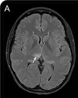
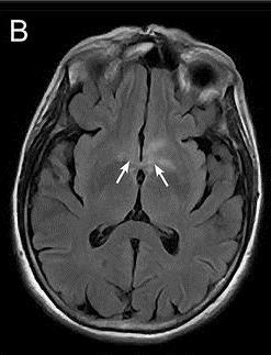

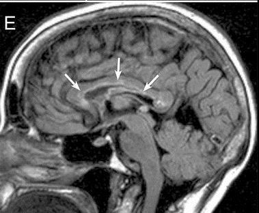
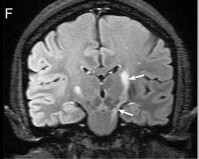

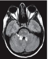
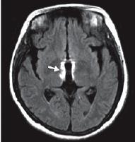
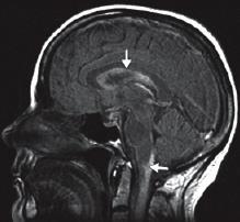
6 NMOSD: BRAIN MRI Thalamus Hypothalamus Subcortical WM Area postrema Corticospinal tract Corpus callosum Periependymal Wingerchuk et al. Neurology 2015
7 NMOSD: BRAIN MRI Extensive brain lesions (EBLs) include: Tumefactive-like lesions ADEM-like lesions MS-like lesions Posterior reversible encephalopathy syndrome (PRES)-like lesions Cheng et al. BMC 2013
 Very rare solid enhancement pattern (indistinguishable from acute MS) Never incomplete ring enhancement Barnett Y et al. AJNR 2013 Ito S. et al. Ann Neurol 2009")
8 NMOSD: BRAIN MRI Enhancement pattern Brain Gadolinium enhancement is rare Frequently patchy enhancement with blurred margins ( cloudlike enhancement ) or pencil-thin ependymal enhancement (similar to that observed in infectious ependymitis) Very rare solid enhancement pattern (indistinguishable from acute MS) Never incomplete ring enhancement Barnett Y et al. AJNR 2013 Ito S. et al. Ann Neurol 2009


9 NMOSD:Spinal Cord MRI Longitudinally extensive transverse myelitis LETM Extension over 3 vertebral segment Frequent Gd enhancement Often necrotizing lesions (T 1 -ipointensity)


10 NMOSD: Spinal Cord MRI LETM lesions have a predilection for central cord Cervical LETM may extend into the medulla Chronic sequelae of LETM may include spinal cord atrophy. Wingerchuk et al. Neurology 2015
11 Acute Disseminated Encephalo-Myelitis-ADEM ADEM is an immune-mediated inflammatory and demyelinating disorder with acute or subacute onset affecting multifocal areas of the CNS. Spinal involvement is much less frequent than brain involvement. ADEM clinical presentation: must be polysimtomatic must include encepahalopathy Encepahalopathy is defined as one or more of the following: Behavioral change (e.g., confusion, excessive irritability) Alteration in consciousness (e.g., lethargy, coma) unexplained by fever Krupp et al. Neurology 2007, Mult Scler 2013

12 ADEM Diagnostic criteria for ADEM Krupp et al 2013 Mult Scler
 located in the")


13 ADEM: BRAIN MRI Large multifocal, T 2 -hyperintense lesions (>1 to 2 cm in size) located in the supratentorial or infratentorial WM regions. T 1 -hypointense lesions are rare. Bilateral diffuse, multifocal, poorly marginated, large asymmetric lesions of the WM, basal ganglia, and cortical GM. Involvement of the thalami Pohl et al. Neurology 2016


14 ADEM: Spinal Cord MRI Spinal cord involvement has been described in up to 1/3 of patients MRI may show confluent intramedullary lesion(s) Often swelling lesions Predominantly affects the thoracic region. Variable enhancement (patchy or pheripheral)
15 ADEM: MRI Complete resolution of MRI abnormalities after treatment has been described in 37% to 75% Partial resolution in 25% to 53% of patients
16 Differential diagnosis Polman C et al 2011


")

17 Multiple Sclerosis Brain lesions characteristics Sites: Periventricular Infratentorial Corpus callosum (sagittal) Juxtacortical (U fibers) Distribution: Asymmetric Shape: Irregular Ovoid Evolution: Variable
 local atrophy (chronic phase) Lycklama et al")
18 Multiple Sclerosis Spinal Cord lesion characteristics Small size: Location: Shape: < 2 vertebral segments < half of cord cross-sectional area cervical > than the rest lateral lateral + posterior columns central GM not spared cigar-like Signal characteristics: Effect on the cord: enhancement uncommon T 1 hypointense rarely swelling (acute phase) local atrophy (chronic phase) Lycklama et al Lancet Neurol 2003


19 NMOSD vs MS Brain MRI MS NMOSD

20 NMOSD vs MS Brain MRI 7.0 Tesla Sinnecker T et al. 2012

21 NMOSD vs MS Spinal Cord MRI NMO central lesion Long spinal cord lesion over 3 vertebral segment Trasversally extensive lesions involving the grey matter surrounding the central canal. SM peripheral lesions Focal, homogeneuos cord lesion Well marginated on sagittal and axial images Peripheral location without involvement of the central canal Nakamura et al. J Neurol 2009; Yonezu et al. Mult Scler 2013

22 NMOSD vs MS Spinal Cord MRI Longitudinally Extensive Transverse Myelitis > 3 segments Centrally located Majority of transverse axis MS risk <2% Acute Partial Transverse Myelitis < 2 segments Eccentric or asymmetric location MS risk: 10% at 61 months in brain MRI negative 88% in brain MRI positive Fisniku LK, Brex PA, Altmann DR, et al. Brain 2008

 BSLs were seen in 63% of the patients without longitudinally extensive spinal cord")
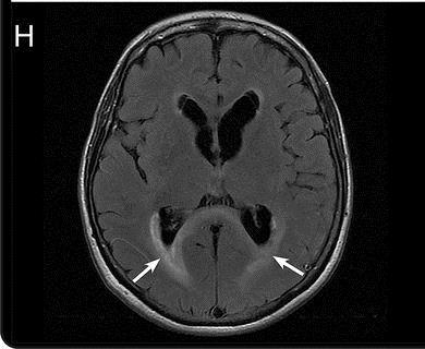
23 NMOSD:Spinal Cord MRI BSLs: very hyperintense spotty lesions on axial T2WI. The signal is visually more hyperintense than that of surrounding cerebrospinal fluid without flow void effects. BSLs were more frequently found in patients with NMO (54%) than in those with MS (3%; p< 0.01). (Sensitivity=54% specificity = 97%) BSLs were seen in 63% of the patients without longitudinally extensive spinal cord lesions (LESCL). BSLs or LESCL were found in 88% of the NMO patients Yonezu T et al. Mult Scler 2013


24 ADEM vs MS MRI characteristics ADEM ADEM SM MS Pohl et al. Neurology 2016

25 Grazie per la vostra attenzione
26 ADEM Typically bilateral lesions on T2/FLAIR sequences, but may be asymmetric Lesions tend to be poorly marginated Almost all patients have multiple lesions in the deep and subcortical WM while the periventricular WM is generally spared Thalami and basal ganglia are frequently affected often symmetrical and brainstem and SC abnormalities are common Contrast enhancement is sometimes seen in acute lesions Differential diagnosis with MS and other demyelinating diseases
27 Number and dimensions of the lesions are variable In the spinal cord large confluent intramedullary that extend over multiple segment are common with variable degree of contrast enhancement. Abnormal findings on MRI may progress over a relatively short period of time, consistent with progression of the disease The occurrence of relapses with new lesions on MRI suggest the diagnosis of multiphase ADEM or MS
28
29 Neuroimaging characteristics of NMOSD Brain MRI Lesion features Large, confluent, unilateral, or bilateral, subcortical or deep WM. Long (1/2 of the length of the corpus callosum or greater), diffuse, heterogeneous, or edematous corpus callosum lesions. Involving: dorsal medulla (especially the area postrema), periependymal surfaces of the IV ventricle in the brainstem/cerebellum, hypothalamus, thalamus, or periependymal surfaces of the III ventricle, long corticospinal tract lesions, unilateral or bilateral, contiguously involving internal capsule and cerebral peduncle. Extensive periependymal brain lesions, often with gadolinium enhancement Wingerchuk et al. Neurology 2015
. Cord expansion/swelling.")
30 Neuroimaging characteristics of NMOSD SPINAL CORD SC MRI, acute Other Increased characteristic signal on features T2-W that (standard may be T2-W, detected PD, or STIR sequences) extending over 3 or more complete vertebral segments. Rostral extension of the lesion into the brainstem. Central cord predominance (more than 70% of the lesion residing within the central GM). Cord expansion/swelling. Gadolinium Decreased signal enhancement on T1-W of corresponding the lesion on T1-W to region (no of specific increased distribution T 2 -W signal. or pattern) SC MRI, chronic Longitudinally extensive cord atrophy (sharply demarcated atrophy extending over 3 complete, contiguous vertebral segments and caudal to a particular segment of the SC), with or without focal or diffuse T 2 signal change involving the atrophic segment OPTIC NERVE Unilateral or bilateral increased T 2 signal or T 1 gadolinium enhancement within ON or optic chiasm; relatively long lesions (e.g., those extending more than half the distance from orbit to chiasm) and those involving the posterior aspects of the ON or the chiasm are associated with NMO Wingerchuk et al. Neurology 2015
Actualização no diagnóstico e tratamento das doenças desmielinizantes na infância. Silvia Tenembaum
 Actualização no diagnóstico e tratamento das doenças desmielinizantes na infância Silvia Tenembaum Acquired CNS inflammatory/demyelinating disorders: Background information More frequent in children than
MRI and differential diagnosis in patients suspected of having MS
 Andrea Falini Italy MRI and differential diagnosis in patients suspected of having MS IMPROVING THE PATIENT S LIFE THROUGH MEDICAL EDUCATION www.excemed.org Outline of presentation - Diagnostic criteria
New Insights on Optic Neuritis in Young People
 Cronicon OPEN ACCESS EC OPHTHALMOLOGY Case Study New Insights on Optic Neuritis in Young People Sergio Carmona 1, Sandra Barbosa 1 and Maria Laura Ortube 2 * 1 Department of Neuro-ophthalmology, Hospital
Myelitis. Myelitis. Multiple Sclerosis (MS) Acute demyelinating syndrome (ADS) Indictions for spinal cord MRI in MS.
 Acute demyelinating syndrome (ADS) Indictions for spinal cord MRI in MS.") Myelitis Myelitis Majda M Thurnher Professor of Radiology Medical University of Vienna University Hospital Vienna Department of Biomedical Imaging and Image-Guided Therapy Vienna Austria Acute demyelinating
Myelitis Myelitis Majda M Thurnher Professor of Radiology Medical University of Vienna University Hospital Vienna Department of Biomedical Imaging and Image-Guided Therapy Vienna Austria Acute demyelinating
COPYRIGHT 2012 THE TRANSVERSE MYELITIS ASSOCIATION. ALL RIGHTS RESERVED
 The Transverse Myelitis Association...advocating for those with acute disseminated encephalomyelitis, neuromyelitis optica, optic neuritis and transverse myelitis ACUTE DISSEMINATED ENCEPHALOMYELITIS (ADEM)
Idiopathic Inflammatory Demyelinating Diseases of the Brainstem
 Idiopathic Inflammatory Demyelinating Diseases of the Brainstem 1 A. Rovira-Cañellas, 2 A. Rovira-Gols, 1 J. Sastre-Garriga, 1 C. Auger, 1 J. Munuera, 1 X. Montalban 1 Hospital Vall d Hebron, Barcelona.
Neuromyelitis optica (NMO), or Devic s disease, is a rare
, or Devic s disease, is a rare") Case Report Neuromyelitis Optica (NMO) Abstract NMO is a is a rare entity which involves the central nervous system acting as an inflammatory process by attacking the optic nerve (ON) and longitudinally
Case Report Neuromyelitis Optica (NMO) Abstract NMO is a is a rare entity which involves the central nervous system acting as an inflammatory process by attacking the optic nerve (ON) and longitudinally
Role of MRI in acute disseminated encephalomyelitis
 Original Research Article Role of MRI in acute disseminated encephalomyelitis Shashvat Modiya 1*, Jayesh Shah 2, C. Raychaudhuri 3 1 1 st year resident, 2 Associate Professor, 3 HOD and Professor Department
Pediatric acute demyelinating encephalomyelitis in Denmark: a nationwide population-based study
 Pediatric acute demyelinating encephalomyelitis in Denmark: a nationwide population-based study Magnus Spangsberg Boesen November, 2016 Supervisors: P. Born, P. Uldall, M. Blinkenberg, M. Magyari, F. Sellebjerg
Myelitis. Case 2. History. Examination. Mahtab Ghadiri
 Case 2 Myelitis Mahtab Ghadiri History A 42-year-old man presented to the emergency department with altered sensation in the lower limbs and difficulty ambulating. He first noted paresthesia in his feet
Magnetic Resonance Imaging in the Acquired Demyelinating Disorders: A Pediatric Cohort Study
 Journal of Pharmacy and Pharmacology 6 (2018) 20-31 doi: 10.17265/2328-2150/2018.01.003 D DAVID PUBLISHING Magnetic Resonance Imaging in the Acquired Demyelinating Disorders: A Pediatric Cohort Study Santa
Neuromyelitis Optica Spectrum Disorder (NMOSD): Brain MRI findings in patients at our institution and literature review.
: Brain MRI findings in patients at our institution and literature review.") Neuromyelitis Optica Spectrum Disorder (NMOSD): Brain MRI findings in patients at our institution and literature review. Poster No.: C-0817 Congress: ECR 2014 Type: Educational Exhibit Authors: G. I. MICHELIN,
Neuromyelitis Optica Spectrum Disorder (NMOSD): Brain MRI findings in patients at our institution and literature review. Poster No.: C-0817 Congress: ECR 2014 Type: Educational Exhibit Authors: G. I. MICHELIN,
MRI in Differential Diagnosis. CMSC, June 2, Jill Conway, MD, MA, MSCE
 MRI in Differential Diagnosis CMSC, June 2, 2016 Jill Conway, MD, MA, MSCE Director, Carolinas MS Center Clerkship Director, UNCSOM-Charlotte Campus Charlotte, NC Disclosures Speaking, consulting, and/or
Magnetic Resonance Imaging of Neuromyelitis Optica (Devic s Syndrome)
") J Radiol Sci 2012; 37: 45-50 Magnetic Resonance Imaging of Neuromyelitis Optica (Devic s Syndrome) Chien-Chuan Huang Tai-Yuan Chen Tai-Ching Wu Yu-Kun Tsui Te-Chang Wu Wen-Sheng Tzeng Chien-Jen Lin Department
J Radiol Sci 2012; 37: 45-50 Magnetic Resonance Imaging of Neuromyelitis Optica (Devic s Syndrome) Chien-Chuan Huang Tai-Yuan Chen Tai-Ching Wu Yu-Kun Tsui Te-Chang Wu Wen-Sheng Tzeng Chien-Jen Lin Department
MYELITIS. A Mochan Neurology
 MYELITIS A Mochan Neurology ATM MS LETM NMOSD ATM LETM MS NMOSD Acute Transverse Myelitis Longitudinally Extensive Transverse Myelitis Multiple Sclerosis Neuromyelitis Optica Spectrum Disorders ATM ADEM
Interactive Cases: Demyelinating Diseases and Mimics. Disclosures. Case 1 25 yo F with nystagmus; look for tumor 4/14/2017
 Interactive Cases: Demyelinating Diseases and Mimics Disclosures None Brad Wright, MD 27 March 2017 Case 1 25 yo F with nystagmus; look for tumor What do you suspect? A. Demyelinating disease B. Malignancy
Neuroimaging and Other Biomarkers. MRI for Diagnosis, Prognosis and Treatment Decisions in MS
 Neuroimaging and Other Biomarkers MRI for Diagnosis, Prognosis and Treatment Decisions in MS Eric Klawiter, MD MSc Massachusetts General Hospital May 30, 2014 Disclosures and Funding Disclosures: Consulting
A pictorial review of neurological complications of systemic lupus erythematosus and antiphospholipid syndrome
 A pictorial review of neurological complications of systemic lupus erythematosus and antiphospholipid syndrome Poster No.: C-2780 Congress: ECR 2010 Type: Educational Exhibit Topic: Neuro Authors: E. Tavernaraki,
Autologous Hematopoietic Stem Cell Transplantation for the Treatment of Neuromyelitis Optica in Singapore
 Case Reports 26 Autologous Hematopoietic Stem Cell Transplantation for the Treatment of Neuromyelitis Optica in Singapore Koh Yeow Hoay, Pavanni Ratnagopal Abstract Introduction: Neuromyelitis optica (NMO)
Heterogeneity of Demyelinating Disease: Definitions and Overlap Overview
 Heterogeneity of Demyelinating Disease: Definitions and Overlap Overview Brian Weinshenker, MD, FRCP(C) Disclosures Royalties related to patent for discovery of NMO-IgG licensed to RSR Ltd; Oxford University
MRI Imaging of Neuromyelitis Optica
 July 2009 MRI Imaging of Neuromyelitis Optica Jenna Nolan, Harvard Medical School Year III Gillian Lieberman, MD Our Patient: Initial Presentation J.H. is a 29 year-old woman who presents with acute vision
RSR RSR RSR RSR RSR. ElisaRSR AQP4 Ab RSR. Aquaporin-4 Autoantibody Assay Kit
 To aid diagnosis of Neuromyelitis Optica (NMO) and NMO spectrum disorder (NMOSD) To confirm diagnosis before initial treatment of patients with demyelinating inflammatory disease NMO, NMOSD and AQP4 Elisa
SWI including phase and magnitude images
 On-line Table: MRI imaging recommendation and summary of key features Sequence Pathologies Visible Key Features T1 volumetric high-resolution whole-brain reformatted in axial, coronal, and sagittal planes
Continuum (Minneap Minn) 2013;19(4):
 2013;19(4):") Review Article Address correspondence to Dr Brian G. Weinshenker, Department of Neurology, Mayo Clinic, 200 First St SW, Rochester, MN 55905, weinb@mayo.edu. Relationship Disclosure: Dr Wingerchuk receives
Review Article Address correspondence to Dr Brian G. Weinshenker, Department of Neurology, Mayo Clinic, 200 First St SW, Rochester, MN 55905, weinb@mayo.edu. Relationship Disclosure: Dr Wingerchuk receives
Tony Traboulsee, MD Associate Professor (Medicine/Neurology) Head, UBC MS and NMO Programs. MRI Diagnostic Red Flags
 Head, UBC MS and NMO Programs. MRI Diagnostic Red Flags") Tony Traboulsee, MD Associate Professor (Medicine/Neurology) Head, UBC MS and NMO Programs MRI Diagnostic Red Flags UBC MS and NMO Research Programs LEARNING OBJECTIVES By the end of this presentation,
Tony Traboulsee, MD Associate Professor (Medicine/Neurology) Head, UBC MS and NMO Programs MRI Diagnostic Red Flags UBC MS and NMO Research Programs LEARNING OBJECTIVES By the end of this presentation,
First clinical attack of inflammatory or demyelinating disease in the CNS. Alteration in consciousness ranging from somnolence or coma
 ADEM Clinical features First clinical attack of inflammatory or demyelinating disease in the CNS Acute or subacute onset Affects multifocal areas of the CNS Polysymptomatic presentation Must include encephalopathy:
Wingerchuk et al, Neurol, 2006
 Current Understanding of Neuromyelitis Optica Jacqueline A. Leavitt, M.D. Mayo Clinic Rochester, MN I have no financial disclosures 46 y/o F Pain in R temple worse with head movements, resolved in days
ORIGINAL CONTRIBUTION. Neuromyelitis Optica Brain Lesions Localized at Sites of High Aquaporin 4 Expression
 ORIGINAL CONTRIBUTION Neuromyelitis Optica Brain Lesions Localized at Sites of High Aquaporin 4 Expression Sean J. Pittock, MD; Brian G. Weinshenker, MD; Claudia F. Lucchinetti, MD; Dean M. Wingerchuk,
Pediatric MS MRI Study Methodology
 General Pediatric MS MRI Study Methodology SCAN PREPARATION axial T2-weighted scans and/or axial FLAIR scans were obtained for all subjects when available, both T2 and FLAIR scans were scored. In order
MRI features and anti-aqp4 antibody status in Idiopathic inflammatory demyelinating CNS disease (IIDCD) in Thai patients
 in Thai patients") Neurology Asia 2013; 18(1) : 73 81 MRI features and anti-aqp4 antibody status in Idiopathic inflammatory demyelinating CNS disease (IIDCD) in Thai patients 1 Naraporn Prayoonwiwat MD, 2 Orasa Chawalparit
Neurology Asia 2013; 18(1) : 73 81 MRI features and anti-aqp4 antibody status in Idiopathic inflammatory demyelinating CNS disease (IIDCD) in Thai patients 1 Naraporn Prayoonwiwat MD, 2 Orasa Chawalparit
1 MS Lesions in T2-Weighted Images
 1 MS Lesions in T2-Weighted Images M.A. Sahraian, E.-W. Radue 1.1 Introduction Multiple hyperintense lesions on T2- and PDweighted sequences are the characteristic magnetic resonance imaging (MRI) appearance
Multiple sclerosis in Japan: Nationwide surveys over 30 years
 Neurology Asia 28; 13 : 131 143 Multiple sclerosis in Japan: Nationwide surveys over 3 years Jun-ichi Kira, Takaaki Ishizu, Manabu Osoegawa, and The Research Committee of Neuroimmunological Diseases Department
Demyelinating Diseases of the Brain
 Department of Radiology University of California San Diego Demyelinating Diseases of the Brain John R. Hesselink, M.D. T1-Weighted Images Normal White Matter Contents Axons with envelope of myelin Neuroglia
Helpful Information for evaluation of new neurological symptoms in patients receiving TYSABRI
 Helpful Information for evaluation of new neurological symptoms in patients receiving TYSABRI This information is provided as an educational resource for healthcare providers and should be considered current
PMH: No medications; Immunizations UTD No hospitalizations or surgeries Speech Delay. Birth Hx: 24 WGA, NICU x6 months
 HPI: 6 months of weakness and parathesias- originally in both feet x 2-3 months, then resolved. Now with parathesias and weakness in fingers x 1 week. Seen by podiatrist and given custom in-soles 1 month
MRI OF THE THALAMUS. Mohammed J. Zafar, MD, FAAN Kalamazoo, MI
 1 MRI OF THE THALAMUS Mohammed J. Zafar, MD, FAAN Kalamazoo, MI Objectives: The thalamic nuclei can be involved in a wide variety of conditions. A systematic imaging approach would be useful for narrowing
Magnetic resonance imaging in multiple sclerosis
 APRIL 2002 103 Magnetic resonance imaging in multiple sclerosis Beside the ability to detect the Olga Ciccarelli and David H. Miller NMR Unit, Institute of Neurology, University College London, Queen Square,
VOLUME 27, NUMERO 1 DECEMBRE 2011 NEUROR ADIOLOGY MUHC MNH VOLUME 27, NUMBER 1 DECEMBER 2011
 VOLUME 27, NUMERO 1 DECEMBRE 2011 NEUROR ADIOLOGY MUHC MNH VOLUME 27, NUMBER 1 DECEMBER 2011 IN THIS ISSUE The new NEURO logo Sandra McPherson Devic s disease Roberta La Piana, Maria Cortés and Donatella
Thashi Chang 1,2* and Milinda Withana 2
 Chang and Withana BMC Research Notes (2015) 8:36 DOI 10.1186/s13104-015-0991-5 CASE REPORT Open Access Gaze palsy, hypogeusia and a probable association with miscarriage of pregnancy - the expanding clinical
NEW DIAGNOSTIC CRITERIA FOR MULTIPLE SCLEROSIS
 NEW DIAGNOSTIC CRITERIA FOR MULTIPLE SCLEROSIS Jeffrey A. Cohen, MD Director, Experimental Therapeutics Mellen MS Center Neurological Institute Cleveland Clinic 2018 Regional MS Summit 30 June 2018 Disclosures
White matter diseases affecting the corpus callosum; demyelinating and metabolic diseases
 White matter diseases affecting the corpus callosum; demyelinating and metabolic diseases Poster No.: C-0199 Congress: ECR 2011 Type: Educational Exhibit Authors: J. H. Yoo; Seoul/KR Keywords: Neuroradiology
Neuromyelitis Optica Spectrum Disorder (NMOSD)
") Neuromyelitis Optica Spectrum Disorder (NMOSD) Neuromyelitis Optica Spectrum Disorder (NMOSD) is a rare relapsing autoimmune disorder that preferentially causes inflammation in the optic nerve and spinal
Neuromyelitis Optica Spectrum Disorder (NMOSD) Neuromyelitis Optica Spectrum Disorder (NMOSD) is a rare relapsing autoimmune disorder that preferentially causes inflammation in the optic nerve and spinal
MRI diagnostic criteria for multiple sclerosis: an update
 MRI diagnostic criteria for multiple sclerosis: an update Poster No.: C-0285 Congress: ECR 2013 Type: Educational Exhibit Authors: L. Valls Masot, A. M. Quiles Granado, J. Puig Alcántara, L. RamióTorrentà,
Neuromyelitis Optica: A Case Report
 Pediatr Neonatol 2010;51(6):347 352 CASE REPORT Neuromyelitis Optica: A Case Report Wei-Chia Chia 1, Jian-Nan Wang 1, Ming-Chi Lai 2 * 1 Department of Family Medicine, Chi-Mei Medical Center, Yong Kang
Sawada J, Orimoto R, Misu T, Katayama T, Aizawa H, Asanome A, Takahashi K, Saito T, Anei R, Kamada K, Miyokawa N, Takahashi T, Fujihara K, Hasebe N.
 Mult Scler (2014.9) 20(10):1413-1416. A case of pathology-proven neuromyelitis optica spectrum disorder with Sjögren syndrome manifesting aphasia and apraxia due to a localized cerebral white matter lesion.
CME Conference: Updates in Neuro ophthalmology and MS March 2017 Teri Schreiner, MD MPH University of Colorado
 CME Conference: Updates in Neuro ophthalmology and MS March 2017 Teri Schreiner, MD MPH University of Colorado I participate in clinical research funded by Biogen, Teva, Novartis, NIH/NINDS, MSDx, ADAMAS
Radiologic-Pathologic Correlation of White Matter Disease
 The Neuroradiology Journal 22 (Suppl. 1): 26-32, 2009 www. centauro. it Radiologic-Pathologic Correlation of White Matter Disease J.G. SMIRNIOTOPOULOS 1,2,3,.. SMITH 1,2, E. RUSHING 2,4, F.M. MURPHY 2,
MRI in MS. MRI in multiple sclerosis. MS T2 Lesions: Pathology. Brain lesions Morphology Matters. Sagittal FLAIR Morphology Matters
 MRI in multiple sclerosis MRI in MS Rohit Bakshi, MD, MA Breakstone Professor of Neurology & Radiology Director, Laboratory for Neuroimaging Research Senior Neurologist, MS Center Brigham & Women s Hospital
Acute disseminated encephalomyelitis in children: differential diagnosis from multiple sclerosis on the basis of clinical course
 Review article DOI: 10.3345/kjp.2011.54.6.234 Korean J Pediatr 2011;54(6):234-240 Acute disseminated encephalomyelitis in children: differential diagnosis from multiple sclerosis on the basis of clinical
The Role of MRI in Multiple Sclerosis
 September 2006 The Role of MRI in Multiple Sclerosis David Cochran, Harvard Medical School Year IV Meet our patient WM 49yoF presents to Multiple Sclerosis Clinic for follow-up regarding worsening symptoms
Clinical and research application of MRI in diagnosis and monitoring of multiple sclerosis
 24-25 February 2016 - Siena, Italy Clinical and research application of MRI in diagnosis and monitoring of multiple sclerosis IMPROVING THE PATIENT S LIFE THROUGH MEDICAL EDUCATION www.excemed.org How
Neurological update: MOG antibody disease
 https://doi.org/10.1007/s00415-018-9122-2 NEUROLOGICAL UPDATE Neurological update: MOG antibody disease Ray Wynford Thomas 1,2 Anu Jacob 3 Valentina Tomassini 1,2,4 Received: 17 August 2018 / Revised:
Neuromyelitis optica mimics the morphology of spinal cord tumors
 The Turkish Journal of Pediatrics 2016; 58: 309-314 Case Report Neuromyelitis optica mimics the morphology of spinal cord tumors İlknur Erol 1, Murat Özkale 2, Tülin Savaş 1, Özlem Alkan 3, Melih Çekinmez
Jarius et al. Journal of Neuroinflammation (2016) 13:280 DOI /s
 13:280 DOI /s") Jarius et al. Journal of Neuroinflammation (2016) 13:280 DOI 10.1186/s12974-016-0718-0 RESEARCH Open Access MOG-IgG in NMO and related disorders: a multicenter study of 50 patients. Part 2: Epidemiology,
Jarius et al. Journal of Neuroinflammation (2016) 13:280 DOI 10.1186/s12974-016-0718-0 RESEARCH Open Access MOG-IgG in NMO and related disorders: a multicenter study of 50 patients. Part 2: Epidemiology,
Content. Polyarteritis nodosa. Vasculitis. Giant cell arteritis. Primary cerebral angiitis. Other autoimmune CNS disease.
 Content Other autoimmune CNS disease Philippe Demaerel Vasculitis Systemic lupus erythematosus Wegener granulomatosis Behçet disease Rhombencephalitis - CLIPPERS Neurosarcoidosis Langerhans cell histiocytosis
Professor Yasser Metwally. Neuromyelitis optica. EPIDEMIOLOGY
 180 Professor Yasser Metwally Neuromyelitis optica www.yassermetwally.com N EPIDEMIOLOGY Afro-Caribbean, and South American descent implying underlying genetic mechanisms in the expression of demyelinating
CONTINUING EDUCATION MRI OF MYELITIS* JBR BTR, 2012, 95: J. Hodel 1,2, O. Outteryck 3, P. Jissendi 1, M. Zins 2, X. Leclerc 1, J.-P.
 JR TR, 2012, 95: 270-276. ONTINUING EDUTION MRI OF MYELITIS* J. Hodel 1,2, O. Outteryck 3, P. Jissendi 1, M. Zins 2, X. Leclerc 1, J.-P. Pruvo 1 MRI of myelitis The diagnosis of myelitis relies on MRI.
Introduction to the Central Nervous System: Internal Structure
 Introduction to the Central Nervous System: Internal Structure Objective To understand, in general terms, the internal organization of the brain and spinal cord. To understand the 3-dimensional organization
NANOS 2018 Literature Review Clinical Papers Neurology. Caroline Tilikete, MD, PhD Hospices Civils de Lyon, University Lyon I Lyon, France
 NANOS 2018 Literature Review Clinical Papers Neurology Caroline Tilikete, MD, PhD Hospices Civils de Lyon, University Lyon I Lyon, France Nystagmus Effect of Gabapentin/Memantine on the Infantile Nystagmus
10/3/2016. T1 Anatomical structures are clearly identified, white matter (which has a high fat content) appears bright.
 appears bright.") H2O -2 atoms of Hydrogen, 1 of Oxygen Hydrogen just has one single proton and orbited by one single electron Proton has a magnetic moment similar to the earths magnetic pole Also similar to earth in that
H2O -2 atoms of Hydrogen, 1 of Oxygen Hydrogen just has one single proton and orbited by one single electron Proton has a magnetic moment similar to the earths magnetic pole Also similar to earth in that
Research Article Optic Nerve and Spinal Cord Are the Major Lesions in Each Relapse of Japanese Multiple Sclerosis
 International Scholarly Research Network ISRN Neurology Volume 211, Article ID 9476, 4 pages doi:1.542/211/9476 Research Article Optic Nerve and Spinal Cord Are the Major Lesions in Each Relapse of Japanese
CASE OF THE WEEK PROFESSOR YASSER METWALLY
 CLINICAL PICTURE CLINICAL PICTURE A 40 Years old female patient, known to be suffering from multiple sclerosis, was presented clinically with painful diminution of vision in the right aye and mild weakness
Anaesthesia recommendations for patients suffering from Neuromyelitis optica spectrum disorder
 orphananesthesia Anaesthesia recommendations for patients suffering from Neuromyelitis optica spectrum disorder Disease name: Neuromyelitis optica spectrum disorder ICD 10: G36.0 Synonyms: Devic's Disease,
Masses of the Corpus Callosum
 Masses of the Corpus Callosum Kesav Raghavan, HMS Year III Dr. Agenda Corpus Callosum Development and Anatomy Our Patient: Clinical Presentation Differential Diagnosis of Masses in the Corpus Callosum
Disclosure. + Outline. Case-based approach to neurological emergencies that might present to the ED
 Kathleen R. Fink, MD University of Washington 5 th Nordic Emergency Radiology Course May 21, 2015 Disclosure My spouse receives research salary support from: Bracco BayerHealthcare Guerbet Outline Case-based
Multiple Sclerosis vs Acute Disseminated Encephalomyelitis in Childhood
 Multiple Sclerosis vs Acute Disseminated Encephalomyelitis in Childhood Steven David Brass, MD, Zografos Caramanos, BA, Carlos Santos, MD, Marie-Emmanuelle Dilenge, MD, Yves Lapierre, MD, and Bernard Rosenblatt,
Susac Syndrome: Brain MRI findings in 3 patients at our institution and review of literature.
 Susac Syndrome: Brain MRI findings in 3 patients at our institution and review of literature. Poster No.: C-0395 Congress: ECR 2014 Type: Educational Exhibit Authors: H. Chaves, M. S. TORONCHIK, P. M.
Essentials of Clinical MR, 2 nd edition. 14. Ischemia and Infarction II
 14. Ischemia and Infarction II Lacunar infarcts are small deep parenchymal lesions involving the basal ganglia, internal capsule, thalamus, and brainstem. The vascular supply of these areas includes the
Transverse Myelitis and Myelopathy in the VA system: Etiology and Epidemiology
 Transverse Myelitis and Myelopathy in the VA system: Etiology and Epidemiology Stacey L. Clardy MD PhD Staff Neurologist, Salt Lake City VA Assistant Professor of Neurology, University of Utah Director,
Tumefactive multiple sclerosis lesions: Diagnostic challenge for the radiologist
 Tumefactive multiple sclerosis lesions: Diagnostic challenge for the radiologist Poster No.: C-0679 Congress: ECR 2015 Type: Educational Exhibit Authors: A. Gargallo Vaamonde, M. Iridoy Zulet, I. Rubio
Roadmap to Pediatric Demyelination: Clinical Features. Teri Schreiner, MD MPH June 1, 2014 CMSC. Disclosures
 Roadmap to Pediatric Demyelination: Clinical Features Teri Schreiner, MD MPH June 1, 2014 Sunday, June 08, 2014 CMSC 1 Disclosures I participate in clinical research funded by Biogen, Teva, Novartis, NIH/NINDS,
Brainstem. By Dr. Bhushan R. Kavimandan
 Brainstem By Dr. Bhushan R. Kavimandan Development Ventricles in brainstem Mesencephalon cerebral aqueduct Metencephalon 4 th ventricle Mylencephalon 4 th ventricle Corpus callosum Posterior commissure
Common Pitfalls in Multiple Sclerosis and CNS Demyelinating Diseases
 Common Pitfalls in Multiple Sclerosis and CNS Demyelinating Diseases Case-Based Learning Common Pitfalls in Multiple Sclerosis and CNS Demyelinating Diseases Case-Based Learning Mayo Clinic College of
Provider Led Entity. CDI Quality Institute PLE Multiple Sclerosis (MS) AUC
 AUC") Provider Led Entity CDI Quality Institute PLE Multiple Sclerosis (MS) AUC Appropriateness of advanced imaging procedures* in patients with the following clinical presentations or diagnoses: 09/04/2018
Provider Led Entity CDI Quality Institute PLE Multiple Sclerosis (MS) AUC Appropriateness of advanced imaging procedures* in patients with the following clinical presentations or diagnoses: 09/04/2018
Setting the Scene: Neuromyelitis Optica epidemiology, population variability, subgroups: relapsing/monophasic, Ab +ve/-ve, NMO/SD, treated/untreated
 UK Nationally Commissioned NMO team Setting the Scene: Neuromyelitis Optica epidemiology, population variability, subgroups: relapsing/monophasic, Ab +ve/-ve, NMO/SD, treated/untreated Jackie Palace Disclosures
MR neuroimaging of HIV infected patients : A pictorial review
 MR neuroimaging of HIV infected patients : A pictorial review Poster No.: R-0198 Congress: 2014 CSM Type: Scientific Exhibit Authors: P. F. Kwan, R. Thomas, A. Dixon; SOUTH YARRA/AU Keywords: Neuroradiology
Paraparesis. Differential Diagnosis. Ran brauner, Tel Aviv university
 Paraparesis Differential Diagnosis Ran brauner, Tel Aviv university Definition Loss of motor power to both legs Paraparesis (paraplegia) refers to partial (- paresis) or complete (-plegia) loss of voluntary
MRI of chronic spinal cord injury
 The British Journal of Radiology, 76 (2003), 347 352 DOI: 10.1259/bjr/11881183 E 2003 The British Institute of Radiology Pictorial review MRI of chronic spinal cord injury 1 K POTTER, FRCR and 1 A SAIFUDDIN,
The current role of MRI in differentiating multiple sclerosis from its imaging mimics
 The current role of MRI in differentiating multiple sclerosis from its imaging mimics Ruth Geraldes 1, Olga Ciccarelli 2,3, Frederik Barkhof 2,3,4, Nicola De Stefano 5, Christian Enzinger 6,7, Massimo
Multiple sclerosis mimics: Diagnostic error and management decisions
 Multiple sclerosis mimics: Diagnostic error and management decisions John R Rinker, II, MD Mitchell Wallin, MD Paralyzed Veterans Summit, Washington Harbor, MD August 30, 2017 Disclosures Dr. Rinker and
Anatomy & Physiology Central Nervous System Worksheet
 1. What are the two parts of the CNS? 2. What are the four functions of the CNS Anatomy & Physiology Central Nervous System Worksheet 3. What are the four functions of the meninges? (p430) 4. Starting
Spinal cord MR imaging in Multiple Sclerosis
 43ème CONGRÈS ANNUEL de la Société Française de NeuroRadiologie 30 mars au 1 er avril 2016 Novotel Paris Tour Eiffel Spinal cord MR imaging in Multiple Sclerosis Àlex Rovira Unitat de Neurorradiología.
References 1. Feng S et al. Journal of Thoracic Oncology 2017; 12: Spain L et al. Annals of Oncology 2017; 28:
 Maulik Shah, MD February 15, 2019 Patient Presentation: Progressive Sensory Disturbance In early 2018, this 57 year old man was sent to the Emergency Department after complaining in the oncology clinic
Transverse myelitis revisited
 Transverse myelitis revisited Poster No.: C-0341 Congress: ECR 2012 Type: Educational Exhibit Authors: V. Lolli, D. Balériaux; Brussels/BE Keywords: Inflammation, Diagnostic procedure, MR, Neuroradiology
Clinical Profiles and Short-Term Outcomes of Acute Disseminated Encephalomyelitis in Adult Chinese Patients
 JCN Open Access pissn 1738-6586 / eissn 2005-5013 / J Clin Neurol 2016;12(3):282-288 / http://dx.doi.org/10.3988/jcn.2016.12.3.282 ORIGINAL ARTICLE Clinical Profiles and Short-Term Outcomes of Acute Disseminated
Ch 13: Central Nervous System Part 1: The Brain p 374
 Ch 13: Central Nervous System Part 1: The Brain p 374 Discuss the organization of the brain, including the major structures and how they relate to one another! Review the meninges of the spinal cord and
Infection-Associated Neurological Syndromes
 Infection-Associated Neurological Syndromes Anand P, MD PhD Medical Director, BloodCenter of Wisconsin Assistant Professor, Medical College of Wisconsin ASFA Annual Meeting San Antonio, TX, May 8th, 2015
Disease of Myelin. Reid R. Heffner, MD Distinguished Teaching Professor Emeritus Department of Pathology and Anatomy January 9, 2019
 Disease of Myelin Reid R. Heffner, MD Distinguished Teaching Professor Emeritus Department of Pathology and Anatomy January 9, 2019 1 I HAVE NO CONFLICTS OF INTEREST OR DISCLOSURES TO DECLARE. I HAVE NO
Chapter 2. Central Nervous System; the brain and spinal cord
 Chapter 2 Central Nervous System; the brain and spinal cord CNS 1. Topography; - what are the main components of the brain - how do you recognize them? 2. The location of the major functional areas of
Chapter 12b. Overview
 Chapter 12b Spinal Cord Overview Spinal cord gross anatomy Spinal meninges Sectional anatomy Sensory pathways Motor pathways Spinal cord pathologies 1 The Adult Spinal Cord About 18 inches (45 cm) long
The Etiological Spectrum of Acute Sensory Myelitis
 Open Access pissn 1738-6586 / eissn 2005-5013 / J Clin Neurol 2015;11(3):227-233 / http://dx.doi.org/10.3988/jcn.2015.11.3.227 ORIGINAL ARTICLE Jae-Won Hyun a,c Jee Young Kim b Kyung Gyu Choi a Ho Jin
Demyelinating Diseases: Multiple Sclerosis January 10, 2018 Dr. Ostrow
 Demyelinating Diseases: Multiple Sclerosis January 10, 2018 Dr. Ostrow Reading: Robbins & Cotran, 9 th edition, pp 1283-1286 Robbins Basic Pathology, 9 th edition, 832-835 Overview: Grossly, myelin is
The central nervous system
 Sectc.qxd 29/06/99 09:42 Page 81 Section C The central nervous system CNS haemorrhage Subarachnoid haemorrhage Cerebral infarction Brain atrophy Ring enhancing lesions MRI of the pituitary Multiple sclerosis
Clinical, Neurodiagnostic, and MR Findings in Children with Spinal and Brain Stem Multiple Sclerosis
 Clinical, Neurodiagnostic, and MR Findings in Children with Spinal and Brain Stem Multiple Sclerosis Charles M. Glasier, Mark B. Robbins, Patricia C. Davis, Elizenda Ceballos, and Stephen R. Bates PURPOSE:
MRI IN THE DIAGNOSIS OF NEUROBEHCET S DISEASE ABOUT 25 CASES
 MRI IN THE DIAGNOSIS OF NEUROBEHCET S DISEASE ABOUT 25 CASES N. Cherif Idrissi El Ganouni1, L. Akka1, H. Jaafari2, M. Zahlane3, L. Essadouni3, N. Kissani2, O. Essadki1, A.Ousehal1. (1) Department of radiology,
Neuroradiological, clinical and genetic characterization of new forms of hereditary leukoencephalopathies
 Neuroradiological, clinical and genetic characterization of new forms of hereditary leukoencephalopathies Principal Investigator: Dr. Donatella Tampieri, MD, FRCPC, Department of Neuroradiology, Montreal
Table of Contents: SKULL AND BRAIN. Scalp, Skull. Anatomically Based Differentials. Skull Normal Variants. Scalp Mass, Child.
 Table of Contents: SKULL AND BRAIN Scalp, Skull Skull Normal Variants Scalp Mass, Child Scalp Mass, Adult Congenital Anomalies of Skull Base Sellar/Parasellar Mass With Skull Base Invasion "Hair on End"
Osmotic Demyelination Syndrome Case Report POPOVA RD, KALCHEV EB, VALCHEV GN, KALOYANOVA DV, TENEVA TG, BALEV BD
 Osmotic Demyelination Syndrome Case Report POPOVA RD, KALCHEV EB, VALCHEV GN, KALOYANOVA DV, TENEVA TG, BALEV BD Once upon a time on a Saturday shift u An emergency head CT scan was ordered from the ICU
Analysis of prognostic significance of clinical and paraclinical indices in case of different types of acute disseminated encephalomyelitis course.
 1 2 Analysis of prognostic significance of clinical and paraclinical indices in case of different types of acute disseminated encephalomyelitis course. 3 4 5 6 7 8 9 10 11 12 13 14 15 16 17 18 19 20 21
Spinal cord tumours Luc van den Hauwe et al.
 overview spinal cord tumours L. van den Hauwe 1,2, D. Balériaux 3, J.W. Van Goethem 2, C. Venstermans 2, F. De Belder 2, P.M. Parizel 2 introduction imaging spinal tumour classification spinal cord tumours