Spinal cord tumours Luc van den Hauwe et al.
|
|
|
- Eileen Cordelia King
- 5 years ago
- Views:
Transcription
1 overview spinal cord tumours L. van den Hauwe 1,2, D. Balériaux 3, J.W. Van Goethem 2, C. Venstermans 2, F. De Belder 2, P.M. Parizel 2 introduction imaging spinal tumour classification spinal cord tumours intradural extramedullary tumours epidural and primary spinal bone tumours differential diagnosis - tumor mimics conclusions 1 AZ KLINA Brasschaat, 2 Universitair Ziekenhuis Antwerpen-University of Antwerp, 3 Hôpital Erasme-Free University of Brussels, Belgium overview introduction imaging spinal cord tumours astrocytoma ependymoma hemangioblastoma ganglioglioma other differential diagnosis - tumour mimics conclusions introduction classification of spinal tumours spinal cord tumours intradural extramedullary tumours epidural and primary spinal bone tumours tumours of the spinal cord are (very) rare may cause significant and longstanding morbidity clinical findings are most often non-specific pain!!! motor and sensory deficits sphincteric disturbances, impotence age some statistics intramedullary spinal cord tumours: 5% within the spinal cord intradural extramedullary tumours: 40% arise from leptomeninges and nerve roots extradural tumours: 55% outside the thecal sac, in vertebral bodies and epidural tissues more statistics intramedullary spinal cord tumours 90% are gliomas ependymomas 60% astrocytomas 30% in children astrocytomas 90% when younger than 10 60% in adolescents 2/3 gangliogliomas ependymomas are very (very) rare (NF2)
 schwannoma (NF-2) 3.")
 is the most important task for the neuroradiologist")


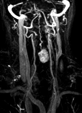
2 solitary vs multiple lesions multiple 1. spinal cord ependymoma (NF-2) hemangioblastoma (VHL syndrome) M + number of lesions? where is the lesion? solitary 2. intradural extramedullary neurofibroma (NF-1) schwannoma (NF-2) 3. epidural space/osseous spine meningioma (NF-2) M + M + lymphoma multiple myeloma hemangioma more statistics myxopapillary ependymomas compose 90% of filum terminale tumours DD paraganglioma, giant schwannoma introduction role of imaging detection differentiating these tumours from other non-tumoural pathology (e.g. demyelination) is the most important task for the neuroradiologist determining the relationship of the tumour with the spinal cord and the extent of the tumour along with associated spinal cord changes such as oedema or syringomyelia is essential in therapy planning and monitoring attempting to diagnose the type of tumor according to imaging criteria may be difficult in some cases but is less critical in patient management MRI imaging modalities CT CR Myelography and myelo-ct DSA PET Tomura et al. AJNR 2012 CR and CT failed to reveal the true extent of intramedullary spinal neoplasms until gross expansion of the spinal canal had occurred straightening of the spine progressive scoliosis widening of the spinal canal myelography myelo-ct complete or partial block in the flow of intrathecal contrast material cannot define the character of the spinal cord lesion in patients in whom MR imaging is contraindicated Abul-Kasim et al. Neuroradiology 2008 Garcia-Morales et al. Neuroradiology Case of the Week, Case 34

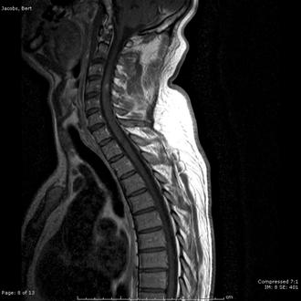
3 MR imaging the whole spinal cord must be visualized imaging protocol sagittal T2, FS-T2 (STIR), T1 -/+ Gd axial T2, T1 + Gd additional sequences: GRE T2*, FLAIR, PRD, DTI, 3 tenets: spinal cord expansion contrast enhancement associated cysts and pseudocysts spinal cord expansion signal changes without cord expansion? consider other, nontumoural lesion demyelinating sarcoid vascular davf in a series of 212 patients suspected of having intramedullary disease, nine (4%) had nonneoplastic lesions; none had imaging evidence of cord expansion Lee et al. Neurosurgery 1998 contrast enhancement almost all spinal cord tumours show some enhancement the absence of enhancement does not exclude an intramedullary neoplasm in the presence of cord expansion not all expansive, enhancing lesions are tumours! contrast enhancement Gd-enhanced MR imaging: accurate delineation of the solid enhancing portion of the tumour tumour vs normal cord on T1-wi tumour vs oedema on T2-wi Parizel et al. AJR 1989 contrast enhancement ependymoma: intense, homogeneous, and sharply marginated focal enhancement occupy the whole width of the spinal cord in the affected segment, centrifugal expansion intratumoural cysts astrocytoma: more patchy, irregular enhancement even low-grade lesions did enhance eccentrical location Parizel et al. AJR 1989 overlap associated cysts tumoural cysts enhancing wall part of the tumour must be resected more frequent in astrocytomas non-tumoural cysts polar or sattelite cysts, rostral or caudal from the tumour non-enhancing wall reactive dilatation of the central canal: syringomyelia


,")

 intense,")

4 MR imaging DTI although MR imaging is a powerful tool, it is not perfect in a series of 171 patients, MR imaging could correctly suggest the histologic diagnosis in only 70% of cases in particular, the differentiation of ependymoma from astrocytoma was the most difficult Brotchi et al. Neurosurgery 1991 Vargas et al. Neuroradiology 2008 ependymomas originate from cells bordering the central ependymal canal: more centromedullary located young patients (mean age is 42), slight male preponderance typically found in the C-spine (50%) associated (large) satellite cysts (60%) intense, mostly homogeneous enhancement well-defined borders cap-sign (27%) low SI areas at both sides of the tumor limits hemosiderin deposits (chronic hemorrhage)


5 Spinal cord tumours myxopapillary ependymomas typical tumor < conus medullaris and filum terminale relatively common in children & young adults WHO, grade I slow growing tumours long standing history of non specific low back pain exacerbated at night may become very large before diagnosis is made hemorrhage may occur; sudden worsening of clinical symptoms LBP after trauma


;")


6 schwannoma hypointense on T1-wi hyperintense on T2-wi astrocytomas pilocytic astrocytoma high-grade and GBM show patchy enhancement, and are more heterogeneous, with necrotic/cystic areas and hemorrhage (T2*-wi) ill-defined borders (infiltrative nature); cleavage plane is rarely found hemangioblastomas benign, usually richly vascularized tumours located superficially, in the sub-pial region, postero-lateral small nodule with extensive edema and cystic components more commonly in adults, with a peak incidence in the fourth decade Baker et al. AJR 2000 Lee et al. 2003


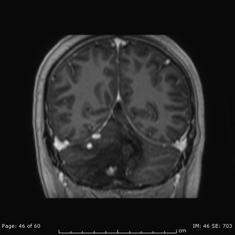


7 hemangioblastomas can be solitary (80%) or multiple (20%), when associated with von Hippel-Lindau syndrome (VHLs) DD with metastases of renal cell carcinoma Wanebo et al Takai et al Case courtesy: D. Balériaux, Brussels, B case courtesy: D. Balériaux, Brussels, B
; mostly")
;")


.")
8 10/09/2012 ganglioglioma rare tumours in adults (1 2% of all spinal cord tumours) 2 nd most common intramedullary tumour in the pediatric age group (15% of cases); mostly affect children between 1 and 5 years of age, as do PA composed of a combination of neoplastic ganglion cells and glial elements typically low-grade tumours (WHO grade 1-2); have a significant propensity for local recurrence, and the glial element may progress to higher grade tumours Rossi et al. Neuroimag Clin N Am 2007 Smith et al. AJR 2012 ganglioglioma cervical and upper thoracic cord may extend over more than 8 vertebral segments typically eccentrically located imaging, scoliosis and remodeling calcification may be seen on CT, is probably the single most suggestive feature of gangliogliomas in the absence of gross calcification, the MR imaging is nonspecific and does not allow differentiation from astrocytomas young patient age, long tumour length, absence of edema, mixed signal intensity on T1-wi, patchy tumor enhancement Rossi et al. Neuroimag Clin N Am 2007 Case courtesy: A. Rossi, Genova, Italy spinal cord metastases considered rare, accounting for only 5% of all intramedullary lesions number is growing fast due to the longer survival of many cancer patients (improved chemotherapy, etc ). less common than leptomeningeal metastases primary tumour: lung and breast carcinoma high sensitivity of MR imaging Case courtesy: A. Rossi, Genova, Italy

 tumefactive demyelinating lesions in multiple sclerosis")
 spinal cord contusion and spinal cord infarction sarcoidosis")


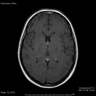
9 M + of breast carcinoma solitary M + of renal cell carcinoma spinal cord tumour mimics in one series of 212 patients undergoing surgery for intramedullary spinal cord tumours, 4% of non-neoplastic lesions were found a variety of lesions may mimic a spinal cord tumour cavernous malformations (cavernomas) tumefactive demyelinating lesions in multiple sclerosis (MS), neuromyelitis optica (NMO) and acute disseminating encephalomyelitis (ADEM) acute transverse myelitis (ATM) spinal cord contusion and spinal cord infarction sarcoidosis vasculitis MR imaging of the brain, follow-up examination spinal cord tumour mimics may enlarge the cord mildly are less extensive may or may not enhance! DD: cavernous malformation ( cavernoma ) inflammatory/demyelinating diseases infectious disease vascular/ischemic lesions MR imaging of the brain, follow-up examination cavernous malformation aka cavernous angioma, or cavernoma represent 7%-10% of all intramedullary tumours most commonly involve the thoracic spine clinical: may remain clinically silent for a long period of time acute and rapidly progressive neurological deficit may occur due to bleeding typical black and white or popcorn appearance due to areas of mixed signal intensity on both T1- and T2- or T2*-wi look also in the brain!!! use SWI!!!


10 MS NMO ADEM tumefactive demyelinating lesions AJNR 2008; 29: neuromyelitis optica: NMO Devic disease


11 Spinal cord tumours Case courtesy: B. Goraj, Nijmegen, The Netherlands conclusion classify spinal tumours spinal cord tumours are rare DD with non-tumoural conditions tumour vs associated changes polar cysts oedema final diagnosis: microscope dural AV-fistula sarcoidosis
Essentials of Clinical MR, 2 nd edition. 51. Primary Neoplasms
 51. Primary Neoplasms As with spinal central canal neoplasms in other regions, those of the lumbar spine may be classified as extradural, intradural extramedullary, and medullary. If an extradural lesion
Pediatric Spine Tumors (and other masses)
") Pediatric Spine Tumors (and other masses) Francisco A Perez, MD, PhD Assistant Professor Neuroradiology and Pediatric Radiology Seattle Children s Hospital University of Washington, Seattle Commercial
Pediatric Spine Tumors (and other masses) Francisco A Perez, MD, PhD Assistant Professor Neuroradiology and Pediatric Radiology Seattle Children s Hospital University of Washington, Seattle Commercial
Introduction to Neuroimaging spine. John J. McCormick MD
 Introduction to Neuroimaging spine John J. McCormick MD Neuroanatomy Netter drawings Radiographic Anatomy Cervical Spine Cervical Spine Oblique View Cervical Spine Dens View Thoracic Spine Lumbar Spine
Kathleen R. Fink, MD Virginia Mason Medical Center. 6 th Nordic Emergency Radiology Course 2017
 Kathleen R. Fink, MD Virginia Mason Medical Center 6 th Nordic Emergency Radiology Course 2017 Disclosure My spouse receives research salary support from: Guerbet Outline Acute neck and back pain Acute
Imaging the Spinal Cord & Intradural Disease
 Department of Radiology University of California San Diego Imaging the Spinal Cord & Intradural Disease John R. Hesselink, M.D. Spinal Cord Diseases Tumors Syringohydromyelia Trauma Ischemia / Infarction
A Journey Down The Canal
 A Journey Down The Canal Radiological Assessment of Spinal Cord Masses John Berry-Candelario HMS III Gillian Lieberman, MD BIDMC Objectives Patient review Anatomy of the spine Imaging techniques Classification
NEURORADIOLOGY. Part III. Angela Csomor University of Szeged Department of Radiology
 NEURORADIOLOGY Part III Angela Csomor University of Szeged Department of Radiology DISEASES OF SPINE AND SPINAL CORD I. Non-tumourous diseases developmental anomalies vascular disorders inflammatory processes
Intradural spinal tumours and their mimics: a review of radiographic features
 Department of Radiology, Royal Melbourne Hospital, Parkville, Victoria, Australia Correspondence to Dr Sara Wein, Radiology Department, Royal Melbourne Hospital, Grattan Street, Parkville, VIC 3050, Australia;
Spinal Vascular Lesions
 Spinal Vascular Lesions Spinal Vascular Lesions Spinal cord infarction Hemangioblastoma Cavernous malformation Vascular malformations (Type 1-4) Spinal artery aneurysm Troy Hutchins, MD Assistant Professor
Spinal Neoplasms. First Things First!! Localize the Lesion!! Ependymomas. Common Intramedullary Lesions
 Acta Radiológica Portuguesa, Vol.XXIII, nº 90, pág. 101-114, Abr.-Jun., 2011 Spinal Neoplasms Bruno A Policeni University of Iowa Hospitals and Clinics Assistant Professor of Radiology Disclosure of Commercial
4/14/2017. Unknown Case #1 Intramedullary Lesion
 4/14/2017 Intradural, Intramedullary Tumor or Mimic Intradural, Extramedullary Tumor or Mimic Extradural Tumor or Mimic Unknown Case #1 Intramedullary Lesion Unknown Case #1 -POST - MAG AXIAL Cranial Caudal
11 May Disclosure. + Outline: Acute Spine Emergencies
 Kathleen R. Fink, MD University of Washington 5 th Nordic Emergency Radiology Course May 21, 2015 Disclosure My spouse receives research salary support from: Bracco BayerHealthcare Guerbet K Fink Nordic
Ependymoma of the spine
 Ependymoma of the spine Tenny Zhang, MS-3 Harvard Medical School 1 Case presentation: history and exam HPI: A 30-year-old man with no significant past medical history presents with one week of bilateral
Spine. Neuroradiology. Spine. Spine Pathology. Distribution of fractures. Radiological algorithm. Role of radiology 18/11/2015
 Spine Neuroradiology Spine Prof.Dr.Nail Bulakbaşı X Ray: AP/L/Oblique Vertebra & disc spaces CT & CTA Vertebra, discs, vessels MRI & MRA Vertebra, disc, vessels, meninges Spinal cord & nerves Myelography
Giant cystic intradural extramedullary pilocytic astrocytoma of Cauda equina
 1 di 7 13/03/2014 09.02 J Neurosci Rural Pract. 2013 Oct-Dec; 4(4): 453 456. doi: 10.4103/0976-3147.120217 PMCID: PMC3858770 Giant cystic intradural extramedullary pilocytic astrocytoma of Cauda equina
Magnetic resonance imaging of intramedullary spinal cord lesions
 Magnetic resonance imaging of intramedullary spinal cord lesions Poster No.: C-1762 Congress: ECR 2014 Type: Educational Exhibit Authors: M. Abdelkafi, H. Derbel, H. Abid, S. Haddar, B. Souissi, N. 1 1
Tumors of the Nervous System
 Tumors of the Nervous System Peter Canoll MD. PhD. What I want to cover What are the most common types of brain tumors? Who gets them? How do they present? What do they look like? How do they behave? 1
SHORT OVERVIEW OF SPINAL CORD TUMORS
 SHORT OVERVIEW OF SPINAL CORD TUMORS 1 INTRODUCTION RARE HETEROGENEOUS GROUP OF TUMORS. 15%OFALLPRIMARYCNSNEOPLASMSARISEINTHESC. INCIDENCE HIGHER IN MALES THAN FEMALES AGE 10TO40YRS MOST PRIMARIES ARE
Neuroimaging. spine / spinal cord
 Neuroimaging spine / spinal cord Spine & spinal cord imaging methodology Plain x-ray of spine Computed tomography CT - traditional ( normal CT) - reconstructions - myelo-ct Magnetic resonance MR - standard
Magnetic Resonance Imaging of Intrinsic Spinal Cord Lesions: A Pictorial Review
 Magnetic Resonance Imaging of Intrinsic Spinal Cord Lesions: A Pictorial Review Award: Guerbet Best Registrar Exhibit Prize Poster No.: R-0024 Congress: RANZCR ASM 2013 Type: Educational Exhibit Authors:
Pathologic Analysis of CNS Surgical Specimens
 2015 Kenneth M. Earle Memorial Neuropathology Review Pathologic Analysis of CNS Surgical Specimens Peter C. Burger, MD Interdisciplinary Quality Control Familiarity with entities Use of diagnostic algorithm
Value of Intravenous Contrast Enhancement in the CT Evaluation of Intraspinal Tumors
 939 Value of Intravenous Contrast Enhancement in the CT Evaluation of Intraspinal Tumors J. S. Lapointe 1 D.. Graeb R.. Nugent W. D. Robertson The usefulness of intravenous contrast-enhanced CT in delineating
Dumbbell Shaped Thoracic Spine Cavernous Hemangioma: A Case Report and Review of the Literature
 ISPUB.COM The Internet Journal of Neurosurgery Volume 3 Number 1 Dumbbell Shaped Thoracic Spine Cavernous Hemangioma: A Case Report and Review of the Literature J Gonzalez-Cruz, A Nanda Citation J Gonzalez-Cruz,
CT & MRI Evaluation of Brain Tumour & Tumour like Conditions
 CT & MRI Evaluation of Brain Tumour & Tumour like Conditions Dr. Anjana Trivedi 1, Dr. Jay Thakkar 2, Dr. Maulik Jethva 3, Dr. Ishita Virda 4 1 M.D. Radiology, Professor and Head, P.D.U. Medical College
IMAGING OF SPINE TUMORS
 IMAGING OF SPINE TUMORS Laszlo Mechtler, MD, FAAN, FASN SUMMARY Historic classification of spine tumors based on computed tomography myelopgraphy. (A) Normal, (B) extradural extramedullary, (C) intradural
CNS TUMORS. D r. Ali Eltayb ( U. of Omdurman. I ). M. Path (U. of Alexandria)
. M. Path (U. of Alexandria)") CNS TUMORS D r. Ali Eltayb ( U. of Omdurman. I ). M. Path (U. of Alexandria) CNS TUMORS The annual incidence of intracranial tumors of the CNS ISmore than intraspinal tumors May be Primary or Secondary
CNS TUMORS D r. Ali Eltayb ( U. of Omdurman. I ). M. Path (U. of Alexandria) CNS TUMORS The annual incidence of intracranial tumors of the CNS ISmore than intraspinal tumors May be Primary or Secondary
Peter Canoll MD. PhD.
 Tumors of the Nervous System Peter Canoll MD. PhD. What I want to cover What are the most common types of brain tumors? Who gets them? How do they ypresent? What do they look like? How do they behave?
A Comprehensive Review of Intraspinal tumors: Diagnostic, classification and radio-pathologic correlation.
 A Comprehensive Review of Intraspinal tumors: Diagnostic, classification and radio-pathologic correlation. Award: Magna Cum Laude Poster No.: C-2112 Congress: ECR 2013 Type: Educational Exhibit Authors:
AMSER Rad-Path Case of the Month January 2019
 AMSER Rad-Path Case of the Month January 2019 Intradural Spinal Tumor Ashley Graziano OMS IV, Lake Erie College of Osteopathic Medicine Dr. Matthew Hartman M.D., Allegheny Health Network Dr. David Oliver-Smith
Case 7391 Intraventricular Lesion
 Case 7391 Intraventricular Lesion Bastos Lima P1, Marques C1, Cabrita F2, Barbosa M2, Rebelo O3, Rio F1. 1Neuroradiology, 2Neurosurgery, 3Neuropathology, Coimbra University Hospitals, Portugal. University
1/9/2013 EXTRAMEDULLARY TUMORS OF THE PEDIATRIC SPINE. Introduction. Classification for Extramedullary Tumors
 EXTRAMEDULLARY TUMORS OF THE PEDIATRIC SPINE Eugene Wang 1/20/12 Dent Neurologic Institute Introduction 2/3 of all intraspinal tumors of childhood are extramedullary 50% Extradural 10-15% Intradural Back
Imaging in neurofibromatosis type 1: An original research article with focus on spinal lesions
 Original Research Article Imaging in neurofibromatosis type 1: An original research article with focus on spinal lesions Kalpesh Patel 1*, Siddharth Zala 2, C. Raychaudhuri 3 1 Assistant Professor, 2 1
RINGS N THINGS: Imaging Patterns in Differential Diagnosis. Anne G. Osborn, M.D.
 RINGS N THINGS: Imaging Patterns in Differential Diagnosis Anne G. Osborn, M.D. ExpDDxs: Intra-axial (Parenchymal) Lesions Ring-enhancing lesions, solitary 1 Ring-enhancing lesion crossing corpus callosum
NEURORADIOLOGY-NEUROPATHOLOGY CONFERENCE
 THE UNIVERSITY OF NORTH CAROLINA at CHAPEL HILL SEPTEMBER 2013 NEURORADIOLOGY-NEUROPATHOLOGY CONFERENCE Claudia da Costa Leite, MD, PhD Thomas Bouldin, MD CASE 1 6 y-o female with headaches and vomiting
Spine and spinal cord
 NEURORADIOLOGY Spine and spinal cord Erika Vörös University of Szeged Department of Radiology SZEGED DISEASES OF SPINE AND SPINAL CORD I. Non-tumourous diseases developmental anomalies vascular disorders
Clinico-pathological study of intradural extramedullary spinal tumors
 International Journal of Research in Medical Sciences Venugopal G et al. Int J Res Med Sci. 2015 Oct;3(10):2795-2799 www.msjonline.org pissn 2320-6071 eissn 2320-6012 Research Article DOI: http://dx.doi.org/10.18203/2320-6012.ijrms20150685
Spinal Cord Tumors of the Thoracolumbar Junction Requiring Surgery: A Retrospective Review of Clinical Features and Surgical Outcome
 Yonsei Med J 48(6):988-993, 2007 DOI 10.3349/ymj.2007.48.6.988 Spinal Cord Tumors of the Thoracolumbar Junction Requiring Surgery: A Retrospective Review of Clinical Features and Surgical Outcome Dong
Imaging of Hearing Loss
 Contemporary Imaging of Sensorineural Hearing Loss Imaging of Hearing Loss Discussion Outline (SNHL) Imaging Approaches Anatomic Relationships Lesions: SNHL KL Salzman, MD University of Utah School of
Case SCIWORA in patient with congenital block vertebra
 Case 15428 SCIWORA in patient with congenital block vertebra Lucas Walgrave 1, Charlotte Vanhoenacker 1-2, Thomas Golinvaux 3, Filip Vanhoenacker3-5 1: Leuven University Hospital, Department of Radiology,
MRI and differential diagnosis in patients suspected of having MS
 Andrea Falini Italy MRI and differential diagnosis in patients suspected of having MS IMPROVING THE PATIENT S LIFE THROUGH MEDICAL EDUCATION www.excemed.org Outline of presentation - Diagnostic criteria
Spinal meningioma imaging
 Spinal meningioma imaging Poster No.: C-0448 Congress: ECR 2018 Type: Educational Exhibit Authors: M. Smoljan, D. Zadravec ; Zagreb/HR, Zageb/HR Keywords: Neoplasia, Imaging sequences, Education, MR, CT,
Neuroimaging Core Curriculum
 Neuroimaging Core Curriculum Program Content The purpose of the training program is to prepare the physician for the independent practice of neuroimaging. Neuroimaging is the subspecialty of Neurology
Patologie infiammatorie encefaliche e midollari
 Patologie infiammatorie encefaliche e midollari Maria Laura Stromillo Department of Medicine, Surgery and Neuroscience Inflammatory disorders of the CNS NMOSD ADEM Multiple Sclerosis Neuro-Myelitis Optica
ISSN X (Print) Original Research Article. DOI: /sjams
 Original Research Article. DOI: /sjams") DOI: 10.21276/sjams Scholars Journal of Applied Medical Sciences (SJAMS) Sch. J. App. Med. Sci., 27; 5(8A):32-31 Scholars Academic and Scientific Publisher (An International Publisher for Academic and
DOI: 10.21276/sjams Scholars Journal of Applied Medical Sciences (SJAMS) Sch. J. App. Med. Sci., 27; 5(8A):32-31 Scholars Academic and Scientific Publisher (An International Publisher for Academic and
Magnetic resonance evaluation of intra dural spinal tumors with histopathology correlation
 International Journal of Research in Medical Sciences Rao UM et al. Int J Res Med Sci. 2015 Nov;3(11):3051-3057 www.msjonline.org pissn 2320-6071 eissn 2320-6012 Research Article DOI: http://dx.doi.org/10.18203/2320-6012.ijrms20151002
NEURORADIOLOGY DIL part 5
 NEURORADIOLOGY DIL part 5 Masses and tumors K. Agyem MD, G. Hall MD, D. Palathinkal MD, Alexandre Menard March/April 2015 OVERVIEW Introduction to Neuroimaging - DIL part 1 Basic Brain Anatomy - DIL part
General: Brain tumors are lesions that have mass effect distorting the normal tissue and often result in increased intracranial pressure.
 1 Lecture Objectives Know the histologic features of the most common tumors of the CNS. Know the differences in behavior of the different tumor types. Be aware of the treatment modalities in the various
IMAGING OF A CASE OF SPINAL MENINGIOMA- A CASE REPORT
 IMAGING OF A CASE OF SPINAL MENINGIOMA- A CASE REPORT Ramneet Wadi 1, Anil Kumar Shukla 2, Seetha Pramila V. V 3, Sabyasachi Basu 4, Sonam Sanjay 5 1Postgraduate Student, Department of Radiodiagnosis,
Tumors and pseudotumors of the spine: a review of the main aspects in computed tomography and magnetic resonance imaging.
 Tumors and pseudotumors of the spine: a review of the main aspects in computed tomography and magnetic resonance imaging. Poster No.: C-1851 Congress: ECR 2012 Type: Educational Exhibit Authors: A. A.
MAGNETIC RESONANCE IMAGING OF SPINAL TUMORS
 MAGNETIC RESONANCE IMAGING OF SPINAL TUMORS A study using a 0.3 T vertical magnetic field MING HUA LI - HEXL- Lund 992 Organization LUND UNIVERSITY Department of Radiology University Hospital S-22 85 Lund,
Spinal tumour: primary cervical extradural meningioma at an unusual location
 Case Report Spinal tumour: primary cervical extradural meningioma at an unusual location Ishita Pant 1, Vinod Kumar Singh Gautam 2, Rima Kumari 3, Sujata Chaturvedi 1 1 Department of Pathology, 2 Department
Case 9087 Retropharyngeal nodular fasciitis
 Case 9087 Retropharyngeal nodular fasciitis Santiago I 1; Cavalheiro F 2; Noruégas MJ 3; Sanches MC3 1 Hospital Infante D. Pedro, Aveiro, Portugal 2 Hospitais da Universidade de Coimbra, Portugal 3 Hospital
Accuracy of intraoperative frozen section diagnosis in spinal cord lesions
 Accuracy of intraoperative frozen section diagnosis in spinal cord lesions Department of Orthopedic Surgery Niigata University Medical and Dental Hospital Toru Hirano, Kei Watanabe, Keiichi Katsumi, Masayuki
ASJ. Myxopapillary Ependymoma of the Cauda Equina in a 5-Year-Old Boy. Asian Spine Journal. Introduction
 Asian Spine Journal 846 Masashi Case Uehara Report et al. Asian Spine J 2014;8(6):846-851 http://dx.doi.org/10.4184/asj.2014.8.6.846 Asian Spine J 2014;8(6):846-851 Myxopapillary Ependymoma of the Cauda
Essentials of Clinical MR, 2 nd edition. 65. Benign Hepatic Masses
 65. Benign Hepatic Masses Pulse sequences acquired for abdominal MRI typically consist of fast acquisition schemes such as single-shot turbo spin echo (i.e. HASTE) and gradient echo schemes such as FLASH
The surgical treatment of metastatic disease of the spine
 The surgical treatment of metastatic disease of the spine Péter Banczerowski National Institute of Neurosurgery, Budapest Spine tumours 15% of the primary tumours of the CNS affect the spine The spine
MRI of chronic spinal cord injury
 The British Journal of Radiology, 76 (2003), 347 352 DOI: 10.1259/bjr/11881183 E 2003 The British Institute of Radiology Pictorial review MRI of chronic spinal cord injury 1 K POTTER, FRCR and 1 A SAIFUDDIN,
Spinal Imaging. Bearbeitet von Herwig Imhof. 1. Auflage Taschenbuch. 312 S. Paperback ISBN Format (B x L): 12,5 x 19 cm
: 12,5 x 19 cm") Spinal Imaging Bearbeitet von Herwig Imhof 1. Auflage 2007. Taschenbuch. 312 S. Paperback ISBN 978 3 13 144071 6 Format (B x L): 12,5 x 19 cm Weitere Fachgebiete > Medizin > Sonstige Medizinische Fachgebiete
Spinal Imaging Bearbeitet von Herwig Imhof 1. Auflage 2007. Taschenbuch. 312 S. Paperback ISBN 978 3 13 144071 6 Format (B x L): 12,5 x 19 cm Weitere Fachgebiete > Medizin > Sonstige Medizinische Fachgebiete
SPINAL CORD DISEASE IN DOGS PART TWO: MOST LIKELY CAUSES
 Vet Times The website for the veterinary profession https://www.vettimes.co.uk SPINAL CORD DISEASE IN DOGS PART TWO: MOST LIKELY CAUSES Author : RITA GONÇALVES Categories : Vets Date : April 7, 2014 RITA
Optic Pathway Gliomas, Germinomas, Spinal Cord Tumours. Colin Kennedy March 2015
 Optic Pathway Gliomas, Germinomas, Spinal Cord Tumours Colin Kennedy March 2015 Glioma of the optic chiasm. T1-weighted MRI with gadolinium enhancement, showing intense irregular uptake of contrast. The
COPYRIGHT 2012 THE TRANSVERSE MYELITIS ASSOCIATION. ALL RIGHTS RESERVED
 The Transverse Myelitis Association...advocating for those with acute disseminated encephalomyelitis, neuromyelitis optica, optic neuritis and transverse myelitis ACUTE DISSEMINATED ENCEPHALOMYELITIS (ADEM)
The Natural History of Cerebellar Hemangioblastomas in von Hippel-Lindau Disease
 AJNR Am J Neuroradiol 24:1570 1574, September 2003 The Natural History of Cerebellar Hemangioblastomas in von Hippel-Lindau Disease Andrew Slater, Niall R. Moore, and Susan M. Huson BACKGROUND AND PURPOSE:
Contents. Basic Ultrasound Principles and Terminology. Ultrasound Nodule Characteristics
 Contents Basic Ultrasound Principles and Terminology Basic Ultrasound Principles... 1 Ultrasound System... 2 Linear Transducer for Superficial Images and Ultrasound-Guided FNA... 3 Scanning Planes... 4
Understanding general brain tumor pathology, Part I: The basics. Craig Horbinski, M.D., Ph.D. Department of Pathology University of Kentucky
 Understanding general brain tumor pathology, Part I: The basics Craig Horbinski, M.D., Ph.D. Department of Pathology University of Kentucky plan of attack what IS a pathologist, anyway? what s so special
Sporadic hemangioblastoma of the filum terminale: Case report and review of literature
 DOI: 10.2478/romneu-2018-0064 Article Sporadic hemangioblastoma of the filum terminale: Case report and review of literature Prashant S. Gade, Vernon Velho, Harish Naik, Laxmikant Bhople INDIA Romanian
Five Most Common Problems in Surgical Neuropathology
 Five Most Common Problems in Surgical Neuropathology If the brain were so simple that we could understand it, we would be so simple that we couldn t Emerson Pugh What is your greatest difficulty in neuropathology?
MR Imaging of Spinal Cord Arteriovenous Malformations at 0.5 T: Study of 34 Cases
 833 MR Imaging of Spinal Cord Arteriovenous Malformations at 0.5 T: Study of 34 Cases D. Dormont 1 F. Gelbert 2 E. Assouline 2 D. Reizine 2 A. Helias 2 M. C. Riche 2 J. Chiras 1 J. Sories 1 J. J. Merland
Posterior fossa tumors: clues to differential diagnosis with case-based review
 Posterior fossa tumors: clues to differential diagnosis with case-based review Poster No.: C-0323 Congress: ECR 2017 Type: Educational Exhibit Authors: H. A. Aboughalia, M. Abdelhady; Doha/QA Keywords:
Tips and traps in spinal cord pathology
 Diagnostic and Interventional Imaging (2012) 93, 975 984 CONTINUING EDUCATION PROGRAM: FOCUS... Tips and traps in spinal cord pathology G. Cosnard Saint-Luc University Clinics, Louvain Catholic University,
Paraparesis. Differential Diagnosis. Ran brauner, Tel Aviv university
 Paraparesis Differential Diagnosis Ran brauner, Tel Aviv university Definition Loss of motor power to both legs Paraparesis (paraplegia) refers to partial (- paresis) or complete (-plegia) loss of voluntary
Pediatric CNS Tumors. Disclosures. Acknowledgements. Introduction. Introduction. Posterior Fossa Tumors. Whitney Finke, MD
 Pediatric CNS Tumors Disclosures Whitney Finke, MD Neuroradiology Fellow PGY-6 University of Utah Health Sciences Center Salt Lake City, Utah None Acknowledgements Introduction Nicholas A. Koontz, MD Luke
Tumors and tumorlike lesions of the craniovertebral junction
 Tumors and tumorlike lesions of the craniovertebral junction Poster No.: C-2632 Congress: ECR 2010 Type: Educational Exhibit Topic: Neuro Authors: S.-C. Hung, Y. L. Chen, W.-Y. Guo, G.-M. Yang ; Taoyuan/
Recommendations for cross-sectional imaging in cancer management, Second edition
 www.rcr.ac.uk Recommendations for cross-sectional imaging in cancer management, Second edition Tumours of the spinal cord Faculty of Clinical Radiology www.rcr.ac.uk Contents Primary spinal cord tumours
Masses of the Corpus Callosum
 Masses of the Corpus Callosum Kesav Raghavan, HMS Year III Dr. Agenda Corpus Callosum Development and Anatomy Our Patient: Clinical Presentation Differential Diagnosis of Masses in the Corpus Callosum
Dr. T. Venkat Kishan Asst. Prof Department of Radiodiagnosis
 Dr. T. Venkat Kishan Asst. Prof Department of Radiodiagnosis Schwannomas (also called neurinomas or neurilemmomas) constitute the most common primary cranial nerve tumors. They are benign slow-growing
Index. aneurysm, 92 carotid occlusion, 94 ICA stenosis, 95 intracranial, 92 MCA, 94
 A ADC. See Apparent diffusion coefficient (ADC) Aneurysm cerebral artery aneurysm, 93 CT scan, 93 gadolinium, 93 Angiography, 13 Anoxic brain injury, 25 Apparent diffusion coefficient (ADC), 7 Arachnoid
APPROPRIATE USE GUIDELINES
 APPROPRIATE USE GUIDELINES Appropriateness of Advanced Imaging Procedures (MRI, CT, Bone Scan/PET) in Patients with Neck Pain CDI QUALITY INSTITUTE: PROVIDER LED ENTITY (PLE) Updated June, 2017 Contents
Intradural and extradural dorsal spinal pediatric lesions
 Intradural and extradural dorsal spinal pediatric lesions Poster No.: C-2520 Congress: ECR 2015 Type: Educational Exhibit Authors: L. Riaza, M. Pérez Rubiralta, C. Perez Balagueró, M. Rebollo, 1 2 4 3
Primary Intradural Conus and Cauda Equina Tumors: Long-time Outcome With 14 Years Follow- up
 Primary Intradural Conus and Cauda Equina Tumors: Long-time Outcome With 14 Years Follow- up Kadir KOT L Turgay B LGE Department of Neurosurgery, Haseki Educational and Research Hospital, Istanbul, Turkey
ADRENAL MEDULLARY DISORDERS: PHAEOCHROMOCYTOMAS AND MORE
 ADRENAL MEDULLARY DISORDERS: PHAEOCHROMOCYTOMAS AND MORE DR ANJU SAHDEV READER AND CONSULTANT RADIOLOGIST QUEEN MARY UNIVERSITY AND ST BARTHOLOMEW S HOSPITAL BARTS HEALTH, LONDON, UK DISCLOSURE OF CONFLICT
Supra- and infratentorial brain tumors from childhood to maternity
 Supra- and infratentorial brain tumors from childhood to maternity What to expect? I am going to show you the characteristic imaging findings of following tumors: Thierry A.G.M. Huisman, MD, FICIS, EQNR
CONTINUING EDUCATION MRI OF MYELITIS* JBR BTR, 2012, 95: J. Hodel 1,2, O. Outteryck 3, P. Jissendi 1, M. Zins 2, X. Leclerc 1, J.-P.
 JR TR, 2012, 95: 270-276. ONTINUING EDUTION MRI OF MYELITIS* J. Hodel 1,2, O. Outteryck 3, P. Jissendi 1, M. Zins 2, X. Leclerc 1, J.-P. Pruvo 1 MRI of myelitis The diagnosis of myelitis relies on MRI.
Tumors of the Central Nervous System
 Tumors of the Central Nervous System 1 Financial Disclosures I have NO SIGNIFICANT FINANCIAL, GENERAL, OR OBLIGATION INTERESTS TO REPORT Introduction General: Brain tumors are lesions that have mass effect
Neuroradiology Subspecialty Exam Study Guide
 Neuroradiology Subspecialty Exam Study Guide The exam will consist of three equal parts; Brain, Spine and Head & Neck. Pediatric cases are included within each exam section. Each section will consist of
CNS pathology Third year medical students. Dr Heyam Awad 2018 Lecture 12: CNS tumours 2/3
 CNS pathology Third year medical students Dr Heyam Awad 2018 Lecture 12: CNS tumours 2/3 Pilocytic astrocytoma Relatively benign ( WHO grade 1) Occurs in children and young adults Mostly: in the cerebellum
Intramedullary Spinal Cord Lesion: A Radiological Review.
 Intramedullary Spinal Cord Lesion: A Radiological Review. Poster No.: C-1899 Congress: ECR 2016 Type: Educational Exhibit Authors: M. C. Gutierrez Ramirez, V. Vázquez Saez, A. Gilabert Úbeda, 1 1 2 1 1
Actualização no diagnóstico e tratamento das doenças desmielinizantes na infância. Silvia Tenembaum
 Actualização no diagnóstico e tratamento das doenças desmielinizantes na infância Silvia Tenembaum Acquired CNS inflammatory/demyelinating disorders: Background information More frequent in children than
Astroblastoma: Radiologic-Pathologic Correlation and Distinction from Ependymoma
 AJNR Am J Neuroradiol 23:243 247, February 2002 Case Report Astroblastoma: Radiologic-Pathologic Correlation and Distinction from Ependymoma John D. Port, Daniel J. Brat, Peter C. Burger, and Martin G.
Management of intramedullary astrocytomas
 Romanian Neurosurgery (2013) XX 2 Daniel Serban, Florin Exergian, R.M. Gorgan Bagdasar-Arseni Clinical Emergency Hospital, Neurosurgery I, Spinal Surgery, Bucharest Abstract Primitive IMT represent 8-10%
Unusual cauda equina syndrome due to multifocal ependymoma infiltrated by lymphoma
 Case Report Unusual cauda equina syndrome due to multifocal ependymoma infiltrated by lymphoma Nassim Bougaci 1, Stephane Litrico 1, Fanny Burel-Vandenbos 2, Philippe Paquis 1 1 Department of Neurosurgery,
Diffusion-weighted MR Imaging Offers No Advantage over Routine Noncontrast MR Imaging in the Detection of Vertebral Metastases
 AJNR Am J Neuroradiol 1:948 953, May Diffusion-weighted MR Imaging Offers No Advantage over Routine Noncontrast MR Imaging in the Detection of Vertebral Metastases Mauricio Castillo, Andres Arbelaez, J.
MARK D. MURPHEY MD, FACR. Physician-in-Chief, AIRP. Chief, Musculoskeletal Imaging
 ALPHABET SOUP AND CYSTIC LESIONS OF THE BONE MARK D. MURPHEY MD, FACR Physician-in-Chief, AIRP Chief, Musculoskeletal Imaging ALPHABET SOUP AND CYSTIC LESIONS OF THE BONE Giant cell tumor (GCT) Unicameral
Key words: Brain, Cysticercosis, Neurocysticercosis, Parasites, Spinal Cord.
 JOURNAL OF CASE REPORTS 2015;5(2):438-442 Disseminated Spinal Cysticercosis: A Rare Intramedullary Ring Enhancing Lesion Avanti Gulhane, Sushil Kumar Kale Department of Radio-Diagnosis, Mahatma Gandhi
Extramedullary Spinal SOL Outcome of Surgery
 TAJ June 2009; Volume 22 Number 1 ISSN 1019-8555 The Journal of Teachers Association RMC, Rajshahi Original Article Extramedullary Spinal SOL Outcome of Surgery M Zahed Hossain 1, Monzurul Hoque 2 Abstract
Case Report Uncommon Progression of an Extradural Spinal Meningioma
 , Article ID 630876, 4 pages http://dx.doi.org/10.1155/2014/630876 Case Report Uncommon Progression of an Extradural Spinal Meningioma Atef Ben Nsir, 1 Mohamed Boughamoura, 1 Houda Mahmoudi, 2 Mohamed
Spinal and para-spinal plexiform neurofibromas in NF1 patients, a clinical-radiological correlation study
 Spinal and para-spinal plexiform neurofibromas in NF1 patients, a clinical-radiological correlation study Poster No.: C-1846 Congress: ECR 2015 Type: Scientific Exhibit Authors: M. Mauda-Havakuk, B. Shofty,
TUMOURS IN THE REGION OF FORAMEN MAGNUM
 TUMOURS IN THE REGION OF FORAMEN MAGNUM Abstract Pages with reference to book, From 119 To 122 Naim-ur-Rahman ( Department of Neurosurgery, Rawalpindi Medical College, Rawalpindi. ) A very unusual case
Vascular Malformations
 Vascular Malformations LTC Robert Shih Chief of Neuroradiology Walter Reed Medical Center Special thanks to LTC Alice Smith (retired) Disclosures: None. This presentation reflects the personal views of
Brain and Spine Tumors
 Brain and Spine Tumors Andrew J. Fabiano, MD FAANS Associate Professor of Neurosurgery Roswell Park Cancer Institute SUNY at Buffalo School of Medicine Brain Tumors Brain Tumor Basics Types of Tumors Cases