Case Report Mixed Large Cell Neuroendocrine Carcinoma and Adenocarcinoma with Spindle Cell and Clear Cell Features in the Extrahepatic Bile Duct
|
|
|
- Baldric Simpson
- 5 years ago
- Views:
Transcription
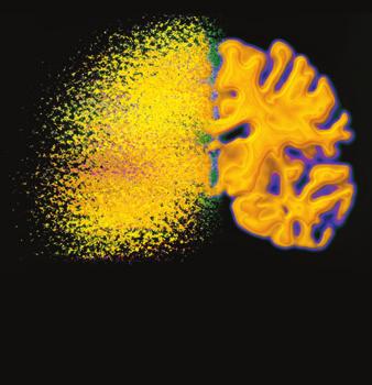
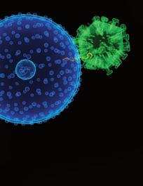
1 Case Reports in Pathology, Article ID , 4 pages Case Report Mixed Large Cell Neuroendocrine Carcinoma and Adenocarcinoma with Spindle Cell and Clear Cell Features in the Extrahepatic Bile Duct John Wysocki, 1 Rishi Agarwal, 1 Laura Bratton, 2 Jeremy Nguyen, 3 Mandy Crause Weidenhaft, 3 Nathan Shores, 1 and Hillary Z. Kimbrell 2 1 Department of Gastroenterology and Hepatology, Tulane University, 1430 Tulane Avenue, New Orleans, LA 70112, USA 2 Department of Pathology and Laboratory Medicine, Tulane University, 1430 Tulane Avenue, SL-79, New Orleans, LA 70112, USA 3 DepartmentofRadiology,TulaneUniversity,1430TulaneAvenue,NewOrleans,LA70112,USA Correspondence should be addressed to Hillary Z. Kimbrell; hkimbrel@tulane.edu Received 6 February 2014; Accepted 11 March 2014; Published 1 April 2014 Academic Editor: Yoji Nagashima Copyright 2014 John Wysocki et al. This is an open access article distributed under the Creative Commons Attribution License, which permits unrestricted use, distribution, and reproduction in any medium, provided the original work is properly cited. Mixed adenoneuroendocrine carcinomas, spindle cell carcinomas, and clear cell carcinomas are all rare tumors in the biliary tract. We present the first case, to our knowledge, of an extrahepatic bile duct carcinoma composed of all three types. A 65-year-old man with prior cholecystectomy presented with painless jaundice, vomiting, and weight loss. CA19-9 and alpha-fetoprotein (AFP) were elevated. Cholangioscopy revealed a friable mass extending from the middle of the common bile duct to the common hepatic duct. A bile duct excision was performed. Gross examination revealed a 3.6 cm intraluminal polypoid tumor. Microscopically, the tumor had foci of conventional adenocarcinoma (CK7-positive and CA19-9-postive) surrounded by malignant-appearing spindle cells that were positive for cytokeratins and vimentin. Additionally, there were separate areas of large cell neuroendocrine carcinoma (LCNEC). Foci of clear cell carcinoma merged into both the LCNEC and the adenocarcinoma. Tumor invaded through the bile duct wall with extensive perineural and vascular invasion. Circumferential margins were positive. The patient s poor performance status precluded adjuvant therapy and he died with recurrent and metastatic disease 5 months after surgery. This is consistent with the reported poor survival rates of biliary mixed adenoneuroendocrine carcinomas. 1. Case Report A 65-year-old man with a remote history of cholecystectomy for benign disease presented with a two-week history of painless jaundice, nausea, vomiting, and an unintentional 40- pound weight loss. His physical exam was within normal limits; specifically he was afebrile and did not have abdominal tenderness. Initial labs included a markedly elevated CA19-9 (2396 U/mL, normal range 0 35 U/mL), mildly elevated alpha fetoprotein (10.1 ng/ml, normal range ng/ml), and normal CEA (1.3 ng/ml, normal range ng/ml for non-smokers). Initial abdominal ultrasound demonstrated diffuse dilatation of the intrahepatic and common hepatic bile ducts. The largest intrahepatic duct had a diameter of 1.8 cm. At the level of the hepatic hilum, the common ducthadamaximumdiameterof2.7cmandaportionof the duct was filled with complex echogenic material. A triple phase liver CT showed a cm enhancing mass in the expected region of the intra- and extrahepatic bile duct. Endoscopic retrograde cholangiopancreatography showed a severe filling defect measuring 1.7 cm in the middle portion of the common bile duct with proximal and distal dilation. Cholangioscopy demonstrated a soft, friable tumor, extending from the mid-common bile duct to the common hepatic duct; the tumor was biopsied and brushed during this procedure, but the specimens contained only necrotic debris. The patient was given a biliary stent and discharged with outpatient follow-up; however, he soon re-presented with worsening jaundice. Therefore, the patient underwent a bile duct excision with creation of a hepaticojejunostomy. A cm segment of bile duct was removed. On gross examination, the bile duct contained a 3.6 cm intraluminal

.")

, and others contained mixtures of all three types.")
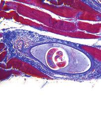
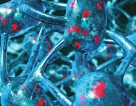
2 2 Case Reports in Pathology Table 1: Immunohistochemical results for each tumor morphology. Morphology Adeno Clear cell LCNEC Spindle AE1/3 CK7 Vim NE Scattered single cells CA19-9 AFP Hep CD117 P53 Ki67 50% 20% 80% 50% Adeno: adenocarcinoma; LCNEC: large cell neuroendocrine carcinoma; spindle: spindle cell carcinoma; AE1/3: cytokeratin cocktail AE1/AE3; CK7: cytokeratin 7; Vim: vimentin; NE: neuroendocrine markers chromogranin, synaptophysin, and CD56; AFP: alpha fetoprotein; Hep: Hep Par 1; LCNEC: large cell neuroendocrine carcinoma. Figure 1 Figure 3 Figure 2 Figure 4 polypoid tumor (Figure 1). Microscopically, the tumor was composed of islands of conventional adenocarcinoma and clear cell carcinoma surrounded by malignant-appearing spindle cells (Figure 2). The spindle cells were positive for cytokeratins and vimentin, consistent with spindle cell carcinoma. Additionally, there were separate areas of large cell neuroendocrine carcinoma (LCNEC), which formed relatively broader sheets with focal rosette-like structures and abundant necrosis (Figure 3). Many tumor nests contained a mixture of LCNEC and clear cell carcinoma (Figure 4), and others contained mixtures of all three types. The tumor invaded through the wall of the bile duct into surrounding soft tissue. Lymphatic and vascular invasion were present, and tumor extended perineurally to the circumferential margins. Proximal and distal bile duct margins were negative. One lymph node was received and was negative for tumor. On immunohistochemistry, the neuroendocrine markers synaptophysin, chromogranin, and CD56 were positive in the LCNEC and in the adenocarcinoma, focally positive in the spindle cells, and showed scattered positive single cells in the clear cell carcinoma, mostly around the edges of the islands (Figure 5). AFP and HepPar 1 were expressed in the clear cell areas and also focally in the adenocarcinoma. CD117 was positive in the LCNEC only. All four components were positive for p53. The Ki-67 index was highest in the LCNEC and lowest in the clear cell carcinoma; interestingly, the Ki67 tended to be positive in the periphery of the clear cell islands, which were the same cells that were positive for neuroendocrine markers. Table 1 summarizes the immunohistochemical results. The patient s performance status was too poor to receive any adjuvant therapy, and he died with recurrent and metastatic disease five months postoperatively.

3 Case Reports in Pathology 3 2. Discussion Figure 5 Our patient s tumor is a combination of LCNEC, spindle cell carcinoma, and clear cell carcinoma. To the best of our knowledge, this combination has not been previously reported. Each component in and of itself is rare in the extrahepatic bile duct and will be discussed individually. It is difficult to determine the exact number of LCNEC reported in the biliary tract because of the evolving terminology for neuroendocrine neoplasms. In the tubular gastrointestinal (GI) tract, neuroendocrine neoplasms are divided into well-differentiated tumors (formerly known as carcinoid tumors) and high-grade carcinomas. High-grade neuroendocrine carcinomas can be further subdivided into small cell carcinomas and LCNEC based on morphologic features: small cell carcinomas are characterized by diffuse growth pattern, markedly high nuclear/cytoplasmic ratio, hyperchromatic nuclei with finely granular salt and pepper chromatin, inconspicuous nucleoli, and nuclear molding. In contrast, LCNECs have a neuroendocrine architecture of organoid or trabecular growth with nuclear palisading and/or rosette formation, relatively monotonous round-to-oval nuclei with more conspicuous nucleoli, moderate amounts of cytoplasm, and generally large foci of necrosis. Unlike small cell carcinoma, which is a morphologic diagnosis, LCNEC must demonstrate immunohistochemical staining for at least one neuroendocrine marker in >20% of the cells [1]. LCNECs in the tubular GI tract often have an associated adenomatous or adenocarcinomatous component [1]. Tumors composed of both adenocarcinoma and a neuroendocrine neoplasm are classified as mixed adenoneuroendocrine carcinoma (MANEC) in the 2010 WHO classification [2]. The WHO does not specify which type of neuroendocrine neoplasm should be present in a MANEC (i.e., well-differentiated versus high-grade). Although there are several reports of biliarymanecswithasmallcellcomponent,therehave been only two previously reported cases of MANECs with a large cell neuroendocrine component in the extrahepatic bile duct [3, 4]. Presenting symptoms of biliary MANECs are nonspecific, including jaundice and abdominal pain. They tend to present at an advanced stage, and the metastases can be composed of adenocarcinoma, neuroendocrine carcinoma, or both [3, 5]. Although LCNEC on its own is a very aggressive tumor in a small series of gallbladder LCNEC, patients showed rapid disease progression and poor response to systemic chemotherapy [6] the prognosis of MANECs might be better than that of pure high-grade neuroendocrine carcinomas. In a series of nearly 5,000 gastroenteropancreatic and hepatobiliary neuroendocrine neoplasms in Korea, the 5- year survival rate for MANECs was 43% versus 35% for pure high-grade neuroendocrine carcinoma; this series included approximately 26 MANECs of the gallbladder and bile duct. Statistically significant prognostic indicators for MANECs in this study were lymph node status, tumor extension, and specific site of tumor, with small intestinal and pancreatic tumors having the best prognosis and esophageal the worst [7]. Similarly, in the tubular GI tract, patients with an adenocarcinoma component had a significantly better survival rate than patients with pure high-grade neuroendocrine carcinoma [1]. Similar to neuroendocrine neoplasms, spindle cell carcinoma has a confusing variety of names in the literature, such as pseudosarcoma and sarcomatoid carcinoma. By the 2010 WHO classification, spindle cell carcinoma falls under the heading of undifferentiated carcinoma, spindle, and giant cell type (UCSGT) [2]. UCSGT has several variants: spindle cell, giant cell, small cell (nonneuroendocrine) and nodular/lobular type. By definition, the tumor cells are positive for cytokeratins by immunohistochemistry and therefore are thought to be carcinomatous in nature. This is in contradistinction to carcinosarcomas, which also occur in the biliary tract [8]. Carcinosarcomas contain a true mesenchymal component (i.e., negative for cytokeratins), often in the form of heterologous elements such as chondrosarcoma, osteosarcoma, or rhabdomyosarcoma. UCSGT can also have areas of adenocarcinoma and squamous cell carcinoma [2, 9]. The prognosis for UCSGT is uncertain due to the rarity of cases. There are only five reported cases of clear cell carcinoma in the extrahepatic bile duct [10 12]. Four of these five cases had foci of conventional adenocarcinoma in addition to the clear cell component. Our case produced and stained for alpha fetoprotein and also stained for HepPar 1, both of which have been reported previously [10, 11]. These features may cause confusion if hepatocellular carcinoma is in the differential diagnosis, especially since hepatocellular carcinoma can also undergo clear cell change. Since the clear cell carcinoma in our case was positive for CK7, it is unlikely to represent a component of clear cell hepatocellular carcinoma. It is not known if the prognosis of clear cell carcinoma is any different from conventional adenocarcinoma because there have been so few cases. Its recognition as an entity is important, however, so it is not confused with metastatic clear cell renal cell carcinoma. Clear cell carcinoma of the biliary tract should be positive for CK7 and negative for RCC and PAX8, whereas metastatic clear cell renal cell carcinoma would show the reverse pattern [2]. It should be mentioned that clear cell change has also been reported in a well-differentiated neuroendocrine tumor (carcinoid) of the distal common bile duct [13]. In our case, the clear cell component showed only scattered single cells that were positive for neuroendocrine markers anddidnothaveanorganoidgrowthpatternsotherefore
4 4 Case Reports in Pathology does not represent a clear cell variant of a neuroendocrine tumor. There are only four other case reports of mixed tumors with multiple morphologies in the gastrointestinal and hepatobiliary tract: a tumor composed of small cell carcinoma, spindle cell carcinoma, adenocarcinoma, squamous cell carcinoma, and something the authors called undifferentiated carcinoma (small tumor cells with clear cytoplasm that were positive for neuroendocrine markers) in the gallbladder [9]; a mixed large cell neuroendocrine carcinoma and sarcomatoid carcinoma in the gastroesophageal junction [1]; a mixed hepatocellular carcinoma, spindle cell carcinoma and adenocarcinoma in the liver [14]; and a mixed neuroendocrine carcinoma, squamous cell carcinoma, and ciliated adenocarcinoma with spindle cell sarcoma in the esophagus [15]. The variety of tumor types seen in these cases and in the currentcasesuggeststhatmixedtumorssuchasthesearise from a stem cell that is capable of differentiating in multiple directions. In the current case, each tumor morphology was positive for p53, which suggests that a p53 mutation could have been an early genetic event in the tumor stem cell. Due to the scarcity of cases, the prognosis of mixed tumors with several different morphologies is uncertain but likely poor. In conclusion, this is the first report of a mixed large cell neuroendocrine carcinoma and adenocarcinoma with spindle cell and clear cell features in the extrahepatic bile duct. The tumor was highly aggressive, and the patient died with recurrent and metastatic disease five months after surgery. Conflict of Interests The authors declare that there is no conflict of interests regarding the publication of this paper. References Computer Assisted Tomography, vol.30,no.4,pp , [7]M.Cho,J.M.Kim,J.H.Sohnetal., Currenttrendsofthe incidence and pathological diagnosis of gastroenteropancreatic neuroendocrine tumors (GEP-NETs) in Korea : multicenter study, Cancer Research and Treatment,vol.44, no. 3, pp , [8] J. Albores-Saavedra, D. E. Henson, and D. S. Klimstra, AFIP Atlas of Tumour Pathology, Third Series Fascicle 27 Tumor of the Gallbladder, Extrahepatic Bile Ducts, and Ampulla of Vater, Armed Forces Institute of Pathology, Washington, DC, USA, [9] Y. Takahashi, J.-I. Fukushima, T. Fukusato, and J. Shiga, Sarcomatoid carcinoma with components of small cell carcinoma and undifferentiated carcinoma of the gallbladder, Pathology International,vol.54,no.11,pp ,2004. [10] K. Tajiri, Y. Shimizu, H. Mihara et al., Cholangiocarcinoma at the cystic duct discovered by lymph node metastases with clear cell transformation, Internal Medicine, vol. 45, no. 18, pp , [11] C. Vardaman and J. Albores-Saavedra, Clear cell carcinomas of the gallbladder and extrahepatic bile ducts, American Surgical Pathology,vol.19,no.1,pp.91 99,1995. [12] L.Gu,C.H.Jiang,Q.Xu,Q.Liu,M.Luo,andZ.Y.Wu, Primary clear cell carcinoma of hilar bile duct: a case report, Digestive Diseases, vol. 11, no. 1, pp , [13]T.Todoroki,T.Sano,S.Yamadaetal., Clearcellcarcinoid tumor of the distal common bile duct, World Surgical Oncology, vol. 5, article 6, [14] M.-J. Kim, H.-L. Koo, S. K. Lee, J.-Y. Ro, and E. Yu, A case of combined hepatocellular and cholangiocarcinoma with neuroendocrine differentiation and sarcomatoid transformation, The Korean Pathology,vol.39,pp ,2005. [15] A. Kanamoto, Y. Nakanishi, A. Ochiai et al., A case of small polypoid esophageal carcinoma with multidirectional differentiation, including neuroendocrine, squamous, ciliated glandular, and sarcomatous components, Archives of Pathology and Laboratory Medicine,vol.124,no.11,pp ,2000. [1] J. Shia, L. H. Tang, M. R. Weiser et al., Is nonsmall cell type high-grade neuroendocrine carcinoma of the tubular gastrointestinal tract a distinct disease entity? American Surgical Pathology,vol.32,no.5,pp ,2008. [2] J. Albores-Saavedra, N. V. Adsay, J. M. Crawford et al., Tumours of the gallbladder and extrahepatic bile ducts, in WHO Classification of Tumours of the Digestive System, F.T.Bosman,F. Carneiro,R.H.Hruban,andN.D.Theise,Eds.,pp , IARC Press, Lyon, France, [3] K. Harada, Y. Sato, H. Ikeda et al., Clinicopathologic study of mixed adenoneuroendocrine carcinomas of hepatobiliary organs, Virchows Archiv, vol. 460, no. 3, pp , [4] K. Sato, R. Waseda, Y. Tatsuzawa et al., Composite large cell neuroendocrine carcinoma and adenocarcinoma of the common bile duct, Clinical Pathology, vol.59,no.1, pp , [5] R. Linder, T. Dorfman, O. Ben-Ishay, E. Kakiashvili, E. Velodavsky, and Y. Kluger, Mixed neuroendocrine tumor of the common bile duct, JournalofthePancreas, vol. 14, no. 1, pp , [6] S. R. Jun, J. M. Lee, J. K. Han, and B. I. Choi, High-grade neuroendocrine carcinomas of the gallbladder and bile duct: report of four cases with pathological correlation,


5 MEDIATORS of INFLAMMATION The Scientific World Journal Gastroenterology Research and Practice Diabetes Research International Endocrinology Immunology Research Disease Markers Submit your manuscripts at BioMed Research International PPAR Research Obesity Ophthalmology Evidence-Based Complementary and Alternative Medicine Stem Cells International Oncology Parkinson s Disease Computational and Mathematical Methods in Medicine AIDS Behavioural Neurology Research and Treatment Oxidative Medicine and Cellular Longevity
Case Report Five-Year Survival after Surgery for Invasive Micropapillary Carcinoma of the Stomach
 Case Reports in Surgery Volume 2013, Article ID 560712, 4 pages http://dx.doi.org/10.1155/2013/560712 Case Report Five-Year Survival after Surgery for Invasive Micropapillary Carcinoma of the Stomach Shigeo
Case Report A Rare Cutaneous Adnexal Tumor: Malignant Proliferating Trichilemmal Tumor
 Case Reports in Medicine Volume 2015, Article ID 742920, 4 pages http://dx.doi.org/10.1155/2015/742920 Case Report A Rare Cutaneous Adnexal Tumor: Malignant Proliferating Trichilemmal Tumor Omer Alici,
Case Report Overlap of Acute Cholecystitis with Gallstones and Squamous Cell Carcinoma of the Gallbladder in an Elderly Patient
 Case Reports in Surgery Volume 2015, Article ID 767196, 4 pages http://dx.doi.org/10.1155/2015/767196 Case Report Overlap of Acute Cholecystitis with Gallstones and Squamous Cell Carcinoma of the Gallbladder
Combined Large Cell Neuroendocrine Carcinoma and Spindle Cell Carcinoma of the Lung
 Case Reports Jpn J Clin Oncol 2011;41(6)797 802 doi:10.1093/jjco/hyr034 Advance Access Publication 16 March 2011 Combined Large Cell Neuroendocrine Carcinoma and Spindle Cell Carcinoma of the Lung Taichiro
Case Report Intrabiliary Hepatic Metastasis of Colorectal Carcinoma Mimicking Primary Cholangiocarcinoma: A Case Report and Review of the Literature
 Case Reports in Pathology Volume 2016, Article ID 4704781, 5 pages http://dx.doi.org/10.1155/2016/4704781 Case Report Intrabiliary Hepatic Metastasis of Colorectal Carcinoma Mimicking Primary Cholangiocarcinoma:
Case Report Uncommon Mixed Type I and II Choledochal Cyst: An Indonesian Experience
 Case Reports in Surgery Volume 2013, Article ID 821032, 4 pages http://dx.doi.org/10.1155/2013/821032 Case Report Uncommon Mixed Type I and II Choledochal Cyst: An Indonesian Experience Fransisca J. Siahaya,
Undifferentiated carcinoma of the gall bladder: a rare entity
 Case Report Undifferentiated carcinoma of the gall bladder: a rare entity Medhi Pranita 1,*, Dowerah Swagata 2, Dutta Utpal 3, Dutta Aparna 4 1 Associate Professor, 2 Demonstrator, 3 PGT, 4 Assistant Professor,
Endoscopic Ultrasonography Assessment for Ampullary and Bile Duct Malignancy
 Diagnostic and Therapeutic Endoscopy, Vol. 3, pp. 35-40 Reprints available directly from the publisher Photocopying permitted by license only (C) 1996 OPA (Overseas Publishers Association) Amsterdam B.V.
Case Scenario 1. Discharge Summary
 Case Scenario 1 Discharge Summary A 69-year-old woman was on vacation and noted that she was becoming jaundiced. Two months prior to leaving on that trip, she had had a workup that included an abdominal
Problem 1: Differential of Neuroendocrine Carcinoma 3/23/2017. Disclosure of Relevant Financial Relationships
 Differential of Neuroendocrine Carcinoma Alain C. Borczuk,MD Weill Cornell Medicine Disclosure of Relevant Financial Relationships USCAP requires that all faculty in a position to influence or control
A 53 year-old woman with a lung mass, right hilar mass and mediastinal adenopathy.
 November 2015 Case of the Month A 53 year-old woman with a lung mass, right hilar mass and mediastinal adenopathy. Contributed by: Rasha Salama, M.D., IU Department of Pathology and Laboratory Medicine
Kidney Case 1 SURGICAL PATHOLOGY REPORT
 Kidney Case 1 Surgical Pathology Report February 9, 2007 Clinical History: This 45 year old woman was found to have a left renal mass. CT urography with reconstruction revealed a 2 cm medial mass which
University Journal of Pre and Para Clinical Sciences
 ISSN 2455 2879 Volume 2 Issue 1 2016 Metaplastic carcinoma breast a rare case report Abstract : Metaplastic carcinoma of the breast is a rare malignancy with two distinct cell lines described as a breast
Intraductal papillary mucinous neoplasm of the bile ducts: a rare form of premalignant lesion of invasive cholangiocarcinoma
 Intraductal papillary mucinous neoplasm of the bile ducts: a rare form of premalignant lesion of invasive cholangiocarcinoma Authors: R. Revert Espí, Y. Fernandez Nuñez, I. Carbonell, D. P. Gómez valencia,
Radiology Pathology Conference
 Radiology Pathology Conference Nadia F. Yusaf, M.D. PGY-3 1/29/2010 Presentation material is for education purposes only. All rights reserved. 2010 URMC Radiology Page 1 of 90 Case 1 60 year- old man presents
Respiratory Tract Cytology
 Respiratory Tract Cytology 40 th European Congress of Cytology Liverpool, UK Momin T. Siddiqui M.D. Professor of Pathology and Laboratory Medicine Director of Cytopathology Emory University Hospital, Atlanta,
Neuroendocrine Lung Tumors Myers
 Diagnosis and Classification of Neuroendocrine Lung Tumors Jeffrey L. Myers, M.D. A. James French Professor Director, Anatomic Pathology & MLabs University of Michigan, Ann Arbor, MI myerjeff@umich.edu
Case Report Tumor-to-Tumor Metastasis: Lung Carcinoma Metastasizing to Thyroid Neoplasms
 Case Reports in Pathology Volume 2015, Article ID 153932, 5 pages http://dx.doi.org/10.1155/2015/153932 Case Report Tumor-to-Tumor Metastasis: Lung Carcinoma Metastasizing to Thyroid Neoplasms Shiuan-Li
Title: Painless jaundice as an initial presentation of lung adenocarcinoma
 Title: Painless jaundice as an initial presentation of lung adenocarcinoma Authors: Irene Andaluz García, Irene González Partida, Javier Lucas Ramos, Jorge Yebra Carmona DOI: 10.17235/reed.2018.5587/2018
Case Report Osteoclastic Giant Cell Rich Squamous Cell Carcinoma of the Uterine Cervix: A Case Report and Review of the Literature
 Case Reports in Pathology, Article ID 415328, 4 pages http://dx.doi.org/10.1155/2014/415328 Case Report Osteoclastic Giant Cell Rich Squamous Cell Carcinoma of the Uterine Cervix: A Case Report and Review
Intrahepatic Sarcomatoid Cholangiocarcinoma with Portal Vein Thrombosis: A Case Report 1
 Intrahepatic Sarcomatoid Cholangiocarcinoma with Portal Vein Thrombosis: A Case Report 1 Jae-Hoon Lim, M.D., Jin Woong Kim, M.D., Suk Hee Heo, M.D., Yong Yeon Jeong, M.D., Heoung Keun Kang, M.D. A 53-year-old
performed to help sway the clinician in what the appropriate diagnosis is, which can substantially alter the treatment of management.
 Hello, I am Maura Polansky at the University of Texas MD Anderson Cancer Center. I am a Physician Assistant in the Department of Gastrointestinal Medical Oncology and the Program Director for Physician
Case Report Heterotopic Pancreas within the Proximal Hepatic Duct, Containing Intraductal Papillary Mucinous Neoplasm
 Case Reports in Surgery Volume 2015, Article ID 816960, 4 pages http://dx.doi.org/10.1155/2015/816960 Case Report Heterotopic Pancreas within the Proximal Hepatic Duct, Containing Intraductal Papillary
Abstracting Upper GI Cancer Incidence and Treatment Data Quiz 1 Multiple Primary and Histologies Case 1 Final Pathology:
 Abstracting Upper GI Cancer Incidence and Treatment Data Quiz 1 Multiple Primary and Histologies Case 1 A 74 year old male with a history of GERD presents complaining of dysphagia. An esophagogastroduodenoscopy
Case Report Synchronous Bilateral Solid Papillary Carcinomas of the Breast
 Case Reports in Surgery Volume 2013, Article ID 812129, 4 pages http://dx.doi.org/10.1155/2013/812129 Case Report Synchronous Bilateral Solid Papillary Carcinomas of the Breast Noriko Yoshimura, 1 Shigeru
R. F. Falkenstern-Ge, 1 S. Bode-Erdmann, 2 G. Ott, 2 M. Wohlleber, 1 and M. Kohlhäufl Introduction. 2. Histology
 Case Reports in Oncological Medicine Volume 2013, Article ID 167585, 4 pages http://dx.doi.org/10.1155/2013/167585 Case Report Late Lung Metastasis of a Primary Eccrine Sweat Gland Carcinoma 10 Years after
Difficult Diagnoses and Controversial Entities in Neoplastic Lung
 Difficult Diagnoses and Controversial Entities in Neoplastic Lung Lynette M. Sholl, M.D. Associate Pathologist, Brigham and Women s Hospital Chief, Pulmonary Pathology Service Associate Professor, Harvard
Case Report Denosumab Chemotherapy for Recurrent Giant-Cell Tumor of Bone: A Case Report of Neoadjuvant Use Enabling Complete Surgical Resection
 Case Reports in Oncological Medicine Volume 2013, Article ID 496351, 4 pages http://dx.doi.org/10.1155/2013/496351 Case Report Denosumab Chemotherapy for Recurrent Giant-Cell Tumor of Bone: A Case Report
Case Scenario 1: Thyroid
 Case Scenario 1: Thyroid History and Physical Patient is an otherwise healthy 80 year old female with the complaint of a neck mass first noticed two weeks ago. The mass has increased in size and is palpable.
담낭에서발생한대세포신경내분비종양 1 예
 대한내과학회지 : 제 85 권제 2 호 2013 http://dx.doi.org/10.3904/kjm.2013.85.2.183 담낭에서발생한대세포신경내분비종양 1 예 서울대학교의과대학보라매병원 1 내과, 2 병리과, 3 외과 김영훈 1 정지봉 1 주세경 1 최민영 1 이국래 1 장미수 2 안영준 3 A Case of Large Cell Neuroendocrine
Differential diagnosis of HCC
 Hepatocellular Carcinoma Quest for an Ideal Immunohistochemical Panel Sanjay Kakar, MD UCSF Differential diagnosis of HCC Hepatocellular lesions Adenoma, FNH, HG dysplasia Adenocarcinoma CholangioCA, metastasis
GOBLET CELL CARCINOID. Hanlin L. Wang, MD, PhD University of California Los Angeles
 GOBLET CELL CARCINOID Hanlin L. Wang, MD, PhD University of California Los Angeles Disclosure of Relevant Financial Relationships USCAP requires that all planners (Education Committee) in a position to
GOBLET CELL CARCINOID
 GOBLET CELL CARCINOID Hanlin L. Wang, MD, PhD University of California Los Angeles Disclosure of Relevant Financial Relationships USCAP requires that all planners (Education Committee) in a position to
Case Report PET/CT Imaging in Oncology: Exceptions That Prove the Rule
 Case Reports in Oncological Medicine Volume 2013, Article ID 865032, 4 pages http://dx.doi.org/10.1155/2013/865032 Case Report PET/CT Imaging in Oncology: Exceptions That Prove the Rule M. Casali, 1 A.
Diplomate of the American Board of Pathology in Anatomic and Clinical Pathology
 A 33-year-old male with a left lower leg mass. Contributed by Shaoxiong Chen, MD, PhD Assistant Professor Indiana University School of Medicine/ IU Health Partners Department of Pathology and Laboratory
Biliary tree dilation - and now what?
 Biliary tree dilation - and now what? Poster No.: C-1767 Congress: ECR 2012 Type: Educational Exhibit Authors: I. Ferreira, A. B. Ramos, S. Magalhães, M. Certo; Porto/PT Keywords: Pathology, Diagnostic
Appendix 4: WHO Classification of Tumours of the pancreas 17
 S3.01 The WHO histological tumour type must be recorded. CS3.01a The histological type of the tumour should be recorded based on the current WHO classification 17 (refer to Appendices 4-7). Appendix 4:
57th Annual HSCP Spring Symposium 4/16/2016
 An Unusual Malignant Spindle Cell Lesion to Involve the Breast Erinn Downs-Kelly, D.O. Associate Professor of Pathology University of Utah & ARUP Laboratories No disclosures Case 39 y/o female with no
Case Report Primary Neuroendocrine Carcinoma of Ocular Adnexa
 Volume 2013, Article ID 281351, 4 pages http://dx.doi.org/10.1155/2013/281351 Case Report Primary Neuroendocrine Carcinoma of Ocular Adnexa Daisuke Yamanouchi, 1 Toshiyuki Oshitari, 1 Yosuke Nakamura,
Papillary Carcinomas of the Gallbladder. Analysis of Noninvasive and Invasive Types
 Papillary Carcinomas of the Gallbladder Analysis of Noninvasive and Invasive Types Jorge Albores-Saavedra, MD; Matthew Tuck, BS; Bernadette K. McLaren, MD; Kelley S. Carrick, MD; Donald Earl Henson, MD
Case Report Features of the Atrophic Corpus Mucosa in Three Cases of Autoimmune Gastritis Revealed by Magnifying Endoscopy
 Volume 2012, Article ID 368160, 4 pages doi:10.1155/2012/368160 Case Report Features of the Atrophic Corpus Mucosa in Three Cases of Autoimmune Gastritis Revealed by Magnifying Endoscopy Kazuyoshi Yagi,
Research Article Papillary Thyroid Cancer, Macrofollicular Variant: The Follow-Up and Analysis of Prognosis of 5 Patients
 yroid Research, Article ID 818134, 4 pages http://dx.doi.org/10.1155/2014/818134 Research Article Papillary Thyroid Cancer, Macrofollicular Variant: The Follow-Up and Analysis of Prognosis of 5 Patients
Enterprise Interest Nothing to declare
 Enterprise Interest Nothing to declare Update of mixed tumours of the GI tract, the pancreas and the liver Introduction to the concept of mixed tumours and clinical implication Jean-Yves SCOAZEC Surgical
Case Report Cytomegalovirus Colitis with Common Variable Immunodeficiency and Crohn s Disease
 Volume 2015, Article ID 348204, 4 pages http://dx.doi.org/10.1155/2015/348204 Case Report Cytomegalovirus Colitis with Common Variable Immunodeficiency and Crohn s Disease Betül Ünal, Cumhur Ebrahim BaGsorgun,
Liver Tumors. Prof. Dr. Ahmed El - Samongy
 Liver Tumors Prof. Dr. Ahmed El - Samongy Objective 1. Identify the most important features of common benign liver tumors 2. Know the risk factors, diagnosis, and management of hepatocellular carcinoma
Correspondence should be addressed to I. Sokolakis;
 Hindawi Case Reports in Urology Volume 2017, Article ID 6597592, 4 pages https://doi.org/10.1155/2017/6597592 Case Report Mucin-Poor Mucinous Tubular and Spindle Cell Carcinoma of the Kidney Presented
Case Scenario 1. The patient has now completed his neoadjuvant chemoradiation and has been cleared for surgery.
 Case Scenario 1 July 10, 2010 A 67-year-old male with squamous cell carcinoma of the mid thoracic esophagus presents for surgical resection. The patient has completed preoperative chemoradiation. This
Case Report Two Cases of Small Cell Cancer of the Maxillary Sinus Treated with Cisplatin plus Irinotecan and Radiotherapy
 Case Reports in Otolaryngology Volume 2013, Article ID 893638, 4 pages http://dx.doi.org/10.1155/2013/893638 Case Report Two Cases of Small Cell Cancer of the Maxillary Sinus Treated with Cisplatin plus
Personal Profile. Name: 劉 XX Gender: Female Age: 53-y/o Past history. Hepatitis B carrier
 Personal Profile Name: 劉 XX Gender: Female Age: 53-y/o Past history Hepatitis B carrier Chief complaint Fever on and off for 2 days Present illness 94.10.14 Sudden onset of epigastric pain 94.10.15 Fever
LUNG CANCER PATHOLOGY: UPDATE ON NEUROENDOCRINE LUNG TUMORS
 LUNG CANCER PATHOLOGY: UPDATE ON NEUROENDOCRINE LUNG TUMORS William D. Travis, M.D. Attending Thoracic Pathologist Memorial Sloan Kettering Cancer Center New York, NY PULMONARY NE TUMORS CLASSIFICATION
Diagnostically Challenging Cases in Gynecologic Pathology
 Diagnostically Challenging Cases in Gynecologic Pathology Eric C. Huang, M.D., Ph.D. Department of Pathology and Laboratory Medicine University of California, Davis Medical Center Case 1 Presentation 38
Case Report Liver Metastases of Unknown Primary: Malignant Melanoma
 Case Reports in Hepatology, Article ID 131708, 4 pages http://dx.doi.org/10.1155/2014/131708 Case Report Liver Metastases of Unknown Primary: Malignant Melanoma Ozgur Bostanci, Kinyas Kartal, and Muharrem
Case Report Contrast Enhanced Ultrasound of a Gallbladder Lesion in a Patient with a History of Renal Cell and Rectal Cancer
 Case Reports in Gastrointestinal Medicine Volume 2013, Article ID 538534, 4 pages http://dx.doi.org/10.1155/2013/538534 Case Report Contrast Enhanced Ultrasound of a Gallbladder Lesion in a Patient with
Congenital dilatation of the common bile duct and pancreaticobiliary maljunction clinical implications
 Langenbecks Arch Surg (2009) 394:209 213 DOI 10.1007/s00423-008-0330-6 CURRENT CONCEPT IN CLINICAL SURGERY Congenital dilatation of the common bile duct and pancreaticobiliary maljunction clinical implications
Case Report Metastatic Malignant Melanoma of Parotid Gland with a Regressed Primary Tumor
 Case Reports in Otolaryngology Volume 2016, Article ID 5393404, 4 pages http://dx.doi.org/10.1155/2016/5393404 Case Report Metastatic Malignant Melanoma of Parotid Gland with a Regressed Primary Tumor
Pancreas Case Scenario #1
 Pancreas Case Scenario #1 An 85 year old white female presented to her primary care physician with increasing abdominal pain. On 8/19 she had a CT scan of the abdomen and pelvis. This showed a 4.6 cm mass
Mody. AIS vs. Invasive Adenocarcinoma of the Cervix
 Common Problems in Gynecologic Pathology Michael T. Deavers, M.D. Houston Methodist Hospital, Houston, Texas Common Problems in Gynecologic Pathology Adenocarcinoma in-situ (AIS) of the Cervix vs. Invasive
Neuroendocrine neoplasms of the lung
 Neuroendocrine neoplasms of the lung M Papotti, L Righi, & M Volante University of Turin at San Luigi Hospital TORINO NETs OF THE LUNG Menu - Spectrum of NE lung tumors - CARCINOID TUMORS - SCLC /LCNEC
Pancreatic Cancer: The ABCs of the AJCC and WHO
 Pancreatic Cancer: The ABCs of the AJCC and WHO Aatur D. Singhi, MD PhD Assistant Professor University of Pittsburgh Medical Center Department of Pathology singhiad@upmc.edu Case presentation Objectives
Neoplasms of the Canine, Feline and Lemur Liver:
 Neoplasms of the Canine, Feline and Lemur Liver: Classification and Prognosis Annual Seminar of the French Society of Veterinary Pathology John M. Cullen VMD PhD DACVP North Carolina State University Primary
American Journals of Cancer Case Reports. A Rare Case of Rectal Metastasis from Sarcomatoid Variant of Urothelial Carcinoma: A Case Report
 American Journals of Cancer Case Reports Lin JYJ et al. American Journals of Cancer Case Reports 2014, 3:1-5 http://ivyunion.org/index.php/ajccr Page 1 of 5 Vol 3 Article ID 20140539, 5 pages Case Report
THYMIC CARCINOMAS AN UPDATE
 THYMIC CARCINOMAS AN UPDATE Mark R. Wick, M.D. University of Virginia Medical Center Charlottesville, VA CARCINOMA OF THE THYMUS General Clinical Features No apparent gender predilection Age range of 35-75
Mandana Moosavi 1 and Stuart Kreisman Background
 Case Reports in Endocrinology Volume 2016, Article ID 6471081, 4 pages http://dx.doi.org/10.1155/2016/6471081 Case Report A Case Report of Dramatically Increased Thyroglobulin after Lymph Node Biopsy in
Research Article Stromal Expression of CD10 in Invasive Breast Carcinoma and Its Correlation with ER, PR, HER2-neu, and Ki67
 SAGE-Hindawi Access to Research International Breast Cancer Volume 20, Article ID 47957, 4 pages doi:0.406/20/47957 Research Article Stromal Expression of CD0 in Invasive Breast Carcinoma and Its Correlation
Case Report Tumor-to-Tumor Metastasis: Lung Carcinoma Metastasizing to Thyroid Neoplasms
 Hindawi Publishing Corporation Volume 2015, Article ID 153932, 5 pages http://dx.doi.org/10.1155/2015/153932 Case Report Tumor-to-Tumor Metastasis: Lung Carcinoma Metastasizing to Thyroid Neoplasms Shiuan-Li
Case Report Renal Cell Carcinoma Metastatic to Thyroid Gland, Presenting Like Anaplastic Carcinoma of Thyroid
 Case Reports in Urology Volume 2013, Article ID 651081, 4 pages http://dx.doi.org/10.1155/2013/651081 Case Report Renal Cell Carcinoma Metastatic to Thyroid Gland, Presenting Like Anaplastic Carcinoma
My Journey into the World of Salivary Gland Sebaceous Neoplasms
 My Journey into the World of Salivary Gland Sebaceous Neoplasms Douglas R. Gnepp Warren Alpert Medical School at Brown University Rhode Island Hospital Pathology Department Providence RI Asked to present
Presentation material is for education purposes only. All rights reserved URMC Radiology Page 1 of 98
 Presentation material is for education purposes only. All rights reserved. 2011 URMC Radiology Page 1 of 98 Radiology / Pathology Conference February 2011 Brooke Koltz, Cytopathology Resident Presentation
Lung Cytology: Lessons Learned from Errors in Practice
 Lung Cytology: Lessons Learned from Errors in Practice Stephen S. Raab, M.D. Department of Laboratory Medicine Eastern Health and Memorial University of Newfoundland, St. John s, NL and University of Washington,
Hepatobiliary and Pancreatic Malignancies
 Hepatobiliary and Pancreatic Malignancies Gareth Eeson MD MSc FRCSC Surgical Oncologist and General Surgeon Kelowna General Hospital Interior Health Consultant, Surgical Oncology BC Cancer Agency Centre
Sarcomatoid (spindle cell) carcinoma of the cricopharynx presenting as dysphagia
 carcinoma of the cricopharynx presenting as dysphagia") Case Report Sarcomatoid (spindle cell) carcinoma of the cricopharynx presenting as dysphagia Jagtap Sunil V. 1, Shukla Dhirajkumar B. 2, Jagtap Swati S. 3, Havle Abhay D. 4 1 Associate Professor, Department
Case Report Sarcomatoid (spindle cell) carcinoma of the cricopharynx presenting as dysphagia Jagtap Sunil V. 1, Shukla Dhirajkumar B. 2, Jagtap Swati S. 3, Havle Abhay D. 4 1 Associate Professor, Department
Bilateral Renal Angiomyolipomas with Invasion of the Renal Vein: A Case Report
 Case Study TheScientificWorldJOURNAL (2008) 8, 145 148 TSW Urology ISSN 1537-744X; DOI 10.1100/tsw.2008.29 Bilateral Renal Angiomyolipomas with Invasion of the Renal Vein: A Case Report C. Blick, N. Ravindranath,
Neuroendocrine Tumor of Unknown Primary Accompanied with Stomach Adenocarcinoma
 J Gastric Cancer 2011;11(4):234-238 http://dx.doi.org/10.5230/jgc.2011.11.4.234 Case Report Neuroendocrine Tumor of Unknown Primary Accompanied with Stomach Adenocarcinoma Ho-Yeun Kim, Sung-Il Choi 1,
Four Cases of Large Cell Neuroendocrine Carcinoma of the Stomach: Findings on CT and Barium Studies 1
 Four Cases of Large Cell Neuroendocrine Carcinoma of the Stomach: Findings on CT and arium Studies 1 Hee Jung Kim, M.D., Dongil Choi, M.D., Soon Jin Lee, M.D., Won Jae Lee, M.D., Sung Kim, M.D. 2, Jae
Endoscopic Resection of Ampullary Neuroendocrine Tumor
 CASE REPORT Endoscopic Resection of Ampullary Neuroendocrine Tumor Hiroyuki Fukasawa, Shigetaka Tounou, Masashi Nabetani and Tomoki Michida Abstract We report the case of a 57-year-old man with a 1.0-cm
Gross appearance of nodular hyperplasia in material obtained from suprapubic prostatectomy. Note the multinodular appearance and the admixture of
 Tiền liệt tuyến Tiền liệt tuyến Gross appearance of nodular hyperplasia in material obtained from suprapubic prostatectomy. Note the multinodular appearance and the admixture of solid and microcystic areas.
Case Report A Case of Primary Submandibular Gland Oncocytic Carcinoma
 Case Reports in Otolaryngology Volume 2013, Article ID 384238, 4 pages http://dx.doi.org/10.1155/2013/384238 Case Report A Case of Primary Submandibular Gland Oncocytic Carcinoma Kunihiko Tokashiki, Kiyoaki
Case Report Intramucosal Signet Ring Cell Gastric Cancer Diagnosed 15 Months after the Initial Endoscopic Examination
 Hindawi Publishing Corporation Case Reports in Medicine Volume 2015, Article ID 479625, 5 pages http://dx.doi.org/10.1155/2015/479625 Case Report Intramucosal Signet Ring Cell Gastric Cancer Diagnosed
3/27/2017. Pulmonary Pathology Specialty Conference. Disclosure of Relevant Financial Relationships. Clinical History:
 Pulmonary Pathology Specialty Conference Saul Suster, M.D. Medical College of Wisconsin Disclosure of Relevant Financial Relationships USCAP requires that all planners (Education Committee) in a position
Note: The cause of testicular neoplasms remains unknown
 - In the 15- to 34-year-old age group, they are the most common tumors of men. - Tumors of the testis are a heterogeneous group of neoplasms that include: I. Germ cell tumors : 95%; all are malignant.
Clinical Study Metastasectomy of Pulmonary Metastases from Osteosarcoma: Prognostic Factors and Indication for Repeat Metastasectomy
 Respiratory Medicine Volume 2015, Article ID 570314, 5 pages http://dx.doi.org/10.1155/2015/570314 Clinical Study Metastasectomy of Pulmonary Metastases from Osteosarcoma: Prognostic Factors and Indication
Small Cell Carcinoma ex-pleomorphic Adenoma of the Parotid Gland
 Head and Neck Pathol (2012) 6:502 506 DOI 10.1007/s12105-012-0376-1 CASE REPORT Small Cell Carcinoma ex-pleomorphic Adenoma of the Parotid Gland Ashley Cimino-Mathews Brian M. Lin Steven S. Chang Kofi
Cholangiocarcinoma. Judy Wyatt Dundee November 2010
 Cholangiocarcinoma Judy Wyatt Dundee November 2010 Making sense of cholangiocarcinoma Difficulties with diagnostic criteria How many entities within cholangiocarcinoma? Rapidly evolving Intrahepatic cholangiocarcinoma
Insulinoma-associated protein (INSM1) is a sensitive and specific marker for lung neuroendocrine tumors in cytologic and surgical specimens
 is a sensitive and specific marker for lung neuroendocrine tumors in cytologic and surgical specimens") Insulinoma-associated protein (INSM1) is a sensitive and specific marker for lung neuroendocrine tumors in cytologic and surgical specimens Kartik Viswanathan, M.D., Ph.D New York Presbyterian - Weill
Insulinoma-associated protein (INSM1) is a sensitive and specific marker for lung neuroendocrine tumors in cytologic and surgical specimens Kartik Viswanathan, M.D., Ph.D New York Presbyterian - Weill
Case Report Sarcomatoid Renal Cell Carcinoma Metastasis to the Penis
 Case Reports in Urology Volume 2015, Article ID 467974, 4 pages http://dx.doi.org/10.1155/2015/467974 Case Report Sarcomatoid Renal Cell Carcinoma Metastasis to the Penis Victor D. Liou, 1 Oussama M. Darwish,
Case Presentation. Maha Akkawi, MD, Fatima Obeidat, MD, Tariq Aladily, MD. Department of Pathology Jordan University Hospital Amman, Jordan
 Case Presentation Maha Akkawi, MD, Fatima Obeidat, MD, Tariq Aladily, MD Department of Pathology Jordan University Hospital Amman, Jordan The 25th Annual Congress of the ADIAP The 8/11/2013 1 5th International
Case 2. Dr. Sathima Natarajan M.D. Kaiser Permanente Medical Center Sunset
 Case 2 Dr. Sathima Natarajan M.D. Kaiser Permanente Medical Center Sunset History 24 year old male presented with a 3 day history of right flank pain, sharp in nature Denies fever, chills, hematuria or
Gastrinoma: Medical Management. Haley Gallup
 Gastrinoma: Medical Management Haley Gallup Also known as When to put your knife down Gastrinoma Definition and History Diagnosis Historic Management Sporadic vs MEN-1 Defining surgical candidates Nonsurgical
SECONDARIES: A PRELIMINARY REPORT
 HPB Surgery, 1990, Vol. 2, pp. 69-72 Reprints available directly from the publisher Photocopying permitted by license only 1990 Harwood Academic Publishers GmbH Printed in the United Kingdom CASE REPORTS
GUT-C 11/30/2017. Debasmita Das, M.D. PGY-1 Danbury Hospital
 GUT-C 11/30/2017 Debasmita Das, M.D. PGY-1 Danbury Hospital CLINICAL SUMMARY 8/2017 59 year old female Presented to the ED with 1 month history of general malaise, fever and weight loss PMH: Significant
Anatomy of the biliary tract
 Harvard-MIT Division of Health Sciences and Technology HST.121: Gastroenterology, Fall 2005 Instructors: Dr. Jonathan Glickman Anatomy of the biliary tract Figure removed due to copyright reasons. Biliary
Solitary Fibrous Tumor of the Kidney with Massive Retroperitoneal Recurrence. A Case Presentation
 246) Prague Medical Report / Vol. 113 (2012) No. 3, p. 246 250 Solitary Fibrous Tumor of the Kidney with Massive Retroperitoneal Recurrence. A Case Presentation Sfoungaristos S., Papatheodorou M., Kavouras
Carcinosarcoma of the Extrahepatic Bile Duct Presenting with Stone-like Radiological Findings
 CASE REPORT Carcinosarcoma of the Extrahepatic Bile Duct Presenting with Stone-like Radiological Findings Shinsuke Kumei 1,2, Yutaka Onishi 1, Takeshi Ogura 1, Chosei Kusumoto 3, Yasuko Matsuno 4, Takashi
sarcomatoid carcinoma with small cell carcinoma component of the urinary bladder: a case report with review of the literature
 Int J Clin Exp Pathol 2013;6(8):1671-1676 www.ijcep.com /ISSN:1936-2625/IJCEP1306009 Case Report Sarcomatoid carcinoma with small cell carcinoma component of the urinary bladder: a case report with review
COLON AND RECTUM SOLID TUMOR RULES ABSTRACTORS TRAINING
 COLON AND RECTUM SOLID TUMOR RULES ABSTRACTORS TRAINING COLON AND RECTUM SOLID TUMOR RULES Separate sections for: Introduction Changes from 2007 MP/H rules Equivalent Terms Terms that are NOT Equivalent
Management of Rare Liver Tumours
 Gian Luca Grazi Hepato-Biliary-Pancreatic Surgery National Cancer Institute Regina Elena Rome Fibrolamellar Carcinoma Mixed Hepato Cholangiocellular Carcinoma Hepatoblastoma Carcinosarcoma Primary Hepatic
Mammary analogue secretory carcinoma of salivary gland A case report of new entity
 Case Report Mammary analogue secretory carcinoma of salivary gland A case report of new entity Vaibhav Bhika Bari 1*, Sandhya Unmesh Bholay 2 1 Assistant Professor, 2 Associate Professor Rajiv Gandhi Medical
The gastric carcinosarcoma with severe venous invasion: a case report
 Bekki et al. Surgical Case Reports (2018) 4:14 DOI 10.1186/s40792-018-0421-8 CASE REPORT The gastric carcinosarcoma with severe venous invasion: a case report Open Access Tomoaki Bekki 1, Nobuaki Fujikuni
Case Report Formation of a Tunnel under the Major Hepatic Vein Mouths during Removal of IVC Tumor Thrombus
 Case Reports in Urology Volume 2013, Article ID 129632, 4 pages http://dx.doi.org/10.1155/2013/129632 Case Report Formation of a Tunnel under the Major Hepatic Vein Mouths during Removal of IVC Tumor Thrombus