Myxoid soft tissue tumor in the loin
|
|
|
- Erica Hope Miller
- 5 years ago
- Views:
Transcription


1 J1 Myxoid soft tissue tumor in the loin Atsuji Matsuyama 1, Masamichi Nakayama 2, Masanori Hisaoka 1 1 Department of Pathology & Oncology, School of Medicine, University of Occupational & Environmental Health, Kitakyushu 2 Department of Pathology, St.Mary's Hospital, Kurume, Japan A 70-year-old man was admitted to a hospital because of a mass in the right loin for six months. CT indicated a well-circumscribed subcutaneous mass, measuring 9x7x6 cm. On coronal T1-weighted magnetic resonance (MR) image, the mass showed mixed hyperintensity and hypointensity. After gadolinium administration, the signal enhancement of the mass was intense and heterogeneous. On T2-weighted MR image, the signal of the mass was of hyperintensity and demonstrated central hypointensity. After the initial surgery and the pathological evaluation of the lesion, the wide resection including residual tumor was carried out. The provided slides are those from the specimen of the second operation. CT Coronal T1-weighted MRI Coronal fat-suppressed postcontrast T1-weighted MRI Axial fat-suppressed T2-weighted MRI Keywords : soft tissue, sarcoma, myxoid, trunk, subcutaneous


2 J2 Tumor of the left thigh Hiroaki Suzuki 1, Toshihisa Osanai 2, Tamotsu Soma 2, Hiroaki Hiraga 2, Hiroko Takeda 1, Katsushige Yamashiro 1, Takayuki Nojima 3, Akifumi Ooi 4 Section of Diagnostic Pathology 1, Section of Orthopedics 2, Hokkaido Cancer Center, Sapporo Department of Pathology, Kanazawa Medical University 3, Kanazawa Department of Molecular and Cellular Pathology, Kanazawa University 4, Kanazawa, Japan A 46-year-old woman noticed a mass in the lt. thigh 14 month before. She felt increase of the size of the mass and went to a regional hospital. She visited our hospital. MRI showed the 7 cm sized mass. Lipoma was suspected and the patient was followed-up. After 10 months the size of the tumor increased in the size to 10 cm. Small area (about 1 cm size) was enhanced by MRI (T1W Gd FS) and dedifferentiated liposarcoma was suspected. Surgical resection of the tumor was performed. MRI T1W T2W T1WGdFS


3 J3 Soft tissue tumor of the right thigh Anna Kobayashi 1, Takuya Fujimoto 2, Toshiko Sakuma 1, Takanori Hirose 1 Departments of 1 Diagnostic Pathology and 2 Orthopedics, Hyogo Cancer Center, Akashi, Japan A 44-year-old male presented with a painless mass of his right thigh. Enhanced Magnetic Resonanse Imaging (MRI) revealed T1-high, T2 high solid tumor, 32x22x32 mm in size, within semimembranosus muscle and the long head of biceps femoris muscle. The clinical diagnosis was solitary fibrous tumor. Ultrasound-guided biopsy was performed, and tumor was histologically diagnosed as rhabdomyosarcoma. The wide excision specimen consisted of a multilobular, white mass with relatively clear borders. The patient has remained disease-free for 10 months after the resection and post-operative chemotherapy. T1W1 T2W1


4 J4 Soft tissue tumor of the left gluteus muscle Hiroki Imada 1, Takehiko Yamaguchi 1, Akihiko Yoshida 2 1 Department of Pathology, Dokkyo Medical University Koshigaya Hospital, Koshigaya 2 Department of Pathology, National Cancer Center Hospital, Tokyo, Japan A 62-year-old man had complained of a soft tissue mass in left part of the buttock for four months. Imaging study revealed a well demarcated mass without mineralization in left gluteus maximus muscle, measuring 6x4.5x3 cm in size. Excisional biopsy was performed for histological diagnosis. X-ray T1 WI enhanced T1 WI


5 T2 WI T1 WI Cut surfaces of excised tumor


6 K1 Sang Kyum Kim Department of Pathology, Yonsei University College of Medicine An eleven-month-old male who was born by normal spontaneous vaginal delivery at gestational age of 40 weeks visited our outpatient department for scrotal swelling. The child s parents said that they first noticed scrotal swelling when he was two months old, and that the swelling seemed to gradually become worse. Ultrasonography revealed an ovoid shape, heterogeneously hypoechoic nodule located near to the left spermatic cord, which demonstrated hypervacular signal on Doppler Ultrasonography. The patient underwent tumor excision. On gross examination of the surgical specimen, the tumor was firm and encapsulated by a thin fibrous tissue and the cut surface was homogeneously white. A representative H&E stained slide is provided. [Ultrasonography] [Gross finding]

7 K2 Kyu Sang Lee Department of Pathology, Seoul National University Bundang Hospital, School of Medicine A 45-year-old man presented with the abdominal pain for 6 months and 8 kg of weight loss. One month before admission, the pain was worse. CT scan revealed markedly thicken ileal wall and numerous irregular peritoneal nodules with mesenteric fat reticulations, concerning for acute Meckel's diverticulitis or TB peritonitis or carcinomatosis peritonei. With the impression of adhesive ileus, he underwent segmental resection of ileum and partial omentectomy. The representative section was taken from omental nodule.
8
 revealed a paraspinal mass at L2-3 level, and the patient was referred to Chonnam National Univertisy Hospital. Computed tomography (CT) demonstrated a 5.5 x 4 x 3.")
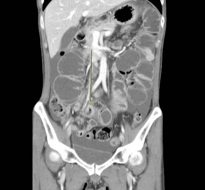
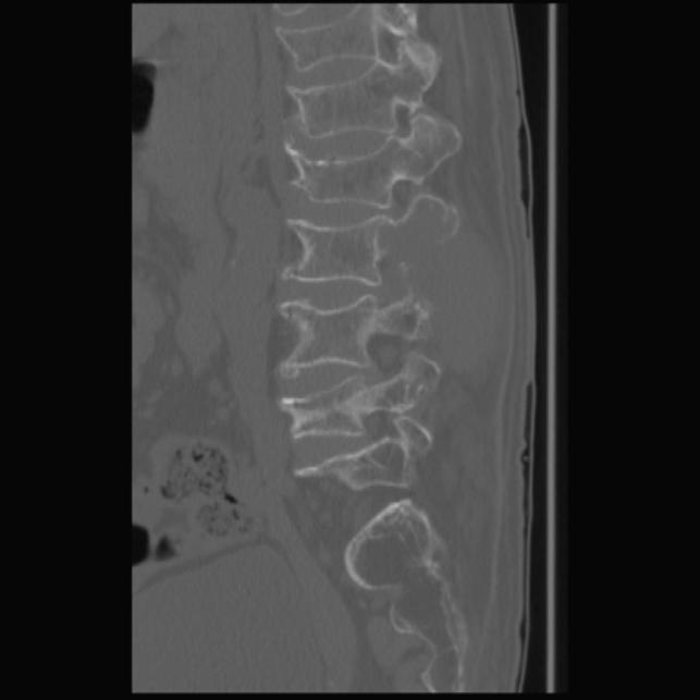
9 K3 Ga-Eon Kim Department of Pathology, Chonnam National University Hostpital A 69 year-old female presented with a noticeable mass in her back and back pain over a one-year period. Magnetic resonance imaging (MRI) revealed a paraspinal mass at L2-3 level, and the patient was referred to Chonnam National Univertisy Hospital. Computed tomography (CT) demonstrated a 5.5 x 4 x 3.5 cm mass involving the right posterior aspect of the L3 with extraosseous mass formation. She underwent surgical excision. The representative section was taken from the mass.
10
K-1 (Kyung Hee University, S )
") Case History K-1 (Kyung Hee University, S12-00829) A 17-year-old male presented with posterior neck pain and numbness in both upper extremities after a diving injury. He had no relevant medical history.
Case History K-1 (Kyung Hee University, S12-00829) A 17-year-old male presented with posterior neck pain and numbness in both upper extremities after a diving injury. He had no relevant medical history.
Takayuki Ohguri 1 Takatoshi Aoki 1 Masanori Hisaoka 2 Hideyuki Watanabe 1 Katsumi Nakamura 1 Hiroshi Hashimoto 2 Toshitaka Nakamura 3 Hajime Nakata 1
 Takayuki Ohguri 1 Takatoshi Aoki 1 Masanori Hisaoka 2 Hideyuki Watanabe 1 Katsumi Nakamura 1 Hiroshi Hashimoto 2 Toshitaka Nakamura 3 Hajime Nakata 1 Received July 1, 2002; accepted after revision November
Recurrent Malignant Fibrous Histiocytoma in Psoas Muscle: A Case Report
 Asian Spine Journal Vol. 6, No. 3, pp 211~215, 2012 Malignant Fibrous Histiocytoma in Psoas Muscle / 211 http://dx.doi.org/10.4184/asj.2012.6.3.211 Recurrent Malignant Fibrous Histiocytoma in Psoas Muscle:
Painless palpable scrotal mass
 Clinical Case - Test Yourself Urogenital Painless palpable scrotal mass Charis Anastasiadis, Georgia Kyriakopoulou, Charikleia Triantopoulou Radiology Department, Konstantopoulio General Hospital of Nea
Scrotum-like protrusion of lipoma arising from the proximal thigh
 Upsala J Med sci 109: 261 265, 2004 Scrotum-like protrusion of lipoma arising from the proximal thigh Report of two cases Koshi Hattori, 1 Masahito Hatori, 1 Mika Watanabe, 2 Toshihisa Osanai, 3 Shoichi
Musculoskeletal Sarcomas
 Musculoskeletal Sarcomas Robert C. Orth, M.D., Ph.D. Edward B. Singleton Department of Pediatric Radiology Texas Children s Hospital Page 0 xxx00.#####.ppt 9/23/2012 9:01:18 AM No disclosures Page 1 xxx00.#####.ppt
Nodular Fasciitis of the Face Diagnosed by US-Guided Core Needle Biopsy: A Case Report 1
 Nodular Fasciitis of the Face Diagnosed by US-Guided ore Needle iopsy: ase Report 1 Sang Kwon Lee, M.D., Sun Young Kwon, M.D. 2 We report here on a case of nodular fasciitis (NF) that was diagnosed by
Case Scenario 1: Thyroid
 Case Scenario 1: Thyroid History and Physical Patient is an otherwise healthy 80 year old female with the complaint of a neck mass first noticed two weeks ago. The mass has increased in size and is palpable.
Taku Naiki, 1 Shuzo Hamamoto, 1 Noriyasu Kawai, 1 Aya Naiki-Ito, 2 Yoshiyuki Kojima, 1 Takahiro Yasui, 1 Keiichi Tozawa, 1 and Kenjiro Kohri 1
 International Scholarly Research Network Volume 2011, Article ID 261735, 4 pages doi:10.5402/2011/261735 Case Report Giant Retroperitoneal Mucinous Tumor Supportively Diagnosed as a Dedifferentiated Liposarcoma
Brief History. Identification : Past History : HTN without regular treatment.
 Brief History Identification : Name : 陳 x - Admission : 94/10/06 Gender : male Age : 75 y/o Chief Complaint : Urinary difficulty for months. Past History : HTN without regular treatment. Brief History
Primary Breast Liposarcoma
 Primary Breast Liposarcoma Bhagyam Nagarajan 1*, GayatriAutkar 1, Keyuri Patel 1, Meghal Sanghvi 1 1. Department of Radiology, Wockhardt Hospital, Mumbai, India * Correspondence: Dr Bhagyam Nagarajan,
Sonography of Pediatric Superficial Lumps and Bumps: Illustrative Examples from Head to Toe
 Sonography of Pediatric Superficial Lumps and umps: Illustrative Examples from Head to Toe nmol Gupta ansal, MD Henrietta Kotlus Rosenberg, MD, FCR, FP Mount Sinai Hospital Icahn School of Medicine at
 INTRAUTERINE DEVICE = IUD INTRAUTERINE DEVICE = IUD CONGENITAL DISORDERS Pyometra = pyometrea is a uterine infection, it is accumulation of purulent material in the uterine cavity. Ultrasound is usually
Surgery for Dedifferentiated Liposarcoma, Presenting Two Radiologically and Pathologically Distinctive Patterns
 Surgery for Dedifferentiated Liposarcoma, Presenting Two Radiologically and Pathologically Distinctive Patterns Manabu Hoshi 1, Seiichi Matsumoto 1, Jun Manabe 1, Taisuke Tanizawa 1, Toshio Shigemitsu
MRI Of Locally Recurrent Soft Tissue Tumors Of The Musculoskeletal System
 ISPUB.COM The Internet Journal of Radiology Volume 5 Number 2 MRI Of Locally Recurrent Soft Tissue Tumors Of The Musculoskeletal System C Costelloe, A Yasko, W Murphy, R Kumar, V Lewis, P Lin, R Stafford,
International Journal of Research in Health Sciences ISSN: Available online at: Case Study
 International Journal of Research in Health Sciences ISSN: 2321-7251 Available online at: http://www.ijrhs.org/ Case Study Foreign body granuloma mimicking a soft tissue neoplasm *Rohan Sawant, Abhishek
Accuracy of transvaginal ultrasound and magnetic resonance imaging in diagnosis and extension of pelvic endometriosis
 Accuracy of transvaginal ultrasound and magnetic resonance imaging in diagnosis and extension of pelvic endometriosis A.Salem, Kh. Fakhfakh, S. Mehiri, Y. Ben Brahim, F. Ben Amara, H. Rajhi, R. Hamza,
Case Report Ileocecal Intussusception due to a Lipoma in an Adult
 Case Reports in Surgery Volume 2012, Article ID 684298, 4 pages doi:10.1155/2012/684298 Case Report Ileocecal Intussusception due to a Lipoma in an Adult Mehmet Bilgin, 1 Huseyin Toprak, 1 Issam Cheikh
Metastatic mechanism of spermatic cord tumor from stomach cancer
 Int Canc Conf J (2013) 2:191 195 DOI 10.1007/s13691-013-0-9 CANCER BOARD CONFERENCE Metastatic mechanism of spermatic cord tumor from stomach cancer Masahiro Seike Yoshikazu Kanazawa Ryuji Ohashi Tadashi
Malignant Focal Liver Lesions
 Malignant Focal Liver Lesions Other Than HCC Pablo R. Ros, MD, MPH, PhD Departments of Radiology and Pathology University Hospitals Cleveland Medical Center Case Western Reserve University Pablo.Ros@UHhospitals.org
Evaluation of Thyroid Nodules
 Evaluation of Thyroid Nodules Stephan Kowalyk, MD January 25 28, 2018 1 Primary goal Exclude malignancy Incidental thyroid nodules If found on CT, MRI, PET scan, carotid Doppler ULTRASOUND!! January 25
Ultrasound screening of soft tissue masses in the trunk and extremity - a BSG guide for ultrasonographers and primary care
 Ultrasound screening of soft tissue masses in the trunk and extremity - a BSG guide for ultrasonographers and primary care Introduction Soft tissue masses in the trunk and extremity are common and most
Intrahepatic Sarcomatoid Cholangiocarcinoma with Portal Vein Thrombosis: A Case Report 1
 Intrahepatic Sarcomatoid Cholangiocarcinoma with Portal Vein Thrombosis: A Case Report 1 Jae-Hoon Lim, M.D., Jin Woong Kim, M.D., Suk Hee Heo, M.D., Yong Yeon Jeong, M.D., Heoung Keun Kang, M.D. A 53-year-old
MRI IN THE CHARACTERIZATION OF SEMINOMATOUS AND NONSEMINOMATOUS GERM CELL TUMORS OF THE TESTIS
 MRI IN THE CHARACTERIZATION OF SEMINOMATOUS AND NONSEMINOMATOUS GERM CELL TUMORS OF THE TESTIS Ambesh Deshar *, Gyanendra KC and Zhang Lopsang *Department of Medical Imaging and Nuclear Medicine, First
Malignant fat-forming solitary fibrous tumor (lipomatous hemangiopericytoma) in the neck: Imaging and histopathological findings of a case
 in the neck: Imaging and histopathological findings of a case") Malignant fat-forming solitary fibrous tumor (lipomatous hemangiopericytoma) in the neck: Alice Duarte de Carvalho 1, Lucas Faria Abrahão-Machado 2, Cristiano Ribeiro Viana 2, Renato de Castro Capuzzo
Malignant fat-forming solitary fibrous tumor (lipomatous hemangiopericytoma) in the neck: Alice Duarte de Carvalho 1, Lucas Faria Abrahão-Machado 2, Cristiano Ribeiro Viana 2, Renato de Castro Capuzzo
Primary Retroperitoneal Myxofibrosarcoma: a case report and review of the literature
 J Radiol Sci 2014; 39: 57-62 Primary Retroperitoneal Myxofibrosarcoma: a case report and review of the literature Chih-Yu Chen 1 Yueh-Min Lin 2 Shang-Yun Ho 1 Kwo-Whei Lee 1 Ching Hsueh 1 Department of
Terumo Scholarship Case Study Dr B Maher, University Hospital Southampton NHS Foundation Trust
 Terumo Scholarship 2015 - Case Study Dr B Maher, University Hospital Southampton NHS Foundation Trust Clinical Presentation A 41year old female presented with pelvic pain and menorrhagia. Pelvic ultrasound
Those strange lipomas!
 Those strange lipomas! Poster No.: C-1498 Congress: ECR 2015 Type: Educational Exhibit Authors: L. Simbula, A. De Marchi, S. Pozza, E. Brach del Prever, 1 2 2 3 2 2 1 2 1 D. Molino, F. Cannone, G. B. Meloni,
Giant Pleomorphic Adenoma of the Parotid gland- A Case Report
 ISPUB.COM The Internet Journal of Otorhinolaryngology Volume 14 Number 1 Giant Pleomorphic Adenoma of the Parotid gland- A Case Report O M.E, U A.N, U Akpan, K J, I Bassey Citation O M.E, U A.N, U Akpan,
CASE REPORT PLEOMORPHIC LIPOSARCOMA OF PECTORALIS MAJOR MUSCLE IN ELDERLY MAN- CASE REPORT & REVIEW OF LITERATURE.
 PLEOMORPHIC LIPOSARCOMA OF PECTORALIS MAJOR MUSCLE IN ELDERLY MAN- CASE REPORT & REVIEW OF LITERATURE. M. Madan 1, K. Nischal 2, Sharan Basavaraj. C. J 3. HOW TO CITE THIS ARTICLE: M. Madan, K. Nischal,
A-005 US DIAGNOSIS OF NONPALPABLE BREAST LESIONS
 A-005 US DIAGNOSIS OF NONPALPABLE BREAST LESIONS Hideaki Shirai M.D., M. Sakurai M.D., K. Yoshida M.D., N. Usuda M.D., H. Masuoka M.D., I. Shimokawara M.D, K. Asaishi M.D. Sapporo Kotoni Breast Clinic,
Imaging in gastric cancer
 Imaging in gastric cancer Gastric cancer remains a deadly disease because of late diagnosis. Adenocarcinoma represents 90% of malignant tumors. Diagnosis is based on endoscopic examination with biopsies.
A CASE OF A Huge Submandibular Pleomorphic Adenoma
 ISPUB.COM The Internet Journal of Head and Neck Surgery Volume 4 Number 2 S VERMA Citation S VERMA.. The Internet Journal of Head and Neck Surgery. 2009 Volume 4 Number 2. Abstract Pleomorphic adenoma
A 25 year old female with a palpable mass in the right lower quadrant of her abdomen
 May 2016 A 25 year old female with a palpable mass in the right lower quadrant of her abdomen Contributed by: Paul Ndekwe, MD, Resident Physician, Indiana University School of Department of Pathology and
Mazabraud s Syndrome Coexisting with a Uterine Tumor Resembling an Ovarian Sex Cord Tumor (UTROSCT): a Case Report
: a Case Report") Mazabraud s Syndrome Coexisting with a Uterine Tumor Resembling an Ovarian Sex Cord Tumor (UTROSCT): a Case Report Cuneyt Calisir, MD 1 Ulukan Inan, MD 2 Ulas Savas Yavas, MD 1 Serap Isiksoy 3 Tamer Kaya
Mazabraud s Syndrome Coexisting with a Uterine Tumor Resembling an Ovarian Sex Cord Tumor (UTROSCT): a Case Report Cuneyt Calisir, MD 1 Ulukan Inan, MD 2 Ulas Savas Yavas, MD 1 Serap Isiksoy 3 Tamer Kaya
Research Article A Clinicopathological Analysis of Soft Tissue Sarcoma with Telangiectatic Changes
 Sarcoma Volume 2015, Article ID 740571, 5 pages http://dx.doi.org/10.1155/2015/740571 Research Article A Clinicopathological Analysis of Soft Tissue Sarcoma with Telangiectatic Changes Hiroshi Kobayashi,
Rare tumors of the heart - angiosarcoma, pericardial lipoma, leiomyosarcoma Three case reports
 , pp.178 182 Rare tumors of the heart - angiosarcoma, pericardial lipoma, leiomyosarcoma Three case reports Ioana Stoian, Ileana Tepes Piser, Iulia Kulcsar, O Chioncel, A Carp, C Macarie Prof Dr CC Iliescu
Five Views of Transitional Cell Carcinoma: One Man s Journey
 September 2006 Five Views of Transitional Cell Carcinoma: One Man s Journey Amsalu Dabela, Harvard Medical School III Outline Overview: Renal Anatomy Our Patient s Story Diagnostic Imaging Studies Appearance
Ultrasonography. Methods. Brief Description. Indications. Device-related Prerequisites. Technical Requirements. Evaluation Criteria
 1 Ultrasonography Brief Description Imaging modality using sound waves Tissue-specific wave reflection. Indications Evaluation of palpable breast nodules Evaluation of clinically occult mammographic findings
GIANT RETROPERITONEAL LIPOSARCOMA: IMAGING AND LITERATURE Amit Kumar 1, Sanjay K. Suman 2, Bipin Kumar 3, Sumit Kumar 4
 GIANT RETROPERITONEAL LIPOSARCOMA: IMAGING AND LITERATURE Amit Kumar 1, Sanjay K. Suman 2, Bipin Kumar 3, Sumit Kumar 4 HOW TO CITE THIS ARTICLE: Amit Kumar, Sanjay K. Suman, Bipin Kumar, Sumit Kumar.
Different Radiologic Appearances of Giant Epidermoid Cysts at the Floor of the Mouth: Three Case Reports ABSTRACT
 Different Radiologic Appearances of Giant Epidermoid Cysts at the Floor of the Mouth: Three Case Reports MS Sakat 1, E Altaş 1, K Kılıç 2, E Demirci 3, H Üçüncü 1* ABSTRACT Epidermoid and dermoid cysts
Soft tissue lipomas, lipoma variants and liposarcomas: MRI evaluation and review of literature
 Soft tissue lipomas, lipoma variants and liposarcomas: MRI evaluation and review of literature Poster No.: R-0122 Congress: RANZCR-AOCR 2012 Type: Authors: Keywords: DOI: Educational Exhibit A. A. Tandon,
Ultrasound Evaluation of Masses
 Ultrasound Evaluation of Masses Jon A. Jacobson, M.D. Professor of Radiology Director, Division of Musculoskeletal Radiology University of Michigan Disclosures: Consultant: Bioclinica Advisory Panel: GE,
Original Report. Imaging Features of Fat Necrosis. Lai Peng Chan 1 R. Gee 2 Ciaran Keogh 2 Peter L. Munk 2
 Lai Peng Chan 1 R. Gee 2 Ciaran Keogh 2 Peter L. Munk 2 Received September 16, 2002; accepted after revision pril 29, 2003. 1 Department of Diagnostic Radiology, Singapore General Hospital, Outram Rd.,
Synovial hemangioma of the suprapatellar bursa
 Synovial hemangioma of the suprapatellar bursa Poster No.: P-0040 Congress: ESSR 2013 Type: Authors: Keywords: DOI: Scientific Exhibit A. YESILDAG, S. Keskin, H. Kalkan, S. Kucuksen, U. Kerimoglu; Konya/TR
Multiple Primary Quiz
 Multiple Primary Quiz Case 1 A 72 year old man was found to have a 12 mm solid lesion in the pancreatic tail by computed tomography carried out during a routine follow up study of this patient with adult
Magnetic resonance imaging of ulnar nerve abscess in leprosy: a case report
 Lepr Rev (2006) 77, 381 385 CASE REPORT Magnetic resonance imaging of ulnar nerve abscess in leprosy: a case report SMRITI HARI, SUBRAMANIAN SUBRAMANIAN & RAJU SHARMA Department of Radiodiagnosis, All
Evaluation of Neck Mass. Disclosure. Learning Objectives 3/24/2014. Karen T. Pitman MD, FACS Banner MDACC, Gilbert AZ. Nothing to disclose
 Evaluation of Neck Mass Karen T. Pitman MD, FACS Banner MDACC, Gilbert AZ Nothing to disclose Disclosure Learning Objectives 1. Describe a systematic method to evaluate a patient with a neck mass 2. Select
Case Scenario 1. Pathology report Specimen from mediastinoscopy Final Diagnosis : Metastatic small cell carcinoma with residual lymphatic tissue
 Case Scenario 1 Oncology Consult: Patient is a 51-year-old male with history of T4N3 squamous cell carcinoma of tonsil status post concurrent chemoradiation finished in October two years ago. He was hospitalized
MRI features of Triple-negative breast cancer: our experience.
 MRI features of Triple-negative breast cancer: our experience. Poster No.: C-1852 Congress: ECR 2013 Type: Scientific Exhibit Authors: V. Bertani, A. Gualano, V. Londero, A. Dal Col, M. Marcon, P. 1 2
Preoperative Diagnosis of Adult Intussusception Caused by Small Bowel Lipoma
 377 Preoperative Diagnosis of Adult Intussusception Caused by Small Bowel Lipoma Hiroaki Shiba a Yoshinobu Mitsuyama a Ken Hanyu a Kenji Ikeuchi b Hirotaka Hayashi c Katsuhiko Yanaga a a Department of
Sclerosing angiomatoid nodular transformation (SANT) of the spleen: a case report with FDG-PET findings and literature review
 of the spleen: a case report with FDG-PET findings and literature review") Case Report Sclerosing angiomatoid nodular transformation (SANT) of the spleen: a case report with FDG-PET findings and literature review Acta Radiologica Open 5(8) 1 6! The Foundation Acta Radiologica
Case Report Sclerosing angiomatoid nodular transformation (SANT) of the spleen: a case report with FDG-PET findings and literature review Acta Radiologica Open 5(8) 1 6! The Foundation Acta Radiologica
Timothy L. Miao 1, Ania Z. Kielar 2,3, Rebecca M. Hibbert 2, Nicola Schieda 2,3
 DOES LESION T1 SIGNAL INTENSITY RELATIVE TO LIVER PARENCHYMA PREDICT VISIBILITY ON ULTRASOUND? A clinical tool to determine feasibility of ultrasound-guided percutaneous interventions Timothy L. Miao 1,
J of Evolution of Med and Dent Sci/ eissn , pissn / Vol. 3/ Issue 46/Sep 22, 2014 Page 11296
 CT SPECTRUM OF GIANT RETROPERITONEAL LIPOSARCOMAS WITH HISTOPATHOLOGICAL CORRELATION Shashikumar M. R 1, Rajendra Kumar N. L 2, C. P. Nanjaraj 3, Nishanth R. K 4, Vishwanath Joshi 5 HOW TO CITE THIS ARTICLE:
Radiologic-Pathologic Correlation of Primary Ovarian Leiomyosarcoma: a Case Report and Review of the Literature
 May, 2017 2017; Vol1; Issue4 http://iamresearcher.online Radiologic-Pathologic Correlation of Primary Ovarian Leiomyosarcoma: a Case Report and Review of the Literature Lama M AlMudaimeegh Department of
ACG Clinical Guideline: Diagnosis and Management of Focal Liver Lesions
 ACG Clinical Guideline: Diagnosis and Management of Focal Liver Lesions Jorge A. Marrero, MD, 1 Joseph Ahn, MD, FACG, 2 K. Rajender Reddy, MD, FACG 3 1 University of Texas at Southwestern, Dallas, Texas,
Case Fibrothecoma of the ovary
 Case 10646 Fibrothecoma of the ovary Elisa Melo Abreu, Teresa Margarida Cunha Section: Genital (Female) Imaging Published: 2015, Jan. 2 Patient: 70 year(s), female Authors' Institution Department of Radiology,
NEW SUBTRACTION ALGORITHMS FOR EVALUATION OF BREAST LESIONS ON DYNAMIC CONTRAST ENHANCED MR MAMMOGRAPHY
 A-056 NEW SUBTRACTION ALGORITHMS FOR EVALUATION OF BREAST LESIONS ON DYNAMIC CONTRAST ENHANCED MR MAMMOGRAPHY So Hee Cho, M.D., Byung Gil Choi, M.D., Hak Hee Kim, M.D., Euy Neyng Kim, M.D., Bum-soo Kim,
Prof. Dr. NAGUI M. ABDELWAHAB,M.D.; MARYSE Y. AWADALLAH, M.D. AYA M. BASSAM, Ms.C.
 Role of Whole-body Diffusion MR in Detection of Metastatic lesions Prof. Dr. NAGUI M. ABDELWAHAB,M.D.; MARYSE Y. AWADALLAH, M.D. AYA M. BASSAM, Ms.C. Cancer is a potentially life-threatening disease,
Thyroid Nodules. Dr. HAKIMI, SpAK Dr. MELDA DELIANA, SpAK Dr. SISKA MAYASARI LUBIS, SpA
 Thyroid Nodules ENDOCRINOLOGY DIVISION ENDOCRINOLOGY DIVISION Dr. HAKIMI, SpAK Dr. MELDA DELIANA, SpAK Dr. SISKA MAYASARI LUBIS, SpA Anatomical Considerations The Thyroid Nodule Congenital anomalies Thyroglossal
Radiology-Pathology Conference
 July 31, 2009 Radiology-Pathology Conference Daniel T Ginat, M.D., M.S. Sharlin Johnykutty,, M.D. Presentation material is for education purposes only. All rights reserved. 2009 URMC Radiology Page 1 of
We are IntechOpen, the world s leading publisher of Open Access books Built by scientists, for scientists. International authors and editors
 We are IntechOpen, the world s leading publisher of Open Access books Built by scientists, for scientists 3,900 116,000 120M Open access books available International authors and editors Downloads Our
Case 9551 Primary ovarian Burkitt lymphoma
 Case 9551 Primary ovarian Burkitt lymphoma Monteiro V, Cunha TM, Saldanha T Section: Genital (Female) Imaging Published: 2011, Nov. 20 Patient: 23 year(s), female Authors' Institution V Monteiro 1, TM
Extraosseous Ewing s Sarcoma Presented as a Rectal Subepithelial Tumor: Radiological and Pathological Features
 pissn 2384-1095 eissn 2384-1109 imri 2017;21:51-55 https://doi.org/10.13104/imri.2017.21.1.51 Extraosseous Ewing s Sarcoma Presented as a Rectal Subepithelial Tumor: Radiological and Pathological Features
Calcifying Aponeurotic Fibroma of the Knee: a Case Report with Radiographic and MRI Finding
 pissn 2384-1095 eissn 2384-1109 imri 2017;21:259-263 Calcifying Aponeurotic Fibroma of the Knee: a Case Report with Radiographic and MRI Finding Seung Hyun Lee 1,2, In Sook Lee 1,2, You Seon Song 1,2,
Myositis Ossificans Mimicking Sarcoma, the Importance of Diagnostic Imaging Case Report
 Signature: Pol J Radiol, 2014; 79: 228-232 DOI: 10.12659/PJR.890209 CASE REPORT Received: 2013.12.18 Accepted: 2014.01.27 Published: 2014.07.28 Authors Contribution: A Study Design B Data Collection C
Gastric Cancer (2002) 5: by International and Japanese Gastric Cancer Associations
 5: by International and Japanese Gastric Cancer Associations") Gastric Cancer (2002) 5: 107 111 2002 by International and Japanese Gastric Cancer Associations Case report Intramuscular metastasis from gastric cancer Shohei Kondo 1, Hisashi Onodera 1, Shugen Kan 1,
Gastric Cancer (2002) 5: 107 111 2002 by International and Japanese Gastric Cancer Associations Case report Intramuscular metastasis from gastric cancer Shohei Kondo 1, Hisashi Onodera 1, Shugen Kan 1,
Pleomorphic adenoma head and neck
 Pleomorphic adenoma head and neck Poster No.: C-1042 Congress: ECR 2015 Type: Educational Exhibit Authors: M. E. Pérez Montilla, I. Bravo Rey, E. Roldán Romero, F. BravoRodríguez; Cordoba/ES Keywords:
A 21 year old woman with a rapidly growing mass on palate. Dr. Elizabeth Bigger and Dr. Memory Bvochora 18 March 2015
 A 21 year old woman with a rapidly growing mass on palate Dr. Elizabeth Bigger and Dr. Memory Bvochora 18 March 2015 History of present illness 21 year old woman G2P1 admitted to the Princess Marina Hospital
Soft Tissue Tumour & Sarcoma Imaging Guidelines 2012
 Soft Tissue Tumour & Sarcoma Imaging Guidelines 2012 Version Control This is a controlled document please destroy all previous versions on receipt of a new version. Date Approved: March 2011 reissued April
1 Uniform hyperintense signal intensity (normal). 2 Linear (arrow), wedge-shaped, or diffuse mild hypointensity, usually indistinct margin.
. 2 Linear (arrow), wedge-shaped, or diffuse mild hypointensity, usually indistinct margin.") Figure 3 PI-RADS assessment for peripheral zone on T2-weighted imaging. 1 Uniform hyperintense signal intensity (normal). 2 Linear (arrow), wedge-shaped, or diffuse mild hypointensity, usually indistinct
Figure 3 PI-RADS assessment for peripheral zone on T2-weighted imaging. 1 Uniform hyperintense signal intensity (normal). 2 Linear (arrow), wedge-shaped, or diffuse mild hypointensity, usually indistinct
Imaging of Liposarcoma: Classification, Patterns of Tumor Recurrence, and Response to Treatment
 Special Article Pictorial Essay O Regan et al. Imaging of Liposarcoma Special Article Pictorial Essay Kevin N. O Regan 1 Jyothi Jagannathan Katherine Krajewski Katherine Zukotynski Frederico Souza Andrew
Elastofibroma dorsi: 8 case reports and a literature review
 DOI 10.1007/s10195-008-0102-7 BRIEF COMMUNICATION Elastofibroma dorsi: 8 case reports and a literature review F. Muratori M. Esposito F. Rosa F. Liuzza N. Magarelli B. Rossi H.M. Folath F. Pacelli G. Maccauro
Chief complaint. A mass at right chest
 Chief complaint A mass at right chest Present illness This 1-year-5-month-old girl had a mass at right side chest since one month ago. flat and not tender at first In the recent 2 days, the mass enlarged
MR Findings in a Rare Case of Sclerosing Mesenteritis of the Mesocolon
 JOURNAL OF MAGNETIC RESONANCE IMAGING 21:632 636 (2005) Clinical Note MR Findings in a Rare Case of Sclerosing Mesenteritis of the Mesocolon Nadir Ghanem, MD,* Gregor Pache, MD, Thorsten Bley, MD, Elmar
MRI XR, CT, NM. Principal Modality (2): Case Report # 2. Date accepted: 15 March 2013
: Case Report # 2. Date accepted: 15 March 2013") Radiological Category: Musculoskeletal Principal Modality (1): Principal Modality (2): MRI XR, CT, NM Case Report # 2 Submitted by: Hannah Safia Elamir, D.O. Faculty reviewer: Naga R. Chinapuvvula, M.D.
Radiological Category: Musculoskeletal Principal Modality (1): Principal Modality (2): MRI XR, CT, NM Case Report # 2 Submitted by: Hannah Safia Elamir, D.O. Faculty reviewer: Naga R. Chinapuvvula, M.D.
A case of pedunculated intraperitoneal leiomyoma
 Jichi Medical University Journal Chio Shuto Kuniyasu Soda Takayoshi Yoshida Fumio Konishi Abstract We report a very rare case of a pedunculated intraperitoneal leiomyoma in the parietal peritoneum of the
Kimura s Disease in the Lower Extremity: A Case Report Mimicking the Malignant Soft Tissue Mass
 Kimura s Disease in the Lower Extremity: A Case Report Mimicking the Malignant Soft Tissue Mass Jee Young Lee, M.D. 1, Kyung Jin Suh, M.D. 2, Hong-Geun Jung, M.D. 3 We present a case of a 37-year-old woman
CLINICO-PATHOLOGICAL CONFERENCE CLASS OF 2007/2012 PHASE IIIB SESSION 2010/2012
 CLINICO-PATHOLOGICAL CONFERENCE CLASS OF 2007/2012 PHASE IIIB SESSION 2010/2012 PRESENTATION OF CASE A 62-year-old woman was seen in the outpatient cancer center of this hospital because of anemia and
Type 1 neurofibromatosis and adult extremity sarcoma A report of two cases
 Acta Orthop. Belg., 2007, 73, 403-407 CASE REPORT Type 1 neurofibromatosis and adult extremity sarcoma A report of two cases Bahtiyar DEMIRALP, M. Taner OZDEMIR, Kaan ERLER, Mustafa BASBOZKURT From Gulhane
Toru Nakamura 1, Takashi Fukutomi 1, Hitoshi Tsuda 2, Sadako Akashi-Tanaka 1, Kaneyuki Matsuo 1, Chikako Shimizu 1 and Kunihisa Miyakawa 3
 Jpn J Clin Oncol 2000;30(10)453 457 Changes in Findings of Mammography, Ultrasonography and Contrast-enhanced Computed Tomography of Three Histological Complete Responders with Primary Breast Cancer Before
Exercise. Discharge Summary
 Exercise Discharge Summary A 32-year-old Brazilian male presented with a 6 month history of right-sided scrotal swelling. Backache was present for 2 months and a history of right epididymitis was present
Frozen recapping laminoplasty: a new technique to treat spinal tumor
 EUROSPINE 2018 19-21 September, Barcelona, Spain Frozen recapping laminoplasty: a new technique to treat spinal tumor Noritaka Yonezawa, Hideki Murakami, Satoru Demura, Satoshi Kato, Katsuhito Yoshioka,
UK Musculoskeletal Oncology: Something for All Ages. Lars Wagner, MD Pediatric Hematology/Oncology University of Kentucky
 UK Musculoskeletal Oncology: Something for All Ages Lars Wagner, MD Pediatric Hematology/Oncology University of Kentucky Pediatric-Type Sarcomas of Bone and Soft Tissue The incidence of sarcoma continues
CLINICAL PRESENTATION AND RADIOLOGY QUIZ QUESTION
 Donald L. Renfrew, MD Radiology Associates of the Fox Valley, 333 N. Commercial Street, Suite 100, Neenah, WI 54956 11/24/2012 Radiology Quiz of the Week # 100 Page 1 CLINICAL PRESENTATION AND RADIOLOGY
Primary Hepatic Undifferentiated Pleomorphic Sarcoma: CT and angiographic findings in two cases
 J Radiol Sci 2013; 38: 15-19 Primary Hepatic Undifferentiated Pleomorphic Sarcoma: CT and angiographic findings in two cases Jan-Wen Ku Ying-Chi Tseng Kuo-Luon Kung Hsien-Chang Shen Yen-Lin Huang Chi-Jen
Kidney Case 1 SURGICAL PATHOLOGY REPORT
 Kidney Case 1 Surgical Pathology Report February 9, 2007 Clinical History: This 45 year old woman was found to have a left renal mass. CT urography with reconstruction revealed a 2 cm medial mass which
Case #1: 75 y/o Male (treated and followed by prostate cancer oncology specialist ).
.") SOLID TUMORS WORKSHOP Cases for review Prostate Cancer Case #1: 75 y/o Male (treated and followed by prostate cancer oncology specialist ). January 2009 PSA 4.4, 20% free; August 2009 PSA 5.2; Sept 2009
SOLID TUMORS WORKSHOP Cases for review Prostate Cancer Case #1: 75 y/o Male (treated and followed by prostate cancer oncology specialist ). January 2009 PSA 4.4, 20% free; August 2009 PSA 5.2; Sept 2009
Triple-negative breast cancer: which typical features can we identify on conventional and MRI imaging?
 Triple-negative breast cancer: which typical features can we identify on conventional and MRI imaging? Poster No.: C-1862 Congress: ECR 2013 Type: Educational Exhibit Authors: V. Bertani 1, A. Gualano
Fat Necrosis: A Grand Imposter
 Fat Necrosis: A Grand Imposter Poster No.: C-0751 Congress: ECR 2015 Type: Educational Exhibit Authors: L. C. Flores Salinas, Y. A. Ramirez Galvan, A. Garza Báez, C. M. Ferrara Chapa; Monterrey/MX Keywords:
Malignant Transformation of Endometriosis: Magnetic Resonance Imaging Aspects
 Malignant Transformation of Endometriosis: Magnetic Resonance Imaging Aspects Poster No.: C-0084 Congress: ECR 2014 Type: Scientific Exhibit Authors: E. A. Yukhno, I. Trofimenko, G. Trufanov; St. Petersburg/RU
Malignant Transformation of Endometriosis: Magnetic Resonance Imaging Aspects
 Malignant Transformation of Endometriosis: Magnetic Resonance Imaging Aspects Poster No.: C-0084 Congress: ECR 2014 Type: Scientific Exhibit Authors: E. A. Yukhno, I. Trofimenko, G. Trufanov; St. Petersburg/RU
Financial Disclosure
 Benign Liver Masses Adil Abdalla, MBBS Creighton University-CHI Health August 25, 2018 Financial Disclosure Nothing to disclose Financial Disclosure 1 Objectives To assess patients with benign liver tumors
Surgical management and neoadjuvant chemotherapy for stage III-IV ovarian cancer
 Ovarian cancer Surgical management and neoadjuvant chemotherapy for stage III-IV ovarian cancer JM. Classe, R. Rouzier, O.Glehen, P.Meeus, L.Gladieff, JM. Bereder, F Lécuru Suitable candidates for neo-adjuvant
X-ray Corner. Imaging of The Peritoneum and Mesentery. Pantongrag-Brown L. Case 1. A 47-year-old woman presenting with abdominal distension.
 X-ray Corner Pantongrag-Brown L THAI J GASTROENTEROL 2016 Vol. 17 No. 3 Sep. - Dec. 2016 187 Pantongrag-Brown L Modern imaging modalities commonly used in peritoneum and mesentery include ultrasound (US),
Ovarian Tumors. Andrea Hayes-Jordan MD FACS, FAAP Section Chief, Pediatric Surgery/Surgical Onc. UT MD Anderson Cancer Center
 Ovarian Tumors Andrea Hayes-Jordan MD FACS, FAAP Section Chief, Pediatric Surgery/Surgical Onc. UT MD Anderson Cancer Center Case 13yo female with abdominal pain Ultrasound shows huge ovarian mass Surgeon
Principles of Surgical Oncology. Winnie Achilles Tierklinik Hollabrunn Lastenstrasse Hollabrunn
 Principles of Surgical Oncology Winnie Achilles Tierklinik Hollabrunn Lastenstrasse 2 2020 Hollabrunn boexi@gmx.de The first surgery provides the best chance for a cure in an animal with a tumor Clinical
Esophageal seeding after endoscopic ultrasound-guided fine-needle aspiration of a mediastinal tumor
 Esophageal seeding after endoscopic ultrasound-guided fine-needle aspiration of a mediastinal tumor Authors Kensuke Yokoyama 1,JunUshio 1,NorikatsuNumao 1, Kiichi Tamada 1, Noriyoshi Fukushima 2, Alan
Detection and Characterization of Hepatocellular Carcinoma by Imaging
 CLINICAL GASTROENTEROLOGY AND HEPATOLOGY 2005;3:S136 S140 Detection and Characterization of Hepatocellular Carcinoma by Imaging OSAMU MATSUI Department of Imaging Diagnosis and Interventional Radiology,
A rare case of cervical epidural extramedullary plasmacytoma presenting with monoparesis
 Romanian Neurosurgery Volume XXXI Number 1 2017 January - March Article A rare case of cervical epidural extramedullary plasmacytoma presenting with monoparesis Okan Turk, Ibrahim Burak Atci, Hakan Yilmaz,