PITUITARY ADENOMAS- CLINICAL, NEURO-OPTHALMIC AND RADIOLOGICAL EVALUATION
|
|
|
- Laurel McGee
- 5 years ago
- Views:
Transcription
1 PITUITARY ADENOMAS- CLINICAL, NEURO-OPTHALMIC AND RADIOLOGICAL EVALUATION
2 PITUITARY GLAND AN OVERVIEW WEIGHS Just 600 mg Cranio caudal dimensions 8-10mm Upper border is usually flat or concave EXERCISES DIRECT OR INDIRECT CONTROL ON EVERY ORGAN SYSTEM
3 PITUITARY GLAND AN OVERVIEW Sella turcica - part of body of sphenoid bone Depth- upper limit 13mm Length- upper limit 17mm Width upperlimit 15 mm volume 1100 mm3
4 ADENOHYPOPHYSIS GLANDULAR COMPONENT BELIEVED TO ARISE FROM STOMODEUM SECRETES GH,PRL,FSH,LH,TSH,ACTH,MSH,ENDORPHINS.
5 ADENOHYPOPHYSIS : DIVIDED INTO PARS TUBERALIS PARS INTERMEDIA PARS DISTALIS
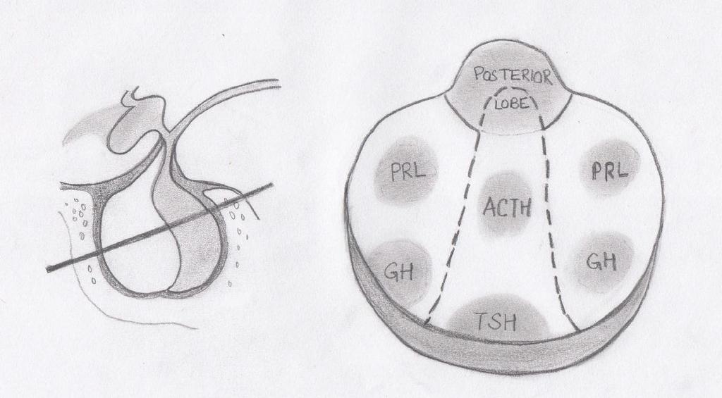
6 ADENOHYPOPHYSIS : DELICATE ACINAR ARCHITECTURE IN HORIZONTAL CROSS SECTION,COMPOSED OF TWO LATERAL WINGS TRAPEZOID CENTRAL MUCOID WEDGE
7 SOMATOTROPHS ANTERIOR PART OF THE LATERAL WINGS LACTOTROPHS POSTERIOR PART OF THE LATERAL WINGS CORTICOTROPHS CENTRAL WEDGE, JUST ANTERIOR TO POSTERIOR LOBE THYROTROPHS GONADOTROPHS ANTEROMEDIAL PART OF CENTRAL WEDGE THROUGH OUT PARS DISTALIS
8
9 NEUROHYPOPHYSIS CONTAINS ONLY AXONS AND FENESTRATED CAPPILARIES DIVIDED INTO MEDIAN EMINENCE INFUNDIBULAR STEM NEURAL LOBE
10 PITUITARY TUMOURS 10-15% *OF ALL PRIMARY BRAIN TUMOURS *kovcks et al.tumours of pituitary gland.atlas of tumour pathology ANNUAL INCIDENCE OF CASE** / POPULATION **annegers et al.report of increasing incidence of diagnosis in women of child bearing age. Mayo clin proc THOUGH INCIDENCE IS EQUAL, IT IS DIAGNOSED MORE COMMONLY IN FEMALES


11 THIRD MOST COMMON PRIMARY BRAINTUMOURS AUTOPSY INCIDENCE: 20-25%* OF POPULATION molitch et al. Incidental pituitary adenomas. Am J Med Sci %* OF ROUTINE MRI SCANS SHOW OCCULT PITUITARY MICROADENOMA. *molitch et al. Incidental pituitary adenomas. Am J Med Sci.1993 BETWEEN 3 RD 6 TH DECADE OF LIFE
12 PITUITARY TUMOURS GENETICS MEN 1 3% OF ALL PITUITARY TUMOURS AUTOSOMAL DOMINANT DISORDER VARIABLE PENETRANCE OCCCURS IN 25% OF AFFECTED PATIENTS with MEN 1 PRL OR GH MACROADENOMAS
13 PITUITARY TUMOURS ADENOHYPOPHYSIS PITUITARY ADENOMAS NEUROHYPOPHYSIS METASTATIC TUMOURS PRIMARY : RARE -GLIOMA S,GRANULAR CELL TUMOURS,HEMARTOMAS

14 PITUITARY ADENOMAS FUNCTIONING YOUNG ADULTS NON FUNCTIONING WITH INCREASING AGE

15 Adenoma type* Prevalence % Prolactin cell adenoma 30 GH cell adenoma 15 ACTH cell adenoma 10 Gonadotroph adenoma 10 GH/PRL cell adenoma 7 TSH cell adenoma 1 Nonfunctioning adenoma 25 *kovcks et al.tumours of pituitary gland.atlas of tumour pathology.1986
16 PITUATARY ADENOMAS GROSS : YELLOWISH GREY TO PURPLE, SOFT FLUID TO CREAMY TEXTURE HISTOLOGICAL: CELLULAR MONOMORPHISM LACK OF ACINAR ORGANIZATION UNIFORM CYTOPLASMIC STAINING, PLEOMORPHIC CELLS, PROMINENT NUCLEOLI, MITOTIC FIGURES.
17 PITUITARY ADENOMAS HYPERSECRETION PRESENTATION MASS EFFECT PITUITARY INSUFFICIENCY
18 HYPERSECRETION 70% OF PITUITARY ADENOMAS ARE ENDOCRINOLOGICALLY ACTIVE MOST COMMON MODE OF PRESENTATION PRESENTATION VARIES ACCORDING TO THE HORMONE IN EXCESS
19 PITUITARY INSUFFICIENCY BY COMPRESSION OF NON TUMOUROUS PITUITARY, PITUITARY STALK,HYPOTHALAMUS. CHRONIC PROCESS, CAN BE ACUTE AS IN PITUITARY APOPLEXY GONADOTROPHS MOST VULNERABLE
20 MASS EFFECT HEADACHE VISUAL LOSS HYDROCEPHALUS INTRACAVERNOUS EXTENSION
21 HARDY S Classification Microadenomas Grades 0 and I Macroadenomas Grades II to IV Grade 0 : Intrapituitary microadenoma with normal sellar floor Grade I : Normal-sized sella with asymmetric floor Grade II : Enlarged sella with an intact floor Grade III : Localized erosion of sellar floor Grade IV : Diffuse destruction of floor
22 Modified Hardy Wilson Classification Type A: Tumor bulges into the chiasmatic cistern Type B: Tumor reaches the floor of the 3 rd ventricle Type C: Tumor is more voluminous with extension into the 3 rd ventricle up to the foramen of Monro Type D: Tumor extends into temporal or frontal fossa TYPE E : Extradural spread ( extension into or out of the cavenous sinus)
23 Pathologic Classification Chromophobic Non-functioning Basophilic Cushing s Acidophilic - Acromegaly Mixed
24 WHO Classification Five-tiered system Clinical presentation and secretory activity Size and invasiveness (e.g. Hardy) Histology (typical vs. atypical) Immunohistologic profile Ultrasturctural subtype
25 PITUITARY ADENOMAS A. PROLACTINOMA Most common primary tumour of pituitary 30% of all pituitary adenoma Female : male = 20: 1 for microadenoma 1:1 for macroadenoma Characterized by hyperprolactinemia Prolactin < 25 ng/ ml - normal ng/ml - prolactinoma, stalk effect, drugs, Hypothyroid > 150ng/ml - prolactinoma(pure or mixed) > 1000 ng/ml - invasive prolactinomas
26
27 HYPOGONADISM PROLACTINOMAS CLINICAL PRESENTATION Menstrual irregularities like secondary amenorrhea, delayed menarche, oligomenorrhea, infertility. Galactorrhea Decreased libido HEADCACHE VISUAL DISTURBANCES HYPOPITUITARISM PSYCHOLOGICAL
28 PITUITARY ADENOMAS B. GROWTH HORMONE SECRETING PITUITARY ADENOMAS Growth hormone Most abundant pituitary hormone Secretion is pulsatile Physiological excess seen in stress, trauma, sepsis, estrogen replacement Exerts it s action through IGF -1
29 GROWTH HORMONE SECRETING PITUITARY ADENOMAS Equal incidence in males and females more than 60% are macroadenomas 4 th and 5 th decade 15% Of all pituitary tumors plurihormonal Overall mortality is increased 3 folds as compared to age matched controls
30 GH excess GROWTH HORMONE SECRETING PITUITARY ADENOMAS Before epiphyseal closure - gigantism Beyond puberty - acromegaly
31 DIVERSE MANISFESTATIONS 2. CARDIOVASCULAR HYPERTENSION CARDIOMYOPATHY ARRHYTHMIAS Malodorous/oily perspiration 1. BONE AND SOFT TISSUE- coarse facial features 3.Musculoskeletal Arthropathies Kyphosis Spinal stenosis Barrel chest Osteoarthritis Spade like enlargement of hand and feet Macroglossia frontal bossing, prognathism, maxillary widening, dental malocclusion Snoring sleep apnea, low voice 4. Increased incidence of premalignant polyps/ colonic cancers 5.Diabetes mellitus
32 DIAGNOSIS Random GH not useful gives false positive and false negative results Insulin like growth factor 1 (IGF-1) best for screening represents average daily GH secretion Insufficient GH suppression on oral glucose tolerance testing gold standard to confirm diagnosis :75 mg of glucose load normally suppresses GH< 2ng/ml RIA. GH nadir >2ng/ml RIA with adenoma confirms it
33 Pituitary adenomas Cushing s disease 5 to 10 times more common in females than males 3 rd and 4 th decade 10-15% of all pituitary tumors Highest morbidity of all pituitary hypersecretory disorders Most common cause of death is cardiovascular complication
34 CUSHING S DISEASE Ch. Exposure of tissues to excessive cortisol Moon facies Centripetal obesity Buffalo hump Thin skin,purple abdominal striae, ecchymosis Psychological Glucose intolerance Hematopoietic features include leukocytosis, lymphopenia, eosinopenia Osteoporosis, proximal myopathy, Impaired immune function Hirsutism, acne menstrual irregularities in females Oligospermia, impotence in males
35 Diagnosis?Primary Cushing syndrome(cushing s disease) Cushing s syndrome??secondary hypercortisolism (ectopic Cushing's syndrome)???primary hypercortisolism(adrenal Cushing syndrome)
36 Diagnosis 24 hr urinary free cortisol(>100mcg)1 and 17 OHcorticosteroids(>12mg) 1 mg overnight dexamethasone test- best screening test Low dose dexamethasone suppression test High dose dexamethasone suppression Plasma ACTH levels Inferior petrosal sinus sampling
37 INVESTIGATION PROTOCOL History and physical examination Neuro- ophthalmology: Acuity, field, fundus and movements Hormone levels basal hormone and dynamic testing Aim- hypersecretory state/insufficiency Radiology (a) X-Rays (b) MRI (c) NCCT/CECT (d)pet/dsa Routine blood investigation
38 NEURO OPTHALMICS OF PITUITARY ADENOMA OPTIC NERVE consists of 1.5 million fibres. Total length is 5 cm of which mm is intracranial. Both optic nerves after coming out of optic canal rise by 45 degrees and meet to form optic chiasm
39 NEURO OPTHALMICS OF PITUITARY ADENOMA OPTIC CHIASM can be Prefixed 15% Normal 70% Post fixed 15% With in the chiasm PMB lies in the middle Temporal hemi retinal fibers pass ipsilateraly Nasal hemi retinal fibers decussate
40 NEURO OPTHALMICS OF PITUITARY ADENOMA Optic chiasm decussation Inferior nasal fibers - anteroinferior Superior nasal fibers - posterosuperior PMB - in the middle primarily postero superiorly
41 NEURO OPTHALMICS OF PITUITARY ADENOMA Enlarging pituitary adenoma may compress Optic chiasm Optic nerve in patients with postfixed chiasm Optic tracts in patients with prefixed chiasm 3 rd, 4 th, 6 th nerves with cavernous sinus extension causing diplopia Diplopia evaluation:: 3 principles abnormal image is always peripheral is always from the paretic eye distance between the image increases on looking in the direction of paretic muscle Third ventricle leading to hydrocephalus
42 NEURO OPTHALMICS OF PITUITARY ADENOMA Visual evaluation in a case of pituitary adenoma includes examination of: Visual acuity Colour vision Visual fields Opthalmoscopy Pupils Extraocular movements
43 NEURO OPTHALMICS OF PITUITARY ADENOMA VISUAL ACUITY Eye s ability to resolve details Neurosurgically, patient s best corrected visual acuity is pertinent Distant vision by Snellen s chart placed at 6 m where accommodation is relaxed and light rays are parallel Near vision by rosenbaum s pocket chart held at a distance of 14 inches
44 NEURO OPTHALMICS OF PITUITARY COLOUR VISION ADENOMA Loss of colour vision precedes other visual deficits In neurosurgical disease, red perception is lost first described as red desaturation or red wash outs Ishihara/hardy ritter rand charts used
45 Visual fields NEURO OPTHALMICS OF PITUITARY deg temporally 60 deg nasally deg superiorly deg inferiorly ADENOMA With binocular vision, VF of both eyes overlap Visual fields are analyzed by Confrontation method Goldman s perimeter Humphrey s field analyzer

46
47 NEURO OPTHALMICS OF PITUITARY ADENOMA Pituitary adenoma can cause primary optic atrophy primary Colour of disc white grey secondary Border of disc Sharp Blurred Arteries and veins Normal or reduced Arteries thin, veins dilated Distribution May affect one sector Entire disc affected Causes Optic nerve/retinal damage Papillitis/papilledema Lamina cribrosa visible Not visible
48 VEP Evoked electro physiological potential that can be extracted using signal averages from EEG activity recorded at the scalp. Provides diagnostic information regarding the functional integrity of visual system. Measures the time taken for visual stimuli to travel from eye to occipital cortex. Particularly useful in infants
49 Radiology X- Rays: Requires proper alignment of posterior clinoid processes widening of sella destruction of sellar floor relation of median sphenoidal septum aeration of sphenoid sinus
50 CT HEAD CT HEAD is especially useful for: Evaluating bony structures adjacent to adenoma Detecting calcifications in association with macro adenoma
51 CT HEAD NCCT+ CECT head/ sella with thin coronal cuts: Neck hyper extended(reduces dental artifacts) mm cuts from tuberculum to dorsum sella MICROADENOMAS Focal hypo intensity Increased vertical height Asymmetrical convexity of superior surface MACROADENOMAS Isodense or heterogenous with mixed iso and hypo areas intense contrast enhancement
52 MRI Better visualization of optic apparatus/carotids Multiplanar display Coronal images Examining asymmetries Minimal volume artifacts Sagittal images Orientation of pituitary in relation to sphenoid sinus Axial images Useful in lesions with parasellar extension Sensitivity for pituitary adenomas 90% Sensitivity post contrast 95%
53 MRI Routine 1-2 T MRI produce 2-3 mm slices Newer techniques : reduce false negatives and can reduce acquisition time I. Volume imaging techniques(3 D Fourier transform) II. Fast spin echo
54 MRI T1W more sensitive Better anatomical details of extra axial structures Obtained in shorter time period Normal anterior lobe is intermediate grey Posterior lobe is bright Paramagnetic contrast agents further improve delineation
55 MRI Microadenoma Seen as area of focal hypo intensity Usually well defined, laterally situated Focal convexity upward Displacement of stalk to opposite side Relative hypo intensity on immediate post contrast sequences

56 PITUITARY ADENOMA RELATIVELY HYPOINTENSE
57 MRI Dynamic imaging Consists of a series of images at the same location to detect temporal changes in the signal intensity Sequential coronal images at sec intervals following contrast injection Slow uptake and slow wash out of contrast by pituitary adenomas *Avg time of enhancement onset in normal pituitary Avg time of enhancement peak in normal pituitary Avg time of enhancement onset in pituitary adenoma Avg time of enhancement peak in pituitary adenoma 43sec 112 sec 110sec 188sec Indrajit et al:value of dynamic mri in imaging of pituitary adenomas;indian journal of radiology and imaging: 2001

58 DYNAMIC SCAN SHOWING DELAYED CONTRAST UPTAKE BY ADENOMA

59 Dynamic MRI
60 MRI Macroadenoma Soft tissue sellar mass of intermediate signal intensity on T1W images Hyperintense on T2W Enhancing diffusely on contrast Superior spread most common (Grows through diaphragma sellae - figure of 8 image )
61 DIFFERENTIALS CRANIOPHARYNGIOMA RATHKE S CLEFT CYST MENINGIOMAS ARISING FROM TUBERCULUM SELLA, PLANUM SPHENOIDALE,ANTERIOR CLINOID,POSTERIOR CLINOID, MEDIAL SPHENOID WING ANEURYSMS OF CAVERNOUS/SUPRACLINOID ICA,RARELY BASILAR TOP EMPTY SELLA TURCICA CHORDOMAS DERMOIDS/EPIDERMOIDS METASTASIS ESPECIALLY IN SKULL BASE


62 CRANIOPHYRNGIOMAS SUPRASELLAR LOCATION ON CT-HETEROGENOUS DENSITY MASSES WITH AREAS OF CYST FORMATION AND CALCIFICATION SOLID TISSUE IS CONTRAST ENHANCING ON MRI VARIABLE SIGNAL INTENSITY LESIONS CYSTS ARE USING HIGH SIGNAL

63 GERMINOMAS SEEN USUALLY IN CHILDREN (PINEAL REGION) WHEN SUPRASELLAR MIDLINE IN LOCATION, BEHIND INFUNDIBULUM HYPO ON T1W, HYPER ON T2W, CONTRAST ENHANCING RATHKE CLEFT CYST ANTERIOR HALF OF SELLA TURCICA IN FRONT OF PITUITARY STALK
64 PITFALLS False negatives Especially with Cushing's disease in conventional spin echo MRI Pneumatized anterior clinoid process False positives Small pars intermedia cysts Clinically silent infarcts Foci of necrosis
65 ROLE OF PET IN PITUITARY ADENOMA Primarily for monitoring treatment 11-C- methionine and 18 FDG for metabolic mapping. Highest metabolic rate with prolactinoma followed by growth hormone tumors.
Diseases of pituitary gland
 Diseases of pituitary gland A brief introduction Anterior lobe = adenohypophysis Posterior lobe = neurohypophysis The production of most pituitary hormones is controlled in large part by positively and
Pituitary gland Pituitary fossa Mass: 5 gms DIMENSIONS 7mm (Ht) 9mm (AP) 11m(transverse) originates from Rathke s pouch and infundibulum
 9mm (AP) 11m(transverse) originates from Rathke s pouch and infundibulum") Pituitary gland Pituitary fossa Mass: 5 gms DIMENSIONS 7mm (Ht) 9mm (AP) 11m(transverse) originates from Rathke s pouch and infundibulum Cell type hormone Clinical syndrome Tumor type Somatotroph Growth
Pituitary gland Pituitary fossa Mass: 5 gms DIMENSIONS 7mm (Ht) 9mm (AP) 11m(transverse) originates from Rathke s pouch and infundibulum Cell type hormone Clinical syndrome Tumor type Somatotroph Growth
Laurie A. Loevner, MD
 Laurie A. Loevner, MD Chief, Division of Neuroradiology UPHS Professor of Radiology, Otorhinolaryngology: Head & Neck Surgery, Neurosurgery, and Ophthalmology University of Pennsylvania Health System Disclosures
Where Has My Vision Gone? Evaluation of Sellar Lesions. Caleb Stowell,, HMS III Gillian Lieberman, MD November 2008
 Where Has My Vision Gone? Evaluation of Sellar Lesions Caleb Stowell,, HMS III Gillian Lieberman, MD November 2008 Objectives Present a case highlighting the clinical presentation and evaluation of a sellar
Pituitary Adenomas: Evaluation and Management. Fawn M. Wolf, MD 10/27/17
 Pituitary Adenomas: Evaluation and Management Fawn M. Wolf, MD 10/27/17 Over 18,000 pituitaries examined at autopsy: -10.6% contained adenomas (1.5-27%) -Frequency similar for men and women and across
Imaging The Turkish Saddle. Russell Goodman, HMS III Dr. Gillian Lieberman
 Imaging The Turkish Saddle Russell Goodman, HMS III Dr. Gillian Lieberman Learning Objectives Review the anatomy of the sellar region Discuss the differential diagnosis of sellar masses Discuss typical
Visual pathways in the chiasm
 Visual pathways in the chiasm Intracranial relationships of the optic nerve Fixation of the chiasm Chiasmatic pathologies The function of the optic chiasm may be altered by the presence of : 4) Artero
Metastasis. 57 year old with progressive Headache and Right Sided Visual Loss
 Metastasis 1% of sellar/parasellar masses Usually occurs with known primary Can involve third ventricle, hypothalamus, infundibular stalk May be both supra-, intrasellar 57 year old with progressive Headache
Pathology of pituitary gland. By: Shifaa Qa qa
 Pathology of pituitary gland By: Shifaa Qa qa Sella turcica Adenohypophysis (80%): - epithelial cells - acidophil, basophil, chromophobe - Somatotrophs, Mammosomatotrophs, Corticotrophs, Thyrotrophs, Gonadotrophs
PITUITARY: JUST THE BASICS PART 2 THE PATIENT
 PITUITARY: JUST THE BASICS PART 2 THE PATIENT DISCLOSURE Relevant relationships with commercial entities none Potential for conflicts of interest within this presentation none Steps taken to review and
Pituitary Tumors and Incidentalomas. Bijan Ahrari, MD, FACE, ECNU Palm Medical Group
 Pituitary Tumors and Incidentalomas Bijan Ahrari, MD, FACE, ECNU Palm Medical Group Background Pituitary incidentaloma: a previously unsuspected pituitary lesion that is discovered on an imaging study
Imaging pituitary gland tumors
 November 2005 Imaging pituitary gland tumors Neel Varshney,, Harvard Medical School Year IV Two categories of presenting signs of a pituitary mass Functional tumors present with symptoms due to excess
(3) Pituitary tumours
 Pituitary tumours") Hypopituitarism Diabetes Insipidus Pituitary tumours (2) Dr T Kemp - Endocrinology and Metabolism Unit - Steve Biko Academic Hospital (3) Pituitary tumours Pituitary microadenoma - intrasellar adenoma
Hypopituitarism Diabetes Insipidus Pituitary tumours (2) Dr T Kemp - Endocrinology and Metabolism Unit - Steve Biko Academic Hospital (3) Pituitary tumours Pituitary microadenoma - intrasellar adenoma
Pituitary gland diseases
 Pituitary gland diseases Pituitary Gland Weight 600 mg Is located within the sella turcica Anatomically and functionally distinct anterior and posterior lobes Pituitary Development The pituitary originate
panhypopituitarism Pattawan Wongwijitsook Maharat Nakhon Ratchasima hospital 17 Nov 2013
 panhypopituitarism Pattawan Wongwijitsook Maharat Nakhon Ratchasima hospital 17 Nov 2013 PITUITARY GLAND (HYPOPHYSIS CEREBRI) The master of endocrine glands master of endocrine glands It is a small oval
Hypothalamus & Pituitary Gland
 Hypothalamus & Pituitary Gland Hypothalamus and Pituitary Gland The hypothalamus and pituitary gland form a unit that exerts control over the function of several endocrine glands (thyroid, adrenals, and
DISCLOSURES LEARNING OBJECTIVES WE WILL NOT DISCUSS. CSB: Birdseye View MESSAGE NAVIGATING THE SELLA AND CENTRAL SKULL BASE
 NAVIGATING THE SELLA AND CENTRAL SKULL BASE Christopher P. Hess, M.D., Ph.D. DISCLOSURES Research Support, General Electric SLIDES: http://www.radiology.ucsf.edu/research/meetings/rsna LEARNING OBJECTIVES
TABLES. Imaging Modalities Evidence Tables Table 1 Computed Tomography (CT) Imaging. Conclusions. Author (Year) Classification Process/Evid ence Class
 Imaging. Conclusions. Author (Year) Classification Process/Evid ence Class") TABLES Imaging Modalities Evidence Tables Table 1 Computed Tomography (CT) Imaging Author Clark (1986) 9 Reformatted sagittal images in the differential diagnosis meningiomas and adenomas with suprasellar
TABLES Imaging Modalities Evidence Tables Table 1 Computed Tomography (CT) Imaging Author Clark (1986) 9 Reformatted sagittal images in the differential diagnosis meningiomas and adenomas with suprasellar
Transplanum Approach for Suprasellar pathology
 Transplanum Approach for Suprasellar pathology Omar A. El-Banhawy Prof. of otorhinolaryngology El Menoufyia University, Egypt Why Endoscopic Approach For Suprasellar Pathology Constant improvements in
PITUITARY PARASELLAR LESIONS. Kim Learned, MD
 PITUITARY PARASELLAR LESIONS Kim Learned, MD DIFFERENTIALS Pituitary Sella Clivus, Sphenoid Sinus Suprasellar Optic chiasm, Hypothalamus, Circle of Willis Parasellar Cavernous Sinus Case 1 17 YEAR-OLD
Mechanism of hyperprolactinemia
 Hyperprolactinemia Mechanism of hyperprolactinemia Causes of hyperprolactinemia Hormone-producing pituitary tumors Prolactinoma Acromegaly Hypothalamic/pituitary stalk lesion Tumors, cysts (craniopharyngeoma,
EXPERT DIFFERENTIAL DIAGNOSIS:
 EXPERT DIFFERENTIAL DIAGNOSIS: Sellar Region Anne G. Osborn, M.D. DISCLOSURE: Published RSNA 2008 SELLA, PITUITARY: Normal Gross, 3T Anatomy SELLA, PITUITARY: Anatomically-Based Differential Diagnoses
Describe the epidemiology and clinical presentations of pituitary tumours:
 Pituitary Tumours: Describe the epidemiology and clinical presentations of pituitary tumours: 10-15% of all primary brain tumours More common in females Unselected autopsy studies 20-25% of population
RADIOANATOMY OF SELLA TURCICA
 RADIOANATOMY OF SELLA TURCICA O.BAKKACHA, H.MALAJATI, M.RHISSASSI, H. BENCHAABOUNE, N.CHAKIR, My R. EL HASSANI,M.JIDDANE Department of Neuroradiology specialties Hospital. Rabat Objective: New imaging
Lecture 03. Hyophyseal Cerebri or Pituitary Gland. By: Dr Farooq Khan PMC Date: 16 th March. 2018
 Lecture 03 Hyophyseal Cerebri or Pituitary Gland By: Dr Farooq Khan PMC Date: 16 th March. 2018 The pituitary gland Also called as Hypophyseal Cerebri. Hypo.Under. Physis..Growth Cerebri Cerebrum. Small
The central nervous system
 Sectc.qxd 29/06/99 09:42 Page 81 Section C The central nervous system CNS haemorrhage Subarachnoid haemorrhage Cerebral infarction Brain atrophy Ring enhancing lesions MRI of the pituitary Multiple sclerosis
Part II - Revising the sellar and parasellar region: differential diagnosis of a sellar region mass
 Part II - Revising the sellar and parasellar region: differential diagnosis of a sellar region mass Poster No.: C-1390 Congress: ECR 2015 Type: Educational Exhibit Authors: I. Candelaria, C. Figueira,
Somatotroph Pituitary Adenomas (Acromegaly) The Diagnostic Pathway (11-2K-234)
 The Diagnostic Pathway (11-2K-234)") Somatotroph Pituitary Adenomas (Acromegaly) The Diagnostic Pathway (11-2K-234) Common presenting symptoms/clinical assessment: Pituitary adenomas are benign neoplasms of the pituitary gland. In patients
Somatotroph Pituitary Adenomas (Acromegaly) The Diagnostic Pathway (11-2K-234) Common presenting symptoms/clinical assessment: Pituitary adenomas are benign neoplasms of the pituitary gland. In patients
JACK L. SNITZER, DO INTERNAL MEDICINE BOARD REVIEW COURSE 2018 PITUITARY
 JACK L. SNITZER, DO INTERNAL MEDICINE BOARD REVIEW COURSE 2018 PITUITARY JACK L. SNITZER, D.O. Peninsula Regional Endocrinology 1415 S. Division Street Salisbury, MD 21804 Phone:410-572-8848 Fax:410-572-6890
Case Studies in Sella/Parasellar Region. Child thirsty, increased urination. Imaging. Suprasellar Germ Cell Tumor (Germinoma) No Disclosures
 No Disclosures") Case Studies in Sella/Parasellar Region No Disclosures 2018 Head and Neck Imaging Conference Child thirsty, increased urination Suprasellar Germ Cell Tumor (Germinoma) Midline Pineal >> Suprasellar > Other
Case Studies in Sella/Parasellar Region No Disclosures 2018 Head and Neck Imaging Conference Child thirsty, increased urination Suprasellar Germ Cell Tumor (Germinoma) Midline Pineal >> Suprasellar > Other
HYPOTHALAMO PITUITARY GONADAL AXIS
 HYPOTHALAMO PITUITARY GONADAL AXIS Physiology of the HPG axis Endogenous opioids and the HPG axis (exerciseinduced menstrual disturbances) Effects of the immune system on the HPG axis (cytokines: interleukins
THE ANTERIOR PITUITARY. Embryology cont. Embryology of the pituitary BY MISPA ZUH HS09A179. Embryology cont. THE PITUIYARY GLAND Anatomy:
 THE ANTERIOR PITUITARY BY MISPA ZUH HS09A179 Embryology of the pituitary The pituitary is formed early in embryonic life from the fusion of the Rathke s pouch (anterior) and the diencephalon ( posterior)
Optic Pathway Gliomas, Germinomas, Spinal Cord Tumours. Colin Kennedy March 2015
 Optic Pathway Gliomas, Germinomas, Spinal Cord Tumours Colin Kennedy March 2015 Glioma of the optic chiasm. T1-weighted MRI with gadolinium enhancement, showing intense irregular uptake of contrast. The
Pituitary apoplexy 台北榮總內分泌新陳代謝科主治醫師林怡君
 Pituitary apoplexy 台北榮總內分泌新陳代謝科主治醫師林怡君 Williams text book of endocrinology 11 th e Anterior pituitary hormone 10-20% of pituitary cells, increase to 40% during AP PRL releasing factors: TRH, oxytocin,
What we will cover. Evaluation of the Child with Suspected Pituitary Disease. ituitary
 Evaluation of the Child with Suspected Pituitary Disease Craig Alter, MD University of Pennsylvania Children s Hospital of Philadelphia What we will cover * What laboratory tests to order * MRI: common
Pituitary Gland Disorders
 Pituitary Gland Disorders 1 2 (GH-RH) (CRH) (TRH) (TRH) (GTRH) (GTRH) 3 Classification of pituitary disorders: 1. Hypersecretory diseases: a. Acromegaly and gigantism: Usually caused by (GH)-secreting
Initials:.. Number of patient in the registry:... Date of visit:.. Gender (genetic): female / male
: female / male") 1. Patient personal details Institute code: Physician code: Initials:.. Number of patient in the registry:... Date of visit:.. Gender (genetic): female / male 2. Changes in acromegaly-specific medical
1. Patient personal details Institute code: Physician code: Initials:.. Number of patient in the registry:... Date of visit:.. Gender (genetic): female / male 2. Changes in acromegaly-specific medical
Non-Functioning Tumours and Pituitary Hormone Testing. Miguel Debono Consultant in Endocrinology
 Non-Functioning Tumours and Pituitary Hormone Testing Miguel Debono Consultant in Endocrinology Agenda Pituitary masses Non functioning pituitary adenomas Testing pituitary function Pituitary Hormone Replacement
Pituitary Tumors: adenoma, craniopharyngioma, rathke cyst
 Pituitary Tumors: adenoma, craniopharyngioma, rathke cyst Overview Tumors that grow from the pituitary gland can affect the whole body by interfering with normal hormone levels. They can also cause headaches
By: Mohammad Jomaa & Amer Al-Salamat. Lec:Pathology of pituitary gland. **Sheet contain the slide in Bold. **some book information in Red.
 By: Mohammad Jomaa & Amer Al-Salamat Lec:Pathology of pituitary gland **Sheet contain the slide in Bold. **some book information in Red. Pathology of pituitary gland Before we start :( from book) The endocrine
Pituitary Macroadenoma with Superior Orbital Fissure Syndrome
 1 CASE REPORT OPEN ACCESS Pituitary Macroadenoma with Superior Orbital Fissure Syndrome Tapan Nagpal, Ankit Singhania ABSTRACT Introduction: Pituitary adenomas are benign tumours which arise within the
Skull-2. Norma Basalis Interna. Dr. Heba Kalbouneh Assistant Professor of Anatomy and Histology
 Skull-2 Norma Basalis Interna Dr. Heba Kalbouneh Assistant Professor of Anatomy and Histology Norma basalis interna Base of the skull- superior view The interior of the base of the skull is divided into
NANOS Patient Brochure
 NANOS Patient Brochure Pituitary Tumor Copyright 2015. North American Neuro-Ophthalmology Society. All rights reserved. These brochures are produced and made available as is without warranty and for informational
Prolactin-Secreting Pituitary Adenomas (Prolactinomas) The Diagnostic Pathway (11-2K-234)
 The Diagnostic Pathway (11-2K-234)") Prolactin-Secreting Pituitary Adenomas (Prolactinomas) The Diagnostic Pathway (11-2K-234) Common presenting symptoms/clinical assessment: Pituitary adenomas are benign neoplasms of the pituitary gland.
Prolactin-Secreting Pituitary Adenomas (Prolactinomas) The Diagnostic Pathway (11-2K-234) Common presenting symptoms/clinical assessment: Pituitary adenomas are benign neoplasms of the pituitary gland.
Pituitary for the General Practitioner. Marilyn Lee Consultant physician and endocrinologist
 Pituitary for the General Practitioner Marilyn Lee Consultant physician and endocrinologist Pituitary tumours Anterior/posterior pituitary Extension of adenoma upwards/downwards/sideways Producing too
GLMS CME- Cell Group 5 10 April Greenlane Medical Specialists Pui-Ling Chan Endocrinologist
 GLMS CME- Cell Group 5 10 April 2018 Greenlane Medical Specialists Pui-Ling Chan Endocrinologist Pituitary case one Mrs Z; 64F Seen ORL for tinnitus wax impaction MRI Head Pituitary microadenoma (3mm)
Functional Pituitary Adenomas. Fawn M. Wolf, MD 2/2/2018
 Functional Pituitary Adenomas Fawn M. Wolf, MD 2/2/2018 Outline Prolactinoma Acromegaly Cushing s disease Thyrotroph adenomas Gonadotroph adenomas Hyperprolactinemia Clinically apparent prolactinomas:
The Endocrine System
 Collin College BIOL 2402 Anatomy/Physiology 2 Chapter 18 The Endocrine System 1 Pituitary Gland or Hypophysis The Pituitary Gland Also called hypophysis Lies within sella turcica Hangs inferior to hypothalamus
Hypophysis or Pituitary Gland
 Hypophysis or Pituitary Gland It is also called master gland because it not only secretes hormones for physiological effects, it also controls the development and functions of other endocrine glands whereas
DIMENSIONS 1 cm in diameter 0.5 to 1 gm in weight. LOCATION Sella turcica A bony cavity. DIVISIONS Anterior lobe Posterior lobe Pars intermedia
 DIMENSIONS 1 cm in diameter 0.5 to 1 gm in weight LOCATION Sella turcica A bony cavity DIVISIONS Anterior lobe Posterior lobe Pars intermedia body temperature autonomic nervous system emotional and food
THE OPTIC CHIASM MAY BE DAMAGED BY A VARIETY
 Clinical Features Associated With Lesions Other Than Pituitary Adenoma in Patients With an Optic Chiasmal Syndrome LUIS J. MEJICO, MD, NEIL R. MILLER, MD, AND LI MING DONG, PHD PURPOSE: Pituitary adenomas
INDEX INTRODUCTION & PATHOLOGY NORMAL ANATOMY PITUITARY MICROADENOMA PITUITARY MACROADENOMA
 INDEX www.yassermetwally.com INTRODUCTION & PATHOLOGY NORMAL ANATOMY PITUITARY MICROADENOMA PITUITARY MACROADENOMA CONTRAST ISSUES IN PITUITARY ADENOMAS PITUITARY APOPLEXY EMPTY SELLA SYNDROME INTRODUCTION
See the latest estimates for new cases of pituitary tumors in the US and what research is currently being done.
 About Pituitary Tumors Overview and Types If you have been diagnosed with a pituitary tumor or worried about it, you likely have a lot of questions. Learning some basics is a good place to start. What
CYSTIC PROLACTINOMA: A SURGICAL DISEASE?
 AACE Clinical Case Reports Rapid Electronic Articles in Press Rapid Electronic Articles in Press are preprinted manuscripts that have been reviewed and accepted for publication, but have yet to be edited,
Anatomy of Pituitary Gland
 Anatomy of Pituitary Gland Please view our Editing File before studying this lecture to check for any changes. Color Code Important Doctors Notes Notes/Extra explanation Objectives At the end of the lecture,
Case Report Rapid Pituitary Apoplexy Regression: What Is the Time Course of Clot Resolution?
 Case Reports in Radiology Volume 2015, Article ID 268974, 5 pages http://dx.doi.org/10.1155/2015/268974 Case Report Rapid Pituitary Apoplexy Regression: What Is the Time Course of Clot Resolution? Devon
SIPAP: A new MR classification for pituitary adenomas
 Acta Radiologica ISSN: 0284-1851 (Print) 1600-0455 (Online) Journal homepage: https://www.tandfonline.com/loi/iard20 SIPAP: A new MR classification for pituitary adenomas A. L. Edal, K. Skjödt & H. J.
Gross Morphology of the Endocrine Glands
 Gross Morphology of the Endocrine Glands A Pituitary Gland (Hypophysis Cerebri) Hypo means below, and physis means growth, so it is the gland that grows from below because it is located below the brain.
Head CT Scan Interpretation: A Five-Step Approach to Seeing Inside the Head Lawrence B. Stack, MD
 Head CT Scan Interpretation: A Five-Step Approach to Seeing Inside the Head Lawrence B. Stack, MD Five Step Approach 1. Adequate study 2. Bone windows 3. Ventricles 4. Quadrigeminal cistern 5. Parenchyma
Pituitary Gland (Hypophysis)
") Endocrine Organs Pituitary Gland (Hypophysis) Function o Production of hormones Location o Connected to the hypothalamus via an infundibulum situated within the sella turcica of the sphenoid bone Structure
Endocrine Organs Pituitary Gland (Hypophysis) Function o Production of hormones Location o Connected to the hypothalamus via an infundibulum situated within the sella turcica of the sphenoid bone Structure
WADE H. RENN, M.D., AND ALBERT L. RHOTON, JR., M.D.
 Microsurgical anatomy of the sellar region WADE H. RENN, M.D., AND ALBERT L. RHOTON, JR., M.D. Division of Neurological Surgery, University of Florida Health Center, Gainesville, Florida v' Fifty adult
33-Year-Old Female With Amenorrhea DISHA KUMAR NARANG, MD PITUITARY ENDORAMA DECEMBER 11, 2014
 33-Year-Old Female With Amenorrhea DISHA KUMAR NARANG, MD PITUITARY ENDORAMA DECEMBER 11, 2014 Our Patient 33-year-old female presents to endocrinology clinic after amenorrhea for 4 years History of Present
Skull-2. Norma Basalis Interna Norma Basalis Externa. Dr. Heba Kalbouneh Associate Professor of Anatomy and Histology
 Skull-2 Norma Basalis Interna Norma Basalis Externa Dr. Heba Kalbouneh Associate Professor of Anatomy and Histology Norma basalis interna Base of the skull- superior view The interior of the base of the
Clinical presentations of endocrine diseases
 Section I Chapter 1 Clinical approaches Clinical presentations of endocrine diseases Karen Gomez-Hernandez and Shereen Ezzat Endocrinology is a fascinating field that covers a wide range of diseases with
Case Report A Rare Corticotroph-Secreting Tumor with Coexisting Prolactin and Growth Hormone Staining Cells
 Case Reports in Endocrinology Volume 2012, Article ID 529730, 5 pages doi:10.1155/2012/529730 Case Report A Rare Corticotroph-Secreting Tumor with Coexisting Prolactin and Growth Hormone Staining Cells
No Financial Interest
 Pituitary Apoplexy Michael Vaphiades, D.O. Professor Department of Ophthalmology, Neurology, Neurosurgery University of Alabama at Birmingham, Birmingham, AL No Financial Interest N E U R O L O G I C
MR Imaging of Pituitary Adenoma: CT, Clinical, and
 07 Imaging of Pituitary denoma:, Clinical, and Surgical Correlation P. C. Oavis J. C. Hoffman, Jr. T. Spencer' G. T. Tindall 2 Ira F. raun Twenty-five patients with suspected pituitary adenoma were evaluated
Introduction to Endocrinology. Hypothalamic and Pituitary diseases Prolactinoma + Acromegaly
 Introduction to Endocrinology. Hypothalamic and Pituitary diseases Prolactinoma + Acromegaly Dr. Peter Igaz MD PhD DSc 2nd Department of Medicine Semmelweis University Fields of Endocrinology Diseases
Endocrine System. Organs and Tissues: Pituitary Adrenals Pancreas Thyroid Parathyroids
 Endocrine System Organs and Tissues: Pituitary Adrenals Pancreas Thyroid Parathyroids Bruce A. Fenderson, Ph.D. Pathology, Anatomy & Cell Biology Sidney Kimmel Medical College Bruce.Fenderson@Jefferson.edu
Pituitary Disorders Suranut Charoensri, MD
 Pituitary Disorders Suranut Charoensri, MD Division of Endocrinology and Metabolism Department of Medicine Faculty of Medicine, Khon Kaen University Anatomical Landmarks Nat Rev Endocrinol 2014;10:423-435
HISTOGENESIS OF HUMAN FOETAL PITUITARY AT VARIOUS GESTATIONAL AGES S. Babu Rao * 1, G. Geetha vani 2, V. Subhadra Devi 3.
 Original Research Article HISTOGENESIS OF HUMAN FOETAL PITUITARY AT VARIOUS GESTATIONAL AGES S. Babu Rao * 1, G. Geetha vani 2, V. Subhadra Devi 3. ABSTRACT International Journal of Anatomy and Research,
Spontaneous remission of acromegaly and Cushing s disease following pituitary apoplexy: Two case reports
 CASE REPORT Spontaneous remission of acromegaly and Cushing s disease following pituitary apoplexy: Two case reports S.H.P.P. Roerink 1 *, E.J. van Lindert 2, A.C. van de Ven 1 Departments of 1 Internal
Neuro - imaging. Sella. ssregypt.com
 Neuro - imaging Sella ssregypt.com Bony Sella AP diameter Depth Contents 16mm 14mm Pituitary gland, part of infundibular stalk, CSF CT Technique 5 mm slices Axial and coronal Contrast injection Bone and
Endocrine System. Dr. Rajaa Ali
 Endocrine System Dr. Rajaa Ali Structure and Function of the Pituitary Gland Anterior Lobe of the Pituitary Gland (Adenohypophysis) The anterior lobe of the pituitary gland regulates other endocrine glands.
The Endocrine System: An Overview
 C H A P T E R 17 The Endocrine System The Endocrine System: An Overview A system of ductless glands Secrete messenger molecules called hormones Hormones travel to distant body cells and signal characteristic
David Rand, MD April 5, 2012
 David Rand, MD April 5, 2012 History 46-yr-old woman presented to SIUH ophthalmology clinic 1/20/12 for initial visit, referred by endocrinology for routine visual field testing for pituitary adenoma No
Chapter 7: Head & Neck
 Chapter 7: Head & Neck Osteology I. Overview A. Skull The cranium is composed of irregularly shaped bones that are fused together at unique joints called sutures The skull provides durable protection from
NEUROENDOCRINOLOGY. Danil Hammoudi.MD
 NEUROENDOCRINOLOGY Danil Hammoudi.MD The hypothalamus and pituitary gland are key regulators of the hormone system. Sensory and endocrine information is processed and integrated t in the brain and hormone
RADIOLOGY TEACHING CONFERENCE
 RADIOLOGY TEACHING CONFERENCE John Athas, MD Monica Tadros, MD Columbia University, College of Physicians & Surgeons Department of Otolaryngology- Head & Neck Surgery September 27, 2007 CT SCAN IMAGING
In some patients with pituitary macroadenoma, visual acuity
 ORIGINAL RESEARCH A.M. Tokumaru I. Sakata H. Terada S. Kosuda H. Nawashiro M. Yoshii Optic Nerve Hyperintensity on T2-Weighted Images among Patients with Pituitary Macroadenoma: Correlation with Visual
HEAD AND NECK IMAGING. James Chen (MS IV)
") HEAD AND NECK IMAGING James Chen (MS IV) Anatomy Course Johns Hopkins School of Medicine Sept. 27, 2011 OBJECTIVES Introduce cross sectional imaging of head and neck Computed tomography (CT) Review head
HEAD AND NECK IMAGING James Chen (MS IV) Anatomy Course Johns Hopkins School of Medicine Sept. 27, 2011 OBJECTIVES Introduce cross sectional imaging of head and neck Computed tomography (CT) Review head
A Combined Case of Macroprolactinoma, Growth Hormone Excess and Graves' Disease
 A Combined Case of Macroprolactinoma, Growth Hormone Excess and Graves' Disease Z Hussein, MRCP*, B Tress**, P G Cohnan, FRACP***... 'Department of Medicine, Hospital Putrajaya, Putrajaya, Presint 7, 62250
Original Article. Abstract. Introduction. Thinesh Kumran 1,2, Saffari Haspani 1,2, Jafri Malin Abdullah 1,4, Azmi Alias 1,2, Fan Rui Ven 3
 Original Article Factors Influencing Disconnection Hyperprolactinemia and Reversal of Serum Prolactin after Pituitary Surgery in a Non-Functioning Pituitary Macroadenoma Thinesh Kumran 1,2, Saffari Haspani
T HE visual field changes that accompany
 J. Neurosurg. / Volume 31 / September, 1969 The Arterial Supply of the Human Optic Chiasm RICHARD BERGLAND, M.D.,* AND BRONSON S. RAY, M.D. Department of Surgery (Neurosurgery), New York Hospital-Cornell
Intrasphenoidal Rathke's Cleft Cyst: Case presentation and review of the literature
 Romanian Neurosurgery Volume XXX Number 4 2016 October - December Article Intrasphenoidal Rathke's Cleft Cyst: Case presentation and review of the literature Umit Kocaman, Muhammet Bahadir Yilmaz, Hakan
Blue-domed cyst with optic nerve compression
 Journal ofneurology, Neurosurgery, and Psychiatry, 1978, 41, 987-991 Blue-domed cyst with optic nerve compression MITCHELL D. BURNBAUM, JOHN W. HARBISON, JOHN B. SELHORST, AND HAROLD F. YOUNG From the
Coincidental aneurysms with tumours of pituitary origin
 Journal ofneurology, Neurosurgery, and Psychiatry, 1978, 41, 972-979 Coincidental aneurysms with tumours of pituitary origin JAN JAKUBOWSKI AND BRIAN KENDALL From the Gough Cooper Department of Neurological
The View through the Nose: ENT considerations for Pituitary/Skull Base Surgery
 The View through the Nose: ENT considerations for Pituitary/Skull Base Surgery Edsel Kim, M.D. Otolaryngology-Head and Neck Surgery The Oregon Clinic Providence Brain and Spine Institute Pituitary, Thyroid
Pituitary adenomas in childhood and adolescence ISABELLE L. RICHMOND, M.D., PH.D., AND CHARLES B. WILSON, M.D.
 J Neurosurg 49:163-168, 1978 Pituitary adenomas in childhood and adolescence ISABELLE L. RICHMOND, M.D., PH.D., AND CHARLES B. WILSON, M.D. Department of Neurological Surgery, University of California
CNS Imaging. Dr Amir Monir, MD. Lecturer of radiodiagnosis.
 CNS Imaging Dr Amir Monir, MD Lecturer of radiodiagnosis www.dramir.net Types of radiological examinations you know Plain X ray X ray with contrast GIT : barium (swallow, meal, follow through, enema) ERCP
SKULL AS A WHOLE + ANTERIOR CRANIAL FOSSA
 SKULL AS A WHOLE + ANTERIOR CRANIAL FOSSA LEARNING OBJECTIVES At the end of this lecture, the student should be able to know: Parts of skeleton (axial and appendicular) Parts of skull Sutures of skull
The Endocrine System Pearson Education, Inc.
 19 The Endocrine System Introduction The nervous system and the endocrine system work together to monitor the body s activities The nervous system: produces short-term, very specific responses The endocrine
Impact of Gamma Knife Radiosurgery on the neurosurgical management of skull-base lesions: The Combined Approach
 Radiosurgery as part of the neurosurgical armamentarium: Educational Symposium November 24 th 2011 Impact of Gamma Knife Radiosurgery on the neurosurgical management of skull-base lesions: The Combined
Sharon maslovitz Lis Maternity Hospital
 Sharon maslovitz Lis Maternity Hospital Case report Chief complaint 27 yo, with PMC @ 31+3w, BCBA twins Complaints of severe rt parietal and retrobulbar headaches Medical background Healthy until 24yo
Professor Ian Holdaway. Endocrinologist Auckland District Health Board
 Professor Ian Holdaway Endocrinologist Auckland District Health Board A land of milk and giants hormonesecreting pituitary tumours I M Holdaway, Endocrinologist, Auckland Acromegaly Prolactinomas Cushing
CUSHING SYNDROME Dr. Muhammad Sarfraz
 Indep Rev Jul-Dec 2018;20(7-12) CUSHING SYNDROME Dr. Muhammad Sarfraz IR-655 Abstract: It is defined as clinical condition in which there are increased free circulating glucocorticoides casused by excessive
Case Studies in the Skull Base
 Case Studies in the Skull Base Amy C Tsai, MD Neuroradiology Fellow Department of Radiology and Imaging Sciences University of Utah Health Sciences Center Salt Lake City, Utah, USA No disclosures related
Craniopharyngioma. Michael Gottschalk, MD,PhD University of California San Diego Rady Children s Hospital
 Craniopharyngioma Michael Gottschalk, MD,PhD University of California San Diego Rady Children s Hospital Objectives Incidence Clinical Presentation Treatment Options Perioperative concerns Long-term endocrine
UW MEDICINE PATIENT EDUCATION. Acromegaly Symptoms and treatments. What is acromegaly? DRAFT. What are the symptoms? How is it diagnosed?
 UW MEDICINE PATIENT EDUCATION Acromegaly Symptoms and treatments This handout explains a health condition called acromegaly. It describes tests that are used to diagnose the condition and gives basic instructions
Dynamic CT of the Laterosellar Extradural Venous Spaces
 535 Dynamic CT of the Laterosellar Extradural Venous Spaces J. F. Bonneville 1 F. Cattin A. Racle M. Bouchareb D. Boulard P. Potelon Y. S.Tang We evaluated the ability of dynamic CT scanning to accurately