Feasibility of Hybrid Conformal technique in treatment of Carcinoma. Esophagus. Dr.Rishan.T.S, MD RT, Cancer Institute(WIA),Adyar, Chennai
|
|
|
- Dayna Holmes
- 5 years ago
- Views:
Transcription
1 Feasibility of Hybrid Conformal technique in treatment of Carcinoma Esophagus Dr.Rishan.T.S, MD RT, Cancer Institute(WIA),Adyar, Chennai Aim of the study: To assess the feasibility of Hybrid conformal technique in treatment of Esophageal cancers thereby achieving a non inferior dose distribution, to reduce the volume of lung treated, and also to calculate the time taken for both. Materials and Methods: Twenty patients with carcinoma Esophagus were taken and their treatment plans were analyzed. All the patients underwent baseline pulmonary function tests, and compared with the same after treatment. The CTV,PTV, spinal cord, heart, and the lungs were contoured and planned. Patients were treated with hybrid technique that combines CT based 2-opposing AP/PA technique and conformal beams. They were compared with the all-imrt plans. Dose volume histograms were calculated for the planning target volume, heart, and lungs. Lung volumes were drawn with volume of the lung receiving 30 Gy (V20) and the mean lung dose (MLD) were calculated. The time taken for the all-imrt and Hybrid-conformal techniques were calculated.
2 Results: Analyzation of both the techniques showed that, taking into account, the constraints for organs at risk, and given the full dose to the planning target volume, the dose distributions achieved was not inferior to that achieved in all IMRT plans. Dose volume histograms revealed that V20 and mean lung dose was lower in Hybrid conformal technique than all-imrt plans, which was found to be statistically significant with a p-value of <0.05. The study also achieved cord and heart doses, as like the control plan. The time taken for Hybrid conformal technique was atleast 50% less compared to IMRT. Conclusion: Hybrid conformal technique will be a good alternative with which dose distributions achieved was not inferior to IMRT, along with reduction of both V20 and mean lung dose. The time taken for completion of treatment was also less compared to all IMRT plans, thereby leading to reduced treatment time. Reduction of lung doses might help in avoiding postoperative pulmonary complications like radiation pneumonitis, thereby improving the quality of life. Key Words: Esophageal cancer, Hybrid conformal technique, IMRT, Two opposing and conformal therapy, Radiation pneumonitis, Treatment related pneumonitis.
3 1.INTRODUCTION: The esophagus is a hollow tubular structure, that bridges three compartments anatomically-neck, thorax and abdomen. It measures about approximately 25cm in length. It extends from the level of cricopharyngeus and extends upto gastro-esophageal junction. The esophagus is further divided into cervical and thoracic esophagus. The thoracic esophagus in turn, is divided into upper thoracic, middle thoracic and lower thoracic esophagus. The cervical esophagus extends from cricopharyngeus muscle upto the level of thoracic inlet, approximately 18 cm from the incisors. The upper thoracic esophagus, extends from thoracic inlet upto the bifurcation of trachea (carina), approximately 18-24cm in length from incisors. The middle thoracic esophagus, extends from carina, upto the level of inferior pulmonary veins, approximately 24-32cm in length from incisors. The lower thoracic esophagus, extends from inferior pulmonary veins, upto gastro-esophageal junction, approximately cm from incisors. Spinal levels: The spinal levels for the same classification is divided as, cervical esophagus from C7 to D3, upper thoracic esophagus, extending from D3-D5, middle thoracic esophagus from D5-D8, and lower thoracic from D9-D11 vertebrae. 1
4 Siewert et al Classification: Siewert classified Adenocarcinoma of the gastro-esophageal junction, which often presents as a challenge for classification into esophageal or gastric cancers. The classification is based on demographics, histopathological variables, and also patterns of lymphatic spread. Based on these factors, Siewert et al classified OG junction tumours into three types: Type I tumours are those adenocarcinomas of the distal esophagus which often arises from an area which has intestinal metaplasia i.e. Barrett's esophagus, which may infiltrate the esophagogastric junction from above. 2
5 Type II tumours are those carcinomas that arise from the gastric cardiac epithelium or short segments with intestinal metaplasia at the esophagogastric junction. These tumours are also called as junctional carcinomas. Type III tumours are those that arise from subcardial gastric region which infiltrates gastroesophageal junction and distal esophagus from below. This was the original Siewert classification that was introduced in This classification was based on location of tumour and morphological characteristics. International Gastric cancer association (IGCA) and the International society for Diseases of the Esophagus (ISDE) consensus conference that held in 1998, agreed that, the classification outlined above should form the basis for defining of disease, assessing the severity and reporting the results of treatment of adenocarcinoma that arises from the vicinity of gastroesophageal junction. This classification was modified later by C.J.Shearer for classifying tumours conveniently. This was later known as Modified Siewert's classification that is being followed widely. This classification classifies tumours as Type I starting >1cm upto 5cm above the OG junction (Z line), and are known as Type I adenocarcinoma of the esophagus. Type II extends within 1cm from OG junction extends upto 2cm caudally. Type III tumours extends more than 2cm 3
, divides the esophagus as Cervical, which extends from the level of cricopharyngeus")
6 upto 5cm distally from OG junction. Type II and Type III are classified as gastric cancers. AJCC classification: On the contrary, the recent classification given by American Joint committee on Cancer (AJCC), divides the esophagus as Cervical, which extends from the level of cricopharyngeus (C7 or 15cm from incisors), upto the level of thoracic inlet (approximately T3 or 20cm from incisors). The thoracic esophagus extends from T3 to T10 or T11. The upper thoracic esophagus 4
7 extends from thoracic inlet upto the level of lower border of azygous vein, extending approximately 20-25cm in length. The middle thoracic esophagus, extends from lower border of the azygous vein, upto the level of inferior pulmonary veins, extending 25-30cm from the level of incisors. The lower thoracic esophagus extends from the inferior pulmonary veins upto the level of stomach, inclusive of gastroesophageal junction. The Gastroesophageal junction can be accurately distinguished by means of squamo-columnar junction. The most recent classification also mentions that, if cancers having an epicentre, either in the lower thoracic esophagus, gastro-esophageal junction, or 5cm into the proximal stomach extending onto the gastro-esophageal junction or esophagus, are classified as Adenocarcinoma of the esophagus. If the epicentre is more than 5cm distal to the OG junction, or within 5 cm but not involving the OG junction, then they are classified as stomach cancers. Moreover, Siewert classification is disregarded in the latest AJCC classification. The AJCC classification is based on adjacent surgical landmarks. 5

8 Lymphatic Drainage: The nodal system that drains the esophagus is essential in understanding the treatment approaches directed towards the various sublevels. Tumours of the cervical and upper thoracic esophagus, drains into cervical and superior mediastinal lymph nodes.tumours of the middle third of esophagus drains both proximally and distally, and drains into paratracheal, hilar, subcarinal, periesophageal and pericardial nodes. Tumours present in the lower third of esophagus, drain mainly towards lower mediastinum and celiac nodal basins. 6
9 Due to the extensive lymphatic network, and because of the rich mucosal and submucosal drainage in the wall of esophagus, skip metastases are common. Tumours in the cervical and upper third can metastasize to celiac axis, and distal tumours can spread to cervical lymph nodes. In addition, around 71% of the frozen sections show negative margins, but show lymphatic micrometastases by immunohistochemistry. These nodal drainage usually drain following their arteries; viz inferior thyroid, esophageal, bronchial or the celiac axis. 7
10 Lymphnode involvement in percentage in various subsites: Nodal Cervical Upper Mid Lower drainage (%) thoracic(%) thoracic(%) thoracic(%) Cervical Mediastinal Abdominal Radiotherapy: The design and delivery of radiation therapy involves appropriate knowledge of the natural history of disease, anatomy, disease extent and nodal involvement, patterns of failure, and radiobiologic principles. Further, the use of proper equipment, implementation of methods to decrease treatment associated toxicity, proper coordination with the physicist and technological staff are essential. General Techniques: Depending on the primary tumour, there may be a number of sensitive organs that might come inside the radiation field. These include, which not only limited to, spinal cord, lungs, heart, intestine, stomach, kidney and liver. Hence, it is essential to reduce the dose to the normal structures to the utmost 8
11 minimum, without compromizing the dose to the primary tumour and the locoregional lymph nodes. These may be aided by means of simulation, CT planning and dose-volume histograms. Conventional Therapy: The target volume should include the primary tumour as defined by barium swallow and oesophagoscopy. It should include margins of about 5cm proximally and distally with the length not exceeding 18cm. The lateral margins should be enough to encompass the soft tissues surrounding the oesophageal wall (usually 6cm) or 8cm if adjacent nodes involved are to be included. In older patients, the superior and inferior margins can be reduced to limit the severity of acute radiation reaction. Anatomical factors like spinal cord, lungs, heart, anterio-posterior diameters constrain the delivery of homogenous dose distribution to the esophagus. The changing postion of esophagus during its course, along with the variation of the contour of the body, often leads to the plane of treatment be inclined, rather than parallel to the couch. It should be borne in mind, that the created volume will be cylindrical in shape. The dose to the spinal cord and lungs should be reduced as much as possible. 9
12 The technique for the cervical and upper third of esophagus will include both the primary tumour and the supraclavicular lymph nodes. Various designs are possible and depends on the geometry of tumour and their relation to the spinal cord. The ideal design would be to design a three field technique that involves an anterior field and two posterior oblique fields. However, since the primary tumour is rarely midline, it is better to design with antero-posterior (AP) and postero-anterior fields (PA) to 39.6 and 41.4 Gy followed by right or left opposed oblique pair with photons to deliver upto 50.4Gy. Since this technique will exclude the supraclavicular fossa, a separate electron field is added to deliver at a depth of 2-3cm depending on the patient's anatomy, delivering thereby upto 50.4Gy. For middle and lower thoracic tumours, the same wedged single anterior and two posterior oblique fields are used. Simulation: The patient is placed supine with the hands resting above the head. Thin barium is used and the patient is advised to swallow the same. This helps in delineating the upper and lower borders of the field. Having defined the upper and lower limits of the field, and the width of volume, the patient is simulated from the side to define the depth in the anteroposterior plane. The field is inclined to exclude the spinal cord. It may not be possible always to exclude the spinal cord if the dose to the primary tumour should not be compromised. 10
13 During such cases, not more than 1cm of the cord should be included in the treatment volume. The posterior oblique fields are viewed to assess adequate coverage. Contours are taken through the top, middle and bottom of the volume. The positions of the spinal cord, heart, lungs and the tumour volume are measured. A correction should be done for the transmission of radiation through the lungs. ( Usually, 3% per centimetre of lung traversed). The beam passing through the lung will give 30-40% higher dose to the oesophagus, than the same beam through the solid tissues. The dose to the spinal cord should not receive more than 40 Gy in 4 weeks. Conformal therapy: Three dimensional conformal therapy links 3D CT visualization of the tumour with the linear accelerator capable of shaping the beam both geometrically and by means of altering the fluence (Intensity modulated radiotherapy). 3D CRT encloses the target volume as close as possible and with minimizing the dose to the adjacent normal structures. The radiation oncologist as well as the physicist agree to the final planning target volume (PTV) which has been created by means of 3D growth algorithms, and protocols followed in the department, keeping in mind the organs at risk (OAR). This ensures understanding of the tumour cell density pattern within the PTV, homogeneity of dose distributions, dose constraints to adjacent OAR, avoidance of maximum 11
14 and minimum dose spots, and review of beam arrangements. Basic conformal radiotherapy consists of coplanar and static beams with the multileaf collimators (MLCs) or cerrobend blocks shaping the tumour volume. For coplanar non standardconfiguration of beams, Dose volume histograms (DVH) may aid in selecting the best plan; but it will not show whether the organ at question will receive a high or low dose. DVH of the CTV, PTV, PRV are all required to clinically correlate and arrive at the precise outcome. Selection of the final plan is made after scrutinizing the PTV and DVH. Good communication is important between radiation oncologist, physicists and the technical staff in ensuring no transfer or setup errors while delivering the treatment. Conformal therapy involves the use of mixed beams like photons and electrons, and then the beams are modified using bolus, wedges, compensators, MLCs, shielding blocks. Optimization of skin dose is achieved by skin sparing using higher megavoltage energies, or by maximising skin dose with tissue equivalent material. Higher dose energy beams are used in treating pelvic patients, and lower dose is used for breast and head and neck treatments in adjusting skin dose to tumour dose as necessary. Based on the tumour consensus and other patterns of spread data available for squamous cell carcinomas of esophagus, general guidelines for 12
15 field design can be as follows. For cervical and upper thoracic esophagus, the CTV encompasses nodal basins from the lower cervical region including the supraclavicular fossa superiorly uptosubcarinal region inferiorly including the upper paraesophageal lymph nodes. For lower esophageal squamous cell carcinomas, lymph nodal basins from subcarinal region superiorly to the left gastric and common hepatic artery / celiac lymph nodal basins inferiorly.for middle esophageal tumours, field design should be individually tailored according to the tumour and complete coverage of paraesophageal nodal basins are necessary. 13

16 In the field design, potential nodal involvement and therefore, the target volumes may be problematic. Most reports state that more than 15% of metastatic nodes are 1cm away and that there is no obvious size difference between involved and uninvolved nodes. To add to this scenario, FDG-PET is only about 67% sensitive in detecting nodal metastases. Even, Endoscopic Ultrasound (EUS), which is considered as most sensitive in picking up lymphatic metastases, detects only about 75% of cases. Hence, these tests should not be relied upon exclusively for defining planning target volumes and understanding the nodal drainage is much important. 14
17 The field design for adenocarcinoma of esophagus, is similar to the lower thoracic squamous cell carcinoma, and deserves a special mention. Periesophageal lymph nodes are generally included for all patients. Because lymph nodal involvement is associated with more depth of penetration of tumour i.e T stage, and because gastroesophageal junction tumours are usually more advanced, inclusion of celiac lymph nodal basins for distal esophagus and OG junction tumours are usually indicated. Studies done by Erlangen et al shows some specific considerations. They are Lymphovascular invasion is predictive of nodal spread. Proximal extension of tumours beyond the Z line, and distal spread in type II and type III tumours predicts an increasing evidence of paraesophageal lymph nodal involvement. Estimated nodal incidence cut-off of 20% for inclusion has special considerations including: The lower paraesophageal, paracardial, lesser curvature, left gastric artery nodes should be included in the CTV. The presence of lymphovascular invasion predicts a nodal spread of more than 20% in left and right gastroepiploic, greater curvature, celiac trunk and splenic hilar regions. 15
18 In T3/T4 disease, the gastroepiploic, greater curvature, celiac trunk, splenic hilar, splenic artery, common hepatic artery should be included. High grade tumours should include left gastroepiploic, greater curvature and celiac nodes should be included. Larger and deeply penetrating tumours should also encompass splenic hilar, splenic artery and also nodes along greater curvature. Tumour extending above diaphragm and those exteding more than 1.5cm beyond the Z line should include midesophageal nodes upto carina. It should be borne in mind, that such extensive field will lead to potential side effects and hence the fields should be decided based on individual build and anatomy of the patient. 16
19 A margin of 5cm above and below the GTV with a cm radial margins are usually given to cover the submucosal and nodal disease. Because of the uncertainty of daily setup errors, and physiological organ motion ( like peristalsis, respiration, cardiac motion), additional margin is given to the CTV, especially to the mobile distal esophagus. More recently, the Internal Target volume (ITV) is used to account for physiologic motion of the organs, which is usually encompassed in the PTV. Varying reports show that esophageal motion varies anteriorly and posteriorly from 0.1-4mm, lateral motion ranging from mm, and superior-inferior motion ranges from 3.7 to 10mm. An analysis evaluating interfractional right-left and anteroposterior movement shows that 17
20 left to right motion ranges from 1.8 to 5.1mm favouring leftward movement; and anteroposterior motion ranges from 0.6 to 4.8mm favouring posterior movement. It was concluded that 12mm left, 10mm posterior, and 9mm anterior margins are appropriate. Intensity Modulated Radiotherapy: IMRT is done using MLCs to define the beam intensity, independently at different regions of the incident beam, thereby producing the desired dose distribution uniformally, or deliberate non uniformal dose distribution within the target volume. The position of the leaves can be modified in time with a fixed or a moving gantry. IMRT can be delivered by means of: Dose compensation Multiple static fields Step and Shoot technique Dynamic MLC Tomotherapy In the Step and shoot technique, the sequence of static beams are used with the beam switched off between changes in position. In the dynamic MLC, there is automatic sequence of beam segments without stopping treatments. Other 18
21 methods like tomotherapy involves intensity modulated rotational delivery with the help of fan beams. Forward planned or Segmental IMRT, involves simple tissue compensation with the beams eye view of PTV and the subsegments are shaped with different MLC to create a uniformal dose distribution. Inverse planning requires dose to the PTV, CTV, OARs in terms of dose volume constraints, optimization of fluence, and 3D dose planning. Careful quality assurance is must in assuring the accuracy of the beam. Dose delivery is verified throughout the course of treatment by using radiographic films or EPIDs. Accurate patient positioning, target volume delineation, reduction of organ and patient movements especially respiration, validates the use of safe and precise IMRT dose delivery. IMRT modulates the intensity of the beam and the geometric conformation, so that, it delivers complex dose distributions with the help of forward and inverse planning. Plans can be produced with concave shapes, and hence critical structures like spinal cord, etc can be spared better, thereby reducing the late toxicities of treatment. However, integral dose is higher, which is a drawback with IMRT, and hence increases the risk of development of second malignancies. IMRT with steep dose gradients, can lead to under dosage of tumour if margins are close and organ movements are present. 19
22 It is difficult to produce evidence of benefit for this new technology, until, wide randomized controlled trials are done to prove its superiority. Furthermore, the unwanted late effects of treatment cannot be predicted, and hence true efficacy of the treatment and to arrive at its therapeutic ratio, is delayed. Organs at Risk (OAR): The International Commission on Radiological Units (ICRU) defines OARs as those normal tissues, which lies adjacent to the tumour site, and may therefore be included in radiation fields, with a risk that radiation might lead to its impaired functioning. Hence, planning involves delineation of not only the tumour site, but also to avoid normal tissues. There is a caveat that, increasing awareness of organ motion and treatment delivery errors might lead to larger PTVs which might overlap normal structures. In practice, the dose limits are usually applied to the organs at risk as defined on planning images. Among them, spinal cord serves as an exception, taking into account, the late effects of its toxicity, especially myelopathy and paralysis. The cord itself can be contoured, and a 3-5mm margins are added isotropically to create a PRV. Alternatively, the spinal canal can be contoured, which will automatically give a cm margin to the 20
23 OAR. Planning spinal canal is more advantageous, as it can compare the dose to that achieved in 2D planning, where spinal canal is always contoured. In Cancers of the Oesophagus, the organs at risk, usually involves the spinal cord, lungs and heart. Hence, planning is done in such a way, that the cord dose is achieved, along with minimal doses to the lungs and heart. The cord dose is important compared to other structures, as the late effect that might occur with spinal cord is usually catastrophic, which will affect the quality of life. The other OARs are taken into consideration to avoid the dose as much as possible, which will reflect in reducing complications with multimodality treatment that is available at present. Tolerance Doses: To assess the acute and late morbidity of normal tissues, that might be affected by radiation., tolerance doses need to be set for each and every organ. Correlating the risk of side effects with 2D planning, the TD 5/5 was generated which is still in use. This gives an estimate of about 5% probability for a given side effect to appear after 5 years of treatment. Similarly, TD 50/5 assesses the 50% probability for a given effect to appear after 5 years of treatment. TD will usually assess the point dose, which are useful in organs like spinal cord. It may not be of much use, when the organs taken into consideration are made up of 21
24 parallel subunits, and hence, 3D volumes are drawn to assess the volume of tissue receiving radiation dose, which can be assessed by means of Dose volume histograms. A dose-volume histogram plots radiation dose on the x-axis, and percent volume of structure of interest on the y-axis. The area under the curve and the shape of DVH is used to ensure homogenous dose is obtained for the target volume, with the dose received by the OARs is within acceptable limits. From this, the percentage of volume of dose received by the particular organ can be read as Vd. If DVH are obtained from series of patients in whom acute and late toxicities are recorded, then it may be useful to predict at what dose, these effects might occur. It must be borne in mind that these limits, essentially simplify the dose into a single value, and it must be correlated with doses from series of patients to arrive at a conclusion. Quantec data is used to arrive at the dose constraint in a better manner. Clinical implications of QUANTEC AND EMAMI: Two of the most commonly used systems for calculating dose volume limits are the QUANTEC and EMAMI; which has some of its own implications. 22
25 Dose- response curves are different for different organs, indicating that there may be different mechanisms for radiation induced damage in different organs, and hence the endpoints will vary depending upon the mechanism. Some organs are considered to be in series; eg. spinal cord, small bowel and optic nerve, have a steep dose response curve beyond a critical dose threshold; and hence damage to a single functional sub unit can lead to dysfunction of entire organ. This is expected because of the anatomy and function of these organs. Moreover, some of the neural structures like brain, brainstem, optic nerve, spinal cord have similar kind of mechanism, with a TD of 55-60Gy, which corresponds to a Biologically equivalent dose of 100 Gy. In other words, it corresponds to an alpha/beta ratio of 3 Gy. As these structures are depending upon the vascular supply for each organ, it clearly implies that, vascular injury will be one of the most possible mechanisms for the above mentioned type of injury. Some organs are considered to be in parallel, for eg. lung, liver, kidneys, parotid; experience radiation induced damage at lower doses, and have a gradual dose response curves in contrast to serial organs. This suggest that, the mechanism of injury that occurs in these organs are different, and one possibility is that, each functional subunit inside the organ behaves differently compared to the structures inside the neuronal tissues. 23
26 Hence, to conclude, all such organs like the nephrons, hepatocytes can be radiosensitive. The concept of series and parallel is important, as the spinal cord has a serial component, and the lung has a parallel component. For example, in lung irradiation, doses to whole lung, if kept from 15 Gy to 23 Gy have a very low risk of developing symptomatic pneumonitis. On contrary, if it was heterogenous lung irradiation, then each lung behaves as a functional subunit, and hence, mean lung doses of 15 to 23 Gy to a single lung will have 10 % to 25 % chances of developing symptomatic pneumonitis. Hence, mean lung dose is just a tool to understand the percentage of lung receiving various doses of irradiation. 24
27 REPORTS CONTRIBUTIONS SHORTCOMINGS Rubin et al, 1975 Emami, 1991 Introduced TD 5/5 and TD 50/5 Concise dose volume summary having most useful endpoints. Based on available clinical data and expert opinion. Minimal dose volume data Limited data available. More expert opinion. QUANTEC, D dose-volume/ outcome data. Systematic review addressing organ changes and confounding factors. Not all organs have dose-volume data. Hence, cannot be used everytime. 25
28 Individual Organ tolerances: Spinal cord: As the effect of late radiation damage to the spinal cord is irreversible paralysis, treatment modalities that include spinal cord have always been cautious; and there is paucity of clinical data, to base on estimation of spinal cord tolerance. Therefore, estimates for the spinal cord have always been conservative. The spinal cord should be either contoured as such, or given a 5mm margin to create the planning organ at risk volume (PRV). Dose to any part of the cord should be less than 46 Gy. If more than 15cm of the cord is treated, it should be less than 44 Gy. A small portion of the cord, may be 1cm can get a dose upto 50 Gy. In cases where hypofractionation are used, the dose to the cord should be lowered to reduce the toxicities. Other method is to calculate the point dose to the spinal cord, which should be less than 45 Gy. Lungs: Late fibrosis is best correlated with a V20 target value of less than 32% percent of the lung receiving less than 20 Gy. Mean lung doses are also used to calculate the constraints. 26
29 Risk of Second malignancies: Any radiation dose that is been delivered, will theoretically increase the chance of second malignancy, and hence, safe dose limits to radiation cannot be prescribed. In practice, the irradiated volume should be kept as small as possible. This is better achieved with the help of newer techniques like the IMRT and VMAT. Even if techniques like IMRT, gives a better dose constraint to normal tissue, it delivers a higher integral dose, and hence, irradiates larger volume of normal tissue. This in turn, leads to increased risk of second malignancy. The long term data for IMRT is not available, and studies collaborate the risk only from biological modelling. The risk of second malignancies in Hodgkins lymphoma receiving mantle field radiotherapy, was estimated to be around 30% at the end of 30 years. Hence, long term follow up is necessary to conclude the best results of newer techniques. Dose limits: OARs: Spinal cord: Point dose of 45Gy Lung: V20 <30% TD 5/5: 45 Gy (1/3), 30 Gy (2/3), 1750cGy (3/3) TD 50/5: 65 Gy (1/3), 45Gy(2/3), 2450 cgy (3/3) 27
30 Heart: V30 <46% TD 5/5: 60Gy (1/3), 45 Gy(2/3), 40 Gy (3/3) TD 50/5: 70Gy(1/3), 55 Gy(2/3), 50Gy (3/3) Mean dose <26 to 30 Gy 28
31 2.REVIEW OF LITERATURE: Radiation Therapy alone: There are no studies comparing surgery vs radiotherapy as a sole modality of treatment. Radiation therapy alone is given in places where treatment is palliative and lesions are deemed inoperable because of the tumour extent and other contraindications. In general, those who receive radiation alone have a median survival of six to twelve months and a 5-year OS of <10%. A meta-analysis of 49 series consisting of more than 8400 patients, treated with only radiotherapy, found overall survival at 1, 2 and 5 years to be 18%, 8% and 6% respectively. Hancock and Glatstein (7) reviewed around 9500 patients and found only 5.8% to survive at the end of 5 years. Another study by Okawa et al reported stagewise 5 year survival rate. For Stage I, 5 year OS was 20%, Stage II 10%, Stage III 3%, and Stage IV 0% with an overall survival rate at 5 years to be 9%. Lederman treated 263 patients with radiation therapy alone and reported 3-year and 5-year survival rates of 11% and 7% respectively. 29
32 Neoadjuvant Chemoradiation: Preoperative radiotherapy to esophagus have some potential advantages: Increased resectability of tumours Increased radioresponsiveness secondary to increased tumour oxygenation Theoretical decrease in dissemination during surgery Avoidance of surgery in patients with rapidly progressive disease. Walsh et al: This study (8) compared the role of concurrent preoperative chemoradiation combined with surgery. A total of 110 patients of adenocarcinoma of esophagus were randomized to receive cisplatin, 5-FU, and concurrent radiation therapy followed by surgery vs surgery alone. Combined modality involved chemotherapy at weeks 1 and 6 followed by radiation therapy which included an anteroposterior and then changed to three field technique to a total dose of around 40 Gy in 15 fractions. Surgery was performed four to six weeks later. Median survival was 16% with preoperative chemoradiation therapy vs 11% with surgery alone. The 1, 2 and 3 year survival was 57%, 37% and 32% with multimodality therapy compared to 44%, 26% and 6% for surgery. Even though, the study concluded 30
33 neoadjuvantchemoradiation showed superior results, it was criticized for its poor surgery alone results, lack of follow-up and historical controls. Urba et al: This study (9) reported the results of 100 nonmetastatic esophageal cancer patients with squamous and adenocarcinoma histologies by comparing concurrent chemoradiation followed by surgery vstranshiatalesophagectomy alone. Chemotherapy regimen consisted of cisplatin, 5-FU, and vinblastine. Radiation included dose of 1.5Gy per fraction twice daily for three weeks to a total dose of 45 Gy followed by surgery on day 42. Tumours more than 5cm, Age more than 70 years, and squamous cell histology were associated with poor survival. There was no statistical difference at the end of 8 years, but the three year survival showed 30% vs 16% benefit respectively. There was reduced local recurrence with the results inclined towards concurrent chemoradiation followed by surgery (40% vs 16%), although the sample size was very small and hence could not be made as standard of care. EORTC trial: Bosset et al (10) randomized 282 patients with thoracic esophageal squamous cell carcinoma to either surgery or preoperative concurrent chemoradiation followed by surgery. Patients were treated with split course 31
34 radiotherapy, with a two weeks interval, giving 3.7 Gy to a total of 37 Gy. The study showed higher postoperative complications of about 12% compared to preoperative mortality of 4%. Also, there was disease free survival, negative margins, cancer related mortality and local control with neoadjuvant chemoradiation, but there was no overall survival. The study also concluded that higher dose per fractionation had a detrimental effect with wound morbidity. CROSS trial: One of the largest randomized controlled trials (11) that compared neoadjuvantchemoradiation followed by surgery versus surgery is the CROSS trial, or the Chemoradiotherapy for Oesophageal cancer followed by Surgery Study. This study included resectable tumours who received Chemotherapy with Paclitaxel 50 mg/m2 weekly with Carboplatin (AUC-2), along with radiotherapy to a total dose of 41.4 Gy followed by surgery versus surgery alone. R0 resection was seen in 92% of patients in the chemoradiation arm versus 69% in surgery arm. Pathological complete response is seen in 29% of patients with chemoradiation. Median survival was 49 months in chemoradiation arm versus 24 months in surgery arm. The 3-year overall survival was 58% and 44% respectively. This study concluded that there is significant overall survival with preoperative chemoradiation, and the only 32
35 study to show the same, and hence can be considered as the treatment of choice for esophageal cancers. Preoperative chemoradiation versus Preoperative chemotherapy: German study group compared neoadjuvant chemoradiation with neoadjuvant chemotherapy by means of POET trial (Preoperative Chemotherapy or Chemoradiotherapy in Esophagogastric Adenocarcinoma). This study (12) randomized patients into two groups- first arm were randomized to receive cisplatin/ 5-FU based chemotherapy alone and second arm received similar induction chemotherapy followed by concurrent cisplatin/etoposide with 30Gy radiation therapy. This study was terminated earlier due to poor accrual. Even so, this study indicated that patients who were randomized to receive preoperative concurrent chemoradiation had: Higher N0 rates (64% vs 37%) Pathological complete response (16% vs 2%) Improved local control (76% vs 59%) 3-year overall survival (47% vs 28%) Hence, the study concluded that preoperative chemoradiation had a better overall survival in locally advanced esophagogastric adenocarcinoma. 33
36 Chemoradiationvs Radiation: RTOG 85-01: This is one of the landmark trials that compared definitive chemoradiation against radiation therapy alone. Herskovic et al (13) randomized patients to receive radiation only to a total dose of 64Gy versus chemoradiation with a total dose of 50 Gy along with cisplatin and 5- Fluorouracil. Even though, chemoradiation arm received less dose of RT, there was significant advantage for the same, with a median survival of about 12.5 months for the chemoradiation arm vs 8.9 months, along with a 2-year survival rate of about 38% vs 10%. Local recurrence was decreased from 24 to 16 % and and two year distant metastates was reduced from 26% to 12%. The study was stopped in between due to this high significant survival difference, and patients in radiation arm were transferred to chemoradiation arm. Updated results indicate 5-year overall survival rate of 26% vs 0%. Local recurrence was about 45% in chemoradiation arm vs 69% in radiation only arm; and distant metastases were 12% vs 40% respectively. Although there was statistical difference with chemoradiation, the rates of acute toxicity, including incidence of life threatening side effects of radiation like hematologic toxicity and fistula formation were more with concurrent chemoradiation versus radiation. Hence it was concluded that, local control, median and overall survival were better with 34
37 chemoradiation arm compared to radiation only arm, at the cost of increased side effects. INT 0123 (RTOG 94-05): This study randomized 236 cases of stages T1-4 N0-1M0 squamous or adenocarcinoma to either high dose radiation 64.8 Gy versus 50.4Gychemoradiation. Chemotherapy consisted of cisplatin and 5- Fluorouracil; Radiation field included superior and inferior margins of about 5cm above and below the tumour upto a TD of 50.4 Gy, followed by tumour boost to about 64.8 Gy with a 2cm margin above and below the tumour. No difference was found between high and low dose arms in median survival (13 vs 18 months), 2-year overall survival (31 % vs 40%) or local recurrence (56% vs 52%). It was noted that there was higher treatment related mortality with high dose radiation arm, but the same occurred before 50.4 Gy. Hence, the finding was found to be controversial. Chemoradiation vs Chemoradiation followed by Surgery: French study: This study (14) randomized 445 patients, who were having clinically resectable tumours, involving both squamous and adenocarcinomas. All patients received cisplatin and 5-FU. Radiation were given as either 46 Gy over 35
38 4.5 weeks (continuous) or 30 Gy over two weeks with 15 Gy per week (split course). Among them, 259 patients who had partial response, were either randomized to surgery or additional chemoradiation with cisplatin and 5-FU. There was no significant difference between median survival (18 vs 19 months), 2 year survival (34% vs 40%) but the 2 year overall survival was better with surgery( 67% vs 57%). The death rates were 9% with surgery arm and 1% with chemoradiation arm, along with a worse quality of life. The study concluded that, surgery in responding patients does not improve survival. German study: Another study (15) from Germany randomized 172 cases of potentially resectable squamous cell carcinoma of the esophagus with 5- FU,leucovorin,etoposide and cisplatin for three cycles followed by concurrent cisplatin and etoposide with 40Gy of External beam radiotherapy. The patients were further randomized to receive either surgery or additional chemoradiotherapy where the dose of radiation is increased to either 60 or 65 Gy, with or without brachytherapy. Although, there was increased local control in surgery arm, there was no overall survival between the two arms, and also, surgical arm was associated with severe postoperative complications (70%) and overall hospital morbidity rate of about 11%. It was found in regression analysis, that tumour response to induction chemotherapy was the only 36
39 prognostic factor. The only drawback of this trial was that around two-thirds of patients in surgery arm alone had surgery. The authors concluded that surgery only improves local control and not overall survival; and also, those patients who did not respond to induction chemotherapy might benefit from surgery,and hence apt tumour response should be noted and patients should be treated according to the same. Adjuvant Chemoradiation: North American Intergroup trial 0116: This study was done to evaluate the efficacy of Postoperative adjuvant chemoradiation in locally advanced gastric and OG junction tumours. The study randomized 556 patients with the above mentioned malignancies to either surgery or surgery combined with postoperative chemoradiation. The multimodality treatment arm included 5-FU/ Leucovorin combined with radiotherapy to a total dose of 45 Gy followed by additional two cycles of 5-FU/ Leucovorin. Median overall survival was found to be 36 months vs 27 months favouring chemoradiation given after surgery. Also, the three year overall survival was 50 % in postoperative chemoradiation against 41% with surgery alone. 37
40 Brachytherapy: Gaspar et al (16) examined and reported the results of intraluminal brachytherapy in patients with non-operable esophageal cancer in a randomized controlled prospective trial. Patients in this study, received 50 Gy of External beam radiotherapy followed by a two week break followed by brachytherapy. Patients then are randomized to either 15 Gy of High dose rate (HDR) brachytherapy in three fractions of 5 Gy each; or a single fraction of 20 Gy low dose rate (LDR) brachytherapy in a single fraction. The dose was then prescribed at 1cm from the source axis. Brachytherapy was accomplished by means of using 10- or 12-size French applicator, inserted through transnasally or transorally. The target length was defined by means of tumour length with a 1cm proximal and distal margin, which is measured by means of computed tomography, barium swallow, and endoscopy. Both external radiation and brachytherapy were given concurrently with 5-FU chemotherapy. The dose in HDR arm was reduced to 10 Gy in two fractions due to the development of fistulas in two patients. The LDR arm was closed because of poor accrual. There was a 11 month median survival in both arms. Local residual disease or recurrence was found in 63% of 49 patients treated, and six patients among them had developed esophageal fistulas. Among these six patients, three patients, had treatment related deaths. It was found that there was 18% chance of developing fistula at the end of one year. Other studies showed fistula 38
41 development rate of about 0-12%. It was concluded that, concurrent chemotherapy along with brachytherapy have increased morbidity, and should be approached with extreme caution depending on the patient. Brachytherapy as a palliative modality: Most of the advanced non-operable esophageal cancer patients, present with total dysphagia. To relieve the same, either a metallic expanding stent or brachytherapy is commonly used. Danish study compared the results of palliation for endoscopic stent placement versus single dose HDR brachytherapy. This study randomized 209 patients, and the exclusion criteria were as follows: Tumours more than 12 cm. Tumours within 3cm of upper esophageal sphincter Deeply ulcerated tumours Tracheo-esophageal fistula Tracheal involvement Patients with pacemakers Patients with stent placement Patients treated with radiation treatment 39
42 Brachytherapy was delivered by means of a 1-cm flexible applicator through which a single fraction dose of about 12 Gy is delivered, which is prescribed at a distance of 1cm from the source axis. This study showed that, patients who had stent insertion had a immediate relief for dysphagia, but on the long run, those patients who received brachytherapy, had a long term relief from dysphagia compared to endoscopic stent placement. Also, complication rates were higher, including bleeding from stent placement in stent arm vs brachytherapy arm (33% vs 24%). Toxicities: Postoperative complications: Pulmonary complications Cardiac morbidity Leak at anastomotic sites Recurrent laryngeal nerve paralysis Stricture formation (14-27%) Radiation induced Toxicity: Acute Toxicities: Esophagitis Dysphagia 40
43 Neutropenia Thrombocytopenia Epidermiditis Fatigue Weight loss Nausea Vomiting Some of the life threatening complications in addition to above are Perforations of esophagus, which presents with retrosternal pain, fever, thready pulse, shock, hemorrhage, etc. The addition of chemotherapy increases the risk of side effects mentioned above, atleast increasing in about 50-70% of patients. In fact, the risk of grade 3 toxicity increases to about 44% compared to 25% with radiation therapy alone. Grade 4 toxicity is shown to be 20% with concurrent chemoradiation in contrast to 3% with radiation therapy alone. The percentage is less, because the number of patients with such toxicities may not survive the same. Late Toxicities: The most common late effects associated with radiotherapy are stenosis and stricture formation. Dysphagia associated with stenosis and stricture occurs in about 10-15% of patient, and can be relieved by Savary-Gilliard dilatation, 41
44 as a temporary basis. Usually three to four dilatations will be required. RTOG trial showed that long term side effects especially Grade 3 toxicity are nearly equal with both concurrent chemoradiation and radiation therapy (29% vs 23%). However, Grade 4 toxicities are more with concurrent chemoradiation arm (20%) versus radiation arm (3%). Radiation pneumonitis: One of the most common under reported complications is Radiation pneumonitis. It can range from minimally symptomatic to fatal disease. The most common presenting features are non productive cough, dyspnea, respiratory distress, etc; which occurs mostly after two to six months of radiation therapy. Some of the most common predictive markers to assess lung toxicities are V20 > 30 percent, mean lung dose of more than 20 Gy, V5 of more than 42%, or an absolute V5 of more than 3000 sq.cm. 42
45 Grading: Radiation Therapy Oncology Group Criteria (RTOG)(17): Acute: Grade 0 Grade 1 Grade 2 No change Mild symptoms like dry cough and dyspnea on exertion Persistent cough requiring narcotics/antitussives; dyspnea with effort but not at rest. Grade 3 Severe cough requiring antitussives, dyspnea at rest, Radiological changes of patchy pneumonitis, might require steroids or oxygen. Grade 4 Severe respiratory compromise requiring assisted ventilation. Late: Grade 0 No change Grade 1 Mild symptoms like dry cough with mild changes radiologically Grade 2 Moderate symptoms like fibrosis and pneumonitis. Presents with severe cough, patchy radiological 43
46 changes, fever Grade 3 Severe fibrosis, Dense radiological changes Grade 4 Severe respiratory insufficiency, continuous assisted ventilation with oxygen. Grade 5 Death Trials from Japan, showed that, in patients (18) treated with concurrent chemoradiation with cisplatin and 5-FU, reported an incidence rate of 27%. The study also concluded that risk of radiation pneumonitis can be judged when V20 was more than 30%. Similarly, in a study done by MD Anderson Cancer Center, 101 patients were studied which included both operative and nonoperative patients having distal esophageal/gastroesophageal junction tumours who underwent a combination of both 3D CRT and IMRT, reported a risk of 59%, 5% and 1% of grade 2, 3 and 5 radiation pneumonitis respectively. On similar aspects, another study from Japan took into account, patients treated with Supraclavicular, mediastinal and celiac regions in both younger and older patients. The study reported that there was about 29% risk of cardiopulmonary toxicities in older (>75 years) patients compared to only 3% in younger patients. Hence, the study concluded, that older patients may not tolerate extensive 44
47 radiation fields, and hence the treatment fields should be tailored according to each individual. Other studies (19) also reported that there was significant decline in diffusion capacity and total lung volume in patients treated with irradiation for esophageal cancer. In a study from MD Anderson Cancer Center (20), 110 patients were treated with preoperative chemoradiation, and the mean lung dose, effective dose and absolute lung dose receiving less than or equal to 5Gy were calculated which proved to predict the risk of developing postoperative pulmonary complications. In this, 18% developed pulmonary complications, with higher rates when the V10 values were more than or equal to 40% (35% vs 8%) and V15 values of more than or equal to 30% (33% vs 11%). The authors concluded that minimization of irradiated lung volumes led to reduced postoperative pulmonary complications. This increase in postoperative pulmonary complications like pneumonitis, acute respiratory distress syndrome, when V10 value was more than 40% suggest that volume of remaining or undamaged lung tissue may predict postoperative pulmonary complications. In other words, patients with smaller lung volume to begin with will experience higher rates of postoperative pulmonary complications. Also patients with less functional reserve may be more susceptible to postoperative pulmonary complications. Hence, it is essential to calculate total lung volume in addition to dose volume histogram. 45
48 A chinese study (21) evaluated patients receiving chemoradiotherapy followed by resection showed that volume of lung sparing more than or equal 5 Gy was the only predicitive independent dosimetric factor in analyzing postoperative pulmonary complications. Wang et al described that the relative V5 of all volumes spared from 5 Gy to 35 Gy correlated significantly with the incidence of postoperative pulmonary complications, although on multivariate analyses, V5 proved to be the only significant predictive prognostic factor, indicating that the volume of unexposed lung during induction therapy was predictive. The majority of patients in this study were treated with induction chemotherapy, mostly paclitaxel, which had shown to increase the rate of pneumonitis in other sites. A significant association of induction chemotherapy prior to concurrent chemoradiotherapy was found to be a predictive factor for Grade 2 or greater pneumonitis (48% vs 13% respectively). Hence, it concluded that, induction chemotherapy alone will sensitize lung tissue to radiation damage. Compared to this, another study evaluated 98 patients, who received preoperative chemoradiotherapy with 5-FU and cisplatin followed by surgery had no significant postoperative pulmonary complications leading to the conclusion that neoadjuvantchemoradiation had no detrimental effect in lung toxicity. Finally, a Taiwanese study (23) using IMRT in esophageal cancer evaluated the preoperative forced expiratory volume in one second (FEV1) and showed that it was a significant independent predictive factor, and that reducing 46
49 the absolute dose to the right lung might reduce the incidence of significant postoperative pulmonary complications. Radiation induced cardiac toxicity: Radiation induced cardiac toxicity involves injury to numerous structures like pericardium, which manifests as effusion or pericarditis, coronary arteries, heart muscle fibres, cardiac valves, or nerve and conduction defects. Radiation mainly leads to fibrosis or small vessel injury. Classic radiation tolerance values i.e TD 5/5 for the heart is about 60 Gy when the irradiated volume is less than or equal to 25%. Similarly, TD 5/5 is 45 Gy when the irradiated volume is about 65%. The mechanism that leads to cardiac injury especially in esophageal cancer is poorly defined. Historically, treated patients with Hodgkin's disease who received more than 40 Gy led to increased cardiac morbidity and mortality. Roughly, V30 of more than 46% predicts an increased risk of having pericardial effusion leading to increased cardiac morbidity. Also, there was reports stating that increased V20 dose to left ventricle led to decrease in ejection fraction, and thereby functioning of the heart. Pulmonary function testing: Pulmonary function test (PFT) is a complete battery of tests that involves patient history, Clinical examination, Chest X-ray, Arterial Blood gas analysis 47
50 and tests of pulmonary function. It is done mainly to assess the functional integrity of lungs and to understand the severity of pulmonary function impairment. It has both diagnostic and therapeutic roles and usually done by a separate technician. Lung volumes: Lung volumes (23) and lung capacities are associated with different phases of respiratory cycle. Lung volumes are directly calculated, and lung capacities are inferred from the same. The average lung capacity for an adult male is about 6 litres of air. Among this, only a few percentage is used for normal breathing. The average respiratory rate is about breaths per minute at birth, reducing to around in adult. Some of the lung volumes that are measured are: Total lung capacity (TLC): This is the measured volume of lungs at maximal inflation, this is the sum of both residual volumes and vital capacity. Tidal volume(tv) is the volume of air that moves in and out during quiet breathing. Residual volume (RV) is the volume of air that remains after a maximal expiration. Expiratory reserve volume (ERV) is the volume of air that can be exhaled from end expiratory position. 48
51 Inspiratory Reserve voume(irv) is maximal volume of air that can be inhaled from end inspiratory level. Inspiratory capacity (IC) is the sum of inspiratory reserve volume and tidal volume. Vital capacity (VC) is the volume of air exhaled after maximum inspiration. Inspiratory vital capacity (IVC) is the maximum volume of air inhaled after maximum expiration. Functional residual capacity (FRC) is the volume of lungs at end expiratory position. Forced vital capacity (FVC) is the vital capacity that is measured after a maximal forced expiratory effort. Forced expiratory volume (FEV1) is the volume of air exhaled at the first second of forced expiration. 49
 Obstructive COPD,Asthma Volumes are Reduced (<0.")
52 Type Examples Volumes FEV1/FVC Restrictive Interstitial lung Volumes are In normal range disease decreased ( ) Obstructive COPD,Asthma Volumes are Reduced (<0.8) normal but flow rates decreased 50

53 Indications for Pulmonary Function Testing: Neuromuscular disorders like Duchenne Muscular dystrophy Chronic dyspnea Asthma Preoperative testing COPD Restrictive lung diseases Disability or functional impairment Spirometry: Spirometry measures Forced vital capacity, Forced expiratory volume FEV1,Forced inspiratory flow rates, etc. By measuring these volumes, spirometry assess the ability of lungs to move air in and out through the airways to identify any airway obstruction or restriction. 51
54 By spirometry, a pneumotachograph is obtained, with which lung conditions like cystic fibrosis, asthma, COPD, pulmonary fibrosis, interstitial lung diseases etc can be identified. It may be also used for diagnosing bronchial hyperresponsiveness to cold air, exercise or drugs. Complications of spirometryinclude syncope, chest pain, paroxysomal cough, pneumothorax, nosocomial infections, increased intracranial pressure, oxygen desaturation and bronchospasm. 52
55 3.ORIGIN OF THE STUDY: Our institute records about 350esophageal cancers per year; among which only 50% of the cases are treated due to various factors. Most of the esophageal cancers present with advanced stage at diagnosis. The treatment modality includes either definitive chemoradiation; neoadjuvant chemoradiation followed by surgery or Surgery followed by postoperative adjuvant chemoradiation. As esophagus is a thoracic and abdominal structure, many of the vital structures come into the field of radiation, and hence, in this modern era of radiation oncology, using sophisticated techniques like the conformal or IMRT have become the modality of choice to deliver radiation, so that, treatment is planned by avoiding dose to the surrounding normal tissues. On such contexts, delivering techniques like IMRT will not be feasible in all places, due to costs and other factors. In places like our institute, where number of patients treated per day per machine is high, treating all patients with IMRT will not be an ideal strategy. Also, due to the high integral dose in IMRT, low dose irradiation to normal tissues might pose a significant health hazard, along with the fact, long term data for IMRT is not available. Hence, the concept of Hybrid conformal technique arose, where tumours can be treated with a better intent of reducing the integral dose, time taken for treatment, without compromising on the dose distribution. 53
56 4.AIM OF THE STUDY: To assess the feasibility of Hybrid conformal technique in treatment of Esophageal cancers thereby achieving a non inferior dose distribution to that of IMRT, to reduce the volume of lung treated, and also to calculate the time taken for both. 54
57 5.MATERIALS AND METHODS: Patients: Twenty patients were considered for this prospective study. Patients were planned for both Hybrid conformal and IMRT techniques from the year December 2013 to September 2014 and their dose volume histograms were calculated. Patient characteristics are as given in the following table. Gender (Male : Female) 12:8 Dose 54Gy Chemotherapy 7 patients Total lung volume 3800cc Ipsilateral lung volume 2000cc Contralateral lung volume 1800cc 55
58 Gender Female 40% Male 60% Chemoradiation and Radiation 35% Concurrent EBRT 65% 56
59 Inclusion Criteria: 1. Age years 2. Both Sexes 3. Histology-Squamous cell carcinoma 4. Carcinomas of thoracic Esophagus 5. Concurrent Chemotherapy 6. Radiation Therapy alone 7. ECOG Performance status 0,1 and Patient who do not have metastatic disease 9. Patients with adequate bone marrow function defined as Absolute peripheral Granulocye count of >2000 cells/mm3; platelet count of > cells/mm3; adequate hepatic function with Serum Bilirubin <1.5mg/dl; Serum Creatinine<1.5mg/dl;creatinine clearance of >50 ml/min; SGOT or SGPT <2x the normal; and normal Serum Calcium. Exclusion Criteria: 1. Patients with prior irradiation to Chest 2. Histology-Non Squamous cell carcinoma 57
60 3. Carcinoma of Cervical Esophagus and involving OG junction. 4. Age<35 and >75 years 5. Metastatic Carcinoma Esophagus 6. Patients with bone marrow suppression, abnormal renal and liver function tests, Creatinine clearance of <50ml/min; and low serum calcium. 7. Patients with ischemic heart disease or myocardial infarction. 8. Pregnant women. 9. Patients with simultaneous primaries. Contouring: All patients underwent CT scans with 5mm slices during normal breathing. No specific measures were taken to control respiratory movements. This was subsequently followed by delineating the gross tumour volume, for which Clinical target volume was made. Furthermore, Planning Target volume (PTV) were drawn with a superior and inferior margin of 5cm and radial margin of 2-2.5cm were done. The spinal cord, heart, left and right lungs were contoured. Patients also underwent Pulmonary function testing before start of the treatment, so that basic functioning of lungs can be assessed and compared at the end of treatment. 58
61 GROSS TUMOUR VOLUME CLINICAL TARGET VOLUME 59

62 GROSS TUMOUR VOLUME CLINICAL TARGET VOLUME 60


63 PLANNING TARGET VOLUME 61
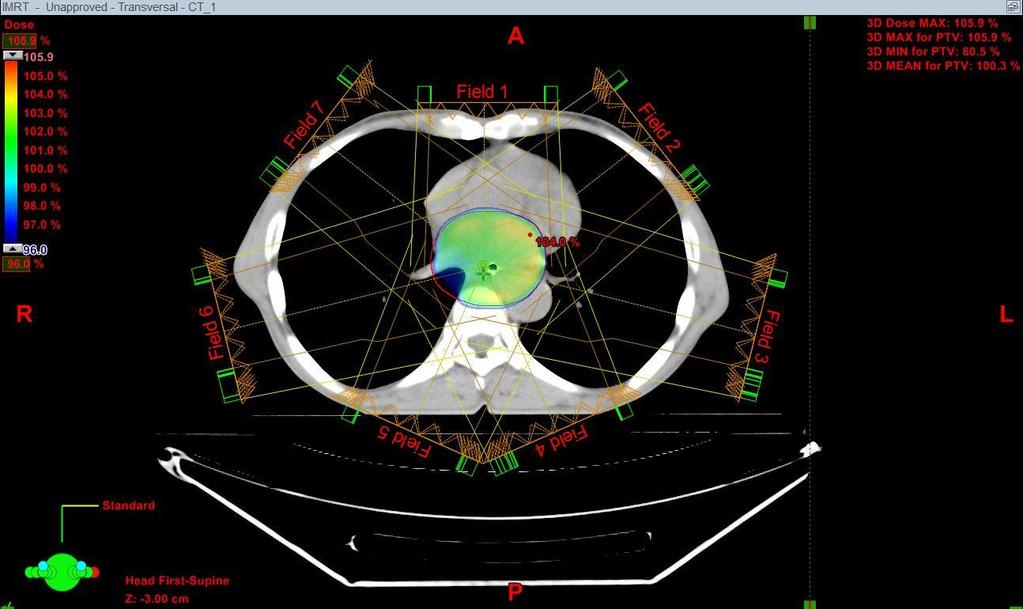
64 Dose Prescription and Planning technique: Treatment planning was done with the help of Varian treatment planning systems. Dose calculations were done with the help of Analytical Anisotropic Algorithm(AAA). Then planning was done for both hybrid conformal and IMRT techniques. Hybrid conformal technique consists of static beams that delivers half the dose of radiation with the help of two opposing Anteroposterior-Posteroanterior fields, followed by conformal technique which delivers the remaining dose. Approximately, the two opposing fields were given upto a total dose of 30 Gy followed by conformal technique which delivers the remaining 24 Gy with the same PTV thereby totalling to a dose of about 54 Gy. Literature reviews indicate a treatment dose of about Gy. But based on our Institute protocol, treatment were delivered to a total dose of 54 Gy. Daily doses were given with 200cGy per fraction per day. Anteroposterior and posteroanterior fields, as the name suggests, were treated with two opposing fields, followed by conformal technique. Conformal technique were delivered with the help of four field technique, with minimization of lung volumes. Comparing to this, an all-imrt plan was made with the help of seven to eight fields, thereby achieving the target volume, minimizing the doses to normal tissues. IMRT beams were oriented in such a 62


65 way that the beams are non opposed and at different angles, thereby avoiding the spinal cord. It is illustrated below. 2-OPPOSING AP/PA 63


66 CONFORMAL TECHNIQUE WITH THREE OF FOUR FIELDS 64

67 IMRT 65
68 Dose volume histograms are then calculated for both the planning target volume (PTV) and organ at risk. The MLD, volume of lung receiving 20 Gy (V20) for both lungs and mean dose to heart are calculated. It is compared with Hybrid Conformal technique and assessed. V30 for heart is also noted. Patients then are treated with two opposing anteroposterior and posteroanterior fields,upto a total dose of 30 Gy followed by conformal technique upto a TD of 54 Gy. Those patients who receive concurrent chemotherapy are treated with three weekly cisplatin with a dose of 70mg/m2 or weekly cisplatin with a dose of 40mg/m2. Patients are carefully noted for side effects of radiation like esophagitis, mucositis, pneumonitis, chest pain,dyspnea or bone marrow suppression. After completion of treatment, patients again are subjected to pulmonary function testing and compared with the initial values. Patient Outcomes: Patients are advised to review after six weeks initially and then followed up at monthly intervals. 66
69 6.RESULTS: Patient characteristics: Among the twenty patients treated, twelve patients were males and eight patients were females. Out of them, twelve patients were carcinomas of middle thoracic esophagus, six involve upper thoracic esophagus and two belong to lower thoracic esophagus without involving gastroesophageal junction. All these patients underwent Hybrid conformal technique and were also planned for all-imrt plans. Patients also underwent pulmonary function testing both before and after treatment. Among the twenty patients, thirteen patients are treated only with radiation therapy and seven patients received concurrent chemoradiation. Out of the chemotherapy patients, four patients received three weekly cisplatin and three patients received weekly cisplatin due to their biological tolerance. Dose volume histogram of these twenty patients were analyzed extensively and found that dose distribution attained by Hybrid conformal technique was not at all inferior when compared to that of IMRT plans. IMRT, being a modern superior technique achieved a uniformal dose distribution for all these patients, confined to the planning target volume. On the other hand, in Hybrid conformal technique, dose distribution achieved in planning target 67
70 volume was not inferior to that of IMRT plans, achieving the same dose without compromising the tumour volume. There was no incidence of pneumonitis in any of these patients. The V20 calculated were less than 30% in all these patients. The lung dose was calculated by taking into account the combination of both right and left lungs. Both Hybrid conformal and IMRT plans were able to achieve a V20 value of less than 30%. Also, the mean lung dose for both Hybrid conformal and IMRT techniques were less than 20 Gy. But noting the difference in both these techniques, Hybrid conformal technique achieved a much lesser lung dose compared to the all-imrt plan in both V20 as well as mean lung dose. It showed that both V20 and mean lung dose achieved in Hybrid conformal technique was atleast 5% lesser in V20, and atleast 1 to 2 Gy lesser than IMRT plans. No of Patients V20 Hybrid conformal(%) V20 IMRT(%)
71 No of Patients MLD Hybrid conformal(gy) MLD IMRT (Gy)
72
73 The cord dose achieved in IMRT is much lesser than than the usual constraint of 45Gy point dose. Similarly, the cord dose achieved in Hybrid conformal technique was much higher than that of IMRT, but still being achieved with in a dose of 45Gy. No of Patients Cord dose Hybrid conformal(gy) Cord dose IMRT (Gy)
74 The other main organ at risk in the treatment field is heart, which usually is in the plane of treatment. The mean dose to heart for these twenty patients under study were less than 30 Gy. The V30 calculated for the heart is less than the usual constraint of 46Gy. No of Patients Heart mean dose Hybrid conformal (Gy) Heart mean dose IMRT (Gy)
75 Pulmonary function testing was done both before and after treatment. The twenty patients under consideration had no abnormal deviation of pulmonary function testing, except for one patient, who had restrictive pattern even before treatment which has not worsened during the course of treatment. Patient had the same restrictive pattern even before and after treatment. Summarizing these, the Dose volume histograms for both Hybrid conformal and IMRT fields are as follows: 73


76 DOSE VOLUME HISTOGRAMS 74


77 75
Evaluation of Whole-Field and Split-Field Intensity Modulation Radiation Therapy (IMRT) Techniques in Head and Neck Cancer
 Techniques in Head and Neck Cancer") 1 Charles Poole April Case Study April 30, 2012 Evaluation of Whole-Field and Split-Field Intensity Modulation Radiation Therapy (IMRT) Techniques in Head and Neck Cancer Abstract: Introduction: This study
1 Charles Poole April Case Study April 30, 2012 Evaluation of Whole-Field and Split-Field Intensity Modulation Radiation Therapy (IMRT) Techniques in Head and Neck Cancer Abstract: Introduction: This study
The objective of this lecture is to integrate our knowledge of the differences between 2D and 3D planning and apply the same to various clinical
 The objective of this lecture is to integrate our knowledge of the differences between 2D and 3D planning and apply the same to various clinical sites. The final aim will be to be able to make out these
The Physics of Oesophageal Cancer Radiotherapy
 The Physics of Oesophageal Cancer Radiotherapy Dr. Philip Wai Radiotherapy Physics Royal Marsden Hospital 1 Contents Brief clinical introduction Imaging and Target definition Dose prescription & patient
First, how does radiation work?
 Hello, I am Prajnan Das, Faculty Member in the Department of Radiation Oncology at The University of Texas MD Anderson Cancer Center. We are going to talk today about some of the basic principles regarding
IMRT - the physician s eye-view. Cinzia Iotti Department of Radiation Oncology S.Maria Nuova Hospital Reggio Emilia
 IMRT - the physician s eye-view Cinzia Iotti Department of Radiation Oncology S.Maria Nuova Hospital Reggio Emilia The goals of cancer therapy Local control Survival Functional status Quality of life Causes
Treatment Planning Evaluation of Volumetric Modulated Arc Therapy (VMAT) for Craniospinal Irradiation (CSI)
 for Craniospinal Irradiation (CSI)") Treatment Planning Evaluation of Volumetric Modulated Arc Therapy (VMAT) for Craniospinal Irradiation (CSI) Tagreed AL-ALAWI Medical Physicist King Abdullah Medical City- Jeddah Aim 1. Simplify and standardize
Treatment Planning Evaluation of Volumetric Modulated Arc Therapy (VMAT) for Craniospinal Irradiation (CSI) Tagreed AL-ALAWI Medical Physicist King Abdullah Medical City- Jeddah Aim 1. Simplify and standardize
The role of chemoradiotherapy in GE junction and gastric cancer. Karin Haustermans
 The role of chemoradiotherapy in GE junction and gastric cancer Karin Haustermans Overview Postoperative chemoradiotherapy Preoperative chemoradiotherapy Palliative radiation Technical aspects Overview
Case Scenario 1. The patient has now completed his neoadjuvant chemoradiation and has been cleared for surgery.
 Case Scenario 1 July 10, 2010 A 67-year-old male with squamous cell carcinoma of the mid thoracic esophagus presents for surgical resection. The patient has completed preoperative chemoradiation. This
Radiotherapy Planning (Contouring Lung Cancer for Radiotherapy dose prescription) Dr Raj K Shrimali
 Dr Raj K Shrimali") Radiotherapy Planning (Contouring Lung Cancer for Radiotherapy dose prescription) Dr Raj K Shrimali Let us keep this simple and stick to some basic rules Patient positioning Must be reproducible Must be
Radiotherapy Planning (Contouring Lung Cancer for Radiotherapy dose prescription) Dr Raj K Shrimali Let us keep this simple and stick to some basic rules Patient positioning Must be reproducible Must be
Controversies in management of squamous esophageal cancer
 2015.06.12 12.47.48 Page 4(1) IS-1 Controversies in management of squamous esophageal cancer C S Pramesh Thoracic Surgery, Department of Surgical Oncology, Tata Memorial Centre, India In Asia, squamous
Head and Neck Service
 Head and Neck Service University of California, San Francisco, Department of Radiation Oncology Residency Training Program Head and Neck and Thoracic Service Educational Objectives for PGY-5 Residents
REVISITING ICRU VOLUME DEFINITIONS. Eduardo Rosenblatt Vienna, Austria
 REVISITING ICRU VOLUME DEFINITIONS Eduardo Rosenblatt Vienna, Austria Objective: To introduce target volumes and organ at risk concepts as defined by ICRU. 3D-CRT is the standard There was a need for a
Case Scenario year-old white male presented to personal physician with dyspepsia with reflux.
 Case Scenario 1 57-year-old white male presented to personal physician with dyspepsia with reflux. 7/12 EGD: In the gastroesophageal junction we found an exophytic tumor. The tumor occupies approximately
Efficient SIB-IMRT planning of head & neck patients with Pinnacle 3 -DMPO
 Investigations and research Efficient SIB-IMRT planning of head & neck patients with Pinnacle 3 -DMPO M. Kunze-Busch P. van Kollenburg Department of Radiation Oncology, Radboud University Nijmegen Medical
Radiotherapy for Gastric Cancer. Nitin Ohri, M.D. Montefiore Medical Center Albert Einstein College of Medicine 12/15/2012
 Radiotherapy for Gastric Cancer Nitin Ohri, M.D. Montefiore Medical Center Albert Einstein College of Medicine 12/15/2012 Disclosures I have no conflicts of interest to disclose. Outline Background Treatment
肺癌放射治療新進展 Recent Advance in Radiation Oncology in Lung Cancer 許峰銘成佳憲國立台灣大學醫學院附設醫院腫瘤醫學部
 肺癌放射治療新進展 Recent Advance in Radiation Oncology in Lung Cancer 許峰銘成佳憲國立台灣大學醫學院附設醫院腫瘤醫學部 Outline Current status of radiation oncology in lung cancer Focused on stage III non-small cell lung cancer Radiation
Defining Target Volumes and Organs at Risk: a common language
 Defining Target Volumes and Organs at Risk: a common language Eduardo Rosenblatt Section Head Applied Radiation Biology and Radiotherapy (ARBR) Section Division of Human Health IAEA Objective: To introduce
9.5. CONVENTIONAL RADIOTHERAPY TECHNIQUE FOR TREATING THYROID CANCER
 9.5. CONVENTIONAL RADIOTHERAPY TECHNIQUE FOR TREATING THYROID CANCER ROBERT J. AMDUR, MD, SIYONG KIM, PhD, JONATHAN GANG LI, PhD, CHIRAY LIU, PhD, WILLIAM M. MENDENHALL, MD, AND ERNEST L. MAZZAFERRI, MD,
Evaluation of Monaco treatment planning system for hypofractionated stereotactic volumetric arc radiotherapy of multiple brain metastases
 Evaluation of Monaco treatment planning system for hypofractionated stereotactic volumetric arc radiotherapy of multiple brain metastases CASE STUDY Institution: Odette Cancer Centre Location: Sunnybrook
BLADDER RADIOTHERAPY PLANNING DOCUMENT
 A 2X2 FACTORIAL RANDOMISED PHASE III STUDY COMPARING STANDARD VERSUS REDUCED VOLUME RADIOTHERAPY WITH AND WITHOUT SYNCHRONOUS CHEMOTHERAPY IN MUSCLE INVASIVE BLADDER CANCER (ISRCTN 68324339) BLADDER RADIOTHERAPY
Medicinae Doctoris. One university. Many futures.
 Medicinae Doctoris The Before and The After: Can chemotherapy revise the trajectory of gastric and esophageal cancers? Dr. David Dawe MD, FRCPC Medical Oncologist Assistant Professor Disclosures None All
NEOADJUVANT THERAPY IN CARCINOMA STOMACH. Dr Jyotirup Goswami Consultant Radiation Oncologist Narayana Superspeciality Hospital, Howrah
 NEOADJUVANT THERAPY IN CARCINOMA STOMACH Dr Jyotirup Goswami Consultant Radiation Oncologist Narayana Superspeciality Hospital, Howrah NEOADJUVANT THERAPY?! Few believers Limited evidence Many surgeons
Sarcoma and Radiation Therapy. Gabrielle M Kane MB BCh EdD FRCPC Muir Professorship in Radiation Oncology University of Washington
 Sarcoma and Radiation Therapy Gabrielle M Kane MB BCh EdD FRCPC Muir Professorship in Radiation Oncology University of Washington Objective: Helping you make informed decisions Introduction Process Radiation
Specification of Tumor Dose. Prescription dose. Purpose
 Specification of Tumor Dose George Starkschall, Ph.D. Department of Radiation Physics U.T. M.D. Anderson Cancer Center Prescription dose What do we mean by a dose prescription of 63 Gy? Isocenter dose
Corporate Medical Policy
 Corporate Medical Policy Intensity Modulated Radiation Therapy (IMRT) of Abdomen and File Name: Origination: Last CAP Review: Next CAP Review: Last Review: intensity_modulated_radiation_therapy_imrt_of_abdomen_and_pelvis
A Comparison of IMRT and VMAT Technique for the Treatment of Rectal Cancer
 A Comparison of IMRT and VMAT Technique for the Treatment of Rectal Cancer Tony Kin Ming Lam Radiation Planner Dr Patricia Lindsay, Radiation Physicist Dr John Kim, Radiation Oncologist Dr Kim Ann Ung,
Corporate Medical Policy
 Corporate Medical Policy Intensity Modulated Radiation Therapy (IMRT) of Head and Neck File Name: Origination: Last CAP Review: Next CAP Review: Last Review: intensity_modulated_radiation_therapy_imrt_of_head_and_neck
SETTING Fudan University Shanghai Cancer Center. RESPONSIBLE PARTY Haiquan Chen MD.
 OFFICIAL TITLE A Phase Ⅲ Study of Left Side Thoracotomy Approach (SweetProcedure) Versus Right Side Thoracotomy Plus Midline Laparotomy Approach (Ivor-Lewis Procedure) Esophagectomy in Middle or Lower
Goals and Objectives: Head and Neck Cancer Service Department of Radiation Oncology
 Goals and Objectives: Head and Neck Cancer Service Department of Radiation Oncology The head and neck cancer service provides training in the diagnosis, management, treatment, and follow-up care of head
PRINCESS MARGARET CANCER CENTRE CLINICAL PRACTICE GUIDELINES GYNECOLOGIC CANCER CERVIX
 PRINCESS MARGARET CANCER CENTRE CLINICAL PRACTICE GUIDELINES GYNECOLOGIC CANCER CERVIX Site Group: Gynecology Cervix Author: Dr. Stephane Laframboise 1. INTRODUCTION 3 2. PREVENTION 3 3. SCREENING AND
A Dosimetric Comparison of Whole-Lung Treatment Techniques. in the Pediatric Population
 A Dosimetric Comparison of Whole-Lung Treatment Techniques in the Pediatric Population Corresponding Author: Christina L. Bosarge, B.S., R.T. (R) (T) Indiana University School of Medicine Department of
Aytul OZGEN 1, *, Mutlu HAYRAN 2 and Fatih KAHRAMAN 3 INTRODUCTION
 Journal of Radiation Research, 2012, 53, 916 922 doi: 10.1093/jrr/rrs056 Advance Access Publication 21 August 2012 Mean esophageal radiation dose is predictive of the grade of acute esophagitis in lung
biij Initial experience in treating lung cancer with helical tomotherapy
 Available online at http://www.biij.org/2007/1/e2 doi: 10.2349/biij.3.1.e2 biij Biomedical Imaging and Intervention Journal CASE REPORT Initial experience in treating lung cancer with helical tomotherapy
Dosimetric Analysis of 3DCRT or IMRT with Vaginal-cuff Brachytherapy (VCB) for Gynaecological Cancer
 for Gynaecological Cancer") Dosimetric Analysis of 3DCRT or IMRT with Vaginal-cuff Brachytherapy (VCB) for Gynaecological Cancer Tan Chek Wee 15 06 2016 National University Cancer Institute, Singapore Clinical Care Education Research
Dosimetric Analysis of 3DCRT or IMRT with Vaginal-cuff Brachytherapy (VCB) for Gynaecological Cancer Tan Chek Wee 15 06 2016 National University Cancer Institute, Singapore Clinical Care Education Research
NCCN GUIDELINES ON PROTON THERAPY (AS OF 4/23/18) BONE (Version , 03/28/18)
 BONE (Version , 03/28/18)") BONE (Version 2.2018, 03/28/18) NCCN GUIDELINES ON PROTON THERAPY (AS OF 4/23/18) Radiation Therapy Specialized techniques such as intensity-modulated RT (IMRT); particle beam RT with protons, carbon ions,
BONE (Version 2.2018, 03/28/18) NCCN GUIDELINES ON PROTON THERAPY (AS OF 4/23/18) Radiation Therapy Specialized techniques such as intensity-modulated RT (IMRT); particle beam RT with protons, carbon ions,
A TREATMENT PLANNING STUDY COMPARING VMAT WITH 3D CONFORMAL RADIOTHERAPY FOR PROSTATE CANCER USING PINNACLE PLANNING SYSTEM *
 Romanian Reports in Physics, Vol. 66, No. 2, P. 394 400, 2014 A TREATMENT PLANNING STUDY COMPARING VMAT WITH 3D CONFORMAL RADIOTHERAPY FOR PROSTATE CANCER USING PINNACLE PLANNING SYSTEM * D. ADAM 1,2,
Dose escalation for NSCLC using conformal RT: 3D and IMRT. Hasan Murshed
 Dose escalation for NSCLC using conformal RT: 3D and IMRT. Hasan Murshed Take home message Preliminary data shows CRT technique in NSCLC allows dose escalation to an unprecedented level maintaining cancer
Protocol of Radiotherapy for Small Cell Lung Cancer
 107 年 12 月修訂 Protocol of Radiotherapy for Small Cell Lung Cancer Indication of radiotherapy Limited stage: AJCC (8th edition) stage I-III (T any, N any, M0) that can be safely treated with definitive RT
Combined modality treatment for N2 disease
 Combined modality treatment for N2 disease Dr Clara Chan Consultant in Clinical Oncology 3 rd March 2017 Overview Background The evidence base Systemic treatment Radiotherapy Future directions/clinical
Evaluation of Three-dimensional Conformal Radiotherapy and Intensity Modulated Radiotherapy Techniques in High-Grade Gliomas
 1 Carol Boyd Comprehensive Case Study July 11, 2013 Evaluation of Three-dimensional Conformal Radiotherapy and Intensity Modulated Radiotherapy Techniques in High-Grade Gliomas Abstract: Introduction:
ARROCase: Borderline Resectable Pancreatic Cancer
 ARROCase: Borderline Resectable Pancreatic Cancer Resident: Jordan Kharofa, MD Staff: Beth Erickson, MD 8/2012 Medical College of Wisconsin Department of Radiation Oncology Case Presentation: 60 year old
Page 1. Helical (Spiral) Tomotherapy. UW Helical Tomotherapy Unit. Helical (Spiral) Tomotherapy. MVCT of an Anesthetized Dog with a Sinus Tumor
 Tomotherapy. UW Helical Tomotherapy Unit. Helical (Spiral) Tomotherapy. MVCT of an Anesthetized Dog with a Sinus Tumor") Helical (Spiral) Tomotherapy Novel Clinical Applications of IMRT Linac Ring Gantry CT Detector X-Ray Fan Beam Binary Multileaf Collimator Binary MLC Leaves James S Welsh, MS, MD Department of Human Oncology
Helical (Spiral) Tomotherapy Novel Clinical Applications of IMRT Linac Ring Gantry CT Detector X-Ray Fan Beam Binary Multileaf Collimator Binary MLC Leaves James S Welsh, MS, MD Department of Human Oncology
Locally advanced disease & challenges in management
 Gynecologic Cancer InterGroup Cervix Cancer Research Network Cervix Cancer Education Symposium, February 2018 Locally advanced disease & challenges in management Carien Creutzberg Radiation Oncology, Leiden
CURRICULUM OUTLINE FOR TRANSITIONING FROM 2-D RT TO 3-D CRT AND IMRT
 CURRICULUM OUTLINE FOR TRANSITIONING FROM 2-D RT TO 3-D CRT AND IMRT Purpose The purpose of this curriculum outline is to provide a framework for multidisciplinary training for radiation oncologists, medical
Chapter 2. Level II lymph nodes and radiation-induced xerostomia
 Chapter 2 Level II lymph nodes and radiation-induced xerostomia This chapter has been published as: E. Astreinidou, H. Dehnad, C.H. Terhaard, and C.P Raaijmakers. 2004. Level II lymph nodes and radiation-induced
Outline. Contour quality control. Dosimetric impact of contouring errors and variability in Intensity Modulated Radiation Therapy (IMRT)
") Dosimetric impact of contouring errors and variability in Intensity Modulated Radiation Therapy (IMRT) James Kavanaugh, MS DABR Department of Radiation Oncology Division of Medical Physics Outline Importance
Dosimetric impact of contouring errors and variability in Intensity Modulated Radiation Therapy (IMRT) James Kavanaugh, MS DABR Department of Radiation Oncology Division of Medical Physics Outline Importance
The role of esophageal brachytherapy
 Esophageal cancer is combination with surgery (stents) or EBRT a better solution? Razvan Galalae, MD, PhD Associate Professor, Medical Faculty, Christian Albrecht University Kiel, Germany, and Head of
Intensity modulated radiotherapy (IMRT) for treatment of post-operative high grade glioma in the right parietal region of brain
 for treatment of post-operative high grade glioma in the right parietal region of brain") 1 Carol Boyd March Case Study March 11, 2013 Intensity modulated radiotherapy (IMRT) for treatment of post-operative high grade glioma in the right parietal region of brain History of Present Illness:
1 Carol Boyd March Case Study March 11, 2013 Intensity modulated radiotherapy (IMRT) for treatment of post-operative high grade glioma in the right parietal region of brain History of Present Illness:
Outcomes of definitive or preoperative IMRT chemoradiation for esophageal cancer
 J Radiat Oncol (2012) 1:347 354 DOI 10.1007/s13566-012-0048-5 ORIGINAL RESEARCH Outcomes of definitive or preoperative IMRT chemoradiation for esophageal cancer Ravi Shridhar & Michael D. Chuong & Jill
Aliu Sanni MD SUNY Downstate Medical Center August 16, 2012
 Aliu Sanni MD SUNY Downstate Medical Center August 16, 2012 Case Presentation 60yr old AAF with PMH of CAD s/p PCI 1983, CVA, GERD, HTN presented with retrosternal chest pain on 06/12 Associated dysphagia
Tracheal Adenocarcinoma Treated with Adjuvant Radiation: A Case Report and Literature Review
 Published online: May 23, 2013 1662 6575/13/0062 0280$38.00/0 This is an Open Access article licensed under the terms of the Creative Commons Attribution- NonCommercial-NoDerivs 3.0 License (www.karger.com/oa-license),
and Strength of Recommendations
 ASTRO with ASCO Qualifying Statements in Bold Italics s patients with T1-2, N0 non-small cell lung cancer who are medically operable? 1A: Patients with stage I NSCLC should be evaluated by a thoracic surgeon,
Clinical indications for positron emission tomography
 Clinical indications for positron emission tomography Oncology applications Brain and spinal cord Parotid Suspected tumour recurrence when anatomical imaging is difficult or equivocal and management will
Clinical experience with TomoDirect System Tangential Mode
 Breast Cancer Clinical experience with TomoDirect System Tangential Mode European Institute of Oncology Milan, Italy Disclosure & Disclaimer An honorarium is provided by Accuray for this presentation The
Potential benefits of intensity-modulated proton therapy in head and neck cancer van de Water, Tara Arpana
 University of Groningen Potential benefits of intensity-modulated proton therapy in head and neck cancer van de Water, Tara Arpana IMPORTANT NOTE: You are advised to consult the publisher's version (publisher's
3D Conformal Radiation Therapy for Mucinous Carcinoma of the Breast
 1 Angela Kempen February Case Study February 22, 2012 3D Conformal Radiation Therapy for Mucinous Carcinoma of the Breast History of Present Illness: JE is a 45 year-old Caucasian female who underwent
TOMOTERAPIA in Italia: Esperienze a confronto
 TOMOTERAPIA in Italia: Esperienze a confronto BARD 20 novembre 2010 L esperienza di Reggio Emilia Testa collo Alessandro Muraglia Reasons for the use of tomotherapy: - Complex tumor geometry and proximity
Quality assurance and credentialing requirements for sites using inverse planned IMRT Techniques
 TROG 08.03 RAVES Quality assurance and credentialing requirements for sites using inverse planned IMRT Techniques Introduction Commissioning and quality assurance of planning systems and treatment delivery
Appendix 1: Regional Lymph Node Stations for Staging Esophageal Cancer
 Appendix 1: Regional Lymph Node Stations for Staging Esophageal Cancer Locoregional (N stage) disease was redefined in the seventh edition of the AJCC Cancer Staging Manual as any periesophageal lymph
Additional Questions for Review 2D & 3D
 Additional Questions for Review 2D & 3D 1. For a 4-field box technique, which of the following will deliver the lowest dose to the femoral heads? a. 100 SSD, equal dmax dose to all fields b. 100 SSD, equal
THORACIC MALIGNANCIES
 THORACIC MALIGNANCIES Summary for Malignant Malignancies. Lung Ca 1 Lung Cancer Non-Small Cell Lung Cancer Diagnostic Evaluation for Non-Small Lung Cancer 1. History and Physical examination. 2. CBCDE,
Protocol of Radiotherapy for Breast Cancer
 107 年 12 月修訂 Protocol of Radiotherapy for Breast Cancer Indication of radiotherapy Indications for Post-Mastectomy Radiotherapy (1) Axillary lymph node 4 positive (2) Axillary lymph node 1-3 positive:
A VMAT PLANNING SOLUTION FOR NECK CANCER PATIENTS USING THE PINNACLE 3 PLANNING SYSTEM *
 Romanian Reports in Physics, Vol. 66, No. 2, P. 401 410, 2014 A VMAT PLANNING SOLUTION FOR NECK CANCER PATIENTS USING THE PINNACLE 3 PLANNING SYSTEM * M. D. SUDITU 1,2, D. ADAM 1,2, R. POPA 1,2, V. CIOCALTEI
THE TRANSITION FROM 2D TO 3D AND TO IMRT - RATIONALE AND CRITICAL ELEMENTS
 THE TRANSITION FROM 2D TO 3D AND TO IMRT - RATIONALE AND CRITICAL ELEMENTS ICTP SCHOOL ON MEDICAL PHYSICS FOR RADIATION THERAPY DOSIMETRY AND TREATMENT PLANNING FOR BASIC AND ADVANCED APPLICATIONS March
Abstracting Upper GI Cancer Incidence and Treatment Data Quiz 1 Multiple Primary and Histologies Case 1 Final Pathology:
 Abstracting Upper GI Cancer Incidence and Treatment Data Quiz 1 Multiple Primary and Histologies Case 1 A 74 year old male with a history of GERD presents complaining of dysphagia. An esophagogastroduodenoscopy
Research and Reviews Journal of Medical and Clinical Oncology
 Comparison and Prognostic Analysis of Elective Nodal Irradiation Using Definitive Radiotherapy versus Chemoradiotherapy for Treatment of Esophageal Cancer Keita M 1,2, Zhang Xueyuan 1, Deng Wenzhao 1,
L impatto dell imaging sulla definizione della strategia terapeutica
 GISCoR L impatto dell imaging sulla definizione della strategia terapeutica M. Galeandro U.C. Radioterapia Oncologica ASMN-IRCCS Reggio Emilia 14 Novembre 2014 Rectal Cancer TNM AJCC-7 th edition 2010
Radiation Oncology MOC Study Guide
 Radiation Oncology MOC Study Guide The following study guide is intended to give a general overview of the type of material that will be covered on the Radiation Oncology Maintenance of Certification (MOC)
A dosimetric comparison of proton and photon therapy in unresectable cancers of the head of pancreas
 A dosimetric comparison of proton and photon therapy in unresectable cancers of the head of pancreas Reid F. Thompson University of Pennsylvania, Philadelphia, Pennsylvania 1914 Sonal U. Mayekar Thomas
Minimally Invasive Esophagectomy- Valuable. Jayer Chung, MD University of Colorado Health Sciences Center December 11, 2006
 Minimally Invasive Esophagectomy- Valuable Jayer Chung, MD University of Colorado Health Sciences Center December 11, 2006 Overview Esophageal carcinoma What is minimally invasive esophagectomy (MIE)?
Therapeutic ratio - An Overview. Past Present Future Prof Ramesh S Bilimaga
 Therapeutic ratio - An Overview Past Present Future Prof Ramesh S Bilimaga Radiation Oncology Discipline of human medicine concerned with the generation, conservation and dissemination of knowledge concerning
Imaging in gastric cancer
 Imaging in gastric cancer Gastric cancer remains a deadly disease because of late diagnosis. Adenocarcinoma represents 90% of malignant tumors. Diagnosis is based on endoscopic examination with biopsies.
Lung Spine Phantom. Guidelines for Planning and Irradiating the IROC Spine Phantom. MARCH 2014
 Lung Spine Phantom Guidelines for Planning and Irradiating the IROC Spine Phantom. MARCH 2014 The study groups are requesting that each institution keep the phantom for no more than 2 week. During this
Radiotherapy Physics and Equipment
 Radiological Sciences Department Radiotherapy Physics and Equipment RAD 481 Lecture s Title: Introduction Dr. Mohammed EMAM Ph.D., Paris-Sud 11 University Vision :IMC aspires to be a leader in applied
Esophageal cancer (EC) is the eighth most common cancer worldwide and the sixth most common cause of cancer-related mortality (Kamangar et al.
 is the eighth most common cancer worldwide and the sixth most common cause of cancer-related mortality (Kamangar et al.") Arch. Biol. Sci., Belgrade, 65 (3), 821-827, 2013 DOI:10.2298/ABS1303821L DETERMINING THE LYMPH NODE CLINICAL TARGET VOLUME OF UPPER ESOPHAGEAL CARCINOMA WITH COMPUTED TOMOGRAPHY MINGHUAN LI 1, YUHUI LIU
Arch. Biol. Sci., Belgrade, 65 (3), 821-827, 2013 DOI:10.2298/ABS1303821L DETERMINING THE LYMPH NODE CLINICAL TARGET VOLUME OF UPPER ESOPHAGEAL CARCINOMA WITH COMPUTED TOMOGRAPHY MINGHUAN LI 1, YUHUI LIU
Dose prescription, reporting and recording in intensity-modulated radiation therapy: a digest of the ICRU Report 83
 Special report Dose prescription, reporting and recording in intensity-modulated radiation therapy: a digest of the ICRU Report 83 Rapid development in imaging techniques, including functional imaging,
SBRT TREATMENT PLANNING: TIPS + TRICKS. Rachel A. Hackett CMD, RT(T)
") SBRT TREATMENT PLANNING: TIPS + TRICKS Rachel A. Hackett CMD, RT(T) OUTLINE Brief radiobiology review 3D CRT Tx Planning VMAT Tx Planning Protocols Other Sites Oligomets Spine Liver Kidney Adrenal Gland
SBRT TREATMENT PLANNING: TIPS + TRICKS Rachel A. Hackett CMD, RT(T) OUTLINE Brief radiobiology review 3D CRT Tx Planning VMAT Tx Planning Protocols Other Sites Oligomets Spine Liver Kidney Adrenal Gland
ASTRO econtouring for Lymphoma. Stephanie Terezakis, MD
 ASTRO econtouring for Lymphoma Stephanie Terezakis, MD Disclosures No conflicts to disclose 1970 Total Lymphoid Irradiation (TLI) 1995 Involved-Field Radiotherapy (IFRT) 2008 Involved Node Radiotherapy
Original Article. Teyyiba Kanwal, Muhammad Khalid, Syed Ijaz Hussain Shah, Khawar Nadeem
 Original Article Treatment Planning Evaluation of Sliding Window and Multiple Static Segments Technique in Intensity Modulated Radiotherapy for Different Beam Directions Teyyiba Kanwal, Muhammad Khalid,
Adenocarcinoma of gastro-esophageal junction - Case report
 Case Report denocarcinoma of gastro-esophageal junction - Case report nupsingh Dhakre 1*, Ibethoi Yengkhom 2, Harshin Nagori 1, nup Kurele 1, Shreedevi. Patel 3 1 2 nd year Resident, 2 3rd year Resident,
Changing Paradigms in Radiotherapy
 Changing Paradigms in Radiotherapy Marco van Vulpen, MD, PhD Mouldroomdag-2015 Towards the elimination of invasion 1 NIH opinion on the future of oncology Twenty-five years from now,i hope that we won
IROC Liver Phantom. Guidelines for Planning and Irradiating the IROC Liver Phantom. Revised July 2015
 IROC Liver Phantom Guidelines for Planning and Irradiating the IROC Liver Phantom. Revised July 2015 The study groups are requests that each institution keep the phantom for no more than 2 weeks. During
MANAGEMENT OF CA HYPOPHARYNX
 MANAGEMENT OF CA HYPOPHARYNX GENERAL TREATMENT RECOMMENDATIONS BASED ON HYPOPHARYNX TUMOR STAGE For patients presenting with early-stage definitive radiotherapy alone or voice-preserving surgery are viable
Radiotherapy physics & Equipments
 Radiotherapy physics & Equipments RAD 481 Lecture s Title: An Overview of Radiation Therapy for Health Care Professionals Dr. Mohammed Emam Vision :IMC aspires to be a leader in applied medical sciences,
JOURNAL OF APPLIED CLINICAL MEDICAL PHYSICS, VOLUME 6, NUMBER 2, SPRING 2005
 JOURNAL OF APPLIED CLINICAL MEDICAL PHYSICS, VOLUME 6, NUMBER 2, SPRING 2005 Advantages of inflatable multichannel endorectal applicator in the neo-adjuvant treatment of patients with locally advanced
Treatment Planning for Breast Cancer: Contouring Targets. Julia White MD Professor
 Treatment Planning for Breast Cancer: Contouring Targets Julia White MD Professor Outline 1. RTOG Breast Cancer Atlas 2. Target development on Clinical Trials Whole Breast Irradiation 2-D Radiotherapy
Esophageal cancer: Biology, natural history, staging and therapeutic options
 EGEUS 2nd Meeting Esophageal cancer: Biology, natural history, staging and therapeutic options Michael Bau Mortensen MD, Ph.D. Associate Professor of Surgery Centre for Surgical Ultrasound, Upper GI Section,
Treatment Planning & IGRT Credentialing for NRG SBRT Trials
 Treatment Planning & IGRT Credentialing for NRG SBRT Trials Hania Al Hallaq, Ph.D. Department of Radiation & Cellular Oncology The University of Chicago Learning Objectives Explain rationale behind credentialing
Esophageal Cancer Staging Essentials: The New TNM Staging System (7th edition) and Clinicoradiologic Implications
 and Clinicoradiologic Implications") Esophageal Cancer Staging Essentials: The New TNM Staging System (7th edition) and Clinicoradiologic Implications Poster No.: E-0060 Congress: ESTI 2012 Type: Scientific Exhibit Authors: K. Lee, T. J.
Esophageal Cancer Staging Essentials: The New TNM Staging System (7th edition) and Clinicoradiologic Implications Poster No.: E-0060 Congress: ESTI 2012 Type: Scientific Exhibit Authors: K. Lee, T. J.
Chapters from Clinical Oncology
 Chapters from Clinical Oncology Lecture notes University of Szeged Faculty of Medicine Department of Oncotherapy 2012. 1 RADIOTHERAPY Technical aspects Dr. Elemér Szil Introduction There are three possibilities
Head and Neck Cancer Treatment
 Scan for mobile link. Head and Neck Cancer Treatment Head and neck cancer overview The way a particular head and neck cancer behaves depends on the site in which it arises (the primary site). For example,
Allan Price NHS Lothian, Edinburgh, UK
 Allan Price NHS Lothian, Edinburgh, UK Radiotherapy Dose Volume Timing Technique PCI Surgery Systemic agents 1 Study Dose Time Induction CT Ann Arbor 65.1-75.6 Gy Duke 73.6-80 Gy RTOG 77.4 Gy 74 Gy 6.5-7.5
Molly Boyd, MD Glenn Mills, MD Syed Jafri, MD 1/1/2010
 LSU HEALTH SCIENCES CENTER NSCLC Guidelines Feist-Weiller Cancer Center Molly Boyd, MD Glenn Mills, MD Syed Jafri, MD 1/1/2010 Initial Evaluation/Intervention: 1. Pathology Review 2. History and Physical
Update on Limited Small Cell Lung Cancer. Laurie E Gaspar MD, MBA Prof/Chair Radiation Oncology University of Colorado Denver
 Update on Limited Small Cell Lung Cancer Laurie E Gaspar MD, MBA Prof/Chair Radiation Oncology University of Colorado Denver Objectives - Limited Radiation Dose Radiation Timing Radiation Volume PCI Neurotoxicity
UPDATE ON RADIOTHERAPY
 1 Miriam Kleiter UPDATE ON RADIOTHERAPY Department for Companion Animals and Horses, Plattform Radiooncology and Nuclear Medicine, University of Veterinary Medicine Vienna Introduction Radiotherapy has
External Beam Radiation Therapy for Thyroid Cancer
 External Beam Radiation Therapy for Thyroid Cancer C. Jillian Tsai, M.D, PH.D. Assistant Attending Director of Head and Neck Cancer Research Department of Radiation Oncology Memorial Sloan Kettering Cancer
is time consuming and expensive. An intra-operative assessment is not going to be helpful if there is no more tissue that can be taken to improve the
 My name is Barry Feig. I am a Professor of Surgical Oncology at The University of Texas MD Anderson Cancer Center in Houston, Texas. I am going to talk to you today about the role for surgery in the treatment
To Reduce Hot Dose Spots in Craniospinal Irradiation: An IMRT Approach with Matching Beam Divergence
 SCIENCE & TECHNOLOGY To Reduce Hot Dose Spots in Craniospinal Irradiation: An IMRT Approach with Matching Beam Divergence Alburuj R. Rahman*, Jian Z. Wang, Dr. Z. Huang, Dr. J. Montebello Department of
Ashley Pyfferoen, MS, CMD. Gundersen Health Systems La Crosse, WI
 Ashley Pyfferoen, MS, CMD Gundersen Health Systems La Crosse, WI 3 Radiation Oncologists 3 Physicists 2 Dosimetrists 9 Radiation Therapists o o o o o o o o o Brachial Plexus Anatomy Brachial Plexopathy
Head and Neck Service
 Head and Neck Service University of California, San Francisco, Department of Radiation Oncology Residency Training Program Head and Neck and Thoracic Service Educational Objectives for PGY-3 Residents