Perioperative management of esophageal cancer
|
|
|
- Dustin Barber
- 5 years ago
- Views:
Transcription
1 Perioperative management of esophageal cancer Lucas Goense
2
3 Perioperative management of esophageal cancer Lucas Goense
4 Perioperative management of esophageal cancer PhD thesis, Utrecht University, The Netherlands Lucas Goense, Utrecht, 2018 All rights reserved. No part of this thesis may be reproduced or transmitted in any form or by any means without prior written permission from the author. The copyright of the papers that have been published or have been accepted for publication has been transferred to the respective journals. For three projects (Chapter 3, 6 and 7) funding was provided in part by the René Vogels Foundation Cover: Lay-out: Joppe Klein Printing: ISBN:
5 Perioperative management of esophageal cancer (met een samenvatting in het Nederlands) Proefschrift ingevolge het besluit van het college voor promoties in het openbaar te verdedigen door Lucas Goense geboren op 2 April 1989 te Wageningen
6 Promotoren: Copromotoren: Dr. J.P. Ruurda
7
8 CONTENTS Chapter 1 General introduction and thesis outline Ann N Y Acad Sci Part 1. Staging diagnosing cervical lymph node metastases in patients with esophageal cancer Eur Radiol Chapter 3 Chapter 4 Chapter 5 Chapter 6 Prediction and diagnosis of interval metastasis after neoadjuvant Eur J Nucl Med Mol Imaging Staging and role of neoadjuvant chemoradiotherapy in clinical Eur J Surg Oncol Diagnostic performance of 18 tection of recurrent esophageal cancer after treatment with curative intent. a systematic review and meta-analysis J Nucl Med Preoperative nomogram to predict early disease recurrence after neoadjuvant chemoradiotherapy and surgery for esophageal adenocarcinoma Ann Surg Oncol in patients after neoadjuvant chemoradiotherapy and surgery for esophageal cancer Ann Thorac Surg Part 2. Treatment response prediction Nucl Med Commun are of complementary value in predicting pathologic response to neoadjuvant chemoradiotherapy for esophageal cancer Acta Oncologica during neoadjuvant treatment of esophageal cancer Br J Radiol
9 Part 3. Postoperative complication management Eur J Surg Oncol esophagectomy for cancer Br J Surg Chapter 14 Chapter 16 Chapter 19 Ann Thorac Surg Generalized cardiovascular disease on a preoperative CT-scan is Eur J Surg Oncol for cancer Dis Esophagus Perioperative chemotherapy versus neoadjuvant chemoradiotherapy outcome, and survival J Surg Oncol Ann Thorac Surg Br J Surg Diagnostic performance of a CT-based scoring system for diagno- subjective CT assessment Eur Radiol Chapter 20 Summary 356 Chapter 21 General discussion 366 Appendices 382 Summary in Dutch Nederlandse samenvatting Review committee List of publications Curriculum Vitae
10
11 1 General introduction esophageal cancer Lucas Goense Peter S.N. van Rossum Daniela Kandioler Jelle P. Ruurda Khean L. Goh Ann N Y Acad Sci. 2016;1381:50 65
12 Chapter 1 ESOPHAGEAL CANCER cancer-related death worldwide, with an estimated new cases and deaths annually 1. The two main histological subtypes of esophageal cancer are squamous cell carcinoma (SCC) and adenocarcinoma. Globally, esophageal squamous cell carcinoma is 2,3 in the last two decades there has been a dramatic increase in the incidence of esophageal 4. This ominous esophageal adenocarcinoma 5 6,7. the surrounding lymph nodes (lymphadenectomy) has been the cornerstone of treatment with curative intent for patients with early and locally advanced esophageal cancer. The relatively strategies during the last decades to improve survival 8 in staging and treatment techniques, patients with esophageal cancer can now be cured by various treatment strategies which requires a multidisciplinary approach. Neoadjuvant therapy with chemotherapy or chemoradiotherapy has supplemented surgery and is now a generally recommended treatment strategy for patients with locally advanced esophageal cancer 9,10. survival is well-recognized, the optimal treatment algorithm to achieve most optimal outcomes The currently available multimodality treatment strategies have been associated with an increase in morbidity resulting in a persistent reduction in health related quality of life, and yet still a relatively poor prognosis 11. Furthermore, while the average patient may respond can be attributed to high inter-patient variability in response to such treatment. Factors that 10
13 General introduction of disease, underlying physical conditions and social circumstances 12,13 single course of treatment that will meet the need of every patient. 1 staging (Part 1), treatment response prediction (Part 2), and in postoperative complication management (Part 3) of patients with esophageal cancer. STAGING Clinical staging gastrointestinal endoscopy with biopsy to verify diagnosis, endoscopic ultrasonography to obtain information on the depth of tumor invasion and loco-regional nodal disease, and loco-regional spread and distant metastases 14 to the 7 th 15. staging modalities is recommended in order to assess the involvement of cervical lymph node metastases 16,17 modality during the last decade has shown to be superior to conventional imaging modalities in the preoperative staging of esophageal cancer 18,19. Therefore, the additional diagnostic value may be limited. As such, it would be of interest to investigate the additional value of cervical Currently there is disagreement between national guidelines whether all patients should be 17,20,21. Detection of metastasis in this phase of treatment would probably prevent a futile attempt at curative esophagectomy. 11
14 Chapter 1 the diagnostic accuracy of this modality for the detection of interval metastasis remains Although a multimodality treatment approach is increasingly recommended for patients with strategy for patients with clinical T2N0 tumors. As T2N0 disease represents an anticipated early stage disease, a surgery alone approach may be regarded as appropriate treatment for these tumors 22,23 studies recommend a multimodality treatment approach for these patients 24,25 available studies on this topic are equivocal, it would be of interest to assess the current status of clinical staging and determine whether the addition of neoadjuvant chemoradiotherapy to surgery is associated with improved outcomes in a large population-based cohort of patients with clinical T2N0 esophageal tumors (Chapter 4). Detection of disease recurrence The relatively poor survival of esophageal cancer patients is partially attributable to the high 26 shown to possess the ability to detect recurrent esophageal cancer in a pre-symptomatic phase after treatment 27 develop recurrent disease are detected by the onset of symptoms between apparently normal surveillance scans 28. As such there is need for accurate prediction of disease recurrence after strategies and prompt earlier initiation of interventions to improve survival. TREATMENT RESPONSE PREDICTION Through tumor downsizing and downstaging, the use of neoadjuvant chemoradiotherapy improves locoregional control and overall survival rates compared to surgery alone 8,9 studies have reported that the degree of tumor regression in response to chemoradiotherapy is directly related to long-term survival, with pathologic complete responders having the most favorable long-term prognosis 29,30. Accurate prediction of pathologic complete responders before surgery would enable investigators to study the feasibility and outcome of an organ-preserving strategy (i.e. omission of surgery and close clinical follow-up) after 12
15 General introduction chemoradiotherapy 31 poor responders early during treatment would enable investigators to study the feasibility of research with the aim to tailor treatment to the individual patient. 1 Various opportunities for improvement in treatment response prediction are currently under microstructural characterization of tumors and visualization of treatment-induced changes of response to treatment in esophageal cancer predictive ability to accurately predict treatment response (Chapters 8 and 9). At the same to improve patient-friendliness of restaging procedures, it is necessary to evaluate the burden of these diagnostic procedures from the perspective of the patient (Chapter 10). POSTOPERATIVE COMPLICATION MANAGEMENT 35. Reported overall frequency rates of complications after being the most common complications 36, Although advances in surgical techniques and perioperative care have reduced the frequency of complications over the years, postoperative morbidity remains high 39 initiatives, complications that have the greatest overall impact on costs (Chapter 11) and patient 13
16 Chapter 1 Currently, accurate prediction of postoperative complications based on standard patient 40 in clinical practice it is necessary to assess its generalizability in an independent cohort for postoperative complications, it is of interest to assess whether the association between the prediction of postoperative complication include intraoperative and postoperative vital parameters (Chapter 15). chemoradiotherapy have demonstrated a survival advantage over surgery alone 8,9,41 after the introduction of neoadjuvant chemoradiotherapy in clinical practice, several hospitals reported a vast increase in their postoperative complication rate 42,43. Therefore, it is of interest neoadjuvant chemoradiotherapy (Chapter 16). Furthermore, it was hypothesized that radiation used for constructing the esophagogastric anastomosis 44 gastric fundus in radiation treatment planning, the potential association between radiation of the esophagogastric anastomosis (i.e. intrathoracic versus cervical) after esophagectomy is 45,46, while other studies did not 47,48 14
17 General introduction 49. Currently, CT to use in critically ill patients 50 1 RESEARCH OBJECTIVE PER CHAPTER Part 1. Staging diagnosing cervical lymph node metastases in patients with newly diagnosed esophageal cancer Chapter 3 metastasis after neoadjuvant chemoradiotherapy and to identify predictors of interval metastases Chapter 4 To evaluate current clinical staging and determine whether the addition of neoadjuvant chemoradiotherapy to surgery is associated with improved survival esophageal cancer after initial treatment with curative intent Chapter 6 To develop a preoperative prediction model for early recurrence after neoadjuvant chemoradiotherapy and surgery for esophageal adenocarcinoma neoadjuvant chemoradiotherapy and surgery for esophageal cancer Part 2. Treatment response prediction 15
18 Chapter 1 and after neoadjuvant chemoradiotherapy to predict pathologic response the patient Part 3. Postoperative complication management Chapter 11 To determine the economic burden of postoperative complications after esophagectomy Chapter 12 To identify which postoperative complications after esophagectomy have the greatest impact on clinical outcomes esophagectomy Chapter 15 To assess the relationship of intraoperative and postoperative vital parameters perioperative chemotherapy and neoadjuvant chemoradiotherapy followed by surgery for patients with esophageal adenocarcinoma followed by esophagectomy Chapter 18 To compare clinical outcome after esophagectomy in patients with an intrathoracic or cervical anastomosis, and to identify predictors of anastomotic 16
19 General introduction esophagectomy and compare it to subjective CT interpretation 1 17
20 Chapter 1 REFERENCES 108. incidence of oesophageal cancer by histological A global assessment of the oesophageal 14. cancer survival in United States adults from al. Survival after neoadjuvant chemotherapy or chemoradiotherapy for resectable oesophageal et al. Neoadjuvant chemoradiotherapy plus surgery versus surgery alone for oesophageal Perioperative chemotherapy versus surgery alone analysis shows clinically relevant and longlasting deterioration in health-related quality of life after esophageal cancer surgery. Qual Life characterization of oesophageal carcinoma Assessment of cervical lymph node metastasis in esophageal carcinoma using ultrasonography. oesofaguscarcinoom. Accessed 7th November reduced early recurrence or increased survival intravenous contrast-enhanced CT for evaluation of metastatic regional lymph nodes in patients with resectable early stage esophageal cancer. 18
21 General introduction 20. NCCN Clinical Practice Guidelines in Accessed January 1, Guidelines for diagnosis, treatment and follow- understaging is seen in clinically staged T2N0 esophageal cancer patients undergoing with suspected recurrence from squamous cancer recurrence patterns and implications for 29. Chirieac LR, Swisher SG, Ajani JA, et al. Posttherapy pathologic stage predicts survival in patients with esophageal carcinoma receiving preoperative chemoradiation. Cancer. Pathological complete response in patients with esophageal cancer after the trimodality al. Active surveillance in clinically complete responders after neoadjuvant chemoradiotherapy for esophageal or junctional cancer. Dis imaging for the prediction of pathologic response to neoadjuvant chemoradiotherapy in esophageal response assessment in patients with oesophageal cancer receiving neoadjuvant chemoradiotherapy. of Pathologic Complete Response to Preoperative and morbidity after resection for adenocarcinoma score-matched analysis of semiprone versus Ann Surg. 2017;
22 Chapter 1 discussion 222. Perioperative Chemotherapy versus Surgery Alone for Resectable Gastroesophageal Cancer. factors associated with clinical response and postoperative complications. Anticancer Res. Neoadjuvant chemoradiation may increase the after transthoracic esophagectomy. J Thorac Lewis esophagectomy in patients treated with neoadjuvant chemoradiation are related to analysis of the society of thoracic surgeons general thoracic database. Ann Thorac Surg. al. Cervical or thoracic anastomosis for 62. or thoracic anastomosis after esophageal prospective randomized trial comparing sutured in Japan. J Laparoendosc Adv Surg Tech A. Computed tomography versus watersoluble contrast swallow in the detection of 20
23 General introduction 1 21
24
25 Part 1 Staging
26
27 2 Cervical ultrasonography has no additional value over a negative PET/CT for diagnosing cervical lymph node metastases in patients with esophageal cancer Lucas Goense * * Peter.S.N. van Rossum Jelle P. Ruurda * European Radiology. 2018;28:
28 Chapter 2 ABSTRACT Objective To investigate the additional value of cervical ultrasonography over 18 diagnosing cervical lymph node metastases in patients with newly diagnosed esophageal cancer. Methods underwent both cervical ultrasonography and 18 the Netherlands. Retrospective clinical data analysis was performed to assess the diagnostic value of cervical ultrasonography and 18 node metastases. Fine needle aspiration or clinical follow-up was used as reference standard. Results of Conclusion Cervical ultrasonography has no additional diagnostic value to a negative integrated 18 F-FDG esophageal cancer. 26
29 Cervical lymph node imaging in esophageal cancer INTRODUCTION of cancer-related death worldwide 1. Surgical resection of the esophagus with en-bloc lymphadenectomy remains the cornerstone treatment with curative intent for patients with nonmetastatic esophageal cancer 2, 3. Currently, a multimodal treatment approach is increasingly over surgery alone for patients with resectable esophageal cancer Accurate staging of esophageal cancer is essential to select patients that are eligible for treatment with curative intent, and to identify patients with distant metastases to prevent a non-curative surgical procedure. Currently recommended staging techniques include F- 7 guidelines the use of cervical ultrasonography is recommended since this is considered an 7, 8 introduction of integrated 18 staging by providing both anatomical and metabolic information 9. Therefore, the additional role of cervical ultrasonography for the detection of cervical lymph node metastases in the current era of routine diagnostic 18 Accordingly, the aim of this study was to investigate the additional value of cervical ultrasonography over 18 patients with newly diagnosed esophageal cancer. METHODS Center Utrecht with newly diagnosed esophageal cancer, between January 2013 and January who were evaluated with both integrated 18 were included. The two investigations of interest were performed in random order. Cervical 18 F-FDG patients were collected to identify cervical metastases that were potentially undetected during clinical staging. Therewith, the composite reference standard of the current study included 27
30 Chapter 2 either positive cytopathology of the aspirated material from a suspicious cervical lymph node during initial staging, or the occurrence of a new cervical lymph node metastasis that were detected within 12 months of clinical follow-up after initial staging and proven malignant with cytology. Cervical lymph node metastases were group into levels corresponding with upper, (level V). After completion of staging all patients with a clinical stage T1N1-3 or T2-4aN0-3, Cervical ultrasonography with FNA loss of fatty hilum, [focal] low echogenicity, or an eccentric mass), and grouped nodes with Integrated F-FDG, and blood glucose intravenously administered 18 administration of All 18 non-physiological 18 Additional FNA was performed in case of suspicious lymph nodes. 28
31 Cervical lymph node imaging in esophageal cancer Follow-up ( 18 metastases, last visit at the outpatient clinic or death. The median follow-up of all eligible of follow-up of patients without lymph node metastases that were still alive during follow up was at least 12 months. 2 Statistical analysis (NPV) of 18 test, and PVV and NPV using the Chi-square test, between 18 ultrasonography, respectively. The additional value of cervical ultrasonography to 18 F-FDG 18 F- RESULTS because 18 both cervical ultrasonography and of the study period. These patients underwent a CT scan only. A total of 163 patients underwent both cervical ultrasonography and 18 patient and treatment-related characteristics. The overall incidence of patients with cervical 29
32 Chapter 2 Cervical ultrasonography and 18 F-FDG PET/CT non-suspected nodes Cervical ultrasonography and 18 During clinical follow-up 4 of these 121 initially non-suspected patients developed cervical respectively. Cervical ultrasonography and 18 F-FDG PET/CT suspected nodes Cervical ultrasonography and 18 two patients it appeared impossible to perform FNA due to small size and inaccessibility lymphadenectomy and had no signs of cervical metastases after 12 and 19 months of clinical Cervical ultrasonography suspected nodes nodes, while 18 had an inconclusive FNA. The patients with inconclusive FNA underwent surgery and had no signs of cervical metastases after 29 and 30 months of clinical follow-up, respectively. 18 F-FDG PET/CT suspected nodes 18 scan showed enlargement of the cervical lymph node 3 months after initial diagnostic chemoradiotherapy and died a few months after diagnosis of the cervical metastasis. The of clinical follow-up. 30
33 Cervical lymph node imaging in esophageal cancer Diagnostic performance The comparison of diagnostic performance between, 18 ultrasonography to detect malignant lymph nodes and subsequent subgroup analysis are 18 of not outperform TABLE 1. Characteristic n (%) 114 (70) Age (years)* 67.2 (±8.6) Tumor location 21 (13) 45 (28) Distal esophagus 74 (45) 23 (14) Adenocarcinoma 94 (58) Squamous cell carcinoma 66 (40) 3 (2) Clinical T-stage T1 16 (10) T2 25 (15) T3 102 (63) T4 18 (11) 2 (1) Clinical N-stage N0 28 (17) N1 59 (36) N2 42 (26) N3 34 (21) 31
34 Chapter 2 TABLE 1 (continued). Characteristic n (%) 143 (88) 20 (12) TABLE 2. Comparison of diagnostic parameters of 18 cluding subgroup analysis 18 F-FDG PET/CT Cervical ultrasonography P-value Squamous cell carcinoma
35 Cervical lymph node imaging in esophageal cancer 2 Figure and cervical ultrasonography between January 2014 and January


36 Chapter 2 Figure 2. A. tumor (arrow). B. Cervical ultrasonography image in the same patient, demonstrating a 10 mm cervical with FNA (arrow). DISCUSSION 18 with newly diagnosed esophageal cancer was evaluated. Results from the current cohort of patients demonstrated no additional value of cervical ultrasonography to integrated 18 F-FDG 34
37 Cervical lymph node imaging in esophageal cancer Several studies suggest that in case of cervical lymph node involvement an aggressive approach 10. node metastases is still considered as systemic disease. Therefore, it is argued that survival will are applied for patients with cervical lymph node metastases 11, 12 decisions, accurate diagnosis of cervical lymph node metastasis is crucial Therefore, the use of integrated 18 staging 13. Due to the improvement in esophageal cancer staging by 18 it has been suggested that the additional role of cervical ultrasonography for the detection of cervical lymph node metastasis may be limited 14 current cohort of patients in which no additional value of cervical ultrasonography was found to integrated value over 18 which ultrasonography outperformed F-FDG value of cervical ultrasonography to integrated 18 respectively) 14, , 16. This may have result in lower diagnostic accuracies compared to integrated 18 this regard, the only study so far that used integrated 18 35
38 Chapter 2 no additional value of cervical ultrasonography for the detection of cervical lymph node for positive cervical lymph nodes on both cervical lymph node staging in daily practice cervical ultrasonography, which reported incidences of cervical lymph node metastases have a higher incidence of cervical lymph node metastases 17 lymph node dissection 18, 19. Potential limitations apply to this study. First, in our cohort study the image analysts were been missed by both imaging modalities, as no pathological evaluation was available for patients with a negative test. These patients were evaluated by clinical follow-up, which is of cervical lymph nodes 20. Third, the time interval (e.g. 6 or 12 months) in which positive 18 diagnostic value over a negative 18 introduction of integrated 18 36
39 Cervical lymph node imaging in esophageal cancer cervical lesions on 18 because of possible false-positive results. 2 37
40 Chapter 2 REFERENCES 412. Neoadjuvant chemoradiotherapy plus surgery versus surgery alone for oesophageal or al. Survival after neoadjuvant chemotherapy or chemoradiotherapy for resectable oesophageal approaches for the curative treatment of esophageal cancer. J Natl Compr Canc Netw 7. Association of Comprehensive Cancer Centers Accessed 13 Feb of ultrasound for the diagnosis of cervical systematic review and meta-analysis. J Thorac reduced early recurrence or increased survival esophagus and gastroesophageal junction in 174 prognostic analysis of chemoradiotherapy in patients with thoracic esophageal squamous carcinoma with cervical lymph nodal metastasis carcinoma metastatic to cervical lymph nodes 490. add diagnostic value to integrated positron emission tomography-computed tomography cervical lymph node metastases in patients Current relevance of cervical ultrasonography in staging cancer of the esophagus and assessment of patients with esophageal cancer. Radical lymph node dissection for cancer of the 38
41 Cervical lymph node imaging in esophageal cancer Assessment of cervical lymph node metastasis in esophageal carcinoma using ultrasonography node metastasis along the recurrent nerve chain is an indication for cervical lymph node dissection 2 39
42
43 3 Prediction and diagnosis of interval metastasis after neoadjuvant chemoradiotherapy for esophageal cancer using PET/CT Lucas Goense Jelle P. Ruurda Penny Fang European Journal of Nuclear Medicine and Molecular Imaging. 2018
44 Chapter 3 ABSTRACT Objective During neoadjuvant chemoradiotherapy for esophageal cancer or in the interval prior to surgery some patients develop systemic metastasis. This study aimed to evaluate the diagnostic performance of 18 predictors of interval metastases in a large cohort of esophageal cancer patients. Methods chemoradiotherapy and pre- and post-treatment 18 were analyzed from a prospectively maintained database. Diagnostic accuracy measures pre-treatment predictors of interval metastasis. A prediction score was developed to predict the probability of interval metastasis. Results 18 tumor histology, and baseline SUV that demonstrated accurate calibration. Conclusion 18 developing interval metastasis, and could be used to prioritize additional restaging modalities 42
45 Detection of interval metastasis INTRODUCTION leading cause of cancer-related mortality worldwide 1. Currently, surgical resection of the esophagus preceded by neoadjuvant chemoradiotherapy is the standard of care for patients with non-metastasized esophageal cancer 1 3 approach for inoperable locally advanced esophageal cancer 4,5 of chemoradiotherapy and subsequent waiting time to surgery, systemic interval metastases may develop that were not visible during baseline staging 6 8 treatment is no longer possible 9. 3 Currently there is disagreement between guidelines whether all patients should be restaged after chemoradiotherapy computed tomography and integrated 18 ( 18 11, or partially recommended for patients with ct3-4 or cn1-3 tumors 12 restaging for all patients who receive preoperative chemoradiotherapy 10. At present little is Several small studies have assessed the role of 18 after neoadjuvant therapy, with reported incidence rates of interval metastases varying from 13, Also, studies that have assessed clinical predictors for interval metastases are scarce 15. Accurate prediction of disease progression during and shortly after chemoradiotherapy would enable surveillance tailored The aim of the current study was two-fold. First, to quantify the incidence of interval metastases after chemoradiotherapy, and evaluate the diagnostic performance of 18 F-FDG identify pre-treatment clinical predictors for interval metastases. METHODS Anderson Cancer Centre and the requirement to obtain informed consent was waived. The 43
46 Chapter 3 16, and the 17. Study population Data from consecutive patients with biopsy-proven adenocarcinoma or squamous cell carcinoma of the esophagus that received chemoradiotherapy (with or without surgery) at or 50.4 Gy with concurrent chemotherapy, staging with 18 chemoradiotherapy and 18 patient selection is summarized in Fig. 1. Disease was staged in accordance with the 7th edition 18 if indicated), and 18 Treatment protocol all patients underwent re-staging procedures and were discussed in multidisciplinary tumor conference. Patients that were deemed eligible for surgical treatment proceeded to esophagectomy. Surgical treatment consisted of transthoracic esophagectomy combined with lymphadenectomy. Image acquisition and analysis Patients were scanned with before and after completion of chemoradiotherapy on a dedicated After fasting for at least 6 hours patients were injected with 18 scans were acquired minutes after administration of 18 F-FDG in either two-dimensional (2-D) or three-dimensional (3D) acquisition mode. 44
47 Detection of interval metastasis 18 images were evaluated for the presence of new lesions with non-physiological 18 F-FDG accumulation. Suspicious lesions on CT scans with increased focal 18 indicated as malignant. 18 metastasis when new malignant lesions were found outside the anatomic dissection plane of 3 Reference standard subsequent radiological follow-up were considered as true-positive (TP). Clinical follow-up was used as reference standard for patients with a negative was considered false negative (FN) in case patients developed new metastatic disease within 3 months after the initial restaging 18 disease progression during follow up were considered as true-negative (TN). Pre-treatment predictors All patient, tumor, and treatment-related characteristics as reported in Table 1 were derived metastasis detected by 18 gender cell carcinoma 3,20 20,21, signet ring cell adenocarcinoma 22,23 24,25, 15,24 junction) 18, clinical T-status (T1b-2 versus T3-4) 19,20, clinical N status (N0 versus N1-3) 20,21, 26,27, and 45
48 Chapter ) of Statistics Patient and treatment-related characteristics were described as frequencies with percentages for categorical variables, mean with standard deviation (SD) for normally distributed variables and (PPV), negative predictive value (NPV) and accuracy of 18 p Model development The association between clinical characteristics and interval metastasis was studied using the multivariable logistic regression model. The initial logistic regression model was reduced using providing the concordance statistic (C-statistic). For internal validation the model was system was developed using the beta probability of interval metastasis versus the observed percentage of interval metastasis for each level of the prediction score. RESULTS 18 characteristic are summarized in Table 1. The study population had a mean age of 62.5 years 18 46
49 Detection of interval metastasis patients underwent esophageal resection. Diagnostic accuracy of new metastatic lesions are presented in Table 2, and the location of false-positives in Fig. months) compared to 59 months for patients without metastatic disease at restaging (47-70 months) of 18 Pre-treatment prediction of interval metastasis The univariable association of clinical factors with interval metastasis after chemoradiotherapy are summarized in Table 1. After multivariable analysis, clinical nodal involvement (odds A practical prediction tool for the development of interval metastasis was developed based each variable was converted into a corresponding number of points (multiplied by 2) rounded 47
50 Chapter 3 + baseline SUV 18 TABLE 1. Patient and treatment-related characteristics and their association with interval metastasis detected by 18 Characteristic All Patients Potentially resectable disease Systemic interval metastases p value n n n Gender Female Age at diagnosis <65 Years status Weight loss AC SCC tion grade a Signet ring cell adenocarcinoma length Nontraversability by Tumour Location SUV primary tumor at baseline erate Poor No Yes <4.0 cm No Yes Upper or middle <
51 Detection of interval metastasis TABLE 1 (continued). Patient and treatment-related characteristics and their association with interval metastasis detected by 18 Characteristic All Patients Potentially resectable disease Systemic interval metastases p value n n n Clinical T status (seventh) b Clinical N status cn <0.001 (seventh) b cn <1.0 cm diameter c mn baseline mn Total radiation dose (Gy) Radiation treatment 3-D CRT modality Proton Therapy Chemotherapy regimen FU FU FU Determined in pre-treatment biopsy 18 ; Lymph node diameter was mea- 3 49
52 Chapter 3 TABLE 2. Location and treatment of interval metastasis, and false positives on 18 neoadjuvant chemoradiotherapy n (%) Location of interval metastasis Lung Liver Retroperitoneal Supraclavicular LN Number of locations with recurrence 1 >1 Type of management Treatment focused on tumor reduction Chemotherapy Radiotherapy Chemoradiation 18 (22) 17 (21) 16 (20) 16 (20) 7 (8) 7 (8) 49 (75) 16 (25) 40 (62) 31 (78) 5 (12) 4 (10) Best supportive care 25 (38) TABLE 3. Diagnostic parameters of 18 Parameter 18 F-FDG PET/CT Diagnostic accuracy 50
53 Detection of interval metastasis TABLE 4. Characteristic Odds-ratio Original regression Adjusted regression p-value Clinical nodal stage (N+ vs. N0 ) 2.91 ( ) ( ) Tumor histology (squamous cell vs. adenocarcinoma) 1.65 ( ) SUV primary tumor at baseline 1.66 ( ) Total number of points: Number of patients at points 3 51
54 Chapter 3 Figure 1. Flowchart of the study. 52
: 80-year- (b) esophagus who had undergone")

55 Detection of interval metastasis 3 Figure (a/c): 80-year- (b) esophagus who had undergone chemoradiotherapy. 18 F-FDG accumulation in the liver and in the thoracic spine at T5. Follow-up CT showed disease progression. 53
: CT image shows new opacities within the left lower with corresponding areas of 18")
 and the appearance was most compatible with radiation- (c): 42-year-old female with adenocarcinoma of the distal")
56 Chapter 3 Figure F-FDG avid lesions detected by 18 (a): CT image shows new opacities within the left lower with corresponding areas of 18 F-FDG activity. The new (b) and the appearance was most compatible with radiation- (c): 42-year-old female with adenocarcinoma of the distal esophagus who had undergone chemoradiotherapy. show linear 18 F-FDG accumulation within the lateral aspect of the left hepatic lobe. The new lesion was (d) and was thought to be related to radiation therapy changes, which 54
57 Detection of interval metastasis 3 Figure 4. versus the observed frequency of interval metastasis. Figure 5. metastasis after 18 55
58 Chapter 3 DISCUSSION Findings in the current study demonstrate that 18 Accurate preoperative detection of (interval) metastasis of esophageal cancer is crucial for quality of life due to highly morbid surgery with subsequent recovery time 28 as a result of our routine 18 guideline recommendations on restaging patients after neoadjuvant chemoradiotherapy for esophageal cancer remain contradictory The incidence of interval metastasis in the current study is consistent with the results of previous reports 6 8, however, there are some important other aspects of 18 lesion will be detected during restaging after chemoradiotherapy, implicating that limited 29. Consequently, a more individualized application of 18 mentioned studies precludes assessment of predictors for interval metastasis after neoadjuvant therapy 6 8,15 metastases, that may guide a more targeted application of 18 56
59 Detection of interval metastasis implemented 18 costs. 3,20,21,24,25,30 has a restaging F-FDG additional scans and biopsies (false positives), however, at the potential cost of missing interval metastasis (false negatives). Determining an appropriate threshold to initiate restaging will depend on patients and physicians judgments about the harm of missed interval metastasis versus unnecessary diagnostic tests and available resources. 3 restaging (Fig. 1) suggests that small distant metastases, which are not detected by 18 F-FDG 31. This indicates that while 18 F- to detect all early disease progression. Therefore, one may consider close monitoring of high- response to neoadjuvant therapy may be another motivation to perform 18 complete response aided by information derived from 18 and-see approach with omission of surgery 19, ,36 and liver and 29,37 57
60 Chapter 3 may falsely indicate disease progression (Fig. 2 and 3). Previous studies evaluating new FDG- metastatic disease restaging Potential limitations of this study are that it used follow-up information as reference standard, which is challenging because follow-up should be long enough to allow hidden cases of disease to progress to a detectable stage, while it should be short enough to prevent new cases that up lengths. Second, histological biopsy was not performed in all patients with suspected interval metastasis, which may have introduced reference test bias. Third, quantitative imaging system settings, many of which are center or manufacturer depended. Therefore, future studies that use quantitative imaging for prognostic modeling are encouraged to control biases through standardization of imaging procedures by using harmonization programs (e.g. Quantitative 40. Furthermore, the current study represents a single-institution generalizability 41. Despite the aforementioned limitations, major strengths of this study include that it is the largest study so far to assess the diagnostic performance of 18 score for interval metastasis after chemoradiotherapy for esophageal cancer. 18 developing interval metastasis, and could be used to prioritize additional restaging modalities 18 58
61 Detection of interval metastasis REFERENCES 412. al. Survival after neoadjuvant chemotherapy or chemoradiotherapy for resectable oesophageal et al. Neoadjuvant chemoradiotherapy plus surgery versus surgery alone for oesophageal chemoradiation versus surgery in patients with results from a randomized controlled trial. Ann Standard of Care in Patients with Non-metastatic 188. detection of interval metastases in esophageal interval metastases and response assessment chemoradiotherapy for esophageal cancer. Clin NCCN Clinical Practice Guidelines in Accessed January 1, Association of Comprehensive Cancer Comprehensive Cancer Center, The Netherlands. oesofaguscarcinoom. Guidelines for diagnosis, treatment and follow Positron emission tomography and pathological evidence of response to neoadjuvant therapy in adenocarcinoma of the esophagus. Dis Restaging oesophageal cancer after neoadjuvant interval metastases and predicting incurable Transparent Reporting of a multivariable Reporting Diagnostic Accuracy Studies. 3 59
62 Chapter 3 Clinical parameters model for predicting pathologic complete response following preoperative chemoradiation in patients with al. Prediction of survival in patients with oesophageal or junctional cancer receiving ring cells in esophageal adenocarcinoma predict poor response to preoperative chemoradiation. Prior to Neoadjuvant Chemoradiotherapy associated with high probability of malignant nodes in the surgical specimen after trimodality partitioning analysis for new classification of patients with esophageal cancer treated by patients with squamous esophageal cancer. Ann JW, et al. Quality of life after transhiatal for adenocarcinoma of the esophagus. J Clin significance of baseline positron emission tomography and importance of clinical complete response in patients with esophageal or gastroesophageal junction cancer treated with definitive chemoradiotherapy. Cancer. early metabolic response and to guide treatment of adenocarcinoma of the oesophagogastric Association between clinical complete response and pathological complete response after preoperative chemoradiation in patients with Cancer with Use of 18 60
63 Detection of interval metastasis 18 tomography predict responses to neoadjuvant therapy in oesophageal cancer patients? A meta- therapy for the treatment of locally advanced induced liver disease as a mimic of liver chemoradiation of distal esophageal cancer. after chemoradiotherapy for esophageal cancer. and hepatic radiation injury in esophageal cancer and interpret a nomogram for cancer prognosis. 3 61
64
65 4 Staging and role of neoadjuvant chemoradiotherapy in clinical T2N0M0 esophageal cancer: a population-based cohort study Lucas Goense * * Peter S.N. van Rossum Jelle P. Ruurda * European Journal of Surgical Oncology. 2018;44:
66 Chapter 4 ABSTRACT Objective The aim of this population-based cohort study was to determine whether the addition of neoadjuvant chemoradiotherapy (ncrt) to surgery is associated with improved pathologic Methods of clinical staging was assessed using the resection specimen as gold standard. After propensity overall survival were compared. Results matching resulted in 78 patients who underwent ncrt plus surgery versus 78 who underwent p Conclusion inaccurate. Compared to surgery alone, neoadjuvant chemoradiotherapy was associated with higher radical resection rates and improved overall survival. 64
67 Treatment of T2N0 esophageal cancer INTRODUCTION the incidence of esophageal carcinoma is increasing 1,2. For patients with locally advanced nonmetastatic esophageal cancer, multimodality treatment with neoadjuvant chemoradiotherapy surgery alone approach 3, 4 4 of nodal disease during clinical staging, a surgery alone approach may be regarded as the designated treatment for these tumors 5,6. Unfortunately, current preoperative staging of patients Due to the limitations of the current clinical staging techniques, a multimodality treatment 7, 8, 10, 12 5,6,11,13-15 postoperative complications esophageal cancer are equivocal. Therefore, the aim of the present nation-wide multi-centre study was to determine whether the addition of ncrt to surgery is associated with improved pathologic outcomes, postoperative mortality, and survival in a large cohort of patients with METHODS Data collection and study population This nation-wide population-based cohort study was conducted with data from the Netherlands Cancer Registry (NCR). This registry serves the total Dutch population of 16.8 million treatment-related characteristics such as gender, date of birth, tumor histology, tumor stage, 65
68 Chapter 4 and primary treatment are routinely obtained from medical records by trained data managers emigrated persons in the Netherlands are registered. According to the Central Committee on from an ethics committee in the Netherlands. This study was approved by the Privacy Review Patients diagnosed with clinical T2 and N0 histologically proven primary esophageal adenocarcinoma or squamous cell carcinoma, who underwent esophagectomy with curative consisted of patients who received ncrt followed by esophagectomy or esophagectomy alone. Clinical and pathological staging After initial diagnosis of esophageal cancer, each patient underwent further investigations 18 of diagnosis. Patients diagnosed between 2005 and 2009 were staged using the 6 th edition, and those diagnosed between 2010 and 2014 according to the 7 th edition 20. Clinical and pathological T and N stage were translated according to the 7 th edition for this study for uniformity purposes. Treatment 23 fractions of 1.8 Gy]) became standard of care for patients with locally advanced esophageal or transhiatal esophagectomy with lymphadenectomy and gastric tube reconstruction with cervical or intrathoracic anastomosis. 66
69 Treatment of T2N0 esophageal cancer Statistical analyses and outcome measures To estimate the accuracy of clinical staging in the surgery alone group, pathologic staging data were used to calculate the respective rates of clinical T- and N- understaging and overstaging. Due to the use of induction therapy in the ncrt group clinical T- and N- underderstaging or overstaging could not be truly assessed. The postoperative pathological stages were reported. two treatment groups regarding baseline characteristics and outcomes (i.e. surgical radicality and 90-day mortality) the Chi square test was used for categorical variables, and Student s U-test for parametric and non-parametric continuous variables, patients in the surgery alone group with a pt2n0-stage versus >pt2n0-stage and for patients in the ncrt group with a pt2n0-stage, <pt2n0-stage or >pt2n0-stage. 4 between the two study groups (ncrt versus surgery alone), propensity score matching was performed to create comparable groups. First, a propensity score was calculated for each patient using logistic regression, based on available patient and treatment-related histology, surgical approach, referral for esophagectomy, year of diagnosis, hospital volume; a caliper of 0.25 of the standard deviation of the logit of the propensity score. All analyses manage missing data, a complete case analysis was carried out. A p-value of less than 0.05 RESULTS Study population and characteristics or squamous cell carcinoma, who underwent esophagectomy with curative intent in The 67
70 Chapter 4 patients received ncrt, and 180 patients underwent surgery alone (Table 1). in the ncrt group (mean 66.0 vs years, p diagnosed in more recent years compared to patients in the surgery alone group ( ; matching was performed. After propensity score matching 78 versus 78 patients remained, and all baseline variables were equally balanced (Table 1). T- and N- staging overstaging was reported. Pathologic T- and N-stage after propensity score matching are presented in Table 3. Pathologic downstaging was more frequently observed in patients who underwent ncrt compared to surgery alone ( p<0.001). Radicality p<0.001). understaged (>(y)pt2n0). After propensity score matching ncrt remained associated with p respectively) (Table 3). 68
71 Treatment of T2N0 esophageal cancer Postoperative mortality Postoperative 30-day and 90-day mortality before and after propensity score matching are p p Overall survival p curves showed that in the surgery alone group clinically understaged patients (>pt2n0) had p test p<0.001, Figure 1). 4 After propensity score matching, median follow-up of all patients was 40.4 months [range p p TABLE 1. Comparison of baseline characteristics of 533 patients who underwent neoadjuvant chemora- Original cohort Propensity score matched cohort ncrt + surgery Surgery ncrt + surgery Surgery n=353 (%) n=180 (%) p-value n=78 (%) n=78 (%) p-value Age [mean ± SD] * 63.5 ± ± ± ± Gender* (73) 138 (77) 63 (81) 63 (81) Female 94 (27) 42 (23) 15 (19) 15 (19) No 295 (84) 142 (79) 60 (77) 63 (81) Yes 58 (16) 38 (21) 18 (23) 15 (19) Squamous cell carcinoma 76 (22) 35 (19) 15 (19) 12 (15) Adenocarcinoma 277 (79) 145 (81) 63 (81) 66 (85) 69
72 Chapter 4 TABLE 1 (continued). Comparison of baseline characteristics of 533 patients who underwent neoadjuvant matching Original cohort Propensity score matched cohort ncrt + surgery Surgery ncrt + surgery Surgery n=353 (%) n=180 (%) p-value n=78 (%) n=78 (%) p-value Surgical approach* < Transhiatal 147 (42) 116 (64) 47 (60) 47 (60) Transthoracal 206 (58) 64 (36) 31 (40) 31 (40) Referral for esophagectomy* No 119 (34) 84 (47) 34 (44) 28 (36) Yes 234 (66) 96 (53) 44 (56) 50 (64) Year of diagnosis* < (3) 70 (39) 9 (12) 8 (10) (19) 82 (46) 38 (49) 42 (54) (79) 28 (16) 31 (40) 28 (36) < (13) 53 (29) 12 (15) 11 (14) (32) 51 (28) 22 (28) 24 (31) > (55) 76 (42) 44 (56) 43 (55) Note. Data are numbers of patients with percentages in parentheses. *Variables used for propensity score TABLE 2. Pathological TN-stage of 533 patients who underwent neoadjuvant chemoradiotherapy or ncrt + surgery Surgery n=353 (%) n=180 (%) (y)ptn stage T0N0 113 (32) n.a. - T1N0 0 (0) 0 (0) T2N0 131 (37) 69 (38) T3-4N0 44 (12) 30 (17) T0N+ 9 (3) 0 (0) T1N+ 0 (0) 0 (0) T2N+ 28 (8) 26 (14) T3-4N+ 28 (8) 55 (31) (y)ptn stage grouped <T2N0 113 (32) 0 (0) T2N0 131 (37) 69 (38) >T2N0 109 (31) 111 (62) Note. Data are numbers of patients with percentages in parentheses. 70
73 Treatment of T2N0 esophageal cancer TABLE 3. Comparison of outcomes after treatment of 533 patients who underwent neoadjuvant chemora- Original cohort Propensity score matched cohort ncrt + surgery Surgery ncrt + surgery n=353 n=180 p- value Surgery n=78 n=78 p- value * <0.001 <0.001 (y)pt0 122 (35) 0 (0) 28 (36) 0 (0) (yp)t1 0 (0) 0 (0) 0 (0) 0 (0) (y)pt2 159 (45) 95 (53) 31 (39) 39 (50) (y)pt3 71 (20) 82 (46) 19 (24) 37 (47) (y)pt4 1 (.3) 3 (2) (3) Pathologic N-stage * <0.001 <0.001 (y)pn0 288 (82) 99 (55) 66 (85) 39 (50) (y)pn1 49 (14) 47 (26) 9 (12) 23 (30) (y)pn2 13 (4) 25 (14) 2 (3) 12 (15) (y)pn3 3 (1) 9 (9) 1 (1) 4 (5) Lymph node yield, median (range) 16 (1-46) 13 (0-47) < (1-33) 14 (3-47) < R1 9 (2) 22 (12) 1 (1) 9 (11) R0 344 (98) 158 (88) 77 (99) 69 (89) 30-day mortality 7 (2) 11 (6) (4) 6 (8) day mortality 7 (2) 17 (9) < (4) < year 3-year 5-year Note. Data are numbers of patients with percentages in parentheses unless indicated otherwise. * Patho- th
74 Chapter 4 Figure 1. alone group whom were clinically understaged (>pt2n0, red line) versus patients with a clinical true stage p p p<0.001). Figure 2. esophageal cancer. 72
75 Treatment of T2N0 esophageal cancer DISCUSSION R0 resection rates and increased overall survival in favor of the ncrt group. Accurate staging of esophageal cancer is essential to select patients for an appropriate treatment Furthermore, the current study found that clinically understaged patients in the surgery alone group had worse overall survival compared to patients with accurate clinical staging in the surgery alone group. Due locally advanced esophageal cancer 4,21, a multimodality treatment approach appears preferable 4 and can result in increased postoperative morbidity and mortality 10,14,22,23. Therefore, it has the argument of clinical understaging alone is not the rightful treatment strategy for these patients 5 mortality between patients treated with ncrt and surgery alone 21. Unfortunately, the current study could not asses postoperative morbidity, as data regarding postoperative outcomes were not available in the Netherlands Cancer Registry database. p ncrt and centralization of esophagectomies in the Netherlands occurred simultaneously, which may have improved postoperative mortality rates in favor of the ncrt group. Also, for multimodality treatment. These patients represent those in whom there may have been a 73
76 Chapter 4 postoperative mortality, because patients who were treated with ncrt and did not proceed to surgery were not included in this analysis. Therefore, this study could not assess mortality 21. of a recent large population based study that found a worse overall survival for understaged chemo(radio)therapy 12. 5,6,11, studies only included a small number of patients (between 27 up to 71 patients) 7,8,10,11,13 resulting treatment-related characteristics 10,11. Third, one larger study corrected for pathological tumor the relationship between downsizing (lower ptn stage) and survival 6. Fourth, many studies used second-best neoadjuvant therapies such as radiotherapy alone 15, or ncrt including the 5,14 which may be inferior with regard to safety and postoperative mortality in comparison with the chemotherapeutic regimen used in 21,24. the comparability between the two groups. Propensity score matching is considered as a high 25 74
77 Treatment of T2N0 esophageal cancer Strengths of this study include the relatively large sample size compared to many previous 4 of this study suggest that neoadjuvant chemoradiotherapy may be preferable as treatment 75
78 Chapter 4 REFERENCES al. Survival after neoadjuvant chemotherapy or chemoradiotherapy for resectable oesophageal Neoadjuvant chemoradiotherapy plus surgery versus surgery alone for oesophageal or clinical stage T2N0 esophageal cancer. J Thorac understaging is seen in clinically staged T2N0 esophageal cancer patients undergoing 9. Crabtree TD, Yacoub WN, Puri V, et al. discussion for clinical stage T2 N0 esophageal cancer patients due to inaccurate preoperative staging esophageal cancer. J Thorac Cardiovasc Surg 12. Samson P, Puri V, Robinson C, et al. Clinical T2N0 Characteristics Associated With Pathologic term outcomes following neoadjuvant chemoradiotherapy in patients with clinical T2N0 esophageal squamous cell carcinoma. Dis alone versus chemoradiotherapy followed by The role of radiation therapy in resected T2 N0 Neoadjuvant chemoradiation may increase the after transthoracic esophagectomy. J Thorac 76
79 Treatment of T2N0 esophageal cancer Surgical mortality in patients with esophageal factors associated with clinical response and postoperative complications. Anticancer Res et al. Preoperative chemoradiotherapy for factors of postoperative mortality after junctional of neoadjuvant chemoradiotherapy followed by surgical intervention. J Thorac Cardiovasc Surg 4 77
80
81 5 Diagnostic performance of 18 F-FDG PET and PET/CT for the detection of recurrent esophageal cancer after treatment with curative intent: a systematic review and meta-analysis Lucas Goense * Peter S.N. van Rossum * Jelle P. Ruurda Journal of Nuclear Medicine. 2015;56:
82 Chapter 5 ABSTRACT Objective The aim of this study was to assess the diagnostic performance of 18 ( tomography (CT) for diagnosing recurrent esophageal cancer after initial treatment with curative intent. Methods routine clinical follow-up or in symptomatic patients suspected of recurrent esophageal cancer were deemed eligible for inclusion. The primary outcome was the presence of recurrent bias and applicability concerns were assessed using the QUADAS-2 tool. Sensitivities and Results with esophageal cancer that underwent 18 with curative intent. The quality of the included studies assessed by the QUADAS-2 tool was test was performed on indication during clinical follow-up. Pooled estimates of sensitivity 18 Conclusion 18 intent. The use of remains required, since a considerable false positive rate is noticed. 80
83 PET/CT for recurrent esophageal cancer INTRODUCTION Surgical resection of the esophagus with en-bloc lymphadenectomy remains the cornerstone of treatment with curative intent for patients with localized esophageal cancer 1. A multimodal with neoadjuvant chemo(radio)therapy over surgery alone 2,3 patients with esophageal cancer who are treated with curative intent remain relatively poor 3,4. This is mainly attributable to the high incidence of recurrent disease early after 5-7 surgery with median time to recurrence of 10 to 12 months 6,7. About half of these patients mainly 5-7. Locoregional recurrence or a combination of locoregional and distant recurrence 7. After diagnosing recurrent esophageal cancer poor median survival rates of 3 to 8 months have been reported 8. 5 Currently, most institutes use conventional imaging modalities such as computed tomography 9 distant recurrent esophageal cancer may be radiological occult on CT or may occur in unusual 10. Whole-body integrated F-FDG metastases 11. Accordingly, 18 detecting recurrent disease in the post-operative follow-up of esophageal cancer patients as recurrences predominantly tend to occur at distant sites 7 have been published on the utility of 18 study design and patient populations may cause heterogeneity in reported outcomes. this study was to systematically review and meta-analyze the diagnostic performance of 18 81
84 Chapter 5 METHODS Search strategy th Study selection After removing duplicates of the retrieved articles, titles and abstracts were screened for relevant articles was retrieved and independently assessed by two authors for inclusion (L.G. and P.S.N.v.R.) in symptomatic patients suspected of recurrent esophageal cancer were deemed eligible for intent for esophageal cancer, and that reported on the diagnostic accuracy of 18 intent should have had at least included surgery, either or not combined with (neo)adjuvant by histopathological biopsy or clinical follow-up. Case reports, studies with less than 10 included patients, reviews, poster abstracts and animal studies authors. References of the included studies and of related review studies were also screened for inclusion. Disagreements regarding the eligibility of a study were resolved by consensus. Study and patient characteristics along with 18 two authors independently (L.G. and P.S.N.v.R.), according to the revised Quality Assessment of Diagnostic Accuracy Studies (QUADAS-2) tool 12 82
85 PET/CT for recurrent esophageal cancer Statistical analysis The target condition consisted of the presence of recurrent esophageal cancer as determined by histopathological biopsy or clinical follow-up. From each included study the number of true positives (TP), false positives (FP), true negatives (TN) and false negatives (FN) were leading to these estimates according to the total number of patients with and without recurrent disease were recalculated to prevent overestimation of the weight of the results. Subsequently, were calculated and depicted in Forest plots The bivariate model also Subgroup analyses were performed by adding the following study characteristics (covariates) N.C., USA) was used to estimate the parameters of the bivariate model. RESULTS Eligible Studies the Cochrane library (Table 1). After removing duplicates, 1867 articles remained of which 83
86 Chapter 5 title and abstract were reviewed. Forty-three articles were deemed potentially relevant for this result 15,16. Screening of references of these eligible articles and related review studies did not yield additional relevant publications. Consequently, eight studies met our inclusion and 18 of study selection is summarized in Figure 1. The general characteristics of the included studies are presented in Table Table 3 outlines the used 18 studies was prospectively designed to answer this research question 22. The duration of clinical follow-up after acquisition of a 18 the included studies 23 17,18,20,21,24, and not described in two other studies 19,22 18 analyzed 17,18,20,21 and three studies analyzed the diagnostic value of standalone ,21-23, whereas in the other studies the diagnostic test was performed on indication during clinical follow-up 17,18,20, basis. Quality assessment The results of the quality assessment using the QUADAS-2 tool are presented in Table
87 PET/CT for recurrent esophageal cancer studies because the study population consisted of patients that underwent a variety of treatment applicability, the quality of the currently available literature was considered reasonable. Diagnostic accuracy The results of two studies that assessed the diagnostic value of 18 17,24. The paired the calculation of the overall pooled estimates only the data of Roedl [1] - and not of Roedl [2] - was used to prevent using the data from this study twice 19. Sensitivity was eventually 5 of origin (Asian versus non-asian) (Table 5). TABLE 1. No. Search query Pubmed Embase Cochrane #1 AND #2 AND #
88 Chapter 5 TABLE 2. Characteristics of the 8 included studies First author, year Country Type of study No. of patients Mean age (range) Gender (M/F) Sharma, 2014 Retro (26-81) Sun, 2009 China Retro (39-68) NR Histology (SCC/ AC/ other) Roedl, 2008 USA Retro 47 NR (NR) S, R Guo, 2007 China Retro 56 NR (38-77) NR Jadvar, 2006 USA Retro 46 NR (47-84) NR Initial treatment S, C, SC, CR, SR, ncrt and S S and adj R ncrt and S S, SC, R, C, SCR Type of scanner, slice thickness of CT, acquisition mode and reconstruction method Reason for imaging CT, 2mm, NR, NR Routine Routine Teyton, 2008 France Prosp (43-83) S, SC Routine Kato, 2004 Japan Retro (36-74) S Routine Flamen, 2000 Retro (NR) S - 86
89 PET/CT for recurrent esophageal cancer TABLE Patients with recurrence (%) Interpreters Reference standard Duration of clinical follow-up Criteria for positive scan FDG dose Time between FDG administration and scanning (min) First author, year NR months follow-up with imaging Two nuclear medicine physicians -Suspicious CT lesions -Suspicious CT lung lesion -FDG hotspot liver Sharma, months follow-up Two nuclear medicine physicians FDG Sun, NR 57.4 follow-up with imaging Nuclear medicine physicians and radiologists. Focal and eccentric Roedl, months follow-up with imaging Three nuclear medical physicians 60 Guo, 2007 Up to 18 months 60.9 follow-up with imaging 60 NR Jadvar, 2006 NR 56.1 follow-up with imaging Two nuclear medicine physicians 60 Teyton, 2008 Within 6 months 49.1 follow-up with imaging Two nuclear medicine physicians 40 NR Kato, months follow-up with imaging Two nuclear medicine physicians 60 NR Flamen,
90 Chapter 5 TABLE 4. Quality assessment of included studies First author, year Risk of bias Applicability concerns Patient selection test Reference standard Flow and timing Patient selection test Sharma, 2014 L L U L L L Sun, 2009 L L U L L L Roedl, 2008 L L U U L L L Guo, 2007 L U L L L Jadvar, 2006 U L U L L Teyton, 2008 L L U U L L L Kato, 2004 L L U L L L Flamen, 2000 L L U L L L L Reference standard TABLE 5. Factor No. of studies p-value Type of scan Routine imaging Clinical suspicion 4 Country of origin Asian Non-Asian 4 sensitivity. 88


91 PET/CT for recurrent esophageal cancer 5 Figure 1. Flowchart summarizing search results and study selection. 89
92 Chapter 5 Figure 2. A: Forest plot of sensitivity of integrated 18- number of FN. 90
93 PET/CT for recurrent esophageal cancer 5 Figure 2. B: 18 + number of FP. 91
94 Chapter 5 Figure 3. DISCUSSION evidence on the accuracy of 18 cancer after primary treatment with curative intent. The methodological quality of the eight studies. Pooled estimates for with esophageal cancer after primary treatment. the reference test, and inclusion of heterogeneous treatment modalities among individual studies. Another limitation is the limited number of included studies in this meta-analysis. Also, in this meta-analysis three of eight studies only included patients with a clinical suspicion of recurrence. This may have led to an overestimation of the diagnostic value of 18 F-FDG 18 92
95 PET/CT for recurrent esophageal cancer 18 reliable and second best reference test (clinical follow-up instead of histopathological biopsy), which may have resulted in a slight overestimation of sensitivity and underestimation of 25. None of the included studies applied a correction method to their results for this potential bias. 5 Conventional imaging modalities for recurrent esophageal cancer include, endoscopy with detect distant metastases 26. Currently, distant metastases are of particular interest since the incidence of locoregional recurrence is substantially reduced by new treatment algorithms, including neoadjuvant chemo(radio)therapy 7. CT scans are commonly used for detection of distant metastases, although the diagnostic value of CT for local recurrence is limited at the site of resection due to anatomic distortion caused by surgery and radiotherapy 9. Furthermore, only limited data on the diagnostic value of CT for detecting recurrent esophageal cancer is 22,23. The pooled sensitivity estimate for F- comparison in two studies 22, ,23 18 pneumonitis or dilation of anastomotic strictures 20,27,28. A combination of metabolic imaging ( 18 15,17,19. To this regard, the only direct comparative study in esophageal cancer recurrence diagnosis found a higher
96 Chapter 5 18 F-FDG were reported on a per-lesion basis in 5 of 8 studies 17,19,20,23, , 19, 20 compared with 18 19,23,24. 17,19,20 19,23,24 dependency on the pre-test probability (e.g. prevalence of true recurrences), which varies of note is the continuous technological progress of 18 reconstruction algorithms, and These developments may prove to further increase the accuracy in diagnosing recurrent esophageal cancer. endoscopy with biopsies after initial treatment for esophageal cancer 30,31 refrain from routine imaging is the limited amount of adequate therapeutic options when recurrence is detected. Current treatment options for recurrent disease consist of salvage chemo(radio)therapy which is associated with symptomatic relief and improved survival rates 32,33 for selected cases of localized recurrence or solitary recurrence in lymph nodes, lungs and subcutaneous lesions is safe and may improve survival This is supported by a recent lymphadenectomy compared to chemo(radio)therapy in patients with cervical lymph node recurrence 37. been shown to possess the ability to detect recurrent esophageal cancer in a pre-symptomatic phase 39,40 94
97 PET/CT for recurrent esophageal cancer and gain of quality of life after early detection of recurrent esophageal disease. Therefore, with the limited evidence available for routine imaging in recurrent esophageal cancer, at this the method of choice is 18 This meta-analysis demonstrates that 18 cancer. The use of F-FDG improving clinical outcome, remains subject of debate. Future studies are warranted to analyze whether earlier detection of recurrent esophageal cancer along with more aggressive therapeutic approaches will improve survival and quality of life. 5 95
98 Chapter 5 REFERENCES 412. al. Survival benefits from neoadjuvant chemoradiotherapy or chemotherapy in et al. Preoperative chemoradiotherapy for limited transhiatal resection for adenocarcinoma of a randomized clinical trial. Ann Surg. The recurrence pattern of esophageal carcinoma after transhiatal resection. J Am Coll Surg. of recurrence following complete resection of esophageal carcinoma and factors predictive of Survival after recurrent esophageal carcinoma has not improved over the past 18 years. Ann al. Recurrence after esophagectomy for up intervals and testing. J Am Coll Surg. with esophageal carcinoma after neoadjuvant of distant metastases in esophageal cancer with assessment of diagnostic accuracy studies. Ann test accuracy. Version The Cochrane informative summary measures in diagnostic of nuclear medicine and molecular imaging. of positron emission tomography-computed tomography imaging in esophageal squamous 17. Sharma P, Jain S, Karunanithi S, et al. Diagnostic of suspected recurrence in patients with cancer after surgical resection and radiotherapy. 96
99 PET/CT for recurrent esophageal cancer Assessment of treatment response and recurrence in esophageal carcinoma based on tumor length emission tomography-computed tomography. with suspected recurrence from squamous positron emission tomography in surgery follow-up of esophageal cancer. Journal of positron emission tomography in the diagnosis utility of positron emission tomography for the diagnosis and staging of recurrent esophageal cancer. J Thorac Cardiovasc Surg. suspected locoregional tumor recurrence. Surg 30. NCCN Clinical practice guidelines in oncology esophagogastric junction cancers. Version 1,. radiochemotherapy for postoperative recurrence radio-chemotherapy for post-operative local recurrence of squamous-cell esophageal cancer. Surgical therapy and chemoradiotherapy for postoperative recurrent esophageal cancer. of esophageal carcinoma after curative resection. significance of surgical resection for the recurrence of esophageal cancer after radical
100 Chapter 5 Chemoradiotherapy for recurrence in cervical lymph node after curative resection of esophageal S, et al. Gastric conduit resection and jejunal interposition for recurrent esophageal cancer. of life and patterns of recurrence following of a prospective follow-up in 50 patients. World al. Recurrence after esophagectomy for up intervals and testing. J Am Coll Surg. 98
101 PET/CT for recurrent esophageal cancer 5 99
102
103 6 Preoperative nomogram to predict early Preoperative nomogram to predict early disease recurrence after neoadjuvant chemoradiotherapy and surgery for esophageal adenocarcinoma Lucas Goense Peter S.N. Lucas van Rossum Goense Peter S.N. van Rossum Mian Xi Dipen M. Maru Brett W. Carter Gert J. Meijer Linus Ho Richard van Hillegersberg Wayne L. Hofstetter Steven H. Lin Annals of Surgical Oncology. 2018;25: Annals of Surgical Oncology. 2018;25:
104 Chapter 6 ABSTRACT Objective To develop a nomogram that estimates 1-year recurrence free survival (RFS) after trimodality Methods who underwent CRT were included for analysis, including 373 patients who underwent esophagectomy after CRT (trimodality therapy), and 195 who did not underwent surgery respectively. Results included male gender, poor histologic grade, signet ring cell adenocarcinoma, cn1, cn2-3, and baseline SUV p p Conclusion is less pronounced. 102
105 Prediction of early recurrence INTRODUCTION Neoadjuvant chemoradiotherapy (CRT) combined with surgical resection of the esophagus (trimodality therapy) is a generally recommended treatment strategy with curative intent for patients with locally advanced esophageal cancer 1,2 is an alternative approach for patients with a poor performance status or inoperable locally advanced esophageal cancer 3,4. Despite recent improvement in multimodality treatment and perioperative care, esophageal cancer remains a devastating condition for the patient with an this setting is generally poor 9 health-related quality of life 10. Despite improvements in (minimally invasive) surgical mortality 11,12. Furthermore, esophagectomy has been associated with a reduction in healthrelated quality of life up to 3-12 months following surgery As such, in the group of patients Some suggest that consideration should therefore be given to less invasive treatment strategies in patients 10 6 Currently most available studies assessing prognosis after trimodality therapy rely on the postoperative available pathology results of the resection specimen, limiting their practical 10,16,17. Additionally, no single clinicopathological characteristic in esophageal cancer can yet optimally predict prognosis preoperatively. 1-year recurrence free survival (RFS) after trimodality therapy for esophageal adenocarcinoma incorporating multiple clinicopathological characteristics and 18 early disease recurrence. 103
106 Chapter 6 METHODS Study population From a prospectively acquired database, all patients with locally advanced potentially resectable adenocarcinoma of the esophagus (ct1n+ or ct2-4an any ) considered eligible for curative resection after initial staging who underwent trimodality therapy or bimodality 18 was not performed, or if restaging after CRT discovered distant metastases. Staging was performed in accordance with the 7 th and cn-status reported in this study were determined before the start of CRT. This study Treatment protocol compound with concurrent radiotherapy (45 or 50.4 Gy in fractions of 1.8 Gy) (Table 1). Patients were considered to have received trimodality therapy if esophagectomy was performed within 4 months after completion of CRT. Reasons to refrain from surgery (bimodality therapy) included patient and physician choice (e.g. physician preference for observation), or a decline in performance status secondary to CRT. Surgical treatment consisted of either with abdominal and thoracic lymphadenectomy. The choice of technique was at the discretion of the treating surgeon. Follow-up 6 months during the second and third year, and 12 months until 5 years after treatment or 18 RFS after trimodality therapy and was calculated from the day of surgery to either the date of recurrence or end of follow-up (censored at 12 months in case of >12 months follow-up). 5 years in case of >5-year follow-up). 104
107 Prediction of early recurrence Preoperative predictors Clinical characteristics were derived from the prospective collected departmental registry. points (Table 1) 10, Statistical analysis 22 p-value of 6 Model development r 23. For internal validation the model was subjected to 200 bootstrap resamples to calculate the optimism of the model, after which the C-statistic used to construct a nomogram. Propensity score matching strata. A propensity score was generated using logistic regression, based on all covariates 24 groups, respectively. 105
108 Chapter 6 RESULTS Patient and treatment-related characteristics criteria, 373 underwent trimodality therapy and 195 underwent bimodality therapy (Figure 1). Preoperative prediction model for early disease recurrence ). A detailed description of the location and treatment of 1-year disease recurrence is The association of clinical characteristics with 1-year RFS in univariable analysis are presented SUV constructed (Figure 2). The discriminative ability of the nomogram was reasonable with an apparent C-statistic of 0.67, and 0.66 after adjustment for optimism. Calibration was accurate, with predictions corresponding closely with the actual observed 1-year RFS probability 106
109 Prediction of early recurrence p<0.001). After applying the p Survival comparison between trimodality and bimodality therapy in low- and highrisk patients After propensity score matching, balance in patient and tumor characteristics between the p p 6 TABLE 1. Patient, tumor, re-staging, and treatment-related characteristics of patients treated with trimodality or bimodality therapy Characteristic Trimodality therapy (n= 373) Bimodality therapy (n= 195) p-value Missing, n d Baseline staging Value Value Gender Female Age (years) a 60 ± ±9 0 2 ) a 25.9 ± ± < Weight loss Poor Signet ring cell adenocarcinoma No Yes
110 Chapter 6 TABLE 1 (continued). Patient, tumor, re-staging, and treatment-related characteristics of patients treated with trimodality or bimodality therapy Characteristic Trimodality therapy (n= 373) Bimodality therapy (n= 195) p-value Missing, n d Baseline staging Value Value <4cm No Yes Clinical T status (seventh) b Clinical N status (seventh) b cn cn cn c <1cm mn mn Celiac lymph node involvement No Yes No Yes Chemotherapy regimen < abine Total Radiation dose (Gy) Postchemoradiation staging Subjective assessment No complete response Clinical complete response
111 Prediction of early recurrence TABLE 1 (continued). Patient, tumor, re-staging, and treatment-related characteristics of patients treated with trimodality or bimodality therapy Characteristic Trimodality therapy (n= 373) Bimodality therapy (n= 195) p-value Missing, n d Postchemoradiation staging Value Value Postchemoradiation endoscopic biopsy No residual cancer Residual cancer Days from completion 60 ± 19 CRT to surgery a 0 Data are numbers, with percentages in parentheses. cation 18 ; CT images; 6 TABLE 2. Patient, tumor, re-staging, and treatment-related characteristics of patients low- and high of 1-year disease recurrence according to nomogram after propensity score matching Propensity score matched low-risk patients Propensity score matched high-risk patients Characteristics TMT (n= 118) BMT (n=118) p-value TMT (n= 54) BMT (n=54) p-value value value value value Gender (male) Age (years) a status (1-2) (Poor) Signet ring cell adenocarcinoma (Yes) Nontraversability by b Clinical N status (cn1) b (cn2-3) FDG avid nodes at baseline (mn+) Celiac lymph node involvement (Yes) therapy (Yes)
112 Chapter 6 TABLE 2 (continued). Patient, tumor, re-staging, and treatment-related characteristics of patients lowand high Propensity score matched low-risk patients Propensity score matched high-risk patients Characteristics TMT BMT p-value TMT BMT (n= 118) (n=118) (n= 54) (n=54) p-value value value value value Postchemoradiation staging Assessment 18 F-FDG (RC) Data are numbers, with percentages in parentheses; b tion 18 residual cancer. Patients with esophageal adenocarcinoma that meet in- and exclusion criteria (n=568) Received trimodality therapy (n=373) Received bimodality therapy (n=195) Nomogram development for early disease recurrence (n=373) Low-risk early disease recurrence: Trimodality therapy (n=256) Bimodality therapy (n=135) High-risk early disease recurrence: Trimodality therapy (n=117) Bimodality therapy (n=60) Propensity score matching Propensity score matching Low-risk early disease recurrence: Trimodality therapy (n=118) Bimodality therapy (n=118) High-risk early disease recurrence: Trimodality therapy (n=54) Bimodality therapy (n=54) Figure 1: 110
113 Prediction of early recurrence Points Gender Female Male Histologic grade Good/Moderate Poor Signet ring cell adenocarcinoma No Yes Clinical N stage cn0 cn1 cn2-3 6 Baseline SUVmax <7 >7 Total Points Probability 1-year RFS Figure 2 A Overall survival (%) Low-risk group after propensity score matching BMT TMT B Overall survival (%) High-risk group after propensity score matching BMT TMT No. at risk TMT: BMT: p= Time (months) No. at risk TMT: BMT: p= Time (months) Figure 3: A) B) groups after propensity score matching, respectively. 111
114 Chapter 6 DISCUSSION this easy-to-use scoring system treating physicians could generate individualized predictions on early disease recurrence after surgery. As such, identifying subgroups of patients with of care. Currently the NCCN guideline recommends preoperative chemoradiation with subsequent 2 7. The location of months 8,9,25 with a median post-recurrence survival of 9 months. The relatively high incidence of early disease recurrence after trimodality therapy suggests that small distant metastases, which are not detected by currently available staging techniques, may already have occurred at the time of esophagectomy 20. Until clinical staging improves CRT (with 50.4 Gy) and closely monitor patients for systemic disease. Salvage surgery could within one year 26,27. Another option would be to avoid chemoradiation due to its considerable morbidity and directly move to esophagectomy
115 Prediction of early recurrence adenocarcinoma, baseline cn1, cn2-3, and baseline SUV 10,19 21,28 compared to patients treated with bimodality therapy. considerably less pronounced, an argument could therefore be made to refrain from surgery patient with an otherwise resectable tumor based on the predicted outcomes of a nomogram Furthermore, potential advances that could improve patient magnetic resonance imaging The latter has shown to have a role in the prediction of pathologic complete response to neoadjuvant CRT 31,32. the developed nomogram is warranted to determine generalizability 33. Second, although propensity score matching was performed to improve the comparability between the two 113
116 Chapter 6 population. Despite these limitations, the major strengths of this study include that it is the RFS after esophagectomy, providing detailed analyses of handling variables, model building, validation and calibration according to a standardized template for conducting and reporting of prognostic studies 34. This will facilitate validation in other populations and incorporation distinction between 1-year RFS in another patient group (i.e. the bimodality group) suggests generalizability of the model. This study demonstrates a novel nomogram that predicts the preoperative probability of early disease recurrence after trimodality therapy for patients with esophageal cancer. The 114
117 Prediction of early recurrence REFERENCES Guidelines for diagnosis, treatment and follow- January 1, Standard of Care in Patients with Non-metastatic 188. chemoradiation versus surgery in patients with results from a randomized controlled trial. Ann et al. Preoperative chemoradiotherapy for al. Survival after neoadjuvant chemotherapy or chemoradiotherapy for resectable oesophageal et al. Neoadjuvant chemoradiotherapy plus surgery versus surgery alone for oesophageal Diagnostic Performance of 18 Prognosis and Treatment After Diagnosis of 10. Davies AR, Pillai A, Sinha P, et al. Factors associated with early recurrence and death oesophagectomy for patients with oesophageal oesophagectomy on major postoperative of neoadjuvant chemoradiotherapy followed by surgical intervention. J Thorac Cardiovasc Surg. cured by surgery for esophageal cancer. Cancer. JW, et al. Quality of life after transhiatal for adenocarcinoma of the esophagus. J Clin on Long-term Survival and Recurrence Rates 6 115
118 Chapter 6 Prognostic Value of Pretreatment Pathological Neoadjuvant Chemoradiotherapy Plus Surgery Clinical predictors of early cancer-related mortality following neoadjuvant therapy and Squamous Cell Carcinoma. Ann Thorac Surg. Prior to Neoadjuvant Chemoradiotherapy imputation for missing data in epidemiological models, evaluating assumptions and adequacy, 24. Austin PC. A comparison of 12 algorithms for Survival after recurrent esophageal carcinoma has not improved over the past 18 years. Ann Surveillance and Success of Salvage Strategies Surgery After Chemoradiotherapy in the et al. Validation of a Nomogram Predicting Circulating Cell-Free DNA Levels Could Predict imaging for the prediction of pathologic response to neoadjuvant chemoradiotherapy in esophageal response assessment in patients with oesophageal cancer receiving neoadjuvant chemoradiotherapy. to build and interpret a nomogram for cancer Transparent Reporting of a multivariable 116
119 Prediction of early recurrence 6 117
120
121 7 Preoperative nomogram to predict early predicting disease overall recurrence survival after in patients neoadjuvant after neoadjuvant chemoradiotherapy chemoradiotherapy and surgery and for surgery esophageal for esophageal adenocarcinoma cancer Lucas Goense Lucas Goense Peter Andrea S.N. van L. Rossum Arnett Jelle P. Ruurda Annals of Thoracic Surgery Annals of Surgical Oncology. 2018;25:
122 Chapter 7 ABSTRACT Objective with neoadjuvant chemoradiotherapy (ncrt) combined with surgery for esophageal or junctional cancer. The nomogram included clinical nodal category (cn), pathological tumour category (ypt) and number of positive lymph nodes in the resection specimen (ypn). The aim progression-free survival (PFS) after ncrt followed by surgery. Methods Patients with potentially resectable esophageal or junctional carcinoma that underwent ncrt plus surgery between 1998 and 2015 at 3 academic centers were included. The discriminative PFS probabilities against predicted probabilities. Results nomogram was also useful for the prediction of PFS (C-statistic of 0.64). Calibration of the with the actual observed estimates. Conclusion independent international cohort of esophageal cancer patients. The current validated model surveillance after treatment. 120
123 External validation of a nomogram INTRODUCTION worldwide 1,2. For patients with locally advanced non-metastatic esophageal cancer neoadjuvant chemoradiotherapy (ncrt) combined with surgical resection of the esophagus is regarded as standard of care in many countries 3,4 treatment with curative intent 5,6. Timely and accurate prediction of disease progression and to improve survival. Previous prediction models and staging tools were mainly based on pathologic data from esophageal cancer patients treated with surgery alone. Therefore, Shapiro and colleagues with ncrt followed by surgery for esophageal or junctional cancer 7. This prediction model is of special interest as it was constructed in a prospectively collectedmulticenter cohort of 6,7. recommendations on model development. The proposed model relies on clinicopathologic of positive lymph nodes in the resection specimen (ypn) 7. Although the nomogram was 8,9. 7 The aim of this study was to investigate whether the recently introduced nomogram is model for the prediction of progression-free survival (PFS) after ncrt followed by surgery in patients with esophageal or junctional cancer, respectively. An international multi-institutional METHODS This retrospective study has been approved by the institutional review boards of each participating institution and the requirement to obtain informed consent was waived. The
124 Chapter 7 Consecutive patients with locally advanced non-metastatic resectable esophageal or gastro-esophageal junction carcinoma (ct1n+ or ct2-4an any ) who underwent neoadjuvant chemoradiotherapy followed by esophagectomy at 3 major academic institutions were selected accordance with the initial published nomogram patients with an incomplete resection (R1) 7. that was valid at the time of diagnosis. Staging variables were translated according to the 7 th edition for this study based on the data available in the prospective databases - for uniformity purposes 11 if indicated), and either standalone computed tomography with contrast (CT) or integrated staging. Pre-treatment staging was reasonably comparable to that of the initial nomogram study 7. Treatment protocol All patients were discussed in a multidisciplinary conference before initiation of treatment. 6 12, with concurrent radiotherapy (41.4, 45 or 50.4 Gy in fractions of 1.8 Gy). After completion of neoadjuvant treatment patients were 122
125 External validation of a nomogram Follow-up After treatment the most common follow-up schedule included outpatient follow-up visits was only performed in case of a clinical indication and in two centers during every follow- esophagectomy to either the date of death or last follow-up, or the date of disease progression initial prediction model. Death from non-disease-related causes (e.g. myocardial infarction) were censored in the PFS analysis. Pathological analysis The resected specimens were processed according to standardized institutional protocols 11. The (circumferential) resection margin was evaluated using the College of American Pathologist criteria 13. The degree of histopathologic tumor regression was graded according to the system proposed by Chirieac and colleagues 14,15. 7 Statistical analysis Patient and treatment-related characteristics were described as count with percentages for categorical variables and mean with standard deviation (SD) or median with range according to the previously published nomogram (Figure 1) 7. The discriminative ability of 16. The cohort was categorized into 5 groups based on the nomogram score in ascending order. For each group the observed 1-year to how closely the predicted probabilities by the nomogram agree with the observed survival probabilities, was visually assessed by plotting actual survival probabilities against predicted assessed by adding an interaction-term between the nomogram score and type of treatment or hospital. 123
126 Chapter 7 model was used with the nomogram points as independent variable. The discriminative ability 16. For the purpose of PFS prediction, the nomogram score was internally validated using 200 bootstrap resamples, after which the C-statistic was adjusted. The accuracy of the nomogram to predict 1-year and 5-year PFS was p-value RESULTS Patient and treatment-related characteristics treatment-related characteristic are shown in Table 1. The study population had a mean age A nomogram was previously published to predict 1-year and 5-year survival after neoadjuvant chemoradiotherapy followed by esophagectomy (Figure 1). The variables included in the nomogram were cn, ypt and ypn-category. The discriminative ability of the nomogram rates at 1-year and 5-year, are presented in Figure 2. The dotted line represents the optimal line in case of complete concordance between predicted and observed PFS. The correspondence or 50.4 Gy [p= p=0.358], and transhiatal versus transthoracic surgery [p= 124
127 External validation of a nomogram Prediction of progression-free survival The discriminative ability of nomogram to predict PFS was reasonable with a C-statistic predictions corresponding closely with the actual observed 1-year and 5-year PFS probabilities (Figure 2). The predicted 1-year and 5-year PFS estimations, based on the points provided by the nomogram are presented in Figure 1. The individual clinical variables constituting the 3 prognostic factors of PFS in univariable and multivariable analysis the current cohort of patients (Table 2). TABLE 1. Patient, tumor, staging, and treatment-related characteristics of patients treated with neoadjuvant chemoradiotherapy followed by surgery for esophageal cancer Characteristic ncrt and surgery (n=975) Missing, n Value Gender 0 Female Age (years) a ± Weight loss Squamous cell carcinoma 121 Adenocarcinoma 854 Tumor Location b 339 Poor ±2.72 Clinical T category (seventh) c 0 T1 14 T2 141 T3 797 T
128 Chapter 7 TABLE 1 (continued). Patient, tumor, staging, and treatment-related characteristics of patients treated with neoadjuvant chemoradiotherapy followed by surgery for esophageal cancer Characteristic ncrt and surgery (n=975) Missing, n Value Clinical N category (seventh) c 0 cn0 305 cn1 439 cn Total Radiation dose (Gy) Chemotherapy regimen Surgical approach 0 Transhiatal 49 Transthoracic 926 Number of resected lymphnodes d Pathological T-category (seventh) c 0 ypt0 304 ypt1 190 ypt2 173 ypt3 302 ypt4 6 Pathological N-category (seventh) c 0 ypn0 642 ypn1 210 ypn2 88 ypn3 35 Time to surgery from CRT 54 ±18 Data are numbers, with percentages in parentheses unless indicated otherwise; Determined in pretreatment biopsy. d: Data are depicted as median (interquartile range) 126
129 External validation of a nomogram TABLE 2. survival in patients treated with neoadjuvant chemoradiotherapy followed by surgery for esophageal cancer Characteristic 1-year PFS estimate a 5-year PFS estimate a p-value a Hazard ratio b dence interval) p-value b Points according to nomogram cn-category <0.001 cn0 reference - 0 cn ( ) pt-category <0.001 ypt0 reference ( ) ypt ( ) pn-category <0.001 ypn0 reference - 0 ypn ( ) < ypn ( ) < ypn ( ) < survival. 7 Points Clinical nodal category cn0 cn1 Pathological tumor category ypt0 ypt1/2 ypt3 Pathological nodal category ypn0 ypn1 ypn2 ypn3 Total points year survival (%) a year survival (%) a year disease-free survival (%) b year disease-free survival (%) b Figure 1. Nomogram for overall survival and disease-free survival in patients with esophageal or esophagogastric junction carcinoma treated with neoadjuvant chemoradiotherapy plus surgery. Figure is adapted from Shapiro et al
130 Chapter 7 Figure 2. Calibration plot showing the predicted versus the observed probability of 1-year and 5-year overall survival and progression-free survival. The grey line represents the optimal line in case of complete concordance between predicted and observed progression-free survival. 128
131 External validation of a nomogram DISCUSSION cancer patients in an independent international cohort of 975 patients. The nomogram showed reasonable discrimination comparable to the initial publication and calibration showed prediction of PFS based on the nomogram score showed reasonable discrimination and accurate calibration. types of cancer 17. Unfortunately, many nomograms show decreased performance when validation) 16. Reasons for this loss in performance include; optimism of the initial model due 18 ), 18,19. is therefore essential to assess their actual performance and to enhance their widespread use in clinical practice 9. To this regard, the results of the current study ensure the reproducibility and reliability of the previously published nomogram to other hospitals and geographical areas, and therewith support its use in clinical practice. 7 surveillance strategies and prompt earlier initiation of interventions to improve survival. Given 5,12 disease progression after treatment with curative intent, there is currently interest in adjuvant ,22. The current NCCN and of their clinicopahtological characteristics 3,4 shown that adjuvant chemotherapy after ncrt and surgery may improve survival in patients with residual nodal disease 21,22 a validated nomogram for treatment selection may increase the precision of patient selection because it incorporates multiple clinicopathological features into one PFS estimation. As such 129
132 Chapter 7 of disease progression after ncrt combined with surgery, as there is currently limited evidence that routine follow-up will impact outcome 3,4 routine imaging is that early detection of disease progression may improve quality of life and survival 23 main challenge for the present clinical practice is that currently available routine follow-up detected by the onset of symptoms between apparently normal surveillance scans 23 given that half of the patients will not develop disease progression after treatment 5,12, it could imaging. For this reason one may suggest that surveillance should be tailored to each patients present study indicates that cn-category holds independent prognostic information beyond ypn-category in patients treated with ncrt followed by esohpagectomy 7 of chemoradiotherapy, cn-category does not necessarily estimate the same disease category as ypn-category. Although initial clinical staging is notoriously imprecise 24,25, the current study determined in the resection specimen after ncrt, and had better prognostic strength compared to cn-category 26 determined in the resection specimen could be a useful novel prognostic factor that bypasses patients in the future. Furthermore, well recognized prognostic factors, such as involvement of signet ring cells in adenocarcinoma 27,28 29,30, and 31,32 may have additional independent prognostic value beyond the current 130
133 External validation of a nomogram primary aim was to validate the nomogram as initially described. As the current nomogram relies on the postoperative available pathology results of the resection specimen to predict prognosis after trimodality therapy, it has limited practicality prior to surgery or for patients who do not undergo surgery at al. A previous study has shown 33. These patients in patients do not proceed to surgery either because they do not have the physiologic capability 34 characteristics that are available prior to surgery have to be used to predict their prognosis. A recent study showed that gender, histologic grade, signet ring cell adenocarcinoma, clinical nodal stage, and baseline SUV can be used as surrogates for prognosis in case no resection specimen is available Several limitations of the current study must be considered. First, the time span for inclusion of patients was relatively long. Therefore clinical and pathological staging approaches may the incorporation of novel prognostic variables. Rather than developing new models from scratch, researchers should consider improving the current validated model by adding novel predictors 35. Despite these limitations, strengths of this study include its relatively large number of patients derived from prospective data registries at 3 academic centers to validate an easy to use nomogram for the prediction of survival after ncrt followed by surgery for patients with esophageal cancer. 131
134 Chapter 7 REFERENCES 12. Guidelines for diagnosis, treatment and follow- esophagogastric Junction Cancers Version Accessed January 1, et al. Neoadjuvant chemoradiotherapy plus surgery versus surgery alone for oesophageal al. Prediction of survival in patients with oesophageal or junctional cancer receiving and interpret a nomogram for cancer prognosis. Transparent Reporting of a multivariable failure rate after preoperative chemoradiation of esophageal adenocarcinoma and the outcomes of 10. resection margin in T3 adenocarcinoma of the 14. Chirieac LR, Swisher SG, Ajani JA, et al. Posttherapy pathologic stage predicts survival in patients with esophageal carcinoma receiving preoperative chemoradiation. Cancer. Pathologic assessment of tumor regression after preoperative chemoradiotherapy of esophageal carcinoma. Clinicopathologic correlations. models, evaluating assumptions and adequacy, practical approach to development, validation, and updating. Springer;
135 External validation of a nomogram systematic review of methodological conduct and of survival in patients with persistent nodal metastases after preoperative chemotherapy for esophageal cancer. J Thorac Cardiovasc Surg. Adjuvant Therapy for Positive Nodes After Adjuvant Chemotherapy After Neoadjuvant 304. cancer recurrence patterns and implications for Prognostic Value of Pretreatment Pathological Neoadjuvant Chemoradiotherapy Plus Surgery ring cells in esophageal adenocarcinoma predict poor response to preoperative chemoradiation. in Primary Surgery Adenocarcinoma of the involvement after neoadjuvant therapy and systematic review and meta-analysis of somatic esophageal cancer survival, therapy response impact on survival of patients with oesophageal and diagnosis of interval metastasis after neoadjuvant chemoradiotherapy for oesophageal
136 PART 2
137 Part 2 Treatment response prediction
138
139 8 Correlation between functional imaging MRI and 18 F-FDG PET/CT in esophageal cancer Lucas Goense Peter S.N. van Rossum Jelle P. Ruurda Nuclear Medicine Communications. 2018;39:60 67
140 Chapter 8 ABSTRACT Objective CT), are well-established functional parameters in cancer imaging. Currently it is unclear esophageal cancer. The aim of this study was to evaluate the correlation between ADC and SUV in patients with esophageal cancer. Methods This prospective study included 76 patients with histologically proven esophageal cancer who 18 and mean ADC values (ADC min and ADC mean and SUV mean ) in the same lesions on 18 Results The tumor ADC and SUV values as measures of cell density and glucose metabolism, min versus SUV r 0.087, p min versus SUV mean r p mean versus SUV r p mean versus SUV mean r p grades. Conclusion measured by 18 esophageal cancer. Therefore, tumor ADC and SUV values may have complementary roles esophageal cancer. 138
141 Functional imaging markers of DW-MRI and PET/CT INTRODUCTION cause of cancer-related mortality worldwide 1. Currently, surgical resection of the esophagus combined with neoadjuvant therapy is the cornerstone of curative treatment for patients with non-metastasized esophageal cancer 1 3 preferred approach for inoperable locally advanced esophageal cancer 4,5. multimodality imaging is applied for staging. Since its clinical introduction, whole-body 18 ( 18 modalities for esophageal cancer 6. The use of the 18 F-FDG tracer allows for quantitative assessment of increased cellular glucose metabolism in tumors by measuring the standardized 18 F-FDG mean setting the most important value of 18 lies in its ability to detect distant metastases and regional lymphadenopathy of response to treatment 10,11. advanced imaging technique in esophageal cancer imaging for tissue density 17 this variation and inversely correlates with tumor cellularity in various tumors 17, including esophageal cancer 18 appears to provide valuable information regarding the assessment of response to treatment in esophageal cancer and in several other malignancies and evaluation of response to treatment in esophageal cancer. Therefore, the purpose of this prospective study was to assess the correlation between SUV and ADC values in order to newly diagnosed esophageal cancer. 139
142 Chapter 8 METHODS Data were gathered from a multicenter prospective study in which three international each center separately and written informed consent was provided by all patients. The study Study population with newly diagnosed biopsy-proven esophageal cancer planned to receive neoadjuvant 18 an endoscopy with biopsy for diagnosis, endoscopic ultrasound, and integrated 18 CT scan for clinical staging. MR image acquisition Tesla scanner equipped with a 16 or 28-element phased-array receive coil for thoracic Netherlands) or on a 3.0 Tesla scanner equipped with a 32-channel torso phased array coil participating centers prior to initiation of the current study 13. Patients were scanned in supine position without administration of anti-peristaltic agents. Transverse DW-images were
143 Functional imaging markers of DW-MRI and PET/CT 18 F-FDG PET/CT image acquisition The 18 underwent injection of 18 of intravenously administered minutes after administration of 18 F-FDG with a CT for attenuation correction with the were reconstructed using iterative 3D-reconstruction. Image analysis 18 data, histopathologic results and the ADC maps when measuring The primary tumor was delineated 2 using automatic contouring, based on 2 b 0 )- b*adc, where S 0 2, 17,20 mean min 8 18 delineated using a semi-automatic gradient-based delineation method followed by manual USA). This method has been validated in a multi-observer study reporting superior accuracy, consistency and robustness compared with manual and threshold methods 25. The 18 F-FDG 141
144 Chapter 8 calculated the metabolic tumor volume (in cm 3 and SUV mean Statistical analysis min, ADC mean ) and 18, SUV mean ) values Pearson and Spearman correlation high; , moderate; , low; and , negligible 26 images and Also, the tumor ADC and SUV were compared between squamous cell carcinomas and adenocarcinomas, between moderately and poorly U test, for parametric and non-parametric variables, respectively. Statistical Prism 6.07 software (GraphPad Software, La Jolla California USA). A p-value of <0.05 was RESULTS Patient and tumor characteristics and population comprised of 76 patients with a mean age of 61.4 years (standard deviation [SD], of the esophageal tumor types corresponds with those of western populations. Patient and tumor-related characteristics are presented in Table 1. Tumor ADC and SUV values The mean ±SD of the ADC mean and ADC min obtained from the 76 esophageal tumors were -3 mm 2-3 mm 2 3 (range, cm 3 ). 142
145 Functional imaging markers of DW-MRI and PET/CT The mean ±SD of the SUV mean and SUV from 18 respectively. Average metabolic tumor volume measured by semi-automatic 18 CT contouring was 25.2 ±21.7 cm 3 18 Correlation analysis of ADC and SUV values min versus SUV r p min versus SUV mean r p mean versus SUV r p mean vs. SUV mean r p Comparison between tumor volumes 18 F-FDG 3 (-26.9 delineation were larger compared to the volumes determined during semi-automatic contouring on 18 8 Association of tumor ADC and SUV with clinical tumor characteristics mean and ADC min T-stage and clinical N-stage. Although a trend towards higher SUV values in esophageal squamous cell carcinoma s was observed, SUV mean and SUV with tumor characteristics. 143
146 Chapter 8 TABLE 1. Patient and tumor-related characteristics Characteristic n (%) Gender Female Age (years) a 61.4 ± 9.4 Adenocarcinoma Squamous cell carcinoma Tumor location Distal third Gastro-esophageal junction Clinical T-stage ct2 ct3 ct4 Clinical N-stage cn0 cn1 cn2 cn3 b 17 (0-30) ADC mean (10-3 mm 2 a 2.05 ± 0.6 ADC min (10-3 mm 2 a 0.75 ± 0.4 a SUV mean 0.77 ± 0.4 a SUV 1.60 ± 1.0 a Data presented as mean ± standard deviation. b Data presented as median with range 144
147 Functional imaging markers of DW-MRI and PET/CT TABLE 2. Association of tumor ADC and SUV with clinical tumor characteristics p p Mean SUV p SUV p Lowest ADC ( *10-3 mm 2 Variable n Mean ADC (*10-3 mm ± ± ± ± ± ± ± ± Adenocarcinoma Squamous cell ca ± ± ± ± ± ± ± ± Poor ± ± ± ± ± ± ± ± Clinical T-stage ct ± ± ± ± ± ± ± ± Clinical N-stage cn0 cn
 (D) and corresponding ADC")
148 Chapter 8 Figure 1. A) demonstrates a 18 C) and the fused images (E). Corresponding tumor on T2-weighted imaging (B 2 ) (D) and corresponding ADC map (F 146
149 Functional imaging markers of DW-MRI and PET/CT A 2.0 B ADC min [*10-3 mm 2 /s] ADC min [*10-3 mm 2 /s] SUV max SUV mean C 5 D ADC mean [x10-3 mm 2 /s] ADC mean [*10-3 mm 2 /s] SUV max SUV mean Figure A ADC min vs. SUV. B min vs. SUV mean. C vs. SUV. D vs. SUV. mean mean mean 147
150 Chapter 8 Figure DISCUSSION 18 current study show that it was feasible to obtain complete data sets of both imaging modalities from most patients with newly diagnosed resectable esophageal cancer. No correlations were found between tumor ADC and SUV, regardless of the underlying histological subtype. Also, prediction of survival or evaluation of response to treatment in esophageal cancer, the current study investigated the correlation between these two parameters. Assessment of treatment response is an important new determinant of prognosis, and guide towards more individualized treatment decisions for patients with locally advanced esophageal cancer 28,29. Due to the unsatisfactory results in treatment response assessment of currently available diagnostic techniques (i.e. endoscopic ultrasonography 30, endoscopy with biopsy 30, and 18 CT 10,11,31 of high clinical interest. Although sequential 18 moderately predictive for treatment response 10,11, ADC changes have shown great potential 20. The combination 148
151 Functional imaging markers of DW-MRI and PET/CT of these two functional imaging techniques may provide additional information with regard to tumor characterization. between pretreatment tumor ADC and SUV values These results suggest that increased metabolic information derived from 18 information 33,36. correlation between tumor ADC and SUV values ) 39,42,43. 8 SUV values Second, several studies calculated the ADC mean by sectional size of the tumor on each slice mean ADC of the whole tumor. evaluation on the ADC maps. Although in general T2-weighted images provide more anatomical details, this method ensured that the calculation of ADC was solely based on 18 3 larger than those measured on 18 37,44,45. For the delineations methods was used
152 Chapter 8 Previous studies have reported associations between pre-treatment tumor ADC or SUV values, and histologic tumor characteristics (i.e. adenocarcinoma versus squamous cell carcinoma, 16,20,46,47. The current study found a trend towards higher SUV values in esophageal squamous cell carcinoma s, which is supported by previous studies 46,47 studies. Concerning ADC values, previous reports so far reported equivocal results, but were 20,45. analysis of ADC tumor delineation with a semi-automatic approach were not part of this (tumor ADC and SUV) and tumor characteristics (i.e. T-stage and histology) was based on small numbers which may have resulted in type 2 errors. Finally, pretreatment ADC, SUV and volume measurements could not be correlated to surgical pathology parameters, as the surgical specimens were obtained after neoadjuvant treatment in the current study. With respect to the aforementioned limitations, future comparative studies are needed to elucidate 18 points during treatment. metabolism by 18 esophageal cancer. Therefore, ADC and SUV values may have complementary roles as esophageal cancer. 150
153 Functional imaging markers of DW-MRI and PET/CT REFERENCES 12. al. Survival after neoadjuvant chemotherapy or chemoradiotherapy for resectable oesophageal et al. Neoadjuvant chemoradiotherapy plus surgery versus surgery alone for oesophageal chemoradiation versus surgery in patients with results from a randomized controlled trial. Ann Standard of Care in Patients with Non-metastatic 188. based guideline recommendations on the use of positron emission tomography imaging in incremental effect of positron emission tomography on diagnostic accuracy in the initial staging of esophageal carcinoma. Cancer. emission tomography in esophageal cancer. J 18 tomography predict responses to neoadjuvant therapy in oesophageal cancer patients? A meta- neoadjuvant therapy in patients with esophageal monitoring tumor response to neoadjuvant magnetic resonance imaging in esophageal diffusion coefficient values measured by diffusion-weighted imaging predict chemoradiotherapeutic effect for advanced weighted magnetic resonance imaging as a cancer 8 151
154 Chapter 8 of the clinical staging of esophageal cancer by response after radiochemotherapy for rectal imaging for the prediction of pathologic response to neoadjuvant chemoradiotherapy in esophageal changes precede size reduction in neoadjuvant early after chemoradiotherapy to monitor User-guided 3D active contour segmentation of efficiency and reliability. Neuroimage. What is the best way to contour lung tumors gradient-based method using a NSCLC digital assessing agreement between two methods of Prognostic classification of histopathologic response to neoadjuvant therapy in esophageal 29. Chirieac LR, Swisher SG, Ajani JA, et al. Posttherapy pathologic stage predicts survival in patients with esophageal carcinoma receiving preoperative chemoradiation. Cancer. of pathologic complete response after neoadjuvant chemoradiotherapy in esophageal of Pathologic Complete Response to Preoperative between diffusion, glucose metabolism and histopathological parameters in patients with appropriate use of correlation coefficient in 152
155 Functional imaging markers of DW-MRI and PET/CT 52. correlation between glucose metabolism and 37. Gu J, Khong P-L, Wang S, et al. Quantitative between apparent diffusion coefficients on prognostic value of pretreatment SUV in 18F tomography in patients with peritoneal lesions evaluated with whole-body simultaneous volume in pharyngolaryngeal squamous cell assessing gastro-oesophageal cancer response Treatment Strategy and Predicting Prognosis in esophageal carcinoma. World J Gastrointest 8 153
156
157 9 DW-MRI and DCE-MRI are of complementary value in predicting pathologic response to neoadjuvant chemoradiotherapy for esophageal cancer Lucas Goense Peter S.N. van Rossum Jelle P. Ruurda Acta Oncologica 2018;21:1 8
158 Chapter 9 ABSTRACT Objective esophageal cancer. Methods Subsequently, histopathologic tumor regression grade (TRG) was assessed. Tumor apparent Results 75 th post-pre per-pre Conclusion cancer. Combining both modalities provides complementary information, resulting in a higher predictive value. 156
159 Multimodality MRI for response prediction INTRODUCTION 1 3. Currently, neoadjuvant chemoradiotherapy (ncrt) followed by surgery is considered standard of care for patients with response to neoadjuvant therapy is associated with patient prognosis, with pathologic complete response (pcr) having the most favorable long-term prognosis 1,4 7. Accurate prediction of good response to ncrt potentially allows a wait-and-see approach, with omission of surgery Unfortunately, diagnostic modalities including endoscopic biopsy, endoscopic ultrasonography and computed tomography (CT) that are currently used to identify pathologic response yield unsatisfactory results 8, , With dynamic contrast-enhanced allowing it to distinguish neoplastic changes in tissue 16. Two recent pilot studies reported that for esophageal cancer are highly predictive for pcr 17,18 9 The purpose of the current study was to evaluate, in a multicenter setting, whether combining yields complementary information for response prediction, and may therefore increase 157
160 Chapter 9 METHODS Study population consent was obtained from all patients and this study was approved by the institutional review contrast agent were not eligible for inclusion. All 46 patients with biopsy-proven esophageal Treatment protocol m 2 of 1.8 Gy 1 completion of neoadjuvant treatment, all patients underwent a transhiatal or transthoracic esophagectomy. Histopathologic assessment 1 (pcr) to 5 (absence of regressive changes) 19. Two approaches were applied to discriminate MRI acquisition 158
161 Multimodality MRI for response prediction 2 with a temporal resolution of 3 seconds. After the 10 th scan the contrast agent was injected 2 -weighted scan was navigator for respiratory triggering 17,18. Detailed scanning parameters are presented in Table 2. 9 Image analysis 0 20 and according to a previously reported protocol 18. Rigid registration was performed to account for 1 time which is required to quantify the contrast agent concentration after injection 21. The area-under-the-concentration versus time curve (AUC), T 2 in Figure 1. To minimize inter-observer variability, the primary tumor was automatically with 2 clusters in pre-segmentation mode and default evolution parameters 22. As a result, a inclusion of lymph nodes), using both the ADC-map and T 2 -weighted scan in Volumetool, an in-house developed image software tool 23 the delineations remained constant over the three time points. All adjustments were performed 159
162 Chapter 9 Data analysis 17,18 larger patient cohort. were visually of poor quality. Scans with poor quality typically showed high noise levels in delineated and mean signal values were divided to reach a quantitative quality measure than mean minus standard deviation (SD) of the whole patient group. Statistical analysis Patient and treatment-related characteristics were described as count with percentages or 24. Predictive values (output of logistic regression) based on a the imaging modalities can then be calculated with (1) constants calculated by the logistic regression model. A p-value of <0.05 was considered 160
163 Multimodality MRI for response prediction software (GraphPad Software, La Jolla, CA, USA). TABLE 1 from the combined analysis are summarized in the third column Study population UMCU Study population NKI Combined analysis DCE/DWI UMCU + NKI Gender Female Age, years (at start ncrt)* 64.5 ± ± ± 10.1 Clinical T-stage T2 T3 Clinical N-stage N0 N1 N2 N3 Type SCC AC ASC Location Distal third of esophagus Gastroesophageal junction Acquisition, nr of days Pre (before start ncrt)* 6 (2-14) 5 (-1-11) 6 (-1-14) Per (after start ncrt)* 10 (8-17) 15 (9-16) 10 (8-17) Post (after completion ncrt)* 41 (17-62) 39 (31-65) 40.5 (17-57) Post scan - surgery interval* 10 (5-48) 11.5 (4-19) 11.5 (4-48) * Data presented as median (range) 9 161

 90 20 90 30 Temporal resolution (sec) N.A. 3 N.A. N.A. Number of time frames N.A. 62 N.")
.")
164 Chapter 9 TABLE 2 DWI EPI (STIR) DCE (3D FFE T 1 W) T 2 W (MS-TSE) B 0 (Dual acquisition) Scan plane transverse coronal transverse Transverse or 1.98* TR (ms) ) (9.2) Temporal resolution (sec) N.A. 3 N.A. N.A. Number of time frames N.A. 62 N.A. N.A. 2 ) 0,200,800 N.A. N.A. N.A. Figure 1. 2 W scan th Corresponding calculated AUC-map is found in (f). RESULTS For the total group of 45 patients, assessment of the histopathologic tumor type revealed 162
165 Multimodality MRI for response prediction per 90 th pcr from no-pcr (p found when comparing the three time points. AUC P90 showed for pcr values of 34.0 ± 9.4 mmol L -1 s [mean ± SD], 36.8 ± 8.3 mmol L -1 s and 29.5 ± 13.9 mmol L -1 s for pre, per and post, respectively. Similarly, AUC P90 for no-pcr was 34.3 ± 9.5 mmol L -1 s, 48.9 ± 16.1 mmol L - 1 s and 32.0 ± 9.6 mmol L -1 per-pre below mean-sd, with a large overlap within the same patients for the three time points. This (Table 1, 3 rd and treatment-related characteristics (e.g. TN-stage, tumor location). For the remaining 34 per P90 (p post P90 (p ADC over time was found for both good and poor responders (median ADC for GR 1.87 ± 0.41*10-3 mm 2-3 mm 2-3 mm 2 and post, respectively, and for nogr 1.96 ± 0.32*10-3 mm 2-3 mm *10-3 mm 2 per-pre post-pre 9 163
166 Chapter 9 per-pre post-pre is presented in Figure 3 in which threshold lines of both univariable models as well as the multivariable model are Figure 1a for both GR and pcr, in which the chosen threshold for the complementary model per-pre and post-pre no complementary value. TABLE 3 n Threshold Sensitivity (%) Spec- (%) PPV (%) NPV (%) c-in- p-value GR vs. nogr per-pre post-pre pcr vs. no-pcr per-pre post-pre Combined * th and 75 th percentiles, respectively AUC ADC per-pre post-pre respectively in multivariable analysis 164
167 Multimodality MRI for response prediction 9 Figure 2. th per-pre is presented as a function of response and in (c) and (d) 75 th post-pre represents the median. Threshold lines are indicated for which predictive values are calculated for separation based on response. 165
168 Chapter 9 Figure th per-pre is plotted as function of 75 th post-pre The solid line represents the threshold line (separating pcr from no-pcr) from the multivariable logistic regression model, for which the probability equals DISCUSSION complementary information for predicting response to ncrt in patients with esophageal 17, other tumor sites multiple studies have reported superior descriptive accuracies of combined analyses compared to separate analyses 26,27. publications concerning other tumor sites 17,18,28,29. Compared to predictive values found for 18 10,11 166
169 Multimodality MRI for response prediction 17 a post-treatment scan can guide the choice to omit surgery and possibly use a wait-and-see approach 30,31. Combining both modalities increased predictive values and showed that they complement to predict response to ncrt in patients with esophageal cancer. To further optimize the other models (e.g. machine-learning methods) 32,33. 9 contouring software 22, which was then transformed to the following time point. Adjustments for possible anatomical changes in esophageal circumference were made using both the ADC-map and anatomical T 2 -weighted images. This method is less user-dependent and more is thus not solely based on the high b800 signal intensity of each time point separately. This post-pre was found to be more sensitive to response prediction compared to our previous study 17 AUC resulted in higher predictive values compared to our previous results, what may be 18. NPV when the goal is to justify therapy discontinuation in poor responders. 167
170 Chapter 9 Some limitations apply to this study. First, the reproducibility within patients of the researched regression analyses, therefore leading to limited clinical impact. Although poor scan quality was found to be patient dependent (i.e. the same patients showed poor scan quality at multiple After the current study, adjustments to the imaging protocol were made. Promising results study. A multicenter prospective validation study in a large cohort of patients is currently underway, which should resolve the limitations in the current literature (ClinicalTrials.gov showed to be of complementary value. Third, the timing of the second scan was based on 11,17,18 our institute is assessing the optimal time point for response prediction during the course of ncrt, which could further improve the reported predictive values. The timing of surgery Due to logistic reasons this date varied. Variations in time between neoadjuvant therapy and 34,35. ncrt in patients with esophageal cancer. Furthermore, both modalities provide distinct predictive information about the tumor, resulting in increased accuracy when using a multiparametric response prediction model. 168
171 Multimodality MRI for response prediction REFERENCES et al. Preoperative chemoradiotherapy for limited transhiatal resection for adenocarcinoma of a randomized clinical trial. Ann Surg pathologic response after neoadjuvant chemoradiotherapy for esophageal cancer is associated with enhanced survival. Ann Thorac 5. Chirieac LR, Swisher SG, Ajani JA, et al. Posttherapy pathologic stage predicts survival in patients with esophageal carcinoma receiving preoperative chemoradiation. Cancer. response to neoadjuvant chemoradiotherapy in esophageal carcinoma is associated with et al. Neoadjuvant chemoradiotherapy plus surgery versus surgery alone for oesophageal or of pathologic complete response after neoadjuvant chemoradiotherapy in esophageal to neoadjuvant therapy--systematic review. Cancer with Use of tomography predict responses to neoadjuvant therapy in oesophageal cancer patients? A meta- weighted magnetic resonance imaging as a cancer cancer treatment response with diffusion- weighted magnetic resonance imaging of rectal predict chemoradiotherapy response and of tumour response to radiotherapy. Lancet imaging for the prediction of pathologic response to neoadjuvant chemoradiotherapy in esophageal 9 169
172 Chapter 9 response assessment in patients with oesophageal cancer receiving neoadjuvant chemoradiotherapy. Pathologic assessment of tumor regression after preoperative chemoradiotherapy of esophageal carcinoma. Clinicopathologic correlations. Rapid calculation of T1 using variable flip User-guided 3D active contour segmentation of efficiency and reliability. Neuroimage. 2015; Response After the First Cycle of Neoadjuvant 27. Chawla S, Kim S. Pretreatment diffusion- for prediction of local treatment response in chemoradiation in locally advanced rectal cancer. during radiochemotherapy for non-small for stage 0 distal rectal cancer following al. Wait-and-see policy for clinical complete responders after chemoradiation for rectal Predicting radiotherapy outcomes using Prolonged time to surgery after neoadjuvant chemoradiotherapy increases histopathological with esophageal or junctional cancer. Ann Surg. chemoradiotherapy and surgery for oesophageal 170
173 Multimodality MRI for response prediction 9 171
174
175 10 Patient perspectives on repeated MRI and treatment of esophageal cancer Lucas Goense Peter S.N. van Rossum Jelle P. Ruurda British Journal of Radiology. 2018
176 Chapter 10 ABSTRACT Objective The perceived burden of diagnostic tests by patients during the assessment of esophageal cancer warrants attention with the current increase in repeated imaging for purposes of disease monitoring during and after treatment. The purpose of this prospective study was to evaluate neoadjuvant treatment for esophageal cancer from the perspective of the patient. Methods Results p p p p p p p Conclusion Repeated imaging with both of response to treatment in esophageal cancer patients. Shorter acquisition times and altered 174
177 Burden of MRI and PET/CT examinations INTRODUCTION cause of cancer-related deaths worldwide 1 endoscopy with biopsy combined with multimodality imaging for staging. Since its clinical introduction, whole-body F-FDG) positron emission tomography 2,3. Currently, high-resolution magnetic resonance types of cancer, including esophageal cancer 4 8. test, however, acceptability to patients is also an essential consideration. This is particularly relevant in esophageal cancer, given the increase in diagnostic testing for purposes of disease monitoring during and after treatment (e.g. response assessment, restaging, and recurrence detection) 4,9,10 diagnostic accuracy and (re)staging procedures, it is necessary to evaluate diagnostic procedures from the perspective of the patient. For this reason the imaging community recently emphasized that 15. Therefore, the purpose of this prospective scanning during preoperative treatment for esophageal cancer, as determined by a questionnaire. METHODS Data were collected in a prospective study evaluating the distinct and combined value of in patients with esophageal cancer. This prospective study was approved by the institutional review board and written informed consent was provided by all patients. The study was registered with ClinicalTrials.gov, number NCT The current study concerns an 175
178 Chapter 10 Study population Patients with newly diagnosed biopsy-proven esophageal cancer planned to receive neoadjuvant 16 followed by surgery were eligible for Diagnostic procedures Magnetic resonance imaging performed on a 1.5 Tesla scanner equipped with a 16 or 28-element phased-array receive coil 2. Patients were scanned in supine position with arms parallel to the body without administration of antiperistaltic agents. Sagittal and transverse T2-weighted images were obtained with a navigator
179 Burden of MRI and PET/CT examinations Positron emission tomography The hyperglycemia. The dose of intravenously administered 18 F-FDG ranged between F-FDG with a CT for attenuation acquisition mode with 2-5 minutes per bed position. Patients were scanned in radiation Data collection module 18 1) Statistical analysis as counts with percentages, mean with standard deviation (SD) or median with range as of patient preferences the non-parametric sign test was used. A p-value of less than 0.05 was 177
180 Chapter 10 TABLE 1. embarrassment and repeated tests, as well as reported willingness to undergo similar tests in the future Item MRI PET/CT (±SD) Range (±SD) Range p-value Discomfort * 1.9 (1.0) (1.0) Pain * 1.1 (0.4) (0.7) (0.2) (0.2) (0.0) (0.2) Additional burden of tests * 1.3 (0.5) (0.6) Willingness to undergo similar tests in the future 4.3 (0.7) (0.8) RESULTS 27 patients were performed without the occurrence of an adverse event. p p 178
181 Burden of MRI and PET/CT examinations patients reported that the main cause of discomfort was the body position in the scanner very uncomfortable while losing weight due to disease and treatment burden. Another patient p p a preference (p 10 being acceptable and comparable between the two modalities (p p 179
182 Chapter 10 TABLE 2. Part of test procedure that caused most discomfort Item MRI PET/CT n n Waiting time before scanning NA NA Scan time Noise of the scanner TABLE 3. Characteristics of the three diagnostic tests Procedure Fasting IV line Sedation Pharmaceutical Duration (min) No Yes No Gadobutrol (Gadovist) 30 Yes Yes No 18 F-FDG (of which 30 minutes actual scanning time) Yes Yes* Yes* 45 Discomfort* Pain* Anxiety* Embarrassment* Additional burden of tests* =0.586 =0.059 =1.000 =0.317 =0.132 Willingness to undergo similar tests in the future =0.317 Score *: 1= none ~ 5= very much : 1= absolutely not ~ 5= absolutely yes. MRI PET/CT Figure
183 Burden of MRI and PET/CT examinations Figure 2. DISCUSSION scanning during preoperative treatment for esophageal cancer was evaluated. The results of 10 and is a promising tool for response to treatment assessment 4,6 8 unsatisfactory results in treatment response assessment of conventional diagnostic techniques (i.e. endoscopic ultrasonography 19, endoscopy with biopsy ), the results of treatment and follow-up in esophageal cancer 2,4,5. 181
184 Chapter 10 indicates that a more comfortable position and faster scanning may improve the acceptance despite measures to reduce the noise levels has been previously noted by several studies 23,24. To this regard, more noise reduction than provided in the current study should be made available, which may be a simple measure to improve patient perception. Third, careful patient preparation, including detailed verbal information compliance required for recording adequate images 25. which has shown to improve the acceptance of gastrointestinal endoscopy 26. Furthermore, 27 future endeavors to compare patient preferences between endoscopy and imaging modalities, Several limitations of the current study must be considered. First, patients did not receive to treatment in esophageal cancer. Previous studies on patient perception of diagnostic modalities, however, have shown that diagnostic performance is a major aspect of overall patient preference 28. Second, it has been indicated that patients that volunteer to participate 29 the burden of the additional scans may be underestimated by the current study, as patients who 182
185 Burden of MRI and PET/CT examinations were informed about the study and found the additional scans too burdensome have probably which may have biased the results when considering the total duration of both procedures generally well-tolerated for the assessment of response to treatment in esophageal cancer
186 Chapter 10 REFERENCES 12. based guideline recommendations on the use of positron emission tomography imaging in for the prediction of pathologic response to neoadjuvant chemoradiotherapy in esophageal response assessment in patients with oesophageal cancer receiving neoadjuvant chemoradiotherapy. magnetic resonance imaging in esophageal of the clinical staging of esophageal cancer by esophageal carcinoma after neoadjuvant therapy. Diagnostic performance of 18 systematic review and meta-analysis. J Nucl reactions associated with magnetic resonance perceptions of magnetic resonance imaging. Clin Patients perception of tests in the assessment of 100. Role in Putting Patients First. Radiology. et al. Preoperative chemoradiotherapy for of pathologic complete response after neoadjuvant chemoradiotherapy in esophageal 184
187 Burden of MRI and PET/CT examinations 18 tomography predict responses to neoadjuvant therapy in oesophageal cancer patients? A meta- neoadjuvant therapy in patients with esophageal of Pathologic Complete Response to Preoperative breast by personal patient care. Clin Radiol. from a Nationwide Survey. Am J Gastroenterol. questionnaire acceptability, completeness of data, reliability and variability of scores. Patient Choosing between CT colonography and qualitative study of influences on patient Generalizability of Randomized Trial Results to
188

189 Part 3 Postoperative complication management
190
191 11 Hospital costs of complications after esophagectomy for cancer Lucas Goense Johannes A. Govaert Peter S.N. van Rossum Jelle P. Ruurda European Journal of Surgical Oncology. 2017;43:
192 Chapter 11 ABSTRACT Objective The purpose of this study was to estimate the economic burden of postoperative complications after esophagectomy for cancer, in order to optimally allocate resources for quality improvement initiatives in the future. Methods esophageal cancer surgery in a tertiary referral center in the Netherlands was performed. multiple linear regression models. Results The average total cost for one patient after esophagectomy was 37,581 (±31,372). The p<0.001). Conclusion Complications and severity of complications after esophageal surgery are associated with a substantial increase in costs. Although not all postoperative complications can be prevented, implementation of preventive measures to reduce complications could result in a considerable cost reduction and quality improvement. 190
193 Costs of complications INTRODUCTION rate has been rising over the past decades 1 cancer, surgical resection of the esophagus with en-bloc lymphadenectomy combined with induction therapy is the recommended treatment strategy 2-4. Survival following esophageal resection for cancer has improved considerably over the past decades due to advances in surgical treatment and perioperative care 5 6. to place emphasis on both improving quality of (cancer) care and reducing costs 7,8 recognized that complications after esophageal surgery are associated with increased length of hospital stay, long-term morbidity and mortality 9. Recent studies analyzing hospital costs with increased resource utilization 10, 11 detailed data regarding the clinical and economic burden of postoperative complications, independent of other baseline co-morbidities, particularly after esophagectomy, remains limited. A clear understanding of the clinical and economic impact of postoperative complications after esophagectomy would allow development of further quality improvement and cost-reduction initiatives, which may improve patient outcomes and reduce hospital costs in an era when cost-reduction is at the foreground of healthcare initiatives. 11 Therefore, the purpose of this study was to evaluate the economic burden of postoperative complications after esophagectomy, in order to optimally allocate resources for quality improvement initiatives in the future. METHODS Study population consent was waived for this study. All consecutive patients who underwent an elective esophagectomy for esophageal or gastro-esophageal junction cancer, between January 1st 2011 and December 31st 2014, at our tertiary referral center were prospectively registered in 12. Surgical treatment consisted of transthoracic or transhiatal esophagectomy with en-bloc lymphadenectomy followed by gastric tube reconstruction with cervical anastomosis end-to-side with handsewn continuous sutures in monolayer
194 Chapter 11 Data collection database (Table 1). Patient and treatment-related parameters included gender, age, body open or minimally invasive), surgical approach (i.e. transhiatal or transthoracic resection), comorbidities are presented in Table 1. All clinical outcomes during admission or readmissions after surgery were prospectively registered in the DUCA. Postoperative complications 14, 15 one patient, the level of severity of the most severe complication was decisive. Patients without Cost analysis costs were assessed from the day of primary surgery until discharge, and 90 days thereafter. Resource utilization at patient level (e.g. laboratory costs, operation room time, ward days, understanding hospitals costs 16 equipment) and indirect costs (e.g. cleaning, catering, and utilization of the hospital building) 192
195 Costs of complications Statistical analysis Descriptive analysis of patient and treatment-related characteristics, and postoperative complications were performed using summary statistics. The association of patient and treatment-related characteristics and complications was studied univariably. Continuous variables were summarized as mean with standard deviation (SD) or median with interquartile range, and categorical variables were presented as frequencies with percentages. Categorical and non-parametric continuous parameters, respectively. The association of patient and treatment-related characteristics and complications with total costs was studied using univariable and multivariable linear regression. Regression models p-values. Logarithmic transformation was applied of the dependent variable (total costs) because of its non-linear distribution. Possible predictors were entered in the multivariable p univariable linear regression analysis. Statistical analysis was performed using SPSS version p 11 RESULTS transthoracic approach. of patient and treatment-related characteristics with hospital costs are presented in Table 1. Patient characteristics associated with additional costs in univariable analysis included female and treatment-related factors (e.g. type of surgery and the use of induction therapy) were not 193
196 Chapter 11 Postoperative complications more complications had a longer postoperative length of hospital stay (median 11 [range complications was 23,476 (± 6,496), compared to 31,529 (±23,359) and 59,167 (±42,615) was caused in particular by higher costs in ward care and intensive care (Table 2). Using severe complications was caused in particular by higher costs for intensive care, ward care, blood products and use of laboratory and radiology (Table 2). Complications associated with additional costs in univariable analysis included anastomotic and cardiac complications) were associated with an increase in both hospital costs and length Adjusted analysis of factors associated with hospital costs Patient and treatment-related characteristics, and complications that showed a potential association with hospital costs in univariable analysis (p linear regression analysis using the logarithm of the total costs (Table 4). Patient related factors that remained independently associated with an increase in hospital costs included age >70 (31,567, p<0.001). 194
197 Costs of complications TABLE 1. tics and postoperative complications after esophagectomy Characteristic n (%) Average costs* p value No. of patients 201 (100) 37,581 ± 31,372 Gender (75.6) 34,823 ± 26,579 Female 51 (25.4) 45,693 ± 41,733 Age at time of surgery <0.001 <70 Years 117 (58.2) 31,126 ± 16,182 >70 years 84 (41.8) 46,572 ± 43, (86.6) 38,470 ± 33, (13.4) 31,849 ± 15,049 ASA score 43 (21.4) 123 (61.2) 35 (17.4) 29,382 ± 16,057 37,890 ± 33,813 46,570 ± 34, Cardiac co-morbidity No 148 (71.1) 36,361 ± 29,392 Yes 53 (28.9) 40,987 ± 36,431 Vascular co-morbidity No 125 (62.2) 34,511 ± 27,223 Yes 76 (37.8) 42,629 ± 36,848 Diabetes mellitus No 173 (86.1) 37,758 ± 32,948 Yes 28 (13.9) 36,486 ± 19,326 <0.001 No 160 (80.1) 33,991 ± 29,218 Yes 41 (19.9) 51,590 ± 35, events No 188 (93.5) 36,394 ± 27,403 Yes 13 ( 7.5) 54,737 ± 66,033 Urological co-morbidity No 182 (90.5) 36,984 ± 31,770 Yes 19 ( 9.5) 27,361 ± 27,360 Any co-morbidity <0.001 No 60 (29.9) 27,375 ± 12,108 Yes 141 (70.1) 41,923 ± 35,785 Surgical approach Transhiatal 42 (20.9) 36,275 ± 23,407 Transthoracic 159 (79.1) 37,926 ± 33,
198 Chapter 11 TABLE 1 (continued). characteristics and postoperative complications after esophagectomy Characteristic n (%) Average costs* p value Neoadjuvant therapy No therapy 37 (18.4) 38,109 ± 29,722 Chemotherapy 55 (27.4) 38,945 ± 36,854 Chemoradiotherapy 109 (54.2) 36,713 ± 29,108 Complications Complication grade <0.001 No complication 48 (23.9) 23,476 ± 6, (47.3) 31,529 ± 23, (28.8) 59,167 ± 42,615 Pneumonia No 123 (61.2) 35,777 ± 33,702 Yes 78 (38.8) 40,426 ± 27, No 147 (73.1) 35,125 ± 31,355 Yes 54 (26.9) 44,268 ± 30,716 # <0.001 No 165 (82.1) 34,446 ± 25,919 Yes 36 (17.9) 51,949 ± 47,090 Cardiac complications No 171 (85.1) 35,995 ± 31,790 Yes 30 (14.9) 46,618 ± 27,643 Wound infection No 184 (91.5) 37,817 ± 32,295 Yes 17 ( 8.5) 35,015 ± 19,121 Laryngeal nerve paresis No 188 (93.5) 36,756 ± 27,421 Yes 13 ( 6.5) 49,501 ± 67,238 Postoperative bleeding <0.001 No 196 (97.5) 35,982 ± 29,458 Yes 5 ( 2.5) 100,232 ± 43,297 Thromboembolic event No 196 (97.5) 37,531 ± 31,484 Yes 5 ( 2.5) 39,503 ± 29,713 Data presented as numbers of patients with percentages in parentheses. *Data presented as mean ± standard deviation. Any record of historical treatment of any cardiac disorder at a cardiology department. with or without repair. 36 # Any new cardiac disorder requiring direct treatment (e.g. cardiac arrhythmia). Data presented as median with interquartile range 196
199 Costs of complications TABLE 2. Detailed analysis of costs for patients without and with minor or major complications No complication minor complications major complications Surgical costs 5,374 5,239 5,552 1,569 1,901 2,542 Ward care 8,524 11,734 19,008 2,524 5,101 18,860 Laboratory costs 1,974 2,518 5,048 Radiology 1,164 1,480 2, ,126 1,316 1,438 1, ,436 1,660 Total 23,476 31,529 59,167 ** TABLE 3. Univariable cost analysis of complications according to severity Total group n Costs ( )* Median length of stay in days Complication grade No complication 48 23, , , Total , Anastomotic leakage 25 43, , Total 54 44, Pneumonia 57 31, , Total 78 40, Chyle leakage 29 39, , Total 36 51, Cardiac complications 28 44, , Total 30 46,
200 Chapter 11 TABLE 4. with postoperative hospital costs after esophagectomy* Characteristic B # (95%CI) Attributable costs ( )* p value Gender Reference Female ( ) 4, Age at time of surgery <70 Years Reference >70 years ( ) 2, ASA score Reference ( ) ( ) 1,866 2, Vascular co-morbidity ( ) 1, Pulmonary co-morbidity ( ) 5, ( ) 6, Complications Pneumonia ( ) 1, ( ) 4, ( ) 6,188 <0.001 Cardiac complications ( ) 5, Postoperative bleeding ( ) 31,567 <0.001 * Adjusted for patient and treatment-related characteristics (i.e. gender, age, ASA score, vascular co-morbidity, pulmonary co-morbidity, history of thromboembolic events) and complications (i.e. pneumonia, # B 198


201 Costs of complications 11 Figure 1. A: of the total hospital costs in this study. B: Figure illustrates the increase in costs for patients with minor and DISCUSSION This study demonstrates that postoperative complications after esophagectomy are common, and associated with worse clinical outcomes and higher costs. Not only the type of complication hospital costs. The results of this study emphasizes the need for detailed cost information 199
202 Chapter 11 of (surgical) treatments in order to optimally allocate resources for quality improvement initiatives in the future. Postoperative complications 17,18, and emphasizes that despite its increase in total hospital costs. This increase in costs is similar to other studies analyzing gastro-intestinal surgical procedures (i.e. liver, pancreas gastric, bypass and colorectal surgery) that also found an increase in hospital costs in case of major complications Complications that were independently contributing to higher costs after esophagectomy attribution to the total hospital costs was highest for the complications with a high incidence esophagectomy 22. Although not all (surgical) complications can be prevented, the results of postoperative complications may hold the potential to improve clinical outcomes and minimize hospital costs 22. esophagectomy, yet the occurrence of pneumonia alone was not independently associated for patients diagnosed with pneumonia resulting in respiratory failure and sepsis requiring prolonged ventilator support, increasing the mean costs per patient by a factor 2 (Table 3). Therefore, the prevention of invasive procedures or organ system failure due to pneumonia is obviously crucial. This is supported by a previous study that did not identify pneumonia as independent contributor to increased hospital costs, but did report an increase in costs and length of hospital stay for patients with respiratory failure due to pneumonia 22. These results underline the importance of recording complications by severity, and not only as binary occurrences (i.e. yes or no) 14,15,
203 Costs of complications length of hospital stay and less postoperative morbidity by averting a thoracotomy 23. Therefore, the transhiatal approach has been associated with lower hospital costs as compared to a transthoracic approach 24 of complications associated with single lung ventilation during transthoracic resection was considered too high. Although the initial surgical costs were 2077 lower for patients treated with transhiatal esophagectomy, they had a longer length of hospital stay (18 versus 15 days) does not allow - to answer the question whether surgical approach per se is associated with hospital costs, but rather to determine costs of complications independent of treatment and patient-related factors. Patient characteristics Analysis of patient demographics demonstrated that a substantial number of patients presenting for esophageal surgery had considerable comorbidities, which was similar undergoing esophagectomy 25. The importance of preoperative management of comorbidities is demonstrated by the observation that several of the patient characteristics (e.g. older patients, costs after esophagectomy. This is also suggested by a previous study demonstrating that costs after surgery whether a postoperative complication occurred 26 state of patients could result in cost savings and quality improvement. Understanding the costs associated with complications may provide the business case for reduce the incidence of complications but also focus on reducing severity of complications, as 201
204 Chapter 11 hospital costs mainly depend on severe complications resulting in a longer length of hospital stay. Proposed opportunities to improve the preoperative physical state of patients include physical therapy to increase cardiorespiratory function 27,28, improve nutritional status 29,30, and 31 enables fast postoperative mobilization has an important role in recovery after esophagectomy. Previous studies have suggested that paravertebral and thoracic epidural analgesia after , 34 needed to elucidate which interventions may result in a lower incidence of complications and reduce costs after esophagectomy. Strengths and limitations claims or hospital records 16. As a result, a detailed overview of costs for every aspect of a patient s hospitalization was available. As shown in Table 2, for instance, the overall increment in costs associated with severe complications depended in particular on ward care and colorectal surgery (from the Dutch Surgical Colorectal Audit) with direct costs from all 29 participating hospitals 19, 35. This methodology is important as it provides relevant information A few study limitations warrant attention. First, this study was based on single center data. costs up to 90 days after discharge. Consequently, costs such as outpatient care and loss of income have not been assessed. esophagectomy are associated with a substantial increase in hospital costs. Anastomotic postoperative complications can be prevented, implementation of preventive measures to reduce complications could result in quality improvement and a considerable cost reduction. 202
205 Costs of complications REFERENCES al. Survival after neoadjuvant chemotherapy or chemoradiotherapy for resectable oesophageal Neoadjuvant chemoradiotherapy plus surgery versus surgery alone for oesophageal or et al. Preoperative chemoradiotherapy for pathways improve outcomes in patients with esophageal cancer. J Gastrointest Surg of Postoperative Complications on Survivals Reporting of short-term clinical outcomes after where should we focus quality improvement anastomoses are associated with a higher number of strictures compared with end-to-side 876. evaluation in a cohort of 6336 patients and results 16. Kaplan RS, Anderson SR. Time-driven activity- Readmission predicts 90-day mortality after postoperative outcomes after esophageal cancer of complications after colorectal cancer surgery
206 Chapter 11 et al. The impact of complications on costs of after major abdominal surgery. Dig Surg of relationship between costs, length of hospital stay, and survival in patients with complications after esophagectomy for cancer. J Am Coll Surg Transthoracic versus transhiatal esophagectomy Transthoracic versus transhiatal resection for esophageal adenocarcinoma of the lower analysis of the society of thoracic surgeons general thoracic database. Ann Thorac Surg are more predictive of costs than postoperative R, et al. Preoperative ambulatory inspiratory muscle training in patients undergoing JT, et al. Comparison of two preoperative inspiratory muscle training programs to prevent pulmonary complications in patients undergoing Randomized clinical trial of the effects of preoperative and postoperative oral nutritional supplements on clinical course and cost of care. controlled trial of preoperative optimisation of for regional anesthesia in multidisciplinary fast- 33. Goense L, van Rossum PS, Weijs TJ, et al. Aortic 34. Kechagias A, van Rossum PS, Ruurda JP, et al. Quality and Reducing Costs. J Am Coll Surg pneumonia following esophagectomy for cancer. 204
207 Costs of complications
208
209 12 Impact of postoperative complications on outcomes after oesophagectomy for cancer Lucas Goense Jelle P. Ruurda British Journal of Surgery. 2018
210 Chapter 12 ABSTRACT Objective should target those complications that have the greatest overall impact on outcomes after surgery. The aim of this study was to identify the most clinically relevant complications following oesophagectomy for cancer in a nationwide cohort study. Methods Consecutive patients who underwent oesophagectomy for cancer between January 2011 and postoperative complications on the clinical outcomes postoperative mortality, reoperation, prolonged hospitalization and hospital readmission in the study population. The PAF represents the percentage reduction in the frequency of a given outcome (e.g. mortality) that would occur be completely prevented in our study population. Results Conclusion greatest clinical impact on outcomes after oesophagectomy. 208
211 Impact of complications INTRODUCTION 1. Reported overall frequency rates of complications after oesophagectomy range from 40 to 60 per cent, with pulmonary and anastomotic complications being the most common complications 2 5. These postoperative complications 6 9. Although advances in surgical techniques and perioperative care have reduced the frequency of complications over the years, postoperative morbidity remains high 10,11. Therefore, further surgery and the associations between these complications and subsequent outcomes 6,7,12,13. overall impact on a patient population. The population-attributable fraction (PAF) is a parameter that has traditionally been used in epidemiologic literature to determine the burden of a given disease (e.g. cancer) that is caused This parameter is also an attractive measure to a given outcome in the presence of that complication 16 present study the PAF represents the percentage reduction in the frequency of a given outcome For this reason surgical quality improvement programs 17,18. Accordingly, the aim of this study was to use the PAF to identify the most clinically relevant complications following oesophagectomy for cancer. 209
212 Chapter 12 METHODS Patient data were obtained from the Dutch Upper Gastrointestinal Cancer Audit (DUCA). The DUCA, founded in 2011, is a nationwide registration of all patients undergoing surgery with curative intent for oesophageal or gastric cancer in the Netherlands 21,22. The DUCA collects preoperative, intraoperative as well as postoperative data to provide surgical teams may improve the quality of cancer care by stimulating quality improvements. Participation is mandatory for all Dutch hospitals performing oesophagectomies and gastrectomies. Data are registered during hospitalization until 30 days after initial discharge in the online registry An independent monitoring team audits the data to evaluate completeness and concordance. not require approval from an ethics committee in the Netherlands. Study population and treatment All consecutive patients that underwent an elective resection for primary oesophageal cancer (ct1n+ or ct2-4an any DUCA. Surgical treatment consisted of an open (both abdomen and chest), hybrid (abdomen minimally invasive and open chest), or totally minimally invasive transthoracic or transhiatal oesophagectomy followed by gastric-tube reconstruction with a cervical or intrathoracic anastomosis. Patients received neoadjuvant treatment according to national guidelines. Patient and treatment-related characteristics, complications and study outcomes American Society of Anaesthesiologists (ASA) score, comorbidity, previous abdominal or thoracic surgery, steroid use, surgical approach, conversion during surgery, location of therapy (Table 1). The most common postoperative complications that occurred during hospital admission or readmission (within 30 days) were retrieved from the DUCA. Selected complications 210
213 Impact of complications or deep venous thrombosis) and postoperative bleeding (Table 2). The study outcomes included postoperative mortality during initial hospital admission or general anaesthesia), prolonged hospitalization, and hospital readmission within 30 days after equal to the 75 th Statistical analysis Patient and treatment-related characteristics were described as count with percentages, 23. The frequency of missing values per variable before p-value of <0.05 was 12 The frequency of complications and of the 4 outcome measures were calculated. Prior to analysis, a directed acyclic graph (DAG) was created to visualize the potential causal pathways from postoperative complications to the study outcomes, and to identify potential 24. The pathways displayed in the graph plausible assumptions 30. The DAGitty web-based software interface (version 2.3) was used to 31. The selected confounders of the comorbidities presented in Table 1 (binary), previous abdominal or thoracic surgery (binary), steroid usage (binary), conversion during surgery (binary), location of anastomosis (binary), surgical approach (open transthoracic, minimally invasive transthoracic, hybrid, open transhiatal, minimally invasive transhiatal), neoadjuvant therapy (none, neoadjuvant 211
214 Chapter 12 confounders and for the presence of other complications. The PAF calculations were performed for cohort studies 32. complications had on each of the study (i.e. postoperative mortality, prolonged hospitalization, reoperation and hospital readmission) prevented in the present study population. RESULTS Patient and treatment characteristics A total of 4096 patients with oesophageal cancer that underwent transthoracic or transhiatal patients, 3168 (77.3 per cent) patients were male and the mean age was 65 years (s.d. 8.8). Patient and treatment characteristics are shown in Table 1. Complications and study outcomes Postoperative complications and outcome data are shown in Table 2. The most common postoperative complications were pulmonary complications that occurred in 1257 of 4096 (30.7 in 555 of 4096 (13.5 per cent). Postoperative mortality occurred in 142 of 4096 of the patients (3.5 per cent), prolonged hospitalization in 1057 of 4096 (25.8 per cent), reoperation in 576 of 4096 (14.1 per cent) and hospital readmission in 546 of 4096 patients(13.3 per cent). The in Table
215 Impact of complications Apart from recurrent nerve paresis, all of the postoperative complications were statistically to 2.46], respectively). Table 4 and summarized in Figure 1. these complications in the present study population would result in an anticipated reduction of 44.1 per cent (95 per cent c.i to 57.2) and 30.4 per cent (19.2 to 41.7) in postoperative the selected study outcomes was relatively small
216 Chapter 12 TABLE 1. Patient and treatment-related characteristics of 4096 patients that underwent oesophagectomy for cancer. Characteristic No. (%) Initial missing values 3168 (77.3) 1 Age (years)* 65± )* 26± (17.3) 2490 (60.8) 881 (21.5) 15 (0.4) 587 (14.3) 0 Coronary artery disease 257 (6.3) (6.3) (8.0) (32.8) 0 Peripheral vascular disease 157 (3.8) 0 Diabetes mellitus 619 (15.1) (3.0) (4.1) (4.0) 0 Previous abdominal or thoracic surgery 1238 (30.2) 0 Steroid use 104 (2.5) 34 0 Transthoracic 636 (15.5) 1981 (48.4) 130 (3.2) Transhiatal 962 (23.5) 387 (9.4) Conversion during surgery 104 (2.5) 0 63 Cervical 2744 (67.0) 1352 (33.0) (1.0) 496 (12.1) Distal oesophagus 2603 (63.5) Gastro-oesophageal junction 955 (23.3) 29 Adenocarcinoma 3201 (78.1) Squamous cell carcinoma 811 (19.8) 84 (2.1) 214
217 Impact of complications TABLE 1 (continued). Patient and treatment-related characteristics of 4096 patients that underwent oesophagectomy for cancer. Characteristic No. (%) Initial missing values 175 T1 225 (5.5) T2 794 (19.4) T (71.9) T4 109 (2.7) 172 N (36.3) N (42.2) N2 761 (18.6) N3 120 (2.9) 21 Chemoradiotherapy 3478 (84.9) Chemotherapy 282 (6.9) No therapy 336 (8.2) Values in parentheses are percentages unless indicated otherwise. Data shown in Table represent the data cm from teeth. Number of missing values for each variable before imputation
218 Chapter 12 TABLE 2. Postoperative complications and clinical outcomes after oesophagectomy in 4096 patients. Outcome No. (%) Initial missing values Postoperative complications Pulmonary * 1257 (30.7) (19.7) (13.5) (7.6) 0 Acute delirium 212 (5.2) 0 Recurrent nerve paresis 201 (4.9) 0 Wound infection 180 (4.4) 0 Thromboembolic 94 (2.3) 0 Post-operative bleeding 46 (1.1) 0 Clinical outcomes Postoperative mortality# 142 (3.5) 0 Duration of hospital stay (days)** 12 ( ) 46 Prolonged hospitalization 1057 (25.8) (14.1) (13.3) 0 Values in parentheses are percentages unless indicated otherwise. Data shown in Table represent the data set - th percentile (for each pital within 30 days after initial discharge. Number of missing values for each variable before imputation. 216
219 Impact of complications TABLE 3. Outcome: postoperative mortality# Outcome: prolonged hospitalization p-value p-value Postoperative complication Pulmonary * 3.98 ( ) < ( ) < ( ) < ( ) < ( ) < ( ) < ( ) ( ) <0.001 Acute delirium 1.33( ) ( ) <0.001 Recurrent nerve paresis 0.37 ( ) ( ) Wound infection 0.78 ( ) ( ) <0.001 Thromboembolic 3.24 ( ) < ( ) <0.001 Post-operative bleeding 3.37 ( ) ( ) <0.001 Outcome: reoperation Outcome: hospital readmission p-value p-value Postoperative complication Pulmonary * 2.45 ( ) < ( ) < ( ) < ( ) < ( ) < ( ) ( ) < ( ) Acute delirium 1.86 ( ) < ( ) Recurrent nerve paresis 1.18 ( ) ( ) 0.76 Wound infection 2.02 ( ) < ( ) <0.001 Thromboembolic 2.03 ( ) < ( ) Post-operative bleeding 5.79 ( ) < ( ) th Postoperative surgical procedure under general anaesthesia. Readmission to hospital within 30 days after initial discharge
220 Chapter 12 TABLE 4. tion-outcome *. %PAF (95%CI) Postoperative complication Postoperative mortality** p-value Prolonged hospitalization p-value Pulmonary 44.1 ( ) < ( ) < ( ) < ( ) <0.001 Cardiac 8.6 ( ) ( ) < ( ) <0.001 Acute delirium ( ) <0.001 Recurrent nerve paresis ( ) Wound infection ( ) Thromboembolic # 3.7 ( ) ( ) <0.001 Post-operative bleeding 2.8 ( ) ( ) <0.001 %PAF (95%CI) Postoperative complication p-value sion p-value Pulmonary # 17.7 ( ) < ( ) ( ) < ( ) <0.001 Cardiac 3.1 ( ) ( ) < Acute delirium 1.7 ( ) Wound infection 2.8 ( ) ( ) Thromboembolic 0.9 ( ) Post-operative bleeding 4.6 ( ) < *PAF represents the percentage reduction in the frequency of a given outcome that would occur in a theo- th percentile of length of hospital stay. after initial discharge. 218


221 Impact of complications Figure 1. each outcome. 12 DISCUSSION the greatest overall impact on postoperative mortality, prolonged hospitalization, reoperations and hospital readmissions The advantage of using complication may have a small population-level impact if it occurs rarely or vice versa. Accordingly, assessing the impact of postoperative complications by using the PAF may 219
222 Chapter 12 postoperative complications on outcomes after elective colon surgery 17. The authors concluded underlines the importance of gaining insight into nationwide outcomes after (oesophageal) surgery by using the PAF as parameter. Not only to stress the negative impact of postoperative complications but also to identify opportunities for improvement. reoperation and hospital readmission would decrease by 31, 47 and 15 per cent, respectively. Pulmonary complications also had a large impact on these outcomes, and had the largest both highly associated with postoperative mortality, reoperation and prolonged hospital stay. Thus, even if it would be possible to reduce the incidence of postoperative bleeding and as targets of complication-related quality improvement initiatives in patients undergoing necessary to determine which outcomes deserve the greatest attention since the impact of a 2 5,8,9. Several strategies have been shown to 33,34 this includes a precise suturing techniques with the prevention of tension and the avoidance of reduction of perfusion of the conduit, reinforcement of the anastomosis with omentoplasty, Pulmonary complications can be prevented 34,35,38,39,40. Furthermore, it has been shown that in high-volume centres complication rates after oesophagectomy are lower and that the use of 22,
223 Impact of complications can be prevented, even a small reduction in these complications could potentially result in large hospital cost savings 9 for such initiatives. Furthermore, monitoring and (publically) reporting of outcomes after oesophagectomy in audits may provide healthcare providers with a very direct and tangible 41 marketbased health care system, hospitals that provide optimal quality of care will increase patient satisfaction and desirability with health care payers (e.g. insurance companies) resulting in outcomes, patient dissatisfaction and loss of future patient referrals 42. patients remains high compared to other international cohort studies 43,44. Some centres in the Netherlands have moved from a cervical to an intrathoracic anastomosis 45 46, the complete and validated prospective data collection and large sample size 21. To correct for potential confounders, adjustments were made for selected patient and treatment-related characteristics. Furthermore, some patients in our study population had more than one type of complication, and therefore all 9 complications were included in our adjusted PAF analysis. Possible limitations apply to this study. First, it was not possible to specify all pulmonary and cardiac complications because the DUCA did not discriminate between pneumonia, pleural complications 6,33,38. Second, it is not always clear whether a postoperative complication (e.g. pulmonary complications) caused a given outcome (e.g. prolonged hospitalization) or conversely the complication occurred as a result of the outcome. Third, there is a possibility that the associations between postoperative Finally, perioperative care and management of postoperative complications change overtime 221
224 Chapter 12 REFERENCES after resection for adenocarcinoma of the 203. The Society of Thoracic Surgeons National Database. Ann Thorac Surg. The Society of Prospective Cohort of 17,395 Patients. J Am Coll costs of complications after esophagectomy for Where should we focus quality improvement Review of the Literature. Thorac Cardiovasc on outcomes after resection for esophageal and gastroesophageal junction carcinoma. J Am Coll misuse of population attributable fractions. Am 16 Uter W PA. The application of methods to important complications in vascular surgery. J 222
225 Impact of complications postoperative complications on the outcomes of emergency general surgery patients. J Trauma from the Dutch Upper Gastrointestinal Cancer imputation for missing data in epidemiological for Cancer With Use of a Nomogram. Ann Armstrong K, Wirtalla C, Rapaport-Kelz R, et Surgeons General Thoracic Surgery Database perioperative mortality following esophagectomy should not be based on observed associations fraction for cross-sectional, case control and invasive versus open oesophagectomy for open-label, randomised controlled trial. Lancet. Complications and management of complications
226 Chapter 12 approach to the management of intrathoracic 543. postoperative pulmonary complications through intensive preoperative respiratory rehabilitation in patients with esophageal cancer. Dis 39 Weijs TJ, Ruurda JP, Nieuwenhuijzen G a P, van pulmonary complications after esophagectomy. operative volume on morbidity, mortality, and hospital use after esophagectomy for cancer. J and Reducing Costs. J Am Coll Surg. American print. print Nieuwenhuijzen GA, van der Peet DL, Daams F, after minimally invasive esophagectomy for
227 Impact of complications
228
229 13 anastomotic leakage after Ivor-Lewis esophagectomy Lucas Goense Peter S.N. van Rossum Teus J. Weijs Grard A. Nieuwenhuijzen Jelle P. Ruurda Annals of Thoracic Surgery 2016;102:
230 Chapter 13 ABSTRACT Objective Methods Consecutive patients who underwent minimally invasive esophagectomy for cancer at two institutes were analyzed. Diagnostic computed tomography images were used to detect logistic regression analysis was used to determine the relationship between vascular Results p Conclusion system may aid in patient selection and lead to earlier diagnosis of this potentially fatal complication. 228
231 INTRODUCTION Surgical resection of the esophagus combined with neoadjuvant chemoradiation or perioperative chemotherapy is the cornerstone of treatment with curative intent for patients with resectable non-metastatic esophageal cancer 1-3 encountered complication after esophagectomy that is associated with increased postoperative morbidity, length of hospital stay and mortality Despite advances in surgical treatment and improvement in perioperative care, incidence rates of up 3,9. characteristics only. Tissue ischemia and a compromised perfusion of the gastric tube are 4,10. As an important contributor healing 11 the gastric tube as determined by routine diagnostic computed tomography (CT) scans was esophagectomy after esophagectomy with an intrathoracic anastomosis has not been elucidated. The shorter length of the gastric tube in case of an intrathoracic anastomosis may cause relatively less ischemia compared to a cervical anastomosis 5. Accordingly, the aim of this study was to METHODS Study population The institutional review board approved this retrospective study and the requirement to obtain informed consent was waived. All consecutive patients who underwent an elective, minimally from prospectively collected institutional databases. Within these databases, patients with 229
232 Chapter 13 an available preoperative thoraco-abdominal contrast-enhanced CT scan were included. All patients underwent a total minimally invasive esophagectomy with gastric tube reconstruction using an intracorporal anastomosis. The intrathoracic anastomosis was created using a sideto-side linear stapling technique or end-to-side hand-sewn technique at the level of the carina. Patients who underwent preoperative vascular conditioning (e.g. stenting of the celiac artery) treatment and anastomotic technique. Image acquisition and evaluation Thoraco-abdominal CT images were acquired using commercially available 16- or 64-section CT scanners at our own or referring centers. All contrast-enhanced routine CT protocols an iodinated contrast bolus was administered intravenously. The CT images were acquired during either the arterial phase or the portal venous phase. The CT images were retrospectively reviewed and scored for location and amount of 12 by the authors that proposed the system using a training set of 25 randomly selected patients who were not and intraobserver reproducibility 12. The reader was blinded to patient and treatment-related characteristics and surgical outcome. 230
233 The right postceliac arteries and left postceliac arteries were scored according to the absence Statistical analysis or endoscopy was performed; no routine diagnostic tests were performed 13. parametric and non-parametric continuous parameters, respectively. Subsequently, variables with p to evaluate whether these factors were independently associated with the occurrence of p-value 13 RESULTS 231
234 Chapter 13 or necrosis of the gastric conduit during postoperative endoscopy. Treatment of anastomotic 66.5 versus 63.5 years, respectively; p p p aorta and left postceliac arteries were selected for multivariable logistic regression analysis 232
235 TABLE 1. Characteristic No anastomotic leakage n=127 Anastomotic leakage n=40 p value 105 (82.7) 34 (85.0) Age (years)* 63.5 ± ± )* 26.3 ± ± ASA score 9 ( 7.1) 86 (67.7) 32 (25.2) 4 (10.0) 29 (72.5) 7 (17.5) (15.7) 7 (17.5) Coronary artery disease 14 (11.0) 8 (20.0) ( 9.4) 4 (10.0) (31.5) 13 (32.5) ( 7.1) 2 ( 2.0) Diabetes mellitus 22 (17.3) 6 (15.0) ( 5.5) 2 ( 5.0) (18.9) 8 (20.0) Neoadjuvant therapy No therapy Chemotherapy Chemoradiotherapy Anastomotic technique Side-to-side stapling 10 ( 7.9) 110 (86.6) 7 ( 5.5) 96 (75.6) 31 (24.4) 4 (10.0) 35 (87.5) 1 ( 2.5) 30 (75.0) 10 (25.0) *Data are depicted as mean ± standard deviation. Requiring percutaneous coronary intervention or coronary artery bypass graft. A record of historical treatment of any cardiac disorder at a cardiology department (other than coronary artery disease).b
236 Chapter 13 TABLE 2. - Artery n Anastomotic leakage p value Thoracic aorta < (40.7) 6 (8.8) 1 56 (33.5) 18 (32.1) 2 43 (25.7) 16 (37.2) (56.9) 22 (23.2) 1 39 (23.4) 7 (17.9) 2 33 (19.8) 11 (33.3) Right postceliac arteries (97.0) 38 (23.5) 1 5 (3.0) 2 (40.0) Left postceliac arteries (75.0) 27 (21.4) 1 42 (25.0) 13 (31.7) Data represent amount of patients with percentages. TABLE 3. - Variable Odds ratio 95% CI p value Thoracic aorta 1 vs vs Left postceliac arteries 1 vs Age Coronary artery disease
, yielding a left postceliac artery")
237 Figure 1. A: arrow B: arrow. C: arrow D: arrow), yielding a left postceliac artery score of DISCUSSION 235
238 Chapter 13 reproducibility 12 prediction of cardiovascular events 14, Similarly, another colorectal surgery 16. Therefore, the current study adds to the increasing body of evidence on the by both compromised local perfusion and generalized vascular disease (indicated by aortic 11,12,17,18. During mobilization of the stomach, the left and right gastric artery, short gastric arteries, and left gastroepiploic artery are ligated, causing the blood supply of the 19. This procedure results in and renal dysfunction 6 20,21. Gastric ischemic preconditioning arterial embolization of the left gastric artery prior to surgery 22,23. Furthermore, recent arterial arcade by leaving the collaterals of the left gastro-epiploic artery in situ (ligating it at the splenic hilus 24, by transient bloodletting of the short gastric vein 25, or by microvascular 236
239 26 is no strong evidence to implement ischemic conditioning and surgical revascularization procedures, which may be due to the inability to adequately identify the patients who may 27 from drain amylase assessment 28 29,30. Selecting unnecessary and invasive endoscopy for a substantial proportion of patients. Therefore, a routine comment on the thoracic aortic calcium burden in the radiology report of the diagnostic thoraco-abdominal CT scan in all patients evaluated for esophageal cancer could in aid pre
240 Chapter 13 of morbidity reduction. Third, a visual grading system may not be the most accurate method used in the current study is easy to use, can be applied on routine diagnostic CT scans, and 12. scoring system may aid in patient selection for interventions that optimize the condition of the anastomosis and lead to earlier diagnosis of this potentially fatal complication. 238
241 REFERENCES Perioperative chemotherapy versus surgery alone al. Survival after neoadjuvant chemotherapy or chemoradiotherapy for resectable oesophageal et al. Preoperative chemoradiotherapy for analysis of the society of thoracic surgeons general thoracic database. Ann Thorac Surg Catastrophic complications of the cervical esophagogastric anastomosis. J Thorac Postoperative complications after esophagectomy for adenocarcinoma of the esophagus are related to timing of death due to recurrence. Ann Surg anastomoses following esophagectomy for 10. Dewar L, Gelfand G, Finley RJ, et al. Factors formation following esophagogastrectomy and gastric tube interposition. Am J Surg Decreased conduit perfusion measured by spectroscopy is associated with anastomotic 385. Diagnostic value of routine aqueous contrast a predictor of esophagogastric anastomotic gastric graft and correlation with anastomotic
242 Chapter 13 al. Conduit vascular evaluation is associated 812. Vascular anatomy of the gastric tube used for esophageal reconstruction. Ann Thorac Surg Utilization of the delay phenomenon improves blood flow and reduces collagen deposition in esophagogastric anastomoses. Ann Surg improves esophagogastric anastomotic wound Laparoscopic ischemic conditioning of the al. Redistribution of gastric blood flow by embolization of gastric arteries before anatomy of the stomach related to gastric tube bloodletting of the short gastric vein in the reconstructed gastric tube improves gastric microcirculation during esophagectomy. World of the supercharge technique in esophageal and pharyngeal reconstruction to augment 424. al.technical factors that affect anastomotic esophagectomy. World J Gastroenterol al. The safety and usefulness of endoscopy for evaluation of the graft and anastomosis early after esophagectomy and reconstruction. Surg
243 13 241
244
245 14 Generalized cardiovascular disease on a preoperative CT scan is predictive for anastomotic leakage after esophagectomy Lucas Goense* Peter S.N. van Rossum, Pim A. de Jong Jelle P. Ruurda. European Journal of Surgical Oncology. 2018;44:
246 Chapter 14 ABSTRACT Objective Methods Consecutive patients who underwent an esophagectomy with gastric tube reconstruction and cervical anastomosis for esophageal cancer were analyzed. Diagnostic CT images were scored Results A total of 406 patients were included for analysis of whom 104 developed anastomotic p p p<.001, respectively) p p p p Conclusions 244
247 INTRODUCTION morbidity and mortality 1-9 2,10,11. Patient-related factors associated with 2. The microvascular perfusion and thus compromise anastomotic healing 2,4. into prediction strategies 11. events ,15. the entire cardiovascular system as determined on routine CT images is associated with 14 METHODS Study population All consecutive patients who underwent elective esophagectomy for cancer with gastric tube 245
248 Chapter 14 Patient and treatment-related characteristics, and surgical outcome data (e.g. anastomotic soluble contrast material during a contrast swallow study or CT scan, or visualization of Image acquisition to 280 days, with a median of 114 days. Image evaluation A detailed visual grading system was developed in order to consistently score CT images subclavian artery), coronary arteries, aortic valve, thoracic aorta (with special attention to a Scores of 0, 1 or 2 were assigned, corresponding with absence, minor presence or major presented in Figure
249 as scored independently by a second reader (L.G.) to determine intra- and interobserver and agreement 12,15,16. The readers were blinded for patient and treatment-related factors, and Statistical analyses 2 in an univariable and multivariable logistic regression model. Variables to be entered into Some CT scans did not include all regions of interest, so not all sites could be assessed for missing sites was applied to replace these missing values for logistic regression analysis, using 20 imputed datasets 20,
250 Chapter 14 poor reliability 23. Anatomical location Coronary arteries absent multiple foci or Supra-aortic arteries absent tic artery Aortic valve absent Thoracic aorta (heart celiac axis) absent or absent or Abdominal aorta (celiac axis bifurcation) absent or Common iliac arteries absent or absent or MCSD: segment of a coronary branch artery >9 foci or slices or or in- distal (hepatosplenic) parts >9 foci or slices >5 foci or slices >5 foci or slices RESULTS A total of 497 patients underwent esophagectomy during the study period, of which
251 (chemotherapy or concurrent chemoradiotherapy) followed by thoracolaparoscopic esophagectomy. (range, 1 to 26 days) postoperatively. Patient and treatment-related characteristics and their p (p older (mean 65.5 versus 63.7 years, p ) and more frequently had a history of diabetes p 14 p p p<0.001, respectively), and thoracic aorta p 249
252 Chapter 14 p p p Reproducibility between and within observers was assessed in 30 randomly selected patients that appeared to be representative of the whole study group. The randomly selected patients and from 0.85 to 0.94, respectively (Table 4). 250
253 TABLE 2. Characteristic Anastomotic leakage No anastomotic leakage p-value Patient-related Age at diagnosis (years) 65.5 ± ± (78.8) 218 (72.2) ) 25.8 ± ± ASA score (22.1) 66 (21.9) 2 55 (52.9) 193 (63.9) 3 26 (25.0) 42 (13.9) 4 0 (0.0) 1 (0.3) Comorbidities 24 (23.1) 37 (12.3).008* Diabetes mellitus 20 (19.2) 37 (12.3).077 Cardiovascular a 46 (44.2) 110 (36.4) Current 22 (21.2) 73 (24.2) Former 44 (42.3) 113 (37.4) Never 38 (36.5) 116 (38.4) Tumor histology.742 Adenocarcinoma 82 (78.8) 227 (75.2) Squamous cell carcinoma 21 (20.2) 71 (23.5) b 1 (1.0) 4 (1.3) Treatment-related Neoadjuvant treatment.368 None 38 (36.5) 93 (30.8) Chemotherapy 26 (25.0) 96 (31.8) Chemoradiotherapy 40 (38.5) 113 (37.4) Surgical approach.024* Thoracolaparoscopic 59 (56.7) 186 (61.6) Laparoscopic transhiatal 24 (23.1) 42 (13.9) Thoracolaparotomic 6 (5.8) 42 (13.9) 12 (11.5) 20 (6.6) Thoracoscopic-laparotomic 3 (2.9) 12 (4.0) (98.1) 298 (98.7) 2 (1.9) 4 (1.3) Duration of surgery (minutes) 343 ± ± Values are numbers of patients, with column-based percentages in parentheses or mean ± SD. * 2 test. a or hypertension. b
254 Chapter 14 TABLE 3. Anatomical location of Score Anastomotic leakage No anastomotic leakage Unadjusted odds-ratio Adjusted odds-ratio* p-value p-value Coronary arteries 0 31 (18.1) 140 (81.9) reference reference 1 42 (36.5) 73 (63.5) 2.60 ( ) ( ) (25.8) 89 (74.2) 1.57 ( ) ( ).432 Supra-aortic arteries 0 30 (16.1) 156 (83.9) reference reference 1 25 (30.9) 56 (69.1) 2.32 ( ) ( ) (35.3) 90 (64.7) 2.83 ( ) < ( ).001 Aortic valve 0 77 (25.1) 230 (74.9) reference reference 1 14 (24.1) 44 (75.9) 0.95 ( ) ( ) (31.7) 28 (68.3) 1.39 ( ) ( ).968 Thoracic aorta 0 28 (19.4) 116 (80.6) reference reference 1 36 (25.4) 106 (74.6) 1.41 ( ) ( ) (33.3) 80 (66.7) 2.07 ( ) ( ) (22.1) 173 (77.9) reference reference 1 34 (29.3) 82 (70.7) 1.46 ( ) ( ) (30.9) 47 (69.1) 1.58 ( ) ( ).552 Abdominal aorta 0 6 (18.2) 27 (81.8) reference reference 1 22 (18.5) 97 (81.5) 0.97 ( ) ( ) (30.1) 174 (69.9) 1.87 ( ) ( ).350 NA 1 (20.0) 4 (80.0) Common iliac arteries 0 9 (15.8) 48 (84.2) reference reference 1 18 (26.1) 51 (73.9) 1.68 ( ) ( ) (27.3) 184 (72.7) 1.91 ( ) ( ).208 NA 8 (29.6) 19 (70.4) 252
255 0 50 (22.1) 176 (77.9) reference reference 1 17 (27.4) 45 (72.6) 1.42 ( ) ( ) (32.1) 55 (67.9) 1.68 ( ) ( ).222 NA 11 (29.7) 26 (70.3) Values in the third and fourth column represent numbers of patients, with row-based percentages in parentheses. CI NA TABLE 4. Intraobserver Interobserver Agreement b a Agreement b a Coronary arteries 0.63 ( ) 73.3 ( ) 0.86 ( ) 90.0 ( ) Supra-aortic arteries 0.87 ( ) 86.7 ( ) 0.90 ( ) 90.0 ( ) Aortic valve 0.87 ( ) 93.3 ( ) 0.94 ( ) 96.7 ( ) Thoracic aorta 0.86 ( ) 86.7 ( ) 0.86 ( ) 86.7 ( ) 0.81 ( ) 86.7 ( ) 0.90 ( ) 96.7 ( ) Abdominal aorta 0.91 ( ) 93.3 ( ) 0.91 ( ) 93.3 ( ) Common iliac arteries 0.96 ( ) 96.7 ( ) 0.88 ( ) 90.0 ( ) 0.96 ( ) 96.7 ( ) 0.85 ( ) 86.7 ( ) a b Proportion

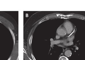
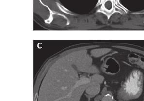
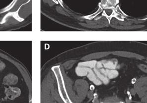
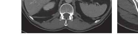
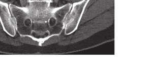
256 Chapter 14 Figure 1. A: the supra-aortic arteries. B: C: D: 254


257 Figure 2. Flowchart of study design DISCUSSION after esophagectomy, and has an adverse impact on prognosis 1-6,9 2,
258 Chapter 14 be more fully optimized before surgery by means of preventive measures to reduce their outcome may include physical therapy to increase cardiorespiratory function and improvement of nutritional status of the current study demonstrate that, not only locoregional vascular disease as demonstrated in previous studies 11,15 which is presumably based on ischemia of the anastomosis 11,15, vascular status could be included in ischemic conditioning trials 10,11 increases perfusion of the gastric tube by partial gastric devascularization through arterial embolization or laparoscopic arterial ligation, followed by esophagectomy and anastomosis at a second stage 10 widespread implementation of ischemic conditioning of the stomach in order to decrease 10,31 10,11. Various limitations apply to this study. First, no internal validation of our results was surgical techniques). Second, there are some potential limitations of the visual grading system. 256
259 Despite these potential limitations, several advantages apply to this approach. The grading studies 12,15,16. Furthermore, for the prediction of cardiovascular events this grading system has irregularities of the vascular wall 12. Second, the scoring system does not require any special CT protocol or calcium-scoring software. Last, a CT or a combined positron emission tomography after esophagectomy may be used in future prediction models, and eventually aid to a more
260 Chapter 14 REFERENCES analysis of the society of thoracic surgeons general thoracic database. Ann Thorac Surg. 5. Raymond DP, Seder CW, Wright CD, et al. Thoracic Surgeons General Thoracic Surgery et al. Quality of Life and Late Complications term Survival and Cancer Recurrence After 10. Kechagias A, van Rossum PSN, Ruurda JP, et 11. Goense L, van Rossum PSN, Weijs TJ, et al. Aortic prediction with ancillary aortic findings on chest CT scans in routine practice. Radiology. of cardiovascular events by using non-vascular Unrequested imaging findings from routine 708. Semiquantitative assessment of cardiovascular intraobserver agreements. J Comput Assist 258
261 al. Transparent Reporting of a multivariable outcome for imputation of missing predictor Development and validation of a prediction with provision for scaled disagreement or partial 23. Landis J, Koch G. The measurement of observer postoperative pulmonary complications through intensive preoperative respiratory rehabilitation in patients with esophageal cancer. Dis during neoadjuvant therapy for esophageal inspiratory muscle training programs to prevent pulmonary complications in patients undergoing healing of cervical oesophagogastrostomies can be predicted by estimation of gastric serosal significance of tissue blood flow during gastric graft and correlation with anastomotic
262
263 15 Intraoperative and postoperative risk factors for anastomotic leakage and pneumonia after esophagectomy for cancer Lucas Goense* Peter S.N. van Rossum* A. Christiaan Kroese Jelle P. Ruurda Diseases of the Esophagus. 2017;30:1 10
264 Chapter 15 ABSTRACT Objective pneumonia. This study aimed to assess the relationship of intraoperative and postoperative Methods Consecutive patients who underwent transthoracic esophagectomy with cervical anastomosis for esophageal cancer from January 2012 to December 2013 were analyzed. Univariable and multivariable logistic regression analyses were used to determine potential associations of Results Conclusion pneumonia. These parameters indicate the importance of setting strict perioperative goals to be protected intensively. 262
265 Risk factors for leakage and pneumonia INTRODUCTION Surgical resection of the esophagus with en-bloc lymphadenectomy is the cornerstone of curative treatment for patients with esophageal cancer 1-3 are the most frequently encountered complications after esophagectomy 4,5 are associated with increased postoperative morbidity, length of hospital stay and mortality ,12. Despite improvement in surgical 4,5,13. Several factors, such as neoadjuvant chemoradiotherapy, anatomic site of the anastomosis, or pneumonia after esophagectomy 5,7,9,14. A reduction in tissue perfusion and subsequent anastomotic healing 6,14 15,16. Also, 17. Perioperative protection intraoperatively and during the early postoperative phase at the intensive care unit 15 the current study was to evaluate associations of intraoperative and postoperative clinical esophagectomy in patients with esophageal cancer. METHODS Study population consent was waived for this study. All patients who underwent elective esophagectomy at 263
266 Chapter 15 our tertiary referral center from January 2012 to December 2013 were included. Data on Anesthetic procedure and postoperative analgesia or T6-7 to provide adequate intraoperative and postoperative analgesia. Pre-procedural medication consisted of prophylactic antibiotics (cefazolin mg, metronidazole 500 mg) 18. Anesthesia was performed using intravenously administered propofol, sufentanil and a muscle 2 2 between 40 and 45 were provided with patient-controlled opioid analgesia. Surgical procedure Robot-assisted minimally invasive thoraco-laparoscopic esophagectomy was the standard procedure for patients with esophageal cancer of all stages in our institute 19 were benign disease and combined laryngeal resection. For the construction of the gastric tube of 4 cm wide, a linear stapling device was used and the staple line was manually oversewn After completion of the anastomosis the surplus of the gastric conduit was removed with a 264
267 Risk factors for leakage and pneumonia and intubation. The perfusion status of patients was routinely assessed based on the urine complications after esophageal surgery 20 to the surgical ward was deemed appropriate. Variables and risk factors retrieved intraoperative and postoperative factors consisted of hemodynamic characteristics, detailed description of studied factors is provided in Table 1. including gastrointestinal defects involving the esophagus, anastomosis, staple line or gastric tube 21,22 endoscopy, visible loss of saliva through the cervical wound, or sepsis caused by mediastinal Utrecht Pneumonia Score (UPS)
268 Chapter 15 Statistical analysis intraoperative and postoperative clinical parameters, using 20 imputed datasets 24. The was studied univariably. Categorical parameters were compared using the Chi-square test or transformations were applied for several parameters to achieve improved linearity on the log odds scale. Also, for several parameters the values were multiplied by 100 to facilitate an Subsequently, parameters with a p logistic regression model to evaluate whether these factors were independently associated 2 r p-value in univariable analysis was pre-selected for the multivariable model. For parameters that remained p-value of <0.05 was considered 266
269 Risk factors for leakage and pneumonia TABLE 1. Studied intraoperative and clinical parameters Intraoperative factors Hemodynamic factors Duration of surgery (minutes) First postoperative systolic RR First postoperative diastolic RR imputed) Number of blood transfusions Total duration systolic RR < 90 (minutes) Time in minutes of inotrope admission Total duration inotrope requirement (minutes) Time in minutes that systolic RR was <90 First measured postoperative hemoglobin Fluid balance (ml) 0-6, 0-12,0-18 hours after surgery Average heart rate Respiratory factors Duration one-lung ventilation (minutes) Average saturation Average saturation ratio Other factors Average temperature measured First postoperative temperature measured
270 Chapter 15 RESULTS time of 8 days (range 3-17) after esophagectomy, whereas pneumonia was diagnosed after encountered for 15 variables, but the percentage of missing values per variable was limited highlighted in Table 1. days [11-18], respectively; p<0.001). Also, in patients with pneumonia the length of hospital versus 13 days [10-18], respectively; p p ). Patient and treatment-related characteristics and their univariable association with anastomotic p Anastomotic leakage p
271 Risk factors for leakage and pneumonia 2 in the p p 2 did not Pneumonia Parameters that showed a potential association with the development of pneumonia in univariable analysis (p 15 2 hours postoperatively and postoperative urine production of <30 ml during at least one hour p 269
272 Chapter 15 p p esophagectomy. TABLE 2 Patient and treatment-related characteristics in relation to postoperative complications No anastomotic leakage Anastomotic leakage p value No pneumonia (n=51) Pneumonia (n=31) p value Characteristic (n=63) (n=19) 50 (79.4) 14 (73.7) (76.5) 25 (80.6) Age (years)* 63.0 ± ± ± ± )* 25.1 ± ± ± ± ASA score 17 (27.0) 34 (54.0) 12 (19.0) 3 (15.8) 13 (68.4) 3 (15.8) (17.6) 33 (64.7) 9 (17.6) 11 (35.5) 14 (45.2) 6 (19.4) 1 ( 1.6) 3 (15.8) ( 2.0) 3 ( 9.7) predicted ± ± ± ± Cardiac co-morbidity 20 (31.7) 5 (26.3) (23.5) 13 (41.9) Vascular co-morbidity 24 (38.7) 8 (41.1) (38.0) 13 (41.9) Diabetes mellitus 6 ( 9.5) 3 (15.8) ( 5.9) 6 (19.4) (23.8) 1 ( 5.3) (15.7) 8 (25.8) Neoadjuvant therapy No therapy Chemotherapy Chemoradiotherapy 8 (12.7) 12 (19.0) 43 (68.3) 1 ( 5.3) 5 (26.3) 13 (68.4) ( 7.8) 13 (25.5) 34 (66.7) 5 (16.1) 4 (12.9) 22 (71.0) Data presented as numbers of patients with percentages in parentheses. *Data presented as mean ± standard 270
273 Risk factors for leakage and pneumonia TABLE 3 Univariable analyses of intraoperative and postoperative clinical parameters potentially asso- p Parameter No anastomotic leakage Anastomotic leakage OR 95% CI p value Intraoperative 7.36 [ ] 7.33 [ ] [ ] 7.22 [ ] in 52.0 [ ] 65.0 [ ] in 43.0 [ ] 46.0 [ ] Postoperative [ ] 7.35 [ ] [ ] 44.0 [ ] Note TABLE 4 Results of multivariable logistic regression analysis Parameter OR 95% CI p value Anastomotic leakage Pneumonia liliter
274 Chapter 15 TABLE 5 Univariable analyses of intraoperative and postoperative clinical parameters potentially associated with pneumonia (p ) Parameter No pneumonia Pneumonia OR 95% CI p value Intraoperative 360 [ ] 460 [ ] ( ) 7.37 ( ) ( ) 39.0 ( ) Postoperative 79.1 ( ) 77.4 ( ) ( ) 74.5 ( ) ( ) 80.0 ( ) ( ) 82.7 ( ) ( ) 75.8 ( ) ( ) 77.8 ( ) TABLE 6 Diagnostic performance of vital parameters resulting from multivariable analysis for predicting the postoperative complications Outcome Parameter SE(%) SP(%) PPV(%) NPV(%) Anastomotic leakage Pneumonia
275 Risk factors for leakage and pneumonia A B C = 0.001* = 0.011* 7.50 = 0.010* Intraoperative minimum ph Average MAP 0-12 hours (mmhg) Intraoperative maximum ph No leakage Leakage 60 No pneumonia Pneumonia 7.10 No pneumonia Pneumonia Figure 1. DISCUSSION occurrence of the two most frequently encountered complications after esophagectomy for cancer could contribute to the improvement of intraoperative and postoperative decision- hemodynamic and respiratory factors during and early after esophagectomy were performed complications , hypotensive events and the need for inotropic support 27 intraoperative blood loss and the need for inotropic support between patients with and 273
276 Chapter 15 frequently be attributed to hypovolemia and tissue hypoperfusion 28. Tissue hypoperfusion 6,14,26. The current the results of a previous study that found an association between hypotension and respiratory complications after esophagectomy 17 17,29 response can cause lung injury by damaging the endothelial and epithelial cells, which predicted the occurrence of pulmonary complications in another study 30. The development of lung injury caused by the described chain of events may contribute to the development of 2 values in these patients, which may suggest ventilationinduced hypocapnia. This might in turn be a consequence of relatively high ventilation frequency or high tidal volumes applied during intraoperative ventilation which can cause ventilation-induced lung injury 31,32 previous studies include increased infusion of crystalloid and colloids, the use of inotropics 17,29, and pneumonia is of importance, since this may lead to prevention and earlier diagnosis 274
277 Risk factors for leakage and pneumonia of hemodynamic and respiratory interventions, during and after esophagectomy on the postoperative course. 4. Certain limitations apply to this study. First, the study was single centered and included a relatively small sample size. Second, this is a hypothesis-generating observational study, and better perioperative cardiorespiratory management reducing postoperative complications. pneumonia and elucidate causality and non-causality of the found associations
278 Chapter 15 REFERENCES 412. Association between clinical complete response and pathological complete response after preoperative chemoradiation in patients with et al. Preoperative chemoradiotherapy for esophagectomy for cancer. J Thorac Cardiovasc analysis of the society of thoracic surgeons general thoracic database. Ann Thorac Surg Catastrophic complications of the cervical esophagogastric anastomosis. J Thorac Pulmonary complications after esophagectomy. of Postoperative Complications on Survivals term Survival and Cancer Recurrence After anastomoses following esophagectomy for 14. Dewar L, Gelfand G, Finley RJ, et al. Factors formation following esophagogastrectomy and gastric tube interposition. Am J Surg significance of tissue blood flow during of esophagogastric anastomotic healing? J Surg glucocorticoids on pulmonary complications 276
279 Risk factors for leakage and pneumonia 19. van der Sluis PC, Ruurda JP, Verhage RJ, et management and major respiratory complications in patients undergoing esophagectomy. Dis Data Collection for Complications Associated pneumonia following esophagectomy for cancer. complete case analysis and the missing-indicator delivery as a factor in the development of fatal postoperative complications after al. Perioperative vasopressors are associated with metabolic acidosis in prolonged surgery. Crit distress syndrome after elective oesophagectomy. predicts the occurrence of pulmonary
280
281 16 Perioperative chemotherapy versus neoadjuvant chemoradiotherapy for esophageal or gastroesophageal junction adenocarcinoma: a propensity score-matched outcome, and survival Lucas Goense* Pieter C. van der Sluis* Peter S.N. van Rossum Jelle P. Ruurda Journal of Surgical Oncology. 2017;115:
282 Chapter 16 ABSTRACT Objective compared to neoadjuvant chemoradiotherapy (ncrt) followed by surgery for patients with Methods by surgery in a tertiary referral center in the Netherlands were compared. Propensity score matching was applied to create comparable groups. Results p p regarding postoperative morbidity and mortality were found. Pathologic complete response p p p p p Conclusion Compared to perioperative chemotherapy, neoadjuvant chemoradiotherapy achieves higher improving survival. 280
283 Multimodality treatment for esophageal cancer INTRODUCTION and the incidence of esophageal adenocarcinoma is rapidly increasing 1,2. Resection of the esophagus with en-bloc lymphadenectomy is the mainstay of curative treatment for patients with esophageal cancer 3 since both perioperative chemotherapy (pct) and neoadjuvant chemoradiotherapy (ncrt) 4-7. Response to neoadjuvant treatment has been associated with a higher percentage of radical surgical resection rates (R0), a reduced Currently, the optimal adenocarcinoma has not been established 5,11. The use of perioperative chemotherapy for esophageal cancer has yielded varying outcomes surgery alone with regard to R0 resection rates and survival 4. Consecutive studies found that 15,16. Nevertheless, thromboembolic events 4,17 was compared to surgery alone for patients with resectable esophageal cancer 10. This trial pathologic complete response (pathcr), favoring the multimodality group. This improvement was found to be clinically relevant for both squamous cell carcinoma and adenocarcinoma 6. Due to the relatively low percentage of adverse events in combination with improved oncologic results, neoadjuvant chemoradiotherapy followed by surgery is now the preferred 18. guidelines Currently, direct comparisons between perioperative chemotherapy with epirubicin, cisplatin adenocarcinoma are limited. Therefore, the current study aimed to compare these two 281
284 Chapter 16 METHODS Patient population the standard treatment with curative intent was switched from perioperative chemotherapy tomography (CT) or integrated F- 0-2 and biopsy-proven resectable adenocarcinoma (clinical stage T1N1-3 or T2-4aN0-3) with no evidence of distant metastases at initial staging. Treatment protocols of capecitabine twice daily for 21 days 17. The chemoradiotherapy regimen consisted of a total radiation dose m 2 of body-surface area) 10. After completion of neoadjuvant treatment, patients were scheduled conduit reconstruction with cervical anastomosis end-to-side with hand-sewn continuous sutures in monolayer 20. Patients with severe cardiopulmonary co-morbidity were scheduled resection was considered too high. Data collection and follow-up Clinical patient characteristics, treatment details (e.g. chemotherapy regimens, surgical 21. After esophagectomy, patients were year, and 12 months until 5 years after surgery. Diagnostic imaging was only performed in 282
285 Multimodality treatment for esophageal cancer follow-up, or the date of death or last follow-up, respectively. Death from non-disease-related causes (e.g. myocardial infarction) were censored in the PFS analysis. Postoperative course 22, 23. Pathological analysis The resected specimens were processed according to a standardized protocol in accordance with the 7 th 24. The (circumferential) resection margin was evaluated using the College of American Pathologist criteria 25. The degree of histopathologic tumor regression was graded according to the system 26. Statistical analysis 16 the two study groups (pct versus ncrt), propensity score matching was used to build comparable groups. First, a propensity score (the probability [ranging from 0 to 1] that a patient was assigned to the chemotherapy or chemoradiotherapy group given the individual 283
286 Chapter Patients who fell outside the joint range of propensity scores (i.e. range of common support) were discarded. RESULTS Patient characteristics characteristics. Using propensity score matching, 86 chemotherapy and 86 chemoradiotherapy patients could be matched without large imbalances of the used covariates. After propensity score matching balance among the two treatment arms improved substantially (Table 1). The results of the propensity score-matched cohort will be discussed here in further detail as this cohort consisted of groups with improved comparability. preoperative chemotherapy cycles. Postoperative continuation of chemotherapy was radiotherapy. The main reason for not completing all chemotherapy cycles in the ncrt group relevant thromboembolic events, which occurred only in the pct group and not in the ncrt p<0.001), 9 of which were symptomatic pulmonary emboli. The remaining 7 thromboembolic evens were asymptomatic (aortic or pulmonary emboli) and were detected p and chemoradiotherapy group (Table 2). preoperative patient-related characteristics, and tumor characteristics between patients who did and did not undergo postoperative chemotherapy. 284
287 Multimodality treatment for esophageal cancer Postoperative course disease progression during therapy (1 patient in the pct group) and diagnosis of metastatic disease during surgery (1 patient in the pct group and 2 patients in the ncrt group). Surgical (p the ncrt group all 6 patients died due to severe postoperative complications. Pathological assessment Pathologic results are presented in Table 4. A pathcr was more frequently observed in patients p<0.001). Also a good group (p Survival resection after propensity score matching), median follow-up was 34 months (range 2-97) in the pct group and 21 months (range 3-47) in the ncrt group, respectively. At 3 years p p analysis showed that at 3 years follow-up, locoregional disease progression occurred less test p respectively
288 Chapter 16 TABLE 1. Comparison of baseline characteristics according to neoadjuvant treatment protocol, before and after propensity score matching Original cohort Propensity score matched cohort Variables pct (n=106) ncrt (n=87) p value pct (n=86) ncrt (n=86) p value 87 (82.1) 77 (88.5) (84.9) 76 (88.4) Age (years)* 62.5 ± ± ± ± )* 26.3 ± ± ± ± ASA score* (28.3) 68 (64.2) 8 ( 7.5) 57 (53.8) 49 (46.2) 16 (18.4) 60 (69.0) 11 (12.6) 36 (41.4) 51 (58.6) (24.4) 58 (67.4) 7 ( 8.1) 45 (52.3) 41 (47.7) 16 (18.6) 59 (68.6) 11 (12.8) 36 (41.9) 50 (58.1) (10.4) 10 (11.5) ( 9.3) 10 (11.6) Cardiac co-morbidity 25 (23.6) 30 (34.5) (23.3) 30 (34.9) Diabetes mellitus 11 (10.4) 13 (14.9) (11.6) 13 (15.1) (55.7) 60 (69.0) (54.7) 59 (68.6) Surgical approach* Transhiatal Transthoracic 23 (21.7) 83 (78.3) 9 (10.3) 76 (89.7) (14.0) 74 (86.0) 9 (10.5) 77 (89.5) Tumor length on endoscopy (cm) 5.3 ± ± ± ± Clinical T-stage* T1 T2 T3 T4 Clinical N-stage* N0 N+ 2 ( 1.9) 9 ( 8.5) 91 (85.8) 4 ( 3.8) 25 (23.6) 81 (76.4) 2 ( 2.3) 15 (17.2) 68 (78.2) 2 ( 2.3) 26 (29.9) 61 (70.1) ( 1.2) 9 (10.5) 73 (84.9) 3 ( 3.5) 24 (27.9) 62 (72.1) 2 ( 2.3) 15 (17.4) 67 (77.9) 2 ( 2.3) 25 (29.1) 61 (70.9) Note. Data are numbers of patients with percentages in parentheses.*variables used for propensity matching. Data are mean ± standard deviation. th edition of 24. Clinical th
289 Multimodality treatment for esophageal cancer TABLE 2. pct ncrt p value (n=86) (n=86) 16 (18.6) 0 ( 0.0) < ( 2.3) 7 ( 8.1) ( 5.8) 2 ( 2.3) ( 3.5) 12 (14.0) ( 1.2) 5 ( 5.8) ( 1.2) 0 ( 0.0) ( 8.1) 5 ( 5.8) ( 9.3) 3 ( 3.5) ( 8.1) 0 ( 0.0) ( 2.3) 3 ( 3.5) ( 1.2) 0 ( 0.0) ( 0.0) 1 ( 1.2) ( 0.0) 1 ( 1.2) ( 0.0) 1 ( 1.2) ( 1.2) 1 ( 1.2) ( 1.2) 0 ( 0.0) (45.3) 34 (39.8) Premature discontinuation 20 (23.3) 23 (26.7) Post-operative continuation 34 (39.5) NA NA Note. Data are numbers of patients with percentages in parentheses.*adverse events graded according to
290 Chapter 16 TABLE 3. Comparative analysis of postoperative course* Outcome measure pct ncrt p value (n=84) (n=84) Complicated postoperative course 58 (69.0) 61 (72.6) (23.8) 24 (28.6) Pneumonia 29 (34.5) 29 (34.5) Cardiac arrhythmia 11 (12.6) 20 (23.8) (16.7) 12 (14.3) Recurrent nerve paresis 6 ( 7.1) 8 (9.5) Wound infection 3 ( 3.6) 5 ( 6.0) Postoperative bleeding 2 ( 1.2) 2 ( 2.4) Thromboembolic event 5 ( 6.0) 5 ( 6.0) Clavien-Dindo grade ( 8.3) 8 ( 9.5) 28 (33.3) 19 (22.6) 0 ( 0.0) 3 ( 3.6) 6 ( 7.1) 14 (16.7) 15 (17.9) 11 (13.1) V 3 ( 3.6) 6 ( 7.1) 23 (27.4) 31 (36.9) Duration of hospital stay (days) # 15 [11-23] 16 [11-27] # 1 [1-4] 1 [1-4] day mortality 1 ( 1.2) 2 ( 2.4) day mortality 5 ( 6.0) 6 ( 7.1) Note. neoadjuvant chemotherapy and chemoradiotherapy, 84 and 84 underwent surgery, respectively. Anastomotic Pneumonia was 40. on an electrocardiogram requiring treatment. system 22,23. # 288
291 Multimodality treatment for esophageal cancer TABLE 4. Comparative analysis of postoperative histopathology* Outcome pct (n=84) ncrt (n=84) Pathologic T-stage ypt0 ypt1b ypt2 ypt3 ypt4 Pathologic N-stage ypn0 ypn1 Tumor regression grade V Radicality of resection R0 R1 9 (10.7) 12 (14.3) 12 (14.3) 48 (57.1) 3 ( 3.6) 36 (42.9) 48 (57.1) 9 (10.7) 5 ( 6.0) 17 (20.2) 18 (21.4) 35 (41.7) 75 (89.3) 9 (10.7) 15 (17.9) 10 (11.9) 19 (22.6) 38 (45.2) 2 ( 2.4) 42 (50.0) 42 (50.0) 15 (17.9) 16 (19.0) 27 (32.1) 22 (26.2) 4 ( 4.8) 80 (95.2) 4 ( 4.8) p value <0.001 Note. neoadjuvant chemotherapy and chemoradiotherapy, 84 and 84 underwent surgery, respectively. Pathological th 24. the 7 th 24. The (circumferential) resection margin was evaluated using the College of American Pathologist (CAP) criteria. The (circumferential) resection margin was evaluated using the College of American Pathologist criteria Figure 1. perioperative chemotherapy and neoadjuvant chemoradiotherapy followed by esophagectomy groups. 289
292 Chapter 16 DISCUSSION ncrt was associated with improved tumor downstaging and a higher pathcr rate compared disease progression in the ncrt group. According to recent literature, pct and ncrt both improve survival compared to surgery 4,6 5,7. Until now, three randomized trials have made a similar attempt to directly compare ncrt with Stahl et al., randomly allocated 119 patients to either chemotherapy (cisplatin, 5-FU, leucovorin) or chemoradiotherapy (cisplatin, 5-FU, leucovorin, 30 and Gy) both followed by surgery 28. The chemoradiotherapy p p p receive either preoperative chemotherapy (cisplatin, 5-FU) or preoperative chemoradiotherapy (cisplatin, 5-FU, and 35 Gy) 29. This study showed a higher histopathological complete response 29. A recent randomized controlled trial of 181 patients by Klevebro et al., again showed a higher p compared to neoadjuvant chemotherapy (platin, 5-FU), with comparable survival between the two groups
293 Multimodality treatment for esophageal cancer tumor progression and a trend towards a higher R0 resection rate was found after ncrt. after neoadjuvant treatment is a major determinant of survival in patients with esophageal cancer 8,26,31-33 ncrt or pct will have distant disease progression, there is need for feasible (adjuvant) control after ncrt is reasonable, currently the interest in adjuvant chemotherapy in these patients to increase systemic control is increasing. A recent cohort study has shown that this approach may improve survival in patients with residual nodal disease 34. Future trials are underway and should answer whether the addition of new adjuvant therapies will improve survival by reducing distant disease progression 35,36. established limitations of perioperative chemotherapy regimens is that adjuvant chemotherapy is less feasible than preoperative chemotherapy 4,13,37 4,13. Also in the current study a limited number of patients chemotherapy over surgery alone has been well established 4,13. The role of postoperative between treatment with perioperative chemotherapy and preoperative chemotherapy only 38. The feasibility problems of the currently available perioperative chemotherapy regimens favor the use of neoadjuvant treatment. 16 contribute to an increase in postoperative morbidity and mortality 39 with earlier reports 10,15,17 of postoperative morbidity or perioperative mortality between the pct and ncrt groups were observed. These results are consistent with two recent meta-analyses that compared postoperative morbidity and mortality between patients treated with pct or ncrt for 291
294 Chapter 16 40,41 randomized controlled trial more severe postoperative complications were observed after ncrt compared to chemotherapy 42. to other studies 40,43, Strengths of this study include the use of predominantly prospective collected data. Furthermore, the sample size of this study is relatively large compared to previous comparative esophageal adenocarcinoma upon recurrence. Recent studies have demonstrated that treatment 45,46. Therefore, treatment of recurrent disease may not have been the same throughout the whole study period, which may have improved the prognosis of the ncrt patients that were included later in this study. Also, the addition of a diagnostic 18 selection for treatment with curative intent. Furthermore, the median-follow up in the ncrt and ncrt regimens are underway and should resolve the limitations in the current literature 47 trial [NCT ] 48 ). 292
295 Multimodality treatment for esophageal cancer Neoadjuvant chemoradiotherapy achieves higher pathologic complete response rates and a chemotherapy
296 Chapter 16 REFERENCES 412. Perioperative chemotherapy versus surgery alone al. Survival after neoadjuvant chemotherapy or chemoradiotherapy for resectable oesophageal Neoadjuvant chemoradiotherapy plus surgery versus surgery alone for oesophageal or R, et al. Preoperative chemo(radio)therapy versus primary surgery for gastroesophageal analysis combining individual patient and Pathologic response after neoadjuvant therapy is the major determinant of survival in patients chemotherapy for resectable thoracic esophageal cancer. Cochrane Database Syst Rev et al. Preoperative chemoradiotherapy for Neoadjuvant chemotherapy compared with surgery alone for locally advanced cancer of the Research and Treatment of Cancer randomized chemotherapy compared with surgery alone for Chemotherapy followed by surgery compared with surgery alone for localized esophageal 15. Cunningham D, Starling N, Rao S, et al. epirubicin, cisplatin and capecitabine for patients with localised gastro-oesophageal junctional evaluation of perioperative epirubicin, Cisplatin, and capecitabine chemotherapy in esophageal 18. NCCN Clinical Practice Guidelines in 294
297 Multimodality treatment for esophageal cancer Guidelines for the management of oesophageal anastomoses are associated with a higher number of strictures compared with end-to-side 876. # Dindo D, Demartines N, Clavien PA. proposal with evaluation in a cohort of 6336 patients and results of a survey. Ann Surg margin in T3 adenocarcinoma of the esophagus. Pathologic assessment of tumor regression after preoperative chemoradiotherapy of esophageal carcinoma. Clinicopathologic correlations. Nonparametric Preprocessing for Parametric 2011;42. compared with chemoradiotherapy in patients with locally advanced adenocarcinoma of patients receiving preoperative chemotherapy for adenocarcinoma of the oesophagus? Wang N, et al. A randomized clinical trial of neoadjuvant chemotherapy versus neoadjuvant chemoradiotherapy for cancer of the oesophagus 31. Chirieac LR, Swisher SG, Ajani JA, et al. Posttherapy pathologic stage predicts survival in patients with esophageal carcinoma receiving preoperative chemoradiation. Cancer pathologic response after neoadjuvant chemoradiotherapy for esophageal cancer is associated with enhanced survival. Ann Thorac response to neoadjuvant chemoradiotherapy in esophageal carcinoma is associated with Adjuvant Chemotherapy After Neoadjuvant
298 Chapter Academic and Community Cancer Research Study of Adjuvant Regorafenib vs Placebo in Adjuvant Nivolumab or Placebo in Subjects nationwide retrospective study of perioperative chemotherapy for gastroesophageal et al. Perioperative chemo(radio)therapy versus primary surgery for resectable adenocarcinoma of the stomach, gastroesophageal junction, and lower esophagus. Cochrane Database Syst Rev is the best neoadjuvant regimen prior analysis of postoperative morbidity and perioperative mortality in patients receiving neoadjuvant chemotherapy or chemoradiotherapy for resectable oesophageal and gastro- chemoradiotherapy in resectable gastric and and mortality after surgery for cancer of the A randomized clinical trial of neoadjuvant chemotherapy vs. neoadjuvant chemoradiation. postoperative outcomes after esophageal cancer following neoadjuvant chemoradiation. Ann Ramucirumab monotherapy for previously treated advanced gastric or gastro-oesophageal international, randomised, multicentre, placebo- advanced gastric or gastro-oesophageal junction induction chemotherapy followed by either based chemoradiation as pre-operative regimen for resectable oesophageal adenocarcinoma. 296
299 Multimodality treatment for esophageal cancer preoperative chemoradiation plus perioperative
300
301 17 Neoadjuvant radiation to the gastric fundus increases the risk of anastomotic leakage after transthoracic esophagectomy for esophageal cancer Lucas Goense* Jelle P. Ruurda Annals of Thoracic Surgery. 2016;102:
302 Chapter 17 ABSTRACT Objective transthoracic esophagectomy. Methods by transthoracic esophagectomy were included in this single-center cohort study. The gastric fundus was contoured on the pre-treatment planning CT. Within this contour, dose-volume histogram parameters were calculated and logistic regression analysis was used to determine Results 35.6Gy versus 24.9Gy, respectively; p clinical T-stage, and radiation modality, the mean radiation dose to the gastric fundus remained p Conclusion ncrt for esophageal cancer, since higher dose levels to the gastric fundus are associated and cervical anastomosis. 300
303 Radiation to the gastric fundus increases the risk of anastomotic leakage INTRODUCTION 1-3 past decades, a steady decrease in postoperative mortality has been achieved by improvements of surgical techniques and perioperative management 4 the esophagogastrostomy remains one of the major complications negatively impacting surgical and oncologic outcomes 4,5 2, increase in surgical morbidity in patients that underwent ncrt Postoperative pulmonary complications have convincingly been related to neoadjuvant radiation dose to the lungs as this part of the stomach is used for the esophagogastric anastomosis. The available evidence on the potential association between neoadjuvant radiation dose to 18,19. the gastric fundus when planning neoadjuvant radiation treatment for esophageal cancer. patients with esophageal cancer undergoing ncrt followed by transthoracic esophagectomy and cervical anastomosis. METHODS Study population This study was approved by our institutional review board, and the informed consent requirement was waived. From a prospectively acquired database, consecutive patients with followed by transthoracic esophagectomy between January 2012 and July 2015 at our tertiary referral center. All patients had biopsy-proven resectable carcinoma with no evidence of distant metastases. Patients who underwent transhiatal esophagectomy, salvage esophagectomy or non-elective surgery and patients in whom no gastric conduit reconstruction was performed
304 Chapter 17 Treatment protocol The ncrt regimen consisted of a total radiation dose of 41.4 Gy in 23 fractions of 1.8 Gy in 2. Some patients (with a clinical T4b tumor) received a total radiation dose of 50.4 Gy in 28 fractions any suspicious regional lymph nodes as determined by all available information (endoscopy, 18 as the CTV plus a margin of 1 cm in all directions. Patients were treated by either three- 80, 3.8 mm, gastric conduit staple line was oversewn by hand. A cervical esophagogastric anastomosis was performed end-to-side with hand-sewn continuous sutures (3-0 PDS) in monolayer. 80, 3.8 mm, Data collection Clinical patient characteristics, treatment details, and surgical outcome data were collected during a contrast swallow study or CT scan, or visualization of anastomotic dehiscence or 8. Image analysis The gastric fundus was retrospectively contoured on the pre-treatment planning CT (section 20. After consultation with the authors of a previous study 18, the boundaries of the delineated gastric fundus were standardized in located within the diaphragmatic dome was determined in the transverse plane. From that point, four consecutive transverse sections in caudal direction were delineated following 302
305 Radiation to the gastric fundus increases the risk of anastomotic leakage percentage of the volume that received at least 20, 25, 30, and 35 Gy, respectively). Statistical analysis compare parametric and non-parametric continuous parameters, respectively. Univariable Three baseline characteristics including tumor location, clinical T-stage and radiation modality, were thought to potentially confound the association between radiation dose to the fundus was entered in a multivariable logistic regression model together with the three dose), resulting in the statistical problem of (multi)collinearity. Therefore, from these highly correlated pairs of parameters only the mean dose was pre-selected for the multivariable p 17 RESULTS 303
306 Chapter 17 p ). A comparison of gastric fundus radiation dose characteristics for patients with versus without 39.9] versus 24.9 Gy [ ], respectively; p p addition, univariable analysis showed that percentages of the gastric fundus volume receiving determined at 31.4 Gy. Patients with a mean dose above versus below this threshold developed junction tumors were 0.95 Gy ± 1.5, 15.6 Gy ± 13.9, 28.9 Gy ± 10.7, and 34.4 Gy ±
307 Radiation to the gastric fundus increases the risk of anastomotic leakage confounded by tumor location, clinical T-stage or radiation modality (Table 4). Adjusted p
308 Chapter 17 TABLE 1. Characteristic Anastomotic leakage No anastomotic leakage p value (n = 25) (n = 72) 19 (76.0) 53 (73.6) Age (years)* 64.9 ± ± )* 25.6 ± ± ASA score 5 (20.0) 12 (48.0) 8 (32.0) 14 (19.4) 48 (66.7) 10 (13.9) (24.0) 9 (12.5) Cardiac co-morbidity 7 (28.0) 25 (34.7) Vascular co-morbidity 12 (48.0) 33 (45.8) Diabetes mellitus 4 (16.0) 10 (13.9) (68.0) 52 (72.2) Clinical T-stage ct1 ct2 ct3 ct4 Clinical N-stage cn0 cn1 cn2 cn3 Tumor histology Adenocarcinoma Squamous cell carcinoma Tumor location Distal third of esophagus Gastro-esophageal junction Radiation modality 3D-CRT Radiation planning modality CT 18 Total radiation dose 0 (0.0) 3 (12.0) 19 (76.0) 3 (12.0) 5 (20.0) 12 (48.0) 7 (28.0) 1 ( 4.0) 17 (68.0) 8 (32.0) 0 ( 0.0) 2 ( 8.0) 5 (20.0) 15 (60.0) 3 (12.0) 11 (44.0) 14 (56.0) 7 (28.0) 18 (72.0) 23 (92.0) 2 ( 8.0) 1 ( 1.4) 19 (26.4) 47 (65.3) 5 ( 6.9) 19 (26.4) 34 (47.2) 15 (20.8) 4 ( 5.6) 43 (59.7) 27 (37.5) 2 ( 2.8) 6 ( 8.3) 18 (25.0) 41 (56.9) 7 ( 9.7) 24 (33.3) 48 (66.7) 19 (26.4) 53 (73.6) 67 (93.1) 5 ( 6.9) ± ± Note. Data are numbers of patients with percentages in parentheses.*data are mean ± standard devia- 306
309 Radiation to the gastric fundus increases the risk of anastomotic leakage TABLE 2. Univariable logistic regression analysis of gastric fundus dose characteristics among patients Characteristic Anastomotic leakage (n = 25) No anastomotic leakage (n = 72) OR (95% CI) p value Volume (ml) 11.1 [ ] 11.8 [ ] 0.92 ( ) [ ] 24.9 [ ] 1.04 ( ) [ ] 8.9 [ ] 1.06 ( ) D50 (Gy) 39.0 [ ] 21.8 [ ] 1.03 ( ) [ ] 41.9 [ ] 1.02 ( ) [ ] 60.0 [ ] 1.12 ( ) [ ] 38.3 [ ] 1.15 ( ) [ ] 26.2 [ ] 1.16 ( ) [ ] 17.4 [ ] 1.16 ( ) in volume-percentage. TABLE 3. Receiver operating characteristics analysis of gastric fundus dose characteristics among Characteristic AUC SE (%) SP (%) PPV (%) NPV (%) Gy Gy V V V TABLE 4. parameter Characteristic OR (95% CI) p value Tumor location Clinical T-stage ct1-2 ct3-4 Radiation modality 3D-CRT 1.00 (ref) 0.60 ( ) (ref) 2.81 ( ) (ref) 0.65 ( ) ( ) ty-modulated radiotherapy

310 Chapter 17 A 50 * * B * * * 0 Figure 1. Figure 2. 20, and 10 Gy, respectively. 308
311 Radiation to the gastric fundus increases the risk of anastomotic leakage DISCUSSION This study demonstrates that the neoadjuvant radiation dose to the gastric fundus in patients transthoracic esophagectomy with cervical anastomosis. Several radiation dose characteristics to the gastric fundus when planning neoadjuvant radiation for esophageal cancer with planned series. According to the results of this study, limiting the mean dose to 31 Gy could decrease Two previous studies have reported on the relationship between neoadjuvant radiation dose 18,19. Similar to the esophagectomy with intrathoracic anastomosis reported that the radiation dose to the gastric 18 were applied 18 the present study is currently regarded as the standard of care in many countries worldwide for patients with resectable locally advanced esophageal cancer That study applied a similar neoadjuvant treatment regimen to the current series, but the surgical procedure (transhiatal esophagectomy 19 bloc radical lymphadenectomy as was performed in the present study is currently considered the preferred approach of oncological esophagectomy 21,22. Therefore, in contrast to other was analyzed under the circumstances of present-day standardized radical surgery and across the two previous reports 18,19 conclusions. This may be the reason for the more nuanced discriminatory ability of the mean
312 Chapter 17 found in the other two studies (AUC ). rates remains equivocal. A recent meta-analysis including 11 randomized controlled trials (RCTs) comparing outcomes of patients undergoing ncrt followed by surgery with patients p 10 surgical morbidity, but that surgical morbidity was inconsistently reported across trials which impeded direct comparisons 1. A recent retrospective analysis of 686 patients reported that p<0.01) (14). The current study was not designed - and hence does not allow - to answer the question whether ncrt per se to determine whether the variability of radiation doses to the gastric fundus relates to the after esophagectomy. 6,7,9, 10 2 which used the same ncrt regimen as the current study. Also, in accordance 2 310
313 Radiation to the gastric fundus increases the risk of anastomotic leakage Sparing of the gastric fundus in radiation treatment planning for esophageal cancer could conformal radiation therapy technique providing greater target volume conformity, greater dose homogeneity, and an increased ability to control dose to adjacent normal structures including the gastric fundus if desired. To this regard, it could be helpful if the radiation in the neoadjuvant setting to spare the gastric fundus, as the microscopic spread beyond the 2, mostly involves microscopically positive surgical margins at the lateral (circumferential) borders rather than the caudal border 23 negatively impact survival 24. Finally, assessment of radiation dose to the gastric fundus could study is limited by the retrospective nature of the analysis, which impedes adjustment for all breathing-induced organ motion, which could have altered radiation dose calculations. Since no daily imaging information was available the authors were not able to compensate for studies 18,19 gastric fundus doses across patients, which increases the statistical power and precision of multivariable analysis. 17 cancer treated with neoadjuvant chemoradiotherapy followed by transthoracic esophagectomy 311
314 Chapter 17 REFERENCES al. Survival after neoadjuvant chemotherapy or chemoradiotherapy for resectable oesophageal et al. Preoperative chemoradiotherapy for Neoadjuvant chemoradiotherapy plus surgery versus surgery alone for oesophageal or complications after esophagectomy for adenocarcinoma of the esophagus are related to timing of death due to recurrence. Ann Surg esophageal resection and reconstruction. Semin limited transhiatal resection for adenocarcinoma after esophagectomy following neoadjuvant 40. end-to-side esophagogastrostomy after neoadjuvant chemoradiotherapy on postoperative Results of a european multicenter study. Ann analysis of postoperative morbidity and perioperative mortality in patients receiving neoadjuvant chemotherapy or chemoradiotherapy for resectable oesophageal and gastro- Neoadjuvant chemoradiation may increase the after transthoracic esophagectomy. J Thorac Surgical mortality in patients with esophageal Factors associated with clinical response and postoperative complications. Anticancer Res Postoperative pulmonary complications after preoperative chemoradiation for esophageal 312
315 Radiation to the gastric fundus increases the risk of anastomotic leakage complications among esophageal cancer patients treated with concurrent chemoradiotherapy associated with postoperative pulmonary complications in esophageal cancer patients treated with concurrent chemoradiotherapy et al. Anastomotic complications after ivor lewis esophagectomy in patients treated with neoadjuvant chemoradiation are related to complications in patients with esophageal cancer treated with neoadjuvant chemoradiation limited transhiatal resection for adenocarcinoma of a randomized clinical trial. Ann Surg What should be the gold standard for the surgical component in the treatment of locally versus transhiatal esophagectomy. Ann Surg JJ, et al. Patterns of recurrence after surgery alone versus preoperative chemoradiotherapy tumor after neoadjuvant chemoradiation outside prognostic factor for survival in esophageal
316
317 18 Intrathoracic versus cervical anastomosis and predictors of anastomotic leakage after esophagectomy for cancer Lucas Goense* Suzanne S. Gisbertz Jelle P. Ruurda British Journal of Surgery. 2018;105:
318 Chapter 18 ABSTRACT Objective versus a cervical anastomosis after esophagectomy are equivocal. The aim of this study was to compare clinical outcome after esophagectomy in patients with an intrathoracic or cervical anastomosis, and Methods Audit. For the comparison between an intrathoracic and cervical anastomosis, propensity Results Some 3348 patients were included. After propensity score matching, 654 patients were included in both the cervical and intrathoracic anastomosis groups. An intrathoracic anastomosis was versus 21.9 per cent; P = 0.025). The percentage of patients with recurrent nerve paresis was also lower (0.6 versus 7.0 per cent; P < 0.001) and an intrathoracic anastomosis was associated with a shorter median hospital stay (12 versus 14 days; P = Conclusion An intrathoracic esophagogastric anastomosis was associated with a lower anastomotic 316
319 Cervical versus intrathoracic anastomosis after esophagectomy INTRODUCTION continues to increase every year 1. According to international guidelines 2, esophagectomy is the cornerstone of curative treatment for non-metastasized esophageal cancer, often combined perioperative management and patient selection have resulted in a reduction in postoperative mortality after esophagectomy 3 and is a major cause of morbidity and mortality. The percentage of patients with anastomotic 4 8. patient-related characteristics 5,9,10, intraoperative factors 11 13, postoperative factors and surgical technique Controversy remains about the optimal anatomical location of the esophagogastric anastomosis (intrathoracic versus cervical) after esophagectomy. Several anastomosis 9,17, , did not show a statistically with a cervical anastomosis, because a wider oncological resection margin can be achieved. anastomosis than for an intrathoracic anastomosis 22 vascularized 9. after esophagectomy is equivocal. Therefore, the primary aim of this study was to assess resection with either an intrathoracic or cervical esophagogastric anastomosis. A secondary 18 METHODS All patient data were obtained from the Dutch Upper Gastrointestinal Cancer Audit (DUCA), a registry of all patients undergoing surgery with curative intent for esophageal or gastric cancer founded in 2011, with the objective to facilitate and organize the initiation of nationwide auditing in a uniform format. The DUCA collects data to monitor national guideline adherence 317
320 Chapter 18 and to provide surgical teams with reliable information on outcome measures. Participation is mandatory for all Dutch hospitals performing esophageal resections, and data are registered for each patient during the hospital stay and until 30 days after discharge. Detailed descriptions uniform data registration. An independent monitoring team audits the data to evaluate completeness and concordance. The organization of the DUCA has been described in more detail previously 23. Patients All patients undergoing esophagectomy for esophageal cancer with gastric tube reconstruction between January 2011 and December 2015 were included. For the comparison between Treatment Surgical treatment consisted of an open (both abdomen and chest), hybrid (abdomen minimally invasive and open chest) or totally minimally invasive transthoracic esophagectomy followed by gastric tube construction with a cervical or intrathoracic anastomosis. The location and technique of the anastomosis (stapled versus handsewn, end-to-side versus side-to-side) are guidelines. Outcome measures surgical and short-term oncological outcomes were analyzed. Surgical outcome parameters days after discharge, positive resection margin and number of retrieved lymph nodes. Statistical analysis Patient and treatment-related characteristics are described as count with percentages, for eight variables; the percentage of missing values per variable was limited (range 0.0 to
321 Cervical versus intrathoracic anastomosis after esophagectomy 24. was performed for the analysis of intrathoracic versus cervical anastomosis. First, propensity scores (the probability, ranging from 0 to 1, that a patient was assigned to an intrathoracic or cervical anastomosis) were derived using a logistic regression model, which included all patient and treatment-related characteristics presented in Table 1 matching was performed with nearest-neighbor matching without replacement, using a caliper width of 0.25 multiplied by the standard deviation of the estimated propensity score 25 in measured patient and treatment-related characteristics of the matched cohort was assessed 26 the two treatment groups, the c 2 test was used for categorical variables, and the Student s t test U respectively. Logistic regression analysis was used to stratify by type of surgery (open versus total minimally invasive approach) in the propensity-matched cohort by adding an interaction term between surgical approach and anastomotic location for each outcome. For this analysis, hybrid procedures were added to the minimally invasive group. patients who underwent a transhiatal resections). Variables with P < in univariable P 18 RESULTS intrathoracic anastomosis (928) and a cervical anastomosis (1158) (Fig. 1). Patients were predominantly men (77.4 per cent), and the mean age was 64.6 (s.d. 9.0) years. The percentage of patients with an intrathoracic anastomosis increased during the study interval from 20.6 per cent in 2011 to 59.3 per cent in Patient and treatment-related characteristics according to location of the anastomosis are shown in Table 1. After propensity matching, 654 patients 319
322 Chapter 18 were included in both groups and all baseline variables including year of surgery were equally Intrathoracic versus cervical anastomoses Postoperative complications and pathological data are shown in Table 2 was less frequent in patients who underwent an intrathoracic anastomosis than in those with a versus 143 of 654 (21.9 per cent) respectively (P = 0.025). Recurrent nerve paresis occurred less often in patients with an intrathoracic versus 46 of 654 (7.0 per cent) respectively (P < 0.001). 12 (range 3 145) versus 14 (range 4 386) days (P < 0.001). Surgical reinterventions, duration two groups. The associations between location of the anastomosis and outcome parameters P for interaction > 0.050) (Table 2). anastomosis groups in the percentage of patients who had a surgical reintervention (53.2 per cent of patients with an intrathoracic anastomosis versus 44.8 per cent with a cervical anastomosis; P = 0.184) or in-hospital mortality (8.1 versus 10.5 per cent respectively; P = 0.520). Duration of hospital stay (median 40 (range 9 132) versus 28 (4 132) days; P < 0.001) versus 4 (1 155) days; P Predictors of anastomotic leakage Table 3, Fig. 1). The median P rates were 9.1 and 2.7 per cent respectively (P = 0.001). Table 3 320
323 Cervical versus intrathoracic anastomosis after esophagectomy P = P = 0.026), P = 0.031), history of cardiac arrhythmia (RR 1.25, 1.01 to 1.55, P = 0.044), diabetes mellitus (RR 1.26, 1.06 to 1.49; P = P = 0.022) (Table 4)
324 Chapter 18 TABLE 1. Patient and treatment-related characteristics according to location of the anastomosis, before and after propensity score matching n After matching (n (n Cervical anastomosis (n (n Cervical anastomosis (n Age (years)* 64.1(9.0) 63.9( (8.6) (8.9) 64.0(8.3) (83.9) 835 (72.1) (80.4) 533 (81.5) 2.9 F 149 (16.1) 323 (27.9) 128 (19.6) 121 (18.5) 2 )* 26.2(4.2) 25.4 (4.2) (4.2) 26.2 (4.2) (16.2) 215 (18.6) (16.8) 109 (16.7) (61.5) 726 (62.7) 423 (64.7) 408 (62.4) 207 (22.3) 212 (18.3) 121 (18.5) 134 (20.5) 0 (0) 5 (0.4) 0 (0) 3 (0.5) No 807 (87.0) 1021 (88.2) (87.8) 568 (86.9) 2.7 Yes 121 (13.0) 137 (11.8) 80 (12.2) 86 (13.1) Coronary artery disease No 830 (89.4) 1063 (91.8) (90.7) 597 (91.3) 2.0 Yes 98 (10.6) 95 (8.2) 61 (9.3) 57 (8.7) No 861 (92.8) 1102 (95.2) (94.2) 614 (93.9) 1.2 infarction Yes 67 (7.2) 56 (4.8) 38 (5.8) 40 (6.1) No 844 (90.9) 1072 (92.6) (91.4) 598 (91.4) 0.0 Yes 84 (9.1) 86 (7.4) 56 (8.6) 56 (8.6) No 643 (69.3) 793 (68.5) (68.7) 449 (68.7) 0.0 Yes 285 (30.7) 365 (31.5) 205 (31.3) 205 (31.3) Peripheral vascular disease No 891 (96.0) 1126 (97.2) (96.9) 634 (96.9) 0.0 Yes 37 (4.0) 32 (2.8) 20 (3.1) 20 (3.1) Diabetes mellitus No 780 (84.1) 1010 (87.2) (84.6) 561 (85.8) 3.3 Yes 148 (15.9) 148 (12.8) 101 (15.4) 93 (14.2) No 872 (94.0) 1102 (95.2) (94.5) 615 (94.0) 1.9 Yes 56 (6.0) 56 (4.8) 36 (5.5) 39 (6.0) 322
325 Cervical versus intrathoracic anastomosis after esophagectomy Thromboembolic events No 887 (95.6) 1120 (96.7) (96.6) 631 (96.5) 0.7 Yes 41 (4.4) 38 (3.3) 22 (3.4) 23 (3.5) No 895 (96.4) 1113 (96.1) (96.3) 628 (96.0) 1.7 Yes 33 (3.6) 45 (3.9) 24 (3.7) 26 (4.0) Previous abdominal or No 657 (70.8) 822 (71.0) (71.9) 475 (72.6) 1.7 thoracic surgery Yes 271 (29.2) 336 (29.0) 184 (28.1) 179 (27.4) ADC 814 (87.7) 723 (62.4) (83.3) 533 (81.5) 5.6 SCC 96 (10.3) 392 (33.9) 92 (14.1) 104 (15.9) 18 (1.9) 43 (3.7) 17 (2.6) 17 (2.6) Tumor location 42 (4.5) 319 (27.5) (6.4) 47 (7.2) 3.7 Distal 886 (95.5) 839 (72.5) 612 (93.6) 607 (92.8) ct category T1 49 (5.3) 64 (5.5) (5.4) 42 (6.4) 0.3 T2 190 (20.5) 216 (18.7) 125 (19.1) 117 (17.9) T3 665 (71.7) 819 (70.7) 474 (72.5) 470 (71.9) T4 24 (2.6) 59 (5.1) 20 (3.1) 25 (3.8) cn category N0 364 (39.2) 370 (32.0) (35.2) 242 (37.0) 0.4 N1 388 (41.8) 503 (43.4) 283 (43.3) 268 (41.0) N2 156 (16.8) 238 (20.6) 125 (19.1) 117 (17.9) N3 20 (2.2) 47 (4.1) 16 (2.4) 27 (4.1) Neoadjuvant therapy No 79 (8.5) 95 (8.2) (7.6) 54 (8.3) 1.3 nct 52 (5.6) 75 (6.5) 49 (7.5) 46 (7.0) ncrt 797 (85.9) 988 (85.3) 555 (84.9) 554 (84.7) Type of surgery 199 (21.4) 355 (30.7) (24.6) 170 (26.0) (77.2) 791 ( (74.5) 479 (73.2) 13 (1.4) 12 (1.0) 6 (0.9) 5 (0.8)
326 Chapter 18 TABLE 1 (continued). Patient and treatment-related characteristics according to location of the anastomosis, before and after propensity score matching n After matching (n (n Cervical anastomosis (n (n Cervical anastomosis (n Year of surgery (35.3) 225 (19.4) (26.6) 164 (25.1) (26.7) 234 (20.2) 157 (24.0) 178 (27.2) (20.5) 212 (18.3) 164 (25.1) 135 (20.6) (11.6) 279 (24.1) 105 (16.1) 125 (19.1) (5.8) 208 (18.0) 54 (8.3) 52 (8.0) - Table 1 TABLE 2. n After matching (n (n Cervical anastomosis (n (n Cervical anastomosis (n P P No 756 (81.5) 919 (79.4) 543 (83.0) 511 (78.1) Yes 172 (18.5) 239 (20.6) 111 (17.0) 143 (21.9) No 598 (64.4) 756 (65.3) 421 (64.4) 432 (66.1) Yes 330 (35.6) 402 (34.7) 233 (35.6) 222 (33.9) Recurrent nerve paresis < < No 924 (99.6) 1063 (91.8) 650 (99.4) 608 (93.0) Yes 4 (0.4) 95 (8.2) 4 (0.6) 46 (7.0) P for interaction by type of surgery 324
327 Cervical versus intrathoracic anastomosis after esophagectomy Surgical reintervention No 783 (84.4) 959 (82.8) 558 (85.3) 548 (83.8) Yes 145 (15.6) 199 (17.2) 96 (14.7) 106 (16.2) Duration of hospital stay (days)* 12 (3 172) 14 (3 386) < (3 145) 14 ( ) < (days)* 2 (0 125) 2 (0 155) (0 125) 2 (0 155) Postoperative death# No 889 (95.8) 1106 (95.5) 633 (96.8) 628 (96.0) Yes 39 (4.2) 52 (4.5) 21 (3.2) 26 (4.0) Readmission** No 794(85.6) 991 (85.6) 556 (85.0) 547 (83.6) Yes 134 (14.4) 167 (14.4) 98 (15.0) 107 (16.4) Positive resection margin No 877 (94.5) 1102 (95.2) 618 (94.5) 622 (95.1) Yes 51 (5.5) 56 (4.8) 36 (5.5) 32 (4.9) No. of lymph nodes harvested < (47.8) 546 (47.2) 356 (54.4) 334 (51.1) 484 (52.2) 612 (52.8) 298 (45.6) 320 (48.9) Any vocal cord dysfunction after resection. Any postoperative surgical reintervention under general anesthesia. #Death during initial hospital admission or within 30 days after surgery. **Readmission to hospital within 30 days after initial discharge. The resection margin was evaluated using the College of American Pathol- 2 U versus minimally invasive approach)
328 Chapter 18 TABLE 3. (n (n P Age (years)* 64.4(9.1) 65.4(8.5) 11 (0.3) 2087 (77.5) 519 (79.1) (0.0) F 605 (22.5) 137 (20.9) 2 )* 25.9(4.4) 26.1(4.3) 30 (0.9) 487 (18.1) 90 (13.7) < (0.7) 1656 (61.5) 369 (56.3) 541 (20.1) 191 (29.1) 8 (0.3) 6 (0.9) No 2348 (87.2) 540 (82.3) (0) Yes 344 (12.8) 116 (17.7) Coronary artery disease No 2440 (90.6) 576 (87.8) (0) Yes 252 (9.4) 80 (12.2) No 2522 (93.7) 597 (91.0) (0) Yes 170 (6.3) 59 (9.0) No 2490 (92.5) 582 (88.7) (0) Yes 202 (7.5) 74 (11.3) No 1840 (68.4) 417 (63.6) (0) Yes 852 (31.6) 239 (36.4) Peripheral vascular disease No 2593 (96.3) 626 (95.4) (0) Yes 99 (3.7) 30 (4.6) Diabetes mellitus No 2302 (85.5) 524 (79.9) < (0) Yes 390 (14.5) 132 (20.1) No 2535 (94.2) 609 (92.8) (0) Yes 157 (5.8) 47 (7.2) 326
329 Cervical versus intrathoracic anastomosis after esophagectomy Thromboembolic events No 2578 (95.8) 630 (96.0) (0) Yes 114 (4.2) 26 (4.0) No 2604 (96.7) 626 (95.4) (0) Yes 88 (3.3) 30 (4.6) Previous abdominal or thoracic surgery No 1881 (69.9) 457 (69.7) (0) Yes 811 (30.1) 199 (30.3) ADC 2095 (77.8) 505 (77.0) (0.6) SCC 528 (19.6) 135 (20.6) 69 (2.6) 16 (2.4) Tumor location 28 (1.0) 16 (2.4) (0.7) 312 (11.6) 81 (12.3) Distal 2352 (87.4) 559 (85.2) ct category T1 154 (5.7) 41 (6.3) (4.9) T2 543 (20.2) 126 (19.2) T (70.4) 458 (69.8) T4 99 (3.7) 31 (4.7) cn category N0 986 (36.6) 234 (35.7) (3.5) N (42.0) 287 (43.8) N2 492 (18.3) 118 (18.0) N3 83 (3.1) 17 (2.6) Neoadjuvant therapy No 260 (9.7) 73 (11.1) (0) nct 205 (7.6) 34 (5.2) ncrt 2227 (82.7) 549 (83.7) - 2 t test. number of missings for each variable before imputation
330 Chapter 18 TABLE 4. P 1.00 (reference) 0.99 (0.77, 1.30) (1.09, 1.78) (1.27, 3.64) (1.02, 1.45) (1.01, 1.55) Diabetes mellitus 1.26 (1.06, 1.49) Tumor location 1.86 (1.25, 2.77) (0.87, 1.32) Distal 1.00 (reference) ease. All oesophageal resections for cancer n = 3348 Eligible for identification of preoperative predictors of anastomotic leakage n = 3348 Excluded n = 1262 All transhiatal resections n =1224 All proximal oesophageal tumours n = 38 Eligible for inclusion and propensity matching n = 2086 Intrathoracic anastomosis (before matching) n = 928 Cervical anastomosis (before matching) n = 1158 Intrathoracic anastomosis (after matching) n = 654 Cervical anastomosis (after matching) n = 654 Figure
331 Cervical versus intrathoracic anastomosis after esophagectomy DISCUSSION This nationwide multicenter cohort study compared clinical outcome in patients with an intrathoracic versus cervical anastomosis following esophagectomy. An intrathoracic tumors. Four RCTS have investigated clinical outcome in patients with an intrathoracic or cervical anastomosis. The results of these studies are equivocal regarding which anastomotic study) in stapled and hand-sutured anastomosis. Two meta-analyses 15,27, including 298 patients, found anastomosis. The present results are in line with these analyses. in the present study are high compared with those in other studies 5,9 intrathoracic and cervical anastomosis range from 9 to 21 per cent 9,17,28,29 and 8 to 35 per cent 9,17,28,30 respectively. Some studies included only clinically relevant or radiologically per cent of anastomoses were intrathoracic in 2011 and 59.3 per cent in The introduction of an intrathoracic anastomosis is associated with a learning curve 32,33. Furthermore, the proportion of minimally invasive procedures increased from 53.1 to 85.4 per cent during the study period. The introduction of minimally invasive esophagectomy is also 329
332 Chapter 18 34,35. present study. some surgeons prefer a cervical anastomosis. The possibility of a wider resection margin and anastomosis 36 intrathoracic anastomosis led to a longer intensive care and hospital stay. This suggests that 37. mortality between intrathoracic and cervical anastomoses. The safety of the intrathoracic technique is supported by a recent meta-analysis 27 mortality between intrathoracic and cervical anastomoses. Previous studies 9,15,27,38 intrathoracic anastomosis. Factors resulting in poor tissue perfusion and vascular impairment are considered important 5,9,10,39 Strengths of this study include the population-based design, the adjustment for important confounders, and the relatively large sample size. Furthermore, data from the DUCA are collected prospectively, and controlled for completeness and validity by an independent monitoring team. There are also limitations to this study, including its retrospective design the anastomosis are not recorded in for the individual centers. At present, a randomized trial 40 comparing the intrathoracic and 330
333 Cervical versus intrathoracic anastomosis after esophagectomy contribution to the current literature
334 Chapter 18 REFERENCES Accessed January 1, Postoperative complications after esophagectomy for adenocarcinoma of the esophagus are related to timing of death due to recurrence. Ann Surg. neoadjuvant chemoradiotherapy on postoperative limited transhiatal resection for adenocarcinoma et al. Preoperative chemoradiotherapy for analysis of postoperative morbidity and perioperative mortality in patients receiving neoadjuvant chemotherapy or chemoradiotherapy for resectable oesophageal and gastro- analysis of the society of thoracic surgeons general thoracic database. Ann Thorac Surg. 10. Goense L, van Rossum PS, Weijs TJ, et al. Aortic delivery as a factor in the development of fatal postoperative complications after anastomoses are associated with a higher number of strictures compared with end-to-side analysis. Journal of Gastrointestinal Surgery. 332
335 Cervical versus intrathoracic anastomosis after esophagectomy analysis of 72 patients in a single center study. al. Cervical or thoracic anastomosis for esophagectomy for carcinoma. Surg Gynecol or thoracic anastomosis after esophageal prospective randomized trial comparing sutured intrathoracic anastomosis after esophagectomy a prospective randomized controlled study. Surg Resection for advanced cancer of the thoracic Late results of a prospective randomized study. imputation for missing data in epidemiological in means and differences in proportions in 26. Austin PC. Assessing balance in measured baseline covariates when using many-toone matching on the propensity-score. or thoracic anastomosis after esophagectomy for Saluja SS, Ray S, Pal S, et al. Randomized trial comparing side-to-side stapled and hand- al. Systematic review of the definition and minimally invasive esophagectomy program
336 Chapter 18 Surgeons National Database. Ann Thorac Surg. cervical anastomoses. Ann Thorac Cardiovasc versus intrathoracic manifestation of cervical reinforcement with omentoplasty following after minimally invasive esophagectomy for 334
337 Cervical versus intrathoracic anastomosis after esophagectomy
338
339 19 Diagnostic performance of a CT based scoring system for diagnosis of anastomotic leakage after esophagectomy: comparison with subjective CT assessment Lucas Goense Peter S.N. van Rossum Jelle P. Ruurda European Radiology. 2017;27:
340 Chapter 19 ABSTRACT Objective compare it to subjective CT interpretation. Methods after esophagectomy with cervical anastomosis between 2003 and 2014 were analyzed. The Results The original interpretation and the systematic subjective CT assessment by two radiologists Conclusion This CT-based score may provide improved diagnostic performance for diagnosis of 338
341 CT-based assessment of anastomotic leakage INTRODUCTION the incidence rate is rapidly increasing 1. Surgical resection of the esophagus with en-bloc lymphadenectomy combined with neoadjuvant chemoradiation or perioperative chemotherapy is the cornerstone of treatment for patients with locally advanced non-metastatic esophageal cancer 2-4. Despite advances in surgical treatment and improvement in perioperative care 2,5 stay, and mortality and provide information on the condition of the gastric tube 14,15 procedure may damage the anastomosis. CT scanning is commonly performed for diagnosis of postoperative complications, since it is non-invasive and safe to use in critically ill patients. Previously, several studies have esophagectomy 13,16-18 during postoperative routine screening and included only a small number of patients 13,16,17. Also a wide range of diagnostic accuracies has been reported, suggesting that the association ,
342 Chapter 19 radiologists and that of the original CT interpretation. METHODS Study population This retrospective cohort study was approved by a institutional review board and the requirement to obtain informed consent was waived. The study was designed and conducted according to Standards for Reporting of Diagnostic Accuracy 21. From a prospectively acquired database, all consecutive patients with esophageal or gastro-esophageal junction cancer who underwent an elective esophagectomy, between 2003 and 2014, at our tertiary referral Surgical treatment consisted of a transthoracic or transhiatal esophagectomy with en-bloc lymphadenectomy and gastric tube reconstruction 22. A cervical esophagogastric anastomosis was performed end-to-side with hand-sewn continuous sutures (3-0 PDS) in monolayer. After surgery two chest tubes were routinely placed, and removed during the following days in case Data collection postoperative demonstration of saliva through the cervical wound, or visualization of up time was truncated to 30 days for all patients. All postoperative complications, including Image acquisition Thoraco-abdominal CT images were acquired using commercially available 16- or 64-section with millimeter section collimation, a tube rotation time of 500 milliseconds, a tube
343 CT-based assessment of anastomotic leakage Variable selection esophageal surgery and variables described previously in the literature were included for discernible wall), and mediastinal induration (whenever the mediastinal fat showed non- collection, and the anatomical region (i.e. above the manubrium, between the manubrium and Image evaluation All CT scans were retrospectively reviewed together by two radiologists in consensus ( esophagectomy, but were blinded for the patients detailed clinical information. The presence or absence of the various selected CT variables were systematically assessed and recorded. 19 Also, all the original CT interpretations rendered as part of the clinical care were reviewed. Statistical analysis and development of a practical scoring system 341
344 Chapter 19 Subsequently, parameters with a p-value below 0.05 in univariable logistic regression analysis to evaluate whether these factors were independently associated with the occurrence of beta-regression 23. Statistical analysis p-value of <0.05 was RESULTS Demographics Clinical and treatment-related patient characteristics and their univariable association with 342
345 CT-based assessment of anastomotic leakage Predictors of anastomotic leakage p p< independent its size, anatomic location within the mediastinum, and the number of days p th postoperative day p p p , p p p p 343
346 Chapter 19 Systematic subjective CT assessment Original clinical CT interpretation Risk scoring system beta- analysis. To this regard it proved feasible to assign one point for the presence of each predictive (Table 3). Therefore, the cumulative amount of points of the ALP score ranges from 0 to 4. presented in Table
347 CT-based assessment of anastomotic leakage to the systematic subjective CT assessment and original CT interpretation, respectively respectively. TABLE 1. Characteristic Anastomotic leakage No anastomotic leakage p value (n = 54) (n = 68) 41 (75.9) 56 (82.4) Age (years)* 65.2 ± ± )* 25.7 ± ± ASA score 12 (22.2) 28 (51.9) 14 (25.9) 0 ( 0.0) 12 (17.6) 40 (58.8) 15 (22.1) 1 ( 1.5) (24.1) 10 (14.7) Cardiac co-morbidity 15 (27.8) 23 (33.8) Diabetes mellitus 9 (16.7) 15 (22.1) (31.5) 21 (30.9) Neoadjuvant therapy None Chemotherapy Chemoradiotherapy 16 (30.9) 12 (22.6) 26 (49.1) 21 (29.6) 14 (21.2) 33 (50.0) ± ± Temperature* 37.7 ± ± ± ± C-reactive protein* 224 ± ± Note. Data are numbers of patients with percentages in parentheses.*data are mean ± standard deviation. Temperature in Celcius ( C). 9 CRP in
348 Chapter 19 TABLE 2. Characteristic Anastomotic leakage (n=54) No anastomotic leakage (n = 68) OR (95% CI) p value 7 (13.0) 6 ( 8.8) 1.5 ( ) Fluid collection 23 (42.6) 13 (19.1) 3.1 ( ) 0.006* Abscess 7 (13.0) 4 ( 5.9) 2.4 ( ) Air 50 (92.6) 36 (52.9) 11.1 ( ) <0.001* Wall discontinuity 27 (50.0) 5 ( 7.4) 12.6 ( ) <0.001* Fistula 15 (27.8) 2 ( 2.9) 12.7 ( ) <0.001* 46 (85.2) 58 (85.3) 1.0 ( ) (20.4) 1 ( 1.5) 17.1 ( ) 0.007* Atelectasis 50 (92.6) 59 (86.8) 1.9 ( ) (20.4) 19 (27.9) 0.7 ( ) p TABLE 3. - Characteristic OR (95% CI) p value Points Fluid collection ( ) 0.016* 1 Air cavity ( ) 0.003* 1 Wall discontinuity ( ) 0.008* 1 Fistula ( ) 0.032* ( ) NA p 346
349 CT-based assessment of anastomotic leakage TABLE 4. Model AUC (95% CI) SE (%) SP (%) PPV (%) NPV (%) NRI (%) reference 0.68 ( ) No AL vs. probable or Systematic assessment 0.75 ( ) No or probable AL vs. ALP-score model 0.86 ( )


350 Chapter 19 TABLE 5. n Anastomotic leakage prediction score ALP score ALP score ALP score ALP score ALP score Systematic subjective assessment Original CT interpretation Figure 1. (AL). 348


.")
 in the")
.")
351 CT-based assessment of anastomotic leakage Figure Figure 3. A: arrow). B: collection (arrow) in the mediastinum. C: (arrow). D: arrow) after esophagectomy. 349
352 Chapter 19 DISCUSSION diagnostic performance. This study demonstrates superior diagnostic accuracy of a CT-based easy to use point-based ALP score may provide radiologists and surgeons a tool to objectively of such complication. esophagectomy, since it is non-invasive, safe in critically ill patients, and aids in the detection 16,17 13, ,17 16,17 after esophagectomy. 13, Another study that included 54 patients found mediastinal air and 18. These observations partially 350
353 CT-based assessment of anastomotic leakage uncertainty of estimates 13,18 and consequently a low sensitivity 13 13, 18 could be used to overcome these limitations and improve diagnostic accuracy of CT scanning after esophagectomy. The developed ALP score has a good predictive value and includes well-recognized CT whether to start treatment could be made quite reliably, without true additional value of other diagnostic tests. This could lead to a reduction in treatment delay that is associated with regard, endoscopy after esophagectomy may be useful in cases where the results of the CT- 14,15. variables (CT parameters). This method allows us to assess the impact of multiple predictor value of empyema - over the other predictor variables - for the prediction of anastomotic fact remains that in clinical practice the presence of empyema on a postoperative CT scan is 19 Various limitations apply to this study. First, this study is limited by its retrospective nature. 351
354 Chapter 19 techniques). systematic subjective CT assessment and original CT interpretation for the detection of 352
355 CT-based assessment of anastomotic leakage REFERENCES et al. Preoperative chemoradiotherapy for Neoadjuvant chemoradiotherapy plus surgery versus surgery alone for oesophageal or al. Survival after neoadjuvant chemotherapy or chemoradiotherapy for resectable oesophageal anastomoses following esophagectomy for 6. Saluja SS, Ray S, Pal S, et al. Randomized trial comparing side-to-side stapled and hand- Catastrophic complications of the cervical esophagogastric anastomosis. J Thorac analysis of the society of thoracic surgeons general thoracic database. Ann Thorac Surg Diagnostic value of routine aqueous contrast 11. Cools-Lartigue J, Andalib A, Abo-Alsaud A, et al. Routine contrast esophagram has minimal impact on the postoperative management of patients undergoing esophagectomy for esophageal swallow in detecting clinically significant Computed tomography versus watersoluble contrast swallow in the detection of al. The safety and usefulness of endoscopy for evaluation of the graft and anastomosis early after esophagectomy and reconstruction. Surg 298. trial comparing contrast swallow, computed tomography and endoscopy to identify Comparison between esophagography and chest after esophagectomy and gastric pull-through. J
356 Chapter 19 Radiological detection of post-oesophagectomy anastomoses are associated with a higher number of strictures compared with end-to-side 876. Reporting Diagnostic Accuracy Studies. 354
357 CT-based assessment of anastomotic leakage
358
359 20 Summary
360 Chapter 20 At present, the incidence of esophageal adenocarcinoma continues to rise and will form an increasingly large health burden in the years ahead. Action is needed to curb this quiet available treatment regimens can be attributed to high inter-patient variability in response treatment plan. The studies presented in this thesis aimed to reduce treatment related morbidity with regard to staging (Part 1), treatment response prediction (Part 2), and in the management in this thesis are summarized. PART I. STAGING Chapter 2. Cervical lymph node imaging metastases was evaluated in a cohort of esophageal cancer patients treated in the University unnecessary. Chapter 3. Detection of interval metastasis Accurate preoperative detection of interval metastasis of esophageal cancer is crucial for optimal selection of patients suitable for surgery. Findings in the current cohort study metastases were clinical nodal involvement, tumor length, tumor histology, and baseline SUV 358
361 Summary Chapter 4. Treatment of clinical T2N0 tumors multi-center cohort study current clinical staging was evaluated and outcomes of neoadjuvant chemoradiotherapy were compared to a surgery alone approach for patients with clinical T2N0 esophageal cancer. Compared to surgery alone, neoadjuvant chemoradiotherapy achieved higher radical resection rates and improved survival. At the same time clinical staging proved to be highly inaccurate. These results indicate that until clinical staging improves treatment strategy for patients with clinical T2N0 esophageal cancer. Chapter 5. PET/CT for the detection of recurrent esophageal cancer The interpretation of conventional diagnostic modalities such as CT and endoscopy for the information. Therefore, this study systematically reviewed and meta-analyzed the diagnostic remains required, since a considerable false positive rate was noticed. Chapter 6. Prediction of early recurrence Therefore, in this chapter a preoperative prediction model was developed based on gender, histologic grade, signet ring cell adenocarcinoma, clinical tumor stage, and baseline SUV predictors is warranted
362 Chapter 20 Chapter 7. Prediction of overall survival nomogram for the prediction of 5-year overall survival after neoadjuvant chemoradiotherapy with subsequent surgery in an independent international cohort of 975 esophageal cancer patients. The nomogram showed reasonable discrimination comparable to the initial observed overall survival estimates. Therefore, the current validated model can be used to after treatment. PART 2. TREATMENT RESPONSE PREDICTION Chapter 8. Correlation between functional imaging markers of DW-MRI and PET/CT prognostic and predictive information in esophageal cancer. Therefore, in this prospective study of 76 patients, correlation between tumor ADC and SUV values as measures of cell cancer. Chapter 9. Multimodality MRI for treatment response prediction highly predictive for pathologic tumor response to neoadjuvant treatment. This study neoadjuvant chemoradiotherapy for esophageal cancer yields complementary information for response prediction, and may therefore increase predictive power compared to single modality 360
363 Summary poor scan quality. Therefore, our research group recently invested in improving scan quality of both modalities in this setting. With the current increase in repeated imaging strategies for disease monitoring during during neoadjuvant treatment was evaluated with a self-report questionnaire. Few patients respectively, p generally well-tolerated and could both be used for the assessment of response to treatment in esophageal cancer patients. PART 3. POSTOPERATIVE COMPLICATION MANAGEMENT Chapter 11. Costs of complications revealed that the average total cost for one esophagectomy was 37,581. The estimated costs of an esophagectomy without complications were 23,476, whereas those with severe prevented, these results indicate that the implementation of preventive measures to reduce complications could also result in a considerable cost reduction. Chapter 12. Clinical impact of complications should target those complications that have the greatest overall impact on outcomes after esophagectomy were assessed in all patients that underwent esophageal surgery in the mortality, prolonged hospitalization, reoperations, and hospital readmission. As such, complication-related quality initiatives that can successfully reduce the incidence of these
364 Chapter 20 leakage lead to earlier diagnosis of this potentially fatal complication. the gastric tube. These results suggest that generalized vascular disease is more indicative for Chapter 15. Perioperative risk factors for anastomotic leakage and pneumonia occurrence of the two most frequently encountered complications after esophagectomy (i.e. and respiratory factors during and early after esophagectomy were performed. A lower that may lead to a reduction in postoperative complications. 362
365 Summary Chapter 16. Perioperative chemotherapy versus neoadjuvant chemoradiotherapy After the introduction of neoadjuvant chemoradiotherapy in clinical practice, several hospitals reported a vast increase in their postoperative complication rate. Therefore, this study compared the two most frequently applied multimodality treatment strategies in locally regarding postoperative morbidity and mortality were found. Although neoadjuvant Chapter 17. Radiation dose to the gastric fundus and the risk of anastomotic leakage As mentioned in the summary of the previous chapter, concerns have been raised that complications. The current Chapter indeed demonstrated that neoadjuvant radiation dose to radiation dose to the gastric fundus when planning neoadjuvant chemoradiotherapy for esophageal cancer. Chapter 18. Cervical versus intrathoracic anastomosis a cervical anastomosis after esophagectomy are equivocal. Accordingly, outcomes after esophagectomy in patients with an intrathoracic or cervical anastomosis were compared in patients that Propensity score matching was used to adjust for potential confounders. An intrathoracic of patients with recurrent nerve paresis was also lower and an intrathoracic anastomosis was associated with a shorter median hospital stay. 20 Chapter 19. Diagnosis of anastomotic leakage with CT 363
366 Chapter 20 compared to conventional interpretation methods. 364
367 Summary
368
369 21 General discussion
370 Chapter 21 The aim of this thesis was to contribute to the improvement of staging (Part 1), treatment response prediction (Part 2) and complication management (Part 3) for patients with STAGING Clinical staging a futile attempt at curative esophagectomy by detecting interval metastasis, has made other diagnostic tests redundant (i.e. cervical ultrasonography), provides prognostic information, and improves detection of disease recurrence. At the same time, however, research in Chapter 4 demonstrated that staging of esophageal cancer patients in current clinical practice remains current available staging techniques will in part result in under- and over-treatment of patients. This emphasizes that improving outcomes for esophageal cancer patients will begin with improving and optimizing staging of esophageal cancer. and improvement in image quality 1. Consequently, the diagnostic and prognostic information 2,3 4,5. because they are all related to the same underlying disease. At the same time, each test 6. For independent predictive or diagnostic value in the staging of esophageal cancer patients. 368
371 General discussion Accordingly, in future research novel staging techniques should demonstrate added value, beyond the test results readily available in clinical practice, for determining the presence or absence of a particular stage or subtype of disease. Furthermore, the burden that novel staging techniques may cause to our patients should always be considered before they are implemented in clinical practice (Chapter 10). Detection of disease recurrence are treated with curative intent remains dismal, is that almost half of patients develop recurrent disease early after treatment 7 test during follow-up is that early detection of recurrence allows for timely salvage therapy with improved quality of life and survival 8 support routine surveillance for the detection of recurrent disease, as the evidence that routine follow-up will lead to favorable results is yet too limited 9,10 as shown in Chapter 6 and 7, is notoriously poor. Nevertheless, recent studies have shown that distant recurrent disease 11,12. This considerable improvement in survival by salvage treatment suggests that the early detection of recurrences is becoming more important. fortunately do not develop disease recurrence after treatment 13,14. Consequently, this group strategy. 21 The current availability of a good diagnostic test for the detection of recurrent disease (Chapter 369
372 Chapter 21 strategies 11,12 in combination with improvement of prognostication by novel predictors, will contribute to TREATMENT RESPONSE PREDICTION chance for cure, patients can now be cured by multiple treatment strategies 9,10. An important powerful tools that can accurately determine the residual cancer status after neoadjuvant chemoradiotherapy. Accurate prediction of pathologic complete response before surgery would enable investigators to study the feasibility and outcome of an organ-preserving strategy discontinuation of neoadjuvant chemoradiotherapy. Unfortunately, most previously studied modalities including endoscopic biopsy, endoscopic ultrasonography yield unsatisfactory results for the evaluation of response to neoadjuvant chemoradiotherapy 15. The promising results of two previously conducted prospective pilot pathologic response 16,17 can be implemented in clinical practice, there are some challenges that need to be overcome. of these scans for the purpose of response assessment deserve improvement. Furthermore, 370
373 General discussion by our research group. A second challenge lies in increasing the reproducibility and practical applicability of this technique in clinical practice. The current manual segmentation procedure of esophageal time-consuming process. Thus, the development of a fully-automated segmentation method to autonomously learn from data and information) the development of such methods is now possible 18. more accurate results than any single modality alone 6 complementary information, resulting in higher predictive value for response assessment compared with single modalities. At the same time, several other novel modalities have previously mentioned poor accuracy of endoscopic biopsy for response prediction can be to complement response prediction 18. Although this phenomenon has been recognized for years, accurate techniques to detect circulating tumor DNA have only become available a multi-center observational study that aims to develop a model that predicts the probability to, during and after administration of neoadjuvant chemoradiotherapy (ClinicalTrials.gov 21 Active surveillance vs. standard esophagectomy Accurate prediction of patients with a pathologic complete response to neoadjuvant chemoradiotherapy would provide the rationale to analyze an organ-sparing active surveillance approach as, possibly, surgical resection of the esophagus in patients with no residual tumor 371
374 Chapter 21 of diagnostic re-evaluations after therapy 18 the absence of distant metastases, surgical therapy will be performed (i.e. salvage surgery). An important caveat of an organ-sparing active surveillance approach will be the ability resectable stage, even in the situation where there is no evidence of distant metastases 18 as the ultimate failure of therapy. Therefore, in the assessment of future active surveillance strategies a very important questions is how many patients will require salvage surgery, and how many will have missed an opportunity for cure because surgery was initially avoided. The latter will also be highly dependent on the accuracy of response prediction after neoadjuvant chemoradiotherapy. As such, there is currently some understandable controversy about trials that are assessing organ-sparing active surveillance strategies with insecure response prediction methods. considering that even in patients with a pathologic complete response who are directly 19. Patients who eventually develop locoregional recurrence in an active surveillance program are confronted with limited curative treatment options, and should absolutely be reviewed by an esophageal 18. With this in mind, the current seemingly inventible movement towards active surveillance approaches dedicated cancer centers 19. be to perform surgery in order to achieve curative control of disease. Unfortunately, however, Chapter 6 we have shown that a group of patients has disease (i.e. occult micrometastsis) that 372
375 General discussion is much to be gained by investing in novel research that aims to identify these patients. could be considered. decade multiple initiatives have been employed to reduce the morbidity associated with esophageal resections. The implementation of minimally invasive (robotic) esophagectomy is at an advanced stage, and has shown to reduce postoperative morbidity, shorten length of hospital stay and improve postoperative quality of life 20,21. Furthermore, the increasing 22,23. All these advances in surgical treatment will increase the percentage of patients who are eligible for surgical resection. Thus, in the decision to initiate either direct surgery or active surveillance, the invasiveness of esophagectomy should be evaluated and re-considered from time to time. POSTOPERATIVE COMPLICATION MANAGEMENT Complications after esophageal resection remain common and problematic with all their potential consequences 24. The research in Chapters 11 and 12 demonstrates that pulmonary receive priority as targets of complication-related quality improvement initiatives. The approaches outlined below may be of use in reducing postoperative complications after esophagectomy. 21 Risk assessment patients perioperative care pathway to be optimized with the aim of reducing morbidity and 373
376 Chapter 21 transhiatal esophagectomy). Chapters 13 through 18 made an important contribution, for that all these novel prognostic factors within a prognostic model to improve patient selection based Radiation treatment planning As shown in chapter 16 and 17, the current radiotherapy of esophageal tumors is not lungs, fundus). Radiation induced adverse events (e.g. radiation pneumonitis, anastomotic to these tissues The recent introduction of improved radiotherapy techniques such as 28. Yet, there is still room for improvement. The recent 28 radiotherapy is another technologically advanced radiation technique of growing interest. The distinct physical characteristics of charged particles in proton beam radiotherapy have the conventional photon therapy 29 protons might have the potential to further improve response to treatment 30. Despite theoretical with esophageal cancer remains to be determined. Surgical techniques with the aim to reduce the relatively high morbidity associated with esophagectomy, while enabling a safe and radical oncologic resection 31,32 lies in its ability of easy manipulation and high three-dimensional quality visualization 374
377 General discussion compared to the conventional minimally invasive approach. These adjustments increase of esophagectomy 33. Yet, robotics for surgery will soon bring more to the table than just improving manipulation and visualization. Robotic systems allow for the collection of data and videos that are generated during surgical procedures. Analyzing all these data from massive databases with machine-learning algorithms may enable robotic systems to aid surgeons techniques can be correlated with an outcome (e.g. a complication or irradical resection), they can inform the surgeon on their technique and provide suggestions for improvement. With this in mind, the combination of robotic systems and machine learning has the potential bring LAST REMARKS or assessment of a particular outcome (e.g. interval metastasis, tumor response, anastomotic hand, if we really want to support physicians in treating their patients it is desirable to generate of life. Nevertheless, the research in this thesis contributes to this development by pointing estimation, and remaining gaps in esophageal cancer research
378 Chapter 21 CONCLUSIONS Part 1. Staging CT for the detection of cervical lymph node metastases in patients with newly diagnosed esophageal cancer Chapter 3 A restaging metastasis Chapter 4 Compared to surgery alone, neoadjuvant chemoradiotherapy followed by surgery is associated with higher radical resection rates and improves overall survival for patients with clinical T2N0 esophageal cancer curative intent Chapter 6 A nomogram based on gender, poor histologic grade, signet ring cell adenocarcinoma, clinical nodal stage, and baseline SUV is predictive for early disease recurrence after neoadjuvant chemoradiotherapy followed by surgery for patients with esophageal cancer Chapter 7 A nomogram based on clinical nodal stage, pathological tumour stage and number of positive lymph nodes in the resection specimen can accurately predict overall survival after neoadjuvant chemoradiotherapy followed by surgery for patients with esophageal cancer Part 2. Treatment response prediction cancer, and may therefore have complementary value in the prediction of treatment response of treatment response to neoadjuvant chemoradiotherapy in esophageal cancer 376
379 General discussion Chapter 10 Repeated imaging with both patients for the assessment of response to neoadjuvant chemoradiotherapy for esophageal cancer Part 3. Postoperative complication management Chapter 11 Complications and severity of complications after esophageal surgery are associated with a substantial increase in healthcare costs complications have the greatest overall impact on postoperative mortality, prolonged hospitalization, reoperations, and hospital readmission after esophagectomy for cancer Chapter 16 Compared to perioperative chemotherapy, neoadjuvant chemoradiotherapy disease progression, without increasing postoperative complications. Chapter 18 Compared to a cervical esophagogastric anastomosis, an intrathoracic recurrent nerve paresis and a shorter hospital stay
380 Chapter
381 General discussion REFERENCES Current evidence and future potential. Transl genomic characterization of oesophageal Research versus Diagnostic Research. Clinical Prognosis and Treatment After Diagnosis of cancer recurrence patterns and implications for 9. NCCN Clinical Practice Guidelines in Natl Compr Cancer Netw Guidelines for diagnosis, treatment and follow- al. Recurrence after esophagectomy for up intervals and testing. J Am Coll Surg. failure rate after preoperative chemoradiation of esophageal adenocarcinoma and the outcomes of 10. et al. Neoadjuvant chemoradiotherapy plus surgery versus surgery alone for oesophageal of pathologic complete response after neoadjuvant chemoradiotherapy in esophageal imaging for the prediction of pathologic response to neoadjuvant chemoradiotherapy in esophageal response assessment in patients with oesophageal cancer receiving neoadjuvant chemoradiotherapy
382 Chapter Luc G, Gronnier C, Lebreton G, et al. Predictive Factors of Recurrence in Patients with Pathological Complete Response After oesophagectomy for patients with oesophageal Lewis esophagectomy in patients treated with neoadjuvant chemoradiation are related to complications among esophageal cancer patients treated with concurrent chemoradiotherapy treatment of esophageal cancer? Radiother institutional analysis of radiation modality use and postoperative outcomes of neoadjuvant chemoradiation for esophageal cancer. Radiother Considerations When Comparing Proton Therapy With Photon Therapy. Semin Radiat assisted thoracoscopic oesophagectomy for S, et al. Robot-assisted minimally invasive
383 General discussion
384
385 Summary in Dutch - Nederlandse samenvatting Review committee List of publications Curriculum Vitae A Appendices
386 Appendices SUMMARY IN DUTCH NEDERLANDSE SAMENVATTING De incidentie van het oesophaguscarcinoom is de afgelopen decennia gestegen, waardoor het oesophaguscarcinoom een toenemend gezondheidsprobleem vormt. Ruim de helft van Neoadjuvante chemoradiotherapie in combinatie met chirurgische resectie van de oesophagus behandelingen, terwijl deze wel beduidende risico s op morbiditeit en zelfs sterfte met zich 2) en preventie en detectie van postoperatieve complicaties (deel 3). worden DEEL 1. STADIËRING huidige stadiëring van het oesophaguscarcinoom onderzocht om daarmee accuraat de optimale behandeling voor de individuele patiënt te selecteren. Hoofdstuk 2. Beeldvorming van cervicale lymfklieren 384
387 Hoofdstuk 3. Detectie van interval metastasen voor het selecteren van patiënten die baat zullen hebben van een oesophagusresectie. De waarde, en het plaveiselcelcarcinoom risicofactoren zijn voor het ontstaan van interval metastasen. risico patiënten. Hoofdstuk 4. Stadiëring en behandeling van klinische T2N0 tumoren behandeling van het potentieel resectabel oesophaguscarcinoom. De rol van chemoradiotherapie gestadieerd T2N0 oesophaguscarcinoom. Hoofdstuk 5. Detectie van het recidief oesophaguscarcinoom middels PET/CT A 385
388 Appendices opzet curatieve behandeling. Door het hoge aantal vals-positieven blijft histopathologische aantonen of vroege detectie van een recidief, samen met agressieve therapeutische strategieën, Hoofdstuk 6. Predictie van een vroeg recidief na een oesophagusresectie neoadjuvante chemoradiotherapie gevolgd, N-stadium en de baseline SUV artsen in staat om vóór de chirurgie een geïndividualiseerde voorspelling te genereren over het Hoofdstuk 7. Predictie van overleving na oesophagusresectie basis van cn, pt en pn-status van patiënten met een oesophaguscarcinoom na behandeling middels neoadjuvante chemoradiotherapie gevolgd door een oesophagusresectie. oesophaguscarcinoom die behandeld worden middels neoadjuvante chemoradiotherapie 386
389 DEEL 2. PREDICTIE VAN RESPONS OP CHEMORADIOTHERAPIE respons op de neoadjuvante behandeling. werd bij patiënten met een oesophaguscarcinoom de correlatie tussen tumor ADC en SUV neoadjuvante behandeling voor het oesophaguscarcinoom. Hoofdstuk 9. Predictie van tumor respons middels MRI Derhalve Hoofdstuk 10. Patiëntperspectieven op herhaalde MRI en PET/CT onderzoeken A 387
390 Appendices neoadjuvante chemoradiotherapie. DEEL 3. POSTOPERATIEVE COMPLICATIES met een oesophaguscarcinoom. Door de nauwe anatomische relatie van de oesophagus met omliggende vitale organen, zoals het hart en de longen, gaat deze procedure gepaard met een hoog risico op postoperatieve complicaties. De gerapporteerde frequentie van postoperatieve oesophagusresectie onderzocht. Hoofdstuk 11. Kosten van postoperatieve complicaties geven aan dat implementatie van preventieve maatregelen voor postoperatieve complicaties Hoofdstuk 12. Klinische gevolgen van postoperatieve complicaties die de zorg rond oesophagusresecties willen verbeteren zich richten op de complicaties 388
391 frequentie van deze complicaties verminderen, de grootste bijdrage leveren aan de verbetering anastomose te optimaliseren. Hoofdstuk 14. Gegeneraliseerde atherosclerose en naadlekkage Hoofdstuk 15. Perioperatieve risicofactoren voor naadlekkage en pneumonie een electieve oesophagusresectie ondergingen uitgebreide metingen van hemodynamische en pneumonie. Deze parameters laten een toestand van cardiorespiratoire instabiliteit zien. Uit A 389
392 Appendices Hoofdstuk 16. Perioperatieve chemotherapie versus chemoradiotherapie perioperatieve chemotherapie zorgt voor een toename in de overleving van patiënten met een met een hogere pathologisch complete respons van de tumor, vertaalde dit resultaat zich niet in een betere algehele overleving. Hoofdstuk 17. Bestralingsdosis op de maagfundus en naadlekkage later de anastomose van wordt geconstrueerd, de maagfundus, tijdens de neoadjuvante suggereert dat de stralingsdosis op de maagfundus tijdens het plannen van de neoadjuvante chemoradiotherapie moet worden geminimaliseerd. Hoofdstuk 18. Cervicale versus intrathoracale anastomose tussen patiënten met een cervicale anastomose en een intrathoracale anastomose. Propensity Hoofdstuk 19. Diagnose van naadlekkage middels een CT 390
393 werd aangetoond dat vocht in het mediastinum, lucht in het mediastinum, wanddiscontinuïteit de conventionele huidige radiologische beoordeling, heeft de nieuwe diagnostische score een beter detecterend vermogen. A 391
394 Appendices AUTHORS AND AFFILIATIONS University Medical Center Utrecht, Utrecht, The Netherlands Department of Surgery Department of Radiation Oncology Department of Medical Oncology Department of Nuclear Medicine Department of Radiology 392
395 Department of Intensive Care Medicine Department of Anesthesiology Julius Center for Health Sciences and Primary Care Netherlands Comprehensive Cancer Organization (IKNL), Utrecht, The Netherlands Department of Research Ziekenhuisgroep Twente, Almelo, The Netherlands Department of Surgery Academic Medical Center Amsterdam, Amsterdam, The Netherlands. Department of Surgery - The Netherlands Cancer Institute, Amsterdam, The Netherlands Department of Radiation Oncology Holland Proton Therapy Center, Delft, The Netherlands A X-IS, Delft, The Netherlands 393
396 Appendices Catharina Hospital Eindhoven, Eindhoven, The Netherlands Department of Surgery Leiden University Medical Center, The Netherlands Department of Surgery Medical University of Vienna, Vienna, Austria Department of Surgery Sun Yat-Sen University, Innovation Centre for Cancer Medicine, Guangzhou, China Department of Radiation Oncology University of Malaya Medical Center, Kuala Lumpur, Malaysia Department Gastroenterology and Hepatology Department of Radiation Oncology Department of Thoracic and Cardiovascular Surgery Department of Pathology Department of Radiology 394
397 Department of gastrointestinal Medical Oncology Jersey Shore University Medical Center, Neptune (New Jersey), USA Department of Surgery Mayo Clinic, Rochester (Minnesota), USA Department of Radiation Oncology A 395
398 Appendices REVIEW COMMITTEE prof. dr. K. Haustermans prof. dr. P.A. de Jong prof. dr. H.M. Verkooijen (Committee chair) Utrecht prof. dr. F.P. Vleggaar prof. dr. M.R. Vriens, 396
399 397 A
400 Appendices LIST OF PUBLICATIONS Goense L 2. Goense L, 3. Goense L 4. Goense L chemoradiotherapy for oesophageal cancer using Goense L authorship] 6. Goense L 7. Goense L JP. Generalized cardiovascular disease on a preoperative CT scan is predictive for 398
401 8. Goense L 9. Goense L a negative Goense L Ruurda JP. Cervical ultrasonography has no additional value over negative 18 CT scans for diagnosing cervical lymph node metastases in patients with oesophageal 11. Goense L Goense L 13. Goense L 14. Goense L Rossum PSN. Detection of distant interval metastases after neoadjuvant therapy for Dis A 399
402 Appendices 15. Goense L predicting pathologic response to neoadjuvant chemoradiotherapy for esophageal cancer. 16. Goense L 17. Grimminger PP, Goense L management of surgical complications following esophagectomy. Ann N Y Acad Sci. 18. Daamen LA, Groot VP, Goense L 19. Goense L variations in intra-fraction motion of esophageal tumors over the course of neoadjuvant 20. Goense L based study. 21. Goense L 400
403 22. Goense L 23. Goense L. Surgical treatment of esophageal cancer in the era of Goense L 25. Goense L 26. Goense L 27. Goense L 28. Goense L A 401
404 Appendices 29. Weijs TJ, Goense L 30. Goense L 31. Goense L 32. Goense L 33. Goense L, Kelly J, Goense L 35. Goense L 402
405 36. Goense L authorship] 37. van Rossum PS, Goense L for the detection of pathologic complete response after neoadjuvant chemoradiotherapy 38. Goense L patients with oesophageal cancer receiving neoadjuvant chemoradiotherapy. Radiother 39. Goense L, Transl Cancer Res. 40. Goense L Goense L* 42. van Rossum PS, Goense L A 403
406 Appendices ACKNOWLEDGMENTS the people who made it possible, both the ones named below and the many people who support in many ways. Without your continuous support, guidance and motivation, this dissertation your encouragements and helicopter view were very valuable for the completion of this thesis. out for opportunities that would enhance my professional and personal development. responses to s and manuscripts by many of your previous PhD students. future research collaborations. 404
407 valuable suggestions which have contributed greatly to the improvement of this thesis. Special the multidisciplinary management of esophageal cancer and your continuing enthusiasm for To all co-authors of the papers assembled in this thesis, who have, on multiple occasions, commented on draft manuscripts, research ideas, rebuttals, and generally done everything of this thesis. generosity with which you have all helped me, and your willingness to muse on the multitude of frustration into laughter. A 405
408 Appendices To the many friends and beloved ones that indirectly contributed to this thesis by inspiring paranimf, Pepijn Polm, for all the joyful moments we have shared and your friendship through everything. To my other paranimf, Peter van Rossum, for pushing me into the world of medical and brilliance continue to amaze me. To my Dad, Daan, for his continuing support, understanding and advice throughout all these years. To the memory of my mother, Liesbeth, for lovingly teaching me many things. To my unconditional support in all its forms and trust in me. And, to my family-in-law for warmly welcoming me in their family. 406
409 407 A
410 Appendices CURRICULUM VITAE Lucas Goense was born on the 2 nd of April 1989 in Wageningen, The Netherlands. After medical school at Utrecht University. During his studies he was an active member of several Conference for medical students in Utrecht in 2012, and he was member of the medical Following graduation as a medical doctor in 2014, he started his interdisciplinary PhD research on the perioperative management of esophageal cancer at the Departments of Surgery and 2017, during his PhD research he was awarded the René Vogels Stipendium that enabled him in Nieuwegein. 408
411

412
Controversies in management of squamous esophageal cancer
 2015.06.12 12.47.48 Page 4(1) IS-1 Controversies in management of squamous esophageal cancer C S Pramesh Thoracic Surgery, Department of Surgical Oncology, Tata Memorial Centre, India In Asia, squamous
Dr Sneha Shah Tata Memorial Hospital, Mumbai.
 Dr Sneha Shah Tata Memorial Hospital, Mumbai. Topics covered Lymphomas including Burkitts Pediatric solid tumors (non CNS) Musculoskeletal Ewings & osteosarcoma. Neuroblastomas Nasopharyngeal carcinomas
Surgical strategies in esophageal cancer
 Gastro-Conference Berlin 2005 October 1-2, 2005 Surgical strategies in esophageal cancer J. Rüdiger Siewert Department of Surgery, Klinikum rechts der Isar Technische Universität München Esophageal Cancer
CT PET SCANNING for GIT Malignancies A clinician s perspective
 CT PET SCANNING for GIT Malignancies A clinician s perspective Damon Bizos Head, Surgical Gastroenterology Charlotte Maxeke Johannesburg Academic Hospital Case presentation 54 year old with recent onset
Overall survival analysis of neoadjuvant chemoradiotherapy and esophagectomy for esophageal cancer
 Original Article Overall survival analysis of neoadjuvant chemoradiotherapy and esophagectomy for esophageal cancer Faisal A. Siddiqui 1, Katelyn M. Atkins 2, Brian S. Diggs 3, Charles R. Thomas Jr 1,
Colorectal Cancer and FDG PET/CT
 Hybrid imaging in colorectal & esophageal cancer Emmanuel Deshayes IAEA WorkShop, November 2017 Colorectal Cancer and FDG PET/CT 1 Clinical background Cancer of the colon and rectum is one of the most
Accuracy of endoscopic ultrasound staging for T2N0 esophageal cancer: a national cancer database analysis
 Review Article Accuracy of endoscopic ultrasound staging for T2N0 esophageal cancer: a national cancer database analysis Ravi Shridhar 1, Jamie Huston 2, Kenneth L. Meredith 2 1 Department of Radiation
Imaging in gastric cancer
 Imaging in gastric cancer Gastric cancer remains a deadly disease because of late diagnosis. Adenocarcinoma represents 90% of malignant tumors. Diagnosis is based on endoscopic examination with biopsies.
Management of Esophageal Cancer: Evidence Based Review of Current Guidelines. Madhuri Rao, MD PGY-5 SUNY Downstate Medical Center
 Management of Esophageal Cancer: Evidence Based Review of Current Guidelines Madhuri Rao, MD PGY-5 SUNY Downstate Medical Center Case Presentation 68 y/o male PMH: NIDDM, HTN, hyperlipidemia, CAD s/p stents,
Determining the Optimal Surgical Approach to Esophageal Cancer
 Determining the Optimal Surgical Approach to Esophageal Cancer Amit Bhargava, MD Attending Thoracic Surgeon Department of Cardiovascular and Thoracic Surgery Open Esophagectomy versus Minimally Invasive
FDG-PET/CT in Gynaecologic Cancers
 Friday, August 31, 2012 Session 6, 9:00-9:30 FDG-PET/CT in Gynaecologic Cancers (Uterine) cervical cancer Endometrial cancer & Uterine sarcomas Ovarian cancer Little mermaid (Edvard Eriksen 1913) honoring
Minimally Invasive Esophagectomy- Valuable. Jayer Chung, MD University of Colorado Health Sciences Center December 11, 2006
 Minimally Invasive Esophagectomy- Valuable Jayer Chung, MD University of Colorado Health Sciences Center December 11, 2006 Overview Esophageal carcinoma What is minimally invasive esophagectomy (MIE)?
A Proposed Strategy for Treatment of Superficial Carcinoma. in the Thoracic Esophagus Based on an Analysis. of Lymph Node Metastasis
 Kitakanto Med J 2002 ; 52 : 189-193 189 A Proposed Strategy for Treatment of Superficial Carcinoma in the Thoracic Esophagus Based on an Analysis of Lymph Node Metastasis Susumu Kawate,' Susumu Ohwada,'
Towards a more personalized approach in the treatment of esophageal cancer focusing on predictive factors in response to chemoradiation Wang, Da
 University of Groningen Towards a more personalized approach in the treatment of esophageal cancer focusing on predictive factors in response to chemoradiation Wang, Da IMPORTANT NOTE: You are advised
Management of Neck Metastasis from Unknown Primary
 Management of Neck Metastasis from Unknown Primary.. Definition Histologic evidence of malignancy in the cervical lymph node (s) with no apparent primary site of original tumour Diagnosis after a thorough
Medicinae Doctoris. One university. Many futures.
 Medicinae Doctoris The Before and The After: Can chemotherapy revise the trajectory of gastric and esophageal cancers? Dr. David Dawe MD, FRCPC Medical Oncologist Assistant Professor Disclosures None All
Utility of 18 F-FDG PET/CT in metabolic response assessment after CyberKnife radiosurgery for early stage non-small cell lung cancer
 Utility of F-FDG PET/CT in metabolic response assessment after CyberKnife radiosurgery for early stage non-small cell lung cancer Ngoc Ha Le 1*, Hong Son Mai 1, Van Nguyen Le 2, Quang Bieu Bui 2 1 Department
The right middle lobe is the smallest lobe in the lung, and
 ORIGINAL ARTICLE The Impact of Superior Mediastinal Lymph Node Metastases on Prognosis in Non-small Cell Lung Cancer Located in the Right Middle Lobe Yukinori Sakao, MD, PhD,* Sakae Okumura, MD,* Mun Mingyon,
Appendix 1: Regional Lymph Node Stations for Staging Esophageal Cancer
 Appendix 1: Regional Lymph Node Stations for Staging Esophageal Cancer Locoregional (N stage) disease was redefined in the seventh edition of the AJCC Cancer Staging Manual as any periesophageal lymph
Esophageal cancer: Biology, natural history, staging and therapeutic options
 EGEUS 2nd Meeting Esophageal cancer: Biology, natural history, staging and therapeutic options Michael Bau Mortensen MD, Ph.D. Associate Professor of Surgery Centre for Surgical Ultrasound, Upper GI Section,
The following slides are provided as presented by the author during the live educa7onal ac7vity and are intended for reference purposes only.
 The following slides are provided as presented by the author during the live educa7onal ac7vity and are intended for reference purposes only. If you have any ques7ons, please contact Imedex via email at:
Exercise 15: CSv2 Data Item Coding Instructions ANSWERS
 Exercise 15: CSv2 Data Item Coding Instructions ANSWERS CS Tumor Size Tumor size is the diameter of the tumor, not the depth or thickness of the tumor. Chest x-ray shows 3.5 cm mass; the pathology report
Clinicopathologic and prognostic factors of young and elderly patients with esophageal adenocarcinoma: is there really a difference?
 Diseases of the Esophagus (2008) 21, 596 600 DOI: 10.1111/j.1442-2050.2008.00817.x Original article Clinicopathologic and prognostic factors of young and elderly patients with esophageal adenocarcinoma:
The CROSS road in neoadjuvant therapy for esophageal cancer: long-term results of CROSS trial
 Editorial The CROSS road in neoadjuvant therapy for esophageal cancer: long-term results of CROSS trial Ian Wong, Simon Law Division of Esophageal and Upper Gastrointestinal Surgery, Department of Surgery,
SETTING Fudan University Shanghai Cancer Center. RESPONSIBLE PARTY Haiquan Chen MD.
 OFFICIAL TITLE A Phase Ⅲ Study of Left Side Thoracotomy Approach (SweetProcedure) Versus Right Side Thoracotomy Plus Midline Laparotomy Approach (Ivor-Lewis Procedure) Esophagectomy in Middle or Lower
Index. Surg Oncol Clin N Am 16 (2007) Note: Page numbers of article titles are in boldface type.
 Note: Page numbers of article titles are in boldface type.") Surg Oncol Clin N Am 16 (2007) 465 469 Index Note: Page numbers of article titles are in boldface type. A Adjuvant therapy, preoperative for gastric cancer, staging and, 339 B Breast cancer, metabolic
Surg Oncol Clin N Am 16 (2007) 465 469 Index Note: Page numbers of article titles are in boldface type. A Adjuvant therapy, preoperative for gastric cancer, staging and, 339 B Breast cancer, metabolic
Position Statement on Management of the Axilla in Patients with Invasive Breast Cancer
 - Official Statement - Position Statement on Management of the Axilla in Patients with Invasive Breast Cancer Sentinel lymph node (SLN) biopsy has replaced axillary lymph node dissection (ALND) for the
Newly Diagnosed Cases Cancer Related Death NCI 2006 Data
 Multi-Disciplinary Management of Esophageal Cancer: Surgical and Medical Steps Forward Alarming Thoracic Twin Towers 200000 150000 UCSF UCD Thoracic Oncology Conference November 21, 2009 100000 50000 0
L impatto dell imaging sulla definizione della strategia terapeutica
 GISCoR L impatto dell imaging sulla definizione della strategia terapeutica M. Galeandro U.C. Radioterapia Oncologica ASMN-IRCCS Reggio Emilia 14 Novembre 2014 Rectal Cancer TNM AJCC-7 th edition 2010
PET CT for Staging Lung Cancer
 PET CT for Staging Lung Cancer Rohit Kochhar Consultant Radiologist Disclosures Neither I nor my immediate family members have financial relationships with commercial organizations that may have a direct
Case Scenario 1. The patient has now completed his neoadjuvant chemoradiation and has been cleared for surgery.
 Case Scenario 1 July 10, 2010 A 67-year-old male with squamous cell carcinoma of the mid thoracic esophagus presents for surgical resection. The patient has completed preoperative chemoradiation. This
EVALUATION OF AXILLARY LYMPH NODES AFTER NEOADJUVANT SYSTEMIC THERAPY KIM, MIN JUNG SEVERANCE HOSPITAL, YONSEI UNIVERSITY
 EVALUATION OF AXILLARY LYMPH NODES AFTER NEOADJUVANT SYSTEMIC THERAPY KIM, MIN JUNG SEVERANCE HOSPITAL, YONSEI UNIVERSITY AXILLARY LYMPH NODE METASTASIS Axillary lymph node metastasis is one of the most
American Head and Neck Society - Journal Club Volume 22, July 2018
 - Table of Contents click the page number to go to the summary and full article link. Location and Causation of Residual Lymph Node Metastasis After Surgical Treatment of Regionally Advanced Differentiated
Index. Note: Page numbers of article titles are in boldface type.
 Index Note: Page numbers of article titles are in boldface type. A Abdominal drainage, after hepatic resection, 159 160 Ablation, radiofrequency, for hepatocellular carcinoma, 160 161 Adenocarcinoma, pancreatic.
A cost analysis of endoscopic ultrasound in the evaluation of esophageal cancer Harewood G C, Wiersema M J
 A cost analysis of endoscopic ultrasound in the evaluation of esophageal cancer Harewood G C, Wiersema M J Record Status This is a critical abstract of an economic evaluation that meets the criteria for
performed to help sway the clinician in what the appropriate diagnosis is, which can substantially alter the treatment of management.
 Hello, I am Maura Polansky at the University of Texas MD Anderson Cancer Center. I am a Physician Assistant in the Department of Gastrointestinal Medical Oncology and the Program Director for Physician
Mediastinal Staging. Samer Kanaan, M.D.
 Mediastinal Staging Samer Kanaan, M.D. Overview Importance of accurate nodal staging Accuracy of radiographic staging Mediastinoscopy EUS EBUS Staging TNM Definitions T Stage Size of the Primary Tumor
Esophageal Cancer. Wesley A. Papenfuss MD FACS Surgical Oncology Aurora Cancer Care. David Demos MD Thoracic Surgery Aurora Cancer Care
 Esophageal Cancer Wesley A. Papenfuss MD FACS Surgical Oncology Aurora Cancer Care David Demos MD Thoracic Surgery Aurora Cancer Care No Disclosures Learning Objectives Review the classification scheme
Abstracting Upper GI Cancer Incidence and Treatment Data Quiz 1 Multiple Primary and Histologies Case 1 Final Pathology:
 Abstracting Upper GI Cancer Incidence and Treatment Data Quiz 1 Multiple Primary and Histologies Case 1 A 74 year old male with a history of GERD presents complaining of dysphagia. An esophagogastroduodenoscopy
GTS. The Journal of Thoracic and Cardiovascular Surgery c Volume 137, Number 3 605
 Change in maximum standardized uptake value on repeat positron emission tomography after chemoradiotherapy in patients with esophageal cancer identifies complete responders Robert J. Cerfolio, MD, FACS,
After primary tumor treatment, 30% of patients with malignant
 ESTS METASTASECTOMY SUPPLEMENT Alberto Oliaro, MD, Pier L. Filosso, MD, Maria C. Bruna, MD, Claudio Mossetti, MD, and Enrico Ruffini, MD Abstract: After primary tumor treatment, 30% of patients with malignant
FDG PET/CT STAGING OF LUNG CANCER. Dr Shakher Ramdave
 FDG PET/CT STAGING OF LUNG CANCER Dr Shakher Ramdave FDG PET/CT STAGING OF LUNG CANCER FDG PET/CT is used in all patients with lung cancer who are considered for curative treatment to exclude occult disease.
General introduction and outline of thesis
 General introduction and outline of thesis General introduction and outline of thesis 11 GENERAL INTRODUCTION AND OUTLINE OF THESIS The incidence of esophageal cancer is increasing in the western world.
Evaluation of Lung Cancer Response: Current Practice and Advances
 Evaluation of Lung Cancer Response: Current Practice and Advances Jeremy J. Erasmus I have no financial relationships, arrangements or affiliations and this presentation will not include discussion of
GUIDELINES ON PROSTATE CANCER
 10 G. Aus (chairman), C. Abbou, M. Bolla, A. Heidenreich, H-P. Schmid, H. van Poppel, J. Wolff, F. Zattoni Eur Urol 2001;40:97-101 Introduction Cancer of the prostate is now recognized as one of the principal
Original articledote_1350. S. P. Mehta, 1 P. Jose, 1,2 A. Mirza, 3 S. A. Pritchard, 3 J. D. Hayden, 1 and H. I. Grabsch 2
 1..7 Diseases of the Esophagus (2012), DOI: 10.1111/j.1442-2050.2012.01350.x Original articledote_1350 Comparison of the prognostic value of the 6th and 7th editions of the Union for International Cancer
Esophageal Cancer. What is the value of performing PET scan routinely for staging of esophageal cancers
 Esophageal Cancer What is the value of performing PET scan routinely for staging of esophageal cancers What is the sensitivity and specificity of PET scan for metastatic lesions When should PET scan be
Prognosis and Treatment After Diagnosis of Recurrent Esophageal Carcinoma Following Esophagectomy with Curative Intent
 Ann Surg Oncol () :S9 S DOI.4/s44--484- ORIGINAL ARTICLE THORACIC ONCOLOGY Prognosis and Treatment After Diagnosis of Recurrent Esophageal Carcinoma Following Esophagectomy with Curative Intent K. Parry,
ESOPHAGEAL CANCER. Dr. Paul Gardiner December 17, 2002 Discipline of Surgery Rounds
 ESOPHAGEAL CANCER Dr. Paul Gardiner December 17, 2002 Discipline of Surgery Rounds ESOPHAGEAL CANCER I. EPIDEMIOLOGY INCIDENCE, DIAGNOSIS & STAGING II. TREATMENT OPTIONS Current role of induction therapies
Best Papers. F. Fusco
 Best Papers UROLOGY F. Fusco Best papers - 2015 RP/RT Oncological outcomes RP/RT IN ct3 Utilization trends RP/RT Complications Evolving role of elnd /Salvage LND This cohort reflects the current clinical
Introduction. Original Article
 Original Article A nomogram that predicts pathologic complete response to neoadjuvant chemoradiation also predicts survival outcomes after definitive chemoradiation for esophageal cancer Steven H. Lin
PET/CT in breast cancer staging
 PET/CT in breast cancer staging Anni Morsing Consultant, PhD, DMSc Rigshospitalet 1 18F- FDG PET/CT for breastcancer staging Where is the clinical impact? To which women should 18F- FDG PET/CT be offered?
Advances in gastric cancer: How to approach localised disease?
 Advances in gastric cancer: How to approach localised disease? Andrés Cervantes Professor of Medicine Classical approach to localised gastric cancer Surgical resection Pathology assessment and estimation
Determining Resectability and Appropriate Surgery for Esophageal Cancer
 Determining Resectability and Appropriate Surgery for Esophageal Cancer Peter Baik, DO, FACOS Thoracic Surgery Cancer Treatment Centers of America 1 Esophageal and Esophagogastric Junction Cancers Siewert
ROLE OF PET-CT IN BREAST CANCER, GUIDELINES AND BEYOND. Prof Jamshed B. Bomanji Institute of Nuclear Medicine UCL Hospitals London
 ROLE OF PET-CT IN BREAST CANCER, GUIDELINES AND BEYOND Prof Jamshed B. Bomanji Institute of Nuclear Medicine UCL Hospitals London CANCER Key facts Estimated 15.2 million new cases per year in 2015 worldwide
Surgical Problems in Proximal GI Cancer Management Cardia Tumours Question #1: What are cardia tumours?
 Surgical Problems in Proximal GI Cancer Management Cardia Tumours Question #1: What are cardia tumours? Question #2: How are cardia tumours managed? Michael F. Humer December 3, 2005 Vancouver, BC Case
Chapter 8 Adenocarcinoma
 Page 80 Chapter 8 Adenocarcinoma Overview In Japan, the proportion of squamous cell carcinoma among all cervical cancers has been declining every year. In a recent survey, non-squamous cell carcinoma accounted
Delay in Diagnostic Workup and Treatment of Esophageal Cancer
 J Gastrointest Surg (2010) 14:476 483 DOI 10.1007/s11605-009-1109-y ORIGINAL ARTICLE Delay in Diagnostic Workup and Treatment of Esophageal Cancer Brechtje A. Grotenhuis & Pieter van Hagen & Bas P. L.
PET/CT Frequently Asked Questions
 PET/CT Frequently Asked Questions General Q: Is FDG PET specific for cancer? A: No, it is a marker of metabolism. In general, any disease that causes increased metabolism can result in increased FDG uptake
Evidence tabel stadiering
 Evidence tabel stadiering Auteurs, T stage Syst Reviews Kwee, 2007 Systematic review Studies included up to aug 2006 Kelly, 2001 Systematic review Studies included from 1991-1996 steekproefgrootte) Included
Intended for use by Clinicians and Health Care Providers involved in the Management or Referral of adult patients with pancreatic
 Intended for use by Clinicians and Health Care Providers involved in the Management or Referral of adult patients with pancreatic cancer Section AA Cancer Centre Referrals In the absence of metastatic
The Learning Curve for Minimally Invasive Esophagectomy
 The Learning Curve for Minimally Invasive Esophagectomy AATS Focus on Thoracic Surgery Mastering Surgical Innovation Las Vegas Nevada Oct. 27-28 2017 Scott J Swanson, M.D. Professor of Surgery Harvard
Utility of PET-CT for detection of N2 or N3 nodal mestastases in the mediastinum in patients with non-small cell lung cancer (NSCLC)
") Utility of PET-CT for detection of N2 or N3 nodal mestastases in the mediastinum in patients with non-small cell lung cancer (NSCLC) Poster No.: C-1360 Congress: ECR 2015 Type: Scientific Exhibit Authors:
Utility of PET-CT for detection of N2 or N3 nodal mestastases in the mediastinum in patients with non-small cell lung cancer (NSCLC) Poster No.: C-1360 Congress: ECR 2015 Type: Scientific Exhibit Authors:
Superior and Basal Segment Lung Cancers in the Lower Lobe Have Different Lymph Node Metastatic Pathways and Prognosis
 ORIGINAL ARTICLES: Superior and Basal Segment Lung Cancers in the Lower Lobe Have Different Lymph Node Metastatic Pathways and Prognosis Shun-ichi Watanabe, MD, Kenji Suzuki, MD, and Hisao Asamura, MD
Staging Accuracy of Computed Tomography and Endoscopic Ultrasound in Preoperative Staging of Esophageal Cancer: Results of an Referral Center
 ARC Journal of Hepatology and Gastroenterology Volume 2, Issue 1, 2017, PP 13-18 www.arcjournals.org Staging Accuracy of Computed Tomography and Endoscopic Ultrasound in Preoperative Staging of Esophageal
Transhiatal Esophagectomy: Lower Mortality, Diminished Morbidity, Equal Effectiveness
 Transhiatal Esophagectomy: Lower Mortality, Diminished Morbidity, Equal Effectiveness Sunil Malhotra, M.D. Department of Surgery University of Colorado Resident Debate April 30, 2007 Esophageal Cancer
Esophageal carcinoma is one of the most tedious
 Subcarinal Node Metastasis in Thoracic Esophageal Squamous Cell Carcinoma Jingeng Liu, MD,* YiHu,MD,* Xuan Xie, MD, and Jianhua Fu, MD Department of Thoracic Oncology, Cancer Center, Sun Yat-sen University,
Case Scenario year-old white male presented to personal physician with dyspepsia with reflux.
 Case Scenario 1 57-year-old white male presented to personal physician with dyspepsia with reflux. 7/12 EGD: In the gastroesophageal junction we found an exophytic tumor. The tumor occupies approximately
The Itracacies of Staging Patients with Suspected Lung Cancer
 The Itracacies of Staging Patients with Suspected Lung Cancer Gerard A. Silvestri, MD,MS, FCCP Professor of Medicine Medical University of South Carolina Charleston, SC silvestri@musc.edu Staging Lung
PROCARE FINAL FEEDBACK
 1 PROCARE FINAL FEEDBACK General report 2006-2014 Version 2.1 08/12/2015 PROCARE indicators 2006-2014... 3 Demographic Data... 3 Diagnosis and staging... 4 Time to first treatment... 6 Neoadjuvant treatment...
Long-term Follow-up for Patients with Papillary Thyroid Carcinoma Treated as Benign Nodules
 Long-term Follow-up for Patients with Papillary Thyroid Carcinoma Treated as Benign Nodules YASUHIRO ITO, TAKUYA HIGASHIYAMA, YUUKI TAKAMURA, AKIHIRO MIYA, KAORU KOBAYASHI, FUMIO MATSUZUKA, KANJI KUMA
De-Escalate Trial for the Head and neck NSSG. Dr Eleanor Aynsley Consultant Clinical Oncologist
 De-Escalate Trial for the Head and neck NSSG Dr Eleanor Aynsley Consultant Clinical Oncologist 3 HPV+ H&N A distinct disease entity Leemans et al., Nature Reviews, 2011 4 Good news Improved response to
Esophageal Cancer: A Multimodality Approach to Detection and Staging
 Esophageal Cancer: A Multimodality Approach to Detection and Staging, MBA Topic: Esophageal Cancer Imaging: A multimodality approach Conference: Society of Thoracic Imaging Location: Date/Time: March 14,
Lymph node metastasis is one of the most important prognostic
 ORIGINAL ARTICLE Comparison of Survival and Recurrence Pattern Between Two-Field and Three-Field Lymph Node Dissections for Upper Thoracic Esophageal Squamous Cell Carcinoma Young Mog Shim, MD, Hong Kwan
Determining the optimal number of lymph nodes harvested during esophagectomy
 Original Article Determining the optimal number of lymph nodes harvested during esophagectomy Khaldoun Almhanna, Jill Weber, Ravi Shridhar, Sarah Hoffe, Jonathan Strosberg, Kenneth Meredith Department
PET/CT in lung cancer
 PET/CT in lung cancer Andrei Šamarin North Estonia Medical Centre 3 rd Baltic Congress of Radiology 08.10.2010 Imaging in lung cancer Why do we need PET/CT? CT is routine imaging modality for staging of
and Strength of Recommendations
 ASTRO with ASCO Qualifying Statements in Bold Italics s patients with T1-2, N0 non-small cell lung cancer who are medically operable? 1A: Patients with stage I NSCLC should be evaluated by a thoracic surgeon,
Stage-directed individualized therapy in esophageal cancer
 Ann. N.Y. Acad. Sci. ISSN 0077-8923 ANNALS OF THE NEW YORK ACADEMY OF SCIENCES Issue: The Esophagiome Stage-directed individualized therapy in esophageal cancer Lucas Goense, 1,2 Peter S.N. van Rossum,
A phase II study of weekly paclitaxel and cisplatin followed by radical hysterectomy in stages IB2 and IIA2 cervical cancer AGOG14-001/TGOG1008
 A phase II study of weekly paclitaxel and cisplatin followed by radical hysterectomy in stages IB2 and IIA2 cervical cancer AGOG14-001/TGOG1008 NCT02432365 Chyong-Huey Lai, MD On behalf of Principal investigator
Title: What is the role of pre-operative PET/PET-CT in the management of patients with
 Title: What is the role of pre-operative PET/PET-CT in the management of patients with potentially resectable colorectal cancer liver metastasis? Pablo E. Serrano, Julian F. Daza, Natalie M. Solis June
Characteristics and prognostic factors of synchronous multiple primary esophageal carcinoma: A report of 52 cases
 Thoracic Cancer ISSN 1759-7706 ORIGINAL ARTICLE Characteristics and prognostic factors of synchronous multiple primary esophageal carcinoma: A report of 52 cases Mei Li & Zhi-xiong Lin Department of Radiation
MUSCLE-INVASIVE AND METASTATIC BLADDER CANCER
 MUSCLE-INVASIVE AND METASTATIC BLADDER CANCER (Text update March 2008) A. Stenzl (chairman), N.C. Cowan, M. De Santis, G. Jakse, M. Kuczyk, A.S. Merseburger, M.J. Ribal, A. Sherif, J.A. Witjes Introduction
Rectal Cancer: Classic Hits
 Rectal Cancer: Classic Hits Charles M. Friel, MD Associate Professor of Surgery Section of Colon and Rectal Surgery University of Virginia September 28, 2016 None Disclosures 1 Objectives Review the Classic
LYMPHATIC DRAINAGE IN THE HEAD & NECK
 LYMPHATIC DRAINAGE IN THE HEAD & NECK Like other parts of the body, the head and neck contains lymph nodes (commonly called glands). Which form part of the overall Lymphatic Drainage system of the body.
Early Rectal Cancer Surgical options Organ Preservation? Chinna Reddy Colorectal Surgeon Western General, Edinburgh
 Early Rectal Cancer Surgical options Organ Preservation? Chinna Reddy Colorectal Surgeon Western General, Edinburgh What is Early rectal cancer? pt1t2n0m0 Predictors for LN involvement Size Depth Intramural
Case Conference. Craig Morgenthal Department of Surgery Long Island College Hospital
 Case Conference Craig Morgenthal Department of Surgery Long Island College Hospital Neoadjuvant versus Adjuvant Radiation Therapy in Rectal Carcinoma Epidemiology American Cancer Society statistics for
Reoperative central neck surgery
 Reoperative central neck surgery R. Pandev, I. Tersiev, M. Belitova, A. Kouizi, D. Damyanov University Clinic of Surgery, Section Endocrine Surgery University Hospital Queen Johanna ISUL Medical University
7/20/2017. Esophageal Cancer: A Less Common But Deadly Cancer. Objectives. Disclosure Statement NYNPA Conference October Saratoga New York
 Esophageal Cancer: A Less Common But Deadly Cancer 2017 NYNPA Conference October 18-22 Saratoga New York Mary McGreal DNP, RN, ANP-C, CCRN, CMC, Adjunct Professor at Stony Brook University School of Nursing
Surgical Management of Pancreatic Cancer
 I Congresso de Oncologia D Or July 5-6, 2013 Surgical Management of Pancreatic Cancer Michael A. Choti, MD, MBA, FACS Department of Surgery Johns Hopkins University School of Medicine, Baltimore, MD Estimated
PAPER. Positron Emission Tomography in the Initial Staging of Esophageal Cancer
 PAPER Positron Emission Tomography in the Initial Staging of Esophageal Cancer Sherry M. Wren, MD; Pascal Stijns, MS; Sandy Srinivas, MD Objective: To assess the value of positron emission tomography (PET)
Tristate Lung Meeting 2014 Pro-Con Debate: Surgery has no role in the management of certain subsets of N2 disease
 Tristate Lung Meeting 2014 Pro-Con Debate: Surgery has no role in the management of certain subsets of N2 disease Jennifer E. Tseng, MD UFHealth Cancer Center-Orlando Health Sep 12, 2014 Background Approximately
Log odds of positive lymph nodes is a novel prognostic indicator for advanced ESCC after surgical resection
 Original Article Log odds of positive lymph nodes is a novel prognostic indicator for advanced ESCC after surgical resection Mingjian Yang 1,2, Hongdian Zhang 1,2, Zhao Ma 1,2, Lei Gong 1,2, Chuangui Chen
Feasibility of Preoperative Axillary Lymph Node Marking with a Clip in Breast Cancer Patients before Neoadjuvant Chemotherapy: A Preliminary Study
 [ABS-0078] GBCC 2018 Feasibility of Preoperative Axillary Lymph Node Marking with a Clip in Breast Cancer Patients before Neoadjuvant Chemotherapy: A Preliminary Study Eun Young Kim 1, Kwan Ho Lee 1, Yong
M D..,., M. M P.. P H., H, F. F A.. A C..S..
 Implications of NSABP B-32 and Loco-Regional Therapy Considerations After Neoadjuvant Chemotherapy Terry Mamounas, M.D., M.P.H, F.A.C.S. Professor of Surgery Northeastern Ohio Medical University Medical
MEDIASTINAL STAGING surgical pro
 MEDIASTINAL STAGING surgical pro Paul E. Van Schil, MD, PhD Department of Thoracic and Vascular Surgery University of Antwerp, Belgium Mediastinal staging Invasive techniques lymph node mapping cervical
Impact of esophageal cancer staging on overall survival and disease-free survival based on the 2010 AJCC classification by lymph nodes
 Journal of Radiation Research, 2013, 54, 307 314 doi: 10.1093/jrr/rrs096 Advance Access Publication 2 November 2012 Impact of esophageal cancer staging on overall survival and disease-free survival based
Outcome of rectal cancer after radiotherapy with a long or short waiting period before surgery, a descriptive clinical study
 Original Article Outcome of rectal cancer after radiotherapy with a long or short waiting period before surgery, a descriptive clinical study Elmer E. van Eeghen 1, Frank den Boer 2, Sandra D. Bakker 1,
Adjuvant Chemotherapy for Rectal Cancer: Are we making progress?
 Adjuvant Chemotherapy for Rectal Cancer: Are we making progress? Hagen Kennecke, MD, MHA, FRCPC Division Of Medical Oncology British Columbia Cancer Agency October 25, 2008 Objectives Review milestones
When to Integrate Surgery for Metatstatic Urothelial Cancers
 When to Integrate Surgery for Metatstatic Urothelial Cancers Wade J. Sexton, M.D. Senior Member and Professor Department of Genitourinary Oncology Moffitt Cancer Center Case Presentation #1 67 yo male
Update on Neoadjuvant Chemotherapy (NACT) in Cervical Cancer
 in Cervical Cancer") Update on Neoadjuvant Chemotherapy (NACT) in Cervical Cancer Nicoletta Colombo, MD University of Milan-Bicocca European Institute of Oncology Milan, Italy NACT in Cervical Cancer NACT Stage -IB2 -IIA>4cm
Update on Neoadjuvant Chemotherapy (NACT) in Cervical Cancer Nicoletta Colombo, MD University of Milan-Bicocca European Institute of Oncology Milan, Italy NACT in Cervical Cancer NACT Stage -IB2 -IIA>4cm