Mammography Education, Inc.
|
|
|
- Conrad Logan
- 5 years ago
- Views:
Transcription

 Course")

1 Mammography Education, Inc D image of the breast tissue Sept Hs-on Screening Course combined with Multimodality Diagnosis of Breast Diseases with Emphasis on Breast MRI SIGTUNA Hotel Kristina Rektor Cullbergs väg 1, Sigtuna, 19323, Sweden The normal TDLUs have bud-like acini Faculty LÁSZLÓ TABÁR, MD,FACR (Hon) Course Director Professor emeritus of Radiology MATS INGVARSSON, MD. Headphysician, Dept. of Mammography, Falun, Sweden Designed for: Radiologists This course teaches how to find breast cancer in its early stages how to arrive at the correct diagnosis of breast diseases using the multimodality approach.
2 Hs-on screening course combined with Course Overview: * This Hs-on Breast Imaging course, led by László Tabár, MD, FACR, (Hon) will offer radiologists 250 full field digital mammography cases for screening during the official course hours. * Normal mammograms will be mixed with proven abnormal cases. * Reading will take place at high resolution work stations. * During the course the attendees will progressively improve their interpretive expertise, as they learn the full spectrum of normal breast images, with all findings explained with the help of 3-dimensional histology images. * These skills will lead to fewer call-backs greater confidence in reading large number of mammograms. * Feedback discussion of every case by the Faculty after every reading session. * Special emphasis will be placed on finding early phase breast cancers. * All abnormal cases are fully worked up the complete imaging workup will be presented in detail, including ultrasound, MRI large section histopathology. * Special sessions will describe the current clinical roles of breast MRI, review the image patterns of malignant breast diseases, correlate the findings with the underlying pathology. * Description of the recent technical advances in breast MRI, including imaging protocols techniques needed to produce high quality breast MRI images. * Teaching how to characterize breast lesions utilizing multimodality imaging, breast MRI included. * Learning MRI reading interpretation at high resolution workstations. II
3 Hs-on screening course combined with Program Objectives: 1. Learn the full spectrum of normal mammograms through detailed explanation of the mammographic images. 2. Progressive improvement of the attendees' interpretive expertise. 3. Increase confidence in reading large numbers of full field digital mammograms at lower call-back rates. 4. Improve skills in detecting early phase breast cancer at digital mammography screening. 5. Greater proficiency in working up screen-detected findings. 6. Appreciate the clinical relevance of unifocal/multifocal/diffusely infiltrating breast cancers. 7. Emphasize the importance of multimodality approach to workup cases in a multidisciplinary environment. 8. Assess the clinical role of breast MRI in patient selection in improving the detection, diagnosis treatment of breast diseases. 9. Characterize breast lesions utilizing multimodality imaging, breast MRI included. The goal is to accurately efficiently identify, interpret report on breast MRI examinations Attendees interpreting 250 digital mammography examinations will receive a Certificate confirming the actual number of mammographic breast MRI examinations read under the direct supervision of an interpreting physician. III
.")


4 Hs-on screening course combined with FACULTY László Tabár, M.D., F.A.C.R. (Hon). Course Director Professor emeritus of Radiology, Department of Mammography, Central Hospital Falun, Sweden. Medical Director Department of Mammography, Central Hospital Falun, Sweden The course organizers would like to thank the support of Carestream for providing the computer software EIZO for providing the monitors IV

5 Hs-on screening course combined with 1st DAY Lectures between 8:30 AM - 12:00 PM for all attendees Break at 10:00 AM 8:30 INTRODUCTION 8:45 DIDACTIC LECTURES COVERING: HOW TO READ A MAMMOGRAM? THE BASIS FOR EFFICIENT INTERPRETATION OF THE MAMMOGRAPHIC IMAGE Correlative 3-dimensional, subgross anatomy mammography of the normal breast How to read a mammogram, how to interpret every nodular linear density we see on the image. MAMMOGRAPHIC PARENCHYMAL PATTERNS Practical implication, problems solutions. Mammographic patterns the risk of developing breast cancer. Understing the mammograms of dense breasts. HOW TO FIND THE INVASIVE BREAST CANCER WHEN IT IS STILL SMALL. SCREENING COMBINED WITH AN ANALYTICAL APPROACH FOR THE DIFFERENTIAL DIAGNOSIS OF STELLATE/SPICULATED AND CIRCULAR/OVAL-SHAPED LESIONS A systematic method for viewing mammograms. Areas on the mammogram where most breast cancers will be found Viewing "difficult to read" mammograms viewing relatively easy-to-read cases THE PROBLEM OF VIEWING THE MAMMOGRAMS OF WOMEN WITH DENSE BREASTS. DEMONSTRATION OF A POTENTIAL SOLUTION Lunch 12:00 PM - 1:00 PM V


6 Hs-on screening course combined with 1st day Afternoon program between 1:00 PM - 5:30 PM Breaks at 2:15 3:30 PM 1:00-2:15 HANDS ON SCREENING. SESSION 1. Break: 2:15-2:30 PM 2:30-3:30 DISCUSSION OF THE SCREENING CASES FROM SESSION 1 Break: 3:30-3:45 PM Hs-on training in screening 3:45-5:00 INTRODUCTION to CONTRAST ENHANCED BREAST MRI. Basic techniques interpretation. Mats Ingvarsson (see page VII) 5:00 PM End of Day 1 7:00 PM Course dinner VI


7 Hs-on screening course combined with 1st day Afternoon program - photographs assisting the MRI session INTRODUCTION to CONTRAST ENHANCED BREAST MRI. Basic techniques. - Mats Ingvarsson BASICS of BREAST MRI INTERPRETATION - Mats Ingvarsson We would like to thank EIZO Carestream for providing the viewing stations the engineering expertise at this teaching seminar. VII


 Duct forming invasive carcinoma / Neoductgenesis cases presenting on")
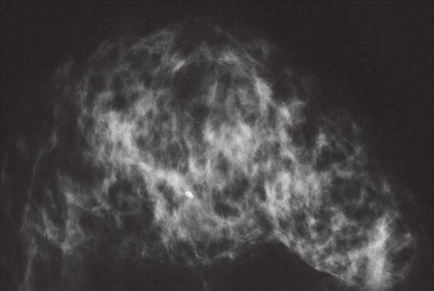
8 Hs-on screening course combined with 2nd DAY AM Hs-on screening didactic lecture series 8:00 AM - 12:00 PM 8:00-9:15 HANDS ON SCREENING WITH MAMMOGRAPHY. SESSION 2. 9:15-9:30 10:30-10:45 AM Breaks 9:30-10:30 DISCUSSION OF THE SCREENING CASES FROM SESSION 2 10:45 DIDACTIC LECTURE SERIES COVERING THE FOLLOWING TOPICS: The site of origin of the breast cancer influences diagnosis, choice of treatment patient outcome. NON-CALCIFIED ASYMMETRIC DENSITIES WITH architectural distortion on the mammogram. ANALYSIS of BENIGN RADIATING STRUCTURES on the mammogram, originating in the ducts: Radial scar Radial scar Neoductgenesis ANALYSIS of MALIGNANT LESIONS PRESENTING as RADIATING STRUCTUREs on the mammogram. Clinical presentation, mammographic appearance outcome 1) Duct forming invasive carcinoma / Neoductgenesis cases presenting on the mammogram as architectural distortion. The role of MRI in diagnosing diffuse breast carcinoma Lunch 12:00 PM-1:00 PM VIII Non-calcified architectural distortion: extensive duct forming invasive cancer


9 Hs-on screening course combined with 2nd day afternoon Mammography MRI Interpretation sessions at workstations between 1:00 PM - 5:00 PM 1:00-2:15 MRI Interpretation sessions at workstations - 2:15-2:30 3:30-4:00 PM Breaks 2:30-3:30 HANDS ON SCREENING WITH MAMMOGRAPHY. SESSION 3. 4:00-5:00 DISCUSSION OF THE SCREENING CASES FROM SESSION 3 5:00 End of Day 2 IX


 dotted, snake skin-like, c) skipping")

10 Hs-on screening course combined with 3rd DAY AM Hs-on screening didactic lecture series 8:00 AM - 12:00 PM 8:00-9:00 HANDS ON SCREENING WITH MAMMOGRAPHY. SESSION 4. 9:00-9:15 10:15-10:30 AM Breaks 9:15-10:15 DISCUSSION OF THE SCREENING CASES FROM SESSION 4 10:00 ALGORITHM FOR CLASSIFYING BREAST DISEASES ACCORDING TO THEIR SITE OF ORIGIN See pages X XI. Benign plasma cell mastitis type calcifications in major ducts BREAST DISEASES ORIGINATING IN THE MAJOR DUCTS Benign type calcifications originating in the major ducts a) Secretory disease type calcifications Malignant type calcifications originating in the major ducts: Four different types of calcifications: a) fragmented casting type, b) dotted, snake skin-like, c) skipping stone-like d) pearl necklace-like a) Fragmented casting type calcifications. Example 1. Example 2. Fragmented casting type calcifications (breast cancer of ductal origin DAB). X Lunch 12:00 PM - 1:00 PM


 Dotted casting type calcifications snake skin-like * The")


11 Hs-on screening course combined with 3rd DAY AM ALGORITHM FOR CLASSIFYING BREAST DISEASES ACCORDING TO THEIR SITE OF ORIGIN Images supporting the morning lecture: ANALYSIS of CALCIFIED MALIGNANT BREAST LESIONS ORIGINATING in the MAJOR DUCTS, cont. Clinical presentation, mammographic appearance patient outcome.the role of MRI in diagnosing diffuse breast carcinoma - Tabar L, Ingvarsson, M. b) Dotted casting type calcifications snake skin-like * The concept of neoductgenesis. Long-term follow-up results. New aspects, correct terminology. * The role of breast MRI examination in demonstrating the extent of Gr 3 in situ carcinoma. - M Ingvarsson * Mammographic /3D histologic correlation helping to explain the underlying pathophysiology outcome. c) Skipping stone-like calcifications d) Pearl necklace-like calcifications Practice of calcification analysis. Faculty-audience interaction. Lunch 12:00 PM - 1:00 PM XI


12 Hs-on screening course combined with 3rd DAY PM MRI interpretation hs-on screening Breaks at 2:15 at 3:30 PM 1:00-2:15 MRI INTERPRETATION SESSION AT WORKSTATIONS. 2:30-3:30 HANDS ON SCREENING WITH MAMMOGRAPHY. SESSION 5. 4:00-5:00 DISCUSSION OF THE SCREENING CASES FROM SESSION 5 5:00 PM End of Day 3 XII


13 Hs-on screening course combined with 3rd DAY PM MRI interpretation hs-on screening Cases supporting the MRI workshop Subtle mammography finding / MRI shows that the entire lobe is filled with a diffuse breast cancer, confirmed at histology Multifocal invasive in situ carcinoma on an area measuring 180X60 mm pn 4/9 5:00 PM End of Day 3 XIII


14 Hs-on screening course combined with 4th day Morning program between 8:00 AM - 12:00 PM Breaks at 9:15 at 10:00 AM 8:00-9:00 HANDS ON SCREENING. SESSION 6 a) Normal cases mixed with b) cases having noncalcified architectural distortion, (both duct forming invasive carcinoma diffusely infiltrating breast caancers) c) calcifications localized within the major ducts TDLUS. d) 1-14 mm unifocal multifocal stellate circular tumors. 9:15-10:00 DISCUSSION OF THE SCREENING CASES FROM SESSION 6 10:15 ALGORITHM FOR CLASSIFYING BREAST DISEASES ACCORDING TO THEIR SITE OF ORIGIN Benign breast diseases originating in the TDLU associated with calcifications on the mammogram - Fibrocystic change. Fibroadenoma. Different types of adenosis. Understing pathophysiology leading to calcified non-calcified hyperplastic breast changes. A - Conventional 3D histology images of small breast cysts containing sediment of psammoma body-like calcifications, seen as "teacup-like calcifications on the mammogram. - Detailed analysis of calcifications associated with hyperplastic breast changes Weddellites (A), powdery calcifications (B), pleomorphic calcifications on the mammogram. B 12:00 Lunch XIV


15 2018 Hs-on screening course combined with 4th day Afternoon program between 1:00 PM - 4:00 PM Breaks at 2:00 at 3:00 PM 1:00 PM THE DIDACTIC LECTURE SERIES WILL COVER THE FOLLOWING TOPICS: Grade 2 cancer in situ: Mammographic / 3-D histologic / MRI correlation of cases with crushed stone-like/pleomorphic calcifications on the mammogram. Mammographic / histologic correlation of pleomorphic calcifications The morphologic analysis of calcifications representing a less aggressive carcinoma: Grade 1 / well differentiated CIS Grade 1 in situ carcinoma: Mammographic / 3D histologic / MRI correlation of cases with powdery calcifications on the mammogram. 4:00 PM End of Course XV

 419 0227, Fax: (480) 419 0219,")


16 Hs-on screening course combined with For more information registration please contact: Mammography Education, Inc., 4429 E. Spur Drive CAVE CREEK, AZ 85331, USA. Ms. Donna Sokolik Phone: (480) , Fax: (480) , Internet: The schedule is subject to change without notice does not represent a commitment on the part of the international center. All rights reserved including the right of reproduction of the teaching material. Computer simulation images of the development of Grade 2 in situ carcinoma within the TDLU. The lobule becomes gradually distended deformed. Calcifications are formed within the necrotic debris are seen on the mammogram as crushed stone-like calcifications. XVI


17 Hs-on screening course combined with


18 Hs-on screening course combined with


19 Hs-on screening course combined with


20 Hs-on screening course combined with


21 2018 Hs-on screening course combined with László Tabár, MD Tibor Tot, MD, Peter B. Dean, MD Ductal Adenocarcinoma of the Breast (DAB), Part 5 Fluid producing DAB subtypes associated with calcifications In 3D László Tabár, MD Tibor Tot, MD, Peter B. Dean, MD Bloody serous nipple discharge Ductal Adenocarcinoma of the Breast (DAB), Part 6 In 3D

, Part 7")


")


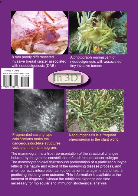
22 2018 Hs-on screening course combined with e László Tabár, MD Tibor Tot, MD, Peter B. Dean, MD In 3D Breast cancer originating from the major ducts Ductal Adenocarcinoma of the Breast (DAB), Part 7 Architectural distorion on the mammogram without calcifications or nipple discharge Mammographic-MRI-subgross (3D) histologic correlation of this extensive micropapillary cancer originating from the major ducts presenting as architectural distortion. Architectural distorion on the mammogram without calcifications or nipple discharge In 3D There are two main groups of diffuse breast cancers presenting on the mammogram as large regions of architectural distortion; these account for about 25% of all breast cancers tend to have a poor outcome: 1) Neoductgenesis, i.e. duct forming invasive carcinoma, the topic of this volume, often erroneously diagnosed as DCIS, 2) Diffusely infiltrating breast cancer, the topic of Vol. XI. This volume demonstrates the DAB subgroup where the unnaturally high concentration of abnormal, tumor-filled ducts results in an asymmetric density with architectural distortion on the mammogram often causes a palpable thickening. Detecting architectural distortion on the mammogram diagnosing the underlying disease correctly is a challenge for the radiologist. Breast cancers originating from the major ducts (DAB) are characterized by the formation of new, duct-like structures through the process of Neoductgenesis.
23 REGISTRATION FORM Screening Course Sept 18-21, 2018 in Sigtuna, Sweden First name: Surname: Complete address, country included: address: Telephone (work or cell): Address / name of Hospital or Breast Center: Tuition: 9,800.- SEK We need your name on the bank statement to let us know who paid Tuition includes: Participation at the hs-on screening course (see attached course schedule), teaching course hout material, breaks lunches course dinner on Sept 18, VAT (moms) registration number of the organization/institute (Hospital, County Council) that paid the tuition. Preferably, please register at MEI ( Paid to: Alternatively, tuition can be paid to Bank account number: Tabár Radiologi AB, Mark: Screening course Account code: 6 Address: Holmgardv. 10, Sigtuna, Sweden Org. Number: IBAN number: SE Bank BIC SWEDSESS Swedbank AB, BOX 717, FALUN, Sweden Please mail this form together with the money transfer receipt to: laszlo@mammographyed.com
2017BREAST SEMINAR SERIES
 Hands-on Breast Screening and Diagnosis Course * Screening of 510 full field digital mammography cases. * Reading a mixture of normals and proven abnormals at high resolution viewing stations. * Immediate
2017BREAST SEMINAR SERIES
 Hands-on Breast Screening and Diagnosis Course * Screening of 510 full field digital mammography cases. * Reading a mixture of normals and proven abnormals at high resolution viewing stations. * Immediate
Mammography Education, Inc.
 Hands-on Breast Screening and Diagnosis Course * Screening of 315 full field digital mammography cases. * Reading a mixture of normals and proven abnormals at high resolution viewing stations. * Immediate
Mammography Education, Inc.
 Mammography Education, Inc. 2018 3D image of the breast tissue BREAST SEMINAR SERIES Faculty LÁSZLÓ TABÁR, MD, FACR (Hon) Professor emeritus of Radiology Using the Multimodality Approach. A FULLY INTERACTIVE,
Mammography Education, Inc.
 Hands-on Breast Screening and Diagnosis Course * Screening of 315 full field digital mammography cases. * Reading a mixture of normals and proven abnormals at high resolution viewing stations. * Immediate
Scottsdale, AZ Plaza Hotel, 7200 N. Scottsdale Rd
 Mammography Education, Inc. 2014 3D image of the breast tissue LÁSZLÓ TABÁR, M.D.,F.A.C.R (Hon) and STAMATIA DESTOUNIS, M.D., F.A.C.R. The normal TDLUs have bud-like acini Multimodality Approach to Detection
Mammography Education, Inc.
 Mammography Education, Inc. 2018 3D image of the breast tissue BREAST SEMINAR SERIES Faculty LÁSZLÓ TABÁR, MD, FACR (Hon) Professor emeritus of Radiology Using the Multimodality Approach A FULLY INTERACTIVE,
Mammography Education, Inc.
 Mammography Education, Inc. 2018 3D image of the breast tissue BREAST SEMINAR SERIES Faculty LÁSZLÓ TABÁR, MD, FACR (Hon) Professor emeritus of Radiology The Critical Role of the Breast Imaging Technologists
Advanced Course on Multimodality Detection and Diagnosis of Breast Diseases
 BREAST SEMINAR Advanced Course on Multimodality Detection and 3D image of the breast tissue Invited speaker LÁSZLÓ TABÁR, MD,FACR (Hon) Falun, Sweden Nov 22-23, 2014 ATHENS, Greece Royal Olympic Hotel
Mammography Education, Inc.
 Mammography Education, Inc. 2018 3D image of the breast tissue BREAST SEMINAR SERIES Faculty LÁSZLÓ TABÁR, MD, FACR (Hon) Professor emeritus of Radiology and GILLIAN NEWSTEAD, MD, FACR Professor emeritus
Mammography Education, Inc. Multimodality approach to screening, diagnosis and differential diagnosis of breast diseases
 Mammography Education, Inc. 2015 BREAST SEMINAR SERIES The Parliament building in Pest viewed from Buda LÁSZLÓ TABÁR, MD, FACR (Hon) Multimodality approach to screening, diagnosis and differential diagnosis
Breast Cancer: Basic and Clinical Research 2014:8
 Open Access: Full open access to this and thousands of other papers at http://www.la-press.com. Breast Cancer: Basic and Clinical Research A Proposal to Unify the Classification of Breast and Prostate
Ana Sofia Preto 19/06/2013
 Ana Sofia Preto 19/06/2013 Understanding the underlying pathophysiologic processes leading to the various types of calcifications Description and illustration of the several types of calcifications, according
Imaging in breast cancer. Mammography and Ultrasound Donya Farrokh.MD Radiologist Mashhad University of Medical Since
 Imaging in breast cancer Mammography and Ultrasound Donya Farrokh.MD Radiologist Mashhad University of Medical Since A mammogram report is a key component of the breast cancer diagnostic process. A mammogram
MEDICAL IMAGING AND BREAST DISEASE HOW CAN WE HELP YOU?
 MEDICAL IMAGING AND BREAST DISEASE HOW CAN WE HELP YOU? Barbara M. Preston, M.D. SCREENING MAMMOGRAPHY AVERAGE RISK PATIENTS KAISER RECOMMENDATION: ALL WOMEN (INCLUDING TRANSGENDER FEMALES) Every 1-21
Multiparameter characterization of breast carcinoma: subgross, microscopy, proteins, and genes
 World Congress on Breast Cancer August 1-3, 2015, Birmingham, UK Multiparameter characterization of breast carcinoma: subgross, microscopy, proteins, and genes Tibor Tot Falun, Sweden Lake Varpan, Falun,
Diagnostic Dilemmas of Breast Imaging
 Diagnostic Dilemmas of Breast Imaging Common Causes of Error in Breast Cancer Detection By: Jason Cord, M.D. Mammography: Initial Imaging The standard for detection of breast cancer Screening mammography
EARLY DETECTION: MAMMOGRAPHY AND SONOGRAPHY
 EARLY DETECTION: MAMMOGRAPHY AND SONOGRAPHY Elizabeth A. Rafferty, M.D. Avon Comprehensive Breast Center Massachusetts General Hospital Harvard Medical School Breast Cancer Screening Early detection of
One or Two Clusters of Crushed Stone like Calcifications on the Mammogram Produced by Malignancy
 66 One or Two Clusters of Crushed Stone like Calcifications on the Mammogram Produced by Malignancy Example 2.13 A 36-year-old woman who recentlyfelt a small hard lump in the upper-outer quadrant of her
EARLY DETECTION: MAMMOGRAPHY AND SONOGRAPHY
 EARLY DETECTION: MAMMOGRAPHY AND SONOGRAPHY Elizabeth A. Rafferty, M.D. Avon Comprehensive Breast Center Massachusetts General Hospital Harvard Medical School Breast Cancer Screening Early detection of
RADIOLOGIC EVALUATION OF BREAST CANCER
 RADIOLOGIC EVALUATION OF BREAST CANCER Orsolya Farkas, Gabriella Bodrogi and Gábor Szalai Department of Radiology, Pécs University Orsifarkas@yahoo.com Complex evaluation of the breast Patient history
DCIS of the Breast--MRI findings with mammographic correlation.
 DCIS of the Breast--MRI findings with mammographic correlation. Poster No.: C-1560 Congress: ECR 2013 Type: Educational Exhibit Authors: N. B. Ibrahim, P. Morris, S. ANANDAN; Burlington, MA/US Keywords:
Non-mass Enhancement on Breast MRI. Aditi A. Desai, MD Margaret Ann Mays, MD
 Non-mass Enhancement on Breast MRI Aditi A. Desai, MD Margaret Ann Mays, MD Breast MRI Important screening and diagnostic tool, given its high sensitivity for breast cancer detection Breast MRI - Indications
Leonard M. Glassman MD Analysis of Breast Calcifications
 Importance of Calcification Leonard M. Glassman MD FACR American Institute for Radiologic Pathology Washington Radiology Associates, PC Washington DC 45% of all breast cancers present as calcification
Lesion Imaging Characteristics Mass, Favoring Benign Circumscribed Margins Intramammary Lymph Node
 Lesion Imaging Characteristics Mass, Favoring Benign Circumscribed Margins Intramammary Lymph Node Oil Cyst Mass, Intermediate Concern Microlobulated Margins Obscured Margins Mass, Favoring Malignant Indistinct
In multiple published papers digital breast
 2015 September 26-27, 2015 San Diego, CA In multiple published papers digital breast tomosynthesis (DBT) has demonstrated the ability to improve the detection of breast cancer while, at the same time,
Breast Imaging Lexicon
 9//201 200 BI RADS th Edition 201 BI RADS th Edition Breast Imaging Lexicon Mammographic Pathology and Assessment Categories Deborah Thames, R.T.(R)(M)(QM) The Advanced Health Education Center Nonmember:
Breast pathology. 2nd Department of Pathology Semmelweis University
 Breast pathology 2nd Department of Pathology Semmelweis University Breast pathology - Summary - Benign lesions - Acute mastitis - Plasma cell mastitis / duct ectasia - Fat necrosis - Fibrocystic change/
Armed Forces Institute of Pathology.
 Armed Forces Institute of Pathology www.radpath.com Armed Forces Institute of Pathology Breast Disease www.radpath.org Armed Forces Institute of Pathology Evaluation of Breast Calcifications Leonard M.
Research Article Ductal Breast Carcinoma In Situ: Mammographic Features and Its Relation to Prognosis and Tumour Biology in a Population Based Cohort
 Hindawi International Journal of Breast Cancer Volume 2017, Article ID 4351319, 9 pages https://doi.org/10.1155/2017/4351319 Research Article Ductal Breast Carcinoma In Situ: Mammographic Features and
Microcalcifications detected on mammography classified as BIRADS 4 and 5 and their correlations with histopatologic findigns
 Microcalcifications detected on mammography classified as BIRADS 4 and 5 and their correlations with histopatologic findigns Poster No.: C-0401 Congress: ECR 2010 Type: Educational Exhibit Topic: Breast
Detailed Program of the second BREAST IMAGING AND INTERVENTIONS PROGRAM am am : Clinician s requirements from breast imaging
 Detailed Program of the second BREAST IMAGING AND INTERVENTIONS PROGRAM 2012 Day one, 2 nd November BREAST IMAGING AND INTERVENTIONS PROGRAM 2012 9.00 AM 9.10 am Introduction 9.10 am - 9.30 am : Clinician
Diseases of the breast (1 of 2)
") Diseases of the breast (1 of 2) Introduction A histology introduction Normal ducts and lobules of the breast are lined by two layers of cells a layer of luminal cells overlying a second layer of myoepithelial
Diseases of the breast (1 of 2) Introduction A histology introduction Normal ducts and lobules of the breast are lined by two layers of cells a layer of luminal cells overlying a second layer of myoepithelial
Here are examples of bilateral analog mammograms from the same patient including CC and MLO projections.
 Good afternoon. It s my pleasure to be discussing Diagnostic Breast Imaging over the next half hour. I m Wei Yang, Professor of Diagnostic Radiology and Chief, the Section of Breast Imaging as well as
Screening Mammograms: Questions and Answers
 CANCER FACTS N a t i o n a l C a n c e r I n s t i t u t e N a t i o n a l I n s t i t u t e s o f H e a l t h D e p a r t m e n t o f H e a l t h a n d H u m a n S e r v i c e s Screening Mammograms:
Imaging the Symptomatic Patient. Avice M.O Connell MD,FACR,FSBI Professor of Imaging Sciences Director, Women s Imaging University of Rochester
 Imaging the Symptomatic Patient Avice M.O Connell MD,FACR,FSBI Professor of Imaging Sciences Director, Women s Imaging University of Rochester The four most common symptoms Mass Pain Discharge Infection
04/10/2018 HIGH RISK BREAST LESIONS. Pathology Perspectives of High Risk Breast Lesions ELEVATED RISK OF BREAST CANCER HISTORICAL PERSPECTIVES
 Pathology Perspectives of High Risk Breast Lesions Savitri Krishnamurthy MD Professor of Pathology Deputy Division Head Director of Clinical Trials, Research and Development The University of Texas MD
In multiple published papers digital breast
 2016 Case-Based Review & Advanced Breast Imaging Course: DIGITAL BREAST TOMOSYNTHESIS NEW FOR 2016! Dedicated case review time on individual DBT workstations* September 17-18, 2016 San Diego, CA Register
ORIGINAL ARTICLE EVALUATION OF BREAST LESIONS USING X-RAY MAMMOGRAM WITH HISTOPATHOLOGICAL CORRELATION
 Available online at www.journalijmrr.com INTERNATIONAL JOURNAL OF MODERN RESEARCH AND REVIEWS IJMRR ISSN: 2347-8314 Int. J. Modn. Res. Revs. Volume 3, Issue 10, pp 807-814, October, 2015 ORIGINAL ARTICLE
BI-RADS and Breast MRI. Kathy Borovicka, M.D. Thursday February 15, 2018
 BI-RADS and Breast MRI Kathy Borovicka, M.D. Thursday February 15, 2018 Learning Objectives Be familiar with the Breast Imaging Reporting and Data System (BI-RADS) Understand the components of a breast
Current Status of Supplementary Screening With Breast Ultrasound
 Current Status of Supplementary Screening With Breast Ultrasound Stephen A. Feig, M.D., FACR Fong and Jean Tsai Professor of Women s Imaging Department of Radiologic Sciences University of California,
Radiologic-pathologic correlation of the mammographic findings retrospectively detected in inflammatory breast cancer: usefulness in clinical practice
 Radiologic-pathologic correlation of the mammographic findings retrospectively detected in inflammatory breast cancer: usefulness in clinical practice Francesca Caumo, Erminia Manfrin, Franco Bonetti,
Stanford Breast in the West: Multi-Modality Breast Imaging Symposium for Radiologists
 Course Overview Stanford Breast in the West: Multi-Modality Breast Imaging Symposium for Radiologists October 18-20, 2018 Monterey Plaza Hotel Monterey, CA A Continuing Medical Education Conference presented
LYMPHATIC DRAINAGE AXILLARY (MOSTLY) INTERNAL MAMMARY SUPRACLAVICULAR
 INTERNAL MAMMARY SUPRACLAVICULAR") BREAST LYMPHATIC DRAINAGE AXILLARY (MOSTLY) INTERNAL MAMMARY SUPRACLAVICULAR HISTOLOGY LOBE: (10 in whole breast) LOBULE: (many per lobe) ACINUS/I, aka ALVEOLUS/I: (many per lobule) DUCT(S): INTRA- or
BREAST LYMPHATIC DRAINAGE AXILLARY (MOSTLY) INTERNAL MAMMARY SUPRACLAVICULAR HISTOLOGY LOBE: (10 in whole breast) LOBULE: (many per lobe) ACINUS/I, aka ALVEOLUS/I: (many per lobule) DUCT(S): INTRA- or
Leonard M. Glassman MD
 BI-RADS The New BI-RADS Leonard M. Glassman MD FACR Former Chief of Breast Imaging American Institute for Radiologic Pathology Washington Radiology Associates, PC Breast Imaging Reporting and Data System
AMSER Case of the Month: November 2018
 AMSER Case of the Month: November 2018 52 year old female with an abnormal screening mammogram Areeg Rehman, MS 4 Nova Southeastern University Rebecca T. Sivarajah, MD Penn State University College of
Mousa. Israa Ayed. Abdullah AlZibdeh. 0 P a g e
 1 Mousa Israa Ayed Abdullah AlZibdeh 0 P a g e Breast pathology The basic histological units of the breast are called lobules, which are composed of glandular epithelial cells (luminal cells) resting on
Breast calcification: Management and Pictorial Review
 Breast calcification: Management and Pictorial Review Poster No.: C-0692 Congress: ECR 2014 Type: Educational Exhibit Authors: V. de Lara Bendahan, M. F. Ramos Solis, A. Amador Gil, C. 1 2 3 2 4 4 Gómez
Malignant transformation of fibroadenomas
 Malignant transformation of fibroadenomas Poster No.: C-2503 Congress: ECR 2013 Type: Educational Exhibit Authors: L. N. Elias, M. A. Rudner, L. M. Yano, P. C. Moraes, Y. 1 1 1 1 1 1 2 1 2 Chang, M. B.
Atypical proliferative lesions diagnosed on core biopsy - 6 year review
 Atypical proliferative lesions diagnosed on core biopsy - 6 year review Dr Angela Harris, Dr Julie Weigner & Dr Ricardo Vilain NSW Health Pathology Pathology North, Hunter Anatomical Pathology & Cytology
Breast Health and Imaging Glossary
 Contact: Lorna Vaughan HerSpace Breast Imaging & Biopsy Associates 300 State Route 35 South W. Long Branch, NJ 07764 732-571-9100, ext. 104 lorna@breast-imaging.com Breast Health and Imaging Glossary Women
Mammographic imaging of nonpalpable breast lesions. Malai Muttarak, MD Department of Radiology Chiang Mai University Chiang Mai, Thailand
 Mammographic imaging of nonpalpable breast lesions Malai Muttarak, MD Department of Radiology Chiang Mai University Chiang Mai, Thailand Introduction Contents Mammographic signs of nonpalpable breast cancer
THE EFFECT OF THE HOMOEOPATHIC SIMILIMUM ON SIDE EFFECTS OF CHEMOTHERAPY IN BREAST CANCER PATIENTS
 THE EFFECT OF THE HOMOEOPATHIC SIMILIMUM ON SIDE EFFECTS OF CHEMOTHERAPY IN BREAST CANCER PATIENTS A Dissertation submitted to the Faculty of Health Sciences, University of Johannesburg, in partial fulfillment
Breast Cancer Management 2014
 Breast Cancer Management 2014 Program Co-Directors: Sheldon Marc Feldman, MD, FACS & Dawn L. Hershman, MD, MS Friday, January 31, 2014 8:45am 5:50pm NewYork-Presbyterian/Columbia University Medical Center
Case study 1. Rie Horii, M.D., Ph.D. Division of Pathology Cancer Institute Hospital, Japanese Foundation for Cancer Research
 NCCN/JCCNB Seminar in Japan April 15, 2012 Case study 1 Rie Horii, M.D., Ph.D. Division of Pathology Cancer Institute Hospital, Japanese Foundation for Cancer Research Present illness: A 50y.o.premenopausal
2016 INTENSIVE BREAST ULTRASOUND
 New lectures based on new classification of breast disease 2016 INTENSIVE BREAST ULTRASOUND A Histopathologically based Approach to Diagnostic and Screening Breast Ultrasound SEPTEMBER 15-18, 2016 THE
Benign, Reactive and Inflammatory Lesions of the Breast
 Benign, Reactive and Inflammatory Lesions of the Breast Marilin Rosa, MD Associate Member Section Head of Breast Pathology Department of Anatomic Pathology Program Director, Breast Pathology Fellowship
Triple Negative Breast Cancer: Clinical Presentation and Multimodality Imaging Characteristics
 Triple Negative Breast Cancer: Clinical Presentation and Multimodality Imaging Characteristics Poster No.: R-0141 Congress: RANZCR-AOCR 2012 Type: Scientific Exhibit Authors: O. H. Woo, S. Jang, K. R.
Mammography. What is Mammography? What are some common uses of the procedure?
 Mammography What is Mammography? Mammography is a specific type of imaging that uses a low-dose x-ray system to examine breasts. A mammography exam, called a mammogram, is used to aid in the early detection
Introduction 1. Executive Summary 5
 Roman_pages 20-09-2005 21:01 Pagina IX Table of contents Introduction 1 Executive Summary 5 1. Epidemiological guidelines for quality assurance in breast cancer screening 15 1.10 Introduction 17 1.20 Local
Breast Disease: What PCPs Need to Know. Eunice Cho MD FACS
 Breast Disease: What PCPs Need to Know Eunice Cho MD FACS New Breast Cancer Screening Guideline for women with average risk Every other year AGE 40 AGE 45 AGE 55 AGE 55 + Talk with your doctor about when
IBCM 2, April 2009, Sarajevo, Bosnia and Herzegovina
 Preoperative diagnosis and treatment planning in breast cancer The pathologist s perspective L. Mazzucchelli Istituto Cantonale di Patologia Locarno, Switzerland IBCM 2, 23-25 April 2009, Sarajevo, Bosnia
1 NORMAL HISTOLOGY AND METAPLASIAS
 1 NORMAL HISTOLOGY AND METAPLASIAS, MD Anatomy and Histology 1 Metaplasias 2 ANATOMY AND HISTOLOGY The female breast is composed of a branching duct system, which begins at the nipple with the major lactiferous
Breast Imaging: Multidisciplinary Approach. Madelene Lewis, MD Assistant Professor Associate Program Director Medical University of South Carolina
 Breast Imaging: Multidisciplinary Approach Madelene Lewis, MD Assistant Professor Associate Program Director Medical University of South Carolina No Disclosures Objectives Discuss a multidisciplinary breast
November 23, Dear Maryland Breast and Cervical Cancer Program Provider:
 STATE OF MARYLAND DHMH Maryland Department of Health and Mental Hygiene 201 W. Preston Street Baltimore, Maryland 21201 Martin O Malley, Governor Anthony G. Brown, Lt. Governor John M. Colmers, Secretary
Armed Forces Institute of Pathology.
 Armed Forces Institute of Pathology www.radpath.com Armed Forces Institute of Pathology Breast Disease www.radpath.org Armed Forces Institute of Pathology Interpretation of Breast MRI Leonard M. Glassman
2016 Clinical Breast Imaging:
 ENTIRE PROGRAM: 22.25 AMA PRA Category 1 Credit(s) TM 11.75 AMA PRA Category 1 Credit(s) TM 8.5 AMA PRA Category 1 Credit(s) TM 5.5 AMA PRA Category 1 Credit(s) TM 10.5 AMA PRA Category 1 Credit(s) TM
Segmental Breast Calcifications
 Residents Section Pattern of the Month Chen et al. Segmental reast Calcifications Residents Section Pattern of the Month Residents inradiology Po-Hao Chen 1 Erica T. Ghosh 1,2 Priscilla J. Slanetz 1,2
Pictorial Essay Singapore Med J 2009; 50(9) :
 :") 907 Pictorial Essay CME Article Breast calcifications: which are malignant? Muttarak M, Kongmebhol P, Sukhamwang N ABSTRACT Most calcifications depicted on mammograms are benign. However, calcifications
907 Pictorial Essay CME Article Breast calcifications: which are malignant? Muttarak M, Kongmebhol P, Sukhamwang N ABSTRACT Most calcifications depicted on mammograms are benign. However, calcifications
Treatment options for the precancerous Atypical Breast lesions. Prof. YOUNG-JIN SUH The Catholic University of Korea
 Treatment options for the precancerous Atypical Breast lesions Prof. YOUNG-JIN SUH The Catholic University of Korea Not so benign lesions? Imaging abnormalities(10% recall) lead to diagnostic evaluation,
Triple-negative breast cancer: which typical features can we identify on conventional and MRI imaging?
 Triple-negative breast cancer: which typical features can we identify on conventional and MRI imaging? Poster No.: C-1862 Congress: ECR 2013 Type: Educational Exhibit Authors: V. Bertani 1, A. Gualano
Innovations in Breast Ultrasound
 Innovations in Breast Ultrasound Interactive learning in breast ultrasound and intervention March 26, 2011 Hyatt Regency Chicago on the Riverwalk Faculty: Terri-Ann Gizienski, MD Kathy Schilling, MD A.
Breast imaging in general practice
 Breast series CLINICAL PRACTICE Breast imaging in general practice Nehmat Houssami, MBBS, FAFPHM, FASBP, PhD, is Associate Clinical Director, NSW Breast Cancer Institute, Westmead Hospital, Honorary Senior
Mammographic Tumor Features Can Predict Long- Term Outcomes Reliably in Women with 1 14-mm Invasive Breast Carcinoma
 1745 Mammographic Tumor Features Can Predict Long- Term Outcomes Reliably in Women with 1 14-mm Invasive Breast Carcinoma Suggestions for the Reconsideration of Current Therapeutic Practice and the TNM
DOCTORAL THESIS SUMMARY
 UNIVERSITY OF MEDICINE AND PHARMACY CRAIOVA FACULTY OF MEDICINE DOCTORAL THESIS SUMMARY CLINICO-IMAGING STUDY OF INVASIVE DUCTAL BREAST CARCINOMAS CORRELATED TO HORMONAL RECEPTORS AND HER2/NEU ONCOPROTEIN
Intracystic papillary carcinoma of the breast
 Intracystic papillary carcinoma of the breast Poster No.: C-1932 Congress: ECR 2011 Type: Educational Exhibit Authors: V. Dimarelos, F. TZIKOS, N. Kotziamani, G. Rodokalakis, 1 2 3 1 1 1 2 T. MALKOTSI
Imaging & Care of the Breast Cancer Patient
 Imaging & Care of the Breast Cancer Patient Presented by Department of Radiology Sponsored by Saturday September 13, 2008 One Baylor Plaza Alkek Building, N315 Houston, TX Saturday, September 13, 2008
FACT: FACT: Breast Cancer Staging. For cancer to occur, something must damage nucleus of the cell. Stage I. Stage II 10/9/2018
 Digital Breast Tomosynthesis (DBT) Pathology Findings: Case Studies and Beyond For cancer to occur, something must damage nucleus of the cell. Advanced Health Education Center www.aheconline.com Copyright
ROLE OF MRI IN SCREENING, DIAGNOSIS AND MANAGEMENT OF BREAST CANCER. B.Zandi Professor of Radiology
 ROLE OF MRI IN SCREENING, DIAGNOSIS AND MANAGEMENT OF BREAST CANCER B.Zandi Professor of Radiology Introduction In the USA, Breast Cancer is : The Most Common Non-Skin Cancer The Second Leading cause of
CLINICAL SIGNIFICANCE OF BENIGN EPITHELIAL CHANGES
 Papillomas. Papillomas are composed of multiple branching fibrovascular cores, each having a connective tissue axis lined by luminal and myoepithelial cells ( Fig. 23-11 ). Growth occurs within a dilated
THE BREAST COURSE 2010
 THE BREAST COURSE 2010 SUNDAY AM 21 FEBRUARY 2010 SESSION 1 BREAST INTERVENTIONS (LIVE CASES) Moderator: R. Kuske 8:00 AM Introduction to The Breast Course R. Kuske Interdisciplinary Breast Care Management
Amammography report is a key component of the breast
 Review Article Writing a Mammography Report Amammography report is a key component of the breast cancer diagnostic process. Although mammographic findings were not clearly differentiated between benign
Promise of a beautiful day
 Promise of a beautiful day Ductal carcinoma in Situ Lobular Carcinoma in Situ Natural History Manosmed Tartous Oct 2009 Gérard ABADJIAN MD Pathology Department Hôtel-Dieu de France. Associate Professor
CPC 4 Breast Cancer. Rochelle Harwood, a 35 year old sales assistant, presents to her GP because she has noticed a painless lump in her left breast.
 CPC 4 Breast Cancer Rochelle Harwood, a 35 year old sales assistant, presents to her GP because she has noticed a painless lump in her left breast. 1. What are the most likely diagnoses of this lump? Fibroadenoma
Prebiopsy Localization of Nonpalpable Breast Lesions
 ORIGINAL ARTICLE Prebiopsy Localization of Nonpalpable Breast Lesions A. Zulfiqar, MMed* v. Param, DMRD** F.A. Meah, FRACS* s. Nair, FRCS** M.A. Siti-Aishah, DCP* A. N orizan, DCP** * Departments of Radiology,
Breast Pathology. Breast Development
 Breast Pathology Lecturer: Hanina Hibshoosh, M.D. Reading: Kumar, Cotran, Robbins, Basic Pathology, 6th Edition, pages 623-635 Breast Development 5th week - thickening of the epidermis - milk line 5th
PELVIC MRI COURSE. November 12-14, 2016
 Memorial Sloan Kettering Cancer Center is pleased to announce: PELVIC MRI COURSE November 12-14, 2016 Conference Location: Memorial Sloan Kettering Cancer Center Rockefeller Research Laboratories 430 East
Ductal carcinoma in situ: ultrasound, mammography and MRI features with pathologic correlation
 Ductal carcinoma in situ: ultrasound, mammography and MRI features with pathologic correlation Poster No.: C-2252 Congress: ECR 2013 Type: Educational Exhibit Authors: L. Fernandes, H. A. M. R. Tinto,
Mammographic evaluation of palpable breast masses with pathological correlation: a tertiary care centre study in Nepal
 Original article 21 Mammographic evaluation of palpable breast masses with pathological correlation: a tertiary care centre study in Nepal G. Gurung, R. K. Ghimire, B. Lohani Department of Radiology and
Mammography is a most effective imaging modality in early breast cancer detection. The radiographs are searched for signs of abnormality by expert
 Abstract Methodologies for early detection of breast cancer still remain an open problem in the Research community. Breast cancer continues to be a significant problem in the contemporary world. Nearly
Melissa Hartman, DO Women s Health Orlando VA Medical Center
 Melissa Hartman, DO Women s Health Orlando VA Medical Center Most common non-skin cancer and Second deadliest cancer in women Majority are diagnosed by abnormal screening study An approach to breast cancer
Courtesy University of Michigan, available under a Creative Commons Attribution-NonCommercial 3.0 license
 HANDS-ON SCANNING WORKSHOPS Courtesy, available under a Creative Commons Attribution-NonCommercial 3.0 license Course Co-Directors:,, Musculoskeletal Ultrasound Course Upper and Lower Extremities - The
Benign Intraparenchymal Scarring in the DBT Era
 Benign Intraparenchymal Scarring in the DBT Era David Gruen, MD, MBA, FACR Director of Women s Imaging, Stamford Health, Stamford, CT With the advent of Digital Breast Tomosynthesis (DBT or 3D mammography),
Diseases of the breast (2 of 2) Breast cancer
 Breast cancer") Diseases of the breast (2 of 2) Breast cancer Epidemiology & etiology The most common type of cancer & the 2 nd most common cause of cancer death in women 1 of 8 women in USA Affects 7% of women Peak at
Diseases of the breast (2 of 2) Breast cancer Epidemiology & etiology The most common type of cancer & the 2 nd most common cause of cancer death in women 1 of 8 women in USA Affects 7% of women Peak at
Multidisciplinary Breast Pathology
 Multidisciplinary Breast Pathology Advanced Learning Series MANUAL This Multidisciplinary Breast Pathology manual is current as of October, 2018. Information is subject to change. CURRICULUM The MBP Advanced
Breast Imaging! Ravi Adhikary, MD!
 Breast Imaging! Ravi Adhikary, MD! ACS Estimated Cancers Statistics 2014! Breast! New Cases in Women! 232,670 (+67,570 in situ)! Deaths in Women! 40,000! Colon! 48,380! 24,040! Cervical! 12,360! 4,020!
Mammography. What is Mammography?
 Scan for mobile link. Mammography Mammography is a specific type of breast imaging that uses low-dose x-rays to detect cancer early before women experience symptoms when it is most treatable. Tell your
Case Scenario 1: This case has been slightly modified from the case presented during the live session to add clarity.
 Case Scenario 1: This case has been slightly modified from the case presented during the live session to add clarity. Background: 46 year old married premenopausal female with dense breasts has noticed
BI-RADS CATEGORIZATION AND BREAST BIOPSY categorization in the selection of appropriate breast biopsy technique is also discussed. Patients and method
 Original Article Positive Predictive Value of BI-RADS Categorization in an Asian Population Yah-Yuen Tan, Siew-Bock Wee, Mona P.C. Tan and Bee-Kiang Chong, 1 Departments of General Surgery and 1Diagnostic