Diseases of the breast (1 of 2)
|
|
|
- Dina Skinner
- 6 years ago
- Views:
Transcription
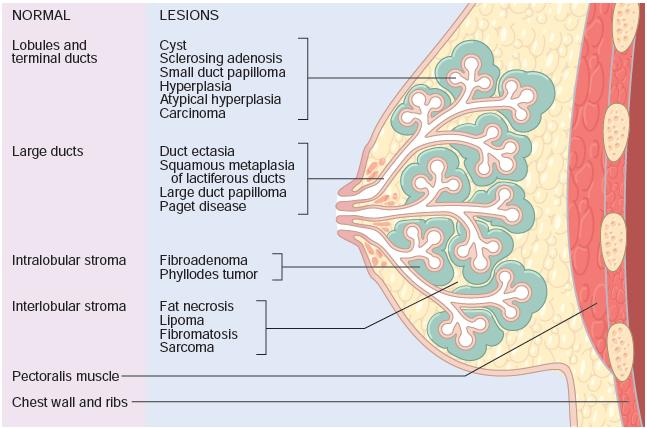
1 Diseases of the breast (1 of 2)
2 Introduction

3 A histology introduction Normal ducts and lobules of the breast are lined by two layers of cells a layer of luminal cells overlying a second layer of myoepithelial cells
4 Fibrocystic changes The most common breast abnormality seen in premenopausal women Most likely a consequence of the cyclic breast changes that occur normally in the menstrual cycle but not associated with OCPs Composed of nonproliferative and proliferative changes the most common type of fibrocystic lesions -fibrosis -cysts due to dilation of ducts -epithelial hyperplasia more than two luminal cell layers typical or atypical hyperplasia -sclerosing adenosis
5 Nonproliferative changes, morphology Usually multifocal and often bilateral Ill-defined, diffusely increased densities and discrete nodularities on mammography Unopened, they are brown to blue (blue dome cysts) and are filled with watery, turbid fluid The secretions within the cysts may calcify, producing microcalcifications on mammograms Histologically: an epithelial lining that in larger cysts may be flattened or even totally atrophic frequently, the lining cells are large and polygonal with abundant granular, eosinophilic cytoplasm and small, round, deeply chromatic nuclei. Such morphology is called apocrine metaplasia A stromal lymphocytic infiltrate is common
 can be seen Sometimes, the proliferating epithelium projects as multiple small papillary excrescences into the ductal lumen (ductal papillomatosis) Occasionally, hyperplasia")
6 Proliferative changes, morphology Coexisting fibrous or cystic changes are usually present The ducts, ductules, or lobules may be filled with orderly cuboidal cells within which small gland patterns (called fenestrations) can be seen Sometimes, the proliferating epithelium projects as multiple small papillary excrescences into the ductal lumen (ductal papillomatosis) Occasionally, hyperplasia produces microcalcifications on mammography There is also atypical ductal hyperplasia (resembles ductal carcinoma in situ) and another lesion called: atypical lobular hyperplasia (resembles lobular carcinoma in situ Both atypical ductal and atypical lobular hyperplasia are associated with an increased risk of invasive carcinoma
7 Proliferative changes, sclerosing adenosis less common than cysts and hyperplasia Significant because its clinical and morphologic features may mimic those of carcinoma

8 Relationship of fibrocystic changes to breast carcinoma *Bilaterality & multifocality of proliferative epithelial changes and related cancers are common
9 Fibroadenoma The most common benign neoplasm of the female breast Typically appear in young women with a peak incidence in the third decade of life A biphasic tumor composed of fibroblastic stroma and epithelium-lined glands only the stromal cells are clonal and truly neoplastic Usually manifest as solitary, discrete, mobile masses There is a role of estrogen: fibroadenomas may enlarge late in the menstrual cycle and during pregnancy after menopause, they may regress and calcify
10 Fibroadenoma, morphology Discrete masses, 1 cm to 10 cm in diameter and of firm consistency Cut section shows a uniform tan-white color, punctuated by softer yellowpink specks representing the glandular areas Histologically: loose fibroblastic stroma containing duct-like, epithelium-lined spaces of various shapes and sizes as in normal breast tissue, these glandular spaces are lined by luminal and myoepithelial cells with a well-defined, intact basement membrane

11 Fibroadenoma, morphology cont d

 Much less common than fibroadenomas Can be benign, borderline or malignant")
12 Phyllodes tumor Biphasic, being composed of neoplastic stromal cells and epithelium-lined glands The stromal element of these tumors is more cellular and abundant, often forming epithelium-lined leaflike projections (phyllodes is Greek for leaflike ) Much less common than fibroadenomas Can be benign, borderline or malignant but mostly benign
13 Phyllodes tumor, cont d Features suggestive of malignancy: -Increased stromal cellularity -Anaplasia -High mitotic activity -Rapid increase in size -Infiltrative margins Benign phyllodes is well-localized and is removed by excision Malignant lesions may recur, but they also tend to remain localized
14 Intraductal papilloma *Clinically: A benign neoplastic papillary growth It is most often seen in premenopausal women Typically solitary and found within the principal lactiferous ducts or sinuses On histologic examination: they are composed of multiple papillae, each having a connective tissue core covered by epithelial cells that are double-layered, with an outer luminal layer overlying a myoepithelial layer The presence of a double-layered epithelium helps to distinguish intraductal papilloma from intraductal papillary carcinoma, which can present with clinical features similar to benign papilloma
15 Acute mastitis Bacteria, usually Staphylococcus aureus, gain access to the breast tissue through the ducts The vast majority of cases arise during the early weeks of nursing, when the skin of the nipple is vulnerable to the development of fissures Clinically, staphylococcal infections induce typical acute inflammatory changes, which can progress to form single or multiple abscesses
16 Mammary duct ectasia A nonbacterial chronic inflammation of the breast associated with inspissation of breast secretions in the main excretory ducts Ductal dilation and eventual rupture leads to reactive changes in the surrounding tissue that may present as a poorly defined periareolar mass with nipple retraction, mimicking the changes caused by some cancers It is an uncommon condition usually encountered in parous women between 40 and 60 years of age Periductal lymphoplasmacytic inflammation is the most distinguishing feature microscopically
17 Fat necrosis Uncommon, innocuous lesion that is significant only because it often produces a mass may be very similar to cancer clinically and radiologically as a painless palpable mass, skin thickening or retraction, or mammographic densities or calcifications Most women with this condition report some antecedent trauma to the breast
18 Fat necrosis, morphology During the early stage of traumatic fat necrosis, the lesion is small, often tender, rarely more than 2 cm in diameter, and sharply localized Eventually is replaced by scar tissue or a cyst consisting of necrotic debris Calcifications may develop in either the scar or the cyst wall
19 Squamous Metaplasia of Lactiferous Ducts (Zuska disease) Painful erythematous subareolar mass that clinically appears to be a bacterial abscess women and sometimes, men More than 90% of the afflicted are smokers Squamous metaplasia causes keratin plugging in the ducts, followed by rupture and superimposed infection recurrent subareolar abscesses With recurrence, a fistula tract opens into the edge of the areola

20
BREAST PATHOLOGY. Fibrocystic Changes
 BREAST PATHOLOGY Lesions of the breast are very common, and they present as palpable, sometimes painful, nodules or masses. Most of these lesions are benign. Breast cancer is the 2 nd most common cause
CLINICAL SIGNIFICANCE OF BENIGN EPITHELIAL CHANGES
 Papillomas. Papillomas are composed of multiple branching fibrovascular cores, each having a connective tissue axis lined by luminal and myoepithelial cells ( Fig. 23-11 ). Growth occurs within a dilated
Breast Pathology. Breast Development
 Breast Pathology Lecturer: Hanina Hibshoosh, M.D. Reading: Kumar, Cotran, Robbins, Basic Pathology, 6th Edition, pages 623-635 Breast Development 5th week - thickening of the epidermis - milk line 5th
Breast pathology. 2nd Department of Pathology Semmelweis University
 Breast pathology 2nd Department of Pathology Semmelweis University Breast pathology - Summary - Benign lesions - Acute mastitis - Plasma cell mastitis / duct ectasia - Fat necrosis - Fibrocystic change/
BREAST PATHOLOGY MCQS
 BREAST PATHOLOGY MCQS 1) :The most important factor in breast enlargement during pregnancy is A. stromal edema B. secretion of chorionic gonadotropin C. glandular hyperplasia D. proliferation of stroma
LYMPHATIC DRAINAGE AXILLARY (MOSTLY) INTERNAL MAMMARY SUPRACLAVICULAR
 INTERNAL MAMMARY SUPRACLAVICULAR") BREAST LYMPHATIC DRAINAGE AXILLARY (MOSTLY) INTERNAL MAMMARY SUPRACLAVICULAR HISTOLOGY LOBE: (10 in whole breast) LOBULE: (many per lobe) ACINUS/I, aka ALVEOLUS/I: (many per lobule) DUCT(S): INTRA- or
BREAST LYMPHATIC DRAINAGE AXILLARY (MOSTLY) INTERNAL MAMMARY SUPRACLAVICULAR HISTOLOGY LOBE: (10 in whole breast) LOBULE: (many per lobe) ACINUS/I, aka ALVEOLUS/I: (many per lobule) DUCT(S): INTRA- or
Mousa. Israa Ayed. Abdullah AlZibdeh. 0 P a g e
 1 Mousa Israa Ayed Abdullah AlZibdeh 0 P a g e Breast pathology The basic histological units of the breast are called lobules, which are composed of glandular epithelial cells (luminal cells) resting on
COMMON BENIGN DISORDERS AND DISEASES OF THE BREAST
 COMMON BENIGN DISORDERS AND DISEASES OF THE BREAST Aberrations of Normal Development and Involution (ANDI). The basic principles underlying the aberrations of normal development and involution (ANDI) classification
Papillary Lesions of the Breast A Practical Approach to Diagnosis. (Arch Pathol Lab Med. 2016;140: ; doi: /arpa.
 Papillary Lesions of the Breast A Practical Approach to Diagnosis (Arch Pathol Lab Med. 2016;140:1052 1059; doi: 10.5858/arpa.2016-0219-RA) Papillary lesions of the breast Span the spectrum of benign,
DISORDERS OF THE BREAST Dated. FIBROADENOSIS Other common names: mastitis, fibrocystic disease, cystic mammary dysplasia.
 DISORDERS OF THE BREAST Dated BENIGN BREAST DISORDERS (Essential Surg 2 nd Ed, pp 540) FIBROADENOSIS Other common names: mastitis, fibrocystic disease, cystic mammary dysplasia. Fibroadenosis is the distortion
Benign, Reactive and Inflammatory Lesions of the Breast
 Benign, Reactive and Inflammatory Lesions of the Breast Marilin Rosa, MD Associate Member Section Head of Breast Pathology Department of Anatomic Pathology Program Director, Breast Pathology Fellowship
Imaging in breast cancer. Mammography and Ultrasound Donya Farrokh.MD Radiologist Mashhad University of Medical Since
 Imaging in breast cancer Mammography and Ultrasound Donya Farrokh.MD Radiologist Mashhad University of Medical Since A mammogram report is a key component of the breast cancer diagnostic process. A mammogram
1 NORMAL HISTOLOGY AND METAPLASIAS
 1 NORMAL HISTOLOGY AND METAPLASIAS, MD Anatomy and Histology 1 Metaplasias 2 ANATOMY AND HISTOLOGY The female breast is composed of a branching duct system, which begins at the nipple with the major lactiferous
CPC 4 Breast Cancer. Rochelle Harwood, a 35 year old sales assistant, presents to her GP because she has noticed a painless lump in her left breast.
 CPC 4 Breast Cancer Rochelle Harwood, a 35 year old sales assistant, presents to her GP because she has noticed a painless lump in her left breast. 1. What are the most likely diagnoses of this lump? Fibroadenoma
SIGNIFICANT OTHERS. Miscellaneous Benign Breast Conditions
 SIGNIFICANT OTHERS Miscellaneous Benign Breast Conditions Epworth HealthCare 1 FAT NECROSIS TRAUMATIC Cell rupture Seat-Belt injury Blunt trauma Iatrogenic injury Surgery, Flaps, Radiotherapy Pathology
Abid Irshad, MD Director Breast Imaging. Medical University of South Carolina Charleston
 Abid Irshad, MD Director Breast Imaging Medical University of South Carolina Charleston Cases Financial disclosure: I or my family have no financial interest related to the material discussed in this presentation
Pathology & Presentation of Benign Breast Disease Zdenek Dubrava - February 2006
 Pathology & Presentation of Benign Breast Disease Zdenek Dubrava - February 2006 Presentation Lump Pain Nipple Discharge Breast Shape/Size Richard J Santen, Robert Mansel. The New England Journal of Medicine.
Treatment options for the precancerous Atypical Breast lesions. Prof. YOUNG-JIN SUH The Catholic University of Korea
 Treatment options for the precancerous Atypical Breast lesions Prof. YOUNG-JIN SUH The Catholic University of Korea Not so benign lesions? Imaging abnormalities(10% recall) lead to diagnostic evaluation,
HISTOPATHOLOGICAL EVALUATION OF BENIGN PROLIFERATIVE BREAST LESIONS
 7 ORIGINAL ARTICLE HISTOPATHOLOGICAL EVALUATION OF BENIGN PROLIFERATIVE BREAST LESIONS DR. VIBHUTI H. CHIHLA*, DR. N N. JAGRIT **, DR. JAYASHREE M. SHAH*** *3 rd year Pathology Resident, **Associate Professor,
Papillary Lesions of the breast
 Papillary Lesions of the breast Emad Rakha Professor of Breast Pathology The University of Nottingham Papillary lesions of the breast are a heterogeneous group of disease, which are characterised by neoplastic
04/10/2018. Intraductal Papillary Neoplasms Of Breast INTRADUCTAL PAPILLOMA
 Intraductal Papillary Neoplasms Of Breast Savitri Krishnamurthy MD Professor of Pathology Deputy Division Head The University of Texas MD Anderson Cancer Center 25 th Annual Seminar in Pathology Pittsburgh,
Proliferative Epithelial lesions of the Breast. Sami Shousha, MD, FRCPath Charing Cross Hospital & Imperial College, London
 Proliferative Epithelial lesions of the Breast Sami Shousha, MD, FRCPath Charing Cross Hospital & Imperial College, London Amman, November2013 Proliferative Epithelial Lesions of the Breast Usual type
Breast Disease: What PCPs Need to Know. Eunice Cho MD FACS
 Breast Disease: What PCPs Need to Know Eunice Cho MD FACS New Breast Cancer Screening Guideline for women with average risk Every other year AGE 40 AGE 45 AGE 55 AGE 55 + Talk with your doctor about when
Inflammatory and Reactive Lesions of the Breast
 Inflammatory and Reactive Lesions of the Breast Laura C. Collins, M.D. Vice Chair of Anatomic Pathology Professor of Pathology Beth Israel Deaconess Medical Center and Harvard Medical School Boston, MA
Lesion Imaging Characteristics Mass, Favoring Benign Circumscribed Margins Intramammary Lymph Node
 Lesion Imaging Characteristics Mass, Favoring Benign Circumscribed Margins Intramammary Lymph Node Oil Cyst Mass, Intermediate Concern Microlobulated Margins Obscured Margins Mass, Favoring Malignant Indistinct
Cytyc Corporation - Case Presentation Archive - March 2002
 FirstCyte Ductal Lavage History: 68 Year Old Female Gail Index: Unknown Clinical History: Negative Mammogram in 1995 6 yrs. later presents with bloody nipple discharge Subsequent suspicious mammogram Suspicious
PRINCIPLES OF BREAST SURGERY & COMPLICATIONS
 PRINCIPLES OF BREAST SURGERY & COMPLICATIONS Adam Cichowitz The Royal Melbourne Hospital ANATOMY Lies in subcutaneous tissue Base: midline to midaxillary line, 2nd to 6th rib Overlies pec major, serratus
Salivary Glands 3/7/2017
 Salivary Glands 3/7/2017 Goals and objectives Focus on the entities unique to H&N Common board type facts Information for your future practice Salivary Glands Salivary Glands Major gland. Paratid. Submandibular.
Ultrasound of the Breast BASICS FOR THE ORDERING CLINICIAN
 Ultrasound of the Breast BASICS FOR THE ORDERING CLINICIAN Breast Ultrasound Anatomy Skin Breast Parenchyma Pectoralis Fascia Pectoralis Breast Ultrasound Anatomy Indications for Breast Ultrasound Palpable
A712(19)- Test slide, Breast cancer tissues with corresponding normal tissues
- Test slide, Breast cancer tissues with corresponding normal tissues") A712(19)- Test slide, Breast cancer tissues with corresponding normal tissues (formalin fixed) For research use only Specifications: No. of cases: 12 Tissue type: Breast cancer tissues with corresponding
A712(19)- Test slide, Breast cancer tissues with corresponding normal tissues (formalin fixed) For research use only Specifications: No. of cases: 12 Tissue type: Breast cancer tissues with corresponding
Pleomorphic adenoma of breast - a case report and distinction with metaplastic carcinoma D Gupta, S Agrawal, N Trivedi, A Tewari
 of breast - a case report and distinction with metaplastic carcinoma D Gupta, S Agrawal, N Trivedi, A Tewari Introduction, also known as mixed tumour, is a benign tumour which typically presents as a painless,
PATHOLOGY OF THE BREAST
 UROGENITAL SYSTEM MBBS 2 nd Yr. Lecture Dr. U.S. Khoo October 3, 2002-9:45 AM Underground Lecture Theatre 1 New Clinical Building, QMH PATHOLOGY OF THE BREAST Learning Objectives 1. To understand the normal
Diseases of the breast (2 of 2) Breast cancer
 Breast cancer") Diseases of the breast (2 of 2) Breast cancer Epidemiology & etiology The most common type of cancer & the 2 nd most common cause of cancer death in women 1 of 8 women in USA Affects 7% of women Peak at
Diseases of the breast (2 of 2) Breast cancer Epidemiology & etiology The most common type of cancer & the 2 nd most common cause of cancer death in women 1 of 8 women in USA Affects 7% of women Peak at
Flat Epithelial Atypia
 Flat Epithelial Atypia Richard Owings, M.D. University of Arkansas for Medical Sciences Department of Pathology Flat epithelial atypia can be a difficult lesion May be a subtle diagnosis Lots of changes
Anatomy of the biliary tract
 Harvard-MIT Division of Health Sciences and Technology HST.121: Gastroenterology, Fall 2005 Instructors: Dr. Jonathan Glickman Anatomy of the biliary tract Figure removed due to copyright reasons. Biliary
Ana Sofia Preto 19/06/2013
 Ana Sofia Preto 19/06/2013 Understanding the underlying pathophysiologic processes leading to the various types of calcifications Description and illustration of the several types of calcifications, according
CURRICULUM FOR THE BREAST PATHOLOGY ROTATION UNIVERSITY OF FLORIDA DEPARTMENT OF PATHOLOGY
 CURRICULUM FOR THE BREAST PATHOLOGY ROTATION UNIVERSITY OF FLORIDA DEPARTMENT OF PATHOLOGY JULY, 2003 The following is a conceptual curriculum and set of guidelines for Pathology Residents on the Breast
Merih Guray, Aysegul A. Sahin. University of Texas M. D. Anderson Cancer Center, Houston, Texas, USA
 This material is protected by U.S. Copyright law. Unauthorized reproduction is prohibited. For reprints contact: Reprints@AlphaMedPress.com Breast Cancer Benign Breast Diseases: Classification, Diagnosis,
Jordan University Faculty Of Medicine. Breast. Dr. Ahmed Salman. Assistant professor of anatomy & embryology
 Jordan University Faculty Of Medicine Breast Dr. Ahmed Salman Assistant professor of anatomy & embryology The breasts are specialized accessory glands of the skin that secretes milk. They are situated
Benign Breast Disease. David Anderson, MD Assistant Professor of Clinical Surgery
 Benign Breast Disease David Anderson, MD Assistant Professor of Clinical Surgery Overview Nipple Discharge Breast infection Breast Pain Gynecomastia Fibroepithelial lesions High Risk Lesions-Papilloma,
Epithelial tumors. Dr. F.F. Khuzin, PhD Dr. M.O. Mavlikeev
 Epithelial tumors Dr. F.F. Khuzin, PhD Dr. M.O. Mavlikeev Epithelial tumors Tumors from the epithelium are the most frequent among tumors. There are 2 group features of these tumors: The presence in most
Case study 1. Rie Horii, M.D., Ph.D. Division of Pathology Cancer Institute Hospital, Japanese Foundation for Cancer Research
 NCCN/JCCNB Seminar in Japan April 15, 2012 Case study 1 Rie Horii, M.D., Ph.D. Division of Pathology Cancer Institute Hospital, Japanese Foundation for Cancer Research Present illness: A 50y.o.premenopausal
Non-mass Enhancement on Breast MRI. Aditi A. Desai, MD Margaret Ann Mays, MD
 Non-mass Enhancement on Breast MRI Aditi A. Desai, MD Margaret Ann Mays, MD Breast MRI Important screening and diagnostic tool, given its high sensitivity for breast cancer detection Breast MRI - Indications
Radiologic and pathologic correlation of non-mass like breast lesions on US and MRI: Benign, high risk, versus malignant
 Radiologic and pathologic correlation of non-mass like breast lesions on US and MRI: Benign, high risk, versus malignant Poster No.: C-1161 Congress: ECR 2013 Type: Educational Exhibit Authors: J. Kwak,
Biliary tract tumors
 Short Course 2010 Annual Fall Meeting of the Korean Society for Pathologists Biliary tract tumors Joon Hyuk Choi, M.D., Ph.D. Professor, Department of Pathology, Yeungnam Univ. College of Medicine, Daegu,
Criteria of Malignancy. Evaluation Score
 30 5 Diagnostic Criteria Criteria of Malignancy Table 5.2 lists criteria in contrast-enhancing MR mammography that strongly indicate the presence of malignancy or are unspecific. Unifactorial evaluation
Columnar Cell Lesions
 Columnar Cell Lesions Laura C. Collins, M.D. Department of Pathology Beth Israel Deaconess Medical Center and Harvard Medical School Boston, MA Question? Columnar cell lesions are: a) Annoying lesions
Gross appearance of nodular hyperplasia in material obtained from suprapubic prostatectomy. Note the multinodular appearance and the admixture of
 Tiền liệt tuyến Tiền liệt tuyến Gross appearance of nodular hyperplasia in material obtained from suprapubic prostatectomy. Note the multinodular appearance and the admixture of solid and microcystic areas.
Breast Cancer. Dr Rodney Itaki Anatomical Pathology Discipline Division of Pathology
 Breast Cancer Dr Rodney Itaki Anatomical Pathology Discipline Division of Pathology Muscles Muscles underneath the breasts separating them from the ribs Breast has no muscle tissue 2 Female Breast Anatomy
High risk lesions of the breast : Review of the current diagnostic and management strategies
 High risk lesions of the breast : Review of the current diagnostic and management strategies Poster No.: C-1204 Congress: ECR 2016 Type: Educational Exhibit Authors: P. Jagmohan, F. J. Pool, P. G. Pillay,
RADIOLOGIC EVALUATION OF BREAST CANCER
 RADIOLOGIC EVALUATION OF BREAST CANCER Orsolya Farkas, Gabriella Bodrogi and Gábor Szalai Department of Radiology, Pécs University Orsifarkas@yahoo.com Complex evaluation of the breast Patient history
-1- Pathology Department (code: 0605) Final Exam for Third year students Date: Time allowed: 2 hours. Paper II (75 marks).
 Final Exam for Third year students Date: Time allowed: 2 hours. Paper II (75 marks).") -1- BENHA UNIVERSITY FACULTY OF MEDICINE Pathology Department (code: 0605) Final Exam for Third year students Date: 28-5-2011 Time allowed: 2 hours. Paper II (75 marks). Please note that this question
-1- BENHA UNIVERSITY FACULTY OF MEDICINE Pathology Department (code: 0605) Final Exam for Third year students Date: 28-5-2011 Time allowed: 2 hours. Paper II (75 marks). Please note that this question
SESSION 1: GENERAL (BASIC) PATHOLOGY CONCEPTS Thursday, October 16, :30am - 11:30am FACULTY COPY
 PATHOLOGY CONCEPTS Thursday, October 16, :30am - 11:30am FACULTY COPY") SESSION 1: GENERAL (BASIC) PATHOLOGY CONCEPTS Thursday, October 16, 2008 9:30am - 11:30am FACULTY COPY GOAL: Describe the basic morphologic (structural) changes which occur in various pathologic conditions.
SESSION 1: GENERAL (BASIC) PATHOLOGY CONCEPTS Thursday, October 16, 2008 9:30am - 11:30am FACULTY COPY GOAL: Describe the basic morphologic (structural) changes which occur in various pathologic conditions.
Ductal Carcinoma in Situ. Laura C. Collins, M.D. Department of Pathology Beth Israel Deaconess Medical Center and Harvard Medical School Boston, MA
 Ductal Carcinoma in Situ Laura C. Collins, M.D. Department of Pathology Beth Israel Deaconess Medical Center and Harvard Medical School Boston, MA Definition of DCIS WHO 2012 A neoplastic proliferation
Vesalius SCALpel : Benign breast disease (see also: breast folios)
") Vesalius SCALpel : Benign breast disease (see also: breast folios) Breast cancer risk Imaging Pain non-proliferative: only fibroadenoma may be associated with a slight risk of cancer proliferative: moderate
Vesalius SCALpel : Benign breast disease (see also: breast folios) Breast cancer risk Imaging Pain non-proliferative: only fibroadenoma may be associated with a slight risk of cancer proliferative: moderate
Maram Abdaljaleel, MD Dermatopathologist and Neuropathologist University of Jordan, School of Medicine
 Maram Abdaljaleel, MD Dermatopathologist and Neuropathologist University of Jordan, School of Medicine The most common non-skin malignancy of women 2 nd most common cause of cancer deaths in women, following
Imaging the Symptomatic Patient. Avice M.O Connell MD,FACR,FSBI Professor of Imaging Sciences Director, Women s Imaging University of Rochester
 Imaging the Symptomatic Patient Avice M.O Connell MD,FACR,FSBI Professor of Imaging Sciences Director, Women s Imaging University of Rochester The four most common symptoms Mass Pain Discharge Infection
Segmental Breast Calcifications
 Residents Section Pattern of the Month Chen et al. Segmental reast Calcifications Residents Section Pattern of the Month Residents inradiology Po-Hao Chen 1 Erica T. Ghosh 1,2 Priscilla J. Slanetz 1,2
Disorders of Cell Growth & Neoplasia. Histopathology Lab
 Disorders of Cell Growth & Neoplasia Histopathology Lab Paul Hanna April 2010 Case #84 Clinical History: 5 yr-old, West Highland White terrier. skin mass from axillary region. has been present for the
Papillary lesions of the breast - Imaging findings and diagnostic challenges
 Papillary lesions of the breast - Imaging findings and diagnostic challenges Poster No.: R-0146 Congress: RANZCR-AOCR 2012 Type: Educational Exhibit Authors: P. Jagmohan, F. J. Pool Keywords: Breast, Mammography,
A study on the usefulness of triple assessment in lumpy breasts in peri-menopausal women
 International Surgery Journal http://www.ijsurgery.com pissn 2349-3305 eissn 2349-2902 Research Article DOI: http://dx.doi.org/10.18203/2349-2902.isj20151249 A study on the usefulness of triple assessment
Benign Mimics of Malignancy in Breast Pathology
 Arthur Purdy Stout Society of Surgical Pathologists Companion Meeting Benign Mimics of Malignancy in Breast Pathology Stuart J. Schnitt, M.D. Beth Israel Deaconess Medical Center and Harvard Medical School,
BENIGN BREAST DISEASES. Dr. B.Naresh Post graduate - S4
 BENIGN BREAST DISEASES ` Dr. B.Naresh Post graduate - S4 BREAST DEVELOPMENT AND FUNCTION Initiated by a variety of hormonal stimuli Estrogen Progesterone Prolactin Oxytocin Thyroid hormone Cortisol Growth
Good afternoon everyone. First of all many thanks to Dr. Bonaventura and Dr. Arn for inviting
 PATHOLOGY IN-SITU CARCINOMA, ROHIT BHARGAVA, MD 1 Good afternoon everyone. First of all many thanks to Dr. Bonaventura and Dr. Arn for inviting me here, it s great to be here and I m going to talk about
CMS Limitations Guide Mammography & Bone Mass Measurement
 Starting October 1, 2015, CMS will update their existing medical necessity limitations on tests and procedures to correspond to ICD-10 codes. This limitations guide provides you with the latest changes.
Synchronous squamous cell carcinoma of the breast. and invasive lobular carcinoma
 Sentani K et al. 1 Letter to the editor Synchronous squamous cell carcinoma of the breast and invasive lobular carcinoma Kazuhiro Sentani, 1 Takashi Tashiro, 2 Naohide Oue, 1 Wataru Yasui 1 1 Department
RSNA, /radiol Appendix E1. Methods
 RSNA, 2016 10.1148/radiol.2016151097 Appendix E1 Methods US and Near-infrared Data Acquisition Four optical wavelengths (740 nm, 780 nm, 808 nm, and 830 nm) were used to sequentially deliver the light
Breast Infections. Epworth Benign Breast Disease Symposium Miss Melanie Walker MBBS(Hons) FRACS Epworth Breast Service
 FRACS Epworth Breast Service") Breast Infections Epworth Miss Melanie Walker MBBS(Hons) FRACS Epworth Breast Service Parenchymal Breast Infection Infections associated with breast feeding Central or subareolar abscesses +/- mammary
Breast Infections Epworth Miss Melanie Walker MBBS(Hons) FRACS Epworth Breast Service Parenchymal Breast Infection Infections associated with breast feeding Central or subareolar abscesses +/- mammary
Breast Evaluation & Management Guidelines
 Breast Evaluation & Management Guidelines Pamela L. Kurtzhals, M.D. F.A.C.S. Head, Dept. of General Surgery Scripps Clinic, La Jolla Objective Review screening & diagnostic guidelines Focused patient complaints
Histopathological Spectrum of Benign Breast Lesions
 Journal of Research in Medical and Dental Sciences Volume 5, Issue 4, Page No: 9-14 Copyright CC BY-NC-ND 4.0 Available Online at: www.jrmds.in eissn No. 2347-2367: pissn No. 2347-2545 Histopathological
Histopathology of benign non-palpable breast lesions
 J Clin Pathol 1988;41:26-30 Histopathology of benign non-palpable breast lesions identified by mammography NCOLA J BARNARD,* B D GEORGE,* A K TUCKER,t 0 J A GLMOREt From the Departments of *Pathology,
M Wani, M Khan, N Ul Gani, S Sangeen, B Singh, M Shafi, A Bilal, S Umer
 ISPUB.COM The Internet Journal of Surgery Volume 12 Number 1 Juvenile Fibroadenoma With Fibroadenomatoid Hyperplasia M Wani, M Khan, N Ul Gani, S Sangeen, B Singh, M Shafi, A Bilal, S Umer Citation M Wani,
Benign Breast Disease and Breast Cancer Risk
 Benign Breast Disease and Breast Cancer Risk Jean F. Simpson, M.D. Vanderbilt University Nashville, Tennessee December 1, 2011 Nashville Nashville Lebanon 1 Cedars of Lebanon State Park The American University
Synonyms. Nephrogenic metaplasia Mesonephric adenoma
 Nephrogenic Adenoma Synonyms Nephrogenic metaplasia Mesonephric adenoma Definition Benign epithelial lesion of urinary tract with tubular, glandular, papillary growth pattern Most frequently in the urinary
Case year female. Routine Pap smear
 Case 1 57 year female Routine Pap smear Diagnosis? 1. Atypical glandular cells of unknown significance (AGUS) 2. Endocervical AIS 3. Endocervical adenocarcinoma 4. Endometrial adenocarcinoma 5. Adenocarcinoma
Normal thyroid tissue
 Thyroid Pathology Overview Normal thyroid tissue Normal thyroid tissue with follicles filled with colloid. Thyroid cells form follicles, spheres of epithelial cells (always single layered in health, usually
Enterprise Interest None
 Enterprise Interest None B3 lesions of the breast What are they at surgery? Case 4 Edi Brogi MD PhD Attending Pathologist - Director of Breast Pathology Memorial Sloan Kettering Cancer Center New York
Proliferative Breast Disease: implications of core biopsy diagnosis. Proliferative Breast Disease
 Proliferative Breast Disease: implications of core biopsy diagnosis Jean F. Simpson, M.D. Breast Pathology Consultants, Inc. Nashville, TN Proliferative Breast Disease Must be interpreted in clinical and
Breast Cancer and its Precursor Lesions
 Breast Cancer and its Precursor Lesions Current Clinical Pathology Antonio Giordano, Md, Phd Series Editor For other titles published in this series, go to www.springer.com/series/7632 Patricia A. Thomas
Lesions Mimicking Adenoid Cystic Carcinoma. Diagnostic Problems in Salivary Gland Pathology An Update 5/29/2009
 Diagnostic Problems in Salivary Gland Pathology An Update Lesions Mimicking Adenoid Cystic Carcinoma Stacey E. Mills, M.D. W.S. Royster Professor of Pathology Director of Surgical and Cytopathology University
Female Genital Tract Lab. Dr. Nisreen Abu Shahin Assistant Professor of Pathology University of Jordan
 Female Genital Tract Lab Dr. Nisreen Abu Shahin Assistant Professor of Pathology University of Jordan Ovarian Pathology A 20-year-old female presented with vague left pelvic pain. Pelvic exam revealed
Index. C Calcifications fat necrosis 1, 61 fat necrosis 4, 69 nipple/peri-areolar involvement 1, 165
 A ADH. See Atypical ductal hyperplasia (ADH) American College of Radiology (ACR), BI-RADS background parenchymal enhancement, 8, 9, 81, 82 fibroglandular tissue guidelines, 6 American Joint Committee on
number Done by Corrected by Doctor Maha Shomaf
 number 16 Done by Waseem Abo-Obeida Corrected by Zeina Assaf Doctor Maha Shomaf MALIGNANT NEOPLASMS The four fundamental features by which benign and malignant tumors can be distinguished are: 1- differentiation
FIBROEPITHELIAL LESIONS
 DEFINITIONS FIBROEPITHELIAL LESIONS Suzanne Moore FIBROADENOMA- A discrete benign tumour showing evidence of connective tissue and epithelial proliferation- WHO Fibrous stromal element of these tumours
The Hot Topic for today is a biopsy from a 58-year-old woman who had worrisome mammographic calcifications on screening.
 The Hot Topic for today is a biopsy from a 58-year-old woman who had worrisome mammographic calcifications on screening. 1 My name is Dan Visscher; I am a consultant in the Division of Anatomic Pathology
Diagnostically Challenging Cases in Gynecologic Pathology
 Diagnostically Challenging Cases in Gynecologic Pathology Eric C. Huang, M.D., Ph.D. Department of Pathology and Laboratory Medicine University of California, Davis Medical Center Case 1 Presentation 38
Giant Ulcerative Lactating Nodule of Ectopic Breast Mimicking Malignancy.
 57 Giant Ulcerative Lactating Nodule of Ectopic Breast Mimicking Malignancy. I. Roy 1 FRCS(C), E Othieno 2 MMed (Path), R Ssentongo 3 FCS (ECSA) 1 Department of Pathology, St. Raphael of St. Francis Hospital,
THE MALE BREAST CARCINOMA: EARLY DETECTION HOPE. Author (s) Supreethi Kohli a, Pragya Garg b
 Supreethi Kohli a, Pragya Garg b") Case Report ABSTRACT - Male breast cancer is exceptionally rare and accounts for less than 0.25% of male malignancies and approximately 0.5-1% of all breast cancer (both genders). Mammography of the male
Case Report ABSTRACT - Male breast cancer is exceptionally rare and accounts for less than 0.25% of male malignancies and approximately 0.5-1% of all breast cancer (both genders). Mammography of the male
INDEX. in this web service Cambridge University Press
 abscess. See also subareolar abscess acute mastitis, 44 lactational/puerperal mastitis, 55 mammary tuberculosis, 42 tuberculous, 43 adeno gastric, 198, 200 invasive, 157 lung, 197, 200 prostatic, 199 200
Note: The cause of testicular neoplasms remains unknown
 - In the 15- to 34-year-old age group, they are the most common tumors of men. - Tumors of the testis are a heterogeneous group of neoplasms that include: I. Germ cell tumors : 95%; all are malignant.
Terminal duct lobular unit (TDLU). A, Diagrammatic representation of this structure. ETD = Extralobular terminal duct; ITD = intralobular terminal
. A, Diagrammatic representation of this structure. ETD = Extralobular terminal duct; ITD = intralobular terminal") Terminal duct lobular unit (TDLU). A, Diagrammatic representation of this structure. ETD = Extralobular terminal duct; ITD = intralobular terminal duct. Mammary gland Mammary gland Terminal duct lobular
Terminal duct lobular unit (TDLU). A, Diagrammatic representation of this structure. ETD = Extralobular terminal duct; ITD = intralobular terminal duct. Mammary gland Mammary gland Terminal duct lobular
Diseases of the vulva
 Diseases of the vulva 1. Bartholin Cyst - Infection of the Bartholin gland produces an acute inflammation within the gland (adenitis) and may result in an abscess. Bartholin duct cysts - Are relatively
Breast Health. Learning Objectives. Breast Anatomy. Poll Question. Breast Anatomy
 Learning Objectives Describe breast anatomy to a patient Breast Health Answer questions about causes of breast pain and masses Explain breast cancer screening/diagnostic modalities Appropriately triage
Basement membrane in lobule.
 Bahram Memar, MD Basement membrane in lobule. Normal lobule-luteal phase Normal lobule-follicular phase Lactating breast Greater than 95% are adenocarcinomas in situ carcinomas and invasive carcinomas.
Oncocytic-Appearing Salivary Gland Tumors. Oncocytic, Cystic, Mucinous, and High Grade Salivary Gland Tumors SALIVARY GLAND FNA: PART II
 William C. Faquin, MD, PhD Professor of Pathology Harvard Medical School Director of Head and Neck Pathology Massachusetts Eye and Ear Massachusetts General Hospital SALIVARY GLAND FNA: PART II Oncocytic,
Imaging of giant breast masses with pathological correlation
 P i c t o r i a l E s s a y Singapore Med J 2004 Vol 45(3) : 132 Imaging of giant breast masses with pathological correlation M Muttarak, B Chaiwun ABSTRACT Ultrasonography (US) and mammography are the
Radiological Appearances of Male Breast Disease
 Radiological Appearances of Male Breast Disease Poster No.: C-1916 Congress: ECR 2017 Type: Educational Exhibit Authors: A. VALDIVIELSO, A. Guma Martinez, R. Ortega Martinez, L. 1 1 1 2 1 1 1 Perez tapia,
Columnar Cell Lesions. Columnar Cell Lesions and Flat Epithelial Atypia
 Columnar Cell Lesions and Stuart J. Schnitt, M.D. Beth Israel Deaconess Medical Center and Harvard Medical School Boston, MA, USA Columnar Cell Lesions Lesions characterized by columnar epithelial cells