Ice recrystallization inhibition as a mechanism for reducing cryopreservation injury in a hematopoietic stem cell model
|
|
|
- Asher Hunter
- 5 years ago
- Views:
Transcription
1 Ice recrystallization inhibition as a mechanism for reducing cryopreservation injury in a hematopoietic stem cell model Luke Wu This thesis is submitted as a partial fulfillment of the M.Sc. program in Biochemistry, Microbiology and Immunology. May 25 th, 2011 Department of Biochemistry Faculty of Medicine University of Ottawa Luke Wu, Ottawa, Canada, 2011
2 Abstract Cryopresevation is the process of cooling biological materials to low sub-zero temperatures for storage purposes. Numerous medical and technical applications, such as hematopoeitic stem cell transplantation and sperm banking, sometimes require the use of cryopreserved cells; however, cryopreservation can induce cell injury and reduce the yields of viable functional cells. Ice recrystallization is a mechanism of cryopreservation injury, but is rarely addressd in strategies to optimize cell cryopreservation. The results from this thesis demonstrate an association between the potency of carbohydrate-mediated ice recrystallization inhibition used in the cryopreservation of umbilical cord blood and recovery of viable non-apoptotic cells and hematopoietic progenitor function. Furthermore, increased numbers of apoptotic cells in hematopoeitic stem cell grafts were associated with reduced hematopoietic function and delayed hematopoietic recovery in patients undergoing blood stem cell transplantation. These findings provide a basis for pursuing further studies assessing ice recrystallization inhibition as a strategy for improving cell cryopreservation. ii
3 Table of contents Abstract. ii List of tables. v List of figures vi List of abbreviations vii Acknowledgments viii Chapter 1 Introduction Cryopreservation methods and associated cellular injury Methods of freezing Methods of thawing Cryoadditives as a strategy for reducing cryopreservation injury Applications of cryopreservation Hematopoietic stem cells and associated medical applications The role of the hematopoietic system Hematopoeisis: the role of stem and progenitor cells Application of hematopoietic stem and progenitor cells for medical applications Statement of the problem Objective Hypothesis,.. 26 Chapter 2 Materials and Methods Collection, Storage and Processing of blood stem cells Peripheral blood stem cell grafts Umbilical cord blood Colony Forming Unit Assays Flow Cytometry Justification for measuring viability with 7AAD and apoptosis with Annexin-V Methodology for flow cytometry of umbilical cord blood Methodology for flow cytometry of peripheral blood Ice Recrystallization-Inhibition (IRI) Assay Statistical analysis iii
4 Chapter 3 Results Measuring mono- and disaccharides ice recrystallization inhibition activity Determining the optimal carbohydrate concentration for sugar cryopreservation Ice recrystallization inhibition capacity of carbohydrates and the yield of viable mononuclear cells and CD34+ progenitors Ice recrystallization inhibition capacity of carbohydrates and the yield of nonapoptotic mononuclear cells and CD34+ progenitors Carbohydrates with greater IRI activity preserve colony forming unit capacity of umbilical cord blood samples Delayed engraftment and case control characteristics The association between viability and delayed neutrophil engraftment in peripheral blood stem cell grafts Apoptosis in peripheral blood stem cell grafts as predictor of delayed neutrophil engraftment Colony forming unit levels in peripheral blood stem cell grafts of patients with delayed neutrophil recovery.. 74 Chapter 4 Discussion References Contributions 105 Appendices Curriculum Vitae iv
5 List of tables Table 1 Delayed engraftment vs control characteristics 62 Table 2 Proportion of patients meeting CD34 threshold dose 64 v
6 List of figures Figure 1 Typical image from the splat-cooling assay. 7 Figure 2 The gating strategy used to identify the cell populations of interest in peripheral blood stem cell samples using flow cytometry Figure 3 Light microscope image of phosphate buffered saline solutions with and without 200mM carbohydrates. 39 Figure 4 Ice recrystallization inhibition activity of mono- and disaccharides at concentrations of 22mM and 220mM Figure 5 Yield of viable nonapoptotic mononuclear cells and CD34 + cells in cord blood units following cryopreservation in various carbohydrate solutions..46 Figure 6 Yield of viable cells vs. ice recrystallization inhibition capacity for mononuclear cells and CD34+ cells...50 Figure 7 Yield of non-apoptotic cells vs. ice recrystallization inhibition capacity for mononuclear cells and CD34+ cells..53 Figure 8 Typical colony-forming unit assay and the densities following cryopreservation in carbohydrates and dimethyl sulfoxide..56 Figure 9 Yield of colony forming units vs. ice recrystallization inhibition capacity...58 Figure 10 Changes in cell viability and apoptosis after thawing cryopreserved peripheral blood stem cell grafts..67 Figure 11 Comparison between delayed engraftment cases and controls for the percentage and absolute number of viable cells infused per kilogram.69 Figure 12 Comparison between delayed engraftment cases and controls for the percentage and absolute number of viable apoptotic and the absolute number of viable nonapoptotic cells infused per kilogram...73 Figure 13 Comparison between delayed engraftment cases and controls for the colony forming unit capacity and absolute number of colony forming unit infused per kilogram...76 vi
7 List of abbreviations 7AAD ALDH BFU-E CD CFU CFU-GM DEAB DMSO GVHD HLA HPCs HSCs HSCT IRI MGS MNCs PBS PBSC ROS UCB 7 actinomycin-d aldehyde dehydrogenase Burst forming unit - erythroid cluster of differentiation marker colony forming unit colony forming unit granulocyte monocyte diethylaminobenzaldehyde Dimethyl sulfoxide graft versus host disease Human leukocyte antigen hematopoietic progenitor cells hematopoietic stem cells hematopoietic stem cell translpant ice recrystallization inhibition mean grain size mononuclear cells phosphate buffered saline peripheral blood stem cell Reactive oxygen species umbilical cord blood vii
8 Acknowledgements The completion of this thesis required the assistance of various people to whom I am greatly thankful for their generosity and assistance. Primarily I wish to thank my supervisor Dr. David S. Allan and my co-supervisor Dr. Robert Ben for their commitment and dedication to assisting me in completing my thesis along with their steadfast support. In particular, I would like to thank Dr. David Allan for acting as a mentor, teacher, role model and a friend throughout the project. His insight, encouragement and guidance were invaluable to my progression and completion of this thesis. In addition, I would also like to specifically thank Dr. Lionel Filion for providing me substantial support, guidance, feedback and wisdom in both the lab and in life. I would also like to thank Dr. Lisheng Wang for his feedback throughout the development of my thesis. I am also very grateful to have worked alongside Yuhua Li, Laura Labonte and Jessie Sun who were enthusiastic, supportive and helpful colleagues throughout the completion of this project Finally, I would like to thank MITACS-ACCELERATE Canada for the financial support they have provided to me throughout this work. viii
9 Chapter 1 Introduction 1.1 Cryopreservation methods and associated cellular injury Methods of freezing Cryopreservation is a technique commonly used to prolong the function and viability of numerous cells and tissues by storing them at subfreezing temperatures below -80 o C, or more commonly below -140 o C, to prevent growth of ice crystals and to slow cell metabolism to the point that cells do not age (1). Most cryopreservation methods use liquid nitrogen to maintain cells and tissues at temperatures below -196 o C (boiling point of nitrogen). Indeed,moderate recovery of viable and functional cells following cryopreservation in liquid nitrogen has been achieved with numerous cell types (2-4). Although most cryopreservation methods involve storing cells at temperatures below -140 o C, the rates that cells are frozen and thawed at are not always the same. Currently, slow rate-controlled freezing and vitrification are the most widely used strategies to cool cells to temperatures below -140 o C. Slow rate-controlled freezing is the more traditional method, and typically requires freezing cells at a controlled rate of around 1 o C / minute. Vitrification, in contrast, is the method of cooling cells at very rapid rates of over 20,000 o C / minute (4). Freezing cells at slower rates of around 1 o C / minute allows for ice to form predominantly in the extracellular compartment. This is primarily due to the intracellular space being more gelatinous compared to the 1
10 extracellular space, and so the intracellular space experiences a greater colligative freezing point depression. As ice forms in the extracellular compartment, solutes are excluded from the ice, which leads to the development of an osmotic pressure gradient across the cellular membrane. As a result, water will diffuse from the intracellular to the extracellular compartment, promoting further external ice crystal formation. This process will continue to cycle, causing the intracellular compartment to become increasingly dehydrated, until the temperature drops below a point called the glass transition temperature, where the remaining solution solidifies (5). Freezing cells at slow rates is beneficial to cell survival because it provides sufficient time for the majority of water to diffuse from the intracellular to extracellular space during freezing, and so reduces the formation of intracellular ice crystals that can be lethal to the cell (6, 7). In addition, freezing cells at slow controlled rates is also beneficial because it allows for the cell to accommodate changes in osmotic pressure without damaging the cell membrane. The freezing rate becomes particularly important throughout a phase of the freezing process, called the thermotropic phase transition, where the plasma membrane undergoes a transition from a gel to a liquid-crystalline state (8). Despite these advantages of slow rate-controlled freezing, there are associated risks. Exposure of cells to excessive dehydration during freezing can cause the cellular membrane to rupture, which can allow for the formation of lethal ice crystals in the intracellular compartment (5). Excessive cell shrinkage from dehydration is thought to 2
11 be a potential mechanism through which membrane damage can occur (9). Dehydration also leads to increased concentrations of intracellular electrolytes, which is another predominating mechanism of cell injury during slow freezing. Injury to cells through increased electrolyte concentrations is commonly reffered to as the solution effect. Another form of cellular damage that can occur during freezing is mechanical stress to cellular membranes from the formation of extracellular ice, commonly reffered to as the mechanical effect (10, 11). A study by Takamatsu & Zawlodska(10) reported that mechanical effects contribute to cryopreservation injury throughout high subzero freezing temperatures (around -10 o C), but solution effects contribute to the majority of cryopreservation injury to cells at lower subzero freezing temperatures (below -20 o C). Increasing the freezing rate is one potential method to reduce damage from these solution effects, since freezing at fast rates will limit the time that water can be transported from the intracellular to extracellular compartments, and so cells will be less dehydrated since they will retain more water. However, increased intracellular water content also promotes the formation of intracellular ice crystals, which are extensively more lethal to cells compared to extracellular ice crystals due to their ability to damage intracellular organelles (6, 7). Freezing cells at excessively slow or fast rates will result in extensive cellular injury (5), and so cells are typically frozen at intermediate rates of approximately 1 o C / minute to minimize cryopreservation injury. The optimal rate for freezing, however, is cell type dependent, and is largely influenced by the cell membrane permeability (9). 3
12 Erythrocytes, for example, have very permeable membranes and optimally freeze at a fast rate of o C/minute, while hematopoietic stem cells and liver cells with relatively non-permeable membranes optimally freeze at speeds approximately 1000x slower (2, 9). Freezing at these rates is typically achieved with rate controlled freezing methods, including rate controlled freezers and storage in closed containers filled with isopropyl alcohol Methods of thawing In addition to cryopreservation-associated injury that can occur from the freezing process, cellular injury can also occur during cold storage and during the thawing process due to dynamic ice shaping and the growth of large ice crystals at the expense of small ice crystals. This phenomenon is termed ice recrystallization, and leads to an increase in the mean ice crystal size which can cause mechanical damage to cell membranes (6, 12, 13). In contrast to the freezing process, cryopreserved cells are commonly thawed in a rapid manner (37 o C water bath) to minimize cellular injury caused by ice recrystallization. Slow thawing of cryopreserved cells has been shown to exacerbate ice recrystallization which in turn reduces cell viability (14, 15). Moreover, improved recovery of functional human β pancreatic cells following cryopreservation and rapid thawing has been demonstrated when ice recrystallization was inhibited with an analogue of antifreeze glycoproteins, an inhibitor of ice recrystallization, compared to controls (15). 4
13 Ice recrystallization associated with specific solutions can be measured with a technique originally described by Knight et al, called the splat cooling assay (16). In brief, a 10 µl droplet of a solution is released 2 m above an aluminum plate that has been cooled to -80 o C. Upon contact with the cooled aluminum plate, the droplet freezes into the shape of wafer which is composed of very fine ice grains. The wafer is then carefully removed from the aluminum plate and transferred to a cryostage dish held at approximately -6.4 o C for annealing, where the ice disc will thaw slowly. The cryostage is then placed under a microscrope equipped with a camera and crossed polaroids to record images of the ice crytsal sizes in the ice disc as it thaws. A typical image obtained using a phosphate buffered saline (PBS) solution is illustrated in figure 1. In addition to the original technique described by Knight et al., a domain recognition software has recently been developed to determine the average size of ice crystals in the images recorded (17). The extent of ice recrystalization that occurs in solution is then represented by the average ice crystal size, or mean grain size. Larger ice crystals signifiy greater ice recrystallization. Depending on the nature of the solution and the solutes present, the extent of ice recrystallization will differ. For instance, Tam et al. demonstrated that the extent of ice recrystallization differed in PBS solutions depending on the concentration of sugars present, in addition to the chemical structure of the carbohydrate present in solution (18). Despite the potential importance of ice 5
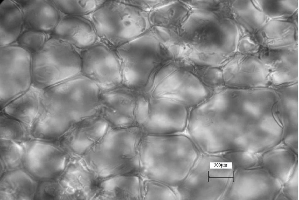
14 Figure 1 Typical image from the splat-cooling assay using phosphate buffered saline solution. Ice crystals are demarcated by the dark edges. The image was obtained at 76x magnification 6
15 7
16 recrystallization as a mechanism of cellular injury, many studies seeking to optimize cryopreservation of specific cell types do not address the issue of ice recrystallization. The accumulation of cryopreservation-associated cellular injuries have been reported to affect primarily the plasma membrane (19) and cellular organelles, particularly the mitcohondria (20). Through these various cryopreservation-associated injuries, cell death can occur. Three predominant modes of cell death have been reported to occur following cryopresevation. The first is cell rupture resulting from intracellular ice formation or large fluctuations in cellular volume, as described earlier. The second mode is cell necrosis, which is characterized by cell swelling, loss of membrane integrity, DNA fragmentation, and cytokine release. Mechanisms responsible for the third mode, cold-induced apoptosis, are not entirely clear. However, cold-induced apoptosis has been reported to be assoicated with changes in the mitochondrial membrane potential, which may activate the caspase dependent apoptotic signalling cascade through release of cytochrome C, along with the production of reactive oxygen species (ROS) that can damage intracellular structures (21, 22) Indeed, both oxidative stress from reactive oxygen species (ROS) and osmotic stressors (eg. dehydration) have been found to induce apoptotic changes in various cell types (23-25). 8
17 1.2 Cryoadditives as a strategy for reducing cryopreservation injury To mitigate cryopreservation-associated cellular injury and cryopreservationassociated cell death, cryoprotectants are added to the cellular suspension before freezing. There are two major groups of cryoprotectants, penetrating cryoprotectants that can pass through the cell membrane unaided, and nonpenetrating cryoprotectants that cannot. Penetrating cryoprotectants generally consist of small amphipathic molecules like dimethylsulfoxide (DMSO) and glycerol, while nonpenetrating cryoprotectants typically consist of large molecules like the polysaccahride hydroxy ethyl starch and proteins. In terms of slow freezing damage, penetrating cryoprotectants such as DMSO are thought to provide the greatest cryoprotective effects by replacing water, which prevents the cell from excessive shrinking and minimizes high electrolyte concentrations in the intracellular compartments(26, 27). In addition, DMSO also provides cryoprotective effects by reducing the ice formation at any temperature and solvates the plasma membrane (28). However, addition of DMSO at high concentrations to cellular suspensions can be toxic to unfrozen cells and may be harmful to patients if infused along with stored cells. For instance, several concerns have emerged in association with the use of DMSO for the storage of hematopoietic stem cells (29, 30). Toxicity related to DMSO infusion has been reported (31-33) and there are increasing concerns regarding the induction of apoptosis in hematopoietic stem cell grafts that are cryopreserved in DMSO (34, 35). Antifreeze glycoproteins are proteins containing numerous disaccharide moieties with ice recrystallization inhibition (IRI) properties and confer protective effects to 9
18 organisms like polar fish when subjected to freezing and sequential thawing conditions (36). Use of antifreeze glycoproteins as cryoprotectants, therefore, has been investigated. Extracting antifreeze glycoproteins from natural sources is resource intensive, therefore, synthesis of antifreeze glycoproteins has emerged as an attractive alternative. Design of synthetic analogues also allows one to enhance the cryoprotective properties and minimize the cytotoxic properties inherent in natural antifreeze glycoproteins. Indeed, Tachibana et al. (37) demonstrated that their synthetic antifreeze glycoprotein analogues were as active as natural antifreeze glycoproteins and have significant antifreeze activity. Matsumoto et al. also demonstrated that their synthetic antifreeze glycoprotein analogues, when in combination with 10%DMSO, were able to reduce cryopreservation-associated damage and improve functionality of cryopreserved β-pancreatic islet cells compared to cryopreservation in 10% DMSO alone (15). For the continued development and optimization of synthetic antifreeze glycoprotein analogues, more insight regarding the specific properties of antifreeze glycoproteins responsible for their cryoprotective effects is needed. Carbohydrates have also been explored as cryoprotectants, based on their protective role in specific frog species that can withstand sub-zero temperatures (38). Furthermore, the addition of carbohydrates, along with 5% DMSO, has provided encouraging results in the cryopreservation of umbilical cord blood (UCB) (39). The mechanism by which carbohydrates exert their beneficial effect has been unclear. It has been shown, however, that carbohydrates possess a natural capacity to inhibit the process of ice recrystallization via a process termed ice recrystallization inhibition (IRI) 10
19 (18), and the extent of IRI is associated with specific structural properties of the carbohydrate, including the hydration status (18, 40). Several studies have reported on the use of carbohydrates to improve UCB cryopreservation (41-43), sperm banking (3), oocyte storage (44), and human hepatocyte cryopreservation (45), but thorough structure-function studies addressing carbohydrate structure and impact on cell viability has not been performed. Furthermore, the mechanism by which the recovery of viable cells is enhanced has not been eluicdated. Greater insight regarding the role of IRI as a mechanism of carbohydrate-mediated cellular cryopreservation will allow the strategic development of novel carbohydrates and carbohyrdate based compounds with enhanced cryoprotective properties. Investigating IRI as a mechanism of carbohyrdatemediated cellular cryopreservation is addressed in this thesis. 1.3 Applications of cryopreservation Cryopreservation of cells and tissues has numerous applications in the fields of reproductive technologies, organ transplantation and cell-based medicine. Indeed, extensive research has been conducted over the past few decades to effectively cryopreserve germline cells (sperm and oocytes) from various species. Such applications allow for reproduction of those species in the future, and hence act as a strategy for the conservation of species, especially those that are endangered (46). Cryopreservation of human sperm and oocytes, along with ovarian tissue and embryos, is also important for the application of assisted reproduction techonolgies. 11
20 Cryopreservation of ovarian tissue, for instance, may allow women to preserve their reproductive functions for extended periods and after cancer treatments like chemotherapy that can threaten reproductive potential (47). Cryopreservation of human embryos is also crucial for in vitro fertilization, which is commonly used as an alternative treatment for infertility (48). Despite these various applications, numerous reports have demonstrated significant cryopreservation injury, particularly through associated apoptosis, in cryopreserved sperm (49) and ovarian tissue (50), which can result in impaired functionality. In addition to reproductive technologies, cryopreservation is widely used for the longterm storage of liver cells and various stem and progenitors cells for cell based treatments. Liver cell transplantation can be used to treat a variety of inborn diseases in liver metabolism by supplying functional liver cells, called hepatocytes. However, cryopreservation injury to hepatocytes markedly reduces cell functionality and viability in comparison to fresh hepatocytes, and hence can affect the success of the transplantation (51). Cryopreservation of various stem cells types, including embryonic stem cells, mesechymal stem cells and hematopoietic stem cells is also widely utilized for the longterm storage of these stem cell types for extended use in medical treatments and for research in the laboratory setting. Research on optimizing the cryopreservation of human embryonic stem cells has been an area of particular interest, since these cells provide a very efficient system to model various disease states, study human 12
21 development, and may be used as part of medical therapies in the future (52). However, human embryonic stem cells are very susceptible to cryopreservation injury, especially through cold-induced apoptotic pathways (53). Mesenchymal and hematopoietic stem cells are other types of stem cells that are stored by cryopreservation and used for medical and research based applications. Similar to embryonic stem cells, these stem cell types are susceptible to cryopreservation injury, particularly through cold-induced apoptosis (54, 55). Umbilical cord blood, an important source of hematopoietic stem cells that is routinely cryopreserved for later use, has been reported to have between 20% -70% of the stem cell population undergoing apoptosis after thawing cryopreserved units (56, 57). Improved understanding of the mechanisms of cryopreservation injury responsible for cell death, especially through cold-induced apoptosis, will allow for the development of strategies to reduce cryopreservation-associated cellular injury and improve the utility of these stored cellular products. This thesis will address the role of IRI as a potential strategy to reduce cryopreservation-associated cellular injury and the associated loss of viable and functional cells. UCB was selected as a model system to study IRI due to the significant levels of apoptosis observed following standard UCB cryopreservation and the presence of functional relevance and importance of hematopoietic stem cells in clinical applications, including hematopoietic stem cell transplantation. 13
22 1.4 Hematopoietic stem cells and associated medical applications The role of the hematopoietic system: The haematopoietic system is an essential physiological system responsible for producing and maintaining all cellular components in the blood, including all of the mature blood and immune cells. In mammals, there is a diverse set of cells produced from the hematopoietic system, the majority of which are bone marrow derived and can be classified as either myeloid or lymphoid. Myeloid cells include progenitors and differentiated cells of the erythroid, megakaryocytic, monocytic, granuloyctic lineages, while lymphocytes and natural killer cells arise from lymphoid tissues (58). Lymphoid cells are predominantly involved in adaptive immunity, but also contribute to innate immunity, as is the case for natural killer cells. Myeloid cells, in contrast, are more diverse in function. Erythrocytes, for example, are the most common cellular component in the blood and are responsible for delivering oxygen to body tissues via blood flow through the circulatory system. Erythrocytes originate from the bone marrow and typically circulate throughout the circulatory system for approximately days before being recycled by macrophages. Granulocytes include neutrophils, eosinophils and basophils. The most abundant type of granulocytes are neutrophils and exert numerous antimicrobial functions, including; phagocytosis, degranulation, neutrophil extracellular traps, and release of cytokines to amplify inflammation reactions. As a result, neutrophils are an essential component in the innate immune system. The life span of neutrophils, however, is limited to approximately 5 days when inactivated, and reduced to about 2 days when activated (59). The role of neutrophils as an essential front line defense against invading pathogens, coupled with limited life span, 14
23 necessitates extensive and continuous production. Platelets arise from megakaryocytes in the bone marrow and are central in the process of blood coagulation and hemostasis. Platelets have a limited lifespan of approximately 5-9 days. These important myeloid blood cell types, together with lymphoid cells, demonstrate the diverse functions of the hematopoietic system and its critical role in maintaining life. Maintenance of the hematopoietic system depends on the constant maturation of functional blood cells from immature stem and progenitor cells Hematopoiesis: the role of stem and progenitor cells Following numerous experiments in the early 1960 s on the transplantation of limited bone marrow cells into mice, Till and McCullough identified a rare specialized group of blood cells, termed hematopoietic stem cells (HSCs), that possessed both the ability to regenerate themselves and produce various types of myeloid and erythroid cells in vitro (60-62). Later studies in the early 1990 s confirmed these same capacities of hematopoietic stem cells in vivo, by demonstrating that a single HSC could generate all of the mature blood cell types in a recipient mouse for over 6 months, and could continue to do so upon secondary transplantation into another mouse (63-65). These capacities to self-regenerate and produce multiple blood cell types came to be later defined as self-renewal and multipotency, respectively. (66). It is through both selfrenewal and differentiation of hematopoietic stem cells (HSCs) into all of the various mature blood cell types that the hematopoietic system is able to produce and maintain a sufficient supply of blood and immune cells throughout an organism s life span (65). 15
24 During normal physiological states, a rapid rate of over 1 million mature blood cells are being produced per second (67). This extensive quantity of mature blood cell production would seem to necessitate an extensive and rapid rate of HSC proliferation and differentiation. Interestingly, however, the majority of hematopoietic stem cells residing in the adult animal bone marrow have been shown to be at rest in the G 0 stage, cycling very rarely (68, 69). Instead, extensive proliferation and differentiation of mature blood cell precursors derived from HSCs, termed hematopoietic progenitor cells (HPCs), is thought to be the predominant mechanism by which the hematopoietic system is able to turn over such vast quantities of new mature blood cells (70). These hematopoietic progenitor cells (HPCs) differ from HSCs in 2 ways: 1) HPCs have lost the capacity for indefinite self-renewal that HSCs possess and 2) HPCs are generally more restricted compared to HSCs in their potency for differentiation into the various mature blood cell types. In addition, HPCs are a heterogeneous population, ranging from oligopotent progenitors that possess the ability to differentiate into several mature blood cell types to unipotent progenitors that are restricted to differentiating into only one type of mature blood cell. Hematopoiesis is hierarchical, with the least lineage-restricted progenitors deriving directly from HSCs to more committed progenitors and mature cells through stages of lineage-restricted differentiation. Moreover, two pools of hematopoietic stem cells have been characterized, termed long-term and short term repopulating HSCs (71-73) with different capacities for self-renewal. Long term HSCs possess life-long self- 16
25 renewal while short term HSC have limited self-renewal capacity. Common myeloid progenitors and common lymphoid progenitors, however, are more committed precursors that give rise to their respective cell lineages. Indeed, it has been shown that common myeloid progenitors give rise to all myeloid cells, but are unlikely to give rise to B lymphocytes (74). Likewise common lymphoid progenitors have been shown to possess the capacity to differentiate into all lymphoid cells, but typically do not differentiate into myeloid cells under normal physiological conditions (75). Certain aspects of this classical model of hematopoiesis, however, have been challenged, more specifically with regards to the lineage restriction of the common myeloid and lymphoid progenitors. For instance, Kawamoto et al. recently demonstrated that early progenitors in the thymus retain myeloid potential. To account for this, they proposed a slightly different model called the myeloid-based model, which posits that myeloid cell differentiation potential is maintained in B and T lymphocyte precursors, even after these lineages segregate from each other (76). HSC and HPC populations differentiate into more mature progenitors or blood cells, the phenotypic expression of many cell surface markers changes. As such, the set of cell surface markers present on a cell can act as a surrogate marker for the stage of cellular differentiation, self-renewal capacity and functionality. One of the most widely used cell surface markers to distinguish HSCs and HPCs from mature blood cells is a cell surface marker called CD34, which is a single-pass transmembrane protein of the sialomucin family that is expressed predominantly in early hematopoietic and vascular tissue, including HSCs and HPCs (77). Although the exact function of CD34 has not 17
26 been definitively determined, CD34 has been shown to be implicated in promoting HSC and HPC self-renweal, while inhibiting those populations from differentiating, enhancing HSC and HPC homing and trafficking to bone marrow for maintenance, and both promoting and blocking cell-cell adhesion (77). Evidently, CD34 is likely an essential protein for proper functioning of HSCs and HPCs, making it an appropriate surrogate for HSC and HPC identification. Indeed, CD34 was the first marker to be used for isolation of HSCs and almost all CD34 + cell populations have been shown to possess multipotency or oligopotency (78-80). Another important surface marker that has been used to discriminate between HSC/HPCs and mature blood cells is the protein tyrosine phosphatase receptor type c, more commonly referred to as CD45 (81, 82). It is a transmembrane protein, similar to CD34, but unlike CD34, CD45 is expressed specifically on all differentiated hematopoietic cells, except erythrocytes and platelets (83). CD45 has been shown to play important roles in regulating a variety of cellular processes, including; cell differentiation and cell growth. In combination with CD34 and CD45, the absence of the cell surface marker CD38, and expression of the cell surface marker CD90, have been used to further identify earlier multipotent HSC populations more specifically (84, 85). In addition, identifying cells with high activity of an enzyme called aldehyde dehydrogenase (ALDH), in combination with all these cell surface markers, has been shown to allow for the isolation of hematopoietic stem cells at very high purities (86). This is likely because ALDH has been shown to play an important role in HSC differentiation by regulating the retinoic acid signaling pathway (87). 18
27 1.4.3 Application of hematopoietic stem and progenitor cells for medical applications Hematopoiesis, although essential to the maintenance of life, can also become aberrant or unregulated and lead to leukemia and lymphoma. Leukemia typically involves the bone marrow compartment causing a marked decrease in normal hematopoieis and lymphoproliferative disorders such as lymphoma typically involve lymph nodes. In either case, patients with leukemia and lymphoma are often treated with systemic chemotherapy and/or radiation treatment. Both chemotherapy and irradiation impair cell replication and induce cell death. While eradicating the malignant cells, chemotherapy and radiation also inhibit cell replication and induce cell damage in the hematopoietic system due to the relatively high rate of cell cycling in the bone marrow. For instance, neutrophils and platelets are especially sensitive to chemotherapy and radiation. In conventional dosing of chemotherapy or irradiation, the patient s blood cells will decrease for a brief period, followed by recovery to normal levels. However, because advanced hematological malignancies may respond favorably to increasing doses of chemotherapy or irradiation, some patients will undergo dose escalations that can be curative but eliminate the bone marrow as a consequence, a treatment called myeloablation. Recovery of mature blood cells after myeloablative treatment requires the transplantation of hematopoietic stem cells, a procedure termed hematopoietic stem cell transplantation (HSCT), to avoid prolonged neutropenia (low neutrophil levels in the blood), and the associated risk of infection from microbial pathogens and to avoid thrombocytopenia (reduced quantities of platelets in the blood), that is associated with bleeding complications. 19
28 Hematopoietic stem cell transplantation (HSCT) following myeloablation is essential for recovery of mature blood cells, especially fast growing cells like neutrophils and platelets. Minimizing the period of insufficient blood cell production, termed cytopenia, following myeloablative treatment reduces the risk of infection and excessive bleeding, and contributes to the success of safe and effective transplantation. The time between HSC transplantation and recovery of sufficient blood cells is termed engraftment. In a patient setting, the exact definition of engraftment can vary, but numerous studies have defined engraftment as the number of days required to reach a concentration of > 0.5x10 9 neutrophils/l blood and > 20x10 9 platelets / L blood for three consecutive days (88-90). The rapid reconstitution rates and critical roles that neutrophils and platelets have in immunity and wound healing, respectively, may explain why these cells have been chosen as markers of engraftment. However, recent studies have reported on other cell types like reticuloytes as earlier markers of hematopoietic engraftment (91). Several sources of cells can be used for HSCT, including bone marrow, mobilized peripheral blood or UCB. In addition, each of these stem cell sources can be collected either from the patient themselves, termed an autologous transplant, or from another person, termed an allogeneic transplant. Autologous HSCT is most commonly performed to allow patients to undergo higher doses of chemotherapy and radiation in an effort to eradicate remaining cancer cells. Typical diseases that can be better controlled using higher doses of treatment that require hematopoietic rescue with autologous HSCs include relapsed or high-grade lymphomas and multiple myeloma, a 20
29 cancer of plasma cells. The process of autologous HSCT requires the collection of the patient s own stem cells, followed by cryopreservation of those stem cells, and then reinfusion of the stem cells at a later date following the completion of myeloablative chemo/radiotherapy. In allogeneic stem cell transplantation, HSCs are collected from human leukocyte antigen (HLA) compatible sibling or unrelated donor, including UCB. Allogeneic transplantsgenerally require strict matching between the recipient and donor for 6 or more genes of the human leukocyte antigen (HLA) cluster. When the recipient of an allogeneic HSC transplant is not sufficiently matched with the donors HLA alleles, there will be an increased risk for two major immune-mediated complications, graft rejection and graft versus host disease. In graft rejection, the HSC graft is rejected by the recipients own immune system, while in graft versus host disease the immune cells within the graft initiate an immune response against the recipient s own tissues. These immunological rejection events, along with transplantation of insufficient cell doses can result in failed engraftment. Despite the increased risk of immune-related complications, allogeneic transplantation can be advantageous since it also provides a platform for beneficial immunological reactions against the tumor, termed graft versus leukemia, where mature immune cells in the graft initiate an immune response against cancer cells. The need for strict HLA matching is less stringent when UCB is used as the source of stem cells, and so UCB has become an attractive alternative source of HSCs for allogeneic stem 21
30 cell transplantation when a matched sibling or unrelated donor cannot be identified. Although enriched for HSCs, UCB units are limited by the total volume available, which can influence the total dose of stem cells and contribute to delayed engraftment. Moreover, as with autologous stem cell grafts, UCB units require storage until use, most commonly by cryopreservation in liquid nitrogen. Cells collected from sibling or unrelated allogeneic donors, however, do not require cryopreservation but instead are collected just prior to their use and typically are infused fresh. As previously mentioned, subjecting UCB grafts to cryopreservation can injure stem and progenitor cells, further exacerbating the problem of a limited dose of viable functional cells that can engraft after transplantation. Particular issues associated with cryopreservation of autologous cells and UCB remain a challenge in HSCT and may limit more widespread use of UCB in particular. Peripheral blood stem cell (PBSC) grafts have become the preferred source of autologous HSCs due to ease of collection and more rapid engraftment rates compared with bone marrow following meyloablative chemotherapy and/or radiation treatment. (92, 93) The most important criteria for predicting engraftment rates following PBSC transplantation has been the dose of hematopoietic stem and progenitor cells infused. More specifically, hematopoietic populations possessing the cell surface marker CD34 correlate with timely engraftment. Indeed, CD34 is a surrogate marker of oligopotent and multipotent hematopoietic populations. Numerous studies have demonstrated the quantity of CD34 + progenitors within a PBSC graft to be the best predictor for the rate of engraftment, as determined by neutrophil and platelet recovery (94-97). Other factors 22
31 including the intensity of chemotherapy and/or radiation, age and sex of the recipient have also been shown to influence neutrophil and platelet engraftment (94, 98, 99). Due to the importance of the CD34 + cell dose infused, multiple studies have identified minimum threshold quantities of CD34 + progenitors that should be infused to ensure rapid engraftment. Typically, infusion of 2.0 x 10 6 CD34 + progenitors/kg has been demonstrated to be the minimum threshold associated with prompt engraftment following PBSC transplantation (100, 101). However, the CD34 content of a PBSC graft alone does not always predict rapid engraftment. In autologous peripheral blood stem cell transplants, the number of viable CD34 + cells following cryopreservation has been shown to be markedly decreased due to the adverse effects of processing and storing the cells and this reduction in the CD34+ cell dose has been shown to prolong engraftment rates (102, 103). Although most patients engraft in a rapid manner when transplanted with PBSC grafts possessing greater than 2.0 x 10 6 CD34 + progenitors/kg, there are still a small number of patients with delayed recovery of blood counts. The percentage of patients that experience delayed engraftment despite receiving sufficient CD34 + progenitors varies. A study by Trébéden-Negre et al.(104) found 16 out of 246 patients developed delayed neutrophil engraftment after receiving at least 3 x10 6 CD34 + progenitors/kg. These 16 patients did not have any known clinical factors that were likely to adversely affect engraftment. Similar findings are further demonstrated in a set of case studies described by Mineishi et al.(105), in which 4 patients receiving a sufficient dose of CD34 + progenitors (>4.0x10 6 CD34 + progenitors / kg) were identified yet experienced 23
32 delayed neutrophil engraftment. It is possible that some of the cells included in their counts were apoptotic yet were still enumerated as viable CD34 + cells. Apoptotic cells could have reduced engrafting potential and contribute to delayed hematopoietic recovery after transplantation.the impact of cold induced apoptosis on engrafting potential of UCB and PBSC requires further study and was addressed in this thesis as a model system of cryopreserved cell storage. 24
33 1.5 Statement of the problem Cryopreservation can injure cells and reduce the yield of viable functional cells. Cold-induced apoptosis has been reported to be a predominant form of cell death in many cryopreserved cells and may reduce the functionality of stored cellular products. Cold-induced apoptosis is particularly relevant for UCB banking where cells are often cryopreserved for extended periods of time, and the levels of cold-induced apoptosis become more prevalent as the duration of cryopreservation increases. Ice recrystallization is a process that occurs during thawing of cryopreserved solutions and is characterized by growth of large ice crystals, which can cause mechanical damage and apoptotis to cells in the cryopreservation solution. Ice recrystallization, therefore, could be a dominant contributor to cold-induced apoptosis and reduced functionality of cryopreserved cells and requires further study. 1.6 Objectives This thesis will address carbohydrate mediated ice recrystallization inhibition as a possible mechanism to reduce cryopreservation injury and loss of cell functionality that occurs from ice recrystallization during the cryopreservation and thawing process. Our studies will focus on the cryopreservation of human UCB. The thesis also aims to determine if increased cold-induced apoptosis in stored HSC products is associated with reduced hematopoietic function. These studies will provide a platform for future research that endeavours to reduce cold-induced apoptosis through inhibition of ice recrystallization. 25
34 1.7 Hypothesis More potent inhibitors of ice recrystallization will improve the cryopreservation of UCB cells, as measured by improved recovery of non-apoptotic (Annexin-V -ve ) cells and greater recovery of hematopoietic function, compared to less potent inhibitors of ice recrystallization. Furthermore, the practical relevance of cold-induced apoptosis will be demonstrated by correlating the number of apoptotic cells in blood stem cell products with delayed hematopoietic recovery in patients undergoing hematopoietic stem cell transplantation. 26
35 Chapter 2: Materials and Methods 2.1 Collection, storage and processing: Umbilical cord blood (UCB) Umbilical UCB was collected following healthy term delivery and informed consent from mothers, in accordance with institutional approval from the Research Ethics Board of The Ottawa Hospital according to research protocol # H (performed by collaborators at The Ottawa Hospital). UCB was decanted into 50ml falcon tubes and centrifuged (Allegra X-15R centrifuge, Beckman Coulter) at 350xg for 10 minutes to remove red blood cells. The remaining buffy coat mononuclear cells (MNCs) and plasma were recovered. MNCs were were then diluted 1:2 in the autologous plasma (15mL of buffy coat MNCs in 15mL autologous plasma) and then layered on ficoll with at 1 part (15mL) ficoll(ficoll-hypaque, GE Healthcare): 2 part plasma/buffy coat (30mL). Samples were then run under ficoll gradient centrifugation at 400xg for 30 minutes and then washed twice in PBS at 300xg for 10 minutes (Allegra X- 15R centrifuge, Beckman Coulter). Total MNCs ( x 10 7 cells/ml) were then suspended in 1 ml media RPMI containing 37% Pentaspan (v/v) (a membrane stabilizer used by Canadian blood services at similar concentrations (37% v/v) for cryopreservation of peripheral blood stem cell grafts) and carbohydrate solutions at concentrations of either 20mM, 200mM or 500mM. A total of 2 replicates were analyzed for 5 different sugars (glucose, galactose, melibiose, trehalose and sucrose) at 20mM, 200mM and 500mM to identify an optimal 27
36 concentration for cryopreservation. An additional 3 replicates for each sugar at 200mM was also analyzed once 200mM was identified as the optimal concentration. 1ml Cell suspensions were cryopreserved in 2 ml cryogenic vials (Corning Incorporated, New York) under rate-controlled conditions to -80 o C (Mr. Freezy, Nalgene Labware) over 16h and then transferred to liquid nitrogen at least 1 week before thawing and analyzing. The duration of 1 week was chosen for logistical reasons and because this duration has been used in previous studies on UCB cryopreservation (106).One sample from each UCB unit was also stored using 5% DMSO in replacement of sugars (Sigma- Aldrich) as an internal standard for each sample. After one week of storage, samples were thawed in a 37 o C water bath (rapid thaw). An additional 2 replicates of each sugar at 200mM were subjected to the same cryopreservation conditions, but thawed at ambient temperatures (slow thaw). Once thawed, 0.8mL of the samples were run through a filter [need microns] (Miltenyi Biotec, Germany) into 15 ml falcon tubes and diluted with 4 ml RPMI solution at 37 o C containing 50x10 3 units/ml DNase I, (Sigma-Aldrich, Germany) and 5 mm MgCl 2 (Sigma-Aldrich, Germany). Samples were incubated in air for 4 minutes and then further diluted with another 5 ml of the RPMI/DNase I solution. Subsequently, samples were centrifuged for 10 min at 300 g s at 4 o C. The supernatant was then removed and the cells were resuspended in 1 ml RPMI media to prevent prolonged exposure to DMSO. Cell concentrations were then determined using a hemocytometer for further analysis by flow cytometry and colony forming unit assays. 28
37 2.1.2 Peripheral blood stem cell grafts Patient selection was performed by Dr. Ayman Al-Hejazi, a bone marrow transplant fellow at The Ottawa Hospital in In brief, patients who underwent autologous PBSC transplantation for non-hodgkin s lymphoma at the Ottawa hospital between January 2000 and December 2005 and who provided consent for the use of medical information for research purposes in accordance with the Ottawa Hospital s research ethics board were included in the study. Research was performed according to research protocol number H Cases were defined as patients with a persistent neutrophil count < 1.0 x 10 9 /L at day 30. Patients from the same cohort that had a neutrophil count > 1.0 x 10 9 /L prior by day 30 were selected as controls. Controls were matched for various factors, including age, sex and disease type. Other factors matched between cases and controls are outlined in table 1. All cases and controls were 18 years of age. A total of 13 delayed neutrophil engraftment cases and 22 matched controls were analyzed. PBSC grafts were volume reduced by centrifugation, transferred into an equal volume of a cryoprotectant solution containing 10%DMSO, 8% human serum albumin, 43.2% pentaspan and 14.8% Plasma-Lyte A and then placed directly into liquid nitrogen vapour overnight for cryopreservation (Performed by collaborators at Canadian Blood Services). Cryopreserved aliquots containing approximately 1mL of a cryopreserved cellular suspension, separate from the PBSC grafts, were thawed in a 37 C water bath until no ice could be seen and then analyzed for this study. Once thawed, 0.5mL of the sample 29
38 was transferred into a 15mL centrifuge tube and diluted 6 fold, dropwise over 5 minutes, with RPMI solution at 37 C containing 5.0 x 10 4 units/ml DNase I, (Sigma-Aldrich, Germany) and 5 mm MgCl2 (Sigma-Aldrich, Germany). Subsequently, samples were centrifuged (Allegra X-15R centrifuge, Beckman Coulter) for 10 minutes at 250 x g at 4 o C. The supernatant was then removed and the cells were resuspended in 2 ml RPMI media to prevent prolonged exposure to DMSO. Cell concentrations were then determined using a hemocytometer for further analysis by flow cytometry and colony forming unit assays. 2.2 Colony forming unit assays: Colony forming unit assay methodology Clonogenic assays were performed on thawed UCB and PBSC aliquots using methylcellulose media (Methocult GF H4434; Stemcell Technologies, Vancouver, Canada) in accordance with manufacturer s instructions. Briefly, cells were suspended in 1 ml of IMDM media with 2% fetal bovine serum (Stemcell Technologies, Vancouver, Canada) at a concentration of 1 x 10 6 cells/ml for peripheral blood stem cell samples and of 5 x 10 5 cells/ml for UCB samples. A volume of 0.3 ml of the cell suspension was transferred into 2.7 ml of the methylcellulose media to obtain a final concentration of 1 x 10 5 cells/ml for peripheral blood stem cell samples and 5 x 10 4 cells/ml for UCB samples. 1.0 ml of the methylcellulose cell suspension was plated in duplicate into 35 mm diameter Petri dishes and incubated at 37 o C and 5% CO 2 for 14 days. Colony 30
39 forming units (CFU) were enumerated by morphology using an inverted microscope to discriminate erythroid (CFU-E), erythroblast-forming units (BFU-E), granulocyte/macrophage (CFU-GM), and granulocyte/erythroid/macrophage/ monocyte (CFU-GEMM) colonies (more than 50 cells/colony). 2.3 Flow cytometry: Justification for measuring viability with 7AAD and apoptosis with Annexin-V Numerous flow cytometric criteria are assessed when transplanting HSC grafts to ensure that the graft will transplant successfully. In HSC grafts that have been cryopreserved, measurement of CD34 + cell viability has been shown by Allan et al. (102) and Lee et al.(103) to be an important criterion for predicting rates of engraftment in autologous peripheral blood stem cell grafts, while Scaradavou et al. (107) has shown CD34 + cell viability to be important for predicting successful engraftment of UCB grafts. All of these studies identified viable cells by measuring the exclusion of a fluorescent DNA binding dye, called 7-actinomycin D (7AAD) via flow cytometry. The use of 7 actinomycin-d as a viability marker was first described by Schmid et al.(108) in 1992 and has become a widely accepted viability marker by numerous transplant centers. Indeed, the International Society for Hematotherapy and Graft engineering (109) recommend measurement of CD34 + cell viability through flow cytometric analysis of CD34 + cells excluding 7AAD, especially in stem cell grafts that have been cryopreserved. 31
40 7AAD permeates into cells that have lost their membrane integrity and are no longer viable. Since the loss of membrane integrity occurs very late in the process of cell death, 7AAD is not able to identify cells in early stages of apoptosis. Multiple studies have identified earlier markers of apoptosis, including annexin V (55, 110). Annexin-V is a protein that binds to a phospholipid called phosphatidyl serine, which is usually only externalized on the outer membrane of cells undergoing apoptosis. Externalization is typically caspase activation dependent, but can also result from caspase independent apoptotic mechanisms. Disruption of the mitochondiral membrane potential also leads to phosphatidyl serine externalization (111). Irrespective of the mechanism, phosphatidyl serine externalization occurs before the loss of cell membrane integrity in cells undergoing apoptosis (112) and measuring Annexin-V binding represents the quantity of cells undergoing early apoptosis. Furthermore, Shin et al. have demonstrated that early apoptotic CD34 + progenitors, as measured by Annexin-V,(56) isolated from umbilical UCB are significantly impaired in their capacity to engraft NOD- SCID mice compared to nonapoptotic CD34 + progenitors (AnnexinV -ve ) (56). Based on the functional relevance of Annexin-V it was selected as a marker for early apoptosis and 7AAD was selected as a marker for cell death (ie. non-viable). As a result, Annexin- V(+) 7AAD(-) cells will be defined as viable apoptotic and cells that are Annexin-V(-) 7AAD(-) will be defined as viable nonapoptotic. 7AAD(+) cells are non-viable, or dead. 32
41 2.3.2 Methodology for flow cytometry of umbilical cord blood Cells were analyzed before and after cryopreservation for viability and apoptosis. MNCs (2x10 6 cells/ml) were suspended in Annexin-V binding buffer (BD Pharmigen, San Diego). 100 µl of the cell suspension was placed into 12x75 polystyrene tubes (Becton Dickinson) and stained with anti-cd34- fluorescein isothiocyanate (BD Pharmigen, San Diego), Annexin V- phycoerythrin (BD Pharmigen, San Diego), and 7AAD (BD Pharmigen), San Diego) and incubated at room temperature for 20 minutes. Samples were diluted up to 500 µl with Annexin-V binding buffer following incubation. Unstained Aliquots were used to set the control gates. Cells were immediately analyzed with a BD LSR I flow cytometer (BD Biosciences). Data analysis was performed using the software Summit (DAKO). Relative viabilities were obtained by dividing the percentage of 7AAD - cells from the respective sample by the percentage of 7AAD - cells from the 5% DMSO control to standardize the viabilities. Relative viable non-apoptotic cells were obtained by dividing the percentage of Annexin-V - / 7AAD - cells from the respective sample by the percentage of Annexin-V - / 7AAD - cells from the 5% DMSO control to standardize the viable nonapoptotic cells Methodology for flow cytometry of peripheral blood stem cell grafts Cells were analysed by 5-color flow cytometry to measure viability and apoptosis in the specific cell populations shown in figure 2. Examples of histograms from these dot plots can be found in Appendix B. All fluorescent markers were incubated together in one tube. Following thawing of cryopreserved samples, cells were washed in 5 volumes 33
42 RPMI and centrifuged at 250xg for 10 minutes to remove DMSO. Cells were then resuspended in 2 ml RPMI. To identify aldehyde dehydrogenase-expressing (ALDH) progenitor cells, 4 x 10 6 cells were suspended in 1mL of Aldecount assay buffer (Stemcell Technologies, Vancouver, Canada). 0.5 ml of the cell suspension was diluted with 1.5 ml activated Biodipy aminoacetaldehyde (BAA) (Stemcell Technologies, Vancouver) into a tube labeled tube µl of this original sample was immediately added to 10 µl of diethylaminobenzaldehyde (Stemcell Technologies) in a separate tube, labeled tube 1, to inhibit ALDH activity and act as a negative control. Both tube 1 and 2 were incubated at 37 O C for 30 minutes, centrifuged at 250 x g for 10 mins and resuspended in 100 µl of Annexin-V binding buffer (BD Pharmigen, Mississauga, Canada). Anti-CD34-phycoerythrin cyanin 7 (clone #581, Beckman Coulter, Mississauga, Canada), Annexin V- phycoerythrin (BD Pharmigen), 7AAD (BD Pharmigen) and CD45-electron coupled dye (clone # J.33, Beckman Coulter) were then added in combination into both tubes 1 and 2, and incubated at room temperature for 20 minutes. Both tubes were then diluted with Annexin-V binding buffer and analyzed with a FC500 flow cytometer (Beckman Coulter). Hematopoietic CD34 + progenitor cells were identified using a modified version of the international standards for hematotherapy and graft engineering (113). Data analysis was performed using the Summit software (DAKO, Carpinteria, United States). The CD34 + CD45 low ALDH high progenitor region was set by the fluorescence of BAA in CD34 + progenitors from the control tube where the biodipy-aminoacetaldehyde catalysis was inhibited. CD34 + CD45 low ALDH high progenitor cells were measured to identify early hematopoietic progenitors as compared to more differentiated CD34 + CD45 low ALDH low progenitors. 34
43 Figure 2. The gating strategy used to identify the various cell populations using flow cytometry. To identify the the ALDH high population, a portion of each original sample (tube 2) was incubated with diethylaminobenzaldehyde (DEAB) to inhibit ALDH catalysis of the fluorescent marker biodipyaminoacetaldehyde (tube 1). Both tubes 1 and 2 were stained with Anti-CD34- phycoerythrin cyanin 7, Annexin V- phycoerythrin, 7AAD, CD45-electron coupled dye and activated biodipyaminoacetaldehyde in combination. A modified version of the International Society for Hematotherapy and Graft Engineering gating strategy was utilized to define CD34+ progenitors. Total events are depicted in A and E and were analyzed for the CD45 + events as defined by R1. R1 was then gated onto B and F and analyzed for CD34 + events as defined by R2. R2 was then gated onto C and G and analyzed for CD34 + CD45 low events as defined by R3. CD34 + progenitors are defined as the CD34+CD45 low population identified in R3 of C and G. R3 was then further gated onto D and H. The CD34 + CD45 low ALDH high progenitor region was defined by R4 in D where ALDH activity was inhibited by (DEAB). CD34 + CD45 low ALDH high cells are defined in H. ALDH aldehyde dehydrogenase DEAB - diethylaminobenzaldehyde 35
44 36
45 2.4 Ice recrystallization-inhibition (IRI) assay: IRI activity of all sugars utilized in the study were determined by collaborators, Jackie Tokarew & Jennifer Chaytor, graduate students in the department of chemistry at University of Ottawa, using the splat cooling assay described earilier in section A total of three images were taken from each wafer using a Nikon CoolPix 5000 equipped to the cryostage. Typical images are shown in Figure 3. During flash freezing, ice crystals spontaneously nucleated from the supercooled solution. These initial crystals are relatively homogeneous in size and quite small. During the annealing cycle, recrystallization occurs, resulting in a dramatic increase in ice crystal size. A quantitative measure of the difference in recrystallization inhibition of two compounds X and Y is the difference in the dynamics of the ice crystal size distribution, termed the mean grain size (MGS). Compounds with stronger IRI activity will result in smaller MGS. Image analysis of the ice wafers was performed using a novel domain recognition software. 37
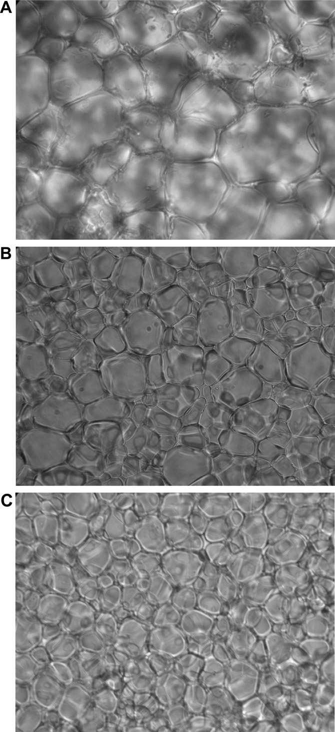
46 Figure 3 Light microscope image of PBS solution following splat-cooling assay (A). Ice crystals are demarcated by the dark edges. Light microscope image of 200 mm (w/v) galactose in PBS solution following splat-cooling assay (B). Ice crystals are demarcated by the dark edges and appear smaller than in image 3SA. Light microscope image of 200 mm (w/v) melibiose in PBS solution following splat-cooling assay (C). Ice crystals appear the smallest in 3C in comparison to 3A and 3B. Images shown are at 76x magnification. Ice crystal sizes were measured with a domain recognition software and used to calculate the IRI capacity of a carbohydrate by dividing the average ice crystal size in the solution containing the carbohydrate by the average ice crystal size in a solution of PBS. 38
47 39
48 2.5 Statistical analyses GraphPad Prism version 5.00 (Graphpad Software, San Diego, California) for Windows was used for statistical analysis. Statistical significance was determined using an unpaired, two-tailed Student's t-test with a significant value of p< Data were plotted using Microsoft Excel or Graphpad. 40
49 Chapter 3: Results 3.1 Measuring mono- and disaccharides ice recrystallization inhibition activity Four monosaccharides (D-galactose, D-glucose, D-mannose, D-talose) and four disaccharides (D-melibiose, D-lactose, D-trehalose and D-sucrose) were measured for their IRI activity at two different concentrations, 22 mm and 220 mm concentrations in PBS. A minimum of 3 replicates were performed for each carbohydrate at 22 mm and 220 mm. Carbohydrate concentrations above 220mM were not assessed because the viscosities of those solutions were too high for the splat-cooling assay to accurately measure the IRI activities. The two different concentrations of carbohydrates resulted in notable differences in the mean grain sizes observed, and hence their IRI activity. First, both mono- and disaccharides resulted in smaller mean grain sizes at 220 mm concentration compared to 22 mm, suggesting that carbohydrate concentration influences IRI activity. The IRI activity of monosaccharides at 220 mm is approximately two times more active than at 22 mm. Secondly, the relative IRI activities of disaccharides compared to monosaccharides are similar at 22mM but different at 220mM. The IRI activities of disaccharides are almost up to three times greater than monosachharides at 220mM, but at 22mM concentrations, these differences were not found. Significant differences between IRI activities of carbohydrates at the same concentration and between the two different concentrations were not calculated due to the low number of replicates performed. 41
50 Figure 4. Ice recrystallization inhibition activity of mono- and disaccharides at concentrations of 22 mm (white bars) and 220 mm (black bars). Y-axis represents the % of the mean grain size (MGS) of ice crystals in the solution relative to a containing only phosphate buffered saline (PBS). A minimum of 3 replicates were measured for each carbohydrate at 22 mm and 220 mm. Significant differences in the IRI activity between samples were not calculated due to the low number of replicates. 42
51 Mean grain size relative to PBS Galactose Glucose Mannose Talose Melibiose Lactose Trehalose Sucrose 22 mm 220 mm 43
52 3.2 Determining the optimal carbohydrate concentration for sugar cryopreservation of umbilical cord blood Optimal carbohydrate concentrations for studying the relationship between carbohydrate mediated IRI activity and recovery of viable and nonapoptotic cells were determined by cryopreserving UCB in 3 different concentrations (20mM, 200mM and 500mM) of the 5 sugars used in the study. 2 replicates were measured for each sugar at the 3 different concentrations. In general, cryopreservation of UCB units in 20mM of sugar resulted in low yields of viable nonapoptotic (7AAD -ve / Annexin-V -ve ) MNCs (range 2-6%) and CD34+ progenitors (range 4-16%). Cryopreservation of UCB units at higher concentrations of carbohydrates (200mM and 500mM) resulted in improved yields of viable nonapoptotic MNCs (range 10-22%) and CD34 + cells (range 10-40%) (See figure 2). Cryopreservation with sugars at 200mM and 500mM generally provided similar yields of viable nonapoptotic MNCs (10-20% for 200mM sugar vs 8-19% for 500mM) and CD34 + cells (13-40% for 200mM sugar vs 10-37% for 500mM). Due to the low number of replicates measured for each carbohydrate and concentration, no significant difference in the recovery of nonapoptotic cells between the 200mM and 500mM carbohydrate conditions can be assumed. Because sugar concentrations of 500mM did not provide any increases in recovering viable nonapoptotic MNC or CD34 + cells, and was difficult to assess for IRI activity, 200mM was selected as the optimal concentration for cryopreservation to avoid unneeded excess use of sugars. 44
53 Figure 5. Yield of viable nonapoptotic MNCs (A) and CD34 + cells(b) in UCB units following cryopreservation in galactose, melibiose, trehalose, glucose and sucrose at 20mM, 200mM and 500mM. Cryopreservation at 20mM resulted in low yields of nonapoptotic MNCs and CD34 + cells compared to 200mM and 500mM (n=2 for each sugar at 20mM,200mM and 500mM). Use of 200mM sugars appeared to be the optimal concentration since cryopreservation in 500mM generally resulted in similar recoveries of non-apoptotic cells as 200mM, and 500mM IRI activity could not be measured with the splat cooling assay. increase yield of viable nonapoptotic CD34 + and MNCs. 45
54 A Recovery of viable nonapoptotic cells [%7AAD(-)AnnexinV(-)] Mononuclear cell population Glucose Melibiose Trehalose Glucose Sucrose 22mM 200mM 500mM B Recovery of viable nonapoptotic cells [%7AAD(-)AnnexinV(-)] Glucose CD34 + cell population Melibiose Trehalose Glucose Sucrose 20mM 200mM 500mM 46
55 3.3 IRI activity of carbohydrates and the yield of viable MNCs and CD34 + cells UCB units were cryopreserved in 200mM of each carbohydrates and then thawed either in a rapid manner (thawed in a 37 ºC water bath for approximately 1 minute) to minimize ice recrystallization or slow manner (air thawed at room temperature for approximately 6 minutes) to exacerbate the occurrence of ice recrystallization. A concentration of 200mM carbohydrate was chosen because IRI activity of carbohydrate concentrations greater than 220mM could not be measured accurately with the splat cooling assay and 220mM carbohydrates appeared to possess greater IRI activity compared to 22mM. Recovery of viable cells was then assessed by flow cytometry for exclusion of 7AAD. When UCB units were cryopreserved using carbohydrates with potent IRI and thawed under conditions of high recrystallization injury (i.e. thawing slowly at ambient temperature), we observed an increase in the yield of viable MNCs and of viable CD34 + cells (see Figure 3). In particular, IRI activity of carbohydrates correlated significantly with improved yield of viable MNCs (r 2 =0.93, p=0.004 for slope different than null hypothesis that slopes are not significantly non zero) and greater yield of viable CD34 + cells (r 2 =0.96, p=0.019). The effect of carbohydrate IRI activity on the yield of total viable MNCs was less apparent under conditions of low recrystallization injury (i.e. thawing rapidly in a 37 ºC 47
56 water bath) (r 2 =0.49) although a trend toward improved yield of viable CD34 + cells was still observed (r 2 =0.705, p=0.07 for the null hypothesis) (see Figure 3). When UCB units were thawed under more clinically relevant conditions of low recrystallization injury (i.e. thawing rapidly in a 37 C water bath), both monosaccharides and disaccharides, at 200 mm in the absence of 5% DMSO, produced yields of viable MNCs that were low in comparison to cells cryopreserved under standard conditions containing 5% DMSO only (ranging from 22 ± 4.8% of input cells for glucose to 36 ± 3.4% for melibiose, compared to 79 ± 3.8% for 5% DMSO, p<0.0001). The yield of viable CD34 + cells, however, was greater than the yield of viable total MNCs for all carbohydrate solutions tested (from 40% ± 6.5% for glucose to 61% ± 8.1% for melibiose, p=0.033 when comparing the mean recovery of viable CD34 + cells to mean yield of viable total MNCs for all samples). The yield of viable CD34 + cells was not different from the viability of total MNCs in the control solution containing 5% DMSO alone (88% ± 3.8% vs. 79 ± 3.8%, p=ns). 48
57 Figure 6. Yield of viable cells vs. IRI activity for MNCs (A) and CD34 + cells (B) UCB MNCs were cryopreserved with 200 mm (w/v) of the carbohydrate and analyzed postthaw for their viability by exclusion of 7-AAD. Samples with each carbohydrate were thawed in either a 37 C water bath for the fast thaw (Black lines, n=5) or at room temperature for the slow thaw (Grey lines, n=2). Resulting viability ratios were then plotted against the IRI activity of the corresponding carbohydrate where MGS represents the mean grain size of ice crystals in a solution of carbohydrate or phosphate buffered saline (PBS). A minimum of 3 replicates for each carbodhyrate was measured for the determination of IRI activity. 49
58 A Viability ratio [%7AAD(-) sample / %7AAD(-)standard] Mononuclear cell population R 2 (slow thaw)=.93 R 2 (fast thaw)=.49 Slow Thaw Fast Thaw IRI ratio (MGS sugar/mgs PBS) B Viability ratio [%7AAD(-) sample / %7AAD(-)standard] R 2 (slow thaw)=.96 R 2 (fast thaw)=.71 CD34 + cell population Slow Thaw Fast Thaw IRI ratio (MGS sugar/mgs PBS) 50
59 3.4 IRI activity of carbohydrates and the yield of nonapoptotic MNCs and CD34 + cells Apoptosis, as measured by annexin-v, was measured in combination with viability in the cryopreserved UCB samples. Overall, apoptosis was found to be affected by thawing rates and appears to be modulated by the presence of carbohydrates with greater IRI activity (Figure 4). In general, under slow thawing which produces a larger amount of ice recrystallization, carbohydrates with greater IRI activity produced higher yields of viable nonapoptotic MNCs (r 2 =0.91, p=0.002) and viable nonapoptotic CD34 + cells (r 2 =0.96, p=0.0001), expressed as a ratio in comparison to standard 5% DMSO conditions. The percentage of apoptotic cells recovered was significantly more reduced within the CD34 + population in comparison to MNCs (p=0.006). Similar to measurements of viability, the effect of carbohydrate IRI activity on the yield of viable nonapoptotic MNCs and CD34 + cells were less apparent under conditions of low recrystallization injury (i.e. thawing rapidly in a 37 ºC water bath). However, there was still a trend towards improved yield of viable nonapoptotic MNCs (r 2 =0.68, p= for null hypothesis) and CD34 + cells (r 2 =0.77, p=0.05 for the null hypothesis) (see Figure 4). The percentage of viable nonapoptotic total MNCs cryopreserved with IRI potent carbohydrates, but without 5% DMSO, and thawed at 37ºC is low (8.2 ± 2.2% for glucose to 22% ± 2.7% for melibiose) in comparison to samples cryopreserved in standard 5% DMSO only (60 ± 9.1%, p= in comparison with carbohydrates). 51
60 Figure 7. A. Yield of viable non-apoptotic (Annexin V -ve / 7AAD -ve ) cells vs. IRI activity for MNCs (A) and CD34 + cells(b). MNCs from UCB samples were cryopreserved with 200 mm (w/v) of the carbohydrate and were analyzed (A) along with the CD34 + population (B) post-thaw for apoptosis using annexin-v. Samples with each carbohydrate were thawed in either a 37 C water bath for the fast thaw (Black lines, n=5) or at room temperature for the slow thaw (Grey lines, n=2). Resulting viable nonapoptotic ratios were then plotted against the IRI activity of the corresponding carbohydrate where MGS represents the mean grain size of ice crystals in a solution of carbohydrate or phosphate buffered saline (PBS). 52
61 A Viable Nonapoptotic ratio [%7AAD(-)AnnexinV(-) sample / %7AAD(-)AnnexinV(-) standard] B Viable Nonapoptotic ratio [%7AAD(-)AnnexinV(-) sample / %7AAD(-)AnnexinV(-) standard] Mononuclear cell population R 2 (slow thaw)=.91 R 2 (fast thaw)=.68 Slow Thaw Fast Thaw R 2 (slow thaw)=.96 R 2 (fast thaw)=.77 IRI ratio (MGS sugar/mgs PBS) CD34 + cell population Slow Thaw Fast Thaw IRI ratio (MGS sugar/mgs PBS) 53
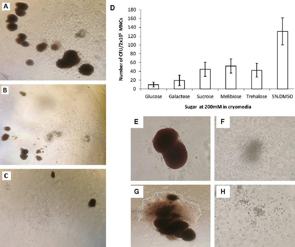
62 3.5 Carbohydrates with greater IRI activity preserve colony forming unit capacity of UCB samples. Colony forming unit (CFU) assays were also performed to provide relevant insight into the relationship between carbohydrate mediated ice recrystallization inhibition and preservation of functional capacity. Example images of the colonies enumerated and their plate densities following cryopreservation in 5% DMSO or 200mM carbohydrates are illustrated in Figure 7. We observed a strong correlation between greater preservation of CFU activity and IRI activity of the carbohydrates used for cryopreservation. The most active ice recrystallization-inhibitors preserved the CFU-forming activity to a greater extent (r 2 =0.92, p=<0.0001) (Figure 5A). Following cryopreservation and fast thawing, the range of total CFUs cryopreserved in carbohydrates was lower (5.7 ± 2.2 per 10 4 cells for glucose to 30 ± 3.5 for melibiose) than the average number of total colonies obtained from samples cryopreserved in 5% DMSO only (76 ± 10 per 10 4 cells (p=0.001)). We observed similar results for the most dominant colony forming unit CFU-GM (Figure 5B). 54
63 Figure 8. Typical colony-forming unit (CFU) assays (40x magnification) revealing colony densities from CB units cryopreserved in (A) 5% DMSO, (B) 200 mm melibiose and (C) 200 mm galactose. (D) Mean number (±SEM) total colonies formed following cryopreservation in 200 mm carbohydrate solution or 5% DMSO. (E H) Typical CFU colonies observed in methylcellulose assays after cryopreservation and thawing of cord blood. CFUs were identified by morphology and enumerated using an inverted microscope to discriminate erythroblast-forming units (BFU-E) (E), granulocyte/macrophage (CFU-GM) (F), granulocyte/erythroid/macrophage/monocyte (CFU-GEMM) (G) and erythroid (CFU-E) (H) colonies. 55
64 56
65 Figure 9. Colony Forming Unit capacity vs. IRI activity. UCB MNCs were cryopreserved with 200 mm (w/v) of the carbohydrate, fast-thawed and cultured for 2 weeks in semisolid methylcellulose media and total colonies (n=3 for each sugar) (A) and CFU-GM (n=3 for each sugar) (B) were enumerated. Samples with each carbohydrate were thawed in a 37 C water bath for the fast thaw (Black lines, n=5) The total colonies obtained from each sample are expressed relative to CFU number from a control sample from the same UCB unit which was cryopreserved with 5% DMSO (v/v). Resulting CFU capacity ratios were then plotted against the IRI activity of the corresponding carbohydrate where MGS represents the mean grain size of ice crystals in a solution of carbohydrate or phosphate buffered saline (PBS). 57
66 A CFU capacity ratio [total CFU sample / total CFU standard] R 2 (fast thaw)=.92 Total CFU IRI ratio (MGS sugar/mgs PBS) B CFU-GM capacity ratio [CFU-GM sample / CFU-GM standard] R 2 (fast thaw)=.86 CFU-GM IRI ratio (MGS sugar/mgs PBS) 58
67 3.6 Delayed engraftment and case control characteristics A nested case control study was designed to assess if cold induced apoptosis was was associated with delayed neutrophil engraftment after autologous HSCT. This type of study design was selected over a retrospective cohort study because retrospective cohort studies require analyzing all patients in a cohort, whereas nested case control studies require analyzing only the patients that developed the disease of interest and a specified number of matched controls within the same cohort that did not develop the disease of interest. Overall, the nested case control study design was utilized to minimize cost and time associated with a full retrospective cohort study. 326 lymphoma patients were identified who received autologous peripheral blood stem cell grafts with a target CD34 + progenitor dose 2.0 x 10 6 cells/kg, with 14 patients out of the 326 experiencing delayed neutrophil engraftment. PBSC graft aliquots from 13 out of the 14 delayed engraftment cases were available for analysis. PBSC graft aliquots from 28 patients in the original cohort that did not experience delayed neutrophil engraftment were also selected for analysis as matched controls. PBSC graft aliquots from 22 out of the 28 matched controls were available for analysis. These 22 matched controls were matched for age, male/female ratio, percentage of hodgkins lymphoma cases, and mean quantity of CD34 + progenitors and MNC harvested/kg. The mean time to initial neutrophil engraftment (1 st of 3 consecutive days with greater than 0.5 x 10 9 neutrophil/l) and the mean length of stay in hospital (LOS), however, was greater in the delayed engraftment case group compared to the matched control group (table 1). Additionally, a small number of patients had < 2.0 x 10 6 CD
68 progenitors/kg at the time of collection. The number was similar between the delayed engraftment cases (2/13, 15%) and control group (2/22, 9%, p=ns), and no significant difference was observed between cases and controls with regard to the number of patients exceeding the optimal threshold of 5.0 x 10 6 CD34 + progenitors/kg at the time of collection (p=ns) (table 2). 60
69 Table 1. Characteristics of study cases with delayed neutrophil engraftment and matched controls. Controls were matched with delayed engraftment cases for gender ratio, age, CD34 + progenitors and MNCs collected / kg patient weight, type of lymphoma (hodgkins or nonhodgkins). Delayed engraftment cases required a significantly longer time to reach an average neutrophil count of > 0.5 x 10 9 cells / L blood and required a length of stay (LOS) in hospital compared to matched controls. 61
70 Delayed engraftment vs control characteristics Delayed engraftment cases Matched controls Number Patients (N) Age (mean±sd) 48±14 47± gender (M/F) 7/6 12/10 N/A Hodgkins Lymphoma / NHL 2/11 3/19 N/A CD34 + progenitors / kg (3.7±2.0) x 10 6 (4.4±1.6) x harvested MNCs/kg harvested (7.9±3.5) x 10 8 (8.1±4.4) x days to ANC > 0.5 (mean SD) 17.6± ± LOS (d, mean +/- SD) 31.2± ± p 62
71 Table 1. Proportion of study cases and controls reaching the minimum CD34 + progenitor dose threshold (2.0x10 6 cells/kg) and the optimal CD34 + progenitor dose threshold (5.0x10 6 cells/kg) at collection, after thawing and considering only viable CD34 + cells, and after thawing and considering only viable nonapoptotic CD34 + cells. 63
72 Proportion of patients meeting CD34 threshold dose Delayed engraftment cases Matched Controls Patients receiving > minimum CD34 dose At collection 11 (85%) 20 (91%) 0.6 Post thaw, viable 10 (77%) 17 (77%) 1.0 Post thaw, viable nonapoptotic 6 (46%) 17 (77%) 0.08 Patients receiving > optimal CD34 dose At collection 2(17%) 8 (36%) 0.3 Post thaw, viable 2 (17%) 6 (27%) 0.7 Post thaw, viable nonapoptotic 2 (17%) 3 (14%) 1.0 P 64
73 3.7 The association between viability and delayed neutrophil engraftment in PBSC grafts The percentage and absolute number of cells excluding 7AAD transplanted into the delayed engraftment and control patients were assessed to determine if loss of viability was associated with reduced functionality in a transplant setting. Patients with delayed neutrophil recovery and controls had similar percentages of viable MNCs and viable CD34 + progenitors following processing and thawing (see Figure 7). In general, PBSC grafts maintained high levels of cell viability in both the MNC population (91.0 ± 1.3% for cases vs 91.6 ± 0.7 % for controls, p=0.7) and the CD34 + progenitor population (84.6 ± 2.7% for cases vs ± 1.4% for controls, p=0.8) after thawing (see Figure 6). In addition, the absolute number of viable MNCs and CD34 + progenitors available for reinfusion and calculated based on patients weight was calculated to determine the actual dose of cells for each patient and were similar for cases and controls (see Figure 7). Further, the proportion of patients with delayed neutrophil recovery who received more than 2.0 x 10 6 CD34 + progenitors/kg was similar to the controls (10/13, 77% vs 17/22, 77%, p=ns) and likewise, the proportion of patients receiving more than 5.0 x 10 6 CD34 + progenitors/kg was the same in cases and controls (2 of 13 cases, 17% vs. 6 of 22 controls, 27%, p=ns) ( see table 2). 65
74 Figure 10. Changes in cell viability and apoptosis after thawing cryopreserved PBSCs. Typical flow cytometry analysis of mononuclear cells (gated from R1 in figure 1) (A), CD34 + progenitors (gated from R3 in figure 1) (B) and CD34 + CD45 low ALDH high progenitors (gated from R4 in figure 1) (C) in freshly collected PBSCs and of mononuclear cells (D), CD34 + progenitors (E) and CD34 + CD45 low ALDH high progenitors (F) after thawing cryopreserved PBSCs. The bottom left and bottom right quadrants of all dot plots represent the viable cells that exclude 7AAD while the bottom right quadrants specifically represents viable apoptotic cells that bind AnnexinV-PE but exclude 7AAD, while the bottom left quadrants specifically represent viable nonapoptotic cells that exclude 7AAD and didn t bind Annexin-V. 66
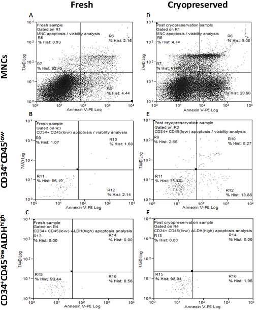
75 67
DISCOVERING ATCC IMMUNOLOGICAL CELLS - MODEL SYSTEMS TO STUDY THE IMMUNE AND CARDIOVASCULAR SYSTEMS
 DISCOVERING ATCC IMMUNOLOGICAL CELLS - MODEL SYSTEMS TO STUDY THE IMMUNE AND CARDIOVASCULAR SYSTEMS James Clinton, Ph.D. Scientist, ATCC February 19, 2015 About ATCC Founded in 1925, ATCC is a non-profit
CD34+ Cells: A Comparison of Stem and Progenitor Cells in Cord Blood, Peripheral Blood, and the Bone Marrow
 White Paper September 2016 CD34+ Cells: A Comparison of Stem and Progenitor Cells in Cord Blood, Peripheral Blood, and the Bone Marrow Lily C. Trajman, PhD Introduction: Hematopoietic Stem Cells (HSCs)
Analysis of the Viability of Umbilical Cord blood Stem Cells. Sachdeva N
 Jsrm/Vol5No.11, 2009 Original Article Analysis of the of Umbilical Cord blood Stem Cells Sachdeva N Dr. Neelam Sachdeva Reader, Department of Zoology, Gargi College, University of Delhi, Siri Fort Road,
Understanding the role of ex vivo T cell depletion
 Prevention of graftversus-host disease (GVHD) Understanding the role of ex vivo T cell depletion Information for patients undergoing allogeneic stem cell transplantation in AML and their families 2 This
Dr.PSRK.Sastry MD, ECMO
 Peripheral blood stem cell transplantation (Haematopoietic stem cell transplantation) Dr.PSRK.Sastry MD, ECMO Consultant, Medical Oncology Kokilaben Dhirubhai Ambani Hospital Normal hematopoiesis Historical
BMTCN REVIEW COURSE PRE-TRANSPLANT CARE
 BMTCN REVIEW COURSE PRE-TRANSPLANT CARE Jennifer Shamai MS, RN, AOCNS, BMTCN Professional Practice Leader Department of Clinical Practice And Professional Education Click How to edit the Master Experts
Hematopoiesis. Hematopoiesis. Hematopoiesis
 Chapter. Cells and Organs of the Immune System Hematopoiesis Hematopoiesis- formation and development of WBC and RBC bone marrow. Hematopoietic stem cell- give rise to any blood cells (constant number,
Cord Blood Bankers are. Angie Yost, MT(ASCP) CM 2016 ASCLS-MO CLMA Spring Meeting
 CM 2016 ASCLS-MO CLMA Spring Meeting") Cord Blood Bankers are Angie Yost, MT(ASCP) CM 2016 ASCLS-MO CLMA Spring Meeting Objectives Cord blood 101 Manufacturing and cryopreservation Characterization testing Why Do We Need Cord Blood? Each year,
Cord Blood Bankers are Angie Yost, MT(ASCP) CM 2016 ASCLS-MO CLMA Spring Meeting Objectives Cord blood 101 Manufacturing and cryopreservation Characterization testing Why Do We Need Cord Blood? Each year,
Hematopoiesis. - Process of generation of mature blood cells. - Daily turnover of blood cells (70 kg human)
") Hematopoiesis - Process of generation of mature blood cells - Daily turnover of blood cells (70 kg human) 1,000,000,000,000 total cells 200,000,000,000 red blood cells 70,000,000,000 neutrophils Hematopoiesis
Hematopoiesis - Process of generation of mature blood cells - Daily turnover of blood cells (70 kg human) 1,000,000,000,000 total cells 200,000,000,000 red blood cells 70,000,000,000 neutrophils Hematopoiesis
Myeloproliferative Disorders - D Savage - 9 Jan 2002
 Disease Usual phenotype acute leukemia precursor chronic leukemia low grade lymphoma myeloma differentiated Total WBC > 60 leukemoid reaction acute leukemia Blast Pro Myel Meta Band Seg Lymph 0 0 0 2
Neutrophil Recovery: The. Posttransplant Recovery. Bus11_1.ppt
 Neutrophil Recovery: The First Step in Posttransplant Recovery No conflicts of interest to disclose Bus11_1.ppt Blood is Made in the Bone Marrow Blood Stem Cell Pre-B White cells B Lymphocyte T Lymphocyte
Hematopoiesis. BHS Liège 27/1/2012. Dr Sonet Anne UCL Mont-Godinne
 Hematopoiesis BHS Liège 27/1/2012 Dr Sonet Anne UCL Mont-Godinne Hematopoiesis: definition = all the phenomenons to produce blood cells Leukocytes = White Blood Cells Polynuclear = Granulocytes Platelet
The Immune System. A macrophage. ! Functions of the Immune System. ! Types of Immune Responses. ! Organization of the Immune System
 The Immune System! Functions of the Immune System! Types of Immune Responses! Organization of the Immune System! Innate Defense Mechanisms! Acquired Defense Mechanisms! Applied Immunology A macrophage
Hematopoietic Stem Cells, Stem Cell Processing, and Transplantation
 Hematopoietic Stem Cells, Stem Cell Processing, and Joseph (Yossi) Schwartz, M irector, Hemotherapy and Stem Cell Processing Facility Bone Marrow Can Cure: Leukemia Lymphoma Multiple Myeloma Genetic iseases:
One Day BMT Course by Thai Society of Hematology. Management of Graft Failure and Relapsed Diseases
 One Day BMT Course by Thai Society of Hematology Management of Graft Failure and Relapsed Diseases Piya Rujkijyanont, MD Division of Hematology-Oncology Department of Pediatrics Phramongkutklao Hospital
What the Transfusion Scientist should know about Stem Cells. Dr Claire Wiggins BBTS September 2017
 What the Transfusion Scientist should know about Stem Cells Dr Claire Wiggins BBTS September 2017 What is a stem cell? Undifferentiated cell Can divide and self renew for long periods Differentiate into
Bone Marrow Transplantation and the Potential Role of Iomab-B
 Bone Marrow Transplantation and the Potential Role of Iomab-B Hillard M. Lazarus, MD, FACP Professor of Medicine, Director of Novel Cell Therapy Case Western Reserve University 1 Hematopoietic Cell Transplantation
XIV. HLA AND TRANSPLANTATION MEDICINE
 XIV. HLA AND TRANSPLANTATION MEDICINE A. Introduction 1. The HLA system includes a complex array of genes and their molecular products that are involved in immune regulation and cellular differentiation.
Corporate Medical Policy
 Corporate Medical Policy Hematopoietic Cell Transplantation for CLL and SLL File Name: Origination: Last CAP Review: Next CAP Review: Last Review: hematopoietic_cell_transplantation_for_cll_and_sll 2/2001
Stem cells: units of development and regeneration. Fernando D. Camargo Ph.D. Whitehead Fellow Whitehead Institute for Biomedical Research.
 Stem cells: units of development and regeneration Fernando D. Camargo Ph.D. Whitehead Fellow Whitehead Institute for Biomedical Research Concepts 1. Embryonic vs. adult stem cells 2. Hematopoietic stem
Rapid antigen-specific T cell enrichment (Rapid ARTE)
") Direct ex vivo characterization of human antigen-specific CD154+CD4+ T cell Rapid antigen-specific T cell enrichment (Rapid ARTE) Introduction Workflow Antigen (ag)-specific T cells play a central role
Direct ex vivo characterization of human antigen-specific CD154+CD4+ T cell Rapid antigen-specific T cell enrichment (Rapid ARTE) Introduction Workflow Antigen (ag)-specific T cells play a central role
Production of the Formed Elements (Chapter 11) *
 *") OpenStax-CNX module: m62120 1 Production of the Formed Elements (Chapter 11) * Ildar Yakhin Based on Production of the Formed Elements by OpenStax This work is produced by OpenStax-CNX and licensed under
OpenStax-CNX module: m62120 1 Production of the Formed Elements (Chapter 11) * Ildar Yakhin Based on Production of the Formed Elements by OpenStax This work is produced by OpenStax-CNX and licensed under
BMTCN Review Course Basic Concepts and Indications for Transplantation How the Experts Treat Hematologic Malignancies Las Vegas, NV, March 10, 2016
 BMTCN Review Course Basic Concepts and Indications for Transplantation How the Experts Treat Hematologic Malignancies Las Vegas, NV, March 10, 2016 David Rice, PhD, RN, NP Director, Professional Practice
Stem cells. -Dr Dinesh Bhurani, MD, DM, FRCPA. Rajiv Gandhi Cancer Institute, Delhi, -Director, Department of Haematology and BMT
 Stem cells -Dr Dinesh Bhurani, MD, DM, FRCPA -Director, Department of Haematology and BMT Rajiv Gandhi Cancer Institute, Delhi, Flow of presentation Update on stem cell uses Haematopoietic stem cell transplantation
Direct ex vivo characterization of human antigen-specific CD154 + CD4 + T cells Rapid antigen-reactive T cell enrichment (Rapid ARTE)
") Direct ex vivo characterization of human antigen-specific CD154 + CD4 + T cells Rapid antigen-reactive T cell enrichment (Rapid ARTE) Introduction Workflow Antigen (ag)-specific T cells play a central
Direct ex vivo characterization of human antigen-specific CD154 + CD4 + T cells Rapid antigen-reactive T cell enrichment (Rapid ARTE) Introduction Workflow Antigen (ag)-specific T cells play a central
Hematopoietic Stem Cells
 Hematopoietic Stem Cells, Stem Cell Processing, and Joseph (Yossi) Schwartz, M irector, Hemotherapy and Stem Cell Processing Facility E-mail: js2745@columbia.edu Hematopoietic Stem Cells Sustain hematopoiesis
CHAPTER 3 LABORATORY PROCEDURES
 CHAPTER 3 LABORATORY PROCEDURES CHAPTER 3 LABORATORY PROCEDURES 3.1 HLA TYPING Molecular HLA typing will be performed for all donor cord blood units and patients in the three reference laboratories identified
Human Umbilical Cord Blood CD34 + /133+ Progenitor Cell Care Manual
 Human Umbilical Cord Blood CD34 + /133+ Progenitor Cell Care Manual INSTRUCTION MANUAL ZBM0065.04 SHIPPING CONDITIONS Human Umbilical Cord Blood CD34+/133+ Progenitor Cells, cryopreserved Cryopreserved
Principles of Anatomy and Physiology
 Principles of Anatomy and Physiology 14 th Edition CHAPTER 19 The Cardiovascular System: The Blood Functions and Properties of Blood Blood is a liquid connective tissue consisting of cells surrounded by
Human CD4+T Cell Care Manual
 Human CD4+T Cell Care Manual INSTRUCTION MANUAL ZBM0067.04 SHIPPING CONDITIONS Human CD4+T Cells, cryopreserved Cryopreserved human CD4+T cells are shipped on dry ice and should be stored in liquid nitrogen
Corporate Medical Policy
 Corporate Medical Policy Hematopoietic Stem-Cell Transplantation for Waldenstrom Macroglobulinemia File Name: Origination: Last CAP Review: Next CAP Review: Last Review: hematopoietic_stem_cell_transplantation_for_waldenstrom_macroglobulinemia
Hematopoietic Stem Cell Therapy
 Hematopoietic Stem Cell Therapy Grace Totoe, MBChB, SBB CME August 2012 Accra-Ghana Hematopoietic Stem Cells Cells capable of self renewal and differentiation into all blood cell lineages Objectives Historical
Corporate Medical Policy
 Corporate Medical Policy File Name: Origination: Last CAP Review: Next CAP Review: Last Review: hematopoietic_stem-cell_ transplantation_for_primary_amyloidosis 2/2001 11/2018 11/2019 11/2018 Description
What s a Transplant? What s not?
 What s a Transplant? What s not? How to report the difference? Daniel Weisdorf MD University of Minnesota Anti-cancer effects of BMT or PBSCT [HSCT] Kill the cancer Save the patient Restore immunocompetence
Bone marrow transplantation for PID
 BMT Bone marrow transplantation for PID hello@piduk.org 0800 987 898 www.piduk.org About this leaflet Bone marrow transplant (BMT; also known as haematopoietic stem cell transplant or HSCT) is increasingly
Hematology. The Study of blood
 Hematology The Study of blood Average adult = 8-10 pints of blood Composition: PLASMA liquid portion of blood without cellular components Serum plasma after a blood clot is formed Cellular elements are
What can we learn from cryopreserved blood cells?
 What can we learn from cryopreserved blood cells? Andreas Sputtek Universitätsklinikum Hamburg-Eppendorf Inst. f. Transfusionsmedizin Hamburg, Germany http://www.sputtek.de Plenary Lecture (Definition)
cryotubes Information from Biochrom AG, May 23, 2011
 Recommendations on how to safely freeze and thaw cell cultures in TPP cryotubes Information from Biochrom AG, May 23, 2011 Cryopreservation can be used to store cell cultures for a virtually indefinite
Unit 6: Circulatory System. 6.1 Blood
 Unit 6: Circulatory System 6.1 Blood Blood Function Function Nutritive Respiratory Excretory Regulatory Protective Effects on Body Transporting nutrient molecules (glucose, amino acids, fatty acids and
UMBILICAL CORD BLOOD STEM CELLS EXPANDED IN THE PRESENCE OF NICOTINAMIDE (NICORD) PROVIDE LONG TERM MULITI-LINEAGE ENGRAFTMENT
 PROVIDE LONG TERM MULITI-LINEAGE ENGRAFTMENT") UMBILICAL CORD BLOOD STEM CELLS EXPANDED IN THE PRESENCE OF NICOTINAMIDE (NICORD) PROVIDE LONG TERM MULITI-LINEAGE ENGRAFTMENT Mitchell E. Horwitz, MD Duke University Medical Center Duke Cancer Institute
UMBILICAL CORD BLOOD STEM CELLS EXPANDED IN THE PRESENCE OF NICOTINAMIDE (NICORD) PROVIDE LONG TERM MULITI-LINEAGE ENGRAFTMENT Mitchell E. Horwitz, MD Duke University Medical Center Duke Cancer Institute
10/18/2012. A primer in HLA: The who, what, how and why. What?
 A primer in HLA: The who, what, how and why What? 1 First recognized in mice during 1930 s and 1940 s. Mouse (murine) experiments with tumors Independent observations were made in humans with leukoagglutinating
Basic information on the cryopreservation process
 COST Action FA1205 AQUAGAMETE 5 th AQUAGAMETE Training School Valencia, Spain, 7-11 th March, 2016 Basic information on the cryopreservation process Ákos Horváth Department of Aquaculture, Szent István
Haplo vs Cord vs URD Debate
 3rd Annual ASBMT Regional Conference for NPs, PAs and Fellows Haplo vs Cord vs URD Debate Claudio G. Brunstein Associate Professor University of Minnesota Medical School Take home message Finding a donor
Blood and Defense. Chapter 11
 Blood and Defense Chapter 11 Functions of Blood 1. Carry nutrients from the small intestine and oxygen from the lung to tissues in the body 2. Transport wastes from tissues to the kidneys and carbon dioxide
The Immune System. These are classified as the Innate and Adaptive Immune Responses. Innate Immunity
 The Immune System Biological mechanisms that defend an organism must be 1. triggered by a stimulus upon injury or pathogen attack 2. able to counteract the injury or invasion 3. able to recognise foreign
Transfer protocol of human HSC into NOG mice
 Transfer protocol of human HSC into NOG mice Mice: Adult NOG mice are aged 8-12 weeks. Newborn mice are 1 2 days old. 8-12 week old NOG mice irradiated with 2.5 Gy Intravenous transfer of 1-0.5 x 10 5
Guidelines for Gamma Irradiation of Blood Components
 AUSTRALIAN & NEW ZEALAND SOCIETY OF BLOOD TRANSFUSION INC. AUSTRALIAN RED CROSS BLOOD SERVICE NEW ZEALAND BLOOD SERVICE Guidelines for Gamma Irradiation of Blood Components Revised 2003 AN ZS B T Australian
Blood consists of red and white blood cells suspended in plasma Blood is about 55% plasma and 45% cellular elements Plasma 90% water 10% dissolved
 Bio 100 Guide 21 Blood consists of red and white blood cells suspended in plasma Blood is about 55% plasma and 45% cellular elements Plasma 90% water 10% dissolved inorganic ions, proteins, nutrients,
M.Sc. III Semester Biotechnology End Semester Examination, 2013 Model Answer LBTM: 302 Advanced Immunology
 Code : AS-2246 M.Sc. III Semester Biotechnology End Semester Examination, 2013 Model Answer LBTM: 302 Advanced Immunology A. Select one correct option for each of the following questions:- 2X10=10 1. (b)
The role of HLA in Allogeneic Hematopoietic Stem Cell Transplantation and Platelet Refractoriness.
 The role of HLA in Allogeneic Hematopoietic Stem Cell Transplantation and Platelet Refractoriness. Robert Liwski, MD, PhD, FRCPC Medical Director HLA Typing Laboratory Department of Pathology Dalhousie
Chapter 13 The Blood
 Chapter 13 The Blood Copyright 2015 Wolters Kluwer Health Lippincott Williams & Wilkins Overview Key Terms agglutination erythrocyte lymphocyte albumin fibrin megakaryocyte anemia hematocrit monocyte antigen
Chapter 19: The Cardiovascular System: The Blood. Copyright 2009, John Wiley & Sons, Inc.
 Chapter 19: The Cardiovascular System: The Blood Blood Liquid connective tissue 3 general functions 1. Transportation Gases, nutrients, hormones, waste products 2. Regulation ph, body temperature, osmotic
The Innate Immune Response
 The Innate Immune Response FUNCTIONS OF THE IMMUNE SYSTEM: Recognize, destroy and clear a diversity of pathogens. Initiate tissue and wound healing processes. Recognize and clear damaged self components.
Question 1. Kupffer cells, microglial cells and osteoclasts are all examples of what type of immune system cell?
 Abbas Chapter 2: Sarah Spriet February 8, 2015 Question 1. Kupffer cells, microglial cells and osteoclasts are all examples of what type of immune system cell? a. Dendritic cells b. Macrophages c. Monocytes
CELLULAR THERAPY QUIZ. Correct answers and rationale
 CELLULAR THERAPY QUIZ Correct answers and rationale In January 2018, registered cellular therapy (CT) facilities were asked to participate in the quiz to determine the educational needs of the users regarding
What is the composition of blood, including blood cells? What organs and structures control the flow of blood throughout the body?
 3 Chapter 10: Circulatory System and Lymphatic System In this chapter, you will learn about the structure and function of the circulatory system and lymphatic system. What is the composition of blood,
Unit 10: Blood. 2. Buffy coat contains leukocytes and platelets (less than 1% of blood)
") Unit 10: Blood I. Blood A. The only fluid tissue in the human body B. Classified as a connective tissue C. Components of blood 1. Living cells a. Formed elements 2. Non-living matrix a. Plasma D. If blood
Unit 10: Blood I. Blood A. The only fluid tissue in the human body B. Classified as a connective tissue C. Components of blood 1. Living cells a. Formed elements 2. Non-living matrix a. Plasma D. If blood
Trends in Hematopoietic Cell Transplantation. AAMAC Patient Education Day Oct 2014
 Trends in Hematopoietic Cell Transplantation AAMAC Patient Education Day Oct 2014 Objectives Review the principles behind allogeneic stem cell transplantation Outline the process of transplant, some of
Manipulation of T Cells in the Thnsplant Inoculum
 International Journal of Cell Cloning 4: 122-126 Suppl 1 (1986) Manipulation of T Cells in the Thnsplant Inoculum J. Kersey Bone Marrow Transplantation Program, University of Minnesota, Minneapolis, MN,
Blood. The only fluid tissue in the human body Classified as a connective tissue. Living cells = formed elements Non-living matrix = plasma
 Blood Blood The only fluid tissue in the human body Classified as a connective tissue Living cells = formed elements Non-living matrix = plasma Blood Physical Characteristics of Blood Color range Oxygen-rich
Chapter 1. Chapter 1 Concepts. MCMP422 Immunology and Biologics Immunology is important personally and professionally!
 MCMP422 Immunology and Biologics Immunology is important personally and professionally! Learn the language - use the glossary and index RNR - Reading, Note taking, Reviewing All materials in Chapters 1-3
Production of the Formed Elements *
 OpenStax-CNX module: m46691 1 Production of the Formed Elements * OpenStax This work is produced by OpenStax-CNX and licensed under the Creative Commons Attribution License 3.0 By the end of this section,
Proper steps for bull semen dilution and freezing. IMV Technologies France
 Proper steps for bull semen dilution and freezing IMV Technologies France Introduction Since Polge reported the first successful cryopreservation of spermatozoa in 1949, spermatozoa from many mammalian
The Texas Medical Center
 ISCT Regional Meeting November 2 4, 2007 Cryopreservation - Evaluation of Different Processes Presenters: Carlos Lee & Renee Smilee The Texas Medical Center Center for Cell & Gene Therapy The Feigin Center
CAN-ASC CONSENSUS PROTOCOL: ISOLATION, CRYOPRESERVATION AND THAWING OF PERIPHERAL BLOOD MONONUCLEAR CELLS
 CAN-ASC CONSENSUS PROTOCOL: ISOLATION, CRYOPRESERVATION AND THAWING OF PERIPHERAL BLOOD MONONUCLEAR CELLS Version March 21, 2019 NOTES Caution: human blood and blood-derived products are a potential source
Immune System AP SBI4UP
 Immune System AP SBI4UP TYPES OF IMMUNITY INNATE IMMUNITY ACQUIRED IMMUNITY EXTERNAL DEFENCES INTERNAL DEFENCES HUMORAL RESPONSE Skin Phagocytic Cells CELL- MEDIATED RESPONSE Mucus layer Antimicrobial
Umbilical Cord Blood Transplantation
 Umbilical Cord Blood Transplantation Current Results John E. Wagner, M.D. Blood and Marrow Transplant Program and Stem Cell Institute University of Minnesota Donor Choices Unrelated Marrow/PBSC Results
Accelerate Your Research with Conversant Bio
 Accelerate Your Research with Conversant Bio 400+ Participating MDs 50+ Partner sites for tissue procurement Continuous expansion of sourcing capabilities Closely monitored chain of custody Full regulatory
What is the immune system? Types of Immunity. Pasteur and rabies vaccine. Historical Role of smallpox. Recognition Response
 Recognition Response Effector memory What is the immune system? Types of Immunity Innate Adaptive Anergy: : no response Harmful response: Autoimmunity Historical Role of smallpox Pasteur and rabies vaccine
12 Dynamic Interactions between Hematopoietic Stem and Progenitor Cells and the Bone Marrow: Current Biology of Stem Cell Homing and Mobilization
 Table of Contents: PART I: Molecular and Cellular Basis of Hematology 1 Anatomy and Pathophysiology of the Gene 2 Genomic Approaches to Hematology 3 Regulation of Gene Expression, Transcription, Splicing,
Instructions for Use. APO-AB Annexin V-Biotin Apoptosis Detection Kit 100 tests
 3URGXFW,QIRUPDWLRQ Sigma TACS Annexin V Apoptosis Detection Kits Instructions for Use APO-AB Annexin V-Biotin Apoptosis Detection Kit 100 tests For Research Use Only. Not for use in diagnostic procedures.
Average adult = 8-10 pints of blood. Functions:
 Average adult = 8-10 pints of blood Functions: Transports nutrients, oxygen, cellular waste products, and hormones Aids in distribution of heat Regulates acid-base balance Helps protect against infection
OneMatch Stem Cell and Marrow Network. Training Guide
 OneMatch Stem Cell and Marrow Network Training Guide What is OneMatch all about? OneMatch is a Canadian Program that matches and coordinates the collection of stem cells from potential donors to help save
Chapter 06 Lecture Outline. See separate PowerPoint slides for all figures and tables preinserted into PowerPoint without notes.
 Chapter 06 Lecture Outline See separate PowerPoint slides for all figures and tables preinserted into PowerPoint without notes. Copyright 2016 McGraw-Hill Education. 2012 Pearson Permission Education,
Chapter 19 Cardiovascular System Blood: Functions. Plasma
 Chapter 19 Cardiovascular System Blood: Functions 19-1 Plasma Liquid part of blood. Colloid: liquid containing suspended substances that don t settle out of solution 91% water. Remainder proteins, ions,
Unit 10 - Blood The only fluid tissue in the human body. c) Plasma rises to the top (55% of blood)
 Plasma rises to the top (55% of blood)") Unit 10 - Blood 1 I. Unit 10: Blood A. Blood 1. The only fluid tissue in the human body 2. Classified as a connective tissue 3. Components of blood a) Living cells (1) Formed elements b) Non-living matrix
Unit 10 - Blood 1 I. Unit 10: Blood A. Blood 1. The only fluid tissue in the human body 2. Classified as a connective tissue 3. Components of blood a) Living cells (1) Formed elements b) Non-living matrix
Lifeblood Lab Activity
 History of Blood: It is the universal symbol of horror, of death, yet it is the one thing that keeps you living. It is the blood that is coursing through your veins. But, what do you really know about
Leukemias and Lymphomas Come From Normal Blood Cells
 Leukemias and Lymphomas Come From Normal Blood Cells by Steve Anderson, Ph.D. Steve Anderson has a Ph.D. in Immunology with 25 years experience in biomedical research. His scientific expertise includes
The National Marrow Donor Program. Graft Sources for Hematopoietic Cell Transplantation. Simon Bostic, URD Transplant Recipient
 1988 199 1992 1994 1996 1998 2 22 24 26 28 21 212 214 216 218 Adult Donors Cord Blood Units The National Donor Program Graft Sources for Hematopoietic Cell Transplantation Dennis L. Confer, MD Chief Medical
immunity defenses invertebrates vertebrates chapter 48 Animal defenses --
 defenses Animal defenses -- immunity chapter 48 invertebrates coelomocytes, amoebocytes, hemocytes sponges, cnidarians, etc. annelids basophilic amoebocytes, acidophilic granulocytes arthropod immune systems
Immunity. ES/RP 531 Fundamentals of Environmental Toxicology. Lecture 14 Immunotoxicity. Instructor: Allan Felsot
 Instructor: Allan Felsot afelsot@tricity.wsu.edu Fall 2005 ES/RP 531 Fundamentals of Environmental Toxicology Lecture 14 Immunotoxicity in Humans Hematopoiesis (generation of blood cells) Differentiation
Transplantation - Challenges for the future. Dr Gordon Cook S t James s Institute of Oncology, Leeds Teaching Hospitals Trust
 Transplantation - Challenges for the future Dr Gordon Cook S t James s Institute of Oncology, Leeds Teaching Hospitals Trust Bone Marrow Transplantation Timeline, 1957-2006 Appelbaum F. N Engl J Med 2007;357:1472-1475
The Main Constituents of Blood
 The Main Constituents of Blood Described as a fluid connective tissue, blood is comprised of approximately 55% plasma (a yellow-ish but transparent fluid) and 45% cellular volume (erythrocytes (red cells),
Immunology Lecture 4. Clinical Relevance of the Immune System
 Immunology Lecture 4 The Well Patient: How innate and adaptive immune responses maintain health - 13, pg 169-181, 191-195. Immune Deficiency - 15 Autoimmunity - 16 Transplantation - 17, pg 260-270 Tumor
Unit Seven Blood and Immunity
 Unit Seven Blood and Immunity I. Introduction A. Definition Blood is a sticky fluid that is heavier and thicker than water. Blood is a type of, whose cells and suspended in a liquid intercellular material.
Pearson's Comprehensive Medical Assisting Administrative and Clinical Competencies
 Pearson's Comprehensive Medical Assisting Administrative and Clinical Competencies THIRD EDITION CHAPTER 27 The Cardiovascular System Lesson 2: Composition and Function of Lesson Objectives Upon completion
4. TEXTBOOK: ABUL K. ABBAS. ANDREW H. LICHTMAN. CELLULAR AND MOLECULAR IMMUNOLOGY. 5 TH EDITION. Chapter 2. pg
 LECTURE: 03 Title: CELLS INVOLVED IN THE IMMUNE RESPONSE LEARNING OBJECTIVES: The student should be able to: Identify the organs where the process of the blood formation occurs. Identify the main cell
Clinical Policy: Donor Lymphocyte Infusion
 Clinical Policy: Reference Number: PA.CP.MP.101 Effective Date: 01/18 Last Review Date: 11/16 Coding Implications Revision Log This policy describes the medical necessity requirements for a donor lymphocyte
Effect of sucrose and propylene glycol on the vitrification of sheep oocytes
 Journal of Cell and Animal Biology Vol. 7 (3), pp. 25-30, March 2013 Available online at http://www.academicjournals.org/jcab DOI: 10.5897/JCAB12.033 ISSN 1996-0867 2013 Academic Journals Full Length Research
Potency & Release Testing for unrelated donor cord blood units
 Potency & Release Testing for unrelated donor cord blood units Joanne Kurtzberg, MD Robertson Cell and Translational Therapy Program Pediatric Blood and Marrow Transplant Program Carolinas Cord Blood Bank
Hematopoiesis i starts from. HPC differentiates t to sequentially more mature. HPC circulate in very low
 Upstate Cord Blood Bank Hematopoietic Progenitor Cells Hematopoiesis i starts from self renewing HPC HPC differentiates t to sequentially more mature blood cells HPC circulate in very low numbers in adults
Immunology. Prof. Nagwa Mohamed Aref (Molecular Virologist & Immunology)
") Host Defenses Overview and Nonspecific Defenses I Immunology Prof. Nagwa Mohamed Aref (Molecular Virologist & Immunology) The Nature of Host Defenses 2 3 4 1st line of defense - intact skin mucous membranes
Host Defenses Overview and Nonspecific Defenses I Immunology Prof. Nagwa Mohamed Aref (Molecular Virologist & Immunology) The Nature of Host Defenses 2 3 4 1st line of defense - intact skin mucous membranes
Innate Immunity: Nonspecific Defenses of the Host
 PowerPoint Lecture Presentations prepared by Bradley W. Christian, McLennan Community College C H A P T E R 16 Innate Immunity: Nonspecific Defenses of the Host Host Response to Disease Resistance- ability
Graft source and Stem cell collection SULADA PUKIAT, MD
 + Graft source and Stem cell collection SULADA PUKIAT, MD + Hematopoietic stem cells Hematopoietic stem cells are CD34 + /CD45 dim /SSC low /FSClow to intermediate Neutrophils Bone NK cells T cells Basophils
Principles of Adaptive Immunity
 Principles of Adaptive Immunity Chapter 3 Parham Hans de Haard 17 th of May 2010 Agenda Recognition molecules of adaptive immune system Features adaptive immune system Immunoglobulins and T-cell receptors
Haematopoietic stem cells
 Haematopoietic stem cells Neil P. Rodrigues, DPhil NIH Centre for Biomedical Research Excellence in Stem Cell Biology Boston University School of Medicine neil.rodrigues@imm.ox.ac.uk Haematopoiesis: An
Blood. Water compartments
 Blood Water compartments 2/8 about 60% of our body is water (young-old, male-female) water is located in compartments, movement is regulated intracellular : extracellular 2:1, i.e. 40:20% interstitial
Automated and Standardized Counting of Mouse Bone Marrow CFU Assays
 Automated and Standardized Counting of Mouse Bone Marrow CFU Assays 2 Automated and Standardized Colony Counting Table of Contents 4 Colony-Forming Unit (CFU) Assays for Mouse Bone Marrow 5 Automated Assay
Blood Product Modifications: Leukofiltration, Irradiation and Washing
 1. Leukocyte Reduction Definitions and Standards: o Process also known as leukoreduction, or leukofiltration o Applicable AABB Standards, 25th ed. Leukocyte-reduced RBCs At least 85% of original RBCs