ETIO-PATHOMORPHOLOGICAL STUDIES ON GASTROINTESTINAL TRACT OF BROILER CHICKENS IN JAMMU By NAVROSE SANGHA (J-15-MV-452)
|
|
|
- Branden Aubrey Scott
- 5 years ago
- Views:
Transcription

1 ETIO-PATHOMORPHOLOGICAL STUDIES ON GASTROINTESTINAL TRACT OF BROILER CHICKENS IN JAMMU By NAVROSE SANGHA (J-15-MV-452) Thesis submitted to Faculty of Postgraduate Studies in partial fulfillment of requirements for the degree of MASTER OF VETERINARY SCIENCE IN VETERINARY PATHOLOGY Division of Veterinary Pathology Sher-e-Kashmir University of Agricultural Sciences & Technology of Jammu Main Campus, Chatha, Jammu
2
3
4
5 ACKNOWLEDGEMENT The Almighty Lord, The Most Compassionate and Most Merciful had blessed me for undertaking this endeavor. I got inspiration during my research work from Bani of Guru Granth Sahib Ji. I don t have words to express my gratitude for my venerated and esteemed major advisor Dr. Shilpa Sood, Assistant Professor, Division of Veterinary Pathology for her expert guidance, keen interest and constant encouragement during my entire degree programme. I simply feel myself blessed being provided with an academic advisor like her. It is my sole prerogative to place on record my indebtedness and everlasting gratitude to the members of my Advisory Committee, Dr. Shagufta Azmi, Ex-Professor and Head, Division of Veterinary Pathology, Dr. Nawab Nashiruddullah, Assoc. Professor & Head, Division of Veterinary Pathology, Dr. Anil Taku, Professor & Head, Division of Veterinary Microbiology and Dr. Mudasir Sultana, Professor & Head, Division of Veterinary Pharmacology & Toxicology for their help, innovative guidance and invaluable suggestions during the course of my study. I do extend my respectful thanks and warm regards to Dr. Rajesh Katoch, (Professor & Head, Division of Vety. Parasitology), Dr. Altaf Bhat, (Professor, Division of Vety. Microbiology) and Dr. Shafiqur Rahman, (Assistant Professor, Division of Vety. Pathology) I am thankful to Hon ble Vice Chancellor of SKUAST-Jammu for allowing me to undertake the study and for providing necessary facilities. It is rearest to thank Director Education, Dean (FVSc. & A.H) and Associate Dean (FVSc. & A.H) for their courteous & indulgent moral, extraordinary help, technical and timely academic support throughout the course of the study. My sincere thanks are due to all technical and non-technical staff of Department of Veterinary Pathology, especially to Kiran Bala, Mr. Joginder, Mr. Mohinder and Mr. Bodraj.
6
7 ABSTRACT Title of Thesis : Etio-pathomorphology of Gastrointestinal Tract of Broiler Chickens in Various Parts of Jammu. Name of Student : Navrose Sangha Registration No. : (J-15-MV-452) Major Subject : Veterinary Pathology Name and Designation of : Major Advisor Dr. Shilpa Sood Assistant Professor, Division of Veterinary Pathology, F.V.SC. & A.H, R.S.Pura, Jammu. Degree to be Awarded : M.V.Sc. (Veterinary Pathology) Year of Award of Degree : 2017 Name of the University : Sher-e-Kashmir University of Agricultural Sciences & Technology of Jammu (J&K). The present study was carried out to study etio-pathomorphology of gastrointestinal tract of broiler chickens where disease occurrence was reported. 200 flocks were surveyed in different areas in and around Jammu. Occurrence of colibacillosis (24.16%) was maximum followed by that of infectious bursal disease (IBD) (7.13%), omphalitis (6.79%), salmonellosis (5.36%), non specific enteritis (4.29%), coccidiosis (2.22%), haemorrhages in proventriculus (1.67%), gout (1.44%), nephro-hepatotoxicity (1.42%), ascites (1.14), fatty liver syndrome (1.03%), caseous nodules in lungs/ suspected brooder pneumonia (0.75%) and non specific liver ailments (0.50%). The maximum mortality was caused by colibacillosis (8.33%). Characteristic lesions in colibacillosis were fibrinous pericarditis, airsacculitis and perihepatitis. Isolates of E.coli belonged to serogroup O1, O22, O37, O114, O118 and O149. Congestion, necrotic foci and bronze discolouration in liver were seen in birds affected with salmonellosis. IBD cases had paint brush haemorrhages on thigh and breast muscles along with enlarged, haemorrhagic, edematous bursa with necrosis of follicles and infiltration of heterophils in interfollicular areas. In suspected cases of Newcastle disease or avian influenza, haemorrhages in proventriculus were found. In cases of coccidiosis, haemorrhages in the small intestine and caecal tonsil were found. Further, degenerating and necrotic intestinal
8
9 DETAILS OF CHAPTERS CHAPTER PARTICULARS PAGE NO. I INTRODUCTION 1-3 II REVIEW OF LITERATURE 4-27 III MATERIALS AND METHODS IV RESULTS 39- V DISCUSSION VI SUMMARY AND CONCLUSIONS REFERENCES VITA
10 LIST OF TABLES TABLE NO. PARTICULARS 3.1 Farm location, total strength of flocks, age, number of sick and dead birds with pathological conditions in monsoon season 3.2 Farm location, total strength of flocks, age, number of sick and dead birds with pathological conditions in autumn (postmonsoon) season. 3.3 Farm location, total strength of flocks, age, number of sick and dead birds with pathological conditions in winter season. 3.4 Farm location, total strength of flocks, age, number of sick and dead birds with pathological conditions in summer season. 4.1 Occurrence (%) and mortality pattern of diseases at different farms in Jammu PAGE NO Age wise distribution of disease conditions Occurrence of pathological lesions of various organs of GIT of broiler chicken Occurrence of various gross lesions in intestine of broiler chicken Occurrence of various microscopic lesions in intestine of broiler chicken Occurrence of various gross lesions in liver of broiler chicken Occurrence of various microscopic lesions in liver of broiler chicken 4.8 Occurrence of various gross lesions in various organs of GIT of broiler chicken 4.9 Occurrence of various microscopic lesions in various organs of GIT of broiler chicken 56 56
11 LIST OF PLATES PLATE NO. PARTICULARS AFTER PAGE NO. 1. Liver showed a thin layer of grayish white material on its surface 2. Proventriculus from a case of colibacillosis showing presence of mucoid exudate adhering to the underlying mucosa and focal haemorrhagic lesions in the proventricular wall. 3. Duodenum from a case of colibacillosis showing congestion. 4. Perihepatitis in case of colibacillosis showing fibrin strands admixed of heterophils forming separate layer over the underlying liver parenchyma. H&E X Congestion, degeneration, necrosis and infiltration of heterophils in a liver section from a case of colibacillosis. H&E X Section of proventriculus from a case of colibacillosis showing necrosis of epithelium of mucosal folds, oedema, congestion and severe infiltration of heterophils in lamina propria and underlying sub mucosa. H&E X Section of proventriculus from a case of colibacillosis showing congestion in tunica muscularis and serosa and fibrinous exudate in serosa. H&E X Section of intestine from a case of colibacillosis showing congestion, necrosis of villi and infiltration of heterophils in lamina propria or sub mucosa and deposition of fibrinous exudate in serosal layer. H&E X E.coli isolates forming pink colored colonies on MacConkey lactose agar.
12 10. E.coli showing characteristics greenish metallic sheen on EMB agar. 11. Gram s staining revealing pink colour, cocco bacilli (Modified Gram s stain, X1000). 12. IMViC pattern characteristic of E.coli. 13. Unabsorbed and haemorrhagic yolk sac in cases of omphalitis. 14. Liver section from a case of omphalitis showing degeneration, atrophy and necrosis of hepatocytes with dilation of sinusoids. H&E X Enteritis in a case of omphalitis revealing desquamation of epithelium and presence of inflammatory cells in lumen of lamina propria. H&E X Greenish-bronze discolouration of liver from a case of Salmonellosis. 17. Swelling, mottling and presence of multiple necrotic foci on the surface of liver in a case of Salmonellosis. 18. Haemorrhages and congestion localized on serosa of proventriculus in a case affected with salmonellosis. 19. Serosal congestion and haemorrhages in intestinal tract in a bird affected with Salmonellosis. 20. Liver section revealing severe congested sinusoids and central veins in a case of Salmonellosis. H&E X Liver section from a Salmonella sp. affected bird showing necrotic hepatocytes floating in pool of heterophils. H&E X Proventriculus showing thickening of mucosal folds, edema, presence of necrotic material, infiltration of heterophils and congestion in lamina propria. H&E X100.
13 23. Intestinal section from a case of Salmonellosis revealing necrotic villi with severe congestion in lamina propria. H&E X Gram s staining rods of Salmonella sp. showing Gram s negative, pale yellow colour, cocco bacilli organisms (Modified Gram s stain, X 1000). 25. Salmonella sp. isolates forming pale-yellow colonies on MacConkey lactose agar. 26. IMViC pattern characteristic of Salmonella sp. 27. Enlarged bursa with haemorrhages on its internal follicles and swollen congested kidneys in a case of IBD. 28. IBD: Swollen congested bursa with presence of serofibrinous exudate in its lumen. 29. Haemorrhages on breast and thigh muscles in a case of IBD. 30. Section of bursa of Fabricius showing congestion and oedema. H&E X Necrosis of bursal follicles, oedema and severe infiltration of heterophils in interfollicular areas. H&E X Presence of serofibrinous exudate in the interfollicular area and on the external bursal surface. H&E X Kidney section showing congestion, haemorrhages, desquamation of degenerating and necrotic tubular epithelial cells in the lumen in a case of IBD. H&E X Intestinal section from a case of IBD showing congestion, haemorrhages, necrosis of villi and infiltration of inflammatory cells in lamina propria. H&E X Hemorrhages in proventriculus from a case suspected of Newcastle disease/avian influenza.
14 36. Mottled appearance of liver from a case suspected of Newcastle disease/avian influenza 37. Severe congestion and presence of MNCs mainly lymphocytes in a section of intestine from cases suspected to be Newcastle disease/avian influenza. H&E X Section of proventriculus revealing congestion, haemorrhage, edema in lamina propria and sub mucosa along with necrosis of mucosal papillae. H&E X Degeneration of hepatocytes, severe congestion along with infiltration of heterophils in portal triad areas from a suspected cases of Newcastle disease/avian influenza. H&E X Severe petechial haemorrhages on mucosal surface of caeca in a case of coccidiosis. 41. Caeca distended with blood clot. 42. Intestinal section from a case of coccidiosis showing severe necrosis and exfoliation of intestinal layers along with infiltration of inflammatory cells mainly heterophils in tunica mucosa and sub mucosa. H&E X Congestion, haemorrhage, edema, infiltration of inflammatory cells and presence of coccidian life-cycle stages in degenerating and necrotic intestinal epithelium. H&E X Presence of developing coccidian parasites in degeneration and necrotic epithelium of crypts and sub mucosal glands. H&E X Presence of developing coccidian stages in epithelium of villi. H&E X Wet mount smear prepared from intestinal scrapings showing oocysts of Eimeria spp. X400.
15 47. Accumulation of straw colored fluid in peritoneal cavity in a case affected with ascites syndrome. 48. Severe vacuolar degeneration and necrosis of hepatocytes in a case of ascites. H&E X Pale and swollen kidneys in a case suffering from hepatotoxicity. 50. Edema and petechial haemorrhages on the surface of kidneys. 51. Edematous and congested kidneys. 52. Mottled liver from a case of hepatotoxicity. 53. Enlarged, pale and mottled liver from a case of hepatotoxicity. 54. Kidney section revealing congestion, haemorrhage, sloughing of tubular epithelial cells into the lumen in case of hepatotoxicity. H&E X Hepatotoxicity. Kidney: Degeneration, desquamation, necrosis and presence of heterophils in intertubular areas. H&E X Liver showing degeneration, necrosis of hepatocytes and infiltration of inflammatory cells mainly heterophils in portal triad areas in case of hepatotoxicity. H&E X100.. Fatty liver syndrome: Enlarged, friable and pale liver. 58. Fatty liver syndrome: Fatty degeneration and characteristic signet ring appearance of hepatocytes. H&E X Liver showing rupture and presence of clotted blood in the abdominal cavity. 60. Presence of congestion, petechial haemorrhages on the yellowish mucosa with mucoid exudate in lumen of intestine. 61. Congestion and haemorrhages on serosa of intestine.
16 62. Photomicrograph of intestine revealing severe necrosis in intestinal villi and thickening of intestinal wall due to serosal congestion. H&E X Necrotic and thickened intestinal wall due to infiltration of lymphocytes and plasma cells in the lamina propria and sub mucosa. H&E X Necrotic enteritis characterised by presence of an admixture of necrotic material, fibrin and inflammatory cells mainly heterophils overlying degenerating and necrotic villous epithelium. H&E X Intestine: Necrosis of villi, hyperplasia of goblet cells and proliferation of MNC s in lamina propria and sub mucosa. H&E X Hyperplasia of crypts epithelium and presence of mitotic figures. H&E X Liver showing diffuse severe congestion and enlargement. 68. Liver showing fatty changes. 69. Liver: Petechial haemorrhages scattered throughout the surface of right lobe and involving only upper half of right left lobe. 70. Liver: Presence of coalescing necrotic foci on surface. 71. Liver showing congestion and dilatation of central veins along with degeneration and necrosis of hepatocytes. H&E X Liver showing necrosis, dissociation and individualization of hepatocytes. H&E X Liver showing necrosis of hepatocytes beneath the capsule. H&E X100.
17 74. Liver showing oedema and inflammatory cells in portal tract. H&E X Liver showing congestion of central vein, dilatation of sinusoids, degeneration and necrosis of surrounding hepatocytes long with infiltration of heterophils. H&E X Liver showing congestion of central vein, dilatation of sinusoids, degeneration and necrosis of surrounding hepatocytes long with infiltration of heterophils. H&E X Liver showing congestion, multifocal areas of hepatic necrosis and infiltration of large number of heterophils. H&E X Liver showing presence of pseudomembrane with fibrin and infiltrating heterophils overlying a congestion and degenerating liver parenchyma. H&E X Liver showed presence of fibrin and infiltration of heterophils in large number. H&E X Liver showed degeneration and hyperplasia of epithelium of bile duct. H&E X Serosal surface of proventriculus showing diffuse severe congestion. 82. Proventriculus: Presence of thick mucoid exudate adhering to underlying congestion of mucosa and erotic mucosa of gizzard. 83. Proventriculus showing swelling and edematous glands. 84. Accumulation of thick tenacious slimy exudate adherent to walls of proventriculus. 85. Proventriculus: Degeneration and necrosis of epithelium of mucosal folds and infiltration of heterophils in lamina propria and sub mucosa. H&E X100.
18 86. Degeneration and necrosis of glands of proventriculus. H&E X Proventriculus: Accumulation of edematous fluid in sub mucosa, proliferation of small arterioles and capillaries and infiltration of inflammatory cells. H&E X Proventriculus: Proliferation and hyalinisation of connective tissue in the interglandular area. H&E X Fibrinous proventriculitis: Congestion of blood vessels, necrotic debris, fibrin and infiltration of inflammatory cells mainly heterophils in serosal layer of proventriculus. H&E X Proventriculus showing necrosis, blunted, fused mucosal folds along with inflammatory cells and fibrous connective tissue proliferation in lamina propria and sub mucosa H&E X Gizzard: Necrosis of overlying keratinized layer revealing the underlying mucosa and sub mucosa. 92. Gizzard showing fragmentation, degeneration and necrosis of smooth muscle fibers along with infiltration of inflammatory cells mostly heterophils. H&E X Gizzard showing degeneration and necrosis of smooth muscle fibers along with infiltration of inflammatory cells mainly lymphocytes. H&E X Gizzard showing proliferation of several congested blood vessel, edema, presence of fibrin in serosa and infiltration of heterophils. 95. Congestion in pancreas. 96. Presence of focal ulceration in pancreas. 97. Pancreas showing congestion and haemorrhages. H&E X100.
19 98. Pancreas: Degeneration and necrosis of acinar cells and heterophilic infiltration. H&E X Pancreas: Necrosis of acini and infiltration of heterophils. H&E X Pancreas showing hyperplasia of pancreatic ducts, congested blood vessels in interlobular areas, degeneration, necrosis of acinar cells and heterophilic infiltration. H&E X Oesophagus showing congestion, edema and inflammation in sub mucosa and serosa. H&E X Oesophagus: Presence of inflammatory cells in sub mucosa around the oesophageal glands. H&E X Oesophagus: infiltration of inflammatory cells mostly heterophils in sub mucosa around the oesophageal glands. H&E X Oesophagus: Expanding micro-abscesses squeezing the oesophageal glands. H&E X100.
20 ABBREVIATION et al. And other people 0 C Degree Celsius E.coli EMB ELISA FCR GIT GPPW H&E IBD ILT KOH MLA MNCs NCD no. Escherichia coli Eosin methylene blue agar Enzyme Linked Immunosorbent Assay Feed conversion ratio Gastrointestinal tract Glucose phosphate peptone water Hematoxylin and eosin Infectious Bursal Disease Infectious Laryngotracheitis Potassium hydroxide MacConkey s Lactose Agar Mononuclear cells Newcastle disease Number % Percent PF Poultry farm ± Plus or minus RSS RVs Spp. viz., Runting and stunting syndrome Rotaviruses Species Videlicet (namely)
21 Introduction
22 CHAPTER 1 INTRODUCTION Poultry production is an important agro-based enterprise of our country. India has witnessed a rapid development of poultry industry and the poultry population has increased from million in 1992 to million in 2012 (Livestock census, 2012). India ranks third in broiler production in the world. Jammu and Kashmir (J&K) being a large meat consuming state a rapidly developing broiler industry provides an excellent means for supplying quality meat and a good source of livelihood to the un-employed youth (Mehta et al., 2003). The J&K state occupies 17 th place in poultry production in the country with the percentage share of The total poultry population of J&K state is 8.3 million. According to the population census , fowl population in Jammu region is 2.2 million. Morbidity and mortality due to various poultry diseases cause great economic losses to the farmers all over the world even though vaccination, proper nutrition and other preventive measures are routinely followed (Tabler et al., 2004). Changing geo-climatic conditions, improper management practices and lack of awareness regarding biosecurity measures serve as major constraints for broiler industry and generally the aetiology of outbreak of diseases remains undiagnosed. The gastrointestinal tract (GIT) performs important functions of digestion, assimilation and absorption of food. Any abnormality in functioning of GIT leads to disturbances with absorption and assimilation of nutrients resulting in decrease in growth, decrease in feed conversion ratio (FCR) and production losses, thereby severely affecting the health status of broiler farming (Ficken and Wages, 1997). Common infectious and non-infectious diseases of poultry cause gastro-intestinal lesions of varying severity. Important infectious diseases of broilers which target GIT are salmonellosis, colibacillosis, mycoplasmosis, coccidiosis, inclusion body hepatitis, Newcastle disease and avian influenza among others (Saif, 2008). Salmonellosis is among the commonest
23 2 gastro-enteric diseases, causing serious losses to the poultry industry in terms of mortality and reduced growth rate (Gupta et al., 1990). Important pathological lesions in the GIT associated with Salmonella infection include congestion, hepatomegaly, bronze discolouration of liver, perihepatitis, necrotic foci on liver, nodular lesions in the gizzard and typhlocolitis with caseous caecal core (Swayne et al., 2013). Colibacillosis is another economically important disease affecting broiler chicken at very young age inflicting severe damage to the GIT. It is prevalent in all over the world including India (Tonu et al., 2011). The lesions include congestion and haemorrhage with excess mucus in the lumen of various organs of GIT along with perihepatitis. Another emerging issue for the broiler industry is necrotic enteritis caused by Clostridium perferingens (Swayne et al., 2013) leading to production losses due to poor digestion and absorption resulting in reduced weight gain (Elwinger et al., 1992; Kaldhusdal et al., 2001). Grossly, it produces a characteristic turkish towel appearance of the mucosa of small intestine as a result of extensive villous necrosis and pseudomembrane formation. In addition, it may cause cholangio-hepatitis, hepatomegaly and hepatic necrosis. Newcastle disease, commonly known as Ranikhet disease in India caused by avian paramyxovirus type 1 (Narayanan et al., 2010) It is a devastating disease of poultry leading to almost 100% morbidity and mortality in a short period of time with huge losses and producing characteristic haemorrhagic and necrotic lesions throughout the GIT. There are pin point haemorrhages in the proventriculus and intestinal mucosa, ulceration of ceacal tonsils and intestines (Swayne et al., 2013). Similar lesions may be seen in avian influenza caused by orthomyxovirus. Adenoviruses have been associated with gastro-intestinal pathology in very young broilers. Lesions in chickens infected include marked gizzard erosions, necrotizing pancreatitis, ecchymotic haemorrhages, proventriculitis, focal hepatic necrosis with intranuclear inclusion bodies in the hepatocytes (Itakura et al., 1974; Lenz et al., 1998; Swayne et al., 2013).
24 3 IBD is an immunesuppressive disease in which haemorrhages at the junction of gizzard and proventriculus are often seen in birds (Swayne et al. 2013). Runting-stunting syndrome, a multifactoral disease condition in chicken is characterized by denuding along with stunting and blunting of villi resulting in malabsorption syndrome (Swayne et al., 2013). Coccidiosis, an important protozoal disease causes severe morbidity and mortality in broiler chicken. The affected birds are anaemic due to bloody diarrhoea and have an impaired growth rate and reduction in body weight gain (Lillehoj et al., 2004). Postmortem examination usually reveals petechial haemorrhages, oedema, necrosis and sloughing of intestinal epithelium (Soomro et al., 2001). Ascaridia galli is a common nematode in broiler chicken causing weight loss, decrease FCR and catarrhal enteritis. So, it is of tremendous importance to study the various diseases of broilers, prevalent in a region which reduce the growth rate as well as feed conversion efficiency. Moreover, knowledge of occurrence of various diseases during the first few weeks of bird s life will permit quick diagnosis of diseases and allow adequate corrective measures to be adopted to minimize losses. Furthermore, an understanding of GIT lesions will serve as guideline in future research and improved management practices. There are only few reports on occurrence of disease of broilers in Jammu. No systematic study has been conducted so far regarding and pathomorphlogy of GIT lesions occurring in broilers in Jammu. Keeping in view the above facts, the present study was undertaken to achieve the following objectives: 1) To study gross & histopathology of gastro-intestinal tract lesions of broilers in Jammu. 2) Attempts to identify/demonstrate causative organisms in these lesions.
25 Review of Literature
26 CHAPTER 2 REVIEW OF LITERATURE A large number of pathological conditions and diseases affect the broilers. These include bacterial, viral, protozoan infection, metabolic diseases and non specific disease conditions. In the present study the literature has been reviewed regarding information on the occurrence of the different pathological conditions in broiler flocks. 2.1 Occurrence and mortality pattern Abroad Reddy and Reddy (1991) studied the mortality pattern in broilers. Commonest cause of death was coccidiosis (21.51%) followed by Ranikhet disease (11.8%) and enteritis (1.95%). Islam et al. (2003) recorded the occurrence of poultry diseases in 1352 birds of Sylhet region, Bangladesh. The occurrence of diseases were IBD (24.26%), Newcastle disease (6.73%), Salmonellosis (6.73%), colibacillosis (5.17%), omphalitis (2.81%), necrotic enteritis (0.44%), aspergillosis (17.53%) and coccidiosis (9.46%). Highest cases were recorded in the age of 8-21 days (42.60%) followed by days age (26.62%) and 0-7 days age (26.10%). Study revealed that poultry diseases occur mostly in rainy season (56.36%) followed by summer (28.11%) and the least in winter season (15.53%). Ahmed et al. (2009) studied the occurrence of infectious diseases in broiler chickens at Kapasia in Gazipur district in Bangladesh. Diseases recorded were colibacillosis 52.26%, salmonellosis 1.01%, omphalitis 11.56%, coccidiosis 4.52%, IBD 11.06% and mixed infection of Gumboro & Coccidiosis 1.51%. Ahmed et al. (2012) reported IBD as most prevalent (29.37%) in broilers followed by colibacillosis (18.61%), coccidiosis (17.38%) and Chronic respiratory disease (CRD) (17.27%) in district of Poonch Azad J&K. On the basis of altitude, the occurrence of various diseases were recorded as IBD (29.22%), colibacillosis (18.53%), CRD (17.89%) and coccidiosis (17.00%) above 4000 feet height, while IBD (32.01%),
27 5 colibacillosis (18.78%), coccidiosis (18.28%) and CRD (15.82%) below 4000 feet were found. Hasan et al. (2010a) reported that a total 47 broilers were collected from 4 different poultry farms of Mymensingh and Gazipur districts by. In clinical diagnosis, colibacillosis was found in 34% and salmonellosis in 23.40% birds. In laboratory, 25.53% cases had colibacillosis and 14.89% had salmonellosis. El-Sayed et al. (2017) observed the pathological lesions in intestine and liver of different breeds of 100 broiler chickens at Alexandria province, Egypt. E.coli and Salmonella gallinarum bacteria were isolated from intestinal samples and hepatic tissues. In parasitological examination only coccidiosis was found. Hepatic necrosis and necrotic enteritis was 29.41% and 67.80% respectively was recorded in broilers. India Chakraborty et al. (1982) investigated causes of broiler chick mortality in and around Calcutta. Various conditions such as infectious agents (40.68%) and nutritional deficiencies (21.19%) were found to be responsible for mortality. Sheriff and Kumaran (1987) studied the mortality pattern in broilers in and around Pudukkottai district of Tamil Nadu during the year Colibacillosis, yolk sac infection, enteritis and intestinal coccidiosis were recorded. Maximum mortality was in 0-3 weeks of life. During 4-6 weeks of age, coccidiosis and heat stroke were the main problems. Lalrintlunga and Baruah (1993) observed that highest mortality was due to Ranikhet disease (25.9%), coccidiosis (15.7%), colisepticaemia (14.6%), aflatoxicosis (10.5%) and omphalitis (5.8%) in broiler chicken in Assam. The highest mortality in broilers was recorded in the month of November to January and again in the month of April.
28 6 Mahajan et al. (1994) studied the major broiler diseases in Haryana and reported the morbidity rate varied from 8.2% to 10.8%. Maximum mortality was caused by fowl typhoid (10.54%) followed by colibacillosis (8.88%), brooder pneumonia (7.69%), coccidiosis (6.85%), Ranikhet disease (6.03%) and other miscellaneous diseases (8.05%). Incidence of diseases was more in monsoon and winter than in summer. Anjaneyulu et al. (1998) while studying broiler mortality in Prakasam district of Andhra Pardesh reported that colisepticaemia (13.4%) followed by coryza (9.6%), gumboro disease (9.1%), respiratory mycoplasmosis (8.4%), coccidiosis (7.0%) and ascites (6.78%) were the major causes of mortality in broilers. Singh et al. (1998) studied the etiopathology of chick mortality in Punjab and recorded the average mortality of 5.69% from 0 to 6 weeks of age. Maximum mortality (1.29%) was caused by omphalitis followed by IBD (0.86%), coccidiosis (0.56%), colibacillosis (0.32%) and fowl typhoid (0.23%). Starvation syndrome, ascites, aspergillosis, hepatitis and gout were other causes of mortality. Mahajan et al. (2002) investigated the outbreak of aflatoxicosis and IBD in two adjoining organized poultry farms in Palampur. Morbidity and mortality was 80-90% among broiler chicks in the 4 to 6 week age group. Samples of vital organs comprising liver, kidney, bursa of Fabricious and intestines were processed microbiologically. Morbid materials yielded E.coli and Proteus spp. Aspergillus flavus was isolated from feed samples collected from feed stocks. Singh et al. (2003) recorded the prevalence and mortality pattern in broilers at selected farms of Punjab, India. They recorded maximum (0.14%) prevalence of omphalitis followed by aflatoxicosis (0.41%), colibacillosis (0.39%), coccidiosis (0.16%), aspergillosis (0.14%), IBD (0.08%) and gout (0.06%). Maximum mortality was recorded in 1-2 weeks age groups and minimum in 6-7 weeks old birds. Goyal (2004) recorded % prevalence of hepatic lesions associated with various diseases of poultry. Diseases affecting the liver were colibacillosis, IBD, nonspecific hepatitis, omphalitis, fatty liver and liver rupture and infarction, gout and ascites syndrome. In ascites syndrome, chronic perihepatitis, fibrosis and pseudolobulation of
29 7 liver were seen. Visceral form of gout was recorded in all age groups affecting mostly the surface of visceral organs. Pugashetti and Shivakumar (2007) observed that maximum deaths in broiler birds in an organised poultry farm of Karnataka were caused by pneumonia (29.60%), enteritis (27.73%), ascites (7.22%) and coccidiosis (2.51%). Balasubramaniam et al. (2009) analyzed the influence of season on the mortality pattern of poultry in Namakkal, Tamil Nadu. The disease diagnosis was based on history, gross and microscopic lesions, isolation and other diagnostic tests. Results revealed that the occurrence of Newcastle disease (15.07%, 14.22% and 17.98%) was higher in winter and rainy seasons than other diseases. Incidence of colibacillosis was high among bacterial disease without any influence of season on the occurrence while necrotic enteritis was high in winter (2.53%) and rainy seasons (1.84%). Buragohain and Kalita (2010) observed the mortality pattern of broiler reared under intensive system (deep litter) in Mizoram. From necropsy of 312 broilers, recorded that ascites syndrome (34.3%) was main cause of mortality followed by colibacillosis (19.23%), omphalitis (12.18%) and caecal coccidiosis (8.33%). Itoo et al. (2013) observed 186 broiler flocks, with a total of birds in and around Srinagar and reported the occurrence of colibacillosis in 26 flocks, Newcastle disease in 2 flocks, aspergillosis in 8 flocks, gout in 4 flocks and ascites in 12 flocks, with respective mortalities of 6.4%, 13.7%, 7.0%, 8.7% and 9.3%. Bhutia and Singh (2016) conducted a survey on the prevalence of viral diseases of poultry in Mizoram. A total of 476 birds were collected from different organized and unorganized poultry farms and 208 cases (43.69%) were diagnosed as viral diseases. IBD was found in 15.13% followed by Newcastle disease in 8.40% cases. Incidence of diseases was found to be highest in winter season in 3-6 week age group. Borah et al. (2017) recorded the occurrence of various infectious diseases in broiler chicken in Kamrup and Kamrup (Metro) districts of Assam. A total of 567 birds were examined from 100 different farms. Highest incidence recorded was of omphalitis
30 8 (13.40%) followed by colibacillosis (11.11%), IBD (10.58%), necrotic enteritis (6.35%), bacillary white diarrhoea (5.11%), Newcastle disease (4.59%) and brooder pneumonia (3.70%). Incidence of omphalitis, colibacillosis and bacillary white diarrhoea were found throughout the year. 2.2 Bacterial diseases Colibacillosis Abroad Nakamura et al. (1985) studied pathology of spontaneous colibacillosis in a broiler flock. They observed fibrinous thrombi in sinusoids of the liver, necrosis of hepatic cells and fibrinopurulent inflammation with granulomatous changes in the serosa of liver. Chowdhury et al. (2009) examined a total 4372 broiler and layer birds to identify the different forms of colibacillosis in commercial broiler and layer birds in Chittagong region of Bangladesh. Among them, 1893 (70.87%) broiler birds were diagnosed as affected with colibacillosis. The most frequent form of colibacillosis was omphalitis, airsacculitis, pericarditis, perihepatitis and peritonitis. Out of total in 30.48% birds, different forms of colibacillosis were recorded. Omer et al. (2010) recorded the outbreak of colibacillosis in broilers in Kassala State, Eastern Sudan. Overall 6.8 % mortality was recorded in broiler flocks. Diagnosis was made on the basis of case history, clinical signs, postmortem findings and laboratory examination. Tonu et al. (2011) studied the pathogenicity of E.coli in birds. Gross examination showed congestion, haemorrhages with excess mucus on the luminal surface of duodenum. Microscopically, the duodenum showed severe infiltration of leukocytes, heterophils, lymphocytes and macrophages in the sub-mucosa of its wall. Samah and Ahmed (2013) collected the 105 carcasses of broilers from farms showing the high mortality in Sharkia Governorate. E.coli was isolated from 84 (80%)
31 9 cases. Total 11 different serotypes were identified, of which O114:K90 was the most detected with 17.9% occurrence. While O125:K70, O55:K59, O111:K58 and O26:K60 were identified in 14.3%, 14.3%, 10.7% and 10.7% birds respectively. Other serotypes (O145, O25:K11, O44:K74, O126:K71, O118) had the same percentage (3.6%) share of occurrence. Abdeltawab et al. (2015) studied incidence of E.coli infection in broiler chickens in winter and summer season. A total of 205 chicken samples were collected from Menofyiea government in provinces of Egypt. Incidence of E.coli infection in healthy broiler chickens, diseased broiler chickens and freshly dead ones was 15.7 %, 37.1 % and 55 % respectively in winter season while in summer season the incidence was 15.8 % in healthy, 17.5 % in diseased and 18.7 % in dead birds. Serogroups of E.coli involved were O1, O2, O78, O55, O111, O114, O124, O128 and O142. Ali and Ali (2015) collected 50 broiler birds from Basra Province which were showing lesions of fibrinous perihepatitis, fibrinous pericarditis and airsacculitis. Bacterological examination revealed presence of E.coli infection in 46% birds. The isolates belonged to the serogroup O78: K80. Matin et al. (2017) recorded that the overall prevalence of colibacillosis in broilers in poultry farms of Mymen Singh and Tangail districts of Bangladesh was 0.84%. Age wise prevalence was 1.0% in days old and 0.5 % in days old broilers. E.coli infection was confirmed by sugar fermentation, biochemical tests and polymerase chain reaction. India Balani (1983) conducted postmortem examination of poultry birds brought to the Rohtak Disease Investigation Laboratory and diagnosed colibacillosis in 38.4% of cases. Ghosh (1987) recorded 37% of mortality in broilers at organized farms in Nagaland and found that E.coli serogroups O61, O143, O147, O91 and O119 were most prevalent.
32 10 Javed et al. (1991) observed colibacillosis in 11.74% birds of a poultry flock in one year and the prevalence was found to be higher in broilers (13.13%) compared with layers (9.40%). Kaul et al. (1992) reported an outbreak of colibacillosis in broiler chicks in North Gujrat in which 16.25% of broilers died at 3-7 weeks of age. E.coli was isolated in pure culture from various organs of dead birds. Mukhopadhyaya and Mishra (1992) isolated and identified E.coli from suspected cases of colibacillosis in West Bengal. 304 strains of E.coli from 508 morbid chicks (59.8%) were isolated which were serotyped as O1, O2, O55, O78, O120, O158 and O162. Baliarsingh et al. (1993) induced experimental colibacillosis in chicks. Histopathological studies revealed severe congestion in liver and small intestine. Necrosis with infiltration of heterophils and a few lymphoid cells was also seen. Mukherjee and Khanapurkar (1994) observed the presence of cheesy yellow pseudomembrane covering the viscera accompanied by perihepatitis and pericarditis in cases affecting with colibacillosis. Histopathological examination revealed congestion, degeneration and infiltration of inflammatory cells in hepatic parenchyma. Pourbakhsh et al. (1997) observed airsacculitis, pericarditis and perihepatitis in E.coli infection in chickens. Microscopically, infiltration of inflammatory cells, serous to fibrinous exudates and cellular debris on the serosal surfaces were present in the liver, spleen and air sacs. Sharada et al. (1999) isolated different serotypes of E.coli of poultry affected with perihepatitis, pericarditis, enteritis, airsacculitis, yolk sac infection and pneumonitis % of cases with perihepatitis and 33.85% of cases with enteritis had E.coli infection. Shankar et al. (2010) collected 162 E.coli isolates from infected cases in Hisar that belonged to 37 different 'O' serogroups and 6 were rough. Serotypes O78 (46), untypable (33), O75 (14), O2 (10), O6 (10) and O111 (10) were recorded.
33 11 Sahoo et al. (2012) collected samples of livers, heart bloods, pericardial fluids, yolk sacs and intestine from different poultry farms of Odisha from suspected cases of colibacillosis and processed them for confirmatory diagnosis. E.coli was isolated from 52.6% yolk sac and 38.4% heart blood samples in 0-4 week birds. In (4-7week) older birds E.coli isolation was done from 35.8% pericardial fluid samples followed by 33.4% heart blood samples. Occurrence of O9 strain was highest (16.7%) followed by O1, O33 & O51 (13.3%), O23 & O119 (10%), O103 & 79 (6.7%) and serotype O90 (3.3%) respectively. Bhalerao et al. (2013) observed that the maximum mortality in 3-4 week old birds occurred due to E.coli infection in Hisar. Gross pathological examination revealed congestion in various organs, accumulation of fibrin on the liver and heart. Microscopically, there was fibrinous pericarditis, myocarditis, fibrinous perihepatitis, fatty changes in hepatocytes, interstitial pneumonia, necrosis and depletion of lymphocytes in spleen and enteritis Sarker et al. (2013) screened 162 samples from different poultry farms of West Bengal, India and 109 (67.3%) were found to be positive for the E.coli. 72.6% and 61.1% samples from intestines and liver tissues were positive for E.coli. Biochemical characteristics of the isolates were indole positive, methyl red postive, nitrate negative and non-reactive to voges proskauer, citrate & urease test. In serotyping revealed presence of O2, O8, O9, O19, O37, O47, O55, O69, O86, O101, O103, O109, O133, O151 and O173 serotypes Omphalitis Abroad Nasrin et al. (2012) identified E.coli, Salmonella sp. and Staphylococci sp. in yolk sac contents from cases of omphalitis in chicks in Bangladesh. India Gross (1964) described the histopathological lesions associated with retained caseous yolk sacs due to E.coli infection. The walls of the yolk sacs were edematous,
34 12 macrophages and giant cells formed a thin layer next to the yolk material. Heterophils and plasma cells were also present. Bhatia et al. (1972) studied the pathological lesions associated with yolk sac infection in young chicks. Pathological examination of dead birds revealed congestion, edema and degeneration of various internal organs most often liver. Srivastava (1990) recorded 23.7% mortality due to omphalitis in 0-4 week old broiler chickens and E.coli was isolated from 45% cases. Ghodasara et al. (1992) observed 26.23% mortality in broilers out of which 31.45% mortality was attributed to omphalitis caused by E.coli Salmonellosis Abroad Barbour et al. (1999) found that mortality percentage in broiler chicks of 8-15 days of age due to Salmonella enteritidis was 4%. The clinical signs included somnolence, profuse diarrhea followed by dehydration, pasting vents, drooping wings and shivering. India Bhattacharyya et al. (1984) investigated broiler chick mortality pattern in West Bengal. Mortality mostly occurred due to pullorum disease and brooder pneumonia. In chicks affected with Salmonella pullorum infection, mortality started on day one and lasted until the 3 rd week of life, killing % of chicks. Most affected organ was liver. Jindal et al. (1999) reported Salmonella gallinarum and Salmonella enteritidis infection in poultry from Haryana. Mortality due to fowl typhoid was 4.82 % during and had gone up to % during the year These infections accounted for 5% of total poultry disease out breaks that were recorded during the period.
35 13 Aetiopathological investigation of Salmonella gallinarum infection in broilers was done by Hafeeji et al. (2000). Average mortality due to fowl typhoid was 8.29 %. Liver showed marked enlargement, congestion, white grey necrotic foci and necrotic patches that were distributed uniformly on their surfaces. In some cases typhlitis was seen. Histopathologically, mild to moderate congestion and haemorrhages, focal to diffuse areas of coagulative necrosis and MNC infiltration in parenchyma were noticed. Occasionally, septic emboli were observed in hepatic parenchyma. Kumar et al. (2010) studied the epidemiological status of fowl typhoid in broilers in Haryana. A total of 227 outbreaks of fowl typhoid were recorded in chickens during the period from 2005 to The maximum number of outbreaks was recorded in the age group of 7-9 days while maximum mortality and case fatality rate were found in 1-2 week old birds. Nazir et al. (2012) encountered presence of salmonellosis in different commercial broiler farms of Srinagar district and adjoining areas. Clinical signs included weakness, droopy wings with ruffled feathers, anorexia, increased thirst, reluctance to move, watery to mucoid greenish yellow diarrhea, reduced growth rate and rarely lameness. Grossly, hepatomegaly, bronze discoloration of liver, congestion and necrotic foci on liver was noticed. Histopathological lesions in liver comprised of congestion, haemorrhages, areas of necrosis, reticular endothelial hyperplasia along with infiltration of MNCs and heterophils. Intestinal changes comprised of congestion of mucosal vessels along with marked hyperplasia of goblet cells and infiltration of heterophils and mononuclear cells in the lamina propria of villi. Detailed patho-microbiological studies were done by Kumari et al. (2013) on Salmonella gallinarum infection in broiler chickens in Haryana. Mortality pattern of 134 birds revealed that maximum mortality occurred in 1-2 week aged birds. 23 Salmonella isolates were identified, out of which 19 samples were identified as S. gallinarum and 4 samples as S. enteritidis. Pathological lesions observed included bronze discoloration of liver and necrotic foci on liver. Microscopically, liver revealed aggregation of heterophils, lymphocytes and macrophages. Necrotic enteritis was also seen.
36 14 Arora et al. (2015) recorded that 309 outbreaks of fowl typhoid occurred in broilers in Haryana. They observed 9.45%, 6.77% and 71.55% of morbidity, mortality and case-fatality rate respectively in broilers. The yearly observations were divided into quarters A (January-March), B (April-June), C (July-September) and D (October- December). Maximum number of outbreaks 106 (34.3%) were recorded in quarter D followed by quarters B -84 (27.3%), C - 64 (20.7%) and A - 55 (17.7%). Typical morphology and colony characters of Salmonella on MacConkeys lactose agar and brilliant green agar, biochemical reactions and serogroups of isolates were recorded. 2.3 Viral diseases Infectious Bursal disease (IBD) India Chauhan et al. (1980) reported the outbreaks of IBD at three poultry farms of Ranchi (Bihar). The disease affected birds of 2-7 weeks of age. Mortality was 17.89% in one poultry farm and 19.8% on the other farms. Gill et al. (1988) observed outbreaks of IBD in broiler chickens in Punjab. Affected birds exhibited characteristic clinical symptoms of anorexia, ruffled feathers, depression and diarrhoea. A total of % mortality was recorded. Rajashewar et al. (1992) reported a mortality rate of 23.2 % while studying the incidence of IBD in chickens in Kanyakumari district of Tamil Nadu. Farooq et al. (2003) investigated the outbreaks of IBD in Mirpur and Kotli districts of Kashmir from 50 broiler farms. Higher losses were found due to IBD in flocks experiencing a concurrent coccidiosis problem. Average mortality due to IBD was 15.31±1.04% with a co-efficient of variation of 48.04%. Higher losses were found in winter than spring season. Significantly, higher losses were found at the age above 32 days than at days of age. Jindal et al. (2004) collected the epidemiological data related to IBD outbreaks in 795 broiler flocks in Hisar. Disease affected 8.89% flocks during the nine year period
37 15 with morbidity, cumulative mortality and case fatality rates of 5.9, 3.63 and 61.43% respectively. Affected birds were dull, depressed and had ruffled feather and yellow white diarrhoea. Postmortem changes were recorded in the bursa of Fabricius followed by changes in thigh and breast muscles. Mor et al. (2010) analysed the epidemiological data on IBD obtained from 483 broiler chicken flocks in Haryana. Overall morbidity, cumulative mortality and case fatality rate were recorded as 4.54 %, 2.34 % and %, respectively. Clinically, affected birds were dull, depressed, had ruffled feathers and suffered from diarrhoea. At necropsy, the gross lesions were observed mainly in bursa of Fabricius and in thigh and breast muscles. Maximum cases (52.80%) were observed in birds of days of age followed by 33.13% cases in the age group of days. Choudhary et al. (2012) recorded that overall incidence of IBD was % in and around Ranchi. Commercial broiler chicks showed higher sero-prevalence (37.97%) than local breeds (29.74%). Incidence was also found to be highest in the age group on 4-7 weeks of chickens (43.18%) with higher rate of infection in male chickens (35.12%). Higher rate of infection was also recorded in the monsoon (36.73%) than the winter season (30.83%). Rathore et al. (2013) recorded 723 cases of IBD in flock of 9000 birds at Verma poultry farm in Panipat, Haryana. Clinically affected birds were dull, depressed, reluctance to move, had ruffled feathers and white diarrhoea with vent pasting. On postmortem examination, haemorrhages were found on leg muscles and bursa along with severe pus accumulation in bursal lumen. Singh et al. (2015) included gross, histopathological and immunopathological approaches in their study for the diagnosis of IBD. A total of 33 samples were collected from the six different poultry farms of Ludhiana. Macroscopic changes were seen in bursa included as swelling, hemorrhages and atrophy. Microscopically, bursa showed prominent fibrotic and atrophic changes, infiltration of MNCs along with chronic cystic changes.
38 Newcastle Disease (NCD) Abroad Rahman et al. (2012) determined the prevalence of Newcastle disease virus using rapid antigen detection kit from field samples of poultry in Bangladesh. The cloacal swabs were collected from 10 randomly selected birds which included broilers, layers, native chickens and ducks in four different districts or areas. A total of 160 field samples were successfully tested. They recorded that prevalence of NDV in broiler bird was 12.5% respectively. Babaca (2015) surveyed 17 farms in and around Erbil City (Iraq) with a history of outbreaks of NCD during late spring, summer and winter of Birds were vaccinated with Lasota strain. They found that fowls 1-8 weeks age were susceptible to ND and that the mortality rate was 8.1% in broilers Kumar et al. (2016) reported that prevalence of Newcastle disease in commercial broiler farms at Bochaganj Upazila of Dinajpur district was 5.35%. Grossly, severe haemorrhages in caecal tonsils and on surface near junction of proventriculus and gizzard were observed. Mortality in non-vaccinated and vaccinated broiler flocks was 20.76% and 4.6% respectively. India Brar et al. (2017) reported the severe enteric form of Newcastle disease virus infections in backyard poultry birds. Grossly, ulcers in intestine, haemorrhages in proventriculus and caecal tonsils, congestion and haemorrhages in liver were noticed. Microscopically, severe haemorrhages in the intestine, degeneration, necrosis of the intestinal villi and fatty changes in hepatocytes were observed. On immunohistochemistry, Newcastle disease viral antigens were found to be localized in the necrotic cells of epithelium of proventriculus, gizzard, liver and intestine.
39 Avian influenza Abroad Rahman et al. (2012) determined the prevalence of avian influenza virus using rapid antigen detection kit from field samples of poultry in Bangladesh. The cloacal swabs were collected from 10 randomly selected birds which included broilers, layers, native chickens and ducks in four different districts or areas. A total of 160 field samples were successfully tested. They recorded that prevalence of AIV in broiler bird was 32.5% respectively. Arif et al. (2015) collected serum samples from 0 broilers from 52 poultry farms of Quetta to determine the seroprevalence of avian influenza virus by Enzyme Linked Immunosorbent Assay (ELISA). The sero-positivity of avian influenza virus was recorded to be 14.03% in broilers. All the positive sera of boilers determined by ELISA were further tested by using H5, H7 and H9 specific strains antigen through haemagglutination inhibition and haemagglutination tests. Only H9 was recognized from the sera of broilers. 2.4 Protozoan disease Coccidiosis Abroad Long et al. (1974) collected 124 intestines of broilers and examined them for presence of necrotic enteritis. Lesions of necrotic enteritis were recorded in one or more areas of the intestine in all but six of 94. Coccidia were found in small numbers in birds. Brown and Brenn stained sections showed Gram-positive bacilli associated with early necrotic lesions on the tips of villi. Tissue sections from the intestines show the lesions starting at the tips of villi. Soomro et al. (2001) observed the symptoms exhibited by birds suffering from coccidiosis included loss of appetite, unthriftiness and greenish or reddish diarrhea.
40 18 Postmortem revealed intestinal ballooning, petechial haemorrhages, edematous walls, necrosis and sloughing of intestinal and caecal epithelium. Nematollahi et al. (2009) recorded an overall prevalence of 55.96% of Eimeria spp. among 218 broiler farms in Tabriz northwest of Iran. Gharekhani et al. (2014) reported that prevalence of coccidiosis in broiler birds in Hamedan province, Western Iran was 31.8%. Khaier et al. (2015) identified Eimeria tenella infection in broiler chickens. They recorded length and width of oocyst. Microscopically examination showed different parasitic stages in chickens mucosa and glandular region Shamim et al. (2015) recorded that overall prevalence of coccidiosis in broilers in Mirpur, Azad Kashmir, Pakistan was 9.59%. Age-wise highest prevalence (10.88%) was seen in 0 to 3 week old birds. Prevalence of coccidiosis observed in spring season was 12.49% as compared with 6.60% in summer season. Eimeria tenella was more prevalent than Eimeria maxima. India Panda et al. (1997) reported the incidence of coccidiosis in broiler birds from Orissa. Mortality due to coccidiosis was found to be 11. % and mortality due to caecal form (7.03%) was more than that from the intestinal form (4.72%). Mortality rate was higher during summer and rainy months. Higher incidence was found in birds of 3-6 weeks of age followed by below 3 weeks and above 6 weeks old birds. Jithendran (2001) studied the prevalence of outbreaks of coccidiosis over a period of ten year ( and ) interval in Himachal Pradesh and found that the disease was more prevalent in rainy and winter season with a slight recession during summer months. Kumar et al. (2008) conducted the study on the outbreak of coccidiosis in broiler farms in Ladakh. The outbreak eventually involved a total of 6,754 birds of 11 different
41 19 batches from different age groups. The mortality was % and majority of birds involved in the outbreak were aged of 35 days and above.. Sood et al. (2009) studied the prevalence of coccidiosis in poultry birds in R.S. Pura, Jammu. 117 faecal samples were examined, 39 were positive for coccidial oocysts, 24 positive for Ascaridia galli and 12 positive for Heterakis gallinarum. Blood tinged mucous exudates clinging to mucosa of intestine and ulceration of mucosa with haemorrhages in different parts of intestine were observed in birds affected with coccidiosis. Histopathologically, intestinal sections were severely inflamed with increased thickness of intestinal wall and infiltration of MNCs. Gamonts and schizonts were numerous in the enteric epithelial cells as well as lumen along with exudates. Multifocal areas of epithelial cell denudation, goblet cell hyperplasia with congestion, haemorrhage and edema in submucosal area was also seen. Patra et al. (2010) observed the Eimeria tenella infection in broiler chicken in Mizoram. Post-mortem examination revealed the distended caeca filled with bloody faeces and mucoid debris with haemorrhages on the mucosa. Histopathological revealed haemorrhages, edema, necrosis and sloughing of caecal epithelium. Datta et al. (2013) studied the epidemiology of enteritis in broiler chickens in Hisar. A total of 481 cases of enteritis were recorded in rainy and winter seasons. Maximum 146 (30.35%) cases were recorded in broiler birds of days of age which was associated with coccidiosis. Naphade (2013) recorded the (4.79%) highest incidence of the coccidiosis in the small poultry farms followed by medium poultry farms (3.04%) and lowest (1.35%) in the largest farms in and around Aurangabad city. Highest incidence was recorded in rainy season followed by winter and summer season. Kala et al. (2013) conduct the survey on 556 poultry birds maintained at Central Poultry Breeding Farm, Patna and in local poultry farms of Patna, Bihar. The incidence of coccidiosis in the study area was found to be 16.54%. It was higher in broilers (21.38%) than in layers (11.27%) and greater in young (22.81%) than in adult (9.3%).
42 20 Incidence was highest in rainy season (32.14%) than other three seasons. Eimeria tenella was the predominant species with higher prevalence. Sharma et al. (2013) recorded coccidiosis with an overall occurrence of 39.58% in both organized and unorganized farms of Jammu region. Maximum cases of coccidiosis were found in monsoon season and least in summer season. Coccidiosis was found to be most prevalent in day old birds. Ahad et al. (2015) found that the prevalence of coccidiosis in broilers in Kashmir valley was 29.87%. Microscopic examination revealed the presence of severe enteritis and presence of coccidial oocysts in intestinal epithelium. Coccidiosis was most prevalent in autumn followed by summer, spring and winter season. 2.5 Metabolic diseases Ascites Abroad Ascites syndrome was noticed by Coello et.al. (1985) as a major cause of mortality for broiler flocks in Mexico with losses averaging 15 %. Anjum et al. (1998) conducted studies on ascites syndrome in and around Faisalabad, during winter season 27 broiler farms were included in the study and recorded an overall mortality was 4.46 %. Affected birds showed clinical signs including dullness, depression, slow movements, ruffled feathers, difficult breathing and distended abdomens. Postmortem examination revealed swelling of liver with smooth or dimpled surfaces, swollen and congested kidneys. Microscopically, liver revealed necrosis and inflammatory changes, kidney showed congestion and degenerative changes in tubular epithelium. Habib-ur-Rehman et al. (1999) made clinical, gross and histopathological observations on spontaneous cases of ascites syndrome in broiler chickens reared at low altitude in Japan. Gross lesions included accumulation of straw yellow colored fluid in the abdominal cavity, hydropericardium and fibrosis of liver. Histopathology showed
43 21 congestion and necrosis with proliferation of connective tissue in sinusoidal space along with infiltration of heterophils and MNCs. Tafti and Karima (2000) observed ascites syndrome in 34 commercial broiler chickens of breeder strain in Shiraz area, Iran. Gross changes included dark breast muscle, marked abdominal distention and presence of clear yellow fluid with fibrin clots in the abdominal cavity. Congestion in liver, kidneys and intestines was also noticed. Histopathologically, dilatation of sinusoids, atrophy and degeneration of hepatocytes, marked thickening of capsule and fatty change of liver were observed. Congestion of glomeruli and urate deposits in the lumen of collecting tubules were noticed in the kidneys. India Davis et al. (2012) studied hepato-renal pathology associated with nutritional and metabolic diseases in chickens. Ascites syndrome was observed in 13 chickens with accumulation of clear yellowish fluid in the abdominal cavity. Microscopically, liver revealed multifocal areas of necrosis and disorganization of hepatic cords. Intertubular hemorrhages and degeneration was seen in kidney sections Gout India Nayak et al. (1988) reported an outbreak of gout in broilers in West Bengal. Total mortality was %. Mortality in age groups 0-7 days, 8-14 days, days and days was 0%, 14%, 20% and 26.33% respectively. Histologically the liver showed multiple necrotic foci throughout the parenchyma and massive deposition of urate crystals in the sinusoids and central veins in the form of fine network of meshes. Kidney also showed complete destruction of both glomeruli and tubular structures with abundant deposition of urate crystals in the form of spongy balls of variable sizes. Rao et al. (1993) investigated an outbreak of gout in broiler birds in organized poultry farm in East Godavari district of Andhra Pradesh and recorded % mortality in the flock of 2000 broilers in a span of 5 days.
44 22 Mir et al. (2005) investigated an outbreak of gout in a flock of Kashmir favorella maintained under intensive managemental system, causing a mortality of 18.76% birds over a period of six months. Severe changes with presence of urate crystals were found in kidneys. Heart, liver and serosal surfaces of the intestines along with proventriculus and gizzard had whitish frosty appearance. Histopathologically, acute to chronic nephritis with hepatitis were predominant lesions. Kumar et al. (2008) reported that mortality due to gout among 40 flocks varied from 2.04 to % with an average of 8.85 % and majority of the flocks showed mortality ranging from 2.04 to 8.00 %. Jana et al. (2009) examined a total of 33,713 broiler birds during 2004 and 2005 in West Bengal. Overall incidence of gout recorded was 7.03%. Davis et al. (2012) studied hepato-renal pathology associated with nutrional and metabolic diseases in chickens. Gout was observed in 18 chickens showing deposition of white chalky crystals on the visceral organs and joints. Microscopically the liver and kidneys showed urate crystal deposition. Singh et al. (2013) recorded that the prevalence of visceral gout in different commercial broiler farms of Chhattisgarh state was around 21.47%. 2.6 Fatty Liver India Fatty liver haemorrhagic syndrome in poultry from Tamil Nadu was reported by Parthasarathy et al. (1979). Grossly, massive blood clots covering the liver and abdominal cavity were seen. Liver revealed rupture of the capsule and was pale yellowish in colour and extremely friable in consistency. In others there were haematomas of varying sizes in the parenchyma and/or ecchymoses over the capsule. Histopathologically, varying degrees of fatty changes, focal parenchymal and/or subcapsular haemorrhages, disruption and disappearance of reticulum fibres along with the sinusoids were noticeable features.
45 23 Lonkar and Prasad (1988) reported fatty liver syndrome in chickens. Grossly liver was enlarged and friable with rounded edges with mild to moderate congestion. Histopathologically, hepatocytes contained fat vacuoles in different cords pushing aside the nucleus. In severely affected cases the normal architecture of liver was disrupted. The cords were distorted and disrupted. Joshi and Bhagwat (1995) recorded incidence of fatty liver syndrome in poultry. Mortality data of 10 years ( ) from AICRP on poultry for meat and of 6 years ( ) from the poultry research station, Akola, was analyzed to find out the incidence of fatty liver syndrome in layers and broilers. The incidence was significantly higher in broilers (8.83%) than layer strain (1.27%). Grossly, liver was enlarged, greasy to touch, pale, fragile with rounded edges and showed capillary haemorrhages and hematoma. Histopathological examination revealed mild to severe fatty changes. Parenchymatous cells showed large fatty vacuoles causing complete distortion of the architecture of hepatic cells and hepatic lobules. Hepatic lobules also revealed haemorrhage. Davis et al. (2012) studied hepato-renal pathology associated with nutrinonal and metabolic diseases in chickens. Fatty liver syndrome was observed in 41 chickens. Histopathologically, liver showed enlargement with yellowish brown discolouration and hepatocytes were distended with fat vacuoles. 2.7 Fungal disease Aspergillosis Abroad Sultana et al. (2015) studied the pathological lesions of aspergillosis in commercial broiler chickens at Chittagong district and recorded overall 6.14 % incidence of aspergillosis. Highest incidence (8.22%) was observed in rainy season and lowest (3.16%) in winter but moderate (5.16%) in summer season. Occurrence of disease was higher (8.27%) in age between 6-10 days and lower (4.11%) in age between 0-5 days.
46 24 India Chaudhury and Kwatra (1977) examined seventy cases of respiratory mycosis in chickens in Assam. Incidence of infection was found to be highest (34.25%) in chickens between 10 days to 1 month old and was comparatively low in adults (3.07%). Study also revealed higher incidence of this condition in winter. Sharma et al. (1979) reported three outbreaks of acute aspergillosis, which occurred concurrently in three separate flocks. A mortality rate up to 75 % was reported in 3-4 days old chicks. Rao et al. (1982) reported an acute outbreak of aspergillosis in chicks at private poultry farm in Hyderabad with a mortality rate of 26 %. Dhaliwal (1989) recorded seventeen outbreak of respiratory mycosis. Mortality in different outbreaks varied from 0.2 to 31.4 % and birds between 5 to 20 days of age were found to be more susceptible. 2.8 Nephro-hepatotoxicity Abroad Lalrintluanga and Baruah (1997) noticed aflatoxicosis in broiler chickens. Liver showed enlargement, congestion and pale appearance with extreme distension of gall bladder with bile. The hepatic surface appeared glistening and fatty with small white nodular foci. Histologically, degenerated, necrosed areas were replaced by newly formed hepatocytes. The biliary epithelium showed active proliferation with hyperplastic changes in hepatocytes in portal region. MNC infiltration in the interlobular connective tissue was also present. Outbreaks of aflatoxicosis in broilers were reported by Al-Sadi et al. (2000) in Iraq. Liver showed swelling and yellowish discoloration. Microscopically, severe fatty degeneration, cytoplasmic vacuolization of hepatocytes and proliferation of bile duct epithelium were detected in periportal zone. In some cases, nodular hyperplasia of liver parenchyma was also seen.
47 25 India Raina (1989) reported the overall 6.22% prevalence of naturally occurring mycotoxicosis in Punjab. Out of 148 outbreaks of mycotoxicosis, 96 were in broilers. Asim et al. (1990) described occurrence of aflatoxins in poultry liver and associated pathological changes. Histopathological changes included fatty change, cellular dissociation, necrosis, cellular infiltration, fibrosis and bile duct hyperplasia. Prevalence and pathology of mycotoxicosis in poultry was recorded by Singh et al. (1994). Liver was pale yellow, mottled, moderate to severely enlarged with subcapsular haemorrhages. Microscopically, congestion, haemorrhages, degeneration and necrosis of hepatocytes, bile duct proliferation with infiltration of MNCs and heterophils and individualization of hepatocytes were observed. 2.9 Intestine Abroad Lenz et al. (1998) isolated adenoviruses and reoviruses from proventriculus and intestinal tract of broiler chickens which were evaluated for gastrointestinal pathogenicity in SPF Leghorn chickens in Alabama. Chickens in all infected groups developed wet and unformed fecal droppings. Lesions found in reovirus-inoculated chickens were hyperplasia of lymphocyte aggregates in various organs and mild gizzard erosions. Adenovirus infected chickens exhibited more severe lesions in the digestive tract which consisted of marked gizzard erosions, necrotizing pancreatitis and mild proventriculitis.. Otto et al. (2006) studied the intestinal tract etio-pathology associated with runting and stunting syndrome (RSS) in Northern Germany with a special focus on rotaviruses (RVs). Severe villous atrophy was seen in chicks with RSS. Lesions were distributed in the middle-to-distal small intestine. In addition, RV particles were observed in intestinal contents of flocks with RSS. In 2011, Daryoush et al. performed a study to find out the presence of different pathological lesions occurring in the intestines of dead fowls suffering with enteritis at
48 26 Tabriz poultry clinics in Iran. Prevalence of the various kinds of enteritis and their association with age, strain and sex was evaluated. Highest rate of occurrence (78.%) was found in broilers. Also, a significant co-relation of occurrence of enteritis with sex and age was seen. Most severe cases were seen in males 4-6 weeks of age and in the females 1-3 weeks of age. Histopathological, enteritis could be categorized as catarrhal, hemorrhagic and necrotic enteritis. Nunez et al. (2016) recorded the signs of enteric disorders in broiler chickens. Grossly, curving of duodenal loop and intestines filled with liquid and gaseous content was observed. Histopathologically, pancreatic atrophy and enteritis characterised by fusion of intestinal villi, hyperplasia of lymphoid follicles, hemorrhage in the lamina propria and infiltration of lymphocytes and plasma cells was seen. India Grossly, accumulation of clear or amber-colored watery or jelly-like fluid in the pericardial sac and discolored, swollen, reticulated and friable liver with focal necrosis were observed in Hydropericardium hepatitis syndrome of broilers by Ganesh and Raghavan (2000). Microscopically, small multifocal areas of coagulative necrosis, mononuclear cell infiltration and the presence of intranuclear basophilic inclusions in the hepatocytes were observed Proventriculus and Gizzard erosions Abroad Nakamura et al. (2002) investigated histopathological lesions in hydropericardium syndrome with pancreatic necrosis and gizzard erosions in 19-day-old broilers. Macroscopically, hydropericardium, pinpoint white foci in the pancreas and ventricular erosions was absorbed. Histopathologically, chicken had multifocal hepatic necrosis with intranuclear inclusions in hepatocytes, multifocal necrosis of pancreatic acinar cells with intranuclear inclusions, focal necrosis of the ventricular koilin layer and degeneration of the ventricular glandular epithelium with intranuclear inclusions was seen.
49 27 In Poland, a study was carried out by Dolka et al. (2012) to estimate the prevalence of histopathological lesions in the different organs of commercial chickens. Out of a total of 189 cases, 66.7% of the affected cases were broiler chickens. The gastrointestinal tract especially liver was found to be the most frequently affected site with regards to the presence of histopathological lesions. In 29% of the cases of hepatic injury, pathognomonic lesions associated with inclusion body hepatitis were found. Also, proventriculitis and gizzard lesions were seen in many cases. Inclusions in the epithelial cells within the proventriculus were also noticed in many cases. Noiva et al. (2015) detected proventricular necrosis virus from outbreak of transmissible viral proventriculitis associated with runting stunting syndrome in day old broiler chickens. At necropsy, enlarged proventriculus with diffusely pale serosa and thickened walls were seen. Microscopically, degeneration and necrosis of the epithelium of the proventricular glands, glandular hyperplasia and formation of lymphoid nodules within the glandular parenchyma was noticed.
50 Materials and Methods
51 Results
52 Discussion
53 Summary and Conclusion
54 References
55 Vita
56 CHAPTER 3 MATERIALS AND METHODS The present investigation was carried out to study the etiology, gross and histopathology of gastrointestinal tract lesions of broilers. 3.1 Study area: To achieve the envisaged objectives of the research, a survey of 200 different poultry flocks in and around Jammu was conducted during the period July 2016 to June The study period was divided into four seasons as per Meteorological Department of India, Pune, viz., monsoon (July to September), post monsoon (October to November), winter (December to February) and summer (March to June) (Table 3.1 to 3.4). Age wise and season wise disease occurrence and mortality pattern at the field level was determined. Occurrence of disease/pathological conditions and mortality pattern was determined as per the method described by Thrusfield (1995). Disease occurrence was calculated as per the formula given below: Occurrence of disease = Total no. of morbidity + Total no. of mortality 100 Total no. of birds in the flock Mortality percentage was calculated as per the formula given below: Mortality (%) = Total no. of dead birds 100 Total no. of sick birds in the flock 3.2 Pathomorphological studies Gross pathology Representative carcasses were necropsied from the mortality in different flocks. Post-mortem examination of a total 632 birds was done. Systemic examination was carried out for the presence of any lesions in the GIT. Oesophagus, proventriculus, gizzard, pancreas, liver and intestine of dead birds were thoroughly examined and visible pathomorphological alterations were recorded.
57 Histopathology After thorough gross examination, representative pieces of less than 5mm thickness from respective visceral organs, viz., oesophagus, proventriculus, gizzard, liver, intestine and pancreas were collected in 10% neutral buffered formalin solution. After 3-4 days of fixation, the tissues were washed in running tap water overnight, dehydrated in ascending grades of ethyl alcohol, cleared in xylene and ultimately embedded with melted paraffin wax. The paraffin blocks were prepared and sections were cut at 4-5 micron thickness with rotary type microtome. The paraffin embedded sections were then passed through sequential steps of deparaffinisation in xylene, rehydration by passing through descending grades of ethyl alcohol to running tap water and stained by routine haematoxylin and eosin stain (Luna, 1968). Occurrence of gross and microscopic lesions was calculated as follows: Occurrence of lesions in an organ = Total no. of carcasses showing one or the other lesion in that organ 100 Total no. of birds necropsied 3.3 Bacteriological studies Collection of tissue samples for bacterial isolation Liver and heart blood swabs were collected from representative dead birds aseptically into sterilized petri-plates. All the samples collected were processed on the same day Isolation and identification of bacteria The organisms were identified on the basis of their morphological, cultural and biochemical characteristics. The procedure for isolation and identification of bacterial culture adopted for the present work was as per Cruickshank et al. (1975). The samples were inoculated in nutrient broth and kept at 37 C. After 24 hours of incubation, the culture was transferred to MacConkey s lactose agar (MLA) kept at 37 C. After 24 hours of incubation the pure colonies were stained with Gram s stain for microscopic examination. Respective cultures were identified on the basis of standard criteria.
58 30 Organisms giving pink coloured colonies on MLA were also cultured on eosin methylene blue agar (EMB). Cultures giving pale-yellow coloured colonies on MLA were also identified. All cultures were subjected to various biochemical tests Biochemical tests E.coli and Salmonella were characterized on the basis of biochemical tests according to standard procedure described by Carter et al. (1994). E.coli and Salmonella isolates were subjected to Indole test, Methyl red test, Voges Proskauer test, Citrate utilization test (IMViC pattern) Indole test A few drops of xylene were added into the two-day-old growth of the isolate in 2ml of tryptone water and mixed thoroughly to dissolve indole and 2 drops of Kovac s reagent was added. Pink layer at the top surface of broth was considered as positive Methyl red test 2-4 drops of methyl red reagent were added to 2 day old growth of the isolate in 5ml of glucose phosphate peptone water (GPPW). Development of pink or bright red colour was considered to be positive and development of yellow colour was considered as negative Voges-Proskauer test (Barrett s method) 3ml of 5% solution of naphthol in absolute ethanol and 1 ml of 40% KOH were added to 2 day growth of the isolate in 5 ml of GPPW. Development of pink colour in the mixture was indicative of a positive test Citrate utilization Slant of Simmon s citrate agar was inoculated with the culture and incubated at 37 C for 2 days. Growth with a development of blue colour of the medium was considered as a positive reaction.
59 Maintenance of cultures On the basis of biochemical tests, the purified cultures were inoculated on maintenance medium (nutrient agar slant) in duplicate and incubated at 37 C for 24 hours. These were sealed with paraffin wax and slants were stored at 4 C in the refrigerator for further identification/conformation. Culture was revived after each month to keep the bacteria alive Serotyping of the E.coli and Salmonella isolates After complete biochemical characterization, all the isolates of E.coli and Salmonella were sent for serotyping to the National Salmonella and Escherichia centre, CRI, Kasauli (HP), India. 3.4 Parasitological examination Scrapings were taken from mucosa of intestine from suspected cases and examined by direct wet mount smear. Table 3.1: Farm location, total strength of flocks, age, number of sick and dead birds with pathological conditions in monsoon season. S. No. Location of poultry farm 1. R.S. Pura Venod poultry farm (PF) 2. R.S. Pura Pankaj PF 3. R.S.pura Balwinder PF 4. R.S.pura Nirmal PF Strength of flock Birds age (weeks) Sick birds number Number of dead birds Pathological conditions diagnosed Omphalitis Coccidiosis Colibacillosis Colibacillosis 5. Sujadpur Coccidiosis 6. Chatha IBD suspected 7. Chatha Salmonellosis 8. Domana Trishla PF Colibacillosis
60 32 9. Domana Raj Kumar PF Colibacillosis 10. Simbal Colibacillosis 11. Simbal Fatty liver syndrome 12. Samba Salmonellosis 13. Samba Gaya PF 14. Samba Sonu PF IBD suspected Colibacillosis 15. Palawala Coccidiosis 16. Bari Brahman Colibacillosis 17. Bari Brahmana Versha PF 18. Satwari Bigani PF 19. Satwari Bittu PF 20. Satwari Yash PF 21. Satwari Arshdeep PF Colibacillosis IBD suspected Fatty liver syndrome Non specific enteritis Non specific enteritis, Proventriculitis 22. Satwari IBD suspected 23. Bakore Fatty liver syndrome 24. Akhnoor Ashwari PF 25. Akhnoor Amar PF 26. Akhnoor Roshan PF Fatty liver syndrome Non specific enteritis Colibacillosis 27. Marh Omphalitis 28. Marh Colibacillosis 29. Raipur Khalsa PF 30. Chawala Sanjeev PF Omphalitis Omphalitis 31. Kud IBD suspected 32. Udampur Non specific enteritis
61 Raya Non specific enteritis and proventriculitis 34. Raya Sawaran PF Colibacillosis 35. Raya Salmonellosis 38 Satwari Yash PF 36. Manda Munir PF 37. Marh Soma PF 38. Marh Radhe Sham PF Coccidiosis Colibacillosis Colibacillosis IBD suspected 39. Jakh Colibacillosis 40. Jakh Taw PF 41. Jakh Uttam PF 42. Bishna Prem PF 43. Bishna GS PF 44. Kotbalwal Subash PF Colibacillosis Colibacillosis Colibacillosis , Omphalitis Colibacillosis 45. Kotbalwal Colibacillosis 46. Ghagwal Bhullar PF Salmonellosis 47. Ghagwal Colibacillosis 48. Rehasi Omphalitis 49. Rehasi Colibacillosis 50. Amphalle Kala PF Colibacillosis 51. Amphalle IBD suspected
62 34 Table 3.2: Farm location, total strength of flocks, age, number of sick and dead birds with pathological conditions in autumn (post-monsoon) season. S. No. Location of poultry farm Strength of flock Birds age (weeks) Sick birds number Number of dead birds Pathological condition diagnosis 1. Manda Hepatotoxicity 2. Chatha Colibacillosis 3. Chatha Omphalitis Khalsa PF 4. R.S.Pura Omphalitis 5. R.S. Pura Omphalitis Parveen PF 6. R.S. Pura Salmonellosis VK PF 7. Marh Coccidiosis 8. Satwari Omphalitis 9. Satwari Omphalitis 10. Ghagwal Colibacillosis 11. Ghagwal Colibacillosis 12. Dhansar Salmonellosis Indepal PF 13. Dhansar Colibacillosis 14. Dhansar Colibacillosis 15. Akhnoor Omphalitis Rajesh PF 16. Akhnoor Salmonellosis 17. Akhnoor Colibacillosis 18. Akhnoor Colibacillosis Deol PF 19. Raya morh Non specific enteritis 20. Raya morh IBD suspected 21. Nanak nagar Colibacillosis Darshan PF 22. Vijaypur Salmonellosis 23. Hira nagar Coccidiosis 24. Vijaypur Colibacillosis 25. Hira nagar Non specific enteritis 26. Bilawar IBD suspected 27. Dansar Colibacillosis 28. Chatha guzra Colibacillosis
63 Chatha guzra Colibacillosis 30. Simbal Colibacillosis 31. Simbal Colibacillosis 32. Talab Tilo Colibacillosis 33. Talab Tilo Salmonellosis 34. Smaylpur Ascites 35. Bishna Colibacillosis 36. Satrayian Non specificenteritis and liver ailments 37. Satrayian Non specificenteritis and liver ailments 38. Marh Non specific enteritis 39. Gajnso Non specificenteritis and liver ailments 40. Gajnso Ascites 41. Ghagwal Colibacillosis 42. Channi Colibacillosis JK PF 43. Channi Gout Rishu Saini PF 44. Sujadpur Salmonellosis 45. Sujadpur Colibacillosis 46. Digyana Hepatotoxicity 47. Digyana Colibacillosis 48. Flyai madal Ascites 49. Flyai madal Colibacillosis Chaudry PF 50. Majalta Ascites 51. Samba Colibacillosis 52. Samba Colibacillosis 53. Ghagwal Colibacillosis 54. Gatala Non specific enteritis and gizzard erosions
64 36 Table 3.3: Farm location, total strength of flocks, age, number of sick and dead S. No. birds with pathological conditions in winter season Location of poultry farm Strength of flock Birds age (days) Sick birds number Number of dead birds Pathological condition diagnosis 1. Kotli Salmonellosis 2. Kotli Colibacillosis 3. Samba Colibacillosis 4. Samba Colibacillosis 5. Satrayian Parveen PF NCD/Avian influenza suspected 6. Satrayian Manhas PF NCD/Avian influenza suspected 7. Akhnoor Colibacillosis 8. Amphalla Colibacillosis 9. Amphalla Salmonellosis 10. R.S.Pura Colibacillosis 11. R.S.Pura Internal haemarrhages 12. Chakmar IBD suspected 13. Kud Brooder pneumonia suspected 14. Kud IBD suspected 15. Satwari Hepatotoxicity 16. Satwari Colibacillosis 17. Manda Colibacillosis Ramesh PF 18. Digyana Omphalitis 19. Smaylpur Ascites 20. Heera Nagar IBD suspected Kala PF 21. Bajalta Omphalitis Vicky PF 22. Ghagwal IBD suspected 23. Ghagwal Colibacillosis 24. Ghagwal NCD/Avian influenza suspected 25. Deoli Colibacillosis 26. Deoli Hepatotoxicity 27. Deoli Colibacillosis 28. Samba Manjoor PF Colibacillosis
65 Palowala IBD suspected 30. Palowal IBD suspected 31. Rehasi Salmonellosis 32. Rehasi IBD suspected 33. Vjaypur Colibacillosis 34. Vijaypur IBD suspected 35. Udampur Colibacillosis 36. Udampur Brooder pneumonia suspected 37. Udampur Colibacillosis 38. Simbal IBD suspected 39. Simbal Colibacillosis 40. Channi Omphalitis 41. Bajalta Salmonellosis 42. Bishna Prem PF Salmonellosis 43. Chawala Salmonellosis 44. Marh Colibacillosis 45. Marh Sanjeev Brooder pneumonia PF suspected 46. Parkhu Colibacillosis 47. Sujadpur Ascites Table 3.4: Farm location, total strength of flocks, age, number of sick and dead S. No. birds with pathological conditions in summer season. Location of poultry farm Strengt h of flock Birds age (weeks) Sick birds number Number of dead birds Pathological condition diagnosis 1. R.S. Pura Colibacillosis 2. R.S. Pura Nephrohepatotoxicity 3. R.S. Pura IBD suspected 4. Mishri Wala Colibacillosis 5. Ghagwal Omphalitis 6. Bishna IBD suspected 7. Satwari Ascitis 8. Raipur Colibacillosis Satwari 9. Sujadpur Gout 10. Chatha IBD suspected 11. Chatha Colibacillosis 12. Chatha Gujra Salmonellosis 13. Samba Colibacillosis 14. Samba Colibacillosis 15. Simbal Salmonellosis
66 Simbal Colibacillosis 17. Hira Nagar Colibacillosis 18. Hira Nagar Hepatotoxicity 19. Jakh Colibacillosis 20. Jakh s Salmonellosis 21. Kotli IBD suspected 22. Majra Gout Radhesham PF 23. Datriyal Salmonellosis 24. Datriyal Colibacillosis 25. Akhnoor Omphalitis 26. Akhnoor Salmonellosis 27. Akhnoor Colibacillosis 28. Digyana Colibacillosis 29. Digyana Colibacillosis 30. Udampur Internal haemorrhages 31. Rehasi Gout 32. Rehasi Ascites 33. Kud Salmonellosis 34. Kud Gout 35. Marh Gout 36. Vijaypur Internal haemorrhages 37. Nanak Nagar Salmonellosis Nirmal PF 38. Nanak Nagar Colibacillosis 39. Amphalla Colibacillosis 40. Amphalla Colibacillosis 41. Barnai Non specific enteritis 42. Gatala Non specific enteritis 43. Sujadpur Colibacillosis 44. Sujadpur Non specific enteritis 45. Palawal Colibacillosis 46. Deoli Salmonellosis 47. Deoli Non specific enteritis
67 Results
68 CHAPTER 4 RESULTS Occurrence of diseases The present study was carried to study etio-pathomorphology of gastrointestinal tract of broiler chickens in various parts of Jammu. Data was collected from 200 flocks in different areas in and around Jammu. The cumulative strength of birds in the farms was 564,753. Necropsy was conducted on representative carcasses of 632 birds. Occurrence and mortality pattern of diseases at different farms is given in Table 4.1. Disease conditions recorded in different age groups is given in Table 4.2. Different diseases/pathological conditions affecting broiler population in and around Jammu were colibacillosis, omphalitis, salmonellosis, IBD, coccidiosis, haemorrhages in proventriculus, caseous nodules in lungs, hepatotoxicity, gout, ascites, internal haemorrhage, fatty liver syndrome, non specific enteritis and non specific liver ailments. In cases where haemorrhages in proventriculus were seen, occurrence of Newcastle disease/avian influenza was suspected. Likewise in cases where caseous nodules were seen, brooder pneumonia was suspected. Table 4.1: Occurrence (%) and Mortality pattern of diseases at different farms in Jammu. S. No. Confirmed and suspected pathological conditions Total no. of flocks Total number of birds Total number of morbid birds (app.) Total number of dead birds Occurrence (%) Mortality (%) 1. Colibacillosis Omphalitis Salmonellosis IBD Coccidiosis Haemorrhages in proventriculus
69 40 7. Caseous nodules in lungs Hepatotoxicity Gout Ascites Non specific enteritis 12. Fatty liver syndrome 13. Non specific liver ailments 14. Internal haemorrhage Total Occurrence of colibacillosis (24.16%) was maximum followed by that of IBD (7.13%), omphalitis (6.79%), salmonellosis (5.36%), non specific enteritis (4.29%), coccidiosis (2.22%), haemorrhages in proventriculus (1.67%), gout (1.44%), hepatotoxicity (1.42%), ascites (1.14%), fatty liver syndrome (1.03%), caseous nodules in lungs (0.75%) and non specific liver ailments (0.50%). The maximum mortality was caused by colibacillosis (8.33%) followed by IBD (5.97%), haemorrhages in proventriculus (5.52%), omphalitis (4.54%) followed salmonellosis (3.66%), non specific enteritis (3.36%), non specific liver ailments (3.03%), hepatotoxicity (2.95%), caseous nodules in lungs (2.00%), coccidiosis (1.69%), ascites (1.12%), gout (0.77%) and fatty liver syndrome (0.41%). In birds 0-1 week of age the pathological conditions recorded were omphalitis and salmonellosis. Colibacillosis, omphalitis, salmonellosis, caseous nodules in lungs, gout and non specific enteritis were seen in birds of 1-2 weeks of age. However, colibacillosis, salmonellosis, haemorrhages in proventriculus, ascites, gout, internal haemorrhage, fatty liver syndrome, proventriculitis, gizzard erosions and non specific enteritis were major conditions observed in 2-3 weeks old birds. Birds in 3-4 weeks of age were found to be affected with ascites, colibacillosis, coccidiosis, IBD, hepatotoxicity, fatty liver syndrome, non specific liver ailments, non specific enteritis whereas 4-5 week old birds
70 41 were found to be affected with colibacillosis, IBD, hepatotoxicity and non specific enteritis (Table 4.2). Table 4.2: Age wise distribution of disease conditions Sr. No. Age of birds (weeks) Pathological conditions Omphalitis (E.coli and Salmonella) and salmonellosis Colibacillosis, omphalitis, salmonellosis, caseous nodules in lungs, gout and non specific enteritis Colibacillosis, salmonellosis, haemorrhages in proventriculus, ascites, gout, internal haemorrhage, fatty liver syndrome, non specific proventriculitis and enteritis Ascites, colibacillosis, IBD, coccidiosis, Hepatotoxicity, fatty liver syndrome, non specific liver ailments and non specific enteritis Colibacillosis, IBD, Hepatotoxicity and non specific enteritis. The following are the details of diseases/pathological conditions recorded in different farms. 4.1 Bacterial diseases Colibacillosis Occurrence of colibacillosis was observed in 82 flocks. It was the major cause of mortality in all age groups with occurrence of 24.16% and mortality of 8.33%. Infection of E.coli was observed throughout the year. Clinically, birds were dull, depressed, listless and huddled together. Birds were anorectic with droopy and ruffled feathers. Some birds were showing signs of gasping and labored breathing. Gross lesions The characteristic lesions recorded were fibrinous pericarditis and perihepatitis (Plate 1). The pericardial sac was opaque, whitish and adherent to the epicardium. The liver was moderately enlarged with whitish thin fibrinous layer on its surface. The air sacs were opaque and thickened. Proventriculus showed presence of mucoid exudates adhering to the underlying mucosa (Plate2). In addition, focal haemorrhagic lesions in
71 42 wall of proventriculus were seen in some birds. Congestion and haemorrhages in intestinal tract was commonly seen especially in duodenum (Plate3). Microscopic lesions Perihepatitis was seen in almost all the cases (Plate 4), where fibrin strands admixed with heterophils formed separate layer over the underlying liver parenchyma which showed varying degrees of degeneration and necrosis. Liver sections were frequently congested. Multifocal areas of degeneration, necrosis, with infiltration of heterophils were commonly seen (Plate 5). Proventriculus revealed necrosis of epithelium of mucosal folds, edema, congestion and severe infiltration of inflammatory cells mainly heterophils in lamina propria and underlying sub mucosa (Plate 6). In some cases tunica muscularis and serosal layers were severely congested. Tunica serosa had large amounts of fibrinous exudate comprising of fibrin strands and heterophilic infiltration (Plate 7). Intestinal sections revealed enteritis characterized by congestion, necrosis of villi and infiltration of heterophils in lamina propria or sub mucosa and occasionally serosal layer was thickened due to severe congestion and deposition of fibrinous exudate (Plate 8). E.coli was isolated from liver and heart of dead birds. Morphologically, E.coli isolates formed pink colonies when cultured on MLA (Plate 9), green metallic sheen on EMB agar (Plate 10) and organisms were Gram ve (Plate 11). Biochemically, all the isolates were positive for indole, methyl red and negative for voges-proskauer and catalase test (Plate12). Isolates of E.coli belonged to serogroup O1, O22, O37, O114, O118 and O Omphalitis Omphalitis was recorded in 17 flocks affecting birds 0-2 weeks of age with an overall occurrence of 6.79% and mortality of 4.54%. Clinically, affected birds were depressed and huddled together near the sources of heat. Abdomen of affected chicks was enlarged and flabby.
72 43 Gross lesions Naval was inflamed and greenish red in colour. Unabsorbed and haemorrhagic yolk sac was found to be attached to intestine through a stalk (Plate 13). The contents of the unabsorbed yolk sac were yellow in colour and foul smelling. In some chicks, the yolk content was caseous and inspissated. Liver was enlarged, congested or pale yellow in colour. Intestine showed congestion and catarrhal exudate in the lumen. Microscopic lesions The yolk sac wall appeared edematous along with an inflammatory zone comprising of heterophils and macrophages. Liver revealed degeneration, atrophy and necrosis of hepatocytes and consequent dilation of sinusoids (Plate 14). Enteritis was characterised by necrosis of villi, desquamation of epithelium inside the lumen of vill, presence of inflammatory cells such as lymphocytes and heterophils in lumen of lamina propria (Plate 15). Salmonella sp. and E.coli was isolated from the chicks affected with omphalitis Salmonellosis Salmonellosis was reported in chicks up to 3 weeks of age. 24 flocks were found to be affected with an overall occurrence of 5.36% and mortality of 3.66%. Clinically, diseased birds were dull, depressed with closed eyes, emaciated and diarrheic. Diarrhoea varied from being thin and watery to mucoid greenish yellow in appearance. Gross lesions Bronze discolouration of liver was the characteristic lesion seen in affected birds (Plate 16). Moreover, swelling, mottling and presence of multiple necrotic foci were frequently observed in the affected birds (Plate 17). In some cases liver revealed severe enlargement and haemorrhagic foci on its surface. Lungs were congested. Proventriculus showed haemorrhages and congestion on serosa (Plate 18). Serosal congestion and haemorrhages throughout the intestinal tract were commonly seen (Plate 19).
73 44 Histopathology Histopathological lesions affecting liver were mild to severe congestion of sinusoids and central veins (Plate 20), haemorrhages, vacuolar degeneration and multifocal areas of coagulative necrosis of hepatocytes. Multifocal areas of suppurative hepatitis characterised by presence of islands of eosinophilic necrotic hepatocytes floating in pool of heterophils was the typical lesion found (Plate 21). Heterophilic infiltration in portal triads along with pyknotic and karyorrhectic nuclei of necrotic hepatocytes was also observed. Thickening of mucosal folds of proventriculus due to edema, presence of necrotic material, infiltration of heterophils and congestion in lamina propria was observed (Plate 22). Intestinal sections revealed necrotic villous epithelium along with severe congestion in lamina propria of villi and infiltration of heterophils (Plate 23). Morphologically, Salmonella sp. isolates were Gram ve rods (Plate 24) and appeared as yellow colonies when cultured on MLA (Plate 25). Biochemically, all isolates exhibited IMViC pattern characteristic of Salmonella spp. (I-M+Vi-C+) (Plate 26). 4.2 Viral diseases Infectious bursal disease (IBD) IBD was recorded in a total of 22 flocks with occurrence and mortality of 7.13% and 5.97%, respectively. Disease was recorded throughout the year in 3-5 week old birds. Affected birds appeared dull, depressed, anorectic had ruffled feathers and were suffering from yellowish white or greenish yellow diarrhoea. Dehydration and prostration was observed in the severely affected birds. Gross lesions Bursa of Fabricius was observed to be enlarged, edematous and had haemorrhages on its serosal and mucosal surfaces (Plate 27). Accumulation of creamy exudate in the lumen was present (Plate 28). Darkening and discolouration of thigh and breast muscles along with presence of paint brush like haemorrhages was noticed in the affected birds
74 45 (Plate 29). Kidneys were congested and swollen (Plate 27). Intestines showed severe enteritis characterised by mild congestion and edema especially in duodenum in many birds. Microscopic lesions Bursa of Fabricius revealed congestion, edema and infiltration of inflammatory cells mainly heterophils in interfollicular areas (Plate 30, 31). Degeneration, necrosis of lymphocytes and presence of esoniphilic debris and cystic cavities in bursal follicles was observed. In addition serofibrinous exudate which was an admixture of fibrin, edema and heterophils was present in the interfollicular area and also adhering to the external bursal surface (Plate 32). Kidney showed congestion, haemorrhages and desquamation of degenerating tubular epithelial cells in the lumen (Plate 33). Liver of many birds showed moderate congestion, degeneration, coagulative necrosis and infiltration of MNCs. Enteritis was seen in most of the cases characterised by congestion, haemorrhages and necrosis of villi with infiltration of inflammatory cells mainly lymphocytes and a few heterophils in lamina propria (Plate 34) Haemorrhages in proventriculus (Suspected cases of Newcastle disease/avian influenza) Haemorrhages in proventriculus in 2-3 week old chickens were recorded with 1.67% occurrence and 5.52% mortality. These cases were recorded during the winter season only. Clinically, severely affected birds exhibited signs of dullness, depression, sneezing, nasal discharge, prostration and greenish watery diarrhoea. Gross lesions Hemorrhages in proventriculus were the most prominent and commonly seen lesion (Plate 35). Severe congestion and haemorrhages were seen involving different parts of intestine particularly caecum. Trachea revealed mild haemorrhages. Liver had mottled appearance in most of the cases (Plate 36).
75 46 Microscopic lesions In the intestine, the lesions consisted of severe congestion, presence of MNCs mainly lymphocytes and few macrophages (Plate 37). Proventriculus revealed congestion, haemorrhage, edema in lamina propria and sub mucosa along with necrosis of mucosal papillae (Plate 38). Degeneration of hepatocytes, severe congestion in sinusoids and blood vessels in portal triad areas and mild infiltration of heterophils in periportal areas was seen in liver (Plate 39). 4.3 Protozoan diseases Coccidiosis Coccidiosis was observed in 3-4 week old birds in six flocks with occurrence of 2.22% and mortality of 1.69%. Clinically, the affected birds voided bloody feces. Comb and wattles of affected birds were pale and anemic. On necropsy, haemorrhagic caecal walls with severe petechial haemorrhages on mucosal surface were seen (Plate 40). Occasionally caeca fully distended with blood were also found (Plate 41). Histopathological examination revealed severe necrosis and exfoliation of intestinal layers exposing tunica muscularis and severe infiltration of inflammatory cells mainly heterophils in tunica mucosa and sub mucosa (Plate 42). Intestinal section typically showed severe congestion, haemorrhage and edema. Necrosis and degeneration of villi and sub-mucosal glandular epithelium; harboring coccidian life-cycle stages was characteristically present (Plate 43). Presence of developing schizonts and oocysts in villi crypts and sub mucosal glandular epithelium caused severe degeneration and necrosis of the epithelium (Plate 44, 45). Scrapings taken from mucosa of intestine from suspected cases followed by direct examination of wet mount smears under microscope, revealed presence of myriad Eimeria oocysts with each oocyst containing four sporocysts and each sporocyst having two sporozoites (Plate 46).
76 Metabolic diseases Ascites Ascites was observed in 2-4 week old birds with total occurrence and mortality of 1.13% with 0.14%, respectively in seven flocks. Clinically, abdominal distension and increase in water consumption was observed in affected birds. Grossly, abdomen was found to be filled with clear straw coloured fluid (Plate 47). Liver was enlarged, pale and had irregular edges. Kidneys were also pale and swollen. Microscopically changes in liver varied from vacuolar degeneration, fatty degeneration to severe coagulative type of necrosis of hepatocytes (Plate 48). Additionally, heterophils were found to have infiltrated in areas undergoing degeneration and necrosis. 4.5 Nephro-hepatotoxicity This was recorded in six different farms affecting 3-6 week old chickens with 1.42% occurrence and causing 2.95% mortality. In most of the cases, kidneys were pale and swollen (Plate 49). Some kidneys had urate crystal deposition. In many cases pin point haemorrhages on the surface of the kidneys were seen (Plate 50). In some cases kidneys appeared dark red due to congestion (Plate 51). In addition liver was pale, mottled (Plate 52, 53) and occasionally also had necrotic foci on its surface. Histopathologically, in kidney sections interstitial capillaries were dilated and engorged with blood. Severe haemorrhages and edematous fluid accumulation was also noticed in intertubular areas. Also degeneration and desquamation of tubular epithelial cells into the lumen was seen (Plate 54). In some cases degeneration and coagulative necrosis of tubular epithelium was seen. The necrotic tubular cells were hypereosinophilic. Such necrotic areas were also infiltrated focally by heterophils in low to moderate numbers (Plate 55). In liver major histopathological changes were degeneration, necrosis and infiltration of inflammatory cells chiefly heterophils and macrophages and lymphocytes in low numbers. Portal triad areas were also infiltrated with inflammatory cells predominantly heterophils (Plate 56).
77 Fatty Liver Syndrome Fatty liver syndrome was seen in 4 flocks accounting for an overall occurrence and mortality of 1.03% and 0.41%, respectively. Broilers in age group 2-4 weeks were mostly affected. Grossly, liver appeared friable and pale yellow in colour (Plate ). Microscopically, fatty degeneration which was characterised by the presence of vacuoles in the cytoplasm pushing the nucleus to the periphery giving signet ring appearance was seen. Hepatocytes became swollen and rounded due to accumulation of fat vacuoles in the cytoplasm (Plate 58). In addition, sinusoids were dilated and congested. Necrosis of hepatocytes was observed at multifocal areas with mild heterophilic infiltration. 4.7 Internal Haemorrhages Internal haemorrhages were recorded in a total of 5 birds belonging to three different farms. Grossly, big clots of blood were seen in abdominal cavity (Plate 59). In two such cases, liver rupture was found to be the cause of internal bleeding. Light pale, anemic liver and kidneys were seen. In one case large hematoma on thigh region was found which correlated with clinical history of lameness in that bird. 4.8 Occurrence and pathology of various lesions of GIT of broilers in Jammu Detailed analysis of pathomorphological lesions occurring in GIT of birds was performed. Out of the 632 birds necropsied, almost all the birds had presence of one or the other lesion in their GIT. Intestines were found to be affected in maximum number of birds. In all, 507 birds had intestinal lesions with a percentage involvement of 80.22%. Liver, proventriculus, gizzard, pancreas and oesophagus were seen to be affected in 454, 135, 56, 53 and 19 cases with percentage involvement of 71.83%, 21.36%, 8.86%, 8.38% and 3.00%, respectively (Table 4.3). The different pathological lesions found in present study with their respective occurrences are described below Intestines Intestines were affected in 80.22% (507/632) cases. Grossly, the lesions consisted of congestion, haemorrhages, necrosis and enteritis which were seen in 40.43%
78 49 (205/507), 21.10% (107/507), 12.42% (63/507) and 68.44% (347/507) cases, respectively as shown in Table 4.4. Microscopic pathological lesions comprising mainly of congestion, haemorrhage, edema, degeneration, necrosis, acute, sub acute and chronic enteritis; were observed in 76.72% (389/507),.79% (293/507), 52.66% (267/507), 90.33% (458/507), 76.72% (389/507), 69.03% (350/507), 2.36% (12/507) and 3.35% (17/507) cases, respectively as depicted in Table 4.5 Upon gross examination, congested intestines appeared red whereas, haemorrhages were either petechial or echymotic and were present both on serosa and mucosa. Enteritis was characterised by thickened intestinal wall, presence of congestion, petechial haemorrhages on the mucosa and mucoid exudate in lumen (Plate 60). Congestion and haemorrhages were frequently seen involving only the serosa of intestine (Plate 61). In such cases catarrhal exudate coating the surface of intestine as well as thickened intestinal wall was frequently noticed. Histopathological examination of intestinal sections from cases of acute enteritis revealed necrosis of mucosal epithelium, blunting-stunting, fusion of villi and thickening because of infiltration of macrophages, heterophils and plasma cells in lamina propria and sub mucosa. Thickening of intestinal wall also due to serosal congestion and presence of heterophils was seen (Plate 62). Acute catarrhal enteritis was characterised by necrosis of epithelium, hyperplasia of goblet cells, presence of edema and inflammatory cells mainly heterophils along with severe congestion and haemorrhages in lamina propria. Necrotic enteritis characterised by severe denudation and necrosis of intestinal epithelium extending upto the crypts along with fusion of adjacent villi also noticed. Severe inflammatory reaction consisting of an admixture of necrotic epithelial cells, fibrin and inflammatory cells mainly heterophils overlying degenerating and necrotic intestinal villi was seen (Plate 63). Sub acute enteritis was observed in many sections characterised by thickened mucosal wall due to infiltration of heterophils, lymphocytes and plasma cells in the lamina propria, sub mucosa and occasionally serosa along with mild to moderate congestion and haemorrhage (Plate 64). Less frequently chronic enteritis was found which was characterised by infiltration of MNCs predominantly lymphocytes, macrophages and few plasma cells in sub mucosa. In addition severe
79 50 necrosis of epithelial cells of villi, hyperplasia of goblet cells and proliferation of MNCs and fibrous connective tissue in lamina propria in some cases was seen (Plate 65). Regeneration characterised by hyperplasia of crypt epithelium as evidenced by the presence of numerous mitotic figures was a common change associated with enteritis (Plate 66) Liver Liver was affected with one or the other lesion in 71.83% (454/632) of the birds. Grossly, congestion, haemorrhages, necrosis, fatty changes and perihepatitis were seen in 45.15% (205/454), 23.56% (107/454), 13.87% (63/454), 39.42% (179/454) and 68.94% (313/454) cases, respectively as depicted in Table 4.6. Microscopically, lesions consisted of congestion, haemorrhage, edema, vacuolar degeneration, necrosis, fatty changes, acute and chronic hepatitis which were seen in 74.22% (337/454), 29.07% (132/454), 58.81% (267/454), 69.82% (317/454), 60.79% (276/454), 39.42% (179/454), 77.09% (350/454) and 3.74% (17/454) cases, respectively as shown in Table 4.7 Grossly, liver was dark red in colour, enlarged and had rounded edges (Plate 67). In some cases, pale and mottled appearance of liver was also noticed (Plate 68). Haemorrhages appeared either as petechial, scattered throughout the liver lobe surface or were present on particular areas of lobes (Plate 69). Bronze discolouration of liver was seen in cases of Salmonellosis. Multifocal necrotic areas appeared as whitish circumscribed nodular areas scattered randomly over surface of liver. In some cases these areas seemed to be coalescing (Plate 70). Microscopically, congested liver sections had dilated and engorged central veins and sinusoids (Plate 71). Hepatocytes surrounding the congested central vein and sinusoids showed varying degree of degenerative changes and necrosis. Many liver sections revealed dissociation and individualization of hepatocytes and coagulative necrosis (Plate 72). Necrotic hepatocytes had deep eosinophilic cytoplasm. Nuclear changes varied from pyknosis to karyorrhexis and karyolysis. Necrosis of hepatocytes beneath the capsule was occasionally seen (Plate 73). Haemorrhages were often focal with few RBC s accumulating in the parenchyma or rarely diffused where RBC s were
80 51 present in large numbers and the latter was mostly seen in sub capsular areas. Edema was mostly seen as accumulation of pink staining fluid in and around central vein, sinusoids or portal tract (Plate 74). In cases of acute hepatitis congestion of central vein, dilatation of sinusoids, focal areas of degeneration and necrosis of hepatocytes with infiltration of inflammatory cells mainly heterophils was seen. (Plate 75, 76). Suppurative hepatitis was characterised by congestion, multifocal areas of vacuolar degeneration, necrosis of hepatocytes and focal infiltration of large number of heterophils in these areas (Plate 77). In case of colibacillosis, fibrinous perihepatitis was seen; characterised by presence of pink strands of fibrin and enterprised between the fibrin were present heterophils in large number (Plate 78, 79). The underlying hepatic capsule was adhering loosely with the overlying fibrin and necrotic debris which formed a pseudo membrane. The hepatocytes adjacent to the capsule mostly exhibited degenerative changes but at some areas the hepatocytes were necrotic. Beneath the capsule, dilatation of blood capillaries was seen (Plate 78). In cases of chronic hepatitis, degeneration, necrosis of hepatocytes along with presence of mononuclear cells mostly lymphocytes, macrophages and few plasma cells was noticed. In addition, infiltration of MNCs chiefly lymphocytes and proliferation of fibrous connective tissue in portal triad areas was also observed. Hepatocholangitis was also encountered concurrently with necrosis and inflammation of liver parenchyma; degeneration, necrosis and hyperplasia of epithelium of bile duct with presence of inflammatory cells consisting of an admixture of heterophils and lymphocytes in portal triad areas (Plate 80) Gizzard and Proventriculus Proventriculus was affected in 21.86% (135/632) cases. Grossly, congestion, haemorrhages, edema and necrosis was seen in 36.29% (49/135), 51.11% (69/135), 85.92% (116/135) and 12.59% (17/135) cases, respectively as given in Table 4.8. Microscopically, lesions consisted of congestion, haemorrhages, edema, degeneration, necrosis and proventriculitis visualized in 56.29% (76/135), 42.22% (/135), 97.77% (132/135), 77.77% (105/135), 77.03% (104/135) and 95.55% (129/135) cases, respectively as presented in Table 4.9.
81 52 Congestion in proventriculus appeared as dark red discolouration of serosal or mucosal surface (Plate 81) while edematous proventriculus appeared swollen with thick walls. In many cases proventricular lumen had presence of thick mucoid exudate adhering to underlying congested mucosa (Plate 82). Swollen and edematous proventricular glands were a common finding (Plate 83). Accumulation of thick tenacious whitish slimy exudate in proventriculus adherent to walls was seen in some birds (Plate 84). Microscopically, congestion and haemorrhages of lamina propria was a common lesion. Degeneration and necrosis of epithelium of mucosal folds and infiltration of heterophils in lamina propria and sub mucosa was also very commonly seen (Plate 85). Likewise, degeneration and necrosis of sub mucosal proventricular glands was also frequently observed (Plate 86). Accumulation of edematous fluid in sub mucosa and interglandular area along with proliferation of small arterioles and capillaries and infiltration of inflammatory cells predominantly heterophils and few lymphocytes was observed in some cases (Plate 87). Proliferation and hyalinisation of connective tissue in the interglandular area was seen in rare cases (Plate 88). Fibrinous proventriculitis with involvement of tunica serosa characterized by presence of congested blood vessels, necrotic debris, fibrin and varying number of inflammatory cells mainly heterophils were observed in some cases (Plate 89). In sub acute infections, thickened sub-mucosa, edema, necrosis of mucosal folds and infiltration of MNCs mainly lymphocytes and few heterophils were seen in lamina propria and sub mucosa. In chronic cases, degeneration and necrosis of glandular epithelium, necrosed, blunted, fused mucosal folds along with inflammatory cells mainly MNC s and fibrous connective tissue proliferation was seen in lamina propria and sub mucosa (Plate 90). Gizzard was affected in 8.86% (56/632) cases. Grossly, haemorrhages and necrosis were seen in 28.% (16/56) and.14% (4/56) cases, respectively as shown in Table 4.8. Microscopically, lesions consisted of congestion, haemorrhages, edema, degeneration, necrosis and ventriculitis as observed in 41.07% (23/56), 33.92% (19/56), 60.71% (34/56), 62.5% (35/56), 76.78% (43/56) and 64.28% (36/56) cases as presented in Table 4.9. Gross lesions in gizzard mostly consisted of haemorrhages and ulcerations.
82 53 Erosions developed as a result of necrosis of overlying koilin layer revealing the underlying mucosa and sub mucosa (Plate 82, 91). Microscopically, these lesions were characterised by necrosis of lining of epithelium with accumulation of inflammatory cells mainly lymphocytes and few heterophils in lamina propria. Fragmentation, degeneration and necrosis of smooth muscle fibers along with infiltration of inflammatory cells mainly heterophils in these areas was observed in two cases (Plate 92). Additionally, focal areas of accumulation of MNCs mostly macrophages, lymphocytes and few heterophils among degenerating and necrotic muscle bundles of gizzard muscular layer was sometimes observed (Plate 93). In serosa, proliferation of several congested blood vessels, edema, presence of fibrin and infiltration of heterophils and this fibrinous exudate encroaching the muscle layer was seen in many cases (Plate 94) Pancreas Pancreas was affected in 2.84% (18/632) cases. Grossly, congestion, haemorrhages and necrosis was seen in 50% (9/18), 27.77% (5/18) and 22.22% (4/18) cases, respectively as depicted in Table 4.8. Microscopically, lesions consisted of congestion, haemorrhages, edema, degeneration, necrosis and pancreatitis which were seen in 33.33% (6/18), 16.66% (3/18), 38.88% (7/18), 83.33% (15/18), 61.11% (11/18) and 44.44% (8/18) cases, respectively as given in Table 4.9 Gross lesions affecting pancreas mostly consisted of congestion where pancreas were dark red in colour (Plate 95). Haemorrhagic and necrotic lesions were seen in few cases and appeared as dark depressed sharply circumscribed erosions (Plate 96). Microscopically, lesions consisted of congestion, haemorrhage (Plate 97), degeneration and necrosis of pancreatic acinar cells and pancreatitis (Plate 98). Focal pancreatitis was occasionally seen which was characterised by necrosis of acini and presence of heterophilic infiltration (Plate 99). Severe pancreatitis was seen in two birds which was characterised by presence of hyperplasia of pancreatic ducts, congested blood vessels in interlobular areas along with degeneration, necrosis of acinar cells and infiltration of large number of heterophilis (Plate 100). Additionally, the inflammatory lesion involved the adjacent adipose tissue causing severe steatitis characterised by proliferation of blood vessels, edema and infiltration of heterophils (Plate 100).
83 Oesophagus Oesophagus was found to be affected in 3% (19/632) of the necropsied birds. No gross lesions could be appreciated. Microscopic lesions in esophagus included congestion 63.15% (12/19), haemorrhage 21.05% (4/19), edema 42.10% (8/19), degeneration 78.94% (15/19), necrosis 84.21% (16/19) and oesophagitis 26.31% (5/19) cases, respectively as depicted in Table 4.9 Microscopically, changes appreciated included congestion, edema and inflammation in sub mucosa and serosa (Plate 101). Suppurative oesophagitis characterised by infiltration of inflammatory cells mostly heterophils in sub mucosa around the oesophageal glands was recorded (Plate 102, 103). Sometimes expanding micro-abscesses were found squeezing the oesophageal glands (Plate 104). Table 4.3 Occurrence of pathological lesions of various organs of GIT of broilers Sr. No. Organ Total affected Percentage 1. Intestine Liver Proventriculus Gizzard Pancreas Oesophagus Table 4.4: Occurrence of various gross lesions in intestine of broilers. Organs Congestion Haemorrhages Necrosis Enteritis No. (%) No. (%) No. (%) No. (%) Intestine
84 55 Table 4.5: Occurrence of various microscopic lesions in intestine of broilers Organs Congestion Haemorrhages Edema Degeneration Necrosis Inflammation No. (%) No. (%) No. (%) No. (%) No. (%) Acute Sub acute Chronic No. % No. % No. % Intestine Table 4.6: Occurrence of various gross lesions in liver of broilers Organ Congestion Haemorrhages Enlargement Necrosis Fatty changes Perihepatitis No. (%) No. (%) No No. (%) No. (%) No. (%) Liver
85 56 Table 4.7: Occurrence of various microscopic lesions in liver of broilers Organ Congestion Haemorrhages Edema Vacuolar degeneration Necrosis Fatty changes Inflammation No. (%) No. (%) No. % No. (%) No. % No. (%) Acute Chronic No. % No. % Liver Table 4.8: Occurrence of various gross lesions in various organs of GIT of broilers S. No. Organs Congestion Haemorrhages Edema Necrosis No. (%) No. (%) No. (%) No. (%) 1. Oesophagus Proventriculus Gizzard Pancreas
86 Table 4.9: Occurrence of various microscopic lesions in various organs of GIT of broilers S. No Organs Congestion Haemorrhages Edema Degeneration Necrosis Inflammation No. (%) No. (%) No. (%) No (%) No. (%) No. (%) 1. Oesophagus Proventriculus Gizzard Pancreas
87 Plate 1: Liver showed a thin layer and grayish white material on its surface. Plate 2: Proventriculus from a case of colibacillosis showing presence of mucoid exudate adhering to the underlying mucosa and focal haemorrhagic lesions (arrow) in the proventricular wall. Plate 3: Duodenum from a case of colibacillosis showing congestion. Plate 4: Perihepatitis in case of colibacillosis showing fibrin strands admixed of heterophils forming separate layer over the underlying liver parenchyma. H&E X40 Plate 5: Congestion, degeneration, necrosis and infiltration of heterophils in a liver section from a case of colibacillosis. H&E X400 Plate 6: Section of proventriculus from a case of colibacillosis showing necrosis of epithelium of mucosal folds, edema, congestion and severe infiltration of heterophils in lamina propria and underlying sub mucosa. H&E X40


88 Plate 1 Plate 2 Plate 3 Plate 4 Plate 5 Plate 6
89 Plate 7: Section of proventriculus from a case of colibacillosis showing congestion in tunica muscularis and serosa and fibrinous exudate in serosa. H&E X400 Plate 8: Section of intestine from a case of colibacillosis showing congestion, necrosis of villi and infiltration of heterophils in lamina propria or sub mucosa and deposition of fibrinous exudate in serosal layer. H&E X40 Plate 9: E.coli isolates forming pink colored colonies on MacConkey lactose agar. Plate 10: E.coli showing characteristics greenish metallic sheen on EMB agar. Plate 11: Gram s staining revealing pink color, cocco bacilli (Modified Gram s stain, X1000). Plate 12: IMViC pattern characteristic of E.coli.


90 Plate 7 Plate 8 Plate 9 Plate 10 Plate 11 Plate 12
91 Plate 13: Unabsorbed and haemorrhagic yolk sac in cases of omphalitis. Plate 14: Liver section from a case of omphalitis showing degeneration, atrophy and necrosis of hepatocytes with dilation of sinusoids. H&E X400 Plate 15: Enteritis in a case of omphalitis revealing desquamation of epithelium and presence of inflammatory cells in lumen of lamina propria. H&E X40 Plate 16: Greenish-bronze discolouration of liver from a case of salmonellosis. Plate 17: Swelling, mottling and presence of multiple necrotic foci on the surface of liver in a case of salmonellosis. Plate 18: Haemorrhages and congestion localized on serosa of proventriculus in a case affected with salmonellosis.


92 Plate 13 Plate 14 Plate 15 Plate 16 Plate 17 Plate 18
93 Plate 19: Serosal congestion and haemorrhages in intestinal tract in a bird affected with Salmonellosis. Plate 20: Liver section revealing severe congested sinusoids and central veins in a case of salmonellosis. H&E X40 Plate 21: Liver section from a Salmonella affected bird showing necrotic hepatocytes floating in pool of heterophils. H&E X100 Plate 22: Proventriculus showing thickening of mucosal folds, edema, presence of necrotic material, infiltration of heterophils and congestion in lamina propria. H&E X100 Plate 23: Intestinal section from a case of Salmonellosis revealing necrotic villi with severe congestion in lamina propria. H&E X100 Plate 24: Gram s staining rods of Salmonella sp. showing negative, pale yellow color, cocco bacilli organisms (Modified Gram s stain, X 1000)


94 Plate 19 Plate 20 Plate 21 Plate 22 Plate 23 Plate 24
95 Plate 25: Salmonella sp. isolates forming pale-yellow colonies on MacConkey lactose agar. Plate 26: IMViC pattern characteristic of Salmonella sp. Plate 27: Enlarged bursa with haemorrhages on its internal follicles and swollen congested kidneys in a case of IBD. Plate 28: IBD: Swollen congested bursa with presence of sero-fibrinous exudate in its lumen. Plate 29: Haemorrhages on breast and thigh muscles in a case of IBD. Plate 30: Section of bursa showing congestion and edema. H&E X40


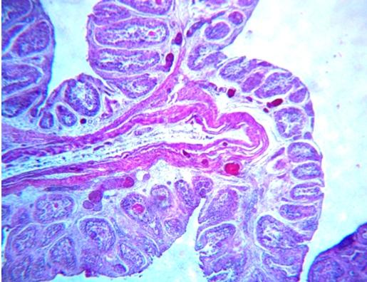
96 Plate 25 Plate 26 Plate 27 Plate 28 Plate 29 Plate 30
97 Plate 31: Necrosis of bursal follicles, edema and severe infiltration of heterophils in interfollicular areas. H&E X400 Plate 32: Presence of serofibrinous exudate in the interfollicular area and on the external bursal surface. H&E X100 Plate 33: Kidney section showing congestion, haemorrhages, desquamation of degenerating and necrotic tubular epithelial cells in the lumen in a case of IBD. H&E X100 Plate 34: Intestinal section from a case of IBD showing congestion, haemorrhages, necrosis of villi and infiltration of inflammatory cells in lamina propria. H&E X100 Plate 35: Hemorrhages in proventriculus from a case suspected of NCD or Avian influenza. Plate 36: Mottled appearance of liver from a case suspected of NCD or Avian influenza.


98 Plate 31 Plate 32 Plate 33 Plate 34 Plate 35 Plate 36
99 Plate 37: Severe congestion and presence of MNCs mainly lymphocytes in a section of intestine from cases suspected to be NCD or Avian influenza. H&E X100 Plate 38: Section of proventriculus revealing congestion, haemorrhage, edema in lamina propria and sub mucosa along with necrosis of mucosal papillae. H&E X100 Plate 39: Degeneration of hepatocytes, severe congestion along with infiltration of heterophils in portal triad areas from a suspected cases of NCD or Avian influenza. H&E X40 Plate 40: Severe petechial haemorrhages on mucosal surface of caeca in a case of coccidiosis. Plate 41: Caeca distended with blood clot. Plate 42: Intestinal section from a case of coccidiosis showing severe necrosis and exfoliation of intestinal layers along with infiltration of inflammatory cells mainly heterophils in tunica mucosa and sub mucosa. H&E X100


100 Plate 37 Plate 38 Plate 39 Plate 40 Plate 41 Plate 42
101 Plate 43: Congestion, haemorrhage, edema, infiltration of inflammatory cells and presence of coccidian life-cycle stages in degenerating and necrotic intestinal epithelium. H&E X100 Plate 44: Presence of developing coccidian parasites (arrow) in degeneration and necrotic epithelium of crypts and sub mucosal glands. H&E X400 Plate 45: Presence of developing coccidian stages in epithelium of villi. H&E X400 Plate 46: Wet mount smear prepared from intestinal scrapings showing oocysts of Eimeria spp. X400 Plate 47: Accumulation of straw colored fluid in peritoneal cavity in a case affected with ascites syndrome. Plate 48: Severe vacuolar degeneration and necrosis of hepatocytes in a case of ascites. H&E X100


102 Plate 43 Plate 44 Plate 45 Plate 46 Plate 47 Plate 48
103 Plate 49: Pale and swollen kidneys in a case suffering from nephro-hepatotoxicity. Plate 50: Edema and petechial haemorrhages on the surface of kidneys. Plate 51: Edematous and congested kidneys. Plate 52: Mottled liver from a case of nephro-hepatotoxicity. Plate 53: Enlarged, pale and mottled liver from a case of nephro-hepatotoxicity. Plate 54: Kidney section revealing congestion, haemorrhage, sloughing of tubular epithelial cells into the lumen in case of nephro-hepatotoxicity. H&E X100


104 Plate 49 Plate 50 Plate 51 Plate 52 Plate 53 Plate 54
105 Plate 55: Nephrotoxicity. Kidney: Degeneration, desquamation, necrosis and presence of heterophils in intertubular areas. H&E X40 Plate 56: Liver showing degeneration, necrosis of hepatocytes and infiltration of inflammatory cells mainly heterophils in portal triad areas in case of nephrohepatotoxicity. H&E X100 Plate : Fatty liver syndrome: Enlargedt, friable and pale liver. Plate 58: Fatty liver syndrome: Fatty degeneration and characteristic signet ring appearance of hepatocytes (arrow). H&E X100 Plate 59: Liver showing rupture and presence of clotted blood in the abdominal cavity. Plate 60: Presence of congestion, petechial haemorrhages on the yellowish mucosa with mucoid exudate in lumen of intestine.


106 Plate 55 Plate 56 Plate Plate 58 Plate 59 Plate 60
107 Plate 61: Congestion and haemorrhages on serosa of intestine. Plate 62: Photomicrograph of intestine revealing severe necrosis in intestinal villi and thickening of intestinal wall due to serosal congestion. H&E X400 Plate 63: Necrotic and thickened intestinal wall due to infiltration of lymphocytes and plasma cells in the lamina propria and sub mucosa. H&E X100 Plate 64: Necrotic enteritis characterised by presence of an admixture of necrotic material, fibrin and inflammatory cells mainly heterophils overlying degenerating and necrotic villous epithelium. H&E X40 Plate 65: Intestine: Necrosis of villi, hyperplasia of goblet cells and proliferation of MNC s in lamina propria and sub mucosa. H&E X400 Plate 66: Hyperplasia of crypts epithelium and presence of mitotic figures. H&E X100


108 Plate 61 Plate 62 Plate 63 Plate 64 Plate 65 Plate 66
109 Plate 67: Liver showing diffuse severe congestion and enlargement. Plate 68: Liver showing fatty changes. Plate 69: Liver: Petechial haemorrhages scattered throughout the surface of right lobe and involving only upper half of right left lobe. Plate 70: Liver: Presence of coalescing necrotic foci on surface. Plate 71: Liver showing congestion and dilatation of central veins along with degeneration and necrosis of hepatocytes. H&E X100 Plate 72: Liver showing necrosis, dissociation and individualization of hepatocytes. H&E X100


110 Plate 67 Plate 68 Plate 69 Plate 70 Plate 71 Plate 72
111 Plate 73: Liver showing necrosis of hepatocytes beneath the capsule. H&E X100 Plate 74: Liver showing oedema and inflammatory cells in portal tract. H&E X100 Plate 75: Liver showing congestion of central vein, dilatation of sinusoids, degeneration and necrosis of surrounding hepatocytes long with infiltration of heterophils. H&E X100 Plate 76: Liver showing congestion of central vein, dilatation of sinusoids, degeneration and necrosis of surrounding hepatocytes long with infiltration of heterophils. H&E X100 Plate 77: Liver showing congestion, multifocal areas of hepatic necrosis and infiltration of large number of heterophils (arrow). H&E X400 Plate 78: Liver showing presence of pseudomembrane with fibrin and infiltrating heterophils overlying a congestion and degenerating liver parenchyma. H&E X100


112 Plate 73 Plate 74 Plate 75 Plate 76 Plate 77 Plate 78
113 Plate 79: Liver showing presence of fibrin and infiltration of heterophils in large number. H&E X400 Plate 80: Liver showing degeneration and hyperplasia of epithelium of bile duct. H&E X100 Plate 81: Serosal surface of proventriculus showing diffuse severe congestion. Plate 82: Proventriculus: Presence of thick mucoid exudate adhering to underlying congestion of mucosa and erotic mucosa of gizzard. Plate 83: Proventriculus showing swelling and edematous glands. Plate 84: Accumulation of thick tenacious slimy exudate adherent to walls of proventriculus.


114 Plate 79 Plate 80 Plate 81 Plate 82 Plate 83 Plate 84
115 Plate 85: Proventriculus: Degeneration and necrosis of epithelium of mucosal folds and infiltration of heterophils in lamina propria and sub mucosa. H&E X100 Plate 86: Degeneration and necrosis of glands of proventriculus. H&E X100 Plate 87: Proventriculus: Accumulation of edematous fluid in sub mucosa, proliferation of small arterioles and capillaries and infiltration of inflammatory cells. H&E X100 Plate 88: Proventriculus: Proliferation and hyalinisation of connective tissue in the interglandular area. H&E X100 Plate 89: Fibrinous proventriculitis: Congestion of blood vessels, necrotic debris, fibrin and infiltration of inflammatory cells mainly heterophils in serosal layer of proventriculus. H&E X100 Plate 90: Proventriculus showing necrosis, blunted, fused mucosal folds along with inflammatory cells and fibrous connective tissue proliferation in lamina propria and sub mucosa H&E X100


116 Plate 85 Plate 86 Plate 87 Plate 88 Plate 89 Plate 90
117 Plate 91: Gizzard showing necrosis of overlying keratinized layer revealing the underlying mucosa and sub mucosa. Plate 92: Gizzard showing fragmentation, degeneration and necrosis of smooth muscle fibers along with infiltration of inflammatory cells mostly heterophils. H&E X400 Plate 93: Gizzard showing degeneration and necrosis of smooth muscle fibers along with infiltration of inflammatory cells mainly lymphocytes. H&E X400 Plate 94: Gizzard showing proliferation of several congested blood vessel, edema, presence of fibrin in serosa and infiltration of heterophils. Plate 95: Congestion in pancreas. Plate 96: Presence of focal ulceration in pancreas.


118 Plate 91 Plate 92 Plate 93 Plate 94 Plate 95 Plate 96
119 Plate 97: Pancreas showing congestion and haemorrhages. H&E X100 Plate 98: Pancreas: Degeneration and necrosis of acinar cells and heterophilic infiltration. H&E X100 Plate 99: Pancreas: Necrosis of acini and infiltration of heterophils. H&E X400 Plate 100: Pancreas showing hyperplasia of pancreatic ducts, congestion of blood vessels in interlobular areas, degeneration, necrosis of acinar cells and heterophilic infiltration. H&E X100 Plate 101: Oesophagus showing congestion, edema and inflammation in sub mucosa and serosa. H&E X40 Plate 102: Oesophagus: Presence of inflammatory cells in sub mucosa around the oesophageal glands. H&E X100


120 Plate 97 Plate 98 Plate 99 Plate 100 Plate 101 Plate 102
121 Plate 103: Oesophagus: infiltration of inflammatory cells mostly heterophils in sub mucosa around the oesophageal glands. H&E X400 Plate 104: Oesophagus: Expanding micro-abscesses squeezing the oesophageal glands. H&E X100

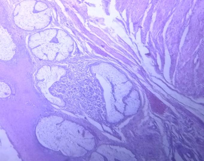
122
Post Mortem Findings in Poultry Birds of Jammu Region
 Post Mortem Findings in Poultry Birds of Jammu Region Shilpa Sood 1, Suresh Kohli 2, Navrose Sangha 1 and D. Basheer Ahamad 3 1 Division of Veterinary Pathology, Shere Kashmir University of Agriculture
PATHOLOGY OF PASTEURELLA MULTOCIDA INFECTION IN CHICKENS
 Indian J. Anim. Res., 40 (1): 15-19, 2006 PATHOLOGY OF PASTEURELLA MULTOCIDA INFECTION IN CHICKENS Shilpa Sood 1 and P.C. Verma CCS Haryana Agricultural University, Hisar - 125 004, India ABSTRACT The
Int. J. BioRes. 1(6): June, 2010 Uddin et al.
: June, 2010 Uddin et al.") Full Length Research Paper PREVALENCE OF POULTRY DISEASES AT NARSINGDI, BANGLADESH *M B Uddin 1, S S U Ahmed 2, 3, M M Hassan 4, S A Khan 5, M A Mamun 6 1 Department of Medicine and Surgery, Sylhet Agricultural
Full Length Research Paper PREVALENCE OF POULTRY DISEASES AT NARSINGDI, BANGLADESH *M B Uddin 1, S S U Ahmed 2, 3, M M Hassan 4, S A Khan 5, M A Mamun 6 1 Department of Medicine and Surgery, Sylhet Agricultural
INCLUSION BODY HEPATITIS AND HYDROPERICARDIUM SYNDROME (ADENOVIRUS INFECTIONS)
") INCLUSION BODY HEPATITIS AND HYDROPERICARDIUM SYNDROME (ADENOVIRUS INFECTIONS) AVIAN ADENOVIRUSES (CHICKEN ADENOVIRUSES, FADV) Adenoviruses are common in poultry. Many replicate in healthy birds without
INCLUSION BODY HEPATITIS AND HYDROPERICARDIUM SYNDROME (ADENOVIRUS INFECTIONS) AVIAN ADENOVIRUSES (CHICKEN ADENOVIRUSES, FADV) Adenoviruses are common in poultry. Many replicate in healthy birds without
2. Avian influenza and the viruses that cause it
 3 2. Avian influenza and the viruses that cause it Avian influenza is caused by influenza viruses that are common in wild birds and occasionally infect poultry. When poultry are infected, they may have
International Journal of Science, Environment and Technology, Vol. 6, No 1, 2017,
 International Journal of Science, Environment and Technology, Vol. 6, No 1, 2017, 662 668 ISSN 2278-3687 (O) 2277-663X (P) INCIDENCE AND RISK FACTORS OF CHRONIC RESPIRATORY DISEASE IN INDIAN POULTRY FLOCKS
This is the second learning component (Learning Component 2) in our first learning module (Learning Module 1). In this component we review a very
 in our first learning module (Learning Module 1). In this component we review a very") This is the second learning component (Learning Component 2) in our first learning module (Learning Module 1). In this component we review a very basic response to injury inflammation. We ll look at examples
This is the second learning component (Learning Component 2) in our first learning module (Learning Module 1). In this component we review a very basic response to injury inflammation. We ll look at examples
GUIDE TO: Diagnosing Coccidiosis & Necrotic Enteritis
 GUIDE TO: Diagnosing Coccidiosis & Necrotic Enteritis Site of Infection Species E. acervulina E. brunetti E. maxima E. mivati E. tenella E. necatrix Oocyst Size 2µ{ 18.3 x 14.6 24.6 x 18.8 30.5 x 20.7
WELCOME TO POULTRY TECHNICAL MEET
 WELCOME TO POULTRY TECHNICAL MEET What is a disease Any condition that results in deviation from normal function How do diseases occur? AGENT BACTERIA OR VIRUS Carrier ENVIRONMENT HOST ETIOLOGY Infectious
Methodology Prevalence:
 Context of Research Free ranging domestic fowl are very susceptible to parasitic infections owing to their exposure to parasites during their roaming in the backyards in search of food. Heterakis gallinarum
Canine Liver Eneku Wilfred Bovine Pathology
 2012-1-3 Canine Liver Eneku Wilfred Bovine Pathology Contributor: New Mexico Department of Agriculture Veterinary Diagnostic Services Signalment: 5 month old male Weimaraner dog (Canis familiaris) History:
Avian Reo-virus infections. Dr./ Wafaa Abd El-ghany Assistant Professor of poultry dis., Fac. Vet. Med., Cairo Univ.
 Avian Reo-virus infections Dr./ Wafaa Abd El-ghany Assistant Professor of poultry dis., Fac. Vet. Med., Cairo Univ. Economic losses Increased mortality. Diminished weight gains, poor feed conversions.
Random Sample. Pages for Preview USF&WS. Mallard with duck plague exhibiting prolapsed penis
 Random Sample Mallard with duck plague exhibiting prolapsed penis USF&WS Random Sample Duck plague: Intestinal tract with hemorrhagic annular bands Cornell Duck Lab. Random Sample Digestive tract from
WSC , Conference 9, Case 1. Tissue from a nyala.
 WSC 2009-2010, Conference 9, Case 1. Tissue from a nyala. MICROSCOPIC DESCRIPTION: Heart, atrium (1 pt.): Approximately 40% of the atrial myocardium is replaced by areas of fibrous connective tissue (1
TOXIC CONDITIONS OF ANIMALS
 TOXIC CONDITIONS OF ANIMALS Prof. R. S. Chauhan Joint Director () Indian Veterinary Research Institute Izatnagar 243 122 (UP) AFLATOXICOSIS Aflatoxicosis is a toxic condition of poultry widely prevalent
Mycotoxin Lesions in the Slaughter House-Broilers
 Mycotoxin Lesions in the Slaughter House-Broilers SPECIAL NUTRIENTS, INC. THE MYCOTOXINS SPECIALIST www.mycotoxin.com INTRODUCTION Traditionally, the presence of mycotoxins capable of causing damage in
The Incidence, Morbidity and Mortality of the Diseases of Broiler Birds in and around NDUAT, Kumarganj, Faizabad
 International Journal of Current Microbiology and Applied Sciences ISSN: 2319-7706 Special Issue-7 pp. 5095-5105 Journal homepage: http://www.ijcmas.com Original Research Article The Incidence, Morbidity
Infectious Bursal Disease: A case compilation study in commercial broiler farms at Mirsarai, Chittagong, Bangladesh.
 International Journal of Advanced Research in Biological Sciences ISSN: 2348-8069 www.ijarbs.com DOI: 10.22192/ijarbs Coden: IJARQG(USA) Volume 5, Issue 4-2018 Research Article DOI: http://dx.doi.org/10.22192/ijarbs.2018.05.04.018
Inflammation Laboratory 1
 Inflammation Laboratory 1 Lab1 Emphasis: The exudates of acute inflammation Descriptions Morphologic Diagnoses Shannon Martinson: http://people.upei.ca/smartinson VPM 152: March 2013 Describing Lesions
Asian-Australasian Journal of Bioscience and Biotechnology ISSN (Print) (Online)
 (Online)") , 173-180 Asian-Australasian Journal of Bioscience and Biotechnology ISSN 2414-1283 (Print) 2414-6293 (Online) www.ebupress.com/journal/aajbb Article Prevalence and pathological investigations of avian
, 173-180 Asian-Australasian Journal of Bioscience and Biotechnology ISSN 2414-1283 (Print) 2414-6293 (Online) www.ebupress.com/journal/aajbb Article Prevalence and pathological investigations of avian
MORPHOLOGIC DIAGNOSIS: Liver: Hepatitis, necrotizing, multifocal to coalescing, severe, with numerous trichomonads. (3 pt)
") Case 1. Tissue from a pelican. MICROSCOPIC DESCRIPTION: Liver: Approximately 80% (1 pt) of the liver is replaced by multifocal to coalescing areas of coagulative and lytic necrosis. Centrally, within these
Case 1. Tissue from a pelican. MICROSCOPIC DESCRIPTION: Liver: Approximately 80% (1 pt) of the liver is replaced by multifocal to coalescing areas of coagulative and lytic necrosis. Centrally, within these
Interaction of Gumboro and other Immunosuppressive Diseases on Respiratory Disease
 Interaction of Gumboro and other Immunosuppressive Diseases on Respiratory Disease FREDERIC J. HOERR II SEMINARIO INTERNACIONAL AVICOLA MERIAL PANAMA CITY, PANAMA JULY 10, 2014 Broiler Disease by Age Rickets
Evaluation of Biosecurity Status in Commercial Broiler Farms in Sri Lanka
 International Journal of Scientific and Research Publications, Volume 7, Issue 4, April 217 114 ISSN 225-3153 Evaluation of Biosecurity Status in Commercial Broiler Farms in Sri Lanka W.M.J.B. Wijesinghe
Inflammation Laboratory 1
 Inflammation Laboratory 1 Lab1 Emphasis: The exudates of acute inflammation Descriptions Morphologic Diagnoses Shannon Martinson: http://people.upei.ca/smartinson VPM 152: February 2012 Describing Lesions
Etiology. Paramyxovirus type 1 = Newcastle disease.
 Newcastle Disease Many strains of similar virus cause signs ranging from mild respiratory signs (pneumotropic) with low mortality to severe neurological (neurotropic) and/or visceral lesions (viscerotropic)
POULTRY DISEASES VIRAL DISEASES
 POULTRY DISEASES VIRAL DISEASES New Castle Disease It is one of the most dreaded diseases of poultry as it can cause very heavy mortality at a very high speed. The disease is caused by a virus of paramyxo
INFLAMMATION & REPAIR
 INFLAMMATION & REPAIR Histopath Laboratory 1 Winter 2013 Chelsea Martin Special thanks to Drs. Hanna and Forzan Goals: Examine Tissue and Identify the Organ Describe the lesion, grossly and histologically
Suggestions to prevent / control Respiratory Disease Complex in poultry
 Suggestions to prevent / control Respiratory Disease Complex in poultry Dr. J. L. Vegad Adviser Phoenix Group 201/15, Gorakhpur, Jabalpur - 482001 Introduction Today, respiratory disease complex has emerged
Necrosis is death of cells and tissues in the living animal. Focal/ Multifocal necrosis- terms used for one
 Necrosis Necrosis Necrosis is death of cells and tissues in the living animal. Focal/ Multifocal necrosis- terms used for one or more, small, clearly defined areas of necrosis. Diffuse necrosis- term used
Avian Pathology. Bacterial diseases: histo slides. ECVP-ESVP Summer School 2012 Frédérique NGUYEN
 Avian Pathology Bacterial diseases: histo slides ECVP-ESVP Summer School 2012 Frédérique NGUYEN Bacterial diseases: histo slides B1. Turkey. Organs? Morphologic diagnosis? Special procedure? B2. Hen. Organ?
ABSTRACT. Keywords: broiler, infections, bacterial microflora
 ABSTRACT Keywords: broiler, infections, bacterial microflora Bacterial infections are often found in broiler chickens, grown on the ground, because bacteria with pathogenic potential exist both in the
Laboratory exercises for abdominal organs
 Laboratory exercises for abdominal organs Slide #77 (C007- H- 107A). Pancreas, dog. pancreatic islets CENTROACINAR CELLS ARE THE BEGINNING CELLS OF THE INTERCALATED DUCTS THAT DRAIN THE SECRETORY ACINI
DUCK VIRUS ENTERITIS (DVE) (Duck Plague) Dr./ Wafaa Abd El-ghany Assistant Professor of poultry dis., Fac. Vet. Med., Cairo Univ.
 (Duck Plague) Dr./ Wafaa Abd El-ghany Assistant Professor of poultry dis., Fac. Vet. Med., Cairo Univ.") DUCK VIRUS ENTERITIS (DVE) (Duck Plague) Dr./ Wafaa Abd El-ghany Assistant Professor of poultry dis., Fac. Vet. Med., Cairo Univ. DISEASES OF WATERFOWL 1. DUCK VIRAL HEPATITIS. 2. DUCK VIRUS ENTERITIS.
DUCK VIRUS ENTERITIS (DVE) (Duck Plague) Dr./ Wafaa Abd El-ghany Assistant Professor of poultry dis., Fac. Vet. Med., Cairo Univ. DISEASES OF WATERFOWL 1. DUCK VIRAL HEPATITIS. 2. DUCK VIRUS ENTERITIS.
Colibacillosis in commercial chickens in Bangladesh
 The Bangladesh Veterinarian (2008) 25(1) : 17 24 Colibacillosis in commercial chickens in Bangladesh R. Khaton, M. G. Haider 1, P. K. Paul, P. M. Das and M. M. Hossain 4 * Department of Pathology, Faculty
Differential Diagnosis of Respiratory Disease in Broilers
 UD Poultry Health System(Lasher and Allen Labs) Department of Animal & Food Sciences College of Agriculture and Natural Resources University of Delaware Newark, DE 19717-1303 USA Differential Diagnosis
Cellular Pathology Gross Pathology Laboratory 2 Cell Injury. VPM 152: General Pathology Instructor: Chelsea Martin Winter 2016
 Cellular Pathology Gross Pathology Laboratory 2 Cell Injury VPM 152: General Pathology Instructor: Chelsea Martin Winter 2016 Gross Specimens The following slides consist of images from the specimens presented
Ceva HAND book of poultry diseases
 IVAN DINEV & CEVA Volume 1 Ceva HAND book of poultry diseases GUMBORO CHICKEN ANaEMIA REOVIRUS MAREK RUNTING-STUNTING SYNDROME NEWCASTLe DISEASE AVIAN INFLUENZA INFECTIOUS BRONCHITIS LARyNGOTRACHEITIS
Nyamdolgor.U, Usuhgerel.S, Baatarjargal.P, others, Journal of agricultural sciences 15 (02): 51-55, 2015
: 51-55, 2015") 51 HISTOPATHOLOGICAL STUDY FOR USING OF POX INACTIVATED VACCINE IN GOATS Nyamdolgor.U 1*, Usuhgerel.S 2, Baatarjargal.P 1, Altanchimeg.A 1, Odbileg.R 1 1-Institute of Veterinary Medicine, MULS, Mongolia
51 HISTOPATHOLOGICAL STUDY FOR USING OF POX INACTIVATED VACCINE IN GOATS Nyamdolgor.U 1*, Usuhgerel.S 2, Baatarjargal.P 1, Altanchimeg.A 1, Odbileg.R 1 1-Institute of Veterinary Medicine, MULS, Mongolia
Poultry Disease Manual Characteristics And
 Poultry Disease Manual Characteristics And Control Of Infections Written by: Dr. Jacquie Jacob, University of Kentucky Pullorum disease, also called Infection by Salmonella pullorum has also been reported
Manal AL khulaifi. Enterobacteriaceae
 Enterobacteriaceae Characteristics E.coli Most significant species in the genus Important potential pathogen in humans Common isolate from colon flora Dry, pink (lactose positive) pink colony with area
Avian encephalomyelitis (AE) Epidemic tremor. Dr./ Wafaa Abd El-ghany Assistant Professor of poultry dis., Fac. Vet. Med., Cairo Univ.
 Epidemic tremor. Dr./ Wafaa Abd El-ghany Assistant Professor of poultry dis., Fac. Vet. Med., Cairo Univ.") Avian encephalomyelitis (AE) Epidemic tremor Dr./ Wafaa Abd El-ghany Assistant Professor of poultry dis., Fac. Vet. Med., Cairo Univ. Definition Avian encephalomyelitis (AE) is a viral infection affecting
Avian encephalomyelitis (AE) Epidemic tremor Dr./ Wafaa Abd El-ghany Assistant Professor of poultry dis., Fac. Vet. Med., Cairo Univ. Definition Avian encephalomyelitis (AE) is a viral infection affecting
Coccidiosis vaccination as a disease model in nutritional research. Rachel Savary and Tim Fiss NSERC-IRC AGM June 4, 2015
 Coccidiosis vaccination as a disease model in nutritional research Rachel Savary and Tim Fiss NSERC-IRC AGM June 4, 2015 Introduction What is Coccidiosis? A parasitic infection of the intestinal tract
General Structure of Digestive Tract
 Dr. Nabil Khouri General Structure of Digestive Tract Common Characteristics: Hollow tube composed of a lumen whose diameter varies. Surrounded by a wall made up of 4 principal layers: Mucosa Epithelial
MYCOTOXICOSIS. Disease related to one or more mycotoxins. Mycotoxin - "mycos" - mold, toxic metabolite
 MYCOTOXICOSIS Disease related to one or more mycotoxins Mycotoxin - "mycos" - mold, toxic metabolite Produced by filamentous fungi while growing on substrate such as feedstuffs and litter. Deleterious
Slide 154: Pancreas, H&E
 Slide 154: Pancreas, H&E the pancreas, located adjacent to the duodenum, is a mixed exocrine and endocrine gland; it is usually readily identifiable by the presence of the interspersed endocrine pancreatic
Pathology of Gout in Commercial Broiler Chicken
 International Journal of Current Microbiology and Applied Sciences ISSN: 2319-7706 Volume 7 Number 07 (2018) Journal homepage: http://www.ijcmas.com Original Research Article https://doi.org/10.20546/ijcmas.2018.707.282
This paper is in two Sections (A and B) and instructions relating to the number of questions to be answered are given at the head of each Section.
 and instructions relating to the number of questions to be answered are given at the head of each Section.") TUESDAY 28 MARCH 2000 PAPER I (3 hours) This paper is in two Sections (A and B) and instructions relating to the number of questions to be answered are given at the head of each Section. SECTION A Two
TUESDAY 28 MARCH 2000 PAPER I (3 hours) This paper is in two Sections (A and B) and instructions relating to the number of questions to be answered are given at the head of each Section. SECTION A Two
Gastrointestinal Pathology of Pigs. Jerome C. Nietfeld, DVM, MS, PhD Kansas State Veterinary Diagnostic Lab Department DMP Kansas State University
 Gastrointestinal Pathology of Pigs Jerome C. Nietfeld, DVM, MS, PhD Kansas State Veterinary Diagnostic Lab Department DMP Kansas State University Neonatal Diarrhea Likely the number 1 killer of neonatal
PRACTICAL APPROACHES FOR DIAGNOSIS AND TREATMENT OF POULTRY DISEASES POULTRY DISEASES FIELD DIAGNOSTIC TREE
 PRACTICAL APPROACHES FOR DIAGNOSIS AND TREATMENT OF POULTRY DISEASES POULTRY DISEASES FIELD DIAGNOSTIC TREE Prof. Paul Gichohi Mbuthia 1 ON THE FARM LEVEL WHAT DO YOU SEE? 1.Something is wrong in the flock
Turkey Corona Virus Enteritis Blue Comb Disease Transmissible enteritis Mud Fever
 Turkey Corona Virus Enteritis Blue Comb Disease Transmissible enteritis Mud Fever Bacterial infections of turkeys Salmonellosis (typhoid and paratyphoid). Mycoplasmosis [M.gallisepticum (infectious sinusitis)
Ischaemia It means local anemia, it is characterized by a decrease amount of blood in an organ or region. Causes of Ischemia: *1.
 المرحلة الثالثة م. هالة عباس ناجي Ischaemia It means local anemia, it is characterized by a decrease amount of blood in an organ or region. Causes of Ischemia: *1.External pressure upon an artery e.g:
Lab activity manual - Histology of the digestive system. Lab activity 1: esophagus stomach - small intestines
 Lab activity manual - Histology of the digestive system Jeanne Adiwinata Pawitan Prerequisite: Histology of the 4 basic tissues In this module we learn about the histology of the digestive system, from
POULTRY DISEASES IN SOME SELECTED AREAS IN SYLHET DISTRICT OF BANGLADESH
 J. Sylhet Agril. Univ. 3(1):1-8, 2016 ISSN: 2308-1597 POULTRY DISEASES IN SOME SELECTED AREAS IN SYLHET DISTRICT OF BANGLADESH M A Rahman* 1 and G N Adhikary 2 1 Department of Genetics and Animal Breeding,
HISTOPATHOLOGICAL CHANGES OF BROILERS IMMUNIZED WITH SONICATED OOCYSTS AGAINST EIMERRIA TENELLA
 HISTOPATHOLOGICAL CHANGES OF BROILERS IMMUNIZED WITH SONICATED OOCYSTS AGAINST EIMERRIA TENELLA a,b 1 Latif Ibrahim Kadhim Department of Pathology and poultry diseases, Faculty of Veterinary Medicine,
Small intestine. Small intestine
 General features Tubular organ longest part; 5-6 m most of chemical digestion absorption of nutrients reabsorption of H2O occurs. Two structural features; maximize the lumenal surface area villi microvilli
09-Mar-15 PNEUMONIA RESPIRATORY SYSTEM L-3
 RESPIRATORY SYSTEM L-3 Professor Department of Pathology, University of Agriculture, Faisalabad. Email: mtjaved@uaf.edu.pk Web: https://sites.geocities.ws/mtjaved PNEUMONIA The pulmonary inflammatory response
Epidemiology of Newcastle Disease. By Dr. Jonathan Amakye Anim & Dr. John Tsitsiwu
 Epidemiology of Newcastle Disease By Dr. Jonathan Amakye Anim & Dr. John Tsitsiwu Definition The current OIE definition (OIE, 2000a) is: Newcastle disease is defined as an infection of birds caused by
Inflammation Laboratory 2. Shannon Martinson: VPM 152: March 2012
 Inflammation Laboratory 2 Shannon Martinson: http://people.upei.ca/smartinson VPM 152: March 2012 Reminder - Creating a Morphologic Diagnosis for Inflammatory Lesions Organ and Process Exudate Distribution
DISEASES OF CHICKENS AND TURKEYS CORRELATED WITH AGE
 By knowing the species affected, salient clinical feature, and age of the flock, it is often possible to make a list of potential differential diagnoses. In the following table, some of the more common
PREVALENCE AND IDENTIFICATION OF COCCIDIAN PARASITE (EIMERIA SPP) IN LAYER CHICKEN OF RATNANAGAR MUNICIPALITY, CHITWAN DISTRICT, NEPAL
 IN LAYER CHICKEN OF RATNANAGAR MUNICIPALITY, CHITWAN DISTRICT, NEPAL") J. Nat. Hist. Mus. Vol. 23, 2008, 45-50 PREVALENCE AND IDENTIFICATION OF COCCIDIAN PARASITE (EIMERIA SPP) IN LAYER CHICKEN OF RATNANAGAR MUNICIPALITY, CHITWAN DISTRICT, NEPAL A. Adhikari, R. Gupta and
J. Nat. Hist. Mus. Vol. 23, 2008, 45-50 PREVALENCE AND IDENTIFICATION OF COCCIDIAN PARASITE (EIMERIA SPP) IN LAYER CHICKEN OF RATNANAGAR MUNICIPALITY, CHITWAN DISTRICT, NEPAL A. Adhikari, R. Gupta and
MYCOPLASMOSIS - A SERIOUS PROBLEM OF POULTRY INDUSTRY
 By, Dr. J. L. Vegad Advisor Phoenix Group MYCOPLASMOSIS - A SERIOUS PROBLEM OF POULTRY INDUSTRY Mycoplasmosis, commonly known as chronic respiratory disease of chickens, has existed in our country since
PLATES 24 TO 26. (Received for publication, December 4, 1935)
") Published Online: 1 March, 1936 Supp Info: http://doi.org/10.1084/jem.63.3.303 Downloaded from jem.rupress.org on January 19, 2019 THE VISCERAL LESIONS PRODUCED IN MICE BY THE SALIVARY GLAND VIRUS OF MICE*
Published Online: 1 March, 1936 Supp Info: http://doi.org/10.1084/jem.63.3.303 Downloaded from jem.rupress.org on January 19, 2019 THE VISCERAL LESIONS PRODUCED IN MICE BY THE SALIVARY GLAND VIRUS OF MICE*
5 DISTURBANCES IN CIRCULATION. Congestion / Hyperemia Haemorrhage Thrombosis Embolism Ischemia Infarction Oedema Shock Sludged blood Model Questions
 5 DISTURBANCES IN CIRCULATION Congestion / Hyperemia Haemorrhage Thrombosis Embolism Ischemia Infarction Oedema Shock Sludged blood Model Questions CONGESTION/ HYPEREMIA Hyperemia is increased amount of
Characteristic. Course of disease:short Days--one month Changes : Alteration, exudation Tissue destruction Inflammation cells: major neutrophils
 ACUTE INFLAMMATION Characteristic Course of disease:short Days--one month Changes : Alteration, exudation Tissue destruction Inflammation cells: major neutrophils TYPES Serous Inflammation Fibrinous Inflammation
Chapter One: Post Mortem Principles for Clinicians and Farmers
 Chapter One: for Clinicians and Farmers INTRODUCTION... EXTERNAL EXAMINATION... RESPIRATORY SYSTEM... LIVER... SPLEEN... HEART... GASTROINTESTINAL TRACT... AIRSACS... BURSA... THYMUS... PROVENTRICULUS...
Pathology of the Liver and Biliary Tract 5 Diseases of the Biliary Tract. Shannon Martinson, April 2016
 Pathology of the Liver and Biliary Tract 5 Diseases of the Biliary Tract Shannon Martinson, April 2016 http://people.upei.ca/smartinson/ OUTLINE Normal anatomy & function Hepatobiliary Injury and responses
Describing and interpreting gross lesions. Prepared for VPM 4600, May 2018; Shannon Martinson
 Describing and interpreting gross lesions Prepared for VPM 4600, May 2018; Shannon Martinson How to Describe (and Interpret) Lesions Step 1 Step 2 Step 3 Step 4 Look at the specimen: Is it normal or abnormal
Compare between two methods for diagnosis of coccidia in broiler chicken
 Compare between two methods for diagnosis of coccidia in broiler chicken O. A. R. AlJumaili Technical institute of Alanbar e-mail:od74ay@yahoo.com Abstract Coccidial infections in broiler cause significant
Recommended Resources: The following resources may be useful in teaching this
 Unit C: Poultry Management Lesson 3: Poultry Health Management Student Learning Objectives: Instruction in this lesson should result in students achieving the following objectives: 1. Demonstrate sanitation
A novel approach to coccidiosis control
 A novel approach to coccidiosis control by Fabio Mello, Novartis Animal Health, Sao Paulo, Brazil, Roberta Alvarez, Novartis Animal Health, Basel, Switzerland, Nick Smith, Institute for the Biotechnology
EXTERNAL SYMPTOMS FOR DIAGNOSING POULTRY DISEASES
 page 1 EXTERNAL SYMPTOMS FOR DIAGNOSING POULTRY DISEASES Emaciation... Stunted, uneven growth... Swellings... Twitching, muscular spasm... Trembling... Paralysis... Dehydration... Nervousness... Lying
Salt Intoxication in Commercial Broilers and Breeders a Clinical and Pathological Description
 Salt Intoxication in Commercial Broilers and Breeders a Clinical and Pathological Description Perelman, B., 1 * Farnoushi, Y., 2 Krispin, H. 3 and Rish, D. 4 1 Poultry Veterinarian-Clinical Consultant,
Cellular Pathology. Histopathology Lab #2 (web) Paul Hanna Jan 2018
 Paul Hanna Jan 2018") Cellular Pathology Histopathology Lab #2 (web) Paul Hanna Jan 2018 Slide #91 Clinical History: a necropsy was performed on an aged cat the gross pathological changes included: widespread subcutaneous edema
Cellular Pathology Histopathology Lab #2 (web) Paul Hanna Jan 2018 Slide #91 Clinical History: a necropsy was performed on an aged cat the gross pathological changes included: widespread subcutaneous edema
Layer/ Pullet Regional Disease Update Northeast US
 2015 MPF Convention Layer/ Pullet Regional Disease Update Northeast US Patricia A. Dunn, DVM, MAM, DACPV Pennsylvania State University, Animal Diagnostic Laboratory 100 Wiley Lane, University Park, PA
UPDATE ON THE RUNTING - STUNTING SYNDROME
 Issue No. 3 / May 2012 UPDATE ON THE RUNTING - STUNTING SYNDROME This article was written in collaboration with Dr. John Rosenberger (AviServe LLC, Delaware Technology Park, USA) & CEVA Scientific Direction
Pathology of the Alimentary Tract
 Pathology of the Alimentary Tract Lab 2: Lower alimentary tract SI, LI, cecum, and peritoneum GIST in the cecum of a dog Shannon Martinson: http://people.upei.ca/smartinson VPM 221: November, 2011 3 year
Pathology of the Liver and Biliary Tract 5 Diseases of the Biliary Tract. Shannon Martinson, March 2017
 Pathology of the Liver and Biliary Tract 5 Diseases of the Biliary Tract Shannon Martinson, March 2017 http://people.upei.ca/smartinson/ OUTLINE Normal anatomy & function Hepatobiliary injury and responses
My presentation is about bovine neonatal diarrhea, more commonly referred to as calf scours. As always, good management is an important factor when
 My presentation is about bovine neonatal diarrhea, more commonly referred to as calf scours. As always, good management is an important factor when dealing with herd health. This applies to calf rearing
HISTOMORPHOLOGICAL STUDY OF THE LYMPHOID TISSUES OF BROILER CHICKENS. S. H. Akter*, M. Z. I. Khan, M. R. Jahan, M. R. Karim and M. R.
 Bangl. J. Vet. Med. (2006). 4 (2): 87 92 HISTOMORPHOLOGICAL STUDY OF THE LYMPHOID TISSUES OF BROILER CHICKENS S. H. Akter*, M. Z. I. Khan, M. R. Jahan, M. R. Karim and M. R. Islam Department of Anatomy
Liver Lab #2. Bacterial Hepatitis
 Liver Lab #2 Bacterial Hepatitis Case: O12561-04. Adult ewe. Describe the lesion: Multifocal large nodules ranging in size from 1-3.5cm in greatest diameter are present within the liver and are filled
Ali M. Ali 1 Hanem F. Khater 2 Shaker A. Seddiek 3 Mai O. Nada 1 Kadarkarai Murugan 4
 Hanem F. Khater Ali M. Ali 1 Hanem F. Khater 2 Shaker A. Seddiek 3 Mai O. Nada 1 Kadarkarai Murugan 4 1 Biochemistry, Toxicology and Nutritional Deficiency Diseases Department 2 Department of Parasitology,
Histopathology: pulmonary pathology
 Histopathology: pulmonary pathology These presentations are to help you identify basic histopathological features. They do not contain the additional factual information that you need to learn about these
Disturbances of Circulation, Lab 1: Edema and Congestion/Hyperemia. Shannon Martinson, Feb
 Disturbances of Circulation, Lab 1: Edema and Congestion/Hyperemia Shannon Martinson, Feb 2017 http://people.upei.ca/smartinson/ Case #1 Signalment and History: 6-month old feeder lamb found dead on pasture
AviagenBrief. Marek s Disease Control in Broiler Breeders
 AviagenBrief January 2018 Marek s Disease Control in Broiler Breeders Author: A. Gregorio Rosales DVM, MS, PhD, DACPV - Poultry Health Consultant Introduction Marek s Disease Virus (MDV), a highly infectious
EVALUATION OF THE EFFECTIVENESS OF COMMERCIALLY AVAILABLE DISINFECTANTS AGAINST SALMONELLAE ISOLATED FROM INTERNAL ORGANS OF DEAD CHICKENS
 Bangl. J. Vet. Med. (2011). 9(1): 43 52 EVALUATION OF THE EFFECTIVENESS OF COMMERCIALLY AVAILABLE DISINFECTANTS AGAINST SALMONELLAE ISOLATED FROM INTERNAL ORGANS OF DEAD CHICKENS N.A. Rumi, M.M. Rahman,
Domain 1b Appendix Example Stephen Hines
 Domain 1b Appendix Example Stephen Hines The Characterization / Sorting Exercise INSTRUCTIONS: 1. If it s not already done for you, cut along the dotted lines to create 3 sets of colored cards Clinical
Exacerbating effect of Newcastle disease virus (NDV) infection on sub clinical caecal coccidiosis in broilers vaccinated against NDV. Shaban, Kh.S.
 infection on sub clinical caecal coccidiosis in broilers vaccinated against NDV. Shaban, Kh.S.") Exacerbating effect of Newcastle disease virus (NDV) infection on sub clinical caecal coccidiosis in broilers vaccinated against NDV Shaban, Kh.S. Department of Poultry Diseases, Faculty of Veterinary
Exacerbating effect of Newcastle disease virus (NDV) infection on sub clinical caecal coccidiosis in broilers vaccinated against NDV Shaban, Kh.S. Department of Poultry Diseases, Faculty of Veterinary
Learning Module 3 Lymphoid System Learning component 1 Basic Responses and Normal Histology
 Learning Module 3 Lymphoid System Learning component 1 Basic Responses and Normal Histology 1 Normal Bursa of Fabricius (BF) A. Normal Bursa showing a plical fold with numerous lymphoid follicles. B. Higher
CLASSIFICATION OF AVIAN MYCOPLASMAS
 Mycoplasmosis CLASSIFICATION OF AVIAN MYCOPLASMAS ** M. galllisepticum M. gallinarum M. iners M. anatis * M. synoviae * M. meleagridis M. pullorum M. gallinaceum M. gallapavonis * M. iowae M. columbinasale
INCIDENCE AND TREATMENT OF ASPERGILLOSIS IN EMUS CH. Sudha Rani Chowdary 1 *, K. Poorani 2 and J. Jaya Lakshmi 3
 International Journal of Science, Environment and Technology, Vol. 5, No 6, 2016, 4555 4559 ISSN 2278-3687 (O) 2277-663X (P) INCIDENCE AND TREATMENT OF ASPERGILLOSIS IN EMUS CH. Sudha Rani Chowdary 1 *,
Digestive system L 4. Lecturer Dr. Firdous M. Jaafar Department of Anatomy/Histology section
 Digestive system L 4 Lecturer Dr. Firdous M. Jaafar Department of Anatomy/Histology section objectives 1-Describe the structure of liver. 2-Define liver lobule, and identify its zones. 3-Define portal
Recognizing African swine fever 13. Post mortem findings
 Recognizing African swine fever 13 Post mortem findings Carcasses of pigs that die in the acute stage of the disease are often in good condition. In white-skinned pigs, bluish-purple discolouration of
Unit II Problem 2 Pathology: Pneumonia
 Unit II Problem 2 Pathology: Pneumonia - Definition: pneumonia is the infection of lung parenchyma which occurs especially when normal defenses are impaired such as: Cough reflex. Damage of cilia in respiratory
Early chick nutrition An approach for enhanced poultry production
 AUGUST 2011 Early chick nutrition An approach for enhanced poultry production The dramatic increase in poultry production and efficiency during the last 50 years is attributable to a number of factors
Outbreak evaluation of highly pathogenic avian influenza in Bangladesh. Mymensingh *Corresponding author:
 Outbreak evaluation of highly pathogenic avian influenza in Bangladesh M. Giasuddin 1*, M.E.Haque 2, A.H.M.Kamal 2, M.R. Islam 2, A. Jahangir 1, E.H. Chowdhury 2 M.J.F.A.Taimur 1 and M. Hafizur Rahman
Technical Report: Coccidiosis in poultry with an emphasis on backyard poultry
 1 2 3 4 5 6 7 8 9 10 11 12 13 14 15 16 17 18 19 20 21 22 23 24 25 26 27 28 29 30 Technical Report: Coccidiosis in poultry with an emphasis on backyard poultry Dr. Geoffrey Lossie, edited by Dr. Pat Wakenell:
Extracellular degeneration
 Extracellular degeneration By Dr. Hemn Hassan Othman PhD, Pathology Fall 2016 1/17/2017 1 Extracellular Degenerations I / Hyaline Degeneration (Hyalinization): The ward hyaline is derived from the Latin
Clostridial Infections. Ulcerative Enteritis (Quail Disease)
") Clostridial Infections Clostridial infections in poultry have 4 pathological forms: 1. Ulcerative enteritis is caused by Clostridium colinum. 2. Necrotic enteritis is caused by C.perfringens. 3. Gangrenous
Clostridial Infections Clostridial infections in poultry have 4 pathological forms: 1. Ulcerative enteritis is caused by Clostridium colinum. 2. Necrotic enteritis is caused by C.perfringens. 3. Gangrenous
HYPEREMIA AND CONGESTION
 HYPEREMIA AND CONGESTION Learning Objectives Define congestion and hyperemia Differentiate between the two with regard to: Mechanisms / underlying causes Appearance (gross and histologic) Effects Differentiate
Adenoviral diseases of poultry are found worldwide but PiAV1 and PiAV2 have not previously been experienced in Australia.
 FINAL REPORT Adenovirus outbreak in WA By Dr Rob Marshall Introduction The recent outbreak of adenovirus infection in Perth Western Australia is of great concern for all Australian pigeon fanciers due