Laboratory exercises for abdominal organs
|
|
|
- Cameron Jordan
- 5 years ago
- Views:
Transcription

1 Laboratory exercises for abdominal organs


2 Slide #77 (C007- H- 107A). Pancreas, dog. pancreatic islets CENTROACINAR CELLS ARE THE BEGINNING CELLS OF THE INTERCALATED DUCTS THAT DRAIN THE SECRETORY ACINI OF THE PANCREAS. THEY SECRETE BICARBONATE serous acini INTERCALATED DUCTS


3 Bile Canaliculi Bile luminal surfaces 155 Bile ducts Blood luminal surface


4 Slide #77 (C007- H- 107A). Pancreas, dog. INTERCALATED DUCTS pancreatic islets Pancreas has Islets and centroacinar cells, but no striated ducts Compared to the salivary glans The salivary gland has striated ducts, but no Islets and centroacinar cells Striations in base of duct cells Demo 186 Salivary gland

5 Slide #193 (GT ). Pancreas, goat. CENTROACINAR CELLS are the beginning of the intercalated duct that originated within the lumen of the ancina pancreatic islets INTERCALATED DUCTS

6 DEMO SLIDE BOX 180 Small intestine (duodenum) and pancreas, dog. (157) CENTROACINAR CELLS pancreatic islet


7 Slide #93 (1017) - Pancreas and duodenum, cat.) Pancreas duodenum Dispersed pancreatic islet cells CENTROACINAR CELLS

8


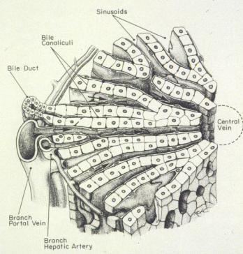
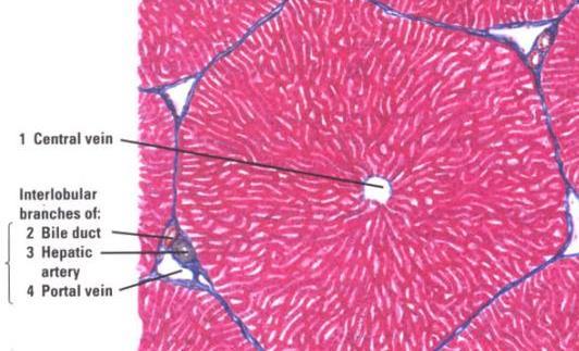
9 155 Portal radicles containing: A bile duct Branch of portal vein, Branch of hepatic artery Lymphatic vessel (usually) or portal canals Liver 155 Cords of hepatocytes


10 Liver
11 Bile Canaliculi Bile luminal surfaces 155 Bile duct Blood luminal surface
 Liver,")

12 DEMO SLIDE BOX 169 (P001- H- 84B) Liver, pig. hepatic lobules hepatocytes Sinusoids discontinuous


13 DEMO SLIDE BOX 169 (P001- H- 84B) Liver, pig. Hepatic sinusoids flow between rows of hepatocytes toward the central vein. venule which is a branch of the portal vein hepatic lobule capsule portal triad. arteriole, which is a branch of the hepatic artery either simple cuboidal or simple columnar epithelium Of bile duct. interlobular connective tissue are larger veins, not accompanied by an artery and a duct, named sublobular veins


14 Slide #45 (Pig 1-87c). Liver, Pig. hepatic lobule portal triad. venule bile duct artery Central vein
. Liver, horse.")


15 DEMO SLIDE BOX 170 (E3- H- 84-1). Liver, horse. Space of Disse Hepatic sinusoids flow between rows of hepatocytes toward the central vein. portal triad.


16 HEPATOCYTE SPACE OF DISSE BILE CANALICULI


17 capsule Sinusoids discontinuous Slide #48 (PF5-76B). Liver & gall bladder, pig. Kupffer cells central vein Mesothelium of capsule portal triad.

. Liver & gall bladder, pig.")
18 Kupffer cells (i.e. the liver macrophages lining the sinusoids) are noticeable because they ingested black Kupffer cells Slide #48 (PF5-76B). Liver & gall bladder, pig. Mesothelium of capsule Kupffer cells portal triad.


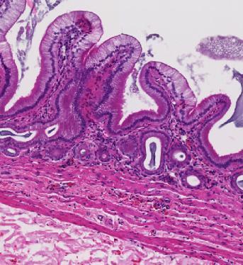
19 Slide #48 (PF5-76B). Liver & gall bladder, pig. Microvilli occur apically but are difficult to see gallbladder No muscularis mucosae occurs so the lamina propria blends with the submucosa. Note the tunica muscularis is not well organized. The thick layer of CCT outside the tunica muscularis is called the perimuscular connective tissue layer.

20 Slide #48 (PF5-76B). Liver & gall bladder, pig. gallbladder
.")

21 Slide #167 (M- 1 87F). Liver, monkey Central vein Kupffer cells


22 Slide #111 (87NG) Liver, goat

23 Slide #117 (SP- 1-61) - Liver and gallbladder, sheep.

24 Slide #117 (SP- 1-61) - Liver and gallbladder, sheep.

25


26 Gallbladder 155 The mucosa is thrown into folds which project into the lumen of the gallbladder. Peritoneal serosal layer Smooth muscle layer or branching layers Lamina propria. A thick perimuscular layer of connective tissue. Simple columnar epithelium


27 The gallbladder stores and concentrates the bile elaborated 155 Mucosa by the liver Plasma cells In the lamina propria Simple columnar epithelium


28 126 Bile duct with portal vein, monkey Portal vein Common hepatic duct Cystic duct The wall of the cystic duct is convoluted and contains abundant smooth muscle fibers which represent the spiral valve preventing distention or collapse of the cystic duct when the latter is subject to sudden changes of pressure.
 Liver,")
29 Slide # 94 (Rbt- 87G) Liver, rabbit.


30 DEMO SLIDE BOX 58 - Liver and gallbladder, dog.
Slide 154: Pancreas, H&E
 Slide 154: Pancreas, H&E the pancreas, located adjacent to the duodenum, is a mixed exocrine and endocrine gland; it is usually readily identifiable by the presence of the interspersed endocrine pancreatic
Digestive system L 4. Lecturer Dr. Firdous M. Jaafar Department of Anatomy/Histology section
 Digestive system L 4 Lecturer Dr. Firdous M. Jaafar Department of Anatomy/Histology section objectives 1-Describe the structure of liver. 2-Define liver lobule, and identify its zones. 3-Define portal
DIGESTIVE SYSTEM II ACCESSORY DIGESTIVE ORGANS
 DIGESTIVE SYSTEM II ACCESSORY DIGESTIVE ORGANS Dr. Larry Johnson Texas A& M University Objectives Distinguish between the parotid and submandibular salivary glands. Understand and identify the structural
Anatomy of the liver and pancreas
 Anatomy of the liver and pancreas Prof. Abdulameer Al-Nuaimi E-mail: a.al-nuaimi@sheffield.ac.uk abdulameerh@yahoo.com Liver Aorta Pulm. Trunk Rt. At, Duct. Art. Lt. Ven. Rt. Ven. Internal Posterior
Oral cavity Lab exercises
 Oral cavity Lab exercises Slide #190 (GT-1-32). Oral cavity, goat. large conical buccal papillae stratified squamous epithelium keratinized or non-keratinized no muscularis mucosae connective tissue represents
HISTOLOGY VIRTUAL LABORATORY GASTROINTESTINAL SYSTEM
 HISTOLOGY VIRTUAL LABORATORY GASTROINTESTINAL SYSTEM LIP (Slides GI 1, 2) Identify the outer portion lined by stratified squamous (keratinized) epithelium. Note the hair follicles and sebaceous glands
Organs Associated with the Digestive Tract. Dr. Emad I H Shaqoura M.D, M.Sc. Anatomy Faculty of Medicine, IUG March, 2016
 Organs Associated with the Digestive Tract Dr. Emad I H Shaqoura M.D, M.Sc. Anatomy Faculty of Medicine, IUG March, 2016 2 Salivary Glands Salivary Glands Major 90% of saliva Minor 10% of saliva Parotid
MICROSCOPIC STRUCTURE OF LIVER, GALLBLADDER, GALL DUCTS, AND PANCREAS OVERVIEW OF DEVELOPMENT OF THE ALIMENTARY CANAL
 Lecture 2 ESS_3rd semester MICROSCOPIC STRUCTURE OF LIVER, GALLBLADDER, GALL DUCTS, AND PANCREAS OVERVIEW OF DEVELOPMENT OF THE ALIMENTARY CANAL MICROSCOPIC STRUCTURE OF LIVER - is the largest gland of
Chapter 12 The Digestive Glands
 Chapter 12 The Digestive Glands Lyu Zhengmei Department of Histology and Embryology, Anhui Medical University Components of digestive glands large salivary glands, pancreas, liver, gallbladder. These organs
12 Digestive system 4 Liver & Pancreas
 12 Digestive system 4 Liver & Pancreas 12-001 Liver Longitudinal section Transverse section 12-01. Scheme showing the 3-dimensional structure of a hepatic lobule. ( Scheme ) Interlobular bile duct, artery
Paneth Cells. Road Map to the Finish. No Review this Friday. Today 11/29 Finish digestion/accessory organs. Wednesday 12/1 Immune System I
 Road Map to the Finish No Review this Friday Today 11/29 Finish digestion/accessory organs Wednesday 12/1 Immune System I Paneth Cells - base of intestinal glands -! large -! intense acidophilic granules
LABORATORY EXERCISES FOR THE URINARY SYSTEM
 LABORATORY EXERCISES FOR THE URINARY SYSTEM cortex Medulla DEMO SLIDE BOX 172 (450-E001-H-76). Kidney, horse. the inner medulla medullary rays, Uriniferous tubules expand both the cortex and medulla corticomedullary
Histology 3. We will continue talking about a few things from last lecture, starting with M cells:
 Histology 3 This is the last Histology lecture in the GI system. Enjoy! There are some extra notes listed as footnotes. We will continue talking about a few things from last lecture, starting with M cells:
Lab activity manual - Histology of the digestive system. Lab activity 1: esophagus stomach - small intestines
 Lab activity manual - Histology of the digestive system Jeanne Adiwinata Pawitan Prerequisite: Histology of the 4 basic tissues In this module we learn about the histology of the digestive system, from
Small intestine. Small intestine
 General features Tubular organ longest part; 5-6 m most of chemical digestion absorption of nutrients reabsorption of H2O occurs. Two structural features; maximize the lumenal surface area villi microvilli
Histology Lab. looking at microscopic pictures of tissues, for more information use Junqueira book and you can use BlueHistolgy website
 Done By: Aseel Twaijer & Laith Sorour Histology Lab *These notes help in differentiating tissues and you must read them while looking at microscopic pictures of tissues, for more information use Junqueira
1. Approximately 21 ft. long: duodenum (one ft.), jejunum (eight ft.), and ileum (twelve ft.)
, jejunum (eight ft.), and ileum (twelve ft.)") IV. Small Intestines A. General features and functions 1. Approximately 21 ft. long: duodenum (one ft.), jejunum (eight ft.), and ileum (twelve ft.) 2. Functions: move forward chyme, continue digestion,
IV. Small Intestines A. General features and functions 1. Approximately 21 ft. long: duodenum (one ft.), jejunum (eight ft.), and ileum (twelve ft.) 2. Functions: move forward chyme, continue digestion,
Large Intestine. The large intestine consists of a mucosal membrane with no folds except in its distal (rectal) portion
 portion") GI Histology 3 Large Intestine The large intestine consists of a mucosal membrane with no folds except in its distal (rectal) portion No villi are present in this portion of the intestine The intestinal
GI Histology 3 Large Intestine The large intestine consists of a mucosal membrane with no folds except in its distal (rectal) portion No villi are present in this portion of the intestine The intestinal
Bio 322 Human Anatomy Objectives for the laboratory exercise Digestive System
 Bio 322 Human Anatomy Objectives for the laboratory exercise Digestive System Required reading before beginning this lab: Saladin, KS: Human Anatomy 5 th ed (2017) Chapter 24 For this lab you will use
PRACTICAL ROADMAP. GLANDS AFFECTING LIFESTYLE WJ van der Spuy & T Tshabalala
 PRACTICAL ROADMAP GLANDS AFFECTING LIFESTYLE WJ van der Spuy & T Tshabalala GLANDS AFFECTING LIFESTYLE Submandibular gland (salivary gland) Liver Pancreas Hypophysis (pituitary gland) Thyroid Suprarenal
terminal portion of the main pancreatic duct (like the the common bile duct) will have longitudinally and circularly arranged smooth muscle which is
 will have longitudinally and circularly arranged smooth muscle which is") Chapter 14 Accessory Organs of the Small Intestine 14.1. The Pancreas The pancreas has both an exocrine and an endocrine nature. These two functions are handled by histologically distinct regions of the
Chapter 14 Accessory Organs of the Small Intestine 14.1. The Pancreas The pancreas has both an exocrine and an endocrine nature. These two functions are handled by histologically distinct regions of the
Gastrointestinal Tract
 CTO Lab #5 GI TRACT & GLANDS; ENDOCRINE SYSTEM Page 1 Gastrointestinal Tract Slide 126 This section through the esophagus shows the characteristic layers of the gastrointestinal tract. Examine the non-keratinized
Respiratory & Digestive Organs of the Head and Neck, Human;
 Name Date Lab Exercise 5: Lab Exercise 6: Lab Exercise 7: Lab Exercise 8: Respiratory & Digestive Organs of the Head and Neck, Human; Histology of the Respiratory System Digestive System Models, Human
Lab 8: Digestive System
 BIOL 221 A&P II Lab 8: Digestive System Become familiar with the gross anatomy of the digestive system (Exercise 38) using the models, Fig. 38.1 (Activity 1), and the rat. Recognize and know the functions
Done by: nisreen obeidat
 Sheet: liver and pancreas Done by: nisreen obeidat Embryology of the liver The liver develops in the ventral mesentery of the foregut and divides the ventral mesentery :into 1)lesser omentum (between the
Small Intestine, Large Intestine and anal cannel
 Small Intestine, Large Intestine and anal cannel 32409 Small intestine Large intestine Small intestine General Structure of the Digestive Tract rat 32409 Epithelium with goblet cells and absorptive cells
LIVER & SPLEEN. Color index: Slides.. Important..Notes..Extra..
 LIVER & SPLEEN Color index: Slides.. Important..Notes..Extra.. Objectives: By the end of this lecture, the student should be able to describe: 1. The histological structure of liver with special emphasis
Chapter 18 Liver and Gallbladder
 Chapter 18 Liver and Gallbladder 解剖學科徐淑媛 本堂重點 1. Liver : functions & histology 2. Gallbladder Physiology Liver Produce circulating plasma proteins Vitamin Iron Degradation Metabolism Bile manufacture (exocrine)
BIOL& 253 Lab Manual for Practical #2 Page 1 Rausch. For all slides, know a function for structures marked with a single asterisk (*).
.") BIOL& 253 Lab Manual for Practical #2 Page 1 Rausch Lab equipment: slides, models SLIDES For all slides, know a function for structures marked with a single asterisk (*). DIGESTIVE SYSTEM Layers of the
BIOL& 253 Lab Manual for Practical #2 Page 1 Rausch Lab equipment: slides, models SLIDES For all slides, know a function for structures marked with a single asterisk (*). DIGESTIVE SYSTEM Layers of the
MICROSTRUCTURES SMALL INTESTIN LARGE INTESTIN PANCREAS LIVER GALLBLADDER SALIVARY GLANDS ADRENALS THYROID AND PARATHYROID GLANDS
 MICROSTRUCTURES SMALL INTESTIN LARGE INTESTIN PANCREAS LIVER GALLBLADDER SALIVARY GLANDS ADRENALS THYROID AND PARATHYROID GLANDS HUMAN ANATOMY: MICROSTRUCTURES CLASSIFICATION: LOCATION AND BOUNDARIES,
The Digestive System
 The Digestive System Identify the Structure and Function. Mesentery of the Large Intestine The mesentery functions to connect the visceral organs to the abdominal wall. Identify the Structure. Nasal Cavity
Epithelia will be discussed according to the following scheme: Type Number of layers Shape Line drawing. Squamous Cuboidal Columnar
 Epithelia Epithelia will be discussed according to the following scheme: Type Number of layers Shape Line drawing Simple Squamous Cuboidal Columnar Covering and Lining epithelium Pseudostratified Stratified
The Digestive System Laboratory
 The Digestive System Laboratory 1 The Digestive Tract The alimentary canal is a continuous tube stretching from the mouth to the anus. Liver Gallbladder Small intestine Anus Parotid, sublingual, and submaxillary
Practical Histology o
 Practical Histology o 1.. Contents: Histology of the : Stomach Esophagus Small intestine Large intestine Liver Gallbladder Exocrine pancreas Spleen GNT Block Things you need to know before the exam : o
The Digestive System. What is the advantage of a one-way gut? If you swallow something, is it really inside you?
 The Digestive System What is the advantage of a one-way gut?! If you swallow something, is it really inside you? Functions and Processes of the Digestive System: Move nutrients, water, electrolytes from
ANATOMY AND BASIC FUNCTION OF THE ENDOCRINE GLANDS
 ANATOMY AND BASIC FUNCTION OF THE ENDOCRINE GLANDS Know these endocrine organs of the cat: thymus, thyroid, pancreas, adrenal glands, ovaries, and testes. Review and know microslides, hormones, and structures
DIGESTIVE. CHAPTER 17 Lecture: Part 1 Part 2 BIO 212: ANATOMY & PHYSIOLOGY II
 BIO 212: ANATOMY & PHYSIOLOGY II CHAPTER 17 Lecture: DIGESTIVE Part 1 Part 2 Dr. Lawrence G. Altman www.lawrencegaltman.com Some illustrations are courtesy of McGraw-Hill. SMALL INTESTINE DUODENUM > JEJUNUM
Chapter 14: The Digestive System
 Chapter 14: The Digestive System Digestive system consists of Muscular tube (digestive tract) alimentary canal Accessory organs teeth, tongue, glandular organs 6 essential activities 1. 2. 3. 4. 5. 6.
Topics and aims. Introduction. Metabolism and Excretion
 Topics and aims Introduction This section contains instructions that are applicable to all material, irrespective of your specific course. Please take note and make sure to comply. Failure to comply could
Lab activity manual Histology of the digestive system
 Lab activity manual Histology of the digestive system Jeanne Adiwinata Pawitan Prerequisite: Histology of the 4 basic tissues In this module we learn about the histology of the digestive system, from the
General Structure of Digestive Tract
 Dr. Nabil Khouri General Structure of Digestive Tract Common Characteristics: Hollow tube composed of a lumen whose diameter varies. Surrounded by a wall made up of 4 principal layers: Mucosa Epithelial
The cardiovascular system
 The cardiovascular system Components of the Cardiovascular system Heart Vessels: Arteries Capillaries Veins Functions of CVS: Transportation system where blood is the transporting vehicle Carries oxygen,
Chapter 9. The digestive system. Glossary. Louise McErlean
 Chapter 9 The digestive system Louise McErlean Glossary Absorption Process whereby the products of digestion move into the blood or lymph fluid. Acini glands Produce pancreatic juice. Amylase Carbohydrate
The stomach is formed of three parts: -
 The stomach is formed of three parts: - (a) CARDIAC STOMACH: - It receives the oesophagus through Cardiac aperture guarded by a cardiac sphincter which prevents regurgitation of food. (b) FUNDIC PART:
Pancreas & Biliary System. Dr. Vohra & Dr. Jamila
 Pancreas & Biliary System Dr. Vohra & Dr. Jamila 1 Objectives At the end of the lecture, the student should be able to describe the: Location, surface anatomy, parts, relations & peritoneal reflection
Urinary system. Urinary system
 Distal convoluted tubule (DCT) Highly coiled, ~ 5 mm in length Last part of the nephron. Wall; simple cuboidal epithelium Less metabolically active than the PCT no brush border light eosinophilic cytoplasm
Gastric Contrac,le Ac,vity. Regula,on of Gastric Emptying
 Gastric Contrac,le Ac,vity Figure 23.18 Regula,on of Gastric Emptying Gastric emptying is regulated by: Neural enterogastric reflex Hormonal (enterogastrone) mechanisms In the presence of gastric gastrin
Histology of the liver, and the biliary system
 Histology of the liver, and the biliary system Dr. Zsuzsanna Tóth Semmelweis University Department of Anatomy, Histology and Embryology http://www.mgtowforums.com/forums/lads-night-inn/3969-give-your-liver-art.html
Connective tissue The Digestive System
 Connective tissue The Digestive System Part 1 Structure of digestive system Functions Basic Structure of the Alimentary Canal Wall Tube is made up of four layers: 1. Mucosa 2. Submucosa 3. Muscularis externa
Tongue In the buccal cavity of the digestive system
 Tongue In the buccal cavity of the digestive system same layers as those of tubular organs Mucosa, submucosa, and muscularis muscularis = the muscularis externa no muscularis mucosa 1 Tongue ling = tongue
Connective tissue The Digestive System
 Connective tissue The Digestive System Part 1 Structure of digestive system Functions Basic Structure of the Alimentary Canal Wall Tube is made up of four layers: 1. Mucosa 2. Submucosa 3. Muscularis externa
ACTIVITY 11: RESPIRATORY AND DIGESTIVE SYSTEMS RESPIRATORY SYSTEM
 ACTIVITY 11: RESPIRATORY AND DIGESTIVE SYSTEMS OBJECTIVES: 1) How to get ready: Read Chapters 25 and 26, McKinley et al., Human Anatomy, 4e. All text references are for this textbook. 2) Identify structures
Exercise. Digestive System. Digestive system function. 1. Define the following terms: a. Chemical digestionb. Mechanical digestionc.
 Exercise 7 The Digestive System NAME: DATE: INSTRUCTOR: SECTION: Digestive system function 1. Define the following terms: a. Chemical digestionb. Mechanical digestionc. Ingestiond. Digestione. Absorptionf.
14 Cardiovascular System
 14 Cardiovascular System The goal of this topic is to examine and understand the structure of the heart, blood vessels and the lymphatic vessels. You should aim to understand how the structure of blood
BIO 139 ANATOMY AND PHYSIOLOGY II. THE DIGESTIVE SYSTEM LAB ANALOGY PAGES MARY CATHERINE FLATH, Ph.D.
 BIO 139 ANATOMY AND PHYSIOLOGY II THE DIGESTIVE SYSTEM LAB ANALOGY PAGES 248-265 MARY CATHERINE FLATH, Ph.D. DIGESTIVE ORGANS ALIMENTARY CANAL MOUTH PHARYNX ESOPHAGUS STOMACH SMALL INTESTINE LARGE INTESTINE
RESPIRATORY SYSTEM. described: pp. 744,746 fig. 25.1, described: p. 746 fig described: p. 776 fig. 26.3
 ACTIVITY 11: RESPIRATORY AND DIGESTIVE SYSTEMS OBJECTIVES: 1) How to get ready: Read Chapters 25 and 26, McKinley et al., Human Anatomy, 5e. All text references are for this textbook. 2) Identify structures
II 212: #9: ANATOMY OF THE DIGESTIVE SYSTEM
 Biology 212: Anatomy and Physiology II Lab #9: ANATOMY OF THE DIGESTIVE SYSTEM ======================================================================================== References: Saladin, KS: Anatomy
Midterm 2 is Tuesday 5/28/13
 Business Reminder: No class Monday (Memorial Day) Midterm 2 is Tuesday 5/28/13 Optional review session tomorrow @ 5pm Homework due in Lab 1. PreLab 8 (1pt) 2. Replace a Missing Assignment (4 pts) Homework
The Digestive system
 The Digestive system The GI tract (gastrointestinal tract) Mouth Pharynx Esophagus Stomach Small intestine Large intestine Anus The accessory digestive organs Supply secretions contributing to the breakdown
Alimentary Canal (I)
") Alimentary Canal (I) Esophagus and Stomach (Objectives) By the end of this lecture, the student should be able to discuss the microscopic structure in correlation with the function of the following organs:
Alimentary Canal (I) Esophagus and Stomach (Objectives) By the end of this lecture, the student should be able to discuss the microscopic structure in correlation with the function of the following organs:
THE ORAL CAVITY
 THE ORAL CAVITY WALL OF ABDOMEN (ANTERIOR) The paraumbilical vein drains into the portal vein and then through the liver. This is an important clinical connection. THE ABDOMINAL VISCERA The small
(b) Stomach s function 1. Dilution of food materials 2. Acidification of food (absorption of dietary Fe in small intestine) 3. Partial chemical digest
 Stomach s function 1. Dilution of food materials 2. Acidification of food (absorption of dietary Fe in small intestine) 3. Partial chemical digest") (1) General features a) Stomach is widened portion of gut-tube: between tubular and spherical; Note arranged of smooth muscle tissue in muscularis externa. 1 (b) Stomach s function 1. Dilution of food
(1) General features a) Stomach is widened portion of gut-tube: between tubular and spherical; Note arranged of smooth muscle tissue in muscularis externa. 1 (b) Stomach s function 1. Dilution of food
-12. -Renad Habahbeh. -Dr Mohammad mohtasib
 -12 -Renad Habahbeh - -Dr Mohammad mohtasib The Gallbladder -The gallbladder has a body, a fundus (a rounded end), a neck, Hartmann s pouch before the neck and a cystic duct that meets the common hepatic
BIO 139 ANATOMY AND PHYSIOLOGY II
 BIO 139 ANATOMY AND PHYSIOLOGY II THE DIGESTIVE SYSTEM LAB ANALOGY PAGES 248-265 MARY CATHERINE FLATH, Ph.D. DIGESTIVE ORGANS ALIMENTARY CANAL MOUTH PHARYNX ESOPHAGUS STOMACH SMALL INTESTINE LARGE INTESTINE
Slide 74: Pituitary (Masson s trichrome)
") histo074 Slide 74: Pituitary (Masson s trichrome) Infundibular stalk Pars tuberlaris Carcinoma Pars nervosa Pars intermedia Pars distalis Pars distalis Pars intermedia Pars nervosa Pars Pars intermedia
histo074 Slide 74: Pituitary (Masson s trichrome) Infundibular stalk Pars tuberlaris Carcinoma Pars nervosa Pars intermedia Pars distalis Pars distalis Pars intermedia Pars nervosa Pars Pars intermedia
HISTOLOGY OF THE RESPIRATORY SYSTEM I. Introduction A. The respiratory system provides for gas exchange between the environment and the blood. B.
 HISTOLOGY OF THE RESPIRATORY SYSTEM I. Introduction A. The respiratory system provides for gas exchange between the environment and the blood. B. The human respiratory system may be subdivided into two
Digestive System 7/15/2015. Outline Digestive System. Digestive System
 Digestive System Biology 105 Lecture 18 Chapter 15 Outline Digestive System I. Functions II. Layers of the GI tract III. Major parts: mouth, pharynx, esophagus, stomach, small intestine, large intestine,
The Digestive System. Chapter 25
 The Digestive System Chapter 25 Introduction Structure of the digestive system A tube that extends from mouth to anus Accessory organs are attached Functions include Ingestion Movement Digestion Absorption
Digestive Anatomy Lab
 Digestive Anatomy Lab In-Lab Exercises I have included the word list in this document. Any descrepencies between this document and the wordlist, you should default to this document. There is a lot of repetition
Objectives. Describe the cells of the GI tract and their function. Differentiate between different parts of the GI tract
 GI Histology 1 Objectives Describe the cells of the GI tract and their function Describe the histological features of each part of the GI tract. Differentiate between different parts of the GI tract Appreciate
Anatomy & Physiology Revealed Instructions. 1. From the Module dropdown menu, chose the 12. Digestive system.
 #10 - Objectives: Examine the histology of selected body organs using Anatomy & Physiology Revealed software and microscope slides. Be able to identify each organ and the specific structures indicated
3/20/2007 Page Mechanisms of Drug Action. The Liver and Metabolism September 30, 2005
 3/20/2007 Page 1 20.201 Mechanisms of Drug Action The Liver and Metabolism September 30, 2005 Distribution of Chemicals to Liver 3/20/2007 Page 2 Chemicals entering blood are distributed in the general
Digestive System Module 6: Accessory Organs in Digestion: The Liver, Pancreas, and Gallbladder
 Connexions module: m49293 1 Digestive System Module 6: Accessory Organs in Digestion: The Liver, Pancreas, and Gallbladder Donna Browne Based on Accessory Organs in Digestion: The Liver, Pancreas, and
Basic Histology. By Mrs. Bailey
 Basic Histology By Mrs. Bailey Primary Tissues 1. Epithelial Tissue 2. Connective Tissue 3. Muscle Tissue 4. Nervous Tissue Very cellular Supported by underlying connective tissue Epithelial & connective
Epithelium. Four primary tissue types:
 Epithelium Four primary tissue types: Epithelial (covering) Connective (support) Nervous (control) Muscular (movement) Smooth muscle Cardiac muscle Skeletal muscle 1 Epithelial Tissue Features Epithelial
Digestive system. Dr. Sami Zaqout. IUG
 Digestive system Digestive system Digestive tract Associated glands Oral cavity Salivary glands Esophagus Liver Stomach Pancreas Small and large intestines Rectum and anus General Structure of the Digestive
DIGESTIVE SYSTEM ALIMENTARY CANAL / GI TRACT & ACCESSORY ORGANS. Mar 16 10:34 PM
 DIGESTIVE SYSTEM ALIMENTARY CANAL / GI TRACT & ACCESSORY ORGANS Mar 16 10:34 PM 1 I. Digestive System Functions > Ingestion the taking in of food > Propulsion movement caused by force > Digestion breakdown
Nutrition. Autotrophs. plants, some protists & bacteria producers
 Nutrition Autotrophs plants, some protists & bacteria producers Nutrition Heterotrophs animals, fungi, some protists & bacteria consumers Animal Nutrition Most obtain food by ingestion take in their food
Epithelial Tissue. Functions include: 1. Protection 4. Absorption 2. Secretion 5. Filtration 3. Sensory reception
 Tissues There are 4 primary tissue types in the human body: 1. Epithelial (covering/lining) 2. Connective (support) 3. Muscle (movement) 4. Nervous (control) Epithelium Epithelial Tissue Covers the surface
The Digestive System. Chapter 16. Introduction. Overview of Digestive System. Histological Organization. Movement and Mixing of Digestive Materials
 The Digestive System Chapter 16 Introduction Structure of the digestive system A tube that extends from mouth to anus Accessory organs are attached Functions include Ingestion Movement Digestion Absorption
GI Histology Lab 1. Prepared by: Zeina Kalaji
 GI Histology Lab 1 Prepared by: Zeina Kalaji Lip ORAL MUCOSA -Arrow shows labial salivary glands in the submucosa. VERMILLION transitional zone. SKIN Stratified Squamous epithelium, keratinized -Arrow
Pancreas and Biliary System
 Pancreas and Biliary System Please view our Editing File before studying this lecture to check for any changes. Color Code Important Doctors Notes Notes/Extra explanation Objectives At the end of the lecture,
Cell and Tissue Types. Epithelial, Connective, Muscle, Nerve
 Cell and Tissue Types Epithelial, Connective, Muscle, Nerve Objectives Explain the major stages of the cell cycle and cellular division (mitosis). Describe specific events occurring in each of the phases
DIGESTIVE SYSTEM. Chapter 25
 DIGESTIVE SYSTEM Chapter 25 DIGESTIVE SYSTEM Digestive Tract Mouth Pharynx Esophagus Stomach Small intestines Large intestines Anus Accessory Organs Teeth Tongue Salivary glands Pancreas Liver Gallbladder
the serous membranes lining the peritoneal cavity continuously produce what?
 Basic A & P II Dr. L. Bacha Chapter Outline (Martini & Nath 2010) - two groups of organs form the digestive system (see Fig. 22-1): 1. digestive tract what is it also called? list the organs that make
The Digestive System and Body Metabolism Premedical Biology
 The Digestive System and Body Metabolism Premedical Biology Copyright 2003 Pearson Education, Inc. publishing as Benjamin Cummings The Digestive System and Body Digestion Metabolism Breakdown of ingested
HISTOLOGY. GIT Block 432 Histology Team. Lecture 1: Alimentary Canal (1) (Esophagus & Stomach) Done by: Ethar Alqarni Reviewed by: Ibrahim Alfuraih
 (Esophagus & Stomach) Done by: Ethar Alqarni Reviewed by: Ibrahim Alfuraih") HISTOLOGY Lecture 1: Alimentary Canal (1) (Esophagus & Stomach) Done by: Ethar Alqarni Reviewed by: Ibrahim Alfuraih Color Guide: Black: Slides. Red: Important. Green: Doctor s notes. Blue: Explanation.
HISTOLOGY Lecture 1: Alimentary Canal (1) (Esophagus & Stomach) Done by: Ethar Alqarni Reviewed by: Ibrahim Alfuraih Color Guide: Black: Slides. Red: Important. Green: Doctor s notes. Blue: Explanation.
Gastrointestinal Anatomy and Physiology. Bio 219 Napa Valley College Dr. Adam Ross
 Gastrointestinal Anatomy and Physiology Bio 219 Napa Valley College Dr. Adam Ross Functions of digestive system Digestion Breakdown of food (chemically) using enzymes, acid, and water Absorption Nutrients,
Development of the Liver and Pancreas
 Development of the Liver and Pancreas Professor Alfred Cuschieri Department of Anatomy University of Malta Three glandular buds arise from the distal end of the foregut during the fourth week Day 22 -The
Ali Yaghi. Yaseen Fatayer. M.Khatatbeh
 6 Ali Yaghi Yaseen Fatayer M.Khatatbeh P a g e 1 pancreatic secretions note: The pancreas has endocrine (secretions are released toward the blood) and exocrine(secretions are released through the canalicular
Epithelium tissue system
 Epithelium tissue system Histology : is the study of the microscopic anatomy (microanatomy) of cells and tissues of plants and animals. It is commonly performed by examining cells and tissues under a light
Tissue: The Living Fabric: Part A
 PowerPoint Lecture Slides prepared by Janice Meeking, Mount Royal College C H A P T E R 4 Tissue: The Living Fabric: Part A Tissues Groups of cells similar in structure and function Types of tissues Epithelial
5. March 23, Seminar: Relationship between structure and function of the liver. Practical class: Gastro-intestinal system, part 3.
 Katedra i Zakład Histologii i Embriologii Centrum Biostruktury Warszawski Uniwersytet Medyczny 2014/2015 SECOND SEMESTER HISTOLOGY & EMBRYOLOGY 6 years MD PROGRAM SEMINARS & PRACTICAL CLASSES SPRING SEMESTER
Tissues Review 4 type
 Tissues Review 4 type Tissues Definition: a group of closely associated cells that perform related functions and are similar in structure Between cells: nonliving extracellular material Four basic types
Epithelial Lecture Test Questions
 Epithelial Lecture Test Questions 1. Which of the following free surfaces lack(s) epithelia: a. lung alveoli (air sacs) b. hard palate c. joint cavities d. abdominal cavity e. salivary gland ducts 2. Which
LECTURE NOTES ANATOMY & PHYSIOLOGY II (A. IMHOLTZ) DIGESTIVE P1 OF 8
 DIGESTIVE P1 OF 8") LECTURE NOTES ANATOMY & PHYSIOLOGY II (A. IMHOLTZ) DIGESTIVE P1 OF 8 I. Function of the digestive system a. Take in food b. Break it down to nutrient molecules c. Absorb nutrient molecules into the bloodstream
LECTURE NOTES ANATOMY & PHYSIOLOGY II (A. IMHOLTZ) DIGESTIVE P1 OF 8 I. Function of the digestive system a. Take in food b. Break it down to nutrient molecules c. Absorb nutrient molecules into the bloodstream
Human Anatomy and Physiology - Problem Drill 20: Immunity and the Lymphatic System
 Human Anatomy and Physiology - Problem Drill 20: Immunity and the Lymphatic System Question No. 1 of 10 The lymphatic system is formed early during human development. Which of the following statements
LABORATORY EXERCISES FOR MALE REPRODUCTIVE SYSTEM
 LABORATORY EXERCISES FOR MALE REPRODUCTIVE SYSTEM Slide #101 (1096). Testis, rat. sustentacular ( Sertoli ) cells Nuclei of Sustentacular cells Leydig cells Spermatogonia Spermatocytes Spermatids pale
1. To describe the gross structure of the pituitary gland and be able to identify the pars nervosa, pars intermedia and pars distalis.
 ENDOCRINE Objectives 1. To describe the gross structure of the pituitary gland and be able to identify the pars nervosa, pars intermedia and pars distalis. 2. Identify and describe the histological features
Dr Nadine Gravett School of Anatomical Sciences Room 2B10B
 Dr Nadine Gravett School of Anatomical Sciences Room 2B10B Nadine.Gravett@wits.ac.za Oral cavity Mechanical breakdown Formation of bolus Oesophagus Conduit from mouth to stomach Stomach Digestion Temporary