Inflammation Laboratory 1
|
|
|
- Joleen Gaines
- 5 years ago
- Views:
Transcription


1 Inflammation Laboratory 1 Lab1 Emphasis: The exudates of acute inflammation Descriptions Morphologic Diagnoses Shannon Martinson: VPM 152: March 2013
2 Describing Lesions tell me what s abnormal! This is simply stating what you see that is abnormal When you describe something - I should be able to make a morphologic diagnosis without ever seeing the sample! IMPORTANCE
3 Describing Lesions tell me what s abnormal! Distribution Spatial arrangement of the lesions : random, focal, multifocal, coalescing, diffuse, symmetrical, etc Contour and shape Raised, depressed, round, square, irregular Colour I m pretty sure you know what these are Size Texture / Consistency Size of the lesions, increases/decreases in organ size What does the surface look like and how does it feel (soft, firm, hard, friable, fluctuant, crepitous)


4 Creating a Morphologic Diagnosis for Inflammatory Lesions Organ and Process Exudate Distribution Duration Severity Generally speaking = Organ + itis Egs: Hepatitis, Pleuritis, Nephritis, Gastritis.. Serous, Fibrinous, Suppurative, Necrotizing, Hemorrhagic, Granulomatous Focal, multifocal, locally extensive, coalescing, diffuse Peracute, Acute, Subacute, Chronic, Chronic-active Mild, Moderate, Severe


5 Fibrinous VS Fibrous! They are different and represent two ends of a spectrum of change Acute inflammation Blood vessels become hyperemic and permeable FIBRIN leaks out Forms strands, sheets, loose adhesions Fibroblasts migrate out organization New blood vessels form granulation tissue Organizes into dense FIBROUS connective tissue (scar)

6 Fibrinous adhesions = ACUTE This is a serofibrinous exudate Delicate and friable strands of yellow material form loose adhesions between the parietal and visceral pleura Abundant clear yellow fluid fills the thorax Fibrin Serous fluid


7 Fibrous adhesions = CHRONIC This is scarring and it is always chronic Tough sheets of pale tan to white material form firm adhesions between the visceral and parietal pleura and the diaphragm Fibrous connective tissue

8 Acute Inflammation Exudates Suppurative exudate = pus (made of neutrophils) It s yellow-tinged, often smells bad, and can be watery, viscous, or crumbly

9 Acute Inflammation Exudates Hemorrhagic exudate = bloody Fibrinous exudate = friable tan-yellow material You can combine the 2: fibrinohemorrhagic

10 Acute Inflammation Exudates Mucoid exudate = gelatinous clear exudate This is typical in mild forms of inflammation


11 Acute Inflammation Exudates Necrotizing - not an exudate per say Fibrinonecrotizing
 - not an exudate per say")
12 Acute Inflammation Exudates Erosive (erosions) - not an exudate per say


13 Acute Inflammation Example Adult, MC, Lab Limping, swelling and pain affecting the right front leg Skin turned black and ulcerated Swelling became marked and the dog became systemically ill
14 Inflammation Example Description? Upon incision and reflection of the skin, abundant viscous yellow opaque fluid exudes from the subcutis and connective tissues of the right front limb

15 Inflammation Example Morphologic Diagnosis? Cellulitis, suppurative, locally extensive, acute, severe Additional Info: Disease name = Necrotizing fasciitis Etiology: Streptococcus sp (Lancefield G)

16 Lungs from a 2 day old calf Inflammation Case 1

17 Lungs from a 2 day old calf Inflammation Case 1

18 Inflammation Case 1 Description? Loosely adhered to the pleura, especially the cranioventral lung fields, is a thin layer of friable yellow material The underlying lung is bright red, firm and consolidated
19 Inflammation Case 1 Description? On cut section the parenchyma is mottled dark red and bright red


20 Inflammation Case 1 Morphologic Diagnosis? Pleuritis, fibrinous, locally extensive, acute, severe Bronchopneumonia, hemorrhagic, locally extensive, acute, severe When the distribution in the lung is cranioventral = Bronchopneumonia

21 Abomasum from a calf Inflammation Case 2
22 Inflammation Case 2 Description? Scattered multifocally within the abomasal mucosa are round to irregular, slightly depressed areas of pale discolouration and mucosal roughening (erosions/ulcers) with a surrounding thin dark rim (hemorrhage).

23 Inflammation Case 2 Morphologic Diagnosis? Additional info: This was caused by a fungal infection Etiologic Diagnosis = Fungal abomasitis Abomasitis, erosive/ulcerative, multifocal to coalescing, acute, severe

24 Small intestine from a snake Inflammation Case 3
25 Inflammation Case 3 Description? Throughout the length of this segment of SI, there is thickening of the wall, roughening of the serosal surface and replacement of the mucosa by an adherent layer of friable tan material. When removed, this leaves a raw ulcerated mucosa. Deeper in the wall, hemorrhage and edema are present.
26 Inflammation Case 3 Morphologic Diagnosis? Additional info: In snakes these lesions can be caused by bacteria (Salmonella) and by parasites (Entamoeba invadens) Enteritis, fibrinonecrotizing, segmental, acute, severe

27 Uterus from a dog Inflammation Case 4
28 Inflammation Case 4 Description? The uterine horns are dilated and the organ is fluctuant The content is viscous yellow tinged opaque fluid.

, diffuse,")
29 Inflammation Case 4 Morphologic Diagnosis? Pyometra, (or suppurative endometritis), diffuse, acute, severe
30 Liver from a lamb Found dead Inflammation Case 5
31 Inflammation Case 5 Description? Numerous, multifocal to coalescing pale, tan, discrete, cm in greatest diameter foci are scattered randomly throughout the liver. These areas extend deep into the parenchyma on cut surface.


32 Inflammation Case 5 Morphologic Diagnosis? Additional info: This was caused by Fusobacterium necrophorum Disease name = Necobacillosis Hepatitis, necrotizing, multifocal to coalescing, acute, severe

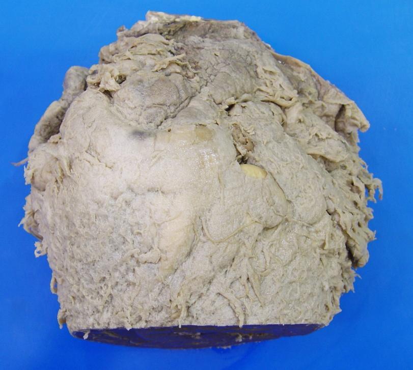
33 Heart from a cow Inflammation Case 6

34 Heart from a cow Inflammation Case 6
35 Inflammation Case 6 Description? The epicardium is thickened and rough with friable to firm, tanyellow sheets and tags covering the entire surface
36 Inflammation Case 6 This is a good example of how fibrin becomes organized with Morphologic time this step precedes fibrosis Diagnosis? Pericarditis, fibrinous, diffuse, subacute (or chronic-active), severe

37 Inflammation Case 6 Additional info: This may occur as a result of ingestion of a nail/wire perforation of the reticulum and penetration of the diaphragm pericardium = Traumatic reticulopericarditis
38 Inflammation Case 7 Kidneys from a cull dairy cow at slaughter Enlarged kidneys noted during processing

39 Inflammation Case 7 Kidneys from a cull dairy cow at slaughter Enlarged kidneys noted during processing


40 Inflammation Case 7 Description? On the capsular surface there are multifocal to coalescing slightly raised rough tan foci. The capsule is firmly adhered in areas
41 Inflammation Case 7 Description? On section, there is dilation and roughening of the renal pelvis with yellow cloudy fluid, and firm yellow material (exudate and stones (uroliths) in the lumen. Fibrosis surrounds these areas
42 Inflammation Case 7 Morphologic Diagnosis? (Pyelo) nephritis, suppurative, bilateral (diffuse), chronic (because of the fibrosis!) Also nephrolithiasis!
43 Questions?
Inflammation Laboratory 1
 Inflammation Laboratory 1 Lab1 Emphasis: The exudates of acute inflammation Descriptions Morphologic Diagnoses Shannon Martinson: http://people.upei.ca/smartinson VPM 152: February 2012 Describing Lesions
Inflammation Laboratory 2. Shannon Martinson: VPM 152: March 2012
 Inflammation Laboratory 2 Shannon Martinson: http://people.upei.ca/smartinson VPM 152: March 2012 Reminder - Creating a Morphologic Diagnosis for Inflammatory Lesions Organ and Process Exudate Distribution
Describing and interpreting gross lesions. Prepared for VPM 4600, May 2018; Shannon Martinson
 Describing and interpreting gross lesions Prepared for VPM 4600, May 2018; Shannon Martinson How to Describe (and Interpret) Lesions Step 1 Step 2 Step 3 Step 4 Look at the specimen: Is it normal or abnormal
Inflammation Laboratory 3 Emphasis: Chronic inflammation and healing. Shannon Martinson: VPM 152: April 2013
 Inflammation Laboratory 3 Emphasis: Chronic inflammation and healing Shannon Martinson: http://people.upei.ca/smartinson VPM 152: April 2013 Example A Reproductive tract and colon/rectum from a sheep Previous
Respiratory Pathology Lab 2: Lung. Shannon Martinson,
 Respiratory Pathology Lab 2: Lung Shannon Martinson, 2017 http://people.upei.ca/smartinson/ Case 1 Signalment: 9 month old DSH cat History: Poor doer with stunted growth One month of lethargy one day the
INFLAMMATION & REPAIR
 INFLAMMATION & REPAIR Histopath Laboratory 1 Winter 2013 Chelsea Martin Special thanks to Drs. Hanna and Forzan Goals: Examine Tissue and Identify the Organ Describe the lesion, grossly and histologically
Pathology of the Alimentary Tract
 Pathology of the Alimentary Tract Lab 2: Lower alimentary tract SI, LI, cecum, and peritoneum GIST in the cecum of a dog Shannon Martinson: http://people.upei.ca/smartinson VPM 221: November, 2011 3 year
Liver Lab #2. Bacterial Hepatitis
 Liver Lab #2 Bacterial Hepatitis Case: O12561-04. Adult ewe. Describe the lesion: Multifocal large nodules ranging in size from 1-3.5cm in greatest diameter are present within the liver and are filled
Disturbances of Circulation, Lab 1: Edema and Congestion/Hyperemia. Shannon Martinson, Feb
 Disturbances of Circulation, Lab 1: Edema and Congestion/Hyperemia Shannon Martinson, Feb 2017 http://people.upei.ca/smartinson/ Case #1 Signalment and History: 6-month old feeder lamb found dead on pasture
WSC , Conference 9, Case 1. Tissue from a nyala.
 WSC 2009-2010, Conference 9, Case 1. Tissue from a nyala. MICROSCOPIC DESCRIPTION: Heart, atrium (1 pt.): Approximately 40% of the atrial myocardium is replaced by areas of fibrous connective tissue (1
Cellular Pathology Gross Pathology Laboratory 2 Cell Injury. VPM 152: General Pathology Instructor: Chelsea Martin Winter 2016
 Cellular Pathology Gross Pathology Laboratory 2 Cell Injury VPM 152: General Pathology Instructor: Chelsea Martin Winter 2016 Gross Specimens The following slides consist of images from the specimens presented
Characteristic. Course of disease:short Days--one month Changes : Alteration, exudation Tissue destruction Inflammation cells: major neutrophils
 ACUTE INFLAMMATION Characteristic Course of disease:short Days--one month Changes : Alteration, exudation Tissue destruction Inflammation cells: major neutrophils TYPES Serous Inflammation Fibrinous Inflammation
GUIDE TO: Diagnosing Coccidiosis & Necrotic Enteritis
 GUIDE TO: Diagnosing Coccidiosis & Necrotic Enteritis Site of Infection Species E. acervulina E. brunetti E. maxima E. mivati E. tenella E. necatrix Oocyst Size 2µ{ 18.3 x 14.6 24.6 x 18.8 30.5 x 20.7
09-Mar-15 PNEUMONIA RESPIRATORY SYSTEM L-3
 RESPIRATORY SYSTEM L-3 Professor Department of Pathology, University of Agriculture, Faisalabad. Email: mtjaved@uaf.edu.pk Web: https://sites.geocities.ws/mtjaved PNEUMONIA The pulmonary inflammatory response
Pathology of the Hematopoietic System - Lab.
 Pathology of the Hematopoietic System - Lab http://people.upei.ca/smartinson/ Shannon Martinson, September 2015 Case #1 Signalment: 96 kg gilt History: Pig from minimal disease herd. Sudden death Case
Respiratory Pathology Lab 1: Upper Respiratory Tract. Shannon Martinson,
 Respiratory Pathology Lab 1: Upper Respiratory Tract Shannon Martinson, 2017 http://people.upei.ca/smartinson/ Case 1 Signalment: 5 year old dog History: 2 month history of nasal discharge Decreased airflow
What s Your Diagnosis?
 What s Your Diagnosis? Signalment: 5 year old MC Belgian Malinois Presenting Complaint: Perineal hernia as well as not eating or defecating History: The patient presented to the KSU VHC on 7/28/2018 for
Cellular Pathology. Histopathology Lab #2 (web) Paul Hanna Jan 2018
 Paul Hanna Jan 2018") Cellular Pathology Histopathology Lab #2 (web) Paul Hanna Jan 2018 Slide #91 Clinical History: a necropsy was performed on an aged cat the gross pathological changes included: widespread subcutaneous edema
Cellular Pathology Histopathology Lab #2 (web) Paul Hanna Jan 2018 Slide #91 Clinical History: a necropsy was performed on an aged cat the gross pathological changes included: widespread subcutaneous edema
HISTOPATHOLOGY. Shannon Martinson
 HISTOPATHOLOGY Shannon Martinson March 2013 Case #1 History: 8 year old beagle Neck pain for the past couple of weeks Paresis, followed by paralysis developed over the past few days Gross Description courtesy
VPM Pigment and other tissue deposits. Shannon Martinson
 VPM 152 - Pigment and other tissue deposits Shannon Martinson http://people.upei.ca/smartinson/ Case 1 Signalment: 2 month old heifer beef calf Clinical History: Lateral recumbency for 4 days Tachycardia,
VPM Pigment and other tissue deposits. Shannon Martinson
 VPM 152 - Pigment and other tissue deposits Shannon Martinson http://people.upei.ca/smartinson/ Case 1: Signalment: 2 month old heifer beef calf Clinical History: Lateral recumbency for 4 days. Tachycardia,
Firm Texture. (chronic) Cut surface: purulent exudate in bronchi Sequels: Abscesses,
 Cut surface: purulent exudate in bronchi Sequels: Abscesses,") 2008 Classification of Pneumonias in Domestic Animals There is no universal classification! Based on texture, distribution of lesions and type of exudate, pneumonias in domestic animals are currently classified
2008 Classification of Pneumonias in Domestic Animals There is no universal classification! Based on texture, distribution of lesions and type of exudate, pneumonias in domestic animals are currently classified
Disturbances of Circulation. Histopathology Lab #2 (Web)
") Disturbances of Circulation Histopathology Lab #2 (Web) Paul Hanna Winter 2015 Slide #96 History: pig was fine in the morning & found dead in the afternoon there was ~100 mls of clear fluid in the pericardial
Disturbances of Circulation Histopathology Lab #2 (Web) Paul Hanna Winter 2015 Slide #96 History: pig was fine in the morning & found dead in the afternoon there was ~100 mls of clear fluid in the pericardial
Pathology of the Hematopoietic System. Case studies
 Pathology of the Hematopoietic System Case studies Shannon Martinson, September 2015 Signalment: 9 yr-old MC cat Case Study 1 History: Cat had been anorexic and developed bleeding in the eyes Physical
Chapter 4 Inflammation and Infection
 Chapter 4 Inflammation and Infection Defense Mechanisms Three lines of defense protect the body against foreign invasion: Physical or surface barriers Inflammation Immune response Inflammation Non-specific
Pathology of the Respiratory System 5: Lung and Thoracic Cavity
 Pathology of the Respiratory System 5: Lung and Thoracic Cavity Shannon Martinson, Jan 2017 http://people.upei.ca/smartinson/ VPM 222 Systemic Pathology DISORDERS OF THE LUNG Congenital Pigmentary deposition
Histopathology: chronic inflammation
 Histopathology: chronic inflammation These presentations are to help you identify, and to test yourself on identifying, basic histopathological features. They do not contain the additional factual information
EXPERIMENTAL THERMAL BURNS I. A study of the immediate and delayed histopathological changes of the skin.
 EXPERIMENTAL THERMAL BURNS I A study of the immediate and delayed histopathological changes of the skin. RJ Brennan, M.D. and B. Rovatti M.D. The purpose of this study was to determine the progressive
Avian Pathology. Bacterial diseases: histo slides. ECVP-ESVP Summer School 2012 Frédérique NGUYEN
 Avian Pathology Bacterial diseases: histo slides ECVP-ESVP Summer School 2012 Frédérique NGUYEN Bacterial diseases: histo slides B1. Turkey. Organs? Morphologic diagnosis? Special procedure? B2. Hen. Organ?
Inflammation. First Lab.
 Inflammation First Lab. The cardinal signs of inflammation are rubor (redness), calor (heat), tumor (swelling), dolor (pain), and loss of function. Seen here is skin with erythema, compared to the more
Canine Liver Eneku Wilfred Bovine Pathology
 2012-1-3 Canine Liver Eneku Wilfred Bovine Pathology Contributor: New Mexico Department of Agriculture Veterinary Diagnostic Services Signalment: 5 month old male Weimaraner dog (Canis familiaris) History:
Pathology of the Hematopoietic System GROSS/HISTO LAB
 Pathology of the Hematopoietic System GROSS/HISTO LAB Paul Hanna (thanks to Dr s Aburto, Martinson & Fenton) Fall 2014 Slide 1 Spleen from a Beaver Give a morphologic diagnosis and possible etiology &
Cardiovascular Pathology Lab. Shannon Martinson,
 Cardiovascular Pathology Lab Shannon Martinson, 2017 http://people.upei.ca/smartinson/ Case 1 Signalment: 10 year old MC DSH Cat History Heart murmur detected on PE recommended cardiac US Blood work was
Liver Pathology Lab 1. Shannon Martinson, 2017
 Liver Pathology Lab 1 Shannon Martinson, 2017 http://people.upei.ca/smartinson/ Case 1 Signalment: 10 year old MC DSH cat History: Inappetence and weight loss Fluid in the abdomen noted on US Esophageal
Etiology. Extreme temperature, electric shock, ionization, physical injury, etc. Metabolic substances, acids, alkalis drugs, tissue necrosis
 INFLAMMATION Inflammation Protective response that is intended to eliminate the initial cause of injury Innate and acquired mechanisms Local or generalized (sepsis) processes Terminology: ~ itis ending
PATHOLOGY Intracellular Degeneration LAB 1
 PATHOLOGY Intracellular Degeneration LAB 1 Cellular swelling Liver Organ :- Liver Lesion :- 1. Narrowing of hepatic sinusoids due to the swelling of hepatocyte. 2. The cytoplasm of affected hepatocyte
SESSION 1: GENERAL (BASIC) PATHOLOGY CONCEPTS Thursday, October 16, :30am - 11:30am FACULTY COPY
 PATHOLOGY CONCEPTS Thursday, October 16, :30am - 11:30am FACULTY COPY") SESSION 1: GENERAL (BASIC) PATHOLOGY CONCEPTS Thursday, October 16, 2008 9:30am - 11:30am FACULTY COPY GOAL: Describe the basic morphologic (structural) changes which occur in various pathologic conditions.
SESSION 1: GENERAL (BASIC) PATHOLOGY CONCEPTS Thursday, October 16, 2008 9:30am - 11:30am FACULTY COPY GOAL: Describe the basic morphologic (structural) changes which occur in various pathologic conditions.
PATHOLOGY OF THE CARDIOVASCULAR SYSTEM
 PATHOLOGY OF THE CARDIOVASCULAR SYSTEM Lecture 3: Pericardium and Endocardium Shannon Martinson, 2018 VPM 2220 Systemic Pathology II http://people.upei.ca/smartinson/ PERICARDIUM AND EPICARDIUM Normal
Menigitidis. Dr Rodney Itaki Lecturer Anatomical Pathology Discipline
 Menigitidis Dr Rodney Itaki Lecturer Anatomical Pathology Discipline University of Papua New Guinea Division of Pathology School of Medicine & Health Sciences Review Normal Microanatomy Image Ref: www.histology-world.com
Necrosis is death of cells and tissues in the living animal. Focal/ Multifocal necrosis- terms used for one
 Necrosis Necrosis Necrosis is death of cells and tissues in the living animal. Focal/ Multifocal necrosis- terms used for one or more, small, clearly defined areas of necrosis. Diffuse necrosis- term used
Systemic Pathology I VPM 221 PATHOLOGY OF THE ALIMENTARY SYSTEM LAB 2
 Systemic Pathology I VPM 221 PATHOLOGY OF THE ALIMENTARY SYSTEM LAB 2 Enrique Aburto Fall 2014 Some advice for your final exam: Focus on the info contained in the pp presentations (on Moodle) Handouts
Endocrine Lab. Heather Fenton VPM 222 November
 Endocrine Lab Heather Fenton VPM 222 November 27 2012 Case 1: Nursery pig Case 1: Nursery pig Description: There are multifocal round (approximately 1cm diameter) firm lesions within the adrenal gland
MORPHOLOGY OF ACUTE INFLAMMATION
 MORPHOLOGY OF ACUTE INFLAMMATION Morphological patterns of acute inflammation Dominating pattern 1. Exudation 2. Necrosis and fibrinous exudation 3. Necrosis 1. DOMINATING PATTERN: EXUDATION Subtypes Serous
This is the second learning component (Learning Component 2) in our first learning module (Learning Module 1). In this component we review a very
 in our first learning module (Learning Module 1). In this component we review a very") This is the second learning component (Learning Component 2) in our first learning module (Learning Module 1). In this component we review a very basic response to injury inflammation. We ll look at examples
This is the second learning component (Learning Component 2) in our first learning module (Learning Module 1). In this component we review a very basic response to injury inflammation. We ll look at examples
Acute and Chronic Inflammation Pathology 1 - Dr. Gary Mumaugh
 Acute and Chronic Inflammation Pathology 1 - Dr. Gary Mumaugh Introduction Injurious stimuli cause a protective vascular connective tissue reaction called inflammation Acute and chronic forms o Inflame
Most abundant and widely distributed tissues in the body Binds, support, and strengthen body tissues, protect and insulate internal organ, serve as
 Connective tissue Most abundant and widely distributed tissues in the body Binds, support, and strengthen body tissues, protect and insulate internal organ, serve as major transport system, compartmentalizes
Table 0: Description of Grading System for Anatomic Severity of Disease in Emergency. Local disease confined to the organ with minimal abnormality
 Table 0: of Grading System for Anatomic Severity of Disease in Emergency Local disease confined to the organ with minimal Local disease confined to the organ with severe Local extension Table 1: Universal
Post Mortal Approach to the Respiratory System Part 1
 Post Mortal Approach to the Respiratory System Part 1 System examination Before the carcass is opened examination of the nasal openings is carried out. Observe for any evidence of nasal discharge or nasal
Lymphoid System: cells of the immune system. Answer Sheet
 Lymphoid System: cells of the immune system Answer Sheet Q1 Which areas of the lymph node have most CD3 staining? A1 Most CD3 staining is present in the paracortex (T cell areas). This is towards the outside
Histopathology: pulmonary pathology
 Histopathology: pulmonary pathology These presentations are to help you identify basic histopathological features. They do not contain the additional factual information that you need to learn about these
Ischaemia It means local anemia, it is characterized by a decrease amount of blood in an organ or region. Causes of Ischemia: *1.
 المرحلة الثالثة م. هالة عباس ناجي Ischaemia It means local anemia, it is characterized by a decrease amount of blood in an organ or region. Causes of Ischemia: *1.External pressure upon an artery e.g:
PATHOLOGICAL STUDY ON SOME RENAL LESION IN SHEEP AND GOATS IN DIYALA PROVINCE
 I.J.S.N., VOL.8 (3) 2017: 611-615 ISSN 2229 6441 PATHOLOGICAL STUDY ON SOME RENAL LESION IN SHEEP AND GOATS IN DIYALA PROVINCE Noura N. Ali & Khalil H. Aljeboori Department of Pathology, college of Vet,
The Alberta Dairy Hoof Health Project. Lesion Severity Scoring Guide
 The Alberta Dairy Hoof Health Project Lesion Severity Scoring Guide Introduction: This lesion severity scoring guide is intended for the use of hoof trimmers, in particular those who are using the Hoof
Pathological Investigations on Bovine Pheumonic Pasteurellosis by Use of Immunoperoxidase Technique
 JARQ 29, 13 1-136 (1995) Pathological Investigations on Bovine Pheumonic Pasteurellosis by Use of Immunoperoxidase Technique Makoto HARITANI Tohoku Branch Laboratory, National Institute of Animal Health
MORPHOLOGIC DIAGNOSIS: Liver: Hepatitis, necrotizing, multifocal to coalescing, severe, with numerous trichomonads. (3 pt)
") Case 1. Tissue from a pelican. MICROSCOPIC DESCRIPTION: Liver: Approximately 80% (1 pt) of the liver is replaced by multifocal to coalescing areas of coagulative and lytic necrosis. Centrally, within these
Case 1. Tissue from a pelican. MICROSCOPIC DESCRIPTION: Liver: Approximately 80% (1 pt) of the liver is replaced by multifocal to coalescing areas of coagulative and lytic necrosis. Centrally, within these
Pathology lab 4 DONE BY : MORAD ABU QAMAR
 Pathology lab 4 DONE BY : MORAD ABU QAMAR Chronic interstitial inflammation, lung Certain etiologic agents such as viruses are more likely to lead to chronic inflammation, as seen here in the lung of a
Histopathology: gastritis and peptic ulceration
 Histopathology: gastritis and peptic ulceration These presentations are to help you identify, and to test yourself on identifying, basic histopathological features. They do not contain the additional factual
Histopathology: healing
 Histopathology: healing These presentations are to help you identify, and to test yourself on identifying, basic histopathological features. They do not contain the additional factual information that
Recognizing African swine fever 13. Post mortem findings
 Recognizing African swine fever 13 Post mortem findings Carcasses of pigs that die in the acute stage of the disease are often in good condition. In white-skinned pigs, bluish-purple discolouration of
Disorders of Cell Growth & Neoplasia. Histopathology Lab
 Disorders of Cell Growth & Neoplasia Histopathology Lab Paul Hanna April 2010 Case #84 Clinical History: 5 yr-old, West Highland White terrier. skin mass from axillary region. has been present for the
CHRONIC INFLAMMATION
 CHRONIC INFLAMMATION Chronic inflammation is an inflammatory response of prolonged duration often for months, years or even indefinitely. Its prolonged course is proved by persistence of the causative
Unit II Problem 2 Pathology: Pneumonia
 Unit II Problem 2 Pathology: Pneumonia - Definition: pneumonia is the infection of lung parenchyma which occurs especially when normal defenses are impaired such as: Cough reflex. Damage of cilia in respiratory
CONNECTIVE TISSUE (C.T.)
") CONNECTIVE TISSUE (C.T.) Objectives: By the end of this lecture, the student should be able to: 1. Enumerate the general characteristics of C.T. 2. Classify C.T into C.T. proper and special types of C.T.
CONNECTIVE TISSUE (C.T.) Objectives: By the end of this lecture, the student should be able to: 1. Enumerate the general characteristics of C.T. 2. Classify C.T into C.T. proper and special types of C.T.
ECVP/ESVP Summer School in Veterinary Pathology Summer School 2015 Histology Case 5 DOG HD: Kidney.
 Case 5 DOG HD: Kidney. 100% of mid to deep renal cortex is characterized by coagulative necrosis/infarction, linear widespread haemorrhages and multifocal vasculitis with thrombosis. Throughout the section
LADIS Case of the Month
 November 2018 LADIS Case of the Month Drs Valentin Janvier and Brieuc Cossic Hospital for Animals and Animal Health Diagnostic Center Signalment and presenting complaint 13 year old Thoroughbred gelding
Pathology of the Liver and Biliary Tract 5 Diseases of the Biliary Tract. Shannon Martinson, April 2016
 Pathology of the Liver and Biliary Tract 5 Diseases of the Biliary Tract Shannon Martinson, April 2016 http://people.upei.ca/smartinson/ OUTLINE Normal anatomy & function Hepatobiliary Injury and responses
Abscess. A abscess is a localized collection of pus in the skin and may occur on any skin surface and be formed in any part of body.
 Abscess A abscess is a localized collection of pus in the skin and may occur on any skin surface and be formed in any part of body. Ethyology Bacteria causing cutaneous abscesses are typically indigenous
5 DISTURBANCES IN CIRCULATION. Congestion / Hyperemia Haemorrhage Thrombosis Embolism Ischemia Infarction Oedema Shock Sludged blood Model Questions
 5 DISTURBANCES IN CIRCULATION Congestion / Hyperemia Haemorrhage Thrombosis Embolism Ischemia Infarction Oedema Shock Sludged blood Model Questions CONGESTION/ HYPEREMIA Hyperemia is increased amount of
Tissues and Membranes
 I. In the Beginning a. Egg + sperm! Tissues and Membranes b. 1 cell divides to make 2, 2 divide to make 4, 4 divide to make 8, and then? c. d. e. Totipotent: f. Pluripotent: II. III. Tissues a. Tissues
Diseases of the breast (1 of 2)
") Diseases of the breast (1 of 2) Introduction A histology introduction Normal ducts and lobules of the breast are lined by two layers of cells a layer of luminal cells overlying a second layer of myoepithelial
Diseases of the breast (1 of 2) Introduction A histology introduction Normal ducts and lobules of the breast are lined by two layers of cells a layer of luminal cells overlying a second layer of myoepithelial
An Uncommon Cardiac Etiology of Liver Cirrhosis, Recurrent Ascites, Atrial Fibrillation and Congestive Heart Failure
 Cronicon OPEN ACCESS EC CARDIOLOGY Case Report An Uncommon Cardiac Etiology of Liver Cirrhosis, Recurrent Ascites, Atrial Fibrillation and Congestive Heart Failure Montaser Y Ismail 1 *, Mohammed I Nassar
Bladder Case 1 SURGICAL PATHOLOGY REPORT. Procedure: Cystoscopy, transurethral resection of bladder tumor (TURBT)
") Bladder Case 1 February 17, 2007 Specimen (s) received: Bladder Tumor Pre-operative Diagnosis: Bladder Cancer Post operative Diagnosis: Bladder Cancer Procedure: Cystoscopy, transurethral resection of
Bladder Case 1 February 17, 2007 Specimen (s) received: Bladder Tumor Pre-operative Diagnosis: Bladder Cancer Post operative Diagnosis: Bladder Cancer Procedure: Cystoscopy, transurethral resection of
Connective Tissue. Consists of two basic elements: Cells and Extra-cellular matrix
 Connective Tissue Consists of two basic elements: Cells and Extra-cellular matrix True Connective Tissue Cells Fibroblasts: Secrete both fibers and ground substance of the matrix (wandering) Macrophages:
Acute Inflammation. Dr. G Mahendra Department of Pathology
 Acute Inflammation Dr. G Mahendra Department of Pathology Inflammation Inflammation is a physiological response to tissue injury. Tissue injury Reaction/response of body Inflammation Inflammation is not
Bio& 241 Unit 1 / Lecture 4
 Bio& 241 Unit 1 / Lecture 4 Connective Tissue Consists of two basic elements: Cells and Extra-cellular matrix 1 True Connective Tissue Cells Fibroblasts: Secrete both fibers and ground substance of the
What s your diagnosis? Malori Marotz. Squirt, an 8month old mix breed puppy. History:
 What s your diagnosis? Malori Marotz Squirt, an 8month old mix breed puppy History: The owner obtained squirt at 12 weeks of age. The owner reported that Squirt was passing soft stools lately and he is
Case: C : Six year old, male castrated, malamute with a history of large bowel diarrhea, tenesmus, mucous and frank blood in the stool.
 Case: E25436-98: 21 year old horse. Describe the lesion: A pedunculated, firm, globoid mass approximately 2cm in greatest diameter that is attached to the serosal surface of the colon. Morphologic diagnosis:
Definition. Terms ending in the suffix itis denote inflammation.
 INFLAMMATION Definition Inflammation is a defensive process that a living body initiates against local tissue damage. It takes the form of a complex reaction of blood vessels, certain plasma components
PROBABLE HODGKIN'S DISEASE IN A DOG: REPORT OF A CASE 1
 PROBABLE HODGKIN'S DISEASE IN A DOG: REPORT OF A CASE 1 LEONARD K. STALKER, M.D. Fellow in Surgery, The Mayo Foundation CARL F. SCHLOTTHAUER, D.V.M. AND WILLIAM H. FELDMAN, D.V.M., M.S. Division of Experimental
ATHEROSCLEROSIS. Secondary changes are found in other coats of the vessel wall.
 ATHEROSCLEROSIS Atherosclerosis Atherosclerosis is a disease process affecting the intima of the aorta and large and medium arteries, taking the form of focal thickening or plaques of fibrous tissue and
TUBERCULOUS PERICARDIAL EFFUSION
 BY S. SUZMAN From the Cardiographic Department, Guy's Hospital Received June 22, 1942 Tuberculous pericardial effusion is not common; and so the case described is of interest on account of the patient's
the urinary system pathology Dr. Fairoz A Eltorgman
 the urinary system pathology Dr. Fairoz A Eltorgman Tumors of the renal pelvis & kidney Benign tumors of the renal pelvis: Hemangioma Leiomyoma Malignant tumors: Transitional cell carcinoma Squamous cell
Proceedings of the 34th World Small Animal Veterinary Congress WSAVA 2009
 www.ivis.org Proceedings of the 34th World Small Animal Veterinary Congress WSAVA 2009 São Paulo, Brazil - 2009 Next WSAVA Congress : Reprinted in IVIS with the permission of the Congress Organizers IMAGING
Gastrointestinal Pathology of Pigs. Jerome C. Nietfeld, DVM, MS, PhD Kansas State Veterinary Diagnostic Lab Department DMP Kansas State University
 Gastrointestinal Pathology of Pigs Jerome C. Nietfeld, DVM, MS, PhD Kansas State Veterinary Diagnostic Lab Department DMP Kansas State University Neonatal Diarrhea Likely the number 1 killer of neonatal
Mediastinum and pericardium
 Mediastinum and pericardium Prof. Abdulameer Al-Nuaimi E-mail: a.al-nuaimi@sheffield.ac.uk E. mail: abdulameerh@yahoo.com The mediastinum: is the central compartment of the thoracic cavity surrounded by
Gastrooesophageal reflux disease. Jera Jeruc Institute of pathology, Faculty of Medicine, Ljubljana, Slovenia
 Gastrooesophageal reflux disease Jera Jeruc Institute of pathology, Faculty of Medicine, Ljubljana, Slovenia Reflux esophagitis (RE) GERD: a spectrum of clinical conditions and histologic alterations resulting
Skin Integrity and Wound Care
 Skin Integrity and Wound Care By Dr. Amer Hasanien & Dr. Ali Saleh Skin Integrity and Wound Care Skin integrity: the presence of normal Skin & Uninterrupted skin layers by wounds. Factors affecting appearance
Pleural Empyema Joseph Junewick, MD FACR
 Pleural Empyema Joseph Junewick, MD FACR 03/19/2010 History Teenager with persistent fever and cough. Pneumonia diagnosed 1 week ago. Diagnosis Pleural Empyema Additional Clinical Surgery-Clear fluid with
2015 Lab Animal 8/22/ Mouse. 2. Rat. 3. Mouse. Tissue from an Mouse: Morphologic Diagnosis: Rectal prolapse
 1. Mouse 2015 Lab Animal 2. Rat Tissue from an Mouse: Morphologic Diagnosis: Rectal prolapse Causes: 1. Syphacia obveluta 2. Citrobacter rodentum 3. Heicobacter sp 3. Mouse 2. Tissue from a Rat: Morphologic
Schistosome life cycle.
 Schistosomiasis infects approximately 200 million persons and kills approximately 280,000 annually. Most of the mortality comes from hepatic granulomas and fibrosis Schistosoma japonicum and Schistosoma
Sheep and Goat Neonate Necropsy Manual
 1 Sheep and Goat Neonate Necropsy Manual Introduction: Why would we want to do a neonatal necropsy? - Performing a necropsy on a neonate is extremely important. Complete examination of the neonate will
P.M Poultry Diseases 4 th year series
 P.M Poultry Diseases 4 th year series By Mohamed Mahmoud Salem gab AllahAssistant lecturer of pathology Faculty of Veterinary Medicine, Benha University, Moshtohor, Tukh; 13736, Qalyuobia, EGYPT FOWL POX
PLEURAE and PLEURAL RECESSES
 PLEURAE and PLEURAL RECESSES By Dr Farooq Aman Ullah Khan PMC 26 th April 2018 Introduction When sectioned transversely, it is apparent that the thoracic cavity is kidney shaped: a transversely ovoid space
Thoracic Cavity and Tumors of Lung and Pleura
 Tutorial Module 6 Thoracic Cavity and Tumors of Lung and Pleura Alfonso López Atlantic Veterinary College University of Prince Edward Island Canada Sept 28, 2014 Thoracic Cavity There are anatomical differences
Liver. Liver. System examination
 Liver System examination Examine the liver in situ after opening the abdominal cavity examine the liver in context with other changes in the abdominal cavity. The image on the left is of a ruptured liver
10/3/2012. Tissue: The Living Fabric: Part B. Extracellular matrix Ground substance Fibers Collagen fiber Elastic fiber Reticular fiber.
 PowerPoint Lecture Slides prepared by Janice Meeking, Mount Royal College C H A P T E R 4 Tissue: The Living Fabric: Part B Copyright 2010 Pearson Education, Inc. Copyright 2010 Pearson Education, Inc.
Pericardial diseases
 Pericardial diseases Anatomy of the pericardium Consists of parietal and visceral membranes. The space between them(pericardial space is normally filled by a lymph like fluid. The fluid s normal quantity
CASE REPORTS. Inflammatory Polyp of the Bronchus. V. K. Saini, M.S., and P. L. Wahi, M.D.
 CASE REPORTS V. K. Saini, M.S., and P. L. Wahi, M.D. I n 1932 Jackson and Jackson [l] first reported a number of clinical cases under the title Benign Tumors of the Trachea and Bronchi with Especial Reference
Urinary Anatomy. Lab 40. Kidneys. Nephrons. Renal Corpuscle
 Urinary Anatomy Lab 40. Urinary Anatomy and Kidney Dissection Kidneys: filters blood, produces urine Ureters: convey urine to bladder Bladder: holding tank Urethra: carries urine to the outside for elimination
PLATES 24 TO 26. (Received for publication, December 4, 1935)
") Published Online: 1 March, 1936 Supp Info: http://doi.org/10.1084/jem.63.3.303 Downloaded from jem.rupress.org on January 19, 2019 THE VISCERAL LESIONS PRODUCED IN MICE BY THE SALIVARY GLAND VIRUS OF MICE*
Published Online: 1 March, 1936 Supp Info: http://doi.org/10.1084/jem.63.3.303 Downloaded from jem.rupress.org on January 19, 2019 THE VISCERAL LESIONS PRODUCED IN MICE BY THE SALIVARY GLAND VIRUS OF MICE*