So now to The Ear. Drawings from Max Brodel, an Austrian artist who came to Johns Hopkins in the 1920s. My point in showing this figure is to
|
|
|
- Rosanna Harrell
- 5 years ago
- Views:
Transcription

1 So now to The Ear. Drawings from Max Brodel, an Austrian artist who came to Johns Hopkins in the 1920s. My point in showing this figure is to emphasize the intricate and well-protected structure of the inner ear, encased in the temporal bone. This combination of fragility and inaccessibility is a large part of the explanation for why knowledge of hearing has somewhat lagged others, such as the vision and olfaction. 1

2 What does it mean to analyze the frequency components of a sound? A spectrogram such as that shown here is the usual display of frequency components as a function of time here during the production of a sentence I can see you. We will see a real-time spectrograph in operation ourselves. 2

3 Today I will provide an overview of cochlear function. Sound enters the external ear, initiating motion of the eardrum and attached ossicular chain, resulting in fluid movement within the inner ear, in the cochlea. 3
4 Structure and Function of the Auditory and Vestibular System - 1 Schematic representation of externally recorded electrical cochlear response waveforms and their relations to each other. The stimulus consists of two bursts lasting about 18 ms. The second tone burst is inverted in polarity with respect to the first. From the bottom up, the cochlear microphonic response CM exactly follows both stimulus waveforms. The summating potential SP is by definition non oscillatory and follows the envelope of the stimulus. The compound or whole nerve action potential CAP is present only at the start and end of stimulation. The top trace shows the composite electrical signal as it would actually be recorded. The CM, SP and CAP traces are simply added. The composite responses to the two stimuli are different only because of the polarity inversion of CM. Subtraction of the two composite waveforms would leave only the CM trace, whereas adding them together would eliminate CM and leave the sum of SP and CAP traces. (From D.T. Kemp, Otoacoustic emissions and evoked potentials, in The Ear, volume 1 of the Oxford University Press Handbook of Auditory Sciences, (P. Fuchs, ed.) to be published in 2010.) 4

5 5

6 Motion of the stapes footplate causes fluid motion, resulting in deflection of the cochlear partition upon which are situated the hair cells and surrounding supporting cells. Frequency selectivity begins with the fact that the mechanics of the cochlear duct vary from end to end. Consequently, lower frequency tones cause maximal vibration at positions near the cochlear apex. High frequency tones deflect the stiffer partition nearer the oval window. This tonotopic pattern of vibration was described first by von Bekesy and is the basis of frequency selectivity in the mammalian cochlea. 6
7 7

8 The tonotopic organization of the cochlea, and the selective innervation of that sensory epithelium, is taken advantage of with the cochlear implant. For the profoundly deaf, a set of electrodes are threaded into the cochlea. A spectrum analyzer provides frequency-dependent stimulation to each electrode, which then stimulates the nearest auditory nerve fibers. With practice this very limited input (usually ~ 10 working electrodes) can be used to understand speech. The best success has been obtained in post-lingually deafened adults, and in congenitally-deaf children who receive the implant in the first year or two of life. In the best cases otherwise deaf children can hit age-appropriate educational milestones. 8

9 Structure and Function of the Auditory and Vestibular System - 1 Example of the human cochlea obtained from a 5 month old fetus. The coil provides a continuous pathway for sound wave propagation. The stapes footplate moves into and out of the oval window (blue arrow), driving fluid motion that is relieved (at steady-state) at the round window (yellow arrow). 9

10 Structure and Function of the Auditory and Vestibular System - 1 Components of the membranous labyrinth 10
 are exposed to these solutions. Perilymph is similar to most extracellular fluids (e.g. CSF), being high in Na and low in K.")
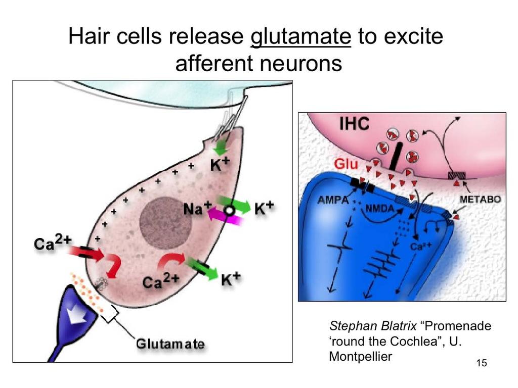
11 Structure and Function of the Auditory and Vestibular System - 1 We have already noted the two types of cochlear fluids, perilymph and endolymph, and how separate portions of the hair cells (in fact, ALL hair cells) are exposed to these solutions. Perilymph is similar to most extracellular fluids (e.g. CSF), being high in Na and low in K. Recall that this solution bathes the basolateral surface of hair cells (i.e. the barrier to endolymph is the reticular lamina, not the basilar membrane). Endolymph is a highly unusual extracellular fluid, being high in K, low in Na, and very low in Ca. This is very similar to most intracellular environments, where potassium is the dominant ion. Thus, the transduction current into hair cells is largely a potassium current. But without a concentration gradient, there must be another driving force to get potassium into the cell. This driving force is generated by two electrical potentials. First, the hair cell s resting potential is ~ -60 mv due to ion channels in the basolateral portion of the membrane. Second, there exists an endolymphatic potential that is +80 mv with respect to the vascular system. This so-called endocochlear potential combines with the hair cell s resting potential for an ~140 mv driving force pushing potassium into the hair cell. The source of the endocochlear potential remains unclear, though it is intimately tied to the stria vascularis which secretes potassium into the endolymphatic space. It should be noted that the endocochlear potential itself is not necessary to proper hair cell function since this potential is only a few mv in the vestibular system (and virtually absent in non-mammalian inner ears). Yet, if the stria is damaged or the endocochlear potential is eliminated, auditory sensitivity is reduced. It may be that the high frequency environment of the cochlea and voltage-dependent motility of some hair cells benefit from the added push of the endocochlear potential. (right slide image: adapted from Promenade, S. Blatrix) 11

12 Structure and Function of the Auditory and Vestibular System - 1 The recycling of potassium and the homeostasis of endolymph is of particular interest, since a stable ionic environment is critical for proper hair cell function. The endolymphatic compartments of each inner ear organ and a separate endolymphatic sack are contiguous. While some secretion and resorption of potassium may occur by the flow of endolymph through this network, there is strong evidence that the recycling of potassium is under local control. The illustrations above depict current theories, where potassium is secreted by the stria, taken up by hair cells in transduction, released from the hair cell s basolateral surface, and recirculated to endolymph via a system of gap junctions between epithelial supporting cells and connective tissues at the spiral ligament. From the fibrocytes of the spiral ligament, potassium presumably enters the stria again by gap junctions. The combination of Na,K- ATPase and Na-K-Cl cotransporters in the marginal cells of the stria enable the accumulation of potassium in these cells (indeed they have a highly positive resting potential). Slowly gating potassium channels on the endolymph-facing surface of marginal cells leads to the accumulation of potassium in endolymph. Immunoreactivity supports connexins as likely components in this network of gap junctions. Mutation of the connexin 26 gene leads to non-syndromic sensorineural hearing loss and the loss of endolymphatic potential. 12
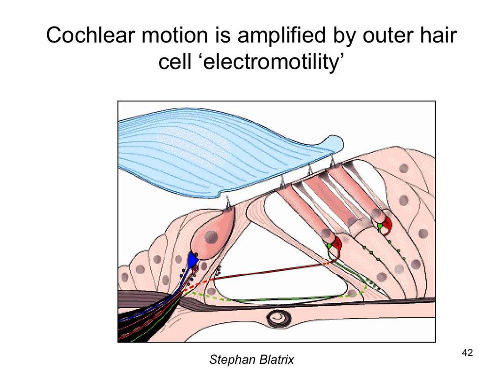
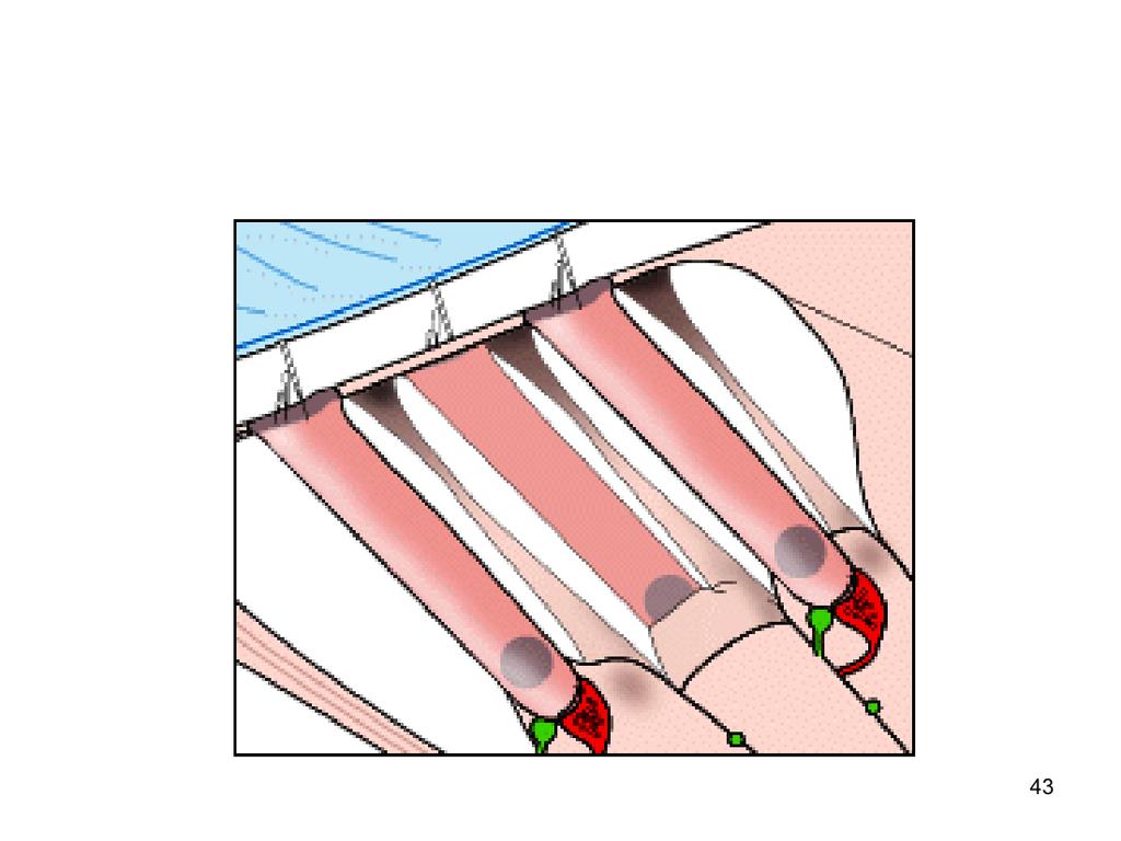
13 Outer hair cells are mechanically active. In response to a sound-induced change in membrane potential, they move. This movement of the outer hair cells adds to that of the cochlear membranes, and so enhances the stimulus energy delivered to the inner hair cell. So, outer hair cell damage deafens the cochlea and is a common form of hearing loss. 13
14 14
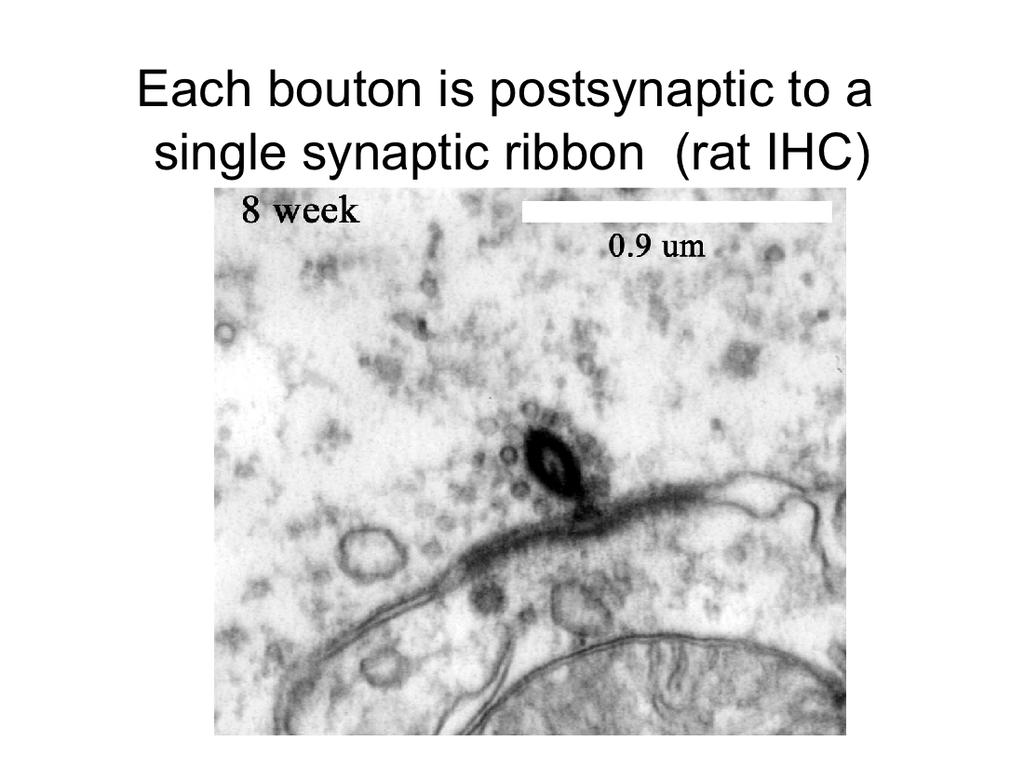
15 15
16 16
17 17
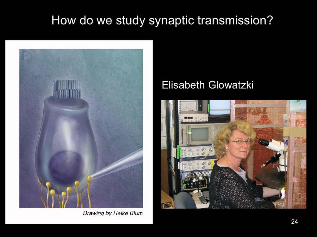
18 Will describe results obtained by intracellular voltage-clamp recording from afferent dendrites at point of contact with IHCs. The peculiar advantages of this experiment provide new insights into ribbon function, and perhaps by extension, into mechanisms of transmitter release more generally. 18
19 Low frequency tones produce phase-locked activity in afferent fibers (turtle). 19
20 Temporal coding results from phase-locking, accurate to submillsecond. 20
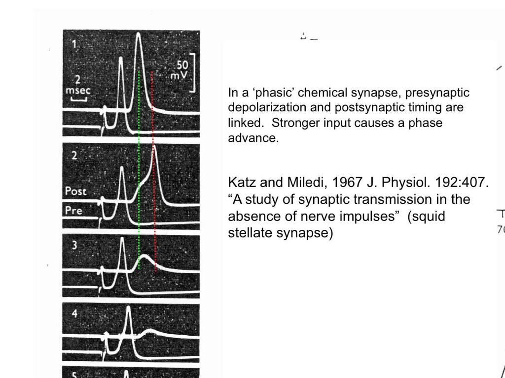
21 At CF phase is independent of intensity. As sound gets louder, hair cell is more strongly depolarized, but spike initiation time does not vary. How does this occur, compare to other chemical synapses? 21
22 22
23 23
24 24

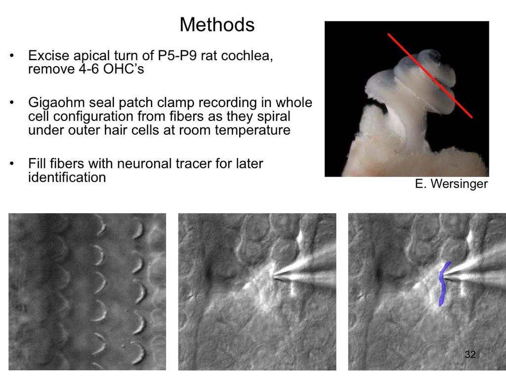
25 This is what the mammalian cochlea (2-3 week old rat) really looks like. These are otic capsules, the bony chamber within the temporal bone of the skull that encloses the inner ear. On the right is the intact capsule. On the left the surgeon (Dr. E. Wersinger, PhD) has dissected away the surrounding bone to reveal the soft tissues of the cochlear spiral. 25
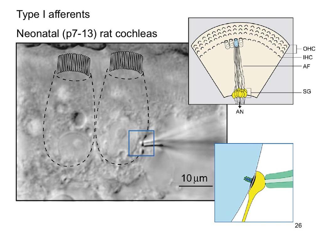
26 26
27 27
28 The synaptic currents reverse in sign near 0 mv, so flow through non-selective cation channels. They are blocked by AMPA/kainate receptor bocker CNQX, and lengthened by cyclothiazide, indicating they are subserved by AMPA receptors. 28
29 29

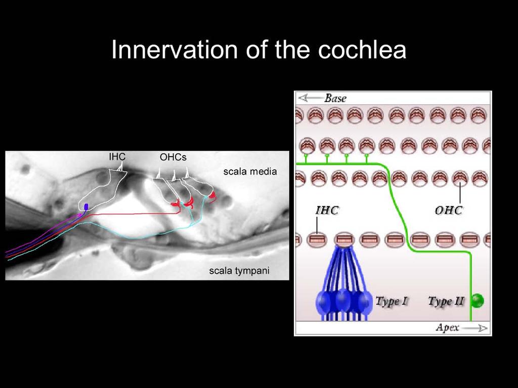
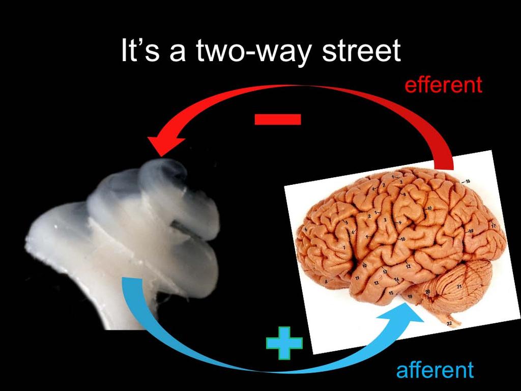
30 The mammalian cochlea possesses two classes of afferent neurons and two classes of efferent neurons. Type I afferents contact single inner hair cells to provide acoustic analysis as we know it. Type II afferents branch extensively to contact numerous outer hair cells. Medial efferent neurons synapse on outer hair cells. Lateral efferent neurons synapse on Type I afferents beneath inner hair cells. 30
31 31
32 32

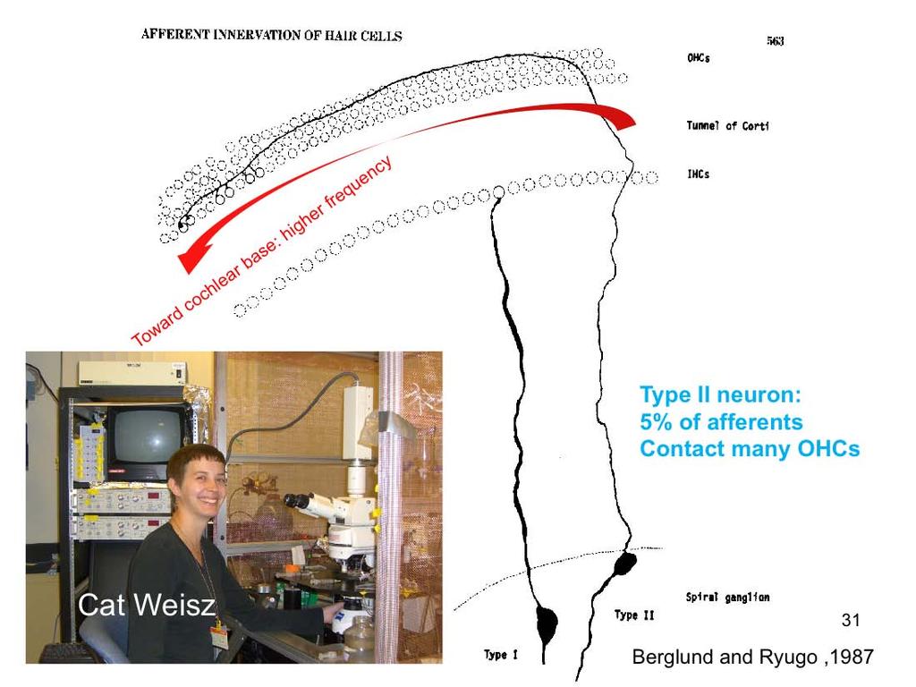
33 I increase the likelihood of recording from a Type II fiber by recording at an age P5-P9 when efferents have not fully innervated the OHC region in rats. Examining the morphology of the labeled fibers gives confirmation that I am recording from Type II s, as the morphology of these cells has been very well described. This image shown a confocal stack of a fiber that I have recorded from and filled with a tracer, in green. The large blob is artifact, due to blowing some of the dye around the recording region. On the left is a tracing of a filled fiber from another recording showing the classic radial projection from the filled soma and a right hand turn towards the base of the cochlea. I should note that I have only observed a soma recording in this one filled neuron. 33
34 We have also determined that the EPSC s are glutamatergic. This figure shows the average EPSC waveform for synaptic events that occur when the membrane holding potential is held at the voltages shown here. Graphing the average peak amplitude of the events by the membrane holding potential illustrates that the currents reverse polarity at a holding potential of 0mV, which is consistent with conductance through a non-selective cation channel. Additionally, the EPSC s are eliminated by the AMPA receptor blocker NBQX at 10uM as seen in the trace on the left. The amplitude by diary plot shown on the right shows the complete blockage of synaptic events upon NBQX application, followed by their return upon washing off the drug. 34
35 35
36 36
37 37
38 38

39 The mammalian cochlea possesses two classes of afferent neurons and two classes of efferent neurons. Type I afferents contact single inner hair cells to provide acoustic analysis as we know it. Type II afferents branch extensively to contact numerous outer hair cells. Medial efferent neurons synapse on outer hair cells. Lateral efferent neurons synapse on Type I afferents beneath inner hair cells. 39
40 Idealized vibration pattern of the basilar membrane (cochlear partition on which hair cells reside). Passive mechanism refers to the vibration pattern produced by a pure tone in a dead cochlea (or one without outer hair cells). Active mechanism refers to the pattern of vibration in a healthy, live cochlea, made 50 db more sensitive and much more sharply-tuned by the active mechanical contribution ( electromotility ) of outer hair cells. 40
41 Outer hair cells are mechanically active. In response to a sound-induced change in membrane potential, they move. This movement of the outer hair cells adds to that of the cochlear membranes, and so enhances the stimulus energy delivered to the inner hair cell. So, outer hair cell damage deafens the cochlea and is a common form of hearing loss. 41
42 42
43 43
 produced by two tones (f1, f2) presented simultaneously to the ear.")
44 Oto-acoustic emissions are ear sounds due to active motility of outer hair cells. They are an indication of normal cochlear function. Typically recorded as the distortion product (2f1-f2) produced by two tones (f1, f2) presented simultaneously to the ear. A sensitive microphone in the ear canal is the detector. 44

45 Otoacoustic emissions are a useful clinical tool, enabling tests for cochlear health in prelingual, or otherwise uncommunicative patients. 45

46 Spontaneous otoacoustic emissions in a newborn. 46
47 47
The mammalian cochlea possesses two classes of afferent neurons and two classes of efferent neurons.
 1 2 The mammalian cochlea possesses two classes of afferent neurons and two classes of efferent neurons. Type I afferents contact single inner hair cells to provide acoustic analysis as we know it. Type
What does it mean to analyze the frequency components of a sound? A spectrogram such as that shown here is the usual display of frequency components
 1 2 3 4 What does it mean to analyze the frequency components of a sound? A spectrogram such as that shown here is the usual display of frequency components as a function of time here during the production
Chapter 3: Anatomy and physiology of the sensory auditory mechanism
 Chapter 3: Anatomy and physiology of the sensory auditory mechanism Objectives (1) Anatomy of the inner ear Functions of the cochlear and vestibular systems Three compartments within the cochlea and membranes
Auditory System Feedback
 Feedback Auditory System Feedback Using all or a portion of the information from the output of a system to regulate or control the processes or inputs in order to modify the output. Central control of
Structure, Energy Transmission and Function. Gross Anatomy. Structure, Function & Process. External Auditory Meatus or Canal (EAM, EAC) Outer Ear
 Outer Ear") Gross Anatomy Structure, Energy Transmission and Function IE N O ME 1 Structure, Function & Process 4 External Auditory Meatus or Canal (EAM, EAC) Outer third is cartilaginous Inner 2/3 is osseous Junction
Gross Anatomy Structure, Energy Transmission and Function IE N O ME 1 Structure, Function & Process 4 External Auditory Meatus or Canal (EAM, EAC) Outer third is cartilaginous Inner 2/3 is osseous Junction
Cochlear anatomy, function and pathology II. Professor Dave Furness Keele University
 Cochlear anatomy, function and pathology II Professor Dave Furness Keele University d.n.furness@keele.ac.uk Aims and objectives of this lecture Focus (2) on the biophysics of the cochlea, the dual roles
The mammalian cochlea possesses two classes of afferent neurons and two classes of efferent neurons.
 1 The mammalian cochlea possesses two classes of afferent neurons and two classes of efferent neurons. Type I afferents contact single inner hair cells to provide acoustic analysis as we know it. Type
Auditory Physiology Richard M. Costanzo, Ph.D.
 Auditory Physiology Richard M. Costanzo, Ph.D. OBJECTIVES After studying the material of this lecture, the student should be able to: 1. Describe the morphology and function of the following structures:
ENT 318 Artificial Organs Physiology of Ear
 ENT 318 Artificial Organs Physiology of Ear Lecturer: Ahmad Nasrul Norali The Ear The Ear Components of hearing mechanism - Outer Ear - Middle Ear - Inner Ear - Central Auditory Nervous System Major Divisions
Cochlear anatomy, function and pathology III. Professor Dave Furness Keele University
 Cochlear anatomy, function and pathology III Professor Dave Furness Keele University d.n.furness@keele.ac.uk Aims and objectives of this lecture Focus (3) on the cochlear lateral wall and Reissner s membrane:
Mechanical Properties of the Cochlea. Reading: Yost Ch. 7
 Mechanical Properties of the Cochlea CF Reading: Yost Ch. 7 The Cochlea Inner ear contains auditory and vestibular sensory organs. Cochlea is a coiled tri-partite tube about 35 mm long. Basilar membrane,
Auditory System. Barb Rohrer (SEI )
") Auditory System Barb Rohrer (SEI614 2-5086) Sounds arise from mechanical vibration (creating zones of compression and rarefaction; which ripple outwards) Transmitted through gaseous, aqueous or solid medium
Auditory System Barb Rohrer (SEI614 2-5086) Sounds arise from mechanical vibration (creating zones of compression and rarefaction; which ripple outwards) Transmitted through gaseous, aqueous or solid medium
Chapter 17, Part 2! The Special Senses! Hearing and Equilibrium!
 Chapter 17, Part 2! The Special Senses! Hearing and Equilibrium! SECTION 17-5! Equilibrium sensations originate within the inner ear, while hearing involves the detection and interpretation of sound waves!
Chapter 17, Part 2! Chapter 17 Part 2 Special Senses! The Special Senses! Hearing and Equilibrium!
 Chapter 17, Part 2! The Special Senses! Hearing and Equilibrium! SECTION 17-5! Equilibrium sensations originate within the inner ear, while hearing involves the detection and interpretation of sound waves!
Anatomy and Physiology of Hearing
 Anatomy and Physiology of Hearing The Human Ear Temporal Bone Found on each side of the skull and contains the organs for hearing and balance Divided into four major portions: - squamous - mastoid - tympanic
What is the effect on the hair cell if the stereocilia are bent away from the kinocilium?
 CASE 44 A 53-year-old man presents to his primary care physician with complaints of feeling like the room is spinning, dizziness, decreased hearing, ringing in the ears, and fullness in both ears. He states
Chapter 11: Sound, The Auditory System, and Pitch Perception
 Chapter 11: Sound, The Auditory System, and Pitch Perception Overview of Questions What is it that makes sounds high pitched or low pitched? How do sound vibrations inside the ear lead to the perception
Cochlear anatomy, function and pathology I. Professor Dave Furness Keele University
 Cochlear anatomy, function and pathology I Professor Dave Furness Keele University d.n.furness@keele.ac.uk Aims and objectives of these lectures Introduction to gross anatomy of the cochlea Focus (1) on
Unit VIII Problem 9 Physiology: Hearing
 Unit VIII Problem 9 Physiology: Hearing - We can hear a limited range of frequency between 20 Hz 20,000 Hz (human hearing acuity is between 1000 Hz 4000 Hz). - The ear is divided into 3 parts. Those are:
Ear. Utricle & saccule in the vestibule Connected to each other and to the endolymphatic sac by a utriculosaccular duct
 Rahaf Jreisat *You don t have to go back to the slides. Ear Inner Ear Membranous Labyrinth It is a reflection of bony labyrinth but inside. Membranous labyrinth = set of membranous tubes containing sensory
Systems Neuroscience Oct. 16, Auditory system. http:
 Systems Neuroscience Oct. 16, 2018 Auditory system http: www.ini.unizh.ch/~kiper/system_neurosci.html The physics of sound Measuring sound intensity We are sensitive to an enormous range of intensities,
Required Slide. Session Objectives
 Auditory Physiology Required Slide Session Objectives Auditory System: At the end of this session, students will be able to: 1. Characterize the range of normal human hearing. 2. Understand the components
Deafness and hearing impairment
 Auditory Physiology Deafness and hearing impairment About one in every 10 Americans has some degree of hearing loss. The great majority develop hearing loss as they age. Hearing impairment in very early
9.01 Introduction to Neuroscience Fall 2007
 MIT OpenCourseWare http://ocw.mit.edu 9.01 Introduction to Neuroscience Fall 2007 For information about citing these materials or our Terms of Use, visit: http://ocw.mit.edu/terms. 9.01 Recitation (R02)
THE COCHLEA AND AUDITORY PATHWAY
 Dental Neuroanatomy Suzanne S. Stensaas, PhD February 23, 2012 Reading: Waxman, Chapter 16, Review pictures in a Histology book Computer Resources: http://www.cochlea.org/ - Promenade around the Cochlea
Before we talk about the auditory system we will talk about the sound and waves
 The Auditory System PHYSIO: #3 DR.LOAI ZAGOUL 24/3/2014 Refer to the slides for some photos. Before we talk about the auditory system we will talk about the sound and waves All waves have basic characteristics:
SPHSC 462 HEARING DEVELOPMENT. Overview Review of Hearing Science Introduction
 SPHSC 462 HEARING DEVELOPMENT Overview Review of Hearing Science Introduction 1 Overview of course and requirements Lecture/discussion; lecture notes on website http://faculty.washington.edu/lawerner/sphsc462/
Hearing. istockphoto/thinkstock
 Hearing istockphoto/thinkstock Audition The sense or act of hearing The Stimulus Input: Sound Waves Sound waves are composed of changes in air pressure unfolding over time. Acoustical transduction: Conversion
Processing of sounds in the inner ear
 Processing of sounds in the inner ear Sripriya Ramamoorthy Associate Professor, IIT Bombay WiSSAP 2018 Cochlea converts sound into electrical signals [Picture courtesy of Northwestern University] von Bekesy
Hearing: Physiology and Psychoacoustics
 9 Hearing: Physiology and Psychoacoustics Click Chapter to edit 9 Hearing: Master title Physiology style and Psychoacoustics The Function of Hearing What Is Sound? Basic Structure of the Mammalian Auditory
to vibrate the fluid. The ossicles amplify the pressure. The surface area of the oval window is
 Page 1 of 6 Question 1: How is the conduction of sound to the cochlea facilitated by the ossicles of the middle ear? Answer: Sound waves traveling through air move the tympanic membrane, which, in turn,
Taste buds Gustatory cells extend taste hairs through a narrow taste pore
 The Special Senses Objectives Describe the sensory organs of smell, and olfaction. Identify the accessory and internal structures of the eye, and explain their function. Explain how light stimulates the
THE COCHLEA AND AUDITORY PATHWAY
 Dental Neuroanatomy Suzanne S. Stensaas, PhD April 14, 2010 Reading: Waxman, Chapter 16, Review pictures in a Histology book Computer Resources: http://www.cochlea.org/ - Promenade around the Cochlea HyperBrain
PSY 214 Lecture # (11/9/2011) (Sound, Auditory & Speech Perception) Dr. Achtman PSY 214
 (Sound, Auditory & Speech Perception) Dr. Achtman PSY 214") PSY 214 Lecture 16 Topic: Sound, Auditory System & Speech Perception Chapter 11, pages 270-289 Corrections: None Announcements: CD is available outside Dr Achtman s office if you would like to see demonstrations
PSY 214 Lecture 16 Topic: Sound, Auditory System & Speech Perception Chapter 11, pages 270-289 Corrections: None Announcements: CD is available outside Dr Achtman s office if you would like to see demonstrations
SPECIAL SENSES: THE AUDITORY SYSTEM
 SPECIAL SENSES: THE AUDITORY SYSTEM REVISION OF PHYSICS: WAVES A wave is an oscillation of power, sound waves have two main characteristics: amplitude, which is the maximum displacement or the power of
Signals, systems, acoustics and the ear. Week 5. The peripheral auditory system: The ear as a signal processor
 Signals, systems, acoustics and the ear Week 5 The peripheral auditory system: The ear as a signal processor Think of this set of organs 2 as a collection of systems, transforming sounds to be sent to
Intro to Audition & Hearing
 Intro to Audition & Hearing Lecture 16 Chapter 9, part II Jonathan Pillow Sensation & Perception (PSY 345 / NEU 325) Fall 2017 1 Sine wave: one of the simplest kinds of sounds: sound for which pressure
Hearing. PSYCHOLOGY (8th Edition, in Modules) David Myers. Module 14. Hearing. Hearing
 David Myers. Module 14. Hearing. Hearing") PSYCHOLOGY (8th Edition, in Modules) David Myers PowerPoint Slides Aneeq Ahmad Henderson State University Worth Publishers, 2007 1 Hearing Module 14 2 Hearing Hearing The Stimulus Input: Sound Waves The
PSYCHOLOGY (8th Edition, in Modules) David Myers PowerPoint Slides Aneeq Ahmad Henderson State University Worth Publishers, 2007 1 Hearing Module 14 2 Hearing Hearing The Stimulus Input: Sound Waves The
HEARING AND COCHLEAR IMPLANTS
 HEARING AND COCHLEAR IMPLANTS FRANCIS CREIGHTON, MD NEUROTOLOGY & SKULL BASE SURGERY FELLOW JOHNS HOPKINS SCHOOL OF MEDICINE NOV 9 TH, 2017 THANKS TO: CHARLIE DELLA SANTINA, HEIDI NAKAJIMA AND DOUG MATTOX
PSY 215 Lecture 10 Topic: Hearing Chapter 7, pages
 PSY 215 Lecture 10 Topic: Hearing Chapter 7, pages 189-197 Corrections: NTC 09-1, page 3, the Superior Colliculus is in the midbrain (Mesencephalon). Announcements: Movie next Monday: Case of the frozen
Representation of sound in the auditory nerve
 Representation of sound in the auditory nerve Eric D. Young Department of Biomedical Engineering Johns Hopkins University Young, ED. Neural representation of spectral and temporal information in speech.
SOLUTIONS Homework #3. Introduction to Engineering in Medicine and Biology ECEN 1001 Due Tues. 9/30/03
 SOLUTIONS Homework #3 Introduction to Engineering in Medicine and Biology ECEN 1001 Due Tues. 9/30/03 Problem 1: a) Where in the cochlea would you say the process of "fourier decomposition" of the incoming
Educational Module Tympanometry. Germany D Germering
 Educational Module anometry PATH medical Germany D-82110 Germering Our educational modules 1 are made for providing information on how the hearing organ works and which test procedures are used to test
The cochlea: auditory sense. The cochlea: auditory sense
 Inner ear apparatus 1- Vestibule macula and sacculus sensing acceleration of the head and direction of gravity 2- Semicircular canals mainly for sensing direction of rotation of the head 1 3- cochlea in
The Structure and Function of the Auditory Nerve
 The Structure and Function of the Auditory Nerve Brad May Structure and Function of the Auditory and Vestibular Systems (BME 580.626) September 21, 2010 1 Objectives Anatomy Basic response patterns Frequency
Acoustics, signals & systems for audiology. Psychoacoustics of hearing impairment
 Acoustics, signals & systems for audiology Psychoacoustics of hearing impairment Three main types of hearing impairment Conductive Sound is not properly transmitted from the outer to the inner ear Sensorineural
Auditory Physiology PSY 310 Greg Francis. Lecture 29. Hearing
 Auditory Physiology PSY 310 Greg Francis Lecture 29 A dangerous device. Hearing The sound stimulus is changes in pressure The simplest sounds vary in: Frequency: Hertz, cycles per second. How fast the
PSY 310: Sensory and Perceptual Processes 1
 Auditory Physiology PSY 310 Greg Francis Lecture 29 A dangerous device. Hearing The sound stimulus is changes in pressure The simplest sounds vary in: Frequency: Hertz, cycles per second. How fast the
Receptors / physiology
 Hearing: physiology Receptors / physiology Energy transduction First goal of a sensory/perceptual system? Transduce environmental energy into neural energy (or energy that can be interpreted by perceptual
PSY 214 Lecture 16 (11/09/2011) (Sound, auditory system & pitch perception) Dr. Achtman PSY 214
 (Sound, auditory system & pitch perception) Dr. Achtman PSY 214") PSY 214 Lecture 16 Topic: Sound, auditory system, & pitch perception Chapter 11, pages 268-288 Corrections: None needed Announcements: At the beginning of class, we went over some demos from the virtual
PSY 214 Lecture 16 Topic: Sound, auditory system, & pitch perception Chapter 11, pages 268-288 Corrections: None needed Announcements: At the beginning of class, we went over some demos from the virtual
College of Medicine Dept. of Medical physics Physics of ear and hearing /CH
 College of Medicine Dept. of Medical physics Physics of ear and hearing /CH 13 2017-2018 ***************************************************************** o Introduction : The ear is the organ that detects
Auditory Physiology PSY 310 Greg Francis. Lecture 30. Organ of Corti
 Auditory Physiology PSY 310 Greg Francis Lecture 30 Waves, waves, waves. Organ of Corti Tectorial membrane Sits on top Inner hair cells Outer hair cells The microphone for the brain 1 Hearing Perceptually,
Hearing. By Jack & Tori
 Hearing By Jack & Tori 3 Main Components of the Human Ear. Outer Ear. Middle Ear. Inner Ear Outer Ear Pinna: >Visible part of ear and ear canal -Acts as a funnel to direct sound Eardrum: >Airtight membrane
Hearing: the function of the outer, the middle and inner ear. Hearing tests. The auditory pathways
 Hearing: the function of the outer, the middle and inner ear. Hearing tests. The auditory pathways Dr. Gabriella Kékesi 74. Hearing: the function of the outer, the middle and inner ear. Hearing tests.
AUDITORY APPARATUS. Mr. P Mazengenya. Tel 72204
 AUDITORY APPARATUS Mr. P Mazengenya Tel 72204 Describe the anatomical features of the external ear Describe the tympanic membrane (ear drum) Describe the walls of the middle ear Outline the structures
STRUCTURAL ELEMENTS OF THE NERVOUS SYSTEM
 STRUCTURAL ELEMENTS OF THE NERVOUS SYSTEM STRUCTURE AND MAINTENANCE OF NEURONS (a) (b) Dendrites Cell body Initial segment collateral terminals (a) Diagrammatic representation of a neuron. The break in
Week 5. Fall 2016 Part 2: Structure and Function of Auditory System 1
 This outline summarizes major points covered in lecture. It is not intended to replace your own lecture notes. Week 5 How sound is heard: EAR Mechanical energy reaches the eardrum, moves to the middle
Can You Hear Me Now?
 An Introduction to the Mathematics of Hearing Department of Applied Mathematics University of Washington April 26, 2007 Some Questions How does hearing work? What are the important structures and mechanisms
= add definition here. Definition Slide
 = add definition here Definition Slide Definition Slides Sensation = the process by which our sensory receptors and nervous system receive and represent stimulus energies from our environment. Perception
MECHANISM OF HEARING
 MECHANISM OF HEARING Sound: Sound is a vibration that propagates as an audible wave of pressure, through a transmission medium such as gas, liquid or solid. Sound is produced from alternate compression
Otoconia: Calcium carbonate crystals Gelatinous mass. Cilia. Hair cells. Vestibular nerve. Vestibular ganglion
 VESTIBULAR SYSTEM (Balance/Equilibrium) The vestibular stimulus is provided by Earth s, and. Located in the of the inner ear, in two components: 1. Vestibular sacs - gravity & head direction 2. Semicircular
Chapter 3. of energy that moves through air, water and other matter, in waves of pressure.
 Chapter 3 Human Hearing Mechanism 3.1 Introduction Audition is the scientific name for the perception of sound. Sound is a form of energy that moves through air, water and other matter, in waves of pressure.
Bioscience in the 21st century
 Bioscience in the 21st century Neurons, Synapses, and Signaling Dr. Michael Burger Outline: 1. Why neuroscience? 2. The neuron 3. Action potentials 4. Synapses 5. Organization of the nervous system 6.
The transformation of sound stimuli into electrical signals
 The transformation of sound stimuli into electrical signals Robert Fettiplace 2 1 Introduction Our sense of hearing depends on the correct performance of about 15 000 hair cells in each cochlea that serve
Definition Slides. Sensation. Perception. Bottom-up processing. Selective attention. Top-down processing 11/3/2013
 Definition Slides Sensation = the process by which our sensory receptors and nervous system receive and represent stimulus energies from our environment. Perception = the process of organizing and interpreting
The lagena a presumptive vestibular epithelium found at the low frequency end of the auditory end organ, is absent from mammals.
 1 Structure and Function of the Auditory and Vestibular System 1 Increasing elaboration of the auditory end organ during evolution. Increasing length of the cochlea associated with higher frequency hearing.
Chapter 13 Physics of the Ear and Hearing
 Hearing 100 times greater dynamic range than vision Wide frequency range (20 ~ 20,000 Hz) Sense of hearing Mechanical system that stimulates the hair cells in the cochlea Sensors that produce action potentials
Salamanca Study Abroad Program: Neurobiology of Hearing
 Salamanca Study Abroad Program: Neurobiology of Hearing Synaptics and the auditory nerve R. Keith Duncan University of Michigan rkduncan@umich.edu Review Resources Reviews: Safieddine et al., 2012, The
Hearing. By: Jimmy, Dana, and Karissa
 Hearing By: Jimmy, Dana, and Karissa Anatomy - The ear is divided up into three parts - Sound enters in through the outer ear and passes into the middle where the vibrations are received and sent to the
COM3502/4502/6502 SPEECH PROCESSING
 COM3502/4502/6502 SPEECH PROCESSING Lecture 4 Hearing COM3502/4502/6502 Speech Processing: Lecture 4, slide 1 The Speech Chain SPEAKER Ear LISTENER Feedback Link Vocal Muscles Ear Sound Waves Taken from:
For this lab you will use parts of Exercise #18 in your Wise lab manual. Please be sure to read those sections before coming to lab
 Bio 322 Human Anatomy Objectives for the laboratory exercise The Eye and Ear Required reading before beginning this lab: Saladin, KS: Human Anatomy 5 th ed (2017) Chapter 17 For this lab you will use parts
Modeling of Mechanoelectrical Transduction of Hair Cells to Action Potentials in the Auditory Nerve
 Modeling of Mechanoelectrical Transduction of Hair Cells to Action Potentials in the Auditory Nerve Anthony Au, Yuchen Wang, Sibai Xie A Term Paper for BENG26 University of California, San Diego San Diego,
Ameen Alsaras. Ameen Alsaras. Mohd.Khatatbeh
 9 Ameen Alsaras Ameen Alsaras Mohd.Khatatbeh Nerve Cells (Neurons) *Remember: The neural cell consists of: 1-Cell body 2-Dendrites 3-Axon which ends as axon terminals. The conduction of impulse through
Printable version - Hearing - OpenLearn - The Open University
 Skip to content Accessibility Sign in Contact Search the OU The Open University Study at the OU Research at the OU OU Community About the OU Hearing Printable page generated Saturday, 12 November 2011,
THE EAR AND HEARING Be sure you have read and understand Chapter 16 before beginning this lab. INTRODUCTION: hair cells outer ear tympanic membrane
 BIOLOGY 211: HUMAN ANATOMY & PHYSIOLOGY ****************************************************************************************************** THE EAR AND HEARING ******************************************************************************************************
Anatomy of the Ear Region. External ear Middle ear Internal ear
 Ear Lecture Objectives Make a list of structures making the external, middle, and internal ear. Discuss the features of the external auditory meatus and tympanic membrane. Describe the shape, position,
Lecture 6 Hearing 1. Raghav Rajan Bio 354 Neurobiology 2 January 28th All lecture material from the following links unless otherwise mentioned:
 Lecture 6 Hearing 1 All lecture material from the following links unless otherwise mentioned: 1. http://wws.weizmann.ac.il/neurobiology/labs/ulanovsky/sites/neurobiology.labs.ulanovsky/files/uploads/purves_ch12_ch13_hearing
For more information about how to cite these materials visit
 Author(s): Matthew Velkey, 2009 License: Unless otherwise noted, this material is made available under the terms of the Creative Commons Attribution Non-commercial Share Alike 3.0 License: http://creativecommons.org/licenses/by-nc-sa/3.0/
Νευροφυσιολογία και Αισθήσεις
 Biomedical Imaging & Applied Optics University of Cyprus Νευροφυσιολογία και Αισθήσεις Διάλεξη 11 Ακουστικό και Αιθουσιαίο Σύστημα (Auditory and Vestibular Systems) Introduction Sensory Systems Sense of
Sound waves from the auditory environment all combine in the ear canal to form a complex waveform. This waveform is deconstructed by the cochlea with
 1 Sound waves from the auditory environment all combine in the ear canal to form a complex waveform. This waveform is deconstructed by the cochlea with respect to time, loudness, and frequency and neural
The Outer and Middle Ear PERIPHERAL AUDITORY SYSTEM HOW WE HEAR. The Ear in Action AUDITORY NEUROPATHY: A CLOSER LOOK. The 3 parts of the ear
 AUDITORY NEUROPATHY: A CLOSER LOOK HOW WE HEAR The 3 parts of the ear The ear consists of three main parts: 1. The outer ear The part you see, which is called the auricle (ohr-a-kal). 2. The middle ear
Electrophysiology. General Neurophysiology. Action Potentials
 5 Electrophysiology Cochlear implants should aim to reproduce the coding of sound in the auditory system as closely as possible, for best sound perception. The cochlear implant is in part the result of
Chapter 7. Audition, the Body Senses, and the Chemical Senses. Copyright Allyn & Bacon 2004
 Chapter 7 Audition, the Body Senses, and the Chemical Senses This multimedia product and its contents are protected under copyright law. The following are prohibited by law: any public performance or display,
Effects of Remaining Hair Cells on Cochlear Implant Function
 Effects of Remaining Hair Cells on Cochlear Implant Function N1-DC-2-15QPR1 Neural Prosthesis Program N. Hu, P.J. Abbas, C.A. Miller, B.K. Robinson, K.V. Nourski, F. Jeng, B.A. Abkes, J.M. Nichols Department
Improving the diagnostic power of otoacoustic emissions. Arturo Moleti Physics Department University of Roma Tor Vergata
 Improving the diagnostic power of otoacoustic emissions Arturo Moleti Physics Department University of Roma Tor Vergata The human ear Ear canal: resonant cavity Middle ear: impedance adapter and pressure
Spectrograms (revisited)
") Spectrograms (revisited) We begin the lecture by reviewing the units of spectrograms, which I had only glossed over when I covered spectrograms at the end of lecture 19. We then relate the blocks of a
Spectrograms (revisited) We begin the lecture by reviewing the units of spectrograms, which I had only glossed over when I covered spectrograms at the end of lecture 19. We then relate the blocks of a
AUDL GS08/GAV1 Signals, systems, acoustics and the ear. Pitch & Binaural listening
 AUDL GS08/GAV1 Signals, systems, acoustics and the ear Pitch & Binaural listening Review 25 20 15 10 5 0-5 100 1000 10000 25 20 15 10 5 0-5 100 1000 10000 Part I: Auditory frequency selectivity Tuning
ID# Final Exam PS325, Fall 1997
 ID# Final Exam PS325, Fall 1997 Good luck on this exam. Answer each question carefully and completely. Keep your eyes foveated on your own exam, as the Skidmore Honor Code is in effect (as always). Have
Essential feature. Who are cochlear implants for? People with little or no hearing. substitute for faulty or missing inner hair
 Who are cochlear implants for? Essential feature People with little or no hearing and little conductive component to the loss who receive little or no benefit from a hearing aid. Implants seem to work
Anatomy of the ear: Lymphatics
 Anatomy of the ear: 1. External ear which consist of auricle and external auditory canal. The auricle has a framework of cartilage except the lobule, the skin is closely adherent to perichonderium at the
Structure and innervation of the cochlea
 Brain Research Bulletin 60 (2003) 397 422 Review Structure and innervation of the cochlea Yehoash Raphael, Richard A. Altschuler Kresge Hearing Research Institute, The University of Michigan, MSRB 3, Rm
Human Acoustic Processing
 Human Acoustic Processing Sound and Light The Ear Cochlea Auditory Pathway Speech Spectrogram Vocal Cords Formant Frequencies Time Warping Hidden Markov Models Signal, Time and Brain Process of temporal
Emissions are low-intensity sounds that may be detected in the external ear canal by a microphone
 OAEs Emissions are low-intensity sounds that may be detected in the external ear canal by a microphone OAE is a pre-neural phenomenon They can be measured even when the 8 th cranial nerve is severely damaged
AUDL GS08 and GAV1: 2013 Final exam page 1/13. You must complete all sections. Label all graphs. Show your work!
 AUDL GS08 and GAV1: 2013 Final exam page 1/13 You must complete all sections. Label all graphs. Show your work! Section A: Short questions concerning Signals & Systems A1. Give the sound pressure levels
A truly remarkable aspect of human hearing is the vast
 AUDITORY COMPRESSION AND HEARING LOSS Sid P. Bacon Psychoacoustics Laboratory, Department of Speech and Hearing Science, Arizona State University Tempe, Arizona 85287 A truly remarkable aspect of human
Who are cochlear implants for?
 Who are cochlear implants for? People with little or no hearing and little conductive component to the loss who receive little or no benefit from a hearing aid. Implants seem to work best in adults who
HST 721 Lecture 4: Mechanics, electromotility and the cochlear amplifier
 HST 721 Lecture 4: Mechanics, electromotility and the cochlear amplifier 1 Cochlear Mechanics: Measures of Basilar Membrane Motion 2 Cochlear Mechanics: Measures of Basilar Membrane Motion Bekesy s experiments
Sense system. Introduction The visual system Hearing. Introduction to sensory mechanisms
 Sense system Introduction The visual system Hearing Introduction to sensory mechanisms Sensory receptors & sense organs Sensory neurons & Receptor cells Adequate stimulus threshold Transduction Receptor
Learning Targets. Module 20. Hearing Explain how the ear transforms sound energy into neural messages.
 Learning Targets Module 20 Hearing 20-1 Describe the characteristics of air pressure waves that we hear as sound. 20-2 Explain how the ear transforms sound energy into neural messages. 20-3 Discuss how
Acquired Deafness Loss of hearing that occurs or develops sometime in the course of a lifetime, but is not present at birth.
 Page 1 of 5 URMC» Audiology Glossary of Terms A Acoustic Neuroma A tumor, usually benign, which develops on the hearing and balance nerves and can cause gradual hearing loss, tinnitus, and dizziness. Acquired