Profound understanding of anatomy
|
|
|
- Frederica Francis
- 5 years ago
- Views:
Transcription


1 ENGLISH


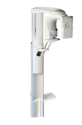
2 Profound understanding of anatomy Planmeca ProMax 3D, the intelligent and multipurpose X-ray unit, is designed to obtain complete information on patient anatomy in the minutest detail. The unit provides digital panoramic, cephalometric, and 3D imaging, as well as advanced imaging software tools to comply with every possible need in dental radiology. The Planmeca ProMax concept is unique in that the 3D imaging modality is reachable with a simple upgrade of a digital Planmeca ProMax. As a result, one intelligent X-ray unit can meet virtually any need in maxillofacial imaging. 2 3


3 Detailed diagnostics with 3D imaging The direct deposit CsI semiconductor flat panel produces accurate, distortion-free images for 3D reconstruction. Unlike image intensifier sensors that use old vacuum tube technology and multi-step focusing, flat panels use single step image readouts with no geometric distortion, no loss of sensitivity, and therefore no need for frequent calibration. Planmeca s proprietary 3D reconstruction algorithm converts the original 2D transillumination images to 3D volume study and is the core component for high quality 3D imaging. It handles high contrast objects, like amalgam fillings, in a special way in order to produce undisturbed study views. The reconstructed image volume consists of more than 120 million voxels. These voxels are isotropic, which enables accurate 1:1 measurements and ensures geometric relations throughout the image. The extremely small voxel size, μm 3, provides a detailed high-resolution 3 lp/mm image without artefacts. In modern dentistry, the demand for surgical implant treatments is steadily growing. The growth trend has created a need for a more advanced X-ray imaging device. Planmeca ProMax 3D, a Cone Beam Volumetric Tomography (CBVT) unit, is expressly designed to comply with the needs of modern surgical dentistry. It supplies clear, dependable imaging in a three-dimensional format with limited patient radiation dose. Thanks to its small footprint, Planmeca ProMax 3D makes effective three-dimensional imaging possible in every dental office. Planmeca ProMax 3D is a genuine all-in-one unit including a digital panoramic, a digital cephalometric, and 3D digital imaging, all in the same unit, saving office space and investment costs. This innovative, versatile, and dynamic imaging device will open up new possibilities for dentists on-site in their offices. Planmeca ProMax 3D uses new Cone Beam Volumetric Tomography technology, in which the X-ray beam used is shaped in the form of a cone or a pyramid. The CBVT technology takes the whole volume needed in a single semicircle scan, as opposed to a medical CT that takes multiple axial slices in multiple full circle scans. During the scan, each image is made using a short X-ray pulse instead of continuous radiation. The total scanning time is 18 seconds for one volume, but the actual exposure time is only 3 seconds at the shortest. This technique reduces patient radiation dose considerably and forms a stroboscopic X-ray effect which, together with the shortened rotation scan (only 194 degrees), virtually eliminates artefacts, contributing to the outstanding image quality. The Planmeca ProMax platform s unique SCARA technology (Selectively Compliant Articulated Robot Arm) enables free formation of the imaging geometry. Planmeca s patented, computer-controlled SCARA robotic arm can produce any movement pattern required, ensuring perfectly accurate and reliable image volume positioning. All controls are made on a full colour graphical user interface in the language of your choice. Thanks to the original, technologically advanced design, any Planmeca ProMax can be upgraded into a 3D Cone Beam Volumetric Tomography unit. 4 5


4 TMJ study The condyle is displayed sharply. The image clearly shows the condition of the temporomandibular joint. Malign finding can be seen inside the condyle head. Implant case The lower first molar on the right is missing. The image clearly shows that there is enough bone to place an implant. Wisdom tooth extraction It is easy to see that the extraction would be difficult. The mandibular canal is located lingually to the roots. Unequalled imaging programs Impacted canine An impacted maxillary right canine found in the wall between sinus and nasal cavity. Large View image Three volumes stitched together for a full view. Planmeca ProMax 3D complies with a multitude of diagnostic requirements: those of endodontics, periodontics, orthodontics, implantology, dental and maxillofacial surgery, and TMJ analysis. It is also an excellent tool in diagnosing ear, maxillary sinus, and respiratory tract diseases. With Planmeca ProMax 3D, study volume sizes are selectable to meet diagnostic needs without excess radiation outside the area of interest. The 80 x 80 mm image size is optimum for most diagnostic applications that require whole dentition, mandible, and maxilla in the same study volume. The 80 x 50 mm volume can be used for single views of the jaw mandible or maxilla, lowering the radiation by almost 40%. The small 40 x 50 mm volume is intended for molar area studies or for planning 3 rd molar extractions. The volumes can also be stitched together to generate an image up to 140 mm in width or 140 mm in height. Planmeca ProMax 3D produces high-resolution volumetric studies of the mandible and maxilla for analysing the bone structure available, the location of the mandibular canal, and the correct position for the implant. Pre-surgical planning will reach a new level of precision, as the prospective site is visible in all three imaging planes: sagittal, axial, and coronal. Third molars, maxillary cuspids, supernumerary teeth, and all of impactions challenge the clinician to identify the tooth s orientation. Planmeca ProMax 3D brings all angles and orientations easily visible. Planmeca ProMax 3D studies accompanied by digital cephalometric images provide the full visualisation of all classes of orthodontic malocclusion. This is highly advantageous for orthodontic planning, as time is saved and patient radiation dose reduced. Unlike traditional orthodontic analyses, Planmeca ProMax 3D provides the orthodontist with image data in the correct anatomic 1:1 ratio, without need to correct for geometric magnification. Planmeca ProMax 3D also provides high-resolution TMJ studies for true and accurate evaluations of the joint arthritides, condylar morphology, and the condyle-fossa relationship. With its high resolution (3 lp/mm) and advanced reconstruction technology, Planmeca ProMax 3D establishes the new standard for 3D dental radiology. 6 7


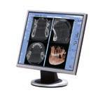
5 Cyst in right mandible A large solitary bone cyst is clearly visible in the right mandible. Planmeca Romexis 3D Implant Planning module Planmeca Romexis for viewing studies Sinus study A cyst and inflammation can be found in the left maxillary sinus. Planmeca Romexis 3D Panoramic module Planmeca Romexis 3D Explorer, the 3D image acquisition software for Planmeca ProMax 3D, enables flexible viewing in all three relevant projections: axial, coronal, and sagittal. The software incorporates a re-slicing feature, which enhances the projections and enables real-time three-dimensional viewing in the desired angle. A rendered 3D view provides a realistic overview of the anatomy. With the Planmeca Romexis 3D Explorer software, each patient study can be stored on a CD with Planmeca Romexis 3D Viewer for others to view. The optional Planmeca Romexis 3D Panoramic module creates a panoramic image from the acquired volume of data without the undesired artefacts, common in normal panoramic images. As the image is reconstructed through software, the user can determine the focal trough location and thickness. The Planmeca Romexis 3D Cross Sections module, which is available as an option, produces cross-sectional images of anatomy along with the defined panoramic curve. The image number and their exact positions can be freely chosen. The 3D Cross Sections module also includes the reconstructed panoramic view. The optional Planmeca Romexis 3D Implant Planning module offers tools for implant placing and nerve drawing. The implant placements are determined with the help of an implant model corresponding to the actual size of the real implant. A drawing tool allows clear marking of the mandibular nerve. Planmeca Romexis software has optional DICOM functionality, which allows 3D studies to be transferred to other implant planning software, such as SimPlant, Nobel Biocare Procera, CyberMed, or any other software that receives images in DICOM format. Studies can also be transferred to PACS or to a high quality DICOM printer in the network. Planmeca Romexis is a pure Java based software that runs in various operating systems and modern web environments. 8 9
 80 GB Minimum set up: Modality Work Station and Database server Planmeca Romexis 3D Explorer (std) Database Server (std, requires Windows)")
6 Computer requirements Example installation Planmeca ProMax 3D with the 3D reconstruction server (included in delivery) Planmeca Romexis database server Processor Pentium 4, 2 Ghz Pentium 4, 2 GHzh RAM 2 GB 2 GB Hard disk space 2 x 320 GB (RAID1 mirroring) 80 GB Minimum set up: Modality Work Station and Database server Planmeca Romexis 3D Explorer (std) Database Server (std, requires Windows) Planmeca Romexis Image Database (std) The Modality Work Station and Database server can also be in separate computers. Additional Diagnostic Work Stations with different software configurations Planmeca Romexis 3D Explorer (opt) Planmeca Romexis Panoramic Reconstruction module (opt) Planmeca Romexis Implant Planning module (opt) Planmeca Romexis DICOM module (opt) Operation system: Windows, Mac OS X or Linux Planmeca Romexis client work station Graphics card 128 MB minimum memory 128 MB minimum memory Monitor SVGA, 1280 x 1024 or more, 24 bit colours or more SVGA, 1280 x 1024 or more, 24 bit colours or more Network card Ethernet 10/100 Mbps Ethernet 10/100 Mbps Peripherals CD R/W and/or DVD R/W drive CD-ROM drive Backup medium DAT or other backup device Operating system Windows XP Pro, Windows 2003 Server, Vista Windows XP, 2003, Vista, Mac OS X*, Linux* Other Java platform (Java Virtual Machine 5.0 or later) Java platform (Java Virtual Machine 5.0 or later) * Mac OS X / Linux support subject to contract X-ray beam Focal spot Image detector Gray scale Detector resolution Number of voxels Voxel size Image acquisition Total scan time Effective exposure time Reconstruction time Standard volumes (height x diam.) Stitched volume 3D reconstruction server Cone 0.5 mm, fixed anode CsI coated CMOS flat panel sensor 12 bit, 4096 gray tones 624 x 624 pixels, pixel size 200 μm x 200 μm 501 x 501 x 501 = 125 M 160 μm x 160 μm x 160 μm, isotropic Single 194 degree rotation 18 s, pulsed X-ray s s 80 mm x 80 mm 50 mm x 80 mm 50 mm x 40 mm width: max 140 mm height: max 140 mm Proprietary Feldkamp based back projection reconstruction algorithm Improved Artifact Removal (IAR) and High Contrast Object Compensation (HCOC) Technical specifications Ethernet Printer (opt) 10 11


7 Planmeca Oy designs and manufactures a full line of high technology dental equipment, including dental care units, panoramic and intraoral X-ray units, and digital imaging products. Planmeca Oy, the parent company of the Finnish Planmeca Group, is strongly committed to R&D, and is the largest privately held company in the field. Planmeca Oy Asentajankatu Helsinki Finland tel fax sales@planmeca.com Images may contain optional items not included in standard delivery. Available configurations and features may have country or area specific variations. Some products displayed above may not be available in all countries or areas. Rights for changes reserved /1008/en
Profound understanding of anatomy
 ENGLISH Profound understanding of anatomy The unique Planmeca ProMax 3D product family offers equipment for all maxillofacial imaging. All volumes sizes from the smallest special cases to whole head images
Profound understanding of anatomy
 ENGLISH Profound understanding of anatomy The unique Planmeca ProMax 3D product family offers equipment for all maxillofacial imaging. All volume sizes from the smallest special cases to whole head images
Planmeca ProMax 3D s Planmeca ProMax 3D ENGLISH
 Planmeca ProMax 3D s Planmeca ProMax 3D ENGLISH Genuine all-in-one unit Planmeca ProMax 3D s and Planmeca ProMax 3D units are designed to obtain complete information on patient anatomy in the minutest
For true visualisation
 ENGLISH For true visualisation Planmeca ProModel is a patient-specific physical model for high-end maxillofacial operations and dental surgery. By reproducing the anatomy of the patient in real-size, Planmeca
Planmeca ProMax 3D s Planmeca ProMax 3D ENGLISH
 Planmeca ProMax 3D s Planmeca ProMax 3D ENGLISH Learn more: Planmeca Imaging for ipad Genuine all-in-one unit Planmeca ProMax 3D s and Planmeca ProMax 3D units are designed to obtain complete information
New era of dental imaging
 ENGLISH New era of dental imaging The revolutionary Planmeca ProMax X-ray unit provides a wide range of extraoral X-ray imaging modalities for the needs of modern dentistry; panoramic radiographs for imaging
Head to new heights with your imaging SCANORA 3D
 SCANORA 3D Head to new heights with your imaging Benefits at a glance The solution for dentomaxillofacial and ENT imaging Easy Patient seated for added stability during exposure. Clear, self-explinatory
CS 9300 Family. The power of flexibility
 CS 9300 Family The power of flexibility The new CS 9300 digital imaging system from Carestream Dental take the guesswork out of examinations The all-in-one CS 9300 is the most versatile multimodality imaging
Low Dose Excellent Image Quality Rapid Reconstruction
 Low Dose Excellent Image Quality Rapid Reconstruction Efficient 3 in 1 Dental X-ray System CBCT > Precise 3-D Anatomical structures - Accurate diagnosis for doctors - Safe implant for patients > Significant
I AM DEMANDING Type CMOS Flat Panel CMOS CMOS ø 40 x 40 mm, ø 60 x 60 mm, ø 80 x 80 mm, ø 110 x 80 mm
 TECHNICAL SPECIFICATIONS 1168 1501 1978 1237 1551-2351 Ø 1090 PANORAMIC CBCT CEPHALOMETRIC X-RAY SOURCE Tube type High frequency DC generator 2.8 mmal / 85 kv 7.0 mmal / 90 kv 2.8 mmal / 85 kv Operation
PAN CEPH 3D CONE BEAM
 PAN CEPH 3D CONE BEAM 2D - 3D panoramic units PANORAMIC CEPHALOMETRIC 3D CONE BEAM IMAGING I-MAX TOUCH Tactile & naturally intuitive panoramic imaging Discover the simplicity and efficiency this unit can
The Quality Leader in 3D Cone Beam CT
 The Quality Leader in 3D Cone Beam CT The Complete 2-in-1 or 3-in-1 Multi-modality Solution PreXion, with over 15 years of innovation in the medical and dental fields, introduces the PreXion3D Eclipse.
Flexible Easy Competitive. SCANORA 3Dx - The in-office large field-of-view Cone Beam CT system for Head and Neck imaging
 Flexible Easy Competitive SCANORA 3Dx - The in-office large field-of-view Cone Beam CT system for Head and Neck imaging SCANORA 3Dx. The solution. SCANORA 3Dx makes advanced 3D imaging easy in the head
Dental Line. 3D digital panoramic system. radiology ahead
 Dental Line 3D digital panoramic system radiology ahead new generation 3D digital panoramic unit 3D imaging s value available for anyone Following the incredible success of the innovative digital panoramic
3D Accuitomo XYZ Slice View Tomograph. Super High-Resolution Images of Region of Interest
 3D Accuitomo XYZ Slice View Tomograph. Super High-Resolution Images of Region of Interest Thinking ahead. Focused on life. 2 Cone Beam X-Ray CT Imaging X-Ray Tube Imaging Intensifier Imaging Volume Voxel
ORTHOPANTOMOGRAPH OP 3D Pro A platform for changing needs
 ORTHOPANTOMOGRAPH OP 3D Pro A platform for changing needs ORTHOPANTOMOGRAPH OP 3D Pro OP 3D Pro is the most comprehensive 3-in-1 platform designed for today and tomorrow, covering the entire maxillofacial
True Low Dose. Exact time to display image on screen may vary upon computer and network configuration.
 RAYSCAN ALPHA PLUS True Low Dose Cone Beam CT Industry Leading Resolution High resolution images provide all the clinical information needed while keeping radiation exposure low. Endodontics - Smallest
Versatility And Expandability In One Panoramic.
 Orthoralix 9200 / 9200 DDE Versatility And Expandability In One Panoramic. Panoramic X-ray Systems Intraoral X-ray Systems Digital Intraoral Sensors Digital X-ray Phosphor Plates Intraoral Cameras Imaging
ENGLISH. Cefla s.c. - Via Selice Provinciale 23/a, Imola - Italy Tel
 09-2017 NVGEGB161S00 According to the regulations in force, some products and/or features may have different availability and characteristics in areas outside of the European Union. Please contact your
THE WAIT IS OVER CS D. 3D imaging is now available for everyone
 THE WAIT IS OVER CS 8100 3D 3D imaging is now available for everyone COMPLEXITY IS NO LONGER THE STANDARD NOW THERE ARE MANY REASONS TO MOVE TO 2D/3D IMAGING Now it s possible to experience nothing but
XPan 3D Plus. FONA Every dental solution you need. Advanced dental technology. Headquarters THE ULTIMATE DIAGNOSTIC SOLUTION DIGITAL DENTISTRY
 FONA Every dental solution you need Through decades of experience and deep understanding of the dental profession, we deliver complete, reliable and accessible solutions.regardless of country or specialisation,
ORTHOPANTOMOGRAPH OP200 D ORTHOCEPH OC200 D. True dynamo leading through the decades.
 ORTHOPANTOMOGRAPH OP200 D ORTHOCEPH OC200 D True dynamo leading through the decades. 1 You can t dublicate the legacy. 1946 Professor Y.V. Paatero publishes his first paper on Panoramic Tomography. 1951
VistaVox S 3D from Dürr Dental
 VistaVox S 3D from Dürr Dental 3D and 2D X-ray images with exceptional image quality COMPRESSED AIR SUCTION IMAGING DENTAL CARE HYGIENE Taking diagnostics to the next level VistaVox S combines diagnostic
ENGLISH. Advanced tools for orthodontics
 ENGLISH Advanced tools for orthodontics A complete solution for orthodontics One unit one software Panoramic Cephalometric CBCT image 3D photo Planmeca ProMax unit 3D model scan Planmeca offers a complete
Cone Beam 3D Imaging
 Cone Beam 3D Imaging NewTom Sets the Standard in 3D Maxillofacial Imaging Cone Beam 3D Imaging The Global Market Leader The Inventors n of Cone Beam 3D In 1996, QR srl developed the first generation of
3D Panoramic Cephalometric. Innovation, in reach. KODAK 9000 Extraoral Imaging System
 3D Panoramic Cephalometric Innovation, in reach 9000 KODAK 9000 Extraoral Imaging System Cephalometric Innovation made simple Innovation made simple We believe in innovation. We always have. In fact, our
CT Imaging at the Point-of-Care
 ENGLISH True Dedication The new Planmed Verity Extremity CT Scanner revolutionizes extremity CT imaging. The compact unit brings 3D imaging at emergency departments, orthopedic clinics or trauma centers
- RCS Paris B
 Technical specifications PANORAMIC CBCT CEPHALOMETRIC X-ray source Tube type High frequency DC generator Total filtration >2.5 mm Al @ 90 kv Mode of operation Continuous Pulsed Continuous Tube voltage
STELLARIS 3D 4 IN 1 CBCT SOLUTION FOR ADVANCED DIAGNOSTICS
 STELLARIS 3D 4 IN CBCT SOLUTION FOR ADVANCED DIAGNOSTICS 3 STELLARIS 3D 4 IN CBCT SOLUTION FOR ADVANCED DIAGNOSTICS Stellaris 3D is a complete and compact, fully upgradeable 3D CBCT for a patient, Panoramic
English. Perfect Vision
 English Perfect Vision Everything becomes clearer and simpler with a big F.O.V. The complete dentomaxillofacial volume ready for your diagnosis One scan provides you with an incredible amount of information
ENGLISH. Distributed by: QR srl - Via Silvestrini, Verona Italy Tel
 GiANO - R15.1 - EN ENGLISH Distributed by: QR srl - Via Silvestrini, 20-37135 Verona Italy Tel. +39 045 8202727-045 583500 info@newtom.it www.newtom.it Manufacturer: CEFLA S.C. - CEFLA DENTAL GROUP Via
ENGLISH. Cone Beam 3D Imaging
 ENGLISH Cone Beam 3D Imaging FIRST USER OF CONE BEAM IN DENTAL FIELD QR s.r.l. is the name that stands behind NewTom Cone Beam 3D imaging units and the creator of Cone Beam technology for the dental field.
OP 3D Vision The upgradable 3D X-ray system for the strictest demands.
 OP 3D Vision The upgradable 3D X-ray system for the strictest demands. The solution for every task: KaVo OP 3D Vision. Regardless of which dental query you may have, the KaVo ORTHOPANTOMOGRAPH OP 3D Vision
DIAGNOSTIC IMAGING. OPTIMIZED.
 ABOUT LED DENTAL SEE THE DIFFERENCE Using our years of business insight and clinical experience as a foundation, LED Dental takes the uncertainty out of your imaging purchase decision. We offer our clients
Dental Cone Beam CT. What is Dental Cone Beam CT?
 Scan for mobile link. Dental Cone Beam CT Dental cone beam computed tomography (CT) is a special type of x-ray equipment used when regular dental or facial x-rays are not sufficient. Your doctor may use
VGi - R EN ENGLISH. QR srl - Via Silvestrini, Verona Italy Tel
 VGi - R15.1 - EN ENGLISH QR srl - Via Silvestrini, 20-37135 Verona Italy Tel. +39 045 8202727-045 583500 info@newtom.it www.newtom.it FIRST IN CONE BEAM, ACCURATE IN RESULTS. 360 degree imaging, reduced
fast accurate safe See the full picture: add a 3rd dimension to your patient evaluation to diagnose more effectively. BENEFITS OF NEWTOM 3G 6" 9" 12"
 See the full picture: add a 3rd dimension to your patient evaluation to diagnose more effectively. Examination Effective Dose Equivalent (ICRP tissue weights 2005) Panoramic Dose (ICRP tissue weights 2005)
ADVANCED 3D IMAGING. CEFLA s.c. Via Selice Provinciale 23/a Imola Italy t newtom.
 CEFLA s.c. Via Selice Provinciale 23/a 40026 Imola Italy t. +39 045 8202727 045 583500 info@newtom.it newtom.it 05/2018 NVGEGB181S00 According to the standards in force, in extra-eu areas the availability
ENGLISH. Advanced tools for orthodontics
 ENGLISH Advanced tools for orthodontics A complete solution for orthodontics One unit one software Panoramic Cephalometric CBCT image 3D photo Planmeca ProMax unit 3D model scan Planmeca offers a complete
ORTHOPANTOMOGRAPH OP300. A platform for changing needs.
 ORTHOPANTOMOGRAPH OP300 A platform for changing needs. 1 Panoramic with cephalometric digital image Xray machine For me, peace of mind means a patient trusting in my care, time after time Leading the way
X X X. GXS-700 Direct USB Digital Intraoral Sensors. Buy a Sensor Combo and a Digital Pan Unit, Receive $600 Off! GO.BENCO benco.
 8 0 0. G O. B E N C O b e n c o. c o m GS-700 Direct USB Digital Intraoral Sensors Designed to make migrating from film, or upgrading an existing digital system, easier than ever High quality image capture
The Optimum Choice for Implantologist
 The Optimum Choice for Implantologist What is essential for your practice? What s the best way to choose a 3D X-ray machine for implant treatment planning? 02 Doctor says.. There are diagnostic limitations
Easy operation. Numerous diagnostic options. X-rays you can rely on: the ORTHOPHOS XG device family. All ORTHOPHOS XG 3Dready programs at a glance.
 CAD /CAM Systems Instruments Hygiene Systems Treatment Centers Imaging Systems Subject to technical changes and errors in the text, Order No. A91100-M47-B346-01-7600, Printed in Germany, Dispo-Nr. 04602,
ORIGINAL PAPERS. Revista Română de Anatomie funcţională şi clinică, macro- şi microscopică şi de Antropologie. Vol. XV Nr
 Revista Română de natomie funcţională şi clinică, macro- şi microscopică şi de ntropologie Vol. XV Nr. 3 2016 ORIGINL PPERS The Utility of CCT Imaging Technique in Maxillofacial Trauma. Dobrovat 1, Roxana
Digital Imaging from a new perspective
 TREATMENT CENTRES HANDPIECES HYGIENE SYSTEMS X-RAY SYSTEMS CEREC TREATMENT CENTRES HANDPIECES HYGIENE SYSTEMS X-RAY SYSTEMS CEREC SIRONA CREATING AND MAINTAINING VALUE. You are right to expect a great
T h e D e n t a l C o m p a n y FROM DIAGNOSTIC SCAN TO SURGERY, WE SHAPE THE FUTURE OF DENTISTRY.
 T h e D e n t a l C o m p a n y FROM DIAGNOSTIC SCAN TO SURGERY, WE SHAPE THE FUTURE OF DENTISTRY. SIDEXIS SOFTWARE ORTHOPHOS SL D/D SEAMLESS THE NEW STANDARD IN CLINICAL DIAGNOSIS AND PATIENT COMMUNICATION
5G XL - R EN ENGLISH
 5G XL - R15.0 - EN ENGLISH Sede legale ed amministrativa - Headquarters QR srl - Via Selice Provinciale, 23/a - 40026 Imola - Bo (Italy) Stabilimento - Plant Via Fermi, 40-37136 Verona (Italy) Tel. +39
NewTom 5G XL EXTRA.VISION
 CEFLA s.c. Via Selice Provinciale 23/a 40026 Imola Italy t. +39 045 8202727 045 583500 info@newtom.it newtom.it 06/2018 N5GXGB181S00 According to the standards in force, in extra-eu areas the availability
3D/2D WALL MOUNTED UNIT
 Product launch document EN Page 1 sur 30 EN PRODUCT LAUNCH DOCUMENT REV03, September 2018 3D/2D WALL MOUNTED UNIT Product launch document EN Page 2 sur 30 INDEX 1. PRODUCT IDENTITY AND POSITIONNING...
ENGLISH VGi - R ENG
 ENGLISH First in Cone Beam, Accurate in Results Cone Beam 3D Imaging First User of Cone Beam in Dental Field QR s.r.l. is the name that stands behind NewTom Cone Beam 3D imaging units and we were the creators
The goal of diagnosis and treatment planning
 Three-dimensional diagnosis & treatment planning: The use of 3D facial imaging and 3D cone beam CT in orthodontics and dentistry By William E. Harrell, Jr, DMD Threedimensional imaging and its use over
The Power Of Choice Pan. Ceph. 3D. Your Imaging Future Starts Today.
 NEW from Gendex! The Power Of Choice Pan. Ceph. 3D. Your Imaging Future Starts Today. Cone Beam 3D Imaging Systems Panoramic X-ray Systems Intraoral X-ray Systems Digital Intraoral Sensors Digital X-ray
I AM EXCLUSIVE TECHNICAL SPECIFICATIONS. The first personal imaging plate scanner IMAGING PLATES. System. Windows recommended configuration
 TECHNICAL SPECIFICATIONS System Resolution... 20 lp/mm Scan Time (fast mode)...1,6s - 2,7s Scan Time (high definition mode)...2,1s - 3,6s Connection... Ethernet RJ-45 Dimensions... L. 154 x D. 204 x H.
Planmeca CBCT units. ENT imaging. Ear Nose Throat ENGLISH
 CBCT units ENT imaging Ear Nose Throat ENGLISH Versatile units for ENT imaging ProMax 3D Max, ProMax 3D Mid and ProMax 3D Plus are our versatile CBCT units for dental and full maxillofacial imaging. With
Clinical details: Details of scan: CONE BEAM CT REPORT: Name: H. B. Gender: Reason for referral: Referred by:
 Name: H. B. Gender: Male DOB: 11/12/1950 Age: 64 Date taken: 16/11/2015 Date reported: 19/11/2015 Clinical details: Reason for referral: Referred by: Investigate symptoms related to left TMJ. Reconstructed
NewTom GiANO HR PERFECT.VISION
 CEFLA s.c. Via Selice Provinciale 23/a 40026 Imola Italy t. +39 045 8202727 045 583500 info@newtom.it newtom.it 06/2018 NHRGB181S00 According to the standards in force, in extra-eu areas the availability
2D AND 3D/2D WALL-MOUNTED PANORAMIC UNITS
 2D AND 3D/2D WALL-MOUNTED PANORAMIC UNITS KEEP YOUR CLINIC ONE STEP AHEAD! Wall-mounted concept: zero foot print 62kg - the lightest unit on the market Face to face positioning High Definition The fruit
Solutions for Large Group Practices
 Solutions for Large Group Practices 2D imaging im aging 3D imaging 3D im aging Infection control ion c CAD/CAM solutions D/C AM solutions Information and monitoring l un ts Dental units One easy-to-use
CT Scanning Protocol For V2R Guided Surgery Solutions
 CT Scanning Protocol For V2R Guided Surgery Solutions 2 V2R CT Scanning Protocol \\ Contents Contents General requirements... 3 V2R Dual Scan Protocol... 5 V2R Single Scan Protocol... 8 Overview... 10
Xelis Dental - What New in
 Xelis Dental - What New in 1.0.6.1 Clinical Needs Why Xelis-Dental? Panorama Cephalography Intra-oral Digital Camera Traditional imaging systems - 2-Dimension view Incorrect anatomical information - Distortion
3D-MODEL CUSTOM-MADE MODELS SEGMENTATION AND PRODUCTION SERVICE OF BONE MODELS WITH HIGHEST 3D PRINTING RESOLUTION
 CUSTOM-MADE MODELS 3D-MODEL SEGMENTATION AND PRODUCTION SERVICE OF BONE MODELS WITH HIGHEST 3D PRINTING RESOLUTION FOLLOW US ON CUSTOM-MADE MODELS 3D-MODEL From a CT or CBCT scan, 3D-model service provides
In-Office Cone Beam Computerized Tomography: Technology Review and Clinical Examples Michael Tischler, DDS
 Page 1 of 8 Issue Date: June 2008, Posted On: 6/26/2008 In-Office Cone Beam Computerized Tomography: Technology Review and Clinical Examples Michael Tischler, DDS Electronic Medical Record Research EMR
Planmeca Ultra Low Dose
 Planmeca Ultra Low Dose 3D imaging with an even lower dose than panoramic imaging ENGLISH Pioneering low dose 3D imaging Planmeca ProMax 3D units offer a unique Planmeca Ultra Low Dose imaging protocol
PERSPECTIVES OF THE TOOTH RESTORATION TECHNOLOGY BASED ON THE COMPUTED TOMOGRAPHY DATA
 PERSPECTIVES OF THE TOOTH RESTORATION TECHNOLOGY BASED ON THE COMPUTED TOMOGRAPHY DATA Maxim Putrik *, Vladimir Ivanov, Igor Antsygin Federal State Autonomous Educational Institution of Higher Education
Extraoral Imaging. Chapter 42. Copyright 2018, Elsevier Inc. All Rights Reserved. 1
 Extraoral Imaging Chapter 42 Copyright 2018, Elsevier Inc. All Rights Reserved. 1 Learning Objectives Lesson 42.1: Panoramic Imaging 1. Pronounce, define, and spell the key terms. 2. Discuss panoramic
User Guide for Dental and Maxillofacial Cone Beam Computed Tomography (CBCT)
") User Guide for Dental and Maxillofacial Cone Beam Computed Tomography (CBCT) Poster No.: C-0756 Congress: ECR 2014 Type: Educational Exhibit Authors: J. Ukkonen, J. Asp; Helsinki/FI Keywords: Education
User Guide for Dental and Maxillofacial Cone Beam Computed Tomography (CBCT) Poster No.: C-0756 Congress: ECR 2014 Type: Educational Exhibit Authors: J. Ukkonen, J. Asp; Helsinki/FI Keywords: Education
ENGLISH. Cefla s.c. - Via Selice Provinciale 23/a, Imola - Italy Tel
 11-2016 N5GGB161S00 According to the regulations in force, some products and/or features may have different availability and characteristics in areas outside of the European Union. Please contact your
Veraviewepocs 3D R100 & F40
 Veraviewepocs 3D R100 & F40 Innovative 3D Reuleaux Full Arch FOV Thinking ahead. Focused on life. Veraviewepocs 3D R100 A New Frontier in X-ray Diagnostics Veraviewepocs 3D R100 has changed the shape of
Interpretation Basics of Cone Beam Computed Tomography COPYRIGHTED MATERIAL
 Interpretation Basics of Cone Beam Computed Tomography COPYIGHTED MATEIA 1 Introduction to Cone Beam Computed Tomography Shawneen M. Gonzalez Introduction This chapter will cover basics of cone beam computed
Cone-beam CT (CB Throne) applied to Title dentomaxillofacial region. Yajima, A; Otonari-Yamamoto, M; San. Yonezu, H; Nakagawa, K; Yajima, Y
 applied to Title dentomaxillofacial region. Yajima, A; Otonari-Yamamoto, M; San. Yonezu, H; Nakagawa, K; Yajima, Y") Cone-beam CT (CB Throne) applied to Title dentomaxillofacial region Yajima, A; Otonari-Yamamoto, M; San Author(s) Y; Otonari, T; Tanabe, K; Wakoh, M; Yonezu, H; Nakagawa, K; Yajima, Y Journal Bulletin
Cone-beam CT (CB Throne) applied to Title dentomaxillofacial region Yajima, A; Otonari-Yamamoto, M; San Author(s) Y; Otonari, T; Tanabe, K; Wakoh, M; Yonezu, H; Nakagawa, K; Yajima, Y Journal Bulletin
EN PRODUCT LAUNCH DOCUMENT. I-MAX 3D 2016 Révision : V Product launch document EN Page 1 sur 29. Version 02, OCTOBER 2016
 Product launch document EN Page 1 sur 29 EN PRODUCT LAUNCH DOCUMENT Version 02, OCTOBER 2016 Product launch document EN Page 2 sur 29 INDEX 1. PRODUCT IDENTITY AND POSITIONNING... 3 2. TECHNICAL CHARACTERISTICS...
ORTHOPHOS XG 3 DS. X-ray systems. ORTHOPHOS XG 3 Digital panoramic X-ray for practical diagnostics.
 ORTHOPHOS XG 3 DS X-ray systems ORTHOPHOS XG 3 Digital panoramic X-ray for practical diagnostics. ORTHOPHOS XG 3 Standard panoramic X-ray with proven technology. competent successful Many years of competence
UNIVERSITY OF MEDICINE AND PHARMACY GR. T. POPA - IASI FACULTY OF DENTAL MEDICINE
 UNIVERSITY OF MEDICINE AND PHARMACY GR. T. POPA - IASI FACULTY OF DENTAL MEDICINE ABSTRACT CONTRIBUTIONS OF THREE-DIMENSIONAL IMAGING TO THE DIAGNOSIS AND MANAGEMENT OF CLEFT LIP AND PALATE PhD ADVISOR,
The Role of. 54 MARCH 2016 // dentaltown.com. radiology feature. by Deepak Gupta, MDS
 The Role of Imaging by Deepak Gupta, MDS Dr. Deepak Gupta completed his post graduation in oral medicine and maxillofacial radiology in 2011. He is currently an Associate Professor/ Reader in the Department
THE USE OF KEYSTONE EASYGUIDE CT SCANNING SOFTWARE FOR DIAGNOSIS, DIRECTION AND DEPTH DETERMINATION
 CT DIAGNOSTICS IN 3D IMPLANT TREATMENT PLANNING THE USE OF KEYSTONE EASYGUIDE CT SCANNING SOFTWARE FOR DIAGNOSIS, DIRECTION AND DEPTH DETERMINATION Timothy Kosinski, DDS, MAGD Assistant Clinical Professor
CBCT: A dentist perspective
 SEGi Review ISSN: 1985.5672 Vol.9, December 2015 CBCT: A dentist perspective Dr. Ranjana Garg, Faculty of Dentistry SEGi University. Dr. Vivek Vijay Gupta Faculty of Dentistry SEGi University Abstract
The agony and ecstasy of buying cone beam technology Part 1: The Ecstasy
 Clinical The agony and ecstasy of buying cone beam technology Part 1: The Ecstasy Dale A. Miles 1 Abstract Background: Since arriving in North America in 2001, cone beam computed tomography (CBCT) has
Using cone beam technology in orthodontics
 PRACTICE US Using cone beam technology in orthodontics Continuing education earn 2 ce credits! Edward Lin explains the benefits of adding cone beam technology to the orthodontic armamentaria Since cone
DE UK. ultra low dose. your dental equipment sales, service and training partner. Planmeca
 DE UK your dental equipment sales, service and training partner Planmeca ultra low dose Pioneering low dose 3D imaging Planmeca ProMax 3D units offer a unique Planmeca Ultra Low Dose imaging protocol that
European Veterinary Dental College
 European Veterinary Dental College EVDC Training Support Document Preparation of Radiograph Sets (Cat and Dog) Document version : evdc-tsd-radiograph_positioning_(dog_and_cat)-20120121.docx page 1 of 13
vertaplan the spine surgeon s software vertaplan System for successful reconstruction of the individual sagittal balance
 the spine surgeon s software System for successful reconstruction of the individual sagittal balance What do you think of patient-specific reconstruction of the spine geometry? Optimum surgical outcome
Dolphin 3D Imaging 11.7 beta
 Dolphin 3D Imaging 11.7 beta Dolphin 3D Surgery Dolphin s 3D Surgery has expanded to include even more features! This revolutionary treatment planning and presentation software module now includes a fully
APW: New Features, New Services, New Update
 APW Dental Services, PC DENTAL CT SCANS APW Dental Services, PC 34 East 62nd Street, New York, NY 10021 Vol. 2, October 2004 IN THIS ISSUE: BioDental Models Surgical Guides Craniofrontal Dysplasia 3D CONE
Planmeca CAD/CAM solutions
 Planmeca CAD/CAM solutions ENGLISH Scan. Design. Manufacture. Chairside workflow We offer a full range of open CAD/CAM solutions for dentists. From ultra-fast intraoral scanning to high-precision chairside
3D Cone beam CT & Digital Radiography Dedicated to Otorhinolaryngology
 3D Cone beam CT & Digital Radiography Dedicated to Otorhinolaryngology Multi-functional imaging solution3 RAYSCAN m is an unique 2-in-1 imaging solution, combining Cone Beam CT and Digital Radiography,
Advantage beyond imaging
 Advantage beyond imaging www.ondemand3d.com About OnDemand3D TM OnDemand3D TM provides sophisticated 3D image processing tools and enables easy sharing of images between practices a nd imaging specialists
Implant Imaging JCD /jp-journals REVIEW ARTICLE ABSTRACT
 JCD 10.5005/jp-journals-10031-1051 REVIEW ARTICLE Ajay R Bhoosreddy, Seema Bhoosreddy, Vinayak Umesh Shirsekar ABSTRACT Implants improve the quality of life for patients who are unable to keep their natural
ENGLISH. Planmeca Compact a
 ENGLISH Planmeca Compact a Affordable approach Planmeca Compact a is the ideal choice for any dentist looking for an affordable high-performing dental unit with pneumatically driven instrument system.
D3D CBCT. See more Do More
 D3D CBCT See more Do More BIOLASE DaVinci Imaging D3D For capturing superior 3D image acquisitions with the lowest minimal dose for patient safety The BIOLASE DaVinci Imaging D3D has one of the lowest
The future of health is digital
 Dated: XX/XX/XXXX Name: XXXXXXXX XXXXXXXXXXX Birth Date: XX/XX/XXXX Date of scan: XX/XX/XXXX Examination of the anatomical volume: The following structures are reviewed and evaluated for bilateral symmetry,
Full Mouth Survey. Delta Dental of Massachusetts DeltaDentalMA.com
 X-Rays Having X-rays taken is a painless procedure that uses small amounts of radiation to capture images of your teeth and bones. Because your dentist takes precautions and the amount of radiation used
CS 8100 FAMILY / CS D FAMILY / CS 9300 FAMILY EXTRAORAL SOLUTIONS EXTRAORDINARY POSSIBILITIES
 CS 8100 FAMILY / CS 8100 3D FAMILY / CS 9300 FAMILY EXTRAORAL SOLUTIONS EXTRAORDINARY POSSIBILITIES A SOLUTION FOR EVERY CLINIC AND ANY CONSULTATION The CS 8100 does more than my old machine and is half
The mission of our company is the development of an individual approach in functional dentistry.
 The Prosystom company started up in 2013. It was created by Russian doctors and IT specialists for carrying out scientific projects and practical work in functional dentistry. The mission of our company
DE UK. 3D imaging. your dental equipment sales, service and training partner. Planmeca
 DE UK your dental equipment sales, service and training partner Planmeca 3D imaging Passion to innovate An introduction from our President Fantastic five... 4 Unique 3D combination an industry first...
ENGLISH. The complete implant workflow easiness with one software
 ENGLISH easiness with one software All in one software 1 Smile designing 8 Final restorations Planmeca CAD/CAM 2 CBCT imaging Planmeca ProMax 3D 7 Guide manufacturing Planmeca Creo 6 Implant guide design
Evaluation of Cone Beam Computed Tomography in Diagnosis and Treatment Plan of Impacted Maxillary Canines
 Original Research Evaluation of Cone Beam Computed Tomography in Diagnosis and Treatment Plan of Impacted Maxillary Canines Seyed Hossein Hoseini Zarch 1, Farzin Heravi 2, Adineh Javadian Langaroodi 3,
Imagine the possibilities with Sirona 3D.
 CAD/CAM SYSTEMS I INSTRUMENTS I HYGIENE SYSTEMS I TREATMENT CENTERS I IMAGING SYSTEMS WELCOME TO THE NEW DIMENSION OF DIAGNOSTIC IMAGING Imagine the possibilities with Sirona 3D. T h e D e n t a l C o
3D/2D. Hyperion X5 Suspended imaging system. Data subject to change without notice. 03/2017 MX53DGB171S00
 www.my-ray.com Data subject to change without notice. 03/2017 MX53DGB171S00 According to the regulations in force, some products and/or features may have different availability and characteristics in areas
3D imaging H IS L G EN
 3D imaging ENGLISH Passion to innovate An introduction from our President Planmeca Viso...4 Planmeca ProMax 3D family...6 Unique 3D combination an industry first...8 Intelligent solutions for the best
Passion to innovate. An introduction from our President. Fantastic five... 4
 3D imaging ENGLISH Passion to innovate An introduction from our President Fantastic five... 4 Unique 3D combination an industry first... 6 CBCT... 8 3D face photo...10 3D model scanning... 12 Planmeca