Središnja medicinska knjižnica
|
|
|
- Allan Morrison
- 6 years ago
- Views:
Transcription
. pp. e109-13. ISSN 1079-2104 http://www.elsevier.com/locate/issn/10792104 http://www.sciencedirect.")
1 Središnja medicinska knjižnica Čuković-Bagić I., Macan D., Dumančić J., Manojlović S., Hat J. (2010) Dilated odontome in the mandibular third molar region. Oral Surgery, Oral Medicine, Oral Pathology, Oral Radiology, and Endodontics, 109 (2). pp. e ISSN University of Zagreb Medical School Repository 1
2 Dilated odontome in the mandibular third molar region Ivana Čuković-Bagić, DMD, PhD, a Darko Macan, DMD, DDS, PhD, b Jelena Dumančić, DMD, PhD, c Spomenka Manojlović, MD, PhD, d and Josip Hat, MD, MSc e a Professor, Department of Paediatric and Preventive Dentistry, School of Dental Medicine, University of Zagreb, Croatia b Professor, Head, Department of Oral Surgery, School of Dental Medicine, University of Zagreb, Department of Oral and Maxillofacial Surgery, University Hospital Dubrava, Zagreb, Croatia c Teaching Assistent, Department of Dental Anthropology, School of Dental Medicine, University of Zagreb, Croatia d Professor, Department of Pathology, University Hospital Dubrava, School of Medicine, University of Zagreb, Croatia e Attending Radiologist, Department of Radiology, University Hospital Sisters of Mercy, Zagreb, Croatia Corresponding Author: Darko Macan, University Hospital Dubrava, Av. G. Suska 6, Zagreb, Croatia. Tel: , Fax: , darkom@kbd.hr 2
3 ABSTRACT The dilated odontome is the most severe form of the dens invaginatus (dens in dente), which is extremely rare in the mandible, especially in the molar region. We report a 28-year-old female with an odd appearance of the mandibular third right molar on panoramic X-ray. CT scan findings were suggestive that the intraosseous circular radiopaque formation was a dilated odontome. The alveotomy of the tooth was performed, and histopathological analysis of the inner and surrounding soft tissue revealed a diagnosis consistent with a dilated odontome. To the best of our knowledge, this paper for the first time presents a CT scan of a dilated odontome. INTRODUCTION Dens invaginatus (dens in dente, tooth within a tooth, dilated odontome) is the tooth anomaly that results from an infolding of the outer surface into the interior of a tooth. It most frequently affects the permanent maxillary lateral incisors, followed by the maxillary central incisors, premolars and canines, and less often the posterior teeth. It is rare in the mandible, especially in the molars, and occurs symmetrically in about half of the cases. 1 The most commonly-used classification is that proposed by Oehlers 2, who described the anomaly as occurring in three forms. In Type 1, which is a minor form, the enamel-lined invagination is confined within the crown of the tooth, not extending beyond the amelocemental junction. In Type 2, the enamel-lined invagination extends apically beyond the cementoenamel junction, but remains within the root. In Type 3, the enamel-lined invagination extends apically beyond the cementoenamel junction and perforates apically to create an apical or periodontal foramen. In the most severe form, named dilated odontome, the tooth has a circular or oval shape with a radiolucent interior and presents a single structure, often with a central soft tissue mass. 3,4 A 3
4 case of a dilated composite odontome in the maxilla was reported at the F.D.I. meeting in Paris in Rushton 6 used the term dilated composite odontoma and proposed that what differentiates dens invaginatus from the former is the time when the disturbance occurs. Dens invaginatus occurs later in odontogenesis and thus forms a recognizable tooth. Thoma and Goldman 7 in 1946 formulated that in dilated odontomes the crown or root part of the tooth shows marked enlargement. We report a 28-year-old female with a dilated odontome originating in the right third molar region of the mandible, discovered incidentally on a routine panoramic radiograph. CASE REPORT A 28-year-old female had an excision of Naevus verrucosus dorsi at the Department of Plastic, Reconstructive and Aesthetic Surgery. Her past medical history included a tonsillectomy at 8 years of age but was otherwise unremarkable. The patient complained to the plastic surgeon of discomfort and crowding of the mandibular anterior teeth, so he referred her to our Department of Oral and Maxillofacial Surgery for evaluation of the development of the mandibular third molar. A panoramic radiograph showed an agenesis of the right third molar and a retained left third molar in the maxilla with an impacted left third molar in the mandible. In the region distal to the mandibular right second molar, an intraosseous spherical radiopaque formation with a radiolucent interior was observed. Because the radiopaque formation did not resemble the regular appearance of a molar, the patient was referred for a micro-slice computerized tomography (MSCT)scan in order to exclude 4
5 an odontogenic tumor. The MSCT lateral slice of the mandible showed a circular formation of mm in diameter positioned above the neural canal. In the horizontal and frontal projections, the formation was of semicircular shape, with the opening facing the lamina corticalis medialis of the mandible. In these projections the enamel was easily distinguished, as it was covering the inner portion of the formation, while the outer portion was of dentine density with a very thin laminar radiolucent area, which was presumably pulp space. The surrounding bone was of normal structure, and there was neither a perforation of the lamina corticalis nor one of the neural canal. This formation was presumed to represent the dilated odontome. Under general anesthesia, a mucoperiosteal flap was raised posterior to the mandibular second molar. The buccal cortical plate was removed, exposing the ovoid mass, which was removed. The surgical flap was repositioned and sutured. Healing was uneventful. Macroscopically, the extirpated formation was a spherical flattened tooth, 17.2 mm in diameter with the diminutive crown (max. height 4.2 mm). Figure 5 shows deep central invagination of the occlusal surface, delineated with irregular pitted enamel. The invagination extended beyond the cementoenamel junction, corresponding to the Type II of dens invaginatus according to the Oehlers 2 classification. It occupied the center of the expanded short root (max. length 4.9 mm). A part of the radicular portion was slightly damaged during the operation, revealing a thin, compressed pulp space between the inner and outer dentinal wall. Two specimens of soft tissue surrounding the anomalous tooth were sent for histopathological examination. The soft tissue surrounding the impacted tooth-like mass was 5
6 composed of loose connective tissue containing capillaries and few islands of regular odontogenic epithelium, consistent with a dental follicle. The tiny fragments taken from the occlusal lining of the mass were composed of amorphous, partly calcified eosinophilic material. The diagnosis was consistent with a dilated odontome. DISCUSSION Dilated odontoma originates during the morphodifferentiation stage of dental development, but its exact etiology and genesis are unknown. Proposed theories include focal growth retardation, focal growth stimulation, and localized external pressure in certain areas of the tooth bud. 8 Most cases of dens invaginatus are usually diagnosed incidentally on routine radiographs. A radiological study by Thomas 9 has revealed an incidence of 7.74%. Another radiographic study reported that the incidence in maxillary lateral incisors, the most frequently-affected teeth, is 9.66%. 10 Some cases at the mandibular arch have also been reported, where the affected teeth were predominantly premolars Hamasha and Alomari 14 investigated the prevalence of dens invaginatus in Jordanian adults, and that study revealed no cases in the mandibular arch. Our case, as far as we know, is the only one presented by MEDLINE search of the last ten years where the most severe form of dens invaginatus, named a dilated odontome, unilaterally affects a mandibular third molar. The molar region is an unusual location for dens invaginatus, though it is a typical location for odontogenic tumors. The differential diagnosis regarding an unusual appearance and localization of the radiopacity in our case included odontoma, cemento-ossifying fibroma, osteoma, and osteoblastoma/osteoid osteoma. 15 Complex odontomas present as amorphous conglomerations of dentin and enamel and are more common 6
7 in the posterior mandible, presenting as a well-defined radiopacity surrounded by a radiolucent rim. Compound odontomas present multiple rudimentary tooth-like structures and are more common in the anterior maxilla. Both are often associated with an unerupted tooth. The shape is usually irregular, but the dilated varieties are often well-defined, corticated, round or oval masses with radiolucent centers. 4 As in our case, Yamamoto et al. 16 also found small root formation. Surgical curettage or enucleation is recommended. Cemento-ossifying fibroma is a benign bone neoplasm more frequently found in the posterior mandible. Its radiographic appearance varies from a unilocular radiolucency to a radiopaque mass surrounded by a well-defined radiolucent rim. Surgical curettage or enucleation is recommended. 17 The osteoma is also a benign bone neoplasm, but it is more frequently found in the mandibular body and the condyle. The endosteal type presents as a small, asymptomatic radiopacity with no radiolucent rim. The peripheral type of osteoid osteoma presents as a radiopacity without a radiolucent rim, and is not associated with root resorption. Superimposition of an osteoma over the tooth roots is a common radiological finding. The osteoblastoma/osteoid osteoma is a larger benign bone neoplasm that typically involves the posterior mandible. Its excision or curettage is recommended. Teeth with invaginations are prone to caries, pulp infection, and periapical pathosis if present in the oral cavity In our case, the unerupted dilated odontome was related to crowding of the mandibular frontal teeth. In case of erupted teeth, various treatment strategies could include preventive and restorative treatment, but in teeth with severe anatomical irregularities, an alveotomy is the only solution. 7
8 REFERENCES 1. White SC, Pharoah MJ. Oral Radiology: Principles and Interpretation, 5 th ed. St. Louis: Mosby; Oehlers FAC. Dens invaginatus (Dilated composite odontome). I. Variations of the invagination process and associated anterior crown forms. Oral Surg Oral Med Oral Pathol 1957;10: Oehlers FAC. Dens invaginatus (Dilated composite odontome).ii. Associated Posterior Crown Forms and Pathogenesis. Oral Surg Oral Med Oral Pathol 1957;10: Pharoah M. Dental Imaging Anonymus. Dilated composite odontome. Proc R Soc Med 1937;30: Rushton MA. A collection of dilated composite odontomes. Br Dent J 1937;63: Thoma KM, Goldman HM. Oral Pathology, 5 th ed. St. Louis: C.V. Mosby; 1960, p Tsurumachi T, Hayashi M, Takeichi O. Non-surgical root canal treatment of dens invaginatus type 2 in a maxillary lateral incisor. Int Endod J 2002;35: Thomas JG. A study of dens in dente. Oral Surg Oral Med Oral Pathol 1974;38: Gotoh T, Kawahara K, Imai K, Kishi K, Fujiki Y. Clinical and radiographic study of dents invaginatus. Oral Surg Oral Med Oral Pathol 1979;48:
9 11. Conklin WW. Bilateral dens invaginatus in the mandibular incisor region. Oral Surg Oral Med Oral Pathol 1978;45: El Deeb ME. Nonsurgical endodontic therapy of a dens invaginatus. J Endodont 1984;10: Banner H. Bilateral dens in dente in mandibular premolars. Oral Surg Oral Med Oral Pathol 1978;45: Hamasha AA, Alomari QD. Prevalence of dens invaginatus in Jordanian adults. Int Endodont J 2004;37: Stavrou E, Tosios KI, Stavrou IE. Globular radiopacity around the apex of an impacted maxillary third molar. Oral Surg Oral Med Oral Pathol Oral Radiol Endod 2007;103: Yamamoto K, Morimoto Y, Kawakami T, Shiotani H, Sugimura M. A case of dilated odontoma arising in buccal region of 3 rd molar tooth of mandible. Journal of the Japanese Stomatological Society 1999;48: Neville BW, Damm DD, Allen CM, Bouquot JE. Oral and Maxillofacial Pathology, 2 nd ed. Philadelphia: Saunders; 2002, p Murphy JB, Doku HC. Dens in dente: An unusual sequela. Abbreviated case report. Oral Surg Oral Med Oral Pathol 1977;43: Samimy B. Dens invaginatus: A potential hazard to the pulp. Quintessence Int 1977;11:
10 20. Taylor GN, Ill M, McDaniel R. Extraradicular communicating dens invaginatus. Oral Surg 1977;44: Šutalo J, Knežević A, Negovetić-Mandić V. Endodontic treatment of dens invaginatuscase report. Acta Stomatol Croat 2004;38:

11 LEGENDS TO ILLUSTRATIONS Fig. 1. Panoramic radiograph showing an intraosseous spherical radiopaque formation distal to the mandibular right second molar. 11
 lateral slices of the mandible")

12 Fig. 2. Micro slice computerized tomography (MSCT) lateral slices of the mandible showing the circular formation 12-13mm in diameter positioned above the neural canal. Fig. 3. MSCT axial (A) and coronal slices (B) of the mandible, showing the semicircular shape with the opening facing the lamina corticalis medialis. 12
 and")

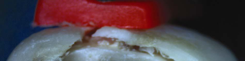
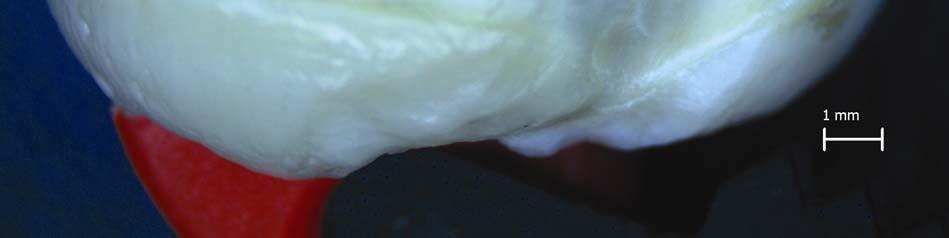
13 Fig. 4. Occlusal (A) and lateral (B) view of the extirpated spherical flattened tooth. 13

.")
.")
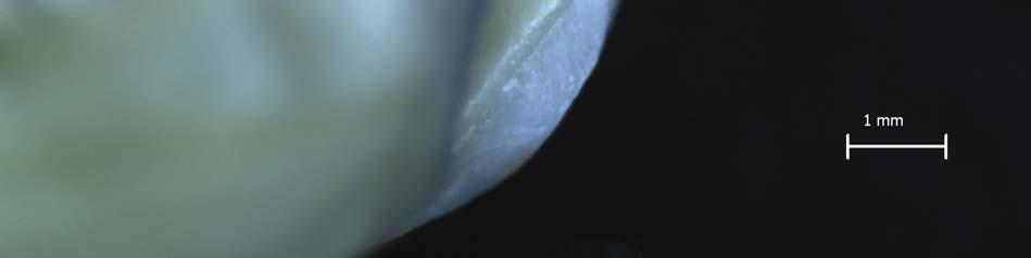
14 Fig. 5. Stereomicroscopic examination of the tooth. Central invagination of the occlusal surface delineated with irregular pitted enamel (A). Compressed pulp space between the inner and outer dentinal wall (B). Cementoenamel junction (C) with the invagination extending beyond it (D). 14

15 15


16 16


17 17

18 Fig. 6. The soft tissue surrounding the "impacted tooth-like mass" was composed of loose connective tissue containing capillaries and few islands of regular odontogenic epithelium, consistent with a dental follicle (hematoxylin and eosin stain, original magnification x 40). 18

19 Fig. 7. Tiny fragments taken from the occlusal lining of the mass were composed of amorphous, partly calcified eosinophilic material (hematoxylin and eosin stain, original magnification x 200). 19
Erupting Compound Odontome - A case report
 IOSR Journal of Dental and Medical Sciences (IOSR-JDMS) e-issn: 2279-0853, p-issn: 2279-0861.Volume 13, Issue 3 Ver. V. (Mar. 2014), PP 26-30 Erupting Compound Odontome - A case report Dr. Sahana Srinath
Surgical Retreatment of an Invaginated Maxillary Central Incisor Following Overfilled Endodontic Treatment: A Case Report
 Surgical Retreatment of an Invaginated Maxillary Central Incisor Following Overfilled Endodontic Treatment: A Case Report Hakan Ozbas a Rustem Kemal Subay b Melike Ordulu c ABSTRACT This case report presents
A Case Report of Odontogenic Keratocyst in Anterior Mandibule Position
 A Case Report of Odontogenic Keratocyst in Anterior Mandibule Position Malihe Moeini 1, Seyed Ehsan Anvar 2, Rasool Barzegari Bafghi 3* 1.Resident of Oral and Maxillofacial Radiology, Faculty of Dentistry,
SURGICAL MANAGEMENT OF A COMPLEX ODONTOMA - A CASE REPORT
 Indian J.Sci.Res. 6(2) : 157-161, 2015 Case Report ISSN : 0976-2876 (Print) ISSN : 2250-0138 (Online) SURGICAL MANAGEMENT OF A COMPLEX ODONTOMA - A CASE REPORT a 1b c d NAVDHA CHAUDHARY, BABITA AHLAWAT,
Management of a Dentigerous Cyst Associated with Inverted and Fused Mesiodens: A Rare Case Report
 Management of a Dentigerous Cyst Associated with Inverted and Fused Mesiodens: A Rare Case Report Kiran Patel 1, Nishtha Patel 2, Karthik Venkataraghavan 3 1 Sr. Lecturer, Department of Oral & Maxillofacial
A Rare Case of Radicular Dens in Dente diagnosed by Clinical, Radiographic, Tomographic, and Histopathological Examinations
 A Rare Case of Radicular Dens in Dente diagnosed by Clinical, Radiographic, 10.5005/jp-journals-10029-1152 Tomographic and Examinations CASE REPORT A Rare Case of Radicular Dens in Dente diagnosed by Clinical,
IMPACTED CANINE ASSOCIATED WITH COMPOUND ODONTOMA: A CASE REPORT
 Maxilo-facial surgery IMPACTED CANINE ASSOCIATED WITH COMPOUND ODONTOMA: A CASE REPORT Carmen Gabriela STELEA 1, Emilia DÎMBU 2, Alexandra Lorina STELEA 3, Alina BOTEZATU 4 1 Lecturer, Gr. T. Popa University
高雄醫學大學 口腔醫學院 口腔病理影像科 牙科 X 光影像判讀 教學範例
 高雄醫學大學 口腔醫學院 口腔病理影像科 牙科 X 光影像判讀 教學範例 Content Image No. 001 Dentigerous cyst over left upper embedded canine--------------------- 頁 1 Image No. 002---------------------------------------------------------------
A Radiographic technique for differentiating enamel and dentin in odontogenic tumors
 ISPUB.COM The Internet Journal of Radiology Volume 12 Number 1 A Radiographic technique for differentiating enamel and dentin in odontogenic tumors D Shetty, A Urs, R Kaur Citation D Shetty, A Urs, R Kaur.
Management of a Type III Dens Invaginatus using a Combination Surgical and Non-surgical Endodontic Therapy: A Case Report
 Management of a Type III Dens Invaginatus using a Combination Surgical and Non-surgical Endodontic Therapy: A Case Report Mithra N. Hegde, BDS, MDS, FPFA; Aditya Shetty, BDS, MDS; Rekha Sagar, BDS, MDS
Unusual transmigration of canines report of two cases in a family
 ISSN: Electronic version: 1984-5685 RSBO. 2014 Jan-Mar;11(1):88-92 Case Report Article Unusual transmigration of canines report of two cases in a family Sulabha A. Narsapur 1 Sameer Choudhari 2 Shrishal
Intraosseous Transmigration of Impacted Canines: Report of Five Cases Sulabha AN, Sachin Deshpande, Sameer C
 International Journal of Oral & Maxillofacial Pathology. 2012;3(3):56-60 ISSN 2231 2250 Available online at http://www.journalgateway.com or www.ijomp.org Case Report Intraosseous Transmigration of Impacted
Odontomes and Odontogenic tumours
 Odontomes and Odontogenic tumours Odontomes Developmental hamartoma Hamartoma: normal tissue in abnormal location Any cells to be neoplastic it must be able to replicate, which is not seen in hamartoma
The future of health is digital
 Dated: XX/XX/XXXX Name: XXXXXXXX XXXXXXXXXXX Birth Date: XX/XX/XXXX Date of scan: XX/XX/XXXX Examination of the anatomical volume: The following structures are reviewed and evaluated for bilateral symmetry,
Prevalence of root dilaceration in adult dental patients in Croatia
 Prevalence of root dilaceration in adult dental patients in Croatia Ana Malčić, DDS, a Silvana Jukić, DDS, PhD, b Valentina Brzović, DDS, a Ivana Miletić, DDS,PhD, b Ivica Pelivan, DDS, d and Ivica Anić,
Pericoronal radiolucency associated with incomplete crown
 Imaging Science in entistry 2013; 43: 295-301 http://dx.doi.org/10.5624/isd.2013.43.4.295 Pericoronal radiolucency associated with incomplete crown Kyung-Soo Nah 1, * 1 epartment of Oral and Maxillofacial
Removal of a Complex Odontoma Associated With an Impacted Third Molar
 Removal of a Complex Odontoma Associated With an Impacted Third Molar Authored by Mohammad Hosein Kalantar Motamedi, DDS Upon successful completion of this CE activity 1 CE credit hour may be awarded A
Incidental finding of dentigerous cyst - a case report
 Case Report Incidental finding of dentigerous cyst - a case report Pulivarthi Sushma 1, Sowbhagya M.B 2, Balaji P 3, Mahesh Kumar T.S 4 1 Postgraduate, 2 Reader, 3 Professor and Head of department, 4 Senior
Only 40% of the Story
 X-RAY, X-RAY, READ ALL ABOUT IT! The Use and Utility of Dental Radiographs in Practice Lisa Fink, DVM, DAVDC Dentistry & Oral Surgery Service October 4, 2015 Only 40% of the Story Radiographs of teeth
Disclosure. Educational Objectives. Terminology. Odontogenic Cysts. Terminology
 Disclosure Lisa J. Koenig BChD, DDS, MS Professor & Program Director, Oral Medicine and Oral Radiology Marquette University School of Dentistry Consultant to Soredex for the Scanora 3D and 3Dx Author/Editor
INFECTED DENTIGEROUS CYST IN IMPACTED CANINE- A case report
 Case Report INFECTED DENTIGEROUS CYST IN IMPACTED CANINE- A case report Gazala Fatima Parveen, M.D. Akheel 1 Department of oral & maxillofacial surgery, MCDRC Lucknow, U.P.,India 1-Department of Oral &
Clinical details: Details of scan: CONE BEAM CT REPORT: Name: H. B. Gender: Reason for referral: Referred by:
 Name: H. B. Gender: Male DOB: 11/12/1950 Age: 64 Date taken: 16/11/2015 Date reported: 19/11/2015 Clinical details: Reason for referral: Referred by: Investigate symptoms related to left TMJ. Reconstructed
Complex Odontoma in Both the Jaws: A Rare Case Report
 10.5005/jp-journals-10026-1013 Adit Srivastava et al CASE REPORT Complex Odontoma in Both the Jaws: A Rare Case Report Adit Srivastava, AG Annaji, Sanjay B Nyamati, Govind Singh, GC Shivakumar, S Sahana
Large Dentigerous Cyst
 Volume 16.2.1 Feb 2016 This Lecture Series qualifies for 0.5 Informal CPD Learning Hours Large Dentigerous Cyst By Dr Hassem Geha A 55 year-old male presented with a painless swelling in the right mandible.
AMELOBLASTIC FIBROMA: A RARE CASE REPORT
 Case Report International Journal of Dental and Health Sciences Volume 04, Issue 03 AMELOBLASTIC FIBROMA: A RARE CASE REPORT Namratha Patil 1 1.Sr lecturer, dept of oral medicine and radiology, KAHES VK
INFLAMMATORY DENTIGEROUS CYST OR INFLAMMATORY CYSTIC LESIONS OF MIXED DENTITION?: A REPORT OF THREE CASES
 Case Report International Journal of Dental and Health Sciences Volume 03, Issue 03 INFLAMMATORY DENTIGEROUS CYST OR INFLAMMATORY CYSTIC LESIONS OF MIXED DENTITION?: A REPORT OF THREE CASES Pritam K Mankapure
Interdisciplinary Treatment of a Fused Lower Premolar with Supernumerary Tooth
 Interdisciplinary Treatment of a Fused Lower Premolar with Supernumerary Tooth Cengiz Gadimli a Zafer Sari b Abstract The objective of this report is to describe combined orthodontic and endodontic treatment
PACIFIC JOURNAL OF MEDICAL SCIENCES ISSN:
 PACIFIC JOURNAL OF MEDICAL SCIENCES {Formerly: Medical Sciences Bulletin} ISSN: 2072 1625 Pac. J. Med. Sci. (PJMS) www.pacjmedsci.com. Email: pacjmedsci@gmail.com. ADENOMATOID ODONTOGENIC TUMOR WITH RARE
An unusual site of Adenomatoid Odontogenic Tumor: A rare case report
 J. Int Oral Health 2010 Case Report All right reserved An unusual site of Adenomatoid Odontogenic Tumor: A rare case report Sapna Panjwani*, Anjana Bagewadi**, Vaishali Keluskar*** *Post Graduate Student
Radicular cyst associated with a primary first molar: A case report
 Case Report Radicular cyst associated with a primary first molar: A case report L. Toomarian 1, M. Moshref 2, M. Mirkarimi 3, A. Lotfi 4, M. Beheshti 5 1 Associate Professor, Department of Pediatric Dentistry,
Buccal approach in surgical removal of lingually embedded teeth: a report of 2 cases
 Dental Journal Mahidol Dental Journal Case report Buccal approach in surgical removal of lingually embedded teeth: a report of 2 cases Thatsanai Tangmankongworakoon 1, Nattamet Wongsirichat 2 1 Lad Yao
Dental Morphology and Vocabulary
 Dental Morphology and Vocabulary Palate Palate Palate 1 2 Hard Palate Rugae Hard Palate Palate Palate Soft Palate Palate Palate Soft Palate 4 Palate Hard Palate Soft Palate Maxillary Arch (Maxilla) (Uppers)
Periapical central giant cell granuloma misdiagnosed as odontogenic cyst
 doi: 10.1111/j.1365-2591.2006.01107.x CLINICAL ARTICLE Periapical central giant cell granuloma misdiagnosed as odontogenic cyst T. Lombardi 1, M. Bischof 1,2, R. Nedir 1,2, D. Vergain 1, C. Galgano 3,
Erupted odontomas: a report of two unusual cases
 ISSN: Printed version: 1806-7727 Electronic version: 1984-5685 RSBO. 2012 Apr-Jun;9(2):199-203 Case Report Article Erupted odontomas: a report of two unusual cases Santosh Patil¹ Farzan Rahman² Shoaib
Adenomatoid odontogenic tumor associated with odontoma: a case report and critical review of the literature
 Gomez et al. Head & Face Medicine 2013, 9:20 HEAD & FACE MEDICINE CASE REPORT Adenomatoid odontogenic tumor associated with odontoma: a case report and critical review of the literature Open Access Ricardo
T O O T H A T L A S C O U R S E G U I D E A S S I S T A N T E D I T I O N
 T O O T H A T L A S C O U R S E G U I D E A S S I S T A N T E D I T I O N The information in this guide was prepared by ehuman with contributions from: Cara Miyasaki, RDHEF, MS, Foothill College Kay Murphy,
Hereditary Opalescent Dentin: A Report of Two Cases
 Hereditary Opalescent Dentin: A Report of Two Cases Siddharth Gupta, BDS, MDS; Rahul R. Bhowate, BDS, MDS; Ashok Bhati, BDS, MDS Abstract Aim: The aim of this case report is to present the clinical and
Tooth Variations. Suruedee Chinthakanan
 Tooth Variations Suruedee Chinthakanan Tooth variations Dental anomalies Cause : hereditary factor Developmental disturbances of teeth www.ectodermaldysplsia.org Tooth variations Enamel is formed from
IMPACTED CANINES. Unfortunately, this important tooth is the second most common tooth to be impacted after third molars
 IMPACTED CANINES After we talked about impacted third molars, today we ll discuss about maxillary impacted canines in upper dental arch, how to manage these cases as a dental surgeon. You will study about
Coexistence of two talon cusps and two dens invaginatus in a single tooth with associated radicular cyst-a case report and review of literature
 Journal section: Oral Medicine and Pathology Publication Types: Case Report doi:10.4317/jced.51421 http://dx.doi.org/10.4317/jced. 51421 Coexistence of two talon cusps and two dens invaginatus in a single
Frequency of Developmental Dental Anomalies in the Indian Population
 Frequency of Developmental Dental Anomalies in the Indian Population Kruthika S Guttal a Venkatesh G Naikmasur b Puneet Bhargava c Renuka J Bathi d ABSTRACT Objectives: To evaluate the frequency of developmental
Maxilla and mandible benign lesions: Radiologic Findings and Differential Diagnosis in CT
 Maxilla and mandible benign lesions: Radiologic Findings and Differential Diagnosis in CT Poster No.: C-0964 Congress: ECR 2012 Type: Scientific Exhibit Authors: N. Lopez 1, E. Marcos Naranjo 2, M. D.
INDIANA HEALTH COVERAGE PROGRAMS
 INDIANA HEALTH COVERAGE PROGRAMS PROVIDER CODE TABLES Note: Due to possible changes in Indiana Health Coverage Programs (IHCP) policy or national coding updates, inclusion of a code on the code tables
Common/Important Radiolucencies. B. Most Common Location Apex of permanent first molar, rare in primary teeth.
 Cincinnati Dental Association Breakfast at Tiffany s: The Jewels and Gems of Oral Pathology November 17, 2010 John A. Svirsky, DDS, MEd Virginia Commonwealth University 804-828-0547 FAX: 804-828-6234 EMAIL:
CASE REPORT A Compound Composite Odontoma Associated with Unerupted Permanent Incisor: A Case Report
 Usha Mohan Das et al CASE REPORT A Compound Composite Odontoma Associated with Unerupted Permanent Incisor: A Case Report 1 Usha Mohan Das, 2 Deepak Viswanath, 3 Umme Azher 1 Principal, Professor and Head,
Radiology. & supporting structures. Lec. 14 Common diseases of teeth Dr. Areej
 Radiology Lec. 14 Common diseases of teeth Dr. Areej & supporting structures A radiograph is only one part of the diagnostic process. Usually one does NOT make a diagnosis solely from a radiograph. A diagnosis
Development of teeth. 5.DM - Pedo
 Development of teeth 5.DM - Pedo Tooth development process of continuous changes in predetermined order starts from dental lamina A band of ectodermal cells growing from the epithelium of the embryonic
Successful Conservative Surgical Treatment of Ameloblastic Fibroma in the Posterior Maxilla : A Case Report
 http://dx.doi.org/10.5933/jkapd.2013.40.4.321 ISSN (print) 1226-8496 Successful Conservative Surgical Treatment of Ameloblastic Fibroma in the Posterior Maxilla : A Case Report Youngeun Lee 1, Hyojung
Periodontal Disease. Radiology of Periodontal Disease. Periodontal Disease. The Role of Radiology in Assessment of Periodontal Disease
 Radiology of Periodontal Disease Steven R. Singer, DDS srs2@columbia.edu 212.305.5674 Periodontal Disease! Includes several disorders of the periodontium! Gingivitis! Marginal Periodontitis! Localized
Unusually large erupted complex odontoma: A rare case report
 Imaging Science in Dentistry 2015; 45: 49-54 http://dx.doi.org/10.5624/isd.2015.45.1.49 Unusually large erupted complex odontoma: A rare case report Shivanand B. Bagewadi 1, *, Rahul Kukreja 1, Gundareddy
Key words: Third molar, Impacted tooth, Tooth Eruption, Molar, Mandible, Unerupted Tooth.
 JOURNAL OF CASE REPORTS 2014;4(2):286-290 OPG and CBCT Finding s of an Ectopic Third Molar in the Sub-condylar Region Tatu Joy E 1, Farakath Khan 1, Shameel Mohammed 2 From the Department of Oral Medicine
Root Canal Treatment of an Immature Dens Invaginatus With Apical Periodontitis: A Case Report
 JDC case report Root Canal Treatment of an Immature Dens Invaginatus With Apical Periodontitis: A Case Report Francisco Wanderley Garcia Paula-Silva, DDS, MSc, PhD Cristiane Tomaz Rocha, DDS, MSc Daniel
Evidence-based decision-making in endodontics
 Clin Dent Rev (2017) 1:6 https://doi.org/10.1007/s41894-017-0006-0 TREATMENT Evidence-based decision-making in endodontics Eyal Rosen 1 Igor Tsesis 1 Received: 15 June 2017 / Accepted: 9 July 2017 / Published
Oral Embryology and Histology
 Oral Embryology and Histology Chapter 8 Copyright 2018, Elsevier Inc. All Rights Reserved. 1 Learning Objectives Lesson 8.1: Oral Embryology 1. Pronounce, define, and spell the key terms. 2. Define embryology
Impeded Eruption of Mandibular Canine
 10.5005/jp-journals-10024-1434 Case Report Gauri S Lele, Darshan Modi Abstract Odontome, tumor of odontogenic origin, is associated with disturbances in the eruption of teeth such as impaction, delayed
 www.oralradiologists.com CONE BEAM CT REPORT CASE XXXX Patient information Patient Name: - Referring Doctor: - Patient DOB: - Scan Date: [Start date] Reason for Exam: Maxillary facial pain Doctor Notes:
Ameloblastic fibro-odontoma in a 9-year-old boy
 Okui et al. Stomatological Dis Sci 2018;2:7 DOI: 10.20517/2573-0002.2017.23 Stomatological Disease and Science Case Report Open Access Ameloblastic fibro-odontoma in a 9-year-old boy Tatsuo Okui 1, Soichiro
Intrabony Migration of Impacted Teeth
 Clinical Report Intrabony Migration of Impacted Teeth Yehoshua Shapira, DMD a ; Mladen M. Kuftinec, DMD, DStom, ScD b Abstract: Intrabony migration of impacted teeth is a rare dental anomaly, which occurs
JOURNAL TAOMFR Mandiblular Ameloblastic fibro-odontoma
 Ameloblastic Fibro-Odontoma of the Mandible with Emphasis on Evaluation Using Cone Beam Computed Tomography Jing-Yi Chen 1, Frank Lei 2, Yu-Fong Chen 3,4 * Divisions of 1 Oral Pathology, 2 Periodotology,
Surgical Uprighting Is a Successful Procedure for Management of Impacted Mandibular Second Molars
 DENTOALVEOLAR SURGERY Surgical Uprighting Is a Successful Procedure for Management of Impacted Mandibular Second Molars Bonnie L. Padwa, DMD, MD,* Rushil R. Dang, BDS, DMD,y and Cory M. Resnick, DMD, MDz
Double Teeth: A challenge for dentists
 Double Teeth: A challenge for dentists Neeraja.R 1, Umapathy 2 Corresponding Author Dr.Neeraja.R No 73, 7 th cross Cambridge layout Bangalore-8 Karnataka ph no- 8197919680 Email id: neeraja_pedo@yahoo.com
Evaluation of Cone Beam Computed Tomography in Diagnosis and Treatment Plan of Impacted Maxillary Canines
 Original Research Evaluation of Cone Beam Computed Tomography in Diagnosis and Treatment Plan of Impacted Maxillary Canines Seyed Hossein Hoseini Zarch 1, Farzin Heravi 2, Adineh Javadian Langaroodi 3,
The process of tooth eruption is very complex, and
 Clinical Section Eruption of a Severely Displaced Second Permanent Molar Following Surgical Removal of an Odontoma Ari Kupietzky, DMD, MSc Catherine M. Flaitz, DDS, MS Rephael Zeltser, DMD, DipOdont Dr.
A case of apical fenestration misdi Title persistent apical periodontitis. Author(s) Furusawa, M; Hayakawa, H; Ida, A; I
 Furusawa, M; Hayakawa, H; Ida, A; I") A case of apical fenestration misdi Title persistent apical periodontitis. Author(s) Furusawa, M; Hayakawa, H; Ida, A; I Journal Bulletin of Tokyo Dental College, 5 URL http://hdl.handle.net/10130/2705
A case of apical fenestration misdi Title persistent apical periodontitis. Author(s) Furusawa, M; Hayakawa, H; Ida, A; I Journal Bulletin of Tokyo Dental College, 5 URL http://hdl.handle.net/10130/2705
Shell teeth m management from the mixed to the permanent dentition: case report Rosamund Harrison, DMD, MS, MRCD(C) CASE REPORTS.
 CASE REPORTS.") Shell teeth m management from the mixed to the permanent dentition: case report Rosamund Harrison, DMD, MS, MRCD(C) David Kennedy, BDS, MSD, FRCD(C) CASE REPORTS Introduction Relatively few cases of shell
Shell teeth m management from the mixed to the permanent dentition: case report Rosamund Harrison, DMD, MS, MRCD(C) David Kennedy, BDS, MSD, FRCD(C) CASE REPORTS Introduction Relatively few cases of shell
Peripheral Odontogenic Fibroma: A rare case report
 Annals of Dental Research (2013) Vol 3 (1): 10-14 HATAM Publishers: All Rights Reserved Annals of Dental Research www.hgpub.com www.adres.yolasite.com Case Report Peripheral Odontogenic Fibroma: A rare
Non-surgical endodontic treatment f invaginatus type III using cone bea Title tomography and dental operating mic report.
 Non-surgical endodontic treatment f invaginatus type III using cone bea Title tomography and dental operating mic report. Author(s) Kato, H. Journal Bulletin of Tokyo Dental College, 5 URL http://hdl.handle.net/10130/3722
Lec. 11 & 12 Dr. Ali H. Murad Dental pulp 1- Coronal pulp
 Lec. 11 & 12 Dr. Ali H. Murad Dental pulp Is the soft connective tissue located in the central portion of each tooth. All pulps have similar morphologic characteristic, such as a soft, gelatinous consistency
Case Report Endodontic Treatment of Fused Teeth with Talon Cusp
 Case Reports in Dentistry, Article ID 738185, 4 pages http://dx.doi.org/10.1155/2014/738185 Case Report Endodontic Treatment of Fused Teeth with Talon Cusp Shima Sadat Miri, 1 Hakimeh Ghorbani, 2 and Anousheh
International Journal of Scientific Research and Innovative Technology ISSN: Vol. 4 No. 4; April 2017
 Frequency of Lesions Associated with Impacted Teeth in Patients Referring to the Department of Oral Pathology, Tabriz Faculty of Dentistry from 2004 to 2013 and Its Relationship with Age, Gender, Location
[ 06-10] Dr. B. Siva Reddy, Dr. B. Ajay Reginald, Dr. D. Sireesha, Dr. Meda Samatha India Abstract: Keywords ARTICLE 20/07/ /09/2018
![[ 06-10] Dr. B. Siva Reddy, Dr. B. Ajay Reginald, Dr. D. Sireesha, Dr. Meda Samatha India Abstract: Keywords ARTICLE 20/07/ /09/2018](/thumbs/87/95905171.jpg "[ 06-10] Dr. B. Siva Reddy, Dr. B. Ajay Reginald, Dr. D. Sireesha, Dr. Meda Samatha India Abstract: Keywords ARTICLE 20/07/ /09/2018") Dentigerous Cyst Associated with an Impacted Mesiodens: A Rare Case Report with Review of Literature [PP: 06-10] Dr. B. Siva Reddy, Dr. B. Ajay Reginald, Dr. D. Sireesha, Dr. Meda Samatha, Department of
Dentigerous Cyst Associated with an Impacted Mesiodens: A Rare Case Report with Review of Literature [PP: 06-10] Dr. B. Siva Reddy, Dr. B. Ajay Reginald, Dr. D. Sireesha, Dr. Meda Samatha, Department of
Autologous Bone Augmentation in Combination with an Ameloblastoma in the Maxillary Region- A Case Report?
 Archives of Clinical and Medical Case Reports doi: 10.26502/acmcr.96550023 Volume 2, Issue 2 Case Report Autologous Bone Augmentation in Combination with an Ameloblastoma in the Maxillary Region- A Case
IN THE NAME OF GOD. Dr.kheirandish DDS,MSC Oral and maxillofacial pathology
 IN THE NAME OF GOD Dr.kheirandish DDS,MSC Oral and maxillofacial pathology ODONTOGENIC CYSTS AND TUMORS Chapter 15 I. DENTIGEROUS CYST II. III. IV. ERUPTION CYST ODONTOGENIC KERATOCYST Orthokeratinized
Complex Odontome: A Review & Case Report Saxena Vasu Siddhartha 1, Karjodkar Freny 2
 Complex Odontome: A Review & Saxena Vasu Siddhartha 1, Karjodkar Freny 2 1 Senior Lecturer, 2 Professor & Head 1 Department of Oral Medicine and Radiology, Career Post Graduate Institute of Dental Sciences
DENTAL RADIOGRAPH INTERPRETATION
 DENTAL RADIOGRAPH INTERPRETATION Brook A. Niemiec, DVM Diplomate, American Veterinary Dental College Fellow, Academy of Veterinary Dentistry www.vetdentaltraning.com www.vetdentalrad.com Interpreting dental
FRACTURES AND LUXATIONS OF PERMANENT TEETH
 FRACTURES AND LUXATIONS OF PERMANENT TEETH 1. Treatment guidelines and alveolar bone Followup Procedures INFRACTION Clinical findings Radiographic findings Treatment Follow-Up Favorable Outcome Unfavorable
Supernumerary Fourth and Fifth Molars: A Report of Two Cases
 Supernumerary Fourth and Fifth Molars: A Report of Two Cases Abstract Panoramic radiographs of two female patients ages 22 and 21 revealed the presence of two impacted bilateral upper fourth molars and
CHAPTER 3 - DEFINITION, SCOPE, AND INDICATIONS FOR ENDODONTIC THERAPY ARNALDO CASTELLUCCI
 Contents Volume I CHAPTER 1 - A BRIEF HISTORY OF ENDODONTICS CHAPTER 2 - EMBRYOLOGY Crown formation Root formation Single- and multiple-root formation The formation of lateral canals Exposed dentin and
Glandular Odontogenic Cyst Coexisting with a Dentigerous Cyst: Case Report
 SmyrnaMed Case 2018;2(1): 1-5 ISSN (Online): 2564-6869 www.smyrnamed.com Glandular Odontogenic Cyst Coexisting with a Dentigerous Cyst: Case Report Assist.Prof.Dr. Serap Keskin Tunç 1, Dt. Erkan Feslihan
Multidisciplinary management of bilateral. symmetrical double maxillary central incisors
 Multidisciplinary management of bilateral symmetrical double maxillary central incisors Othman M. Yassin DDS/ MSc* * Consultant in Pediatric Dentistry, King Hussein Medical Center, Royal Medical Services,
Newport News Public Schools Summary Schedule of Services Delta Dental PPO EPO Plan
 Newport News Public Schools Summary of Services Delta Dental PPO EPO Plan Services In-Network Out-of-Network PPO Premier All Other Diagnostic & Preventive Oral Exams & Teeth Cleanings Fluoride Applications
Prevalence and Pattern of Congenital Missing Teeth in a Group of Iranian Adolescents
 Prevalence and Pattern of Congenital Missing Teeth in a Group of Iranian Adolescents Ajami B. a, Shabzendedar M. a*, Afzal Agaee M. b, Mehrjerdian M. c a Dept. of Pediatric Dentistry, Faculty of Dentistry,
RETIREE DENTAL PLAN. RETIREE DENTAL PLAN FEE SCHEDULE Page 1 of 8
 D0120 periodic oral evaluation $ 30.50 D0140 limited oral evaluation problem focused $ 30.50 D0150 comprehensive oral evaluation - new or established patient $ 30.50 D0160 detailed and extensive oral evaluation
Dentigerous cyst associated with an impacted mesiodens: report of 2 cases
 Imaging Science in Dentistry 2012; 42 : 255-60 http://dx.doi.org/10.5624/isd.2012.42.4.255 Dentigerous cyst associated with an impacted mesiodens: report of 2 cases Neha Khambete, Rahul Kumar*, Mukund
Fundamental & Preventive Curvatures of Teeth and Tooth Development. Lecture Three Chapter 15 Continued; Chapter 6 (parts) Dr. Margaret L.
 Dr. Margaret L.") Fundamental & Preventive Curvatures of Teeth and Tooth Development Lecture Three Chapter 15 Continued; Chapter 6 (parts) Dr. Margaret L. Dennis Proximal contact areas Contact areas are on the mesial and
Fundamental & Preventive Curvatures of Teeth and Tooth Development Lecture Three Chapter 15 Continued; Chapter 6 (parts) Dr. Margaret L. Dennis Proximal contact areas Contact areas are on the mesial and
The influence of sensor size and orientation on image quality in intra-oral periapical radiography
 Clinical The influence of sensor size and orientation on image quality in intra-oral periapical radiography Tony Druttman 1 The periapical view is one of the standard intra-oral radiographs by which diagnostic
Inter-radicular Radiolucencies
 Inter-radicular Radiolucencies Differential Diagnosis Laterally Displaced Radicular Cyst Accessory canals Root fracture Lateral Periodontal Cyst Botryoid variant Odontogenic Keratocyst Incisive Canal Cyst
Dr.Sepideh Falah-kooshki
 Dr.Sepideh Falah-kooshki MAXILLA Premaxillary/median palatal suture (radiolucent). Incisive fossa and foramen (radiolucent). Nasal passages (radiolucent). Nasal septum (radiopaque). Anterior nasal spine
A Case of Complex Odontoma Assosiated with Supplemental Supernumerary Teeth
 Annals of Dental Research (2011) Vol 1 (1): 67-72 Mind Reader Publications: All Rights Reserved Annals of Dental Research www.adres.yolasite.com www.journalshub.com Case Report A Case of Complex Odontoma
SQUAMOUS ODONTOGENIC TUMOUR: REPORT OF FIVE CASES FROM NIGERIA AND REVIEW OF LITERATURE
 African Journal of Oral Health Volume 3 Numbers 1&2, 2006:1-5 REFEREED ARTICLE SQUAMOUS ODONTOGENIC TUMOUR: REPORT OF FIVE CASES FROM NIGERIA AND REVIEW OF LITERATURE Adebiyi K.E., Odukoya O., Taiwo, E.O.
 This article was published in an Elsevier journal. The attached copy is furnished to the author for non-commercial research and education use, including for instruction at the author s institution, sharing
Periapical Radiography
 Periapical Radiography BARBARA E. DIXON B.D.S., M.Sc., D.P.D.S. Main Indications Detection of Apical infection/inflammation Assessment of the periodontal status After trauma Assessment of Unerupted teeth
Cone Beam Computed Tomography Findings in Calcifying Cystic Odontogenic Tumor Associated with Odontome: A Case Report
 Phulambrikar T., et al. Dent Shiraz Univ Med Sci., December 2015; 16(4): 374-379. Case Report Cone Beam Computed Tomography Findings in Calcifying Cystic Odontogenic Tumor Associated with Odontome: A Case
Fee Schedule Detail Procedure Procedure Description Code Fee
 Fee Schedule Detail Procedure Procedure Description Code Fee D0120 PERIODIC ORAL EVALUATION - ESTABLISHED PATIENT $ 32.29 D0140 LIMITED ORAL EVALUATION-PROBLEM FOCUSED $ 53.02 D0150 COMPREHENSIVE ORAL
Employee Benefit Fund July 2018 ADA Codes and Plan Fees
 CSEA Employee Benefit Fund July 2018 ADA Codes and Plan Fees DIAGNOSTIC D0120 periodic oral examination 40 34 42 45 48 38 30 32 31 D0140 limited oral examination (Does not look at 9110) 40 34 42 45 48
Human, Child (7 years +/- 2 years)
") Human, Child (7 years +/- 2 years) Product Number: Specimen Evaluated: Skeletal Inventory: BC-276 Natural bone specimen One panoramic radiograph (Panorex) 1 intact cranium 1 intact mandible General observations:
Human, Child (7 years +/- 2 years) Product Number: Specimen Evaluated: Skeletal Inventory: BC-276 Natural bone specimen One panoramic radiograph (Panorex) 1 intact cranium 1 intact mandible General observations:
Ghost Teeth: A Rare Developmental Anomaly of the Dental Tissues
 Science Journal of Medicine and Clinical Trials ISSN: 2276-7487 http://www.sjpub.org Author(s) 2014. CC Attribution 3.0 License. Published By Science Journal Publication International Open Access Publisher
Radiographic Assessment of the Incidence of Supernumerary teeth in a population of 1683 patients (Preliminary study)
") Radiographic Assessment of the Incidence of Supernumerary teeth in a population of 1683 patients (Preliminary study) Saleh I. Hijawi *, Ziyad K.M. Mohammad 1 Conservative and Prosthodontic Department,
Radiographic Assessment of the Incidence of Supernumerary teeth in a population of 1683 patients (Preliminary study) Saleh I. Hijawi *, Ziyad K.M. Mohammad 1 Conservative and Prosthodontic Department,
Dental Anatomy High Yield Notes. **Atleast 35 questions comes from these areas of old lectures**
 Dental Anatomy High Yield Notes **Atleast 35 questions comes from these areas of old lectures** This review notes compiled and prepared by my sister for her own study, as a last day review session for
Formation of supernumerary tooth one year after enucleation of adjacent dentigerous cyst in a 9-year-old boy
 Iranian Journal of Orthodontics Shirani, Arshad, Asefi 43 Formation of supernumerary tooth one year after enucleation of adjacent dentigerous cyst in a 9-year-old boy Gholamreza Shirani a, Mahnaz Arshad