Prospective, comparative assessment of alveolar ridge preservation using Guidor Easy-Graft Classic in atrumatic extraction socket
|
|
|
- Rosemary Haynes
- 6 years ago
- Views:
Transcription


1 University of Iowa Iowa Research Online Theses and Dissertations Summer 2017 Prospective, comparative assessment of alveolar ridge preservation using Guidor Easy-Graft Classic in atrumatic extraction socket Kelsey S. Tengan University of Iowa Copyright 2017 Kelsey S. Tengan This thesis is available at Iowa Research Online: Recommended Citation Tengan, Kelsey S.. "Prospective, comparative assessment of alveolar ridge preservation using Guidor Easy-Graft Classic in atrumatic extraction socket." MS (Master of Science) thesis, University of Iowa, Follow this and additional works at: Part of the Oral Biology and Oral Pathology Commons
2 PROSPECTIVE, COMPARATIVE ASSESSMENT OF ALVEOLAR RIDGE PRESERVATION USING GUIDOR EASY-GRAFT CLASSIC IN ATRUMATIC EXTRACTION SOCKET by Kelsey S. Tengan A thesis submitted in partial fulfillment of the requirements for the Master of Science degree in Oral Science in the Graduate College of The University of Iowa August 2017 Thesis Supervisor: Associate Professor Gustavo Avila-Ortiz
3 Copyright by KELSEY S. TENGAN 2017 All Rights Reserved
4 Graduate College The University of Iowa Iowa City, Iowa CERTIFICATE OF APPROVAL MASTER S THESIS This is to certify that the Master s thesis of Kelsey S. Tengan has been approved by the Examining Committee for the thesis requirement for the Master of Science degree in Oral Science at the August 2017 graduation. Thesis Committee: Gustavo Avila-Ortiz, Thesis Supervisor Georgia K. Johnson Veeratrishul Allareddy Christopher A. Barwacz
5 ACKNOWLEDGMENTS I would first like to thank Dr. Gustavo Avila-Ortiz for being a great mentor and for providing me with the opportunity to grow in our specialty. His clinical and didactic knowledge base has been a great resource for me over the last 3 years. Much more than that, he has been a source of continuous support and encouragement. It has truly been a pleasure working with Dr. Avila-Ortiz and his friendship is one I hope to have forever. I would also like to thank Dr. Georgia Johnson and the Department of Periodontics at the University of Iowa College of Dentistry and Dental Clinics for helping make this project possible. Thanks go to Dr. Veeratrishul Allareddy and the Department of Oral Pathology, Radiology and Medicine for all their help with the CBCT scans and Dr. Chris Barwacz for his mentorship and friendship throughout this process. I would like to also thank the Periodontics residents, past and present, for their support throughout this process. A huge debt of gratitude to Dr. Miguel Romero-Bustillos for not only his help with the statistical analysis, but his friendship over the last three years. And thank you to Dr. Christopher Hutton and Dr. Steven Clark for helping me get through the rough and tough times. There were numerous people who, without, this project would not have been possible. This includes Rick Barwacz for his meticulousness in keeping our study streamlined. Not enough can be said about Amber Marolf. This project would not be possible without her organization and her tireless patient recruitment. The department assistants and staff, Sheena Schumacher, Emily Meyer, Kelsey Fisk, and Terri Shay, should be recognized for not only assuring the cases ran smoothly, but also their assistance in everyday matters. Finally, and most importantly, I would like to thank my fiancé Nicole Del Carpio for her constant support and encouragement throughout this entire project and in my life. ii
6 ABSTRACT Objectives: Tooth extraction initiates a cascade of biological events leading to the reduction of alveolar ridge volume. Alveolar ridge preservation (ARP) is a surgical treatment which aims at minimizing hard and soft tissue changes following tooth extraction. Several techniques and materials have been studied and used clinically in ARP. The selection of the biomaterials used for this technique is determined by several factors, such as features of the extraction site, inherent biomaterial properties and handling preferences by the surgeon, among others. The purpose of this study was to evaluate the efficacy of alveolar ridge preservation via the application of Easy-graft CLASSIC (Sunstar Americas Inc.), an alloplastic bone substitute with unique handling features, following flapless posterior single tooth extraction compared to a particulate freeze-dried bone allograft (FDBA) covered with a collagen wound dressing, which has been advocated as a predictable treatment modality. The primary outcome in this study was bone volumetric reduction of the alveolar ridge assessed using cone beam computed tomography (CBCT) scans obtained at baseline and 16 weeks after tooth extraction and ARP. Methods: This study is part of a multicenter study in collaboration with the University of Maryland School of Dentistry. Seventeen healthy adults treatment planned for a single tooth implants in the area posterior to the canines, excluding third molars, were recruited on the basis of an eligibility criteria. Patients were randomly assigned to the control group or the experimental group. Minimally traumatic extraction of the tooth was completed and the presence of an intact buccal plate of bone was verified. The control group received FDBA and the site was stabilized with a collagen wound dressing and sutures. The experimental group received Easy-graft CLASSIC with no attempt to approximate the marginal mucosa. Healing was assessed after 1, 2, 4, 8, and 16 weeks. CBCT scans were made at baseline prior to treatment and 16 weeks postoperative; DICOM data from the CBCT volume was imported into Anatomage (Invivo, CA) and was used to assess the alveolar ridge volume and linear changes. Clinical measurements of the buccal gingival thickness, buccal alveolar bone thickness, keratinized gingiva, and socket dimensions were made at the time iii
7 of the extraction and were subsequently analyzed for possible influences on the observed volumetric and linear outcomes. Results: The mean alveolar ridge volume reduction from baseline to 16 weeks post operatively for the control and the experimental group was mm 3 and mm 3, respectively. These values correspond to a reduction of 9.59% for the control group and 13.04% for the experimental group. This difference did not reach statistical significance. The average loss of ridge width was 1.10mm for the FDBA and 1.24mm for the Easy-graft CLASSIC with no statically significant differences between the two groups. The average loss of buccal bone height and lingual bone height in the FDBA group was 1.12mm and 0.63mm, respectively. Similarly, the average loss of buccal bone height and lingual bone height in the Easy-graft CLASSIC was 1.19mm and 0.67mm, respectively. There was a weak to moderate positive correlation between buccal tissue thickness and the thickness of the buccal bone and a weak negative correlation between buccal bone thickness and alveolar ridge width reduction. Conclusions: Within the limitations of this study, both treatment groups appear to be effective in alveolar ridge preservation and are associated with similar volumetric and linear bone reduction patterns. iv
8 PUBLIC ABSTRACT Following tooth extraction, the bone supporting the tooth resorbs to an extent. For tooth replacement with a dental implant, a sufficient amount of bone is needed to provide functional support and to aid in an esthetic outcome. To minimize the amount of bone loss after tooth extraction, bone grafting procedures have been developed and many different grafting materials and clinical protocols have been investigated. The use of sterilized and processed human cadaver bone with a collagen seal is generally considered one of the standard methods to graft the socket after tooth extraction. The use of this human-sourced grafting material is challenging to contain within the socket and may incur more cost due to the increased time and materials required. A novel bone substitute material with unique handling properties has been introduced to decrease the time required for grafting sockets. The purpose of this study was to evaluate the changes that take place at the extraction sites when treated with either a human-sourced bone graft with a collagen seal or the aforementioned bone substitute. The radiographic data and clinical measurements suggest that both graft types performed well with no significant statistical differences. In light of these results, the human-sourced bone graft and the novel bone substitute may be used interchangeably to help preserve bone for future implant placements. v
9 TABLE OF CONTENTS LIST OF FIGURES... viii LIST OF TABLES... ix CHAPTER 1. Introduction Periodontal Anatomy Periodontal Structures and Components Width of Keratinized Gingiva Periodontal Biotype Tooth Extraction Prevalence of Edentulism Indications for Tooth Extraction Tooth Extraction Wound Healing Alveolar Ridge Dimension Changes after Extraction Alveolar Ridge Preservation Rationale Tooth Extraction Wound Healing with Graft Efficacy of Alveolar Ridge Preservation Graft Sources Control Material Easy-Graft CLASSIC CHAPTER 1I. Hypothesis and Specific Aims CHAPTER III. Material and Methods Center and Ethical Approval Pre-screening, Clinical Screening and Enrollment Randomization Presurgical Measurements Surgical Intervention Post-operative Care Follow-up Visits and Assessments Dimensional Osseous Changes Statistical Analysis Sample Size Determination 20 CHAPTER IV. Results Subject Population Subject Characteristics Baseline Data Volumetric Changes Linear Ridge Width Changes Linear Ridge Height Changes Correlations 23 CHAPTER V. Discussion CHAPTER VI. Conclusions APPENDIX A Figures vi
10 APPENDIX B Tables APPENDIX C Additional Forms BIBLIOGRAPHY vii
11 LIST OF FIGURES Figure A1. Study Timeline Figure A2. Events Schedule Figure A3. Pre-Operative Radiographs Figure A4. Pre-Operative Clinical Presentation Figure A5. Clinical Measurement Figure A6. Intact Socket after Extraction Figure A7. Buccal Plate Thickness Measurement Figure A8. Graft Procedure Figure A9. 16-Week Follow Up Figure A Week Follow Up Radiographs Figure A11. Volumetric Analysis Figure A12. Ridge Width Changes Figure A13. Ridge Height Changes viii
12 LIST OF TABLES Table B1. Subject Characteristics Table B2. Extraction Sites Table B3. Baseline Clinical Measurements Table B4. Volumetric Data Table B5. Ridge Width Data Table B6. Buccal Ridge Height Data Table B7. Lingual Ridge Height Data Table B8. Correlation of Buccal Tissue Thickness and Buccal Bone Thickness Table B9. Correlation of Buccal Bone Thickness and Alveolar Ridge Volume Reduction Table B10. Correlation of Buccal Bone Thickness and Alveolar Ridge Reduction ix
13 CHAPTER 1. Introduction 1.1 Periodontal Anatomy Periodontal Structures and Components The periodontium functions to provide support to the teeth and anchor them to the alveolar bone. It is comprised of the gingiva, periodontal ligament, root cementum, bundle bone, and alveolar bone. The gingiva is part of the oral masticatory mucosa which covers the alveolar process and hard palate and terminates at the cervical region of the teeth. The oral masticatory mucosa is mostly keratinized tissue and is continuous apically with the non-keratinized oral lining mucosa. The junction between these different mucosal tissues is demarcated by the mucogingival junction. There is no mucogingival junction on the palatal aspect of the maxilla as the gingiva is continuous with the masticatory mucosa of the hard palate. The gingiva can be divided by anatomic features into the marginal or free gingiva, the interdental gingiva, and the attached gingiva. The marginal gingiva surrounds the cervical region of teeth and terminates coronally at the free gingival margin. The free gingival margin helps to form the sulcus, a V-shaped, shallow crevice between the tooth and soft tissues. In health, the clinical measurement of the gingival sulcus depth is less than 3mm. Histologic data from cadaver studies indicates the depth of the sulcus to be about 0.69mm (Gargiulo et al., 1961), though this value varies between individuals and between sites in the same individual. The marginal gingiva is not attached to the tooth root and is demarcated by the free gingival groove, a small depression on the surface of the facial and lingual gingiva aligning with the tooth s cementoenamel junction. Apically, the marginal gingiva is continuous with the attached gingiva. The marginal gingiva can be further separated into the oral epithelium, which faces away from the tooth and continues as part of the attached gingiva, the sulcular epithelium, which lines the surface directly adjacent to the tooth enamel but not adhered, and the junctional epithelium, which is apical to the sulcular epithelium and contacts the tooth surface via hemidesmosomes and forms the base of the sulcus. The sulcular 1
14 epithithelium is non-keratinized, however, it has been shown to become keratinized in animal models with strict oral hygiene measures (Kristoffersen et al., 1983). The junctional epithelium is non-keratinized and is derived from the reduced enamel epithelium, forming during tooth eruption into the oral cavity. The junctional epithelium forms a physical barrier to protect against bacterial plaque and is continuously renewed through cell division at the basal layer. The cells migrate coronally until they are shed at the base of the gingival sulcus. The junctional epithelium also functions to secrete gingival crevicular fluid. The gingival crevicular fluid functions to cleanse material from the sulcus and has antimicrobial properties. The interdental gingiva is the non-keratinized tissue that is contained within the interproximal embrasure space created apical to the contact area between adjacent teeth. The morphology of the interdental gingiva is dependent upon the location and size of the contact area. Typically, in the anterior region of the dental arch where the contact areas are more coronal and narrow, the morphology of the interdental gingiva is pyramidal while the broader and flatter contacts in the posterior region form a col, or saddle-shaped interdental gingiva. The buccal and lingual gingival tissues converge through the interdental gingiva and form the papilla. The apical portion of the interdental gingiva is attached by connective tissue to the crestal bone of the alveolus. The marginal gingiva is continuous with the attached gingiva apically which, in turn, continues apically to the mucogingival junction. The attached gingiva is firmly fixed to the underlying periosteum of the alveolar bone and the root cementum by connective tissue fibers. Apical to and continuous with the attached gingiva, in all areas except the hard palate, is the oral lining mucosa. The oral lining mucosa is non-keratinized and mobile and lines the floor of the mouth, cheeks, soft palate, and the ventral surface of the tongue. The connective tissue, or lamina propria, is the predominant tissue component in the gingiva. It consists of collagen fibers, fibroblasts, vessels, nerves, and extracellular matrix. Type 1 collagen forms the majority of the lamina propria. It provides the tensile strength of the tissues. Type 4 collagen is present between the Type 1 collagen bundles and is continuous with the basement membrane. Subjacent to the junctional epithelium, collagen bundles are anchored to the cementum and form the connective tissue attachment. 2
15 Histological measurements of the connective tissue attachment to the tooth root in cadaver models reveal a relatively stable dimension of about 1mm in apico-coronal height. There are several distinct arrangements of the connective tissue fibers in the gingiva. They can be classified into the dentogingival fibers, dentoperiosteal fibers, alveologingival fibers, transeptal fibers, and circular fibers. The dentogingival fibers attach to the cementum of the supracrestal root cementum and fan out into the surrounding gingiva. The dentoperiosteal fibers attach at the same region on the root and traverse apically and attach to the periosteum of the bone crest adjacent to the tooth. The alveologingival fibers attach from the periosteum of the bone crest and fan out coronally into the connective tissue of the gingiva. The transeptal fibers traverse the interdental gingiva and link the supracrestal cementum of two adjacent teeth. The circular fibers do not attach to the root but encircle it in the free gingiva. The attachment apparatus coronal to the alveolar crest of bone adjacent to the tooth is termed the biologic width. It is a summation of the length of the epithelial attachment and the connective tissue attachment. Gargiulo et al. (1961) found the average dimensions of the epithelial attachment and connective tissue attachment were 0.97mm and 1.07mm, respectively. Vacek et al. (1994) found the average epithelial attachment to be 1.14mm and the average connective tissue attachment to be 0.77mm. Both of these studies reported the epithelial attachment had strong individual variations while the most stable dimension was the connective tissue. More recently, a systematic review and meta-analysis of biologic width reported a range of 2.15 to 2.30mm but cautioned that there was a significant variability between individuals and even between sites in the same individual (Schmidt et al., 2013). Following the attachment apically, the periodontal ligament is the connective tissue that forms between the root cementum and the alveolar bone. These fibers, termed Sharpey s fibers, terminate in the root cementum and the adjacent bundle bone. It is hourglass shaped, maintaining its smallest dimension at the mid-root level and occupying a space approximately 0.2 to 0.4mm in width. The periodontal ligament can adapt in response to forces applied to the tooth. In the presence of increased forces to the tooth, the width of the periodontal ligament increases and with disuse, it narrows. The periodontal ligament primarily serves a supportive 3
16 function by firmly attaching to the tooth to the surrounding alveolar bone but also plays a role in remodeling, sensation, and providing nutrients to supporting cells. The Sharpey s fibers are Type 1 collagen bundles that are organized along the tooth surface in orientations that provide resistance and retention of the tooth to occlusal forces. They can be classified by their location and direction of orientation. The most coronal Sharpey s fibers are the alveolar crest fibers that extend between the supracrestal cementum and the alveolar crestal bone. The horizontal group runs perpendicularly from the alveolar bone to the root surface. The oblique group forms the majority of the periodontal fibers and is oriented between the cementum and the bundle bone in a coronal to apical direction. These fibers act to resist compression forces. The apical fibers are those near the apices of the tooth root and provide resistance to tensile forces. In multi-rooted teeth, the radicular fiber group fans out from the bone beneath the furcation and attach to the cementum of the furcation. The alveolar process forms on basal bone of the body of the maxillae and mandible and houses the teeth. It develops in conjunction with tooth eruption and consists of the buccal and lingual cortical plates which form the outer surface, cancellous bone contained between them, and the alveolar bone proper, also known as bundle bone. The coronal termination of the bone around teeth in health is about 1 to 2mm apical to the tooth s cementoenamel junction. The buccal and lingual cortical plates are formed from compact lamellar bone that is covered by the periosteum, a dense fibrous connective tissue that contains osteoblasts, osteoclasts, pluripotential cells, fibroblasts, vessels, and nerves (Zuhr, 2012). The periosteum provides the blood supply to the outer surface of bone and is one of the main blood supplies to the gingival tissues. The bundle bone is a thin cortical plate of bone which lines the tooth socket. It is where connective tissue fibers from the periodontal ligament insert and is the site through which blood vessels from the marrow spaces in the cancellous bone that supplies the periodontal ligament traverse. 4
17 Width of Keratinized Gingiva The gingival tissue measured from the free gingival margin and terminating at the mucogingival junction is known as the keratinized gingiva width. This measurement is highly variable from patient to patient and from site to site within the same patient. The widest dimension of the keratinized gingiva width is found at the buccal of maxillary anterior teeth and the narrowest occurs at the lingual of the mandibular anterior teeth (Bowers, 1963). These dimensions may increase with age (Ainamo and Loe, 1966) and are also dependent upon the position of the tooth within the arch. Studies have suggested that there is a minimum width of keratinized gingiva that is required to sustain gingival health. In a study by Lang and Loe (1972), all sites that had less than 2 mm of keratinized gingiva had signs of gingival inflammation and varying levels of gingival exudate while a majority of sites with two or more millimeters of keratinized gingiva were clinically healthy. Contrasting evidence from Kennedy et al. (1985), demonstrated that, in the presence of good oral hygiene and control of gingival inflammation, periodontal health can be maintained without attached gingiva Periodontal Biotype The horizontal dimension of the gingival tissues is important in many clinical outcomes. Restoratively, sites with thin tissues are more prone to recession when compared to thicker tissues (Weisgold, 1977). Complete root coverage after mucogingival procedures tend to favor thicker tissues (Baldi et al., 1999, Huynh-Ba et al., 2010, Hwang and Wang, 2006). Esthetic outcomes after implant placement are also dependent upon the thickness of the periodontium (Kois, 2004, Lee et al., 2011). This bucco-lingual thickness is referred to as the gingival biotype and describes the dimensions and morphology of the marginal gingiva and the underlying alveolar bone. Ochsenbein and Ross (1969) found that the gingival anatomy mimics the underlying bone and described these gingival contours as scalloped or flat. The term periodontal biotype was used by Seibert JL (1989) who classified the gingiva into thin-scalloped or thick-flat. The thin, scalloped biotype is characterized by thin and fragile gingival tissue with a narrow band of keratinized gingiva. Thin 5
18 biotypes usually present with thin buccal alveolar bone with dehiscence or fenestrations and have triangular tooth forms with minimal cervical convexity and contact points near the incisal edges. Thick biotypes have a flat soft tissue contour with dense and fibrotic gingival tissues and a wider band of keratinized gingiva. Thick biotypes are associated with a thick buccal plate of alveolar bone and a squared tooth form with distinct cervical convexity and a broader contact area more apically located (Ochsenbein and Ross, 1973, Seibert JL, 1989, Weisgold, 1977). The gingival biotypes association with the thickness of the underlying bone has been investigated. From CBCT images, study casts, and clinical examinations, Cook et al. (2011) reported that there was a significant association between the gingival biotype and the buccal plate thickness such that thin biotypes had thin buccal bone. From a study on cadavers, Fu et al. (2010) found a moderate association between the gingival biotype and the thickness of the buccal bone. Frost et al. (2015) found an association of probe visibility through the buccal tissues with a buccal plate less than 0.21mm, however these findings were not statistically significant. In a study by Chappuis et al. (2015), the soft tissue thickness was similar between patients with either thick or thin buccal bone. The thickness of the soft tissues was about 0.8mm for each group. Compared to the thick buccal plate group, the loss of bone height and width was greater in the thin buccal plate group. Interestingly, the thin buccal plate group had a more robust soft tissue response after tooth extraction than the thick buccal plate group. The soft tissue response in the thin buccal plate group masked the large bony height lost following extraction. Ridge resorption following tooth extraction tends to be more pronounced when there is a thin or absent buccal bone. Therefore, the use of classifications of gingival biotypes is important during treatment planning. Many methods used for the classification of gingival biotypes have been proposed. Studies have looked at the use of direct measurements, probe transparency, ultrasonic devices, and CBCT images to determine the gingival biotype. From direct measurements using a periodontal probe penetration, Claffey and Shanley (1986) considered thin biotypes as a gingival thickness of <1.5mm and a thick biotype as tissues 2mm while others classified gingival tissues as thin when it was 1.5mm and thick if it exceeded 1.5mm. Direct 6
19 measurements with the use of periodontal probe has been shown to be accurate however this method can have errors due to limited ability to standardize angulation and apico-coronal position of the probe during penetration. Transparency classifications were dependent upon the visualization of the outline of the periodontal probe on the outer surface of the gingival margins such that thick gingival biotypes obscured the gray of the probe (Kan et al., 2003, De Rouck et al., 2009). This method of classification was determined to correlate with direct measurements (Kan et al., 2010) however there may be subjective differences when using this method. Ultrasonic devices have also been proposed to assess gingival thickness. This ultrasonic device used pluses transmitted through the mucosa that were reflected off the alveolar bone and had a resolution of 0.1mm (Muller et al., 2000). The reproducibility of the device was proven by Eger et al. (1996) however the difficulties in using the device, scarcity, and high costs have limited the use of this ultrasonic device (De Rouck et al., 2009). The use of CBCTs to measure the biotype has been explored (Cook et al., 2011, Fu et al., 2010) and has been shown to be an accurate method to measure both hard and soft tissues, however many times, the tooth root and the presence or absence of a buccal plate are difficult to distinguish. Though there are limitations and difficulties classifying gingival biotypes, the importance of the gingival thickness is clear Tooth Extraction Prevalence of Edentulism Tooth loss is a worldwide public health issue and has been shown to be associated with negative effects on general health. Tooth loss has been associated with effects on blood pressure (Peres et al., 2012), obesity (De Marchi et al., 2012), malnutrition (Mojon et al., 1999, Sheiham et al., 2001), and cardiovascular health (Polzer et al., 2012). Aside from negative health effects, there are also effects on the quality of life (Gerritsen et al., 2010), function (Naka et al., 2014), and esthetics (Gotfredsen and Walls, 2007). The prevalence of edentulism worldwide has been estimated to be between 7% and 69% of adults (Petersen et al., 2005). In the U.S. population between the ages of 65 and 74, the prevalence of edentulism is 26% (Felton et al., 2011), a 7
20 decline of about 6% since the late 1980s (Beltran-Aguilar et al., 2005). However, due to advances in medicine, patient life expectancy has increased and subsequently, the proportion of elderly patients will increase. This progressive increase in the elderly population may increase the prevalence of edentulism Indications for Tooth Extraction When teeth cannot be maintained in an acceptable condition of health or function, extraction is indicated. Common indications for extractions are if the tooth is deemed non-restorable after loss of tooth structure from caries or trauma, periodontal disease, periradicular infections, or for orthodontic or prosthodontic reasons. The two most common reasons for tooth extraction are periodontal disease and dental caries. Untreated caries in permanent teeth was present in about 35% of the global population, affecting about 2.4 billion people worldwide and peaked around the age of 25 (Kassebaum et al., 2015). For severe periodontitis, a recent study reported a prevalence of 10.8% of the global population, affecting about 743 million people aged between 15 and 99 (Frencken et al., 2017). The global prevalence is slightly higher compared to the 8.7% prevalence reported with data collected from the United States population by Eke et al. (2012) Tooth Extraction Wound Healing Following tooth extraction, the alveolar bone undergoes a remodeling process that results in a significant loss of alveolar ridge volume. This process begins at the time of the tooth extraction and data from canine model studies demonstrated that a blood clot forms within the socket and stabilizes the site within the first few hours. The blood clot consists of erythrocytes and leukocytes that are embedded in a fibrin network. There is a concomitant inflammatory reaction which stimulates the formation of granulation tissues, which is rich with newly formed vascular structures. Within 48 to 72 hours, the blood clot begins to degrade as the newly formed granulation tissues infiltrate the area. By 4 days, the epithelium from the wound edges proliferates along the socket periphery and immature connective tissue is apparent. By 1 to 2 weeks, the granulation tissue has replaced the blood clot entirely and the formation of osteoid, a bone precursor, is present at the base of the extraction socket. By 2 to 4 weeks, there is mineralization of the osteoid that forms 8
21 from the base of the socket and lateral walls toward the coronal aspect of the socket. The newly formed bone is classified as woven bone. This mineralization and maturation continues over time and ultimately forms dense lamellar bone, the principle load-bearing tissue. This complete maturation process takes over 3 to 6 months to complete (Amler et al., 1960, Cardaropoli et al., 2005) Alveolar Ridge Dimension Changes after Extraction A classic preclinical study using a canine model (Araujo and Lindhe, 2005) reported an intense resorption of the buccal plate within the first 8 weeks characterized by marked osteoclastic activity. They observed that there was more pronounced resorption at the crestal level of the facial wall when compared to the lingual wall. There was also a loss of ridge height on both bony walls, however the buccal wall crest was consistently more apical to the lingual wall crest. The pattern of resorption followed two overlapping phases: the resorption of the bundle bone with replacement with woven bone, and the resorption at the outer surfaces of both buccal and lingual walls. This distinct resorptive pattern is a result of a generally thinner buccal plate of alveolar bone compared to the lingual plate of alveolar bone. Often, the buccal plate is fused directly with the bundle bone directly adjacent to the tooth root. The blood supply to this bundle bone is severed after tooth extraction, leaves the buccal plate with less nutrient supply, and thus results in more significant resorption. The magnitude of the ridge dimension changes after tooth extraction in humans was explored by Schropp et al. (2003) with study casts. They observed that the ridge dimensions changed over the 1 year study period and that the ridge width had reduced by approximately 50% with the loss of ridge height to be less substantial. Though the changes occurred over the 1 year study period, approximately 2/3rds of the dimensional changes occurred within the first 3 months following tooth extraction. Systematic reviews of the data from alveolar ridge dimensional changes after extractions in humans report approximately 3.5mm of ridge width reduction and about 1.5mm of ridge height reduction following tooth extraction (Van der Weijden et al., 2009, Vignoletti et al., 2012). 9
22 One of the factors that may influence the healing of extraction sockets is the surgical technique used. The choice between a flapless tooth extraction and a flapped procedure may affect the alveolar ridge dimensions. After a full-thickness flap reflection, meaning elevation of the periosteum from the cortical plate of the alveolus, a loss of 0.6mm of crestal height was reported in humans (Wood et al., 1972). When comparing volumetric changes after tooth extraction, Fickl et al. (2008) found that, in a canine model, there was 0.5 to 0.7mm of additional volumetric shrinkage when utilizing flap elevation compared to flapless extractions after 4 months. Interestingly, a study by (Araujo and Lindhe, 2009) in a canine model, found no differences in volumetric changes after tooth extraction in a flapped or flapless manner after 6 months of healing Alveolar Ridge Preservation Rationale Dental implants have become an excellent long-term treatment option to replace missing teeth with survival rates of about 93% to 95% (Hjalmarsson et al., 2016, Moraschini and Barboza, 2016). Adequate ridge volume is an integral criterion for a successfully functioning and esthetically pleasing dental implant solution. A deficiency of the buccal bone following tooth extraction may make the placement of a dental implant unfeasible, by not allowing proper three dimensional positioning of the implant, which may also negatively impact the esthetics of the final restorative complex (Buser et al., 2004, Garber and Belser, 1995) Tooth Extraction Wound Healing with Graft Similar to non-grafted socket healing, within the first few hours, there is the establishment of the blood clot. This fibrin clot entraps the graft particles. The blood clot is replaced with granulation tissues over the following week. There is a recruitment of inflammatory cells such as polymorphonuclear cells to the area and are shortly followed by osteoclasts. These osteoclasts migrate to the surface of the graft particles and being to resorb the particles. After 1 to 2 weeks, there is a replacement of the osteoclasts with osteoblasts that begin to deposit osteoid tissues into the collagen that forms the lattice work for bone maturation. This healing and immature newly formed bone can be visualized at 2 weeks along the apical and lateral walls of 10
23 the grafted site. By 3 months, the presence of the graft did not affect the processes of bone modeling and remodeling (Cardaropoli et al., 2005). Though wound healing following tooth extraction has been well documented in both animal and human models, there is a high degree of variability between patients. The resorptive process can be affected by many different factors such as the presence of infection, loss of bone height due to previous periodontal disease, trauma to the site during extraction, tooth position, as well as, the presence of a dehiscence or fenestration following the procedure (Adriaens, 1999, Chen et al., 2004) Efficacy of Alveolar Ridge Preservation Recent systematic reviews have reported the efficacy of alveolar ridge preservation and the procedures ability to limit the resorption of the alveolar bone. Vignoletti et al. (2012) reported that sites that received a bone graft had less alveolar ridge height and width loss when compared to a tooth extraction alone. They reported that these sites lost 1.47mm less in height, and 1.83mm less in width and also found that there was a significant difference that favored a flapped approach rather than a flapless extraction. Vittorini Orgeas et al. (2013) reported that sites that were augmented had about 0.96mm of less ridge height reduction and 1.99mm of less ridge width reduction when compared to extraction sites alone. Similar results were reported by Avila-Ortiz et al. (2014a) such that the ridge preservation sites had about 2.07mm less buccal height reduction and 1.89mm less ridge width reduction when compared to extractions alone. The study also reported that the use of a flapped approach, use of a membrane, and use of a xenograft or an allograft was more beneficial. Overall, ARP techniques appear to be beneficial by limiting the magnitude of ridge dimension changes, but do not entirely prevent it Graft Sources There have been many different bone grafting materials used for alveolar ridge preservation. The most common sources are autogenous, allographic, xenographic, and alloplastic. These materials may be osteogenic, osteoinductive, and/or osteoconductive. Osteogenic grafts supply viable cells that may participate in the process of osteogenesis. Osteoinductive grafts stimulate the host s pluripotential 11
24 mesenchymal cells to differentiate into osteoblasts. Osteoconductive grafts act as a lattice for cell growth and permits the host s osteoblasts from the wound margins to infiltrate into the defect. Autogenous bone is harvested from another site in the same individual that requires the graft. Autogenous bone is an osteogenic bone graft because the graft contains bone forming cells within. Using an autogenous bone graft tends to increase surgical morbidity because it requires a secondary surgical site. Also, the amount of autogenous bone is limited. Allogenic bone grafts are sourced from human cadavers and may be osteoinductive and/or osteoconductive, depending on the processing of the graft. Allografts may be more costly to the patient than other graft types. Xenografts are sourced from non-human animals, most often from a bovine source. Both allografts and xenografts have had ethical and disease transmission concerns in the past however both are very effective bone grafting materials. Alloplastic bone grafting materials are synthetic. Alloplastic materials, such as hydroxyapatite, bioactive glass, beta-tricalcium phosphate, and others, have been used to eliminate the need of bone harvesting and risk of disease transmission, and decrease costs to the patient Control Material The allograft used in this study was a mineralized cortical freeze-dried particulate bone allograft (Lifenet Health, Virginia Beach, VA) with a particle size of µm. It is sterilized via Allowash XG technology, consisting of rigorous screening and assessment of donor tissues; cleaning with hypotonic solutions and antimicrobial reagents; decontamination, disinfection, and cleaning regimens with hydrogen peroxide and isopropanol alcohol solutions; and terminal sterilization with gamma irradiation. The collagen wound dressing is made from type 1 bovine collagen. Collagen Plug (Zimmer Dental Inc., Palm Beach Gardens, FL) is a biocompatible, sterile resorbable highly porous collagen wound dressing for bleeding control, stabilizes the blood clot, and protects the wound bed. It resorbs in days (Almazrooa et al., 2014). 12
25 Easy-Graft CLASSIC Easy-graft CLASSIC is a commercially available bioresorbable, alloplastic bone graft that consists of betatricalcium phosphate granules that have an extremely thin coating of poly(lactic-co-glycolic acid). When combined with the BioLinker, which consists of water and N-Methyl-2-pyrrolidone (NMP), a non-volatile solvent, the loose granules become a moldable mass. When the hydrated grafting material contacts blood or tissue fluids it hardens within a few minutes to form a stable porous scaffold. According to the manufacturer, this unique handling property allows the clinician to forgo the use of a collagen wound dressing seal with sutures, saving time and reducing costs to the patient. 13
26 CHAPTER 1I. Hypothesis and Specific Aims The purpose of this study was to evaluate two different treatment approaches using Easy-graft CLASSIC for comparative effectiveness to FDBA with collagen wound dressing in post extraction ridge preservation. It was hypothesized that the use of Easy-graft CLASSIC will lead to similar clinical outcomes as compared to the use of a mineralized cortical FDBA with a collagen wound dressing for alveolar ridge preservation after single tooth extraction. The primary objective was to assess the clinical efficacy in terms of horizontal and vertical linear, and volumetric osseous changes after single tooth extraction and the application of Easy-graft CLASSIC compared to conventional particulate FDBA with collagen wound dressing, via DICOM data analysis. 14
27 CHAPTER III. Material and Methods 3.1. Center and Ethical Approval The clinical component of this study was conducted in the Graduate Periodontics clinic at the University of Iowa College of Dentistry and Dental Clinics from August 2016 to April The University of Iowa Institutional Review Board approval was obtained in June 2016 (IRB approval # ). This study was registered at clinicaltrials.gov under code NCT This study is part of a multicenter study in collaboration with the University of Maryland School of Dentistry 3.2 Pre-screening, Clinical Screening and Enrollment Patients needing a single tooth extraction of maxillary or mandibular premolars and molars, excluding third molars were recruited. Subjects were pre-screened by a telephone interview. Individuals who met the initial eligibility criteria and were interested in participating in the study were scheduled for a clinical screening. The clinical screening consisted of an in-depth consultation and a clinical exam. The screening process included an evaluation of general systemic and dental health. Clinical and radiographic assessment of the study site and adjacent teeth was completed. If available, pre-existing intraoral radiographs not older than 6 months were used to assess the site, otherwise a new diagnostic periapical radiograph was obtained. All female subjects, except postmenopausal females, were required to have a negative serum human chorionic gonadotropin test result prior to inclusion in the study to rule out pregnancy. Those eligible after the screening were immediately scheduled for enrollment provided they fulfilled all inclusion criteria and none of the exclusion criteria. Inclusion criteria encompassed: 18 years or older, provision of informed consent, in need of one or more tooth extraction and subsequent dental implant in premolar or molar sites except for third molars (if more than one tooth required extraction in the same quadrant, only one site was used for the study), intact alveolar 15
28 ridge structure verified by clinical and radiographic exam at time of screening, at least one stable, natural tooth adjacent to study site. Exclusion criteria were any of the following: insufficient inter-occlusal distance for implant placement and restoration at study site, large bone dehiscence or fenestration identified at time of screening, previous site development (soft and/or bone tissue grafting) at study site, untreated caries on adjacent teeth and/or untreated periodontal disease, adjacent tooth with active periapical endodontic lesion confirmed by endodontics specialist, adjacent tooth extracted within the last 6 months, adjacent teeth or tooth to study site with more than 3mm of vertical bone loss as measured form the mid-buccal crest of bone on the adjacent teeth and/or significant soft tissue loss, smokers using more than 10 cigarettes or equivalent per day, smokeless tobacco use or e-cigarette use, current alcohol or drug abuse, systemic or local disease or conditions that are known to compromise post-operative healing and/or osseointegration e.g. uncontrolled diabetes, need for any medications that are known to compromise post-operative healing and/or osseointegration of dental implants, pregnancy, as indicated by positive serum human chorionic gonadotropin test result, unable or unwilling to return for follow-up visits for the study period, unlikely to be able to comply with study procedures according to the judgment of the investigators, or currently enrolled in other clinical trials that may affect the course of therapy and/or outcomes of this study. Consent was obtained by both written and verbal explanations of the potential risks and benefits of participating, along with other possible treatment options. Ample time was designated for questions and answers, if needed. After enrollment, cone beam computed tomography (CBCT) images were obtained to provide baseline data for horizontal and vertical bone locations in the site of interest. CBCT scans were made using an i-cat Next Generation (Imaging Sciences International Inc., Hatfield, PA, USA). Only the arch that contained the site of interest was scanned to minimize radiation exposure. The field of view was approximately 6 cm with settings fixed at 120kVp and 18.66mAs for all scans at 0.3mm resolution. CBCT scans were reviewed to verify the presence of an intact ridge. If present, patient was eligible to continue in study (Figure A1, A2) 16
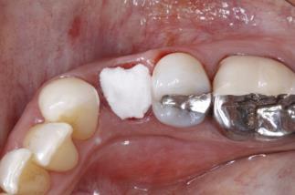
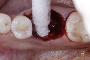
29 In order to standardize radiographs, a radiographic jig was fabricated by using a bite registration material (Regisil Rigid, Milford, DE) on periapical tab attached to dental periapical film holder (Schick, Wickford, United Kingdom) to make a record of adjacent teeth and restorations. Long-cone parallel technique was then used to capture a periapical radiograph of the study site (Figure A3) Randomization Prior to surgical intervention, computer software was used to randomly allocate each subject into the control group of FDBA with a collagen wound dressing, or the experimental group of Easy-graft CLASSIC. The randomization was withheld from the surgeon (K.T.) until shortly before the surgical visit. Given the nature of the study, the surgeon could not be masked to the study intervention Presurgical Measurements Pre-surgical antibiotics, analgesics, sedation, and anesthesia was given at the discretion of the surgeon. Within the study population, no pre-surgical antibiotics analgesics, or sedation was used. Buccal and occlusal photographs of the surgical site were obtained (Figure A4). The width of keratinized mucosa on the buccal of the tooth to be extracted was measured with periodontal probe (UNC-15). Local anesthesia was administered and time was allowed for anesthesia to take effect. Buccal and lingual mucosal thickness taken at the mid-facial and mid-lingual surface of the tooth was measured at approximately 1 mm apical to free gingival margin with the use of endodontic finger spreader and rubber stopper. The finger spreader penetrated the tissues until a hard stop was felt tactically and the rubber stopped was adjusted to be in intimate contact with the surface mucosa. The finger spreader was then removed and a millimeter ruler was used to measure to the nearest 0.5 mm (Figure A5) Surgical Intervention After the baseline measurements were recorded and adequate local anesthesia was confirmed, a circumferential sulcular incision was made to release soft tissue attachment to the study tooth. Surgical hand 17
30 instruments were used to perform a minimally traumatic extraction, limiting the forces applied to the buccal bone and soft tissues. After extraction, a gentle, but thorough curettage of the socket was completed with hand instruments (Figure A6). A periodontal probe (UNC-15) and tactile sensation was used to determine if a fenestration and/or dehiscence was present. If fenestration and/or dehiscence was present, the patient was excluded from the study and was treated as a normal patient of the clinic, receiving a bone allograft with collagen wound dressing, in an intent-to-treat fashion. If there was no fenestration and/or dehiscence, the computer-assisted randomization result was provided to the surgeon (K.T.). Horizontal bucco-lingual socket width at the crest and the depth of the socket was measured using the same periodontal probe utilized in previous measurements. Iwanson calipers (1-Iwanson Spring Caliper, Chicago, IL) with markings to 0.1 mm were used to measure the buccal bone thickness 1 mm apical to the level of the alveolar crest by penetrating the buccal soft tissue until it reached a hard stop (Figure A7). The Easy-graft CLASSIC was prepared according to manufacturer s recommendations. The FDBA material was rehydrated by sterile saline prior to grafting. Both the experimental and control grafts were condensed and filled to the crestal bone level. Sites grafted with FDBA had a collagen wound dressing placed on top of the graft and secured with cross mattress sutures (Figure A8) Post-operative Care Buccal and occlusal photographs of the surgical site was obtained and a post-operative radiograph was made with the radiographic jig that was fabricated earlier. Subjects received detailed written and verbal postoperative instructions. Subjects were instructed to avoid mechanical disturbance of the surgical site, use a 0.12% chlorhexidine mouth rinse, and no direct brushing of the surgical site for the first week. All subjects were prescribed oral antibiotics. Amoxicillin 500 mg every 8 hours for 7 days was the medication of choice. If an allergy to this antibiotic was reported Clindamycin 300 mg every 6 hours for 7 days was prescribed. Also, an anti-inflammatory and pain reliever drug (Ibuprofen 600 mg every 4-6 hours for first 3 days and only as needed thereafter), and narcotic pain reliever (Hydrocodone/APAP 5/325 mg every 6-8 hours as 18
31 needed for pain, up to 4 days) was prescribed to all subjects, unless contraindicated for individual medical reasons Follow-up Visits and Assessments Subjects returned to the clinic at 1, 2, 4, 8 and 16 weeks post operatively. Sutures, if present in the control group, were removed at 2 weeks postoperatively. At each follow-up visit, intraoral photographs were made of the buccal and occlusal surfaces of the surgical site. Gentle debridement of the adjacent teeth was also done. Any adverse events were documented and recorded. At the 16 weeks post-operative visit, in addition to the photographs (Figure A9), a second CBCT scan was obtained for all patients, using the same settings as employed at baseline (Figure A10) 3.8. Dimensional Osseous Changes Volumetric analysis of the data from DICOM files obtained from the initial scan and the 16 week follow up. The volumetric assessments were performed using a specific software (InVivo5.4, Anatomage, San, Jose, CA) via selection of a region of interest that included only hard tissues. The region of interest cropping from the baseline image were then transferred to the 16 week follow up scan in order to generate comparable volumes, using a constant threshold. The volumetric measurements were calculated using the volumetric tool in Anatomage and was expressed in mm 3. Linear measurements were also made in the CBCT data obtained from the initial and 16 week follow up scan. The ridge width dimensional changes, and buccal and lingual ridge height changes were calculated from the difference of two measurements. For each measurement, a reference line was created from a fixed and reproducible point on the reference tooth. Measurements were made from this line to the mid-buccal and mid-lingual crest at the site of interest (Figure A11). 19
32 3.9. Statistical Analysis Statistical analyses were performed for the change in the primary outcome comparing dimensional bone changes from baseline to 16 week follow up occurring in a single tooth extraction socket following ridge preservation using either FDBA with a collagen dressing or the Easy-graft CLASSIC. A p-value of <0.05 was considered statistically significant. Wilcoxon rank sum test was used to analyze volumetric and linear changes. The Wilcoxon rank sum test was chosen because normal distribution was not assumed, the sample sizes in the control and experimental groups were small, and the standard deviation in measurements were relatively large. In addition, a Pearson correlation coefficient calculation was performed to assess various correlations between buccal tissue thickness and buccal bone thickness, buccal bone thickness and volumetric reduction, buccal bone thickness and ridge width reductions Sample Size Determination For the multicenter study, a sample size of 22 participants in each treatment group was determined following power calculations and allowing for a 10% patient drop out. The sample size of 20 patients per group was calculated for the horizontal bone changes with the assumption that the detectable difference would amount to 0.7mm with a standard deviation of 0.8. The type 1 error probability was set at 0.05 and the statistical power was set at 80%. 20
33 CHAPTER IV. Results 4.1. Subject Population Twenty-nine patients were appointed for clinical screening. Two patients were not accepted due to clinical and radiographic indications of loss of alveolar ridge. One patient was not accepted due to adjacent teeth presenting severe pathology that required treatment. One patient was undergoing active periodontal therapy at the time of screening and was later accepted. One patient s treatment had to be delayed due to untreated hypertension. One patient was accepted, but later chose to exit the study citing ailing health. Twenty-three patient were enrolled in the study. Seventeen patients have completed the study to date and were included in the data analysis (9 control, 8 experimental) Subject Characteristics The mean age of the subjects were 54.4 for the control group and 61.1 for the experimental group (p-value = ). Eleven females and 6 males comprised the analyzed group. In the control group, 7 were female and 2 were male. In the experimental group, 4 were females and 4 were males. (Table B1) Baseline Data Of the teeth extracted, 12 were premolars (8 maxillary, 4 mandibular) and 5 were molars (2 maxillary, 3 mandibular) (Table B2). The average width of keratinized gingiva was 3.36mm for the control group and 3.75mm for the experimental group (p-value = ). The average buccal tissue thickness was 1.39mm for the control group and 1.56mm for the experimental group (p-value = ). The average lingual tissue thickness was 1.78mm for the control group and 2.06mm for the experimental group (p-value = ). Following tooth extraction, the buccal bone thickness for the control group was 0.59mm and the experimental group was 0.73mm (p-value = ). No significant difference between groups from baseline data was found. 21
34 The mean volume of the region of interest in the initial CBCT for the FDBA group was mm 3 and for the Easy-graft group was mm 3. The bucco-lingual ridge width for the FDBA group at baseline was mm and for the Easy-graft group at baseline was mm (Table B3) Volumetric Changes Volumetric data can be found in Table B4. The average ridge volume for the FDBA group from the initial scan was mm 3. At the 16 week follow up, the ridge volume had reduced to mm 3. This was a mean of ± mm 3 of reduction, a 9.59% reduction, however, this was not considered statistically significant between the initial and 16 week follow up for the control group (p-value = ). The average ridge volume of the Easy-graft CLASSIC group from the initial scan was mm 3. By week 16, the ridge volume reduced to mm 3. The average loss of ridge volume was ± 79.65mm 3, a 13.04% reduction. There was no statistically significant difference in ridge volume between the initial and the 16 week follow up for the experimental group (p-value = ). When comparing the average ridge volume reduction for both the experiemental and control, there was no statistically significant difference between either group (p-value = ) 4.5. Linear Ridge Width Changes Ridge width data can be found in Table B5. The average ridge width for the FDBA group from the initial scan was 10.29mm. At the 16 week follow up, the ridge width was reduced to 9.19mm. This was a reduction of 1.10 ± 0.87mm, or 10.72% reduction. This reduction was not considered statistically significant (p-value = ). The average ridge width for the Easy-graft group from the initial scan was 10.09mm. At the 16 week follow up, the ridge width was reduced to 8.85mm. This was a reduction of 1.23 ± 1.17mm, or 12.23% reduction. This reduction was not considered statistically significant (p-value = ) 22
35 When comparing the average ridge width reduction for both the FDBA group and the Easy-graft group, there was no statistically significant difference between the two groups (p-value = ). 4.6 Linear Ridge Height Changes Ridge height data can be found in Tables B6 and B7. From the initial CBCT scan to the 16 week follow up CBCT scan, the buccal ridge height had been reduced 1.12mm for the FDBA group and 1.19mm for the Easy-graft group. These values were not statistically different from one another (p-value = ). From the initial CBCT scan to the 16 week follow up CBCT scan, the lingual ridge height had been reduced 0.63mm for the FDBA group and 0.67mm for the Easy-graft group. These values were not statistically different from one another (p-value = ) Correlations In general, there was a weak to moderate positive correlation between the buccal tissue thickness and the thickness of the buccal bone (R-value = 0.30) (Table B8). There was a weak positive correlation between the buccal bone thickness and the amount of volume reduction for the FDBA group but not the Easy-graft group (R-value FDBA = 0.25, R-value Easy-graft = 0.09) (Table B9). There was a weak negative correlation between the buccal bone thickness and the ridge width reduction for the FDBA group (R-value = -0.44) (Table B10) but no correlation of the same parameters for the Easy-graft group (R-value = -0.14) 23
36 CHAPTER V. Discussion The primary aim of this randomized clinical trial was to assess the clinical efficacy in terms of volumetric ridge dimension changes, and horizontal and vertical linear changes, after single tooth extraction and the application of Easy-graft CLASSIC compared to conventional particulate FDBA with collagen wound dressing, via volumetric data analysis. The average change in alveolar ridge volume for the FDBA group and the Easy-graft group was 9.59% and 13.04%, respectively, after 16 weeks of healing. Linear measurements utilizing the CBCT data were made to determine the ridge width reduction and buccal and lingual ridge height reductions. The present study showed that both ridge preservation methods are associated similar remodeling pattern; such that there are significant, but attenuated changes in volume between the baseline and the 16 week follow up, and a loss of both ridge width and height. The control group had a volumetric reduction of mm 3, whereas the experimental group had a change of 94.87mm 3. These reductions were not statistically significantly different between the groups, however, due to the small sample size, the difference may become statistically significant with a sample size increase, once the data from all participating centers is combined. Nevertheless, the measured difference of approximately 20mm 3 could be considered clinically insignificant. The percent change in alveolar ridge volume in this study appears to be similar to previous studies utilizing DICOM data from CBCT scans, although, there appears to be a paucity of studies that analyze alveolar ridge volume changes utilizing three dimensional imaging technology. A previous pilot study by Avila-Ortiz et al. (2014b) compared three different ridge preservation techniques to a collagen wound dressing control utilizing CBCT scans immediately after the surgical procedure and again after 16 weeks. The result of alveolar ridge volume reduction reported by the group were similar to our result. They found that the percent of volumetric ridge reduction for the various ridge preservation techniques was 5% when using an allograft with a nonresorbable membrane, 7% when using an allograft with collagen barrier in addition to a non-resorbable 24
37 membrane, and 16% when overbuilding the buccal aspect with the allograft and using a non-resorbable membrane. In another human study of alveolar ridge preservation, computed tomography was utilized to compare ridge dimension changes after placement of a xenograft with a collagen membrane and naturally healing sockets (Pang et al., 2014). After 3 months they found the volume of preserved sites had resorbed mm 3 while the untreated sites resorbed by mm 3. No percent changes in alveolar bone volume were reported. Our data demonstrated significantly lower volumetric changes than that reported by Pang et al. (2014). Variance in the method used to determine the region of interest may explain the differences in numeric volumetric changes. However, the condition of sockets after extraction was not described in their study and the magnitude of the volumetric changes may be due to the loss of the buccal plate of alveolar bone. When the buccal plate is not present, the socket most often fills with fibrous reparative tissues, which usually leads to a reduced net bone volume (Darby et al., 2009). Barone et al. (2008) utilized study casts and CBCT scans to measure volumetric changes over 3 months following tooth extraction with alveolar ridge preservation using two different xenograft materials. They found volumetric changes ranging from 244 to 349mm 3. The values from our study for this parameter are significantly smaller. This may be due to methodologic differences in calculating the volumetric changes. By using study casts scanned by a CBCT, they analyzed the surface area of the region of interest rather than the hard tissue volume. Thus, there may be more significant soft tissue dimensional changes that occur after extraction that we did not account for in the study. In the same study, linear measurements were also made from their CBCT scans of study casts. They reported a width reduction of about 1.2 to 1.6mm and a height loss of 1.8 to 2.0 mm. Pang et al. (2014) reported values of width and height reduction as 1.11mm and 1.05mm respectively. Our results of width loss of 1.10mm for the FDBA group and 1.24mm for the Easy-graft group are in agreeance with these studies. The ridge height loss of 0.63 to 1.12mm for the FDBA group and 0.67 to 1.19mm for the Easy-graft group 25
38 appears to be smaller than the magnitudes reported in previous studies however the differences between the two groups in our study is negligible. The pattern of resorption following tooth extraction has been well documented (Cardaropoli et al., 2003, Schropp et al., 2003, Araujo and Lindhe, 2009). There are both horizontal and vertical ridge dimension changes. In untreated sites, the healing can be described in two overlapping phases. The first phase is characterized by the resorption of the bundle bone and replacement with woven bone. The second phase shows marked resorption of the outer surfaces of both the buccal and lingual walls. The buccal plate is usually thinner and it is comprised of mostly bundle bone. Therefore, the buccal plate experiences a greater magnitude of resorption than the lingual plate. The result of the ridge resorption is a more lingually and apically positioned residual ridge (Lekovic et al., 1998, Lekovic et al., 1997, Araujo and Lindhe, 2009). The amount of ridge width loss is around 3 to 4mm while the ridge height loss is about 1.5 to 2mm (Van der Weijden et al., 2009). To combat the magnitude of alveolar ridge resorption, the use of bone grafting has been advocated. When compared to untreated controls, there was about 1.5-2mm less ridge width reduction and about 1.0 to 2mm less ridge height when using a bone replacements graft (Avila-Ortiz et al., 2014a, Iocca et al., 2017, Vittorini Orgeas et al., 2013). Overall, the use of alveolar bone grafting lead to reduction in the amount of ridge width and height loss, but was unable to fully prevent these changes. From our data, it is clear that the resorption of the buccal plate was more significant than the resorption of the lingual plate for both the experimental and the control groups. The buccal ridge height resorption was about twice that of the lingual ridge height resorption. Though our results follow the same pattern or resorption, the magnitude appears to be much less within our study. This may be due, in part, to the tooth types analyzed in ridge preservation studies mentioned earlier. A recent systematic review Avila-Ortiz et al. (2014a) on the effects of alveolar ridge preservation reported a 1.89mm clinical benefit on ridge width preservation, 2.07mm clinical benefit on the buccal bone height, and 1.18mm clinical benefit on the lingual bone height when compared to tooth extraction alone. These results were based on 6 studies of non-molar tooth extraction. Another systematic review by Vignoletti et al. 26
39 (2012) reported a 1.83mm clinical benefit on ridge width preservation and 1.47mm clinical benefit on ridge height preservation when compared to tooth extraction alone. Results from that study was based 9 studies, of which, only 2 were studies that included molars. Another systematic review by Vittorini Orgeas et al. (2013) reported an overall benefit of 1.99mm clinical benefit on the ridge width, and 0.96mm clinical benefit on the ridge height when compared to tooth extraction alone. Results were from analysis of 13 studies, of which, only 1 study reported on posterior teeth only. This study, by Cardaropoli and Cardaropoli (2008) treated 10 patients with a xenograft and collagen membrane. After 4 months of healing, they reported a ridge width dimensional change of about 1.85mm, or about 15%. Data from the present study found slightly less ridge width reduction for either the FDBA or the Easy-graft group. The difference may be explained by the surgical technique utilized. In the study by Cardaropoli and Cardaropoli (2008) a collagen membrane was to cover the xenograft. Though not described in detail, the placement of the membrane requires that the buccal and lingual tissues be reflected off the alveolar bone. As previously reported, soft tissue reflection may increase the amount of bone resorption (Wood et al., 1972). Though the effectiveness of alveolar ridge preservation is well established, there is a scarcity of studies that analyze the effects of the initial morphology of the socket, the thickness of the buccal plate, and the soft tissue phenotype, on clinical outcomes. A few studies have advocated the use of CBCT scans to determine the buccal plate thickness. These studies focus on the anterior maxilla. After analyzing 125 CBCT scans, Braut et al. (2011) found that 26% of these teeth present with no radiographic presence of the buccal plate, 63% present with a buccal plate that is less than 1mm, and 11% presented with a buccal plate thickness of more than 1mm. Januario et al. (2011) analyzed 250 CBCT scans and found that a majority of sites had a buccal plate thickness of less than 1mm and about half of sites had a buccal plate thickness of less than 0.5mm. Utilizing direct measurements, Huynh-Ba et al. (2010) reported on the buccal plate thickness of 93 extraction sites. The anterior sites had an average buccal thickness of 0.8mm, with 87% of sites with thickness less than 1mm. Premolar sites had a thickness of 1.1mm, with 59% of sites with thickness less than 1mm. Due to the resorption patterns described earlier, the resorption of the anterior sextant is more dramatic 27
40 than the posterior sextant. Thus, results from our study may not be as comparable to reported effects of alveolar ridge preservation that is pervasive in the literature. Our study found a weak positive correlation between buccal bone thickness and buccal tissue thickness. This finding is similar to that of Cook et al. (2011). Analyzing 60 subjects (26 with thin biotypes, 34 with thick biotypes), they found that there was a significant correlation between the thickness of the biotype and the presence of thick bundle bone verified via CBCT. This correlation was also seen in a study by Fu et al. (2010) on cadavers. They found a moderate association between the presence of a thick buccal plate measure with a CBCT and the thickness of the buccal tissues. There was a weak positive correlation between the buccal bone thickness and the magnitude of alveolar ridge volume reduction. This correlation appears to contradict the generally held belief that the presence of a thick buccal plate will attenuate the effects of ridge resorption when compared to the presence of a thin buccal plate. The negative correlation of the buccal bone thickness and the magnitude of ridge width reduction is more in line with previous studies. Following tooth extraction, sites with a thicker buccal plate are more resistant to the effects of bundle bone resorption that occurs. In a study by Spinato et al. (2014), single rooted teeth were extracted and grafted with an allograft and compared to sites treated with an extraction with no ridge preservation. Sites were classified into either thick or thin buccal plate groups using 1mm as a cut off between the two groups. They found that the thin buccal plate group demonstrated worse outcomes on socket healing. These correlations may be seen as contradictory to one another. With an increase in the thickness of the buccal plate, our correlations seem to suggest that there will be greater volumetric reduction, yet the ridge width will be protected. We feel that these findings can be explained due to the methodology of analyzing the DICOM data. The volumetric measurements takes into account the entire region of interest while the linear measurements are based on comparing measurements at a specific, repeatable location on two separate CBCT scans. Because these linear measurements were not made at the immediate crest of the residual alveolar ridge and were, instead, measures slightly subcrestally, the magnitude of the resorption at the more 28
41 apical position would be significantly less than the magnitude of the crestal resorption. Therefore, the correlation between buccal bone thickness and alveolar ridge volume reduction should be expected to be more representative of the morphological changes than the linear measurements, however due to the limited number of patients in each group, the conclusions drawn from the correlations should be taken with caution. Another source of discrepancy may be from the lack of use of a barrier membrane in our study. Using barrier membranes to exclude cells from infiltrating the bone graft after ridge preservation procedures has been advocated (Buser et al., 2004, Camargo et al., 2003, Cardaropoli et al., 2005, Lekovic et al., 1997, Simion et al., 1994). Results from using these occlusive membranes has shown that significant reduction of the alveolar bone could be avoided but the resorption of the buccal plate is still unavoidable (Fickl et al., 2008). In a study by Thalmair et al. (2013), 3 different ridge preservation techniques were compared to a tooth extraction with no treatment. The first experimental group was the use of a xenograft within the socket and sealed with a free gingival graft. The second experimental group was the use of the gingival graft alone. The third experimental group was the use of the xenograft alone. From their results, all treatment modalities could not prevent the resorption of the buccal plate but all fared better than the sites not treated. The range of ridge dimension shrinkage was 0.8 to 2.3mm. The least shrinkage was from the xenograft with the gingival graft while the control group had the most. The magnitude of the ridge width changes are comparable to those found in the treated sites in their study, however Thalmair et al. performed measurements on study casts, combining soft and hard tissue changes. Of more interest from the study was the results from the use of the xenograft alone. The xenograft used in the study was a particulate and no soft tissue seal or collagen wound dressing was applied to contain the graft material within the socket. They found ridge contour shrinkage of about 1.45mm, and was unable to be distinguished statistically from the untreated control and yielded the second worst results. Our study data demonstrates that, despite being left exposed to the oral cavity during the healing phase, similar results to the FDBA group were found after 16 weeks of healing. This can be attributed to the unique handling capability of Easy-graft CLASSIC, whose graft dimensions 29
42 are stable after just a few minutes exposed to bodily fluids rather than a particulate bone graft that would be more likely to be washed out from the socket. The present study used DICOM data from CBCT scans taken at baseline and after 16 weeks of healing. This 16 week healing period was selected based on biologic reasoning and common clinical practice. A study of healing extraction sockets in humans demonstrated that new bone proliferation occurs from 4 to 8 weeks. It is characterized by the proliferation of osteogenic cells and immature bone formation. From 8 to 12 weeks the osteogenesis decreases and the trabeculae matures and increases in volume. By 12 to 16 weeks the extraction sockets usually presents with boney trabeculae that is mature with little osteoid and osteoblasts (Evian et al., 1982). Future studies into this novel, alloplastic, bone grafting material should focus on implant survival and success in the grafted sites. Human histological data should be collected after healing of the graft to determine the composition of the bone and the fate of the particles. 30
43 CHAPTER VI. Conclusions The purpose of this randomized clinical study was to determine, utilizing DICOM data obtained from CBCT scans, the alveolar ridge volumetric changes, and horizontal and vertical linear changes after posterior single tooth extraction sites treated with either FDBA with a collagen wound dressing or Easy-graft CLASSIC, a novel alloplastic bone substitute. No statistically significant differences were observed between the two groups in alveolar ridge volume changes, alveolar ridge width loss, and buccal and lingual alveolar ridge height loss, however there is a slight positive correlation between buccal tissue thickness and buccal bone thickness, a slight positive correlation between buccal bone thickness and alveolar ridge volume reduction, and a slight negative correlation between buccal bone thickness and ridge width reduction. It appears that both grafting materials perform well in alveolar ridge preservation. 31


44 APPENDIX A Figures Figure A1. Study Timeline 32
45 Figure A2. Events Schedule Visit Number Visit Description Screening Randomization Extraction, Ridge Preservation 1 Week Post Op 2 Week Post Op 4 Week Post Op 8 Week Post Op 16 Week Post Op, CBCT Patient Information Informed Consent Patient Demographics Medical/Surgical History Oral Examination Inclusion/Exclusion Criteria X X X X X Treatment Clinical Photography X X X X X X Tooth Extraction, Ridge Preservation Suture Removal X Assessment CBCT X X Standardized PA Radiograph X Horizontal Ridge Width X Other Wound Dehiscence X X X X X Adverse Events X X X X X X 33


46 Figure A3. Pre-Operative Radiographs a. FDBA Group b. Easygraft Group Figure A4. Pre-Operative Clinical Presentation a. FDBA Group b. Easygraft Group 34


47 Figure A5. Clinical Measurement a. FDBA Group b. Easygraft Group Figure A6. Intact Socket after Extraction a. FDBA Group b. Easygraft Group 35


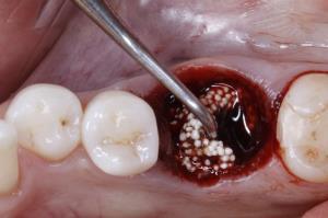
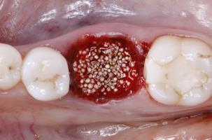
48 Figure A7. Buccal Plate Thickness Measurement a. FDBA Group c. Easygraft Group Figure A8. Graft Procedure a. FDBA Group b. Easygraft Group 36


49 Figure A9. 16-Week Follow Up a. FDBA Group b. Easygraft Group 37


50 Figure A Week Follow Up Radiographs a. FDBA Group b. Easygraft Group Figure A11. Volumetric Analysis a. FDBA Group b. Easygraft Group 38


51 Figure A12. Ridge Width Changes a. FDBA Group b. Easy-graft Group Figure A13. Ridge Height Changes a. FDBA Group b. Easy-graft Group 39
52 APPENDIX B Tables Table B1. Subject Characteristics Subject Characteristics Control Experimental Age Gender F 7 / M 2 F 4 / M - 4 Table B2. Extraction Sites Control Experimental Maxillary First Premolar 1 1 Maxillary Second Premolar 1 5 Maxillary First Molar 1 1 Mandibular First Premolar 0 1 Mandibular Second Premolar 3 0 Mandibular First Molar 2 0 Mandibular Second Molar 1 0 Table B3. Baseline Clinical Measurements Control Experimental Keratinized Gingiva Width (mm) Volume (mm 3 ) Ridge Width (mm) Buccal Height from Reference (mm) Lingual Height from Reference (mm) Buccal Bone Thickness (mm) Buccal Tissue Thickness (mm) Lingual Tissue Thickness (mm) Table B4. Volumetric Data Ridge Volume (mm 3 ) Control Experimental Comparison p-value Baseline Week Change ± % ± % p-value
53 Table B5. Ridge Width Data Ridge Width (mm) Control Experimental Comparison p-value Baseline Week Change, Percent Change ± % ± % p-value, Baseline to Week Table B6. Buccal Ridge Height Data Buccal Ridge Height (mm from reference) Control Experimental Comparison p-value Baseline Week Change ± ± p-value, Baseline to Week Table B7. Lingual Ridge Height Data Lingual Ridge Height (mm from reference) Control Experimental Comparison p-value Baseline Week Change ± ± p-value, Baseline to Week


54 Table B8. Correlation of Buccal Tissue Thickness and Buccal Bone Thickness R-value = 0.30 Table B9. Correlation of Buccal Bone Thickness and Alveolar Ridge Volume Reduction Ridge Volume Reduction (mm 3 ) R-value = 0.25 Buccal Bone Thickness 42

55 Table B10. Correlation of Buccal Bone Thickness and Alveolar Ridge Width Reduction R-value = Buccal Bone Thickness 43
ANATOMY OF THE PERIODONTIUM. Dr. Fatin Awartani
 ANATOMY OF THE PERIODONTIUM Part II Cementum and Alveolar bone Associate Professor Periodontal division King Saud university Cementum Calcified mesenchymal tissue that forms the outer covering of the anatomic
Fundamental & Preventive Curvatures of Teeth and Tooth Development. Lecture Three Chapter 15 Continued; Chapter 6 (parts) Dr. Margaret L.
 Dr. Margaret L.") Fundamental & Preventive Curvatures of Teeth and Tooth Development Lecture Three Chapter 15 Continued; Chapter 6 (parts) Dr. Margaret L. Dennis Proximal contact areas Contact areas are on the mesial and
Fundamental & Preventive Curvatures of Teeth and Tooth Development Lecture Three Chapter 15 Continued; Chapter 6 (parts) Dr. Margaret L. Dennis Proximal contact areas Contact areas are on the mesial and
Advanced Probing Techniques
 Module 21 Advanced Probing Techniques MODULE OVERVIEW The clinical periodontal assessment is one of the most important functions performed by dental hygienists. This module begins with a review of the
Dental Implants: A Predictable Solution for Tooth Loss. Reena Talwar, DDS PhD FRCD(C) Oral & Maxillofacial Surgeon Associate Clinical Professor
 Oral & Maxillofacial Surgeon Associate Clinical Professor") Dental Implants: A Predictable Solution for Tooth Loss Reena Talwar, DDS PhD FRCD(C) Oral & Maxillofacial Surgeon Associate Clinical Professor What are Dental Implants? Titanium posts used to replace missing
Dental Implants: A Predictable Solution for Tooth Loss Reena Talwar, DDS PhD FRCD(C) Oral & Maxillofacial Surgeon Associate Clinical Professor What are Dental Implants? Titanium posts used to replace missing
REGENERATIONTIME. A Case Report by. Ridge Augmentation and Delayed Implant Placement on an Upper Lateral Incisor
 A Case Report by Dr. Daniele Cardaropoli Ridge Augmentation and Delayed Implant Placement on an Upper Lateral Incisor The Situation An adult female patient presented with an endodontic/prosthetic failure
Periodontal Disease. Radiology of Periodontal Disease. Periodontal Disease. The Role of Radiology in Assessment of Periodontal Disease
 Radiology of Periodontal Disease Steven R. Singer, DDS srs2@columbia.edu 212.305.5674 Periodontal Disease! Includes several disorders of the periodontium! Gingivitis! Marginal Periodontitis! Localized
Case Report. RapidSorb Rapid Resorbable Fixation System. Ridge augmentation in a one-step surgical protocol.
 Case Report RapidSorb Rapid Resorbable Fixation System. Ridge augmentation in a one-step surgical protocol. RapidSorb Rapid Resorbable Fixation System. Ridge augmentation in a one-step surgical protocol.
Masking Buccal Plate Remodeling in the Esthetic Zone with Connective Tissue Grafts: Concepts and Techniques with Immediate Implants
 Peer-Reviewed and Indexed Annual Implant Issue Masking Buccal Plate Remodeling in the Esthetic Zone with Connective Tissue Grafts: Concepts and Techniques with Immediate Implants of Continuing Education
Principles of Periodontal flap surgery. Dr.maryam khosravi
 Principles of Periodontal flap surgery Dr.maryam khosravi Goals of periodontal SURGICAL phase 1 - Controlling or eliminating periodontal disease. 2 Correcting anatomic conditions that may a. favor periodontal
Efficacy of socket grafting for alveolar ridge preservation: a randomized clinical trial
 University of Iowa Iowa Research Online Theses and Dissertations Summer 2015 Efficacy of socket grafting for alveolar ridge preservation: a randomized clinical trial Mitchell Miles Gubler University of
Rehabilitating a Compromised Site for Restoring Form, Function and Esthetics- A Case Report
 Research & Reviews: Journal of Dental Sciences Rehabilitating a Compromised Site for Restoring Form, Function and Esthetics- A Case Report Priyanka Prakash* Division of Periodontology, Department of Dental
Evaluation of a Combination Allograft Material Compared to DFDBA in Alveolar Ridge Preservation. Sanju P. Jose
 Evaluation of a Combination Allograft Material Compared to DFDBA in Alveolar Ridge Preservation by Sanju P. Jose B.S., University of Maryland Baltimore County, 2008 D.D.S., University of Maryland School
Socket preservation in the daily practice: A clinical case report
 Clinical Socket preservation in the daily practice: A clinical case report Rabih Abi Nader 1 and Carine Tabarani 2 Abstract Soft tissue contour depends on the underlying bone anatomy. Following tooth extraction,
14/09/15. Assessment of Periodontal Disease. Outline. Why is Periodontal assessment needed? The Basics of Periodontal assessment
 Assessment of Periodontal Disease Dr Wendy Turner Outline Why is Periodontal assessment needed? The Basics of Periodontal assessment Probing: Basic Periodontal Examination for adults and children. Detailed
A Clinical Evaluation of Anatomic Features of Gingiva in Dental Students in Tabriz, Iran
 Received 26 October 2007; Accepted 11 February 2008 A Clinical Evaluation of Anatomic Features of Gingiva in Dental Students in Tabriz, Iran Adileh Shirmohammadi 1 Masoumeh Faramarzie 1 * Ardeshir Lafzi
Bone Grafting for Socket Preservation
 Bone Grafting for Socket Preservation Dr. Karl R. Koerner Normal extraction facial bone loss. Excessive force. Commonly the thickness of facial bone. Hussain, A. et al. Ridge preservation comparing a nonresorbable
Periodontal ligament
 Periodontal ligament The periodontium The periodontium includes: The gingiva Cementum Periodontal ligament Alveolar bone Def: The periodontal ligament is the dense fibrous connective tissue that occupies
Surgical Therapy. Tuesday, April 2, 13. Alessan"o Geminiani, DDS, MS
 Surgical Therapy Alessan"o Geminiani, DDS, MS Periodontal Flap: a surgical procedure in which incisions are made in the gingiva or mucosa to allow for separation of the epithelium and connective tissues
Cytoflex Barrier Membrane Clinical Evaluation
 Cytoflex Barrier Membrane Clinical Evaluation Historical Background Guided tissue regeneration is a well established concept in the repair of oral bone defects. The exclusion of soft tissue epithelial
Immediate Implant Placement:
 Immediate Implant Placement: Parameters Influencing Tissue Remodeling Bernard Touati, DDS and Mario Groisman, DDS In esthetic implant therapy, the patient s objective is to obtain an imperceptible, natural-looking
More than bone regeneration. A total solution.
 More than bone regeneration. A total solution. More than a dental implant company. A total solution. When it comes to treatment options, your patients want positive results both functionally and esthetically.
REGENERATIONTIME. A Case Report by. Geistlich Mucograft for the treatment of multiple adjacent recession defects: A more palatable option
 A Case Report by Dr. Daniel Gober Geistlich Mucograft for the treatment of multiple adjacent recession defects: A more palatable option The Situation A 35 year old male presented in my practice with a
Bringing you Geistlich biocompatibility with improved application and handling benefits. Your combination for success
 Bringing you Geistlich biocompatibility with improved application and handling benefits Your combination for success Geistlich Combi-Kit Collagen: Combining ease and predictablility Geistlich Combi-Kit
Management of a complex case
 2 Soft- and hard-tissue reconstruction of a severely deficient site prior to implant placement: a case report Management of a complex case Younes Khosroshahy, DDS, MFDS RCS (Eng), Dip Imp Dent RCSEd, Blue
Case Study. Case # 1 Author: Dr. Suheil Boutros (USA) 2013 Zimmer Dental, Inc. All rights reserved. 6557, Rev. 03/13.
 2013 Zimmer Dental, Inc. All rights reserved. 6557, Rev. 03/13.") Placement of a Zimmer Trabecular Metal Dental Implant with Simultaneous Ridge Augmentation and Immediate Non-Functional Loading Following Tooth Extraction and Orthodontic Treatment for Implant Site Development
Placement of a Zimmer Trabecular Metal Dental Implant with Simultaneous Ridge Augmentation and Immediate Non-Functional Loading Following Tooth Extraction and Orthodontic Treatment for Implant Site Development
Procedure Manual and Catalog
 Procedure Manual and Catalog TM Why SynthoGraft? SynthoGraft offers a unique structure which provides stability, while its micro-porosity allows for rapid vascularization and subsequent resorption. Although
Contemporary Implant Dentistry
 Contemporary Implant Dentistry C H A P T ER 1 4 O F C O N T E M P OR A R Y O R A L A N D M A X I L L OFA C IA L S U R G E RY B Y : D R A R A S H K H O J A S T EH Dental implant is suitable for: completely
Gum Graft? Patient Need a. Does My. 66 JANUARY 2017 // dentaltown.com. by Dr. Brian S. Gurinsky
 by Dr. Brian S. Gurinsky Dr. Brian S. Gurinsky was born in Dallas and attended college at the University of Texas at Austin. He continued his education at Baylor College of Dentistry in Dallas, where he
Over the years, mucogingival surgery
 The Use of DynaMatrix Extracellular Membrane for Gingival Augmentation and Root Coverage: A Case Series Saroff Stephen Andrew Saroff, DDS, MSD 1 Abstract Over the years, mucogingival surgery has developed
PERIODONTAL REVIEW DENTALELLE TUTORING ANDREA TWAROWSKI
 PERIODONTAL REVIEW DENTALELLE TUTORING ANDREA TWAROWSKI Periodontics Questions 1. What is the periodontium composed of? 2. What type of mucosa is the gingiva composed of? 3. Is it normal to have melanin
Horizontal bone augmentation by means of guided bone regeneration
 Periodontology 2000, Vol. 66, 2014, 13 40 2014 John Wiley & Sons A/S. Published by John Wiley & Sons Ltd Printed in Singapore. All rights reserved PERIODONTOLOGY 2000 Horizontal bone augmentation by means
Townie Guest Editorial. Gingival Attachment Loss: Evaluation and Surgical Options. Daniel J. Melker, DDS. fig. 1
 Gingival Attachment Loss: Evaluation and Surgical Options Daniel J. Melker, DDS Attached connective tissue (a.k.a. attached tissue) in the simplest terms is the body s only barrier between the underlying
Consensus Report Tissue augmentation and esthetics (Working Group 3)
") B. Klinge Thomas F. Flemmig Consensus Report Tissue augmentation and esthetics (Working Group 3) Members of working group: Matteo Chiapasco Jan-Eirik Ellingsen Ronald Jung Friedrich Neukam Isabella Rocchietta
B. Klinge Thomas F. Flemmig Consensus Report Tissue augmentation and esthetics (Working Group 3) Members of working group: Matteo Chiapasco Jan-Eirik Ellingsen Ronald Jung Friedrich Neukam Isabella Rocchietta
Extraction with Immediate Implant Placement and Ridge Preservation in the Posterior
 Extraction with Immediate Implant Placement and Ridge Preservation in the Posterior by Timothy F. Kosinski, DDS, MAGD The following case presentation illustrates the diagnosis, planning and treatment for
Clinical Case Reports using Cytoplast GTR Barrier Membranes
 Clinical Case Reports using Cytoplast GTR Barrier Membranes Barry K. Bartee, DDS, MD The Cytoplast Technique: Extraction Site Grafting Without Primary Closure 1. 1. Preoperative view. To maximize the result
Periodontal phenotype and supracrestal soft tissue dimensions clinical correlations and their impact on post-extraction volumetric changes
 University of Iowa Iowa Research Online Theses and Dissertations Summer 2017 Periodontal phenotype and supracrestal soft tissue dimensions clinical correlations and their impact on post-extraction volumetric
The Original remains unique.
 The Original remains unique. Geistlich leading regeneration 2A, 2B Geistlich is the world leader in regenerative dentistry. We transform natural biomaterials into safe and reliable treatment methods that
Prosthetic Options in Implant Dentistry. Hakimeh Siadat, DDS, MSc Associate Professor
 Prosthetic Options in Dentistry Hakimeh Siadat, DDS, MSc Associate Professor Dental Research Center, Department of Prosthodontics & Dental s Faculty of Dentistry, Tehran University of Medical Sciences
Limited bone availability makes implant placement challenging
 Bone Grafting: Essential Indications and Techniques in Implant Dentistry Limited bone availability makes implant placement challenging and sometimes unpredictable. Candidates for implant therapy must have
Thick vs. Thin Gingival Biotypes: A Key Determinant in Treatment Planning for Dental Implants
 r s Thick vs. Thin Gingival Biotypes: A Key Determinant in Treatment Planning for Dental Implants richard t. kao, dds, phd; mark c. fagan, ms, dds; and gregory j. conte, ms, dmd abstract During the treatment
Educational Training Document
 Educational Training Document Table of Contents Part 1: Resource Document Disclaimer Page: 2 Part 2: Line Item Grade Sheets Page: 3 Release: 11/2016 Page 1 of 6 Part 1: Resource Document Disclaimer The
A new approach with an in-situ self-hardening grafting material
 74 Bone grafting with simultaneous early implant placement A new approach with an in-situ self-hardening grafting material MINAS LEVENTIS 1,2, PHD; PETER FAIRBAIRN 1,3, BDS; ORESTIS VASILIADIS 2,4, DDS
Purpose: To assess the long term survival of sites treated by GTR.
 Cortellini P, Tonetti M. Long-term tooth survival following regenerative treatment of intrabony defects. J Periodontol 2004; 75:672-8. (28 Refs) Purpose: To assess the long term survival of sites treated
Surgical Procedure in Guided Tissue Regeneration with the. Inion GTR Biodegradable Membrane System
 Surgical Procedure in Guided Tissue Regeneration with the Inion GTR Biodegradable Membrane System 1 Introduction This presentation familiarizes you with the basic steps how to use the Inion GTR membrane
CASE REPORT. CBCT-Assisted Treatment of the Failing Long Span Bridge with Staged and Immediate Load Implant Restoration
 Computer Aided Implantology Academy Newsletter - Newsletter 20 - July 2009 CASE REPORT CBCT-Assisted Treatment of the Failing Long Span Bridge with Staged and Immediate Load Implant Restoration Case Report
Maryland AGD AE and Socket Grafting 2015
 The Goodacre Study 5 year retrospective study looked at Crown & Bridge Caries Single crowns 1% FPD abutments 17% Periodontal Involvement Single crowns
Alveolar ridge preservation techniques
 Alveolar ridge preservation techniques Semmelweis University, Department of Periodontology, Budapest Dr. Windisch Péter Head of Department of Periodontology Changes of the alveolar ridge dimensions after
Pre op Failed endodontic treatment with sinus involvement.
 Case #1 of 10 consecutive extraction sockets grafted with Socket Graft Putty, covered with Socket Seal and sealed with Periacryl. I D # HEU This patient is a 66 year old female. Pre op Failed endodontic
Labial and lingual/palatal bone thickness of maxillary and mandibular anteriors in human cadavers in Koreans
 Research Article J Periodontal Implant Sci 2011;41:60-66 doi: 10.5051/jpis.2011.41.2.60 Labial and lingual/palatal bone thickness of maxillary and mandibular anteriors in human cadavers in Koreans Ji Young
BONE AUGMENTATION AND GRAFTING
 1 A Computer-Guided Bone Block Harvesting Procedure: A Proof-of-Principle Case Report and Technical Notes Effectiveness of Lateral Bone Augmentation on the Alveolar Crest Dimension: A Systematic Review
Dental Morphology and Vocabulary
 Dental Morphology and Vocabulary Palate Palate Palate 1 2 Hard Palate Rugae Hard Palate Palate Palate Soft Palate Palate Palate Soft Palate 4 Palate Hard Palate Soft Palate Maxillary Arch (Maxilla) (Uppers)
Replacement of a congenitally missing lateral incisor in the maxillary anterior aesthetic zone using a narrow diameter implant: A case report
 C A S E R E P O R T Replacement of a congenitally missing lateral incisor in the maxillary anterior aesthetic zone using a narrow diameter implant: A case report Rhoodie Garrana 1 and Govindrau Mohangi
( ) 2009;28(2):89-94
 2009;28(2):89-94") ( ) 2009;28(2):89-94 Osseointegration is important in the functional aspect, however, esthetics is also important, especially in the maxillary anterior region. An adequate surgical technique is necessary
( ) 2009;28(2):89-94 Osseointegration is important in the functional aspect, however, esthetics is also important, especially in the maxillary anterior region. An adequate surgical technique is necessary
Management of millers class III marginal tissue recession associated with endodontic lesion: Report of two cases managed using second stage surgery
 CASE SERIES 1 OPEN ACCESS Management of millers class III marginal tissue recession associated with endodontic lesion: Report of two cases managed using second stage surgery Sangeeta ABSTRACT Introduction:
Oral Embryology and Histology
 Oral Embryology and Histology Chapter 8 Copyright 2018, Elsevier Inc. All Rights Reserved. 1 Learning Objectives Lesson 8.1: Oral Embryology 1. Pronounce, define, and spell the key terms. 2. Define embryology
ijcrr Vol 04 issue 12 Category: Case Report Received on:22/04/12 Revised on:07/05/12 Accepted on:22/05/12
 SURGICAL RECONSTRUCTION OF INTERDENTAL PAPILLA USING AN INTERPOSED SUBEPITHELIAL CONNECTIVE TISSUE GRAFT: A CASE REPORT ijcrr Vol 04 issue 12 Category: Case Report Received on:22/04/12 Revised on:07/05/12
MANAGEMENT OF ATROPHIC ANTERIOR MAXILLA USING RIDGE SPLIT TECHNIQUE, IMMEDIATE IMPLANTATION AND TEMPORIZATION
 Case Report International Journal of Dental and Health Sciences Volume 02, Issue 06 MANAGEMENT OF ATROPHIC ANTERIOR MAXILLA USING RIDGE SPLIT TECHNIQUE, IMMEDIATE IMPLANTATION AND TEMPORIZATION Rakshith
Extraction socket preservation using β tricalcium phosphate bone graft plug and platelet rich fibrin membrane A case series
 2014; 1(1): 39-43 ISSN Print: 2394-7489 ISSN Online: 2394-7497 IJADS 2014; 1(1): 39-43 2014 IJADS www.oraljournal.com Received: 02-01-2015 Accepted: 07-02-2015 Shantipriya Reddy Prof and Head, department
SOCKET WHETHER TO PRESERVE IT NOW OR TO CREATE LATER? - A CASE REPORT
 MAVEN CASE REPORT SOCKET WHETHER TO PRESERVE IT NOW OR TO CREATE LATER? - A CASE REPORT Dr. Parthasarathi Biswas 1, Dr. Debajyoti Mondal 1, Dr. B Praveena Devi 1, Dr. Indrasri Das 2, Dr. Somen Bagchi 3,
1984 Lubow et al Endodontic flap design: analysis and recommendations for current usage
 Surgical Endodontics Endodontic flap design 1984 Lubow et al Endodontic flap design: analysis and recommendations for current usage Most important factor in flap design is blood supply therefore widest
International Journal of Health Sciences and Research ISSN:
 International Journal of Health Sciences and Research www.ijhsr.org ISSN: 2249-9571 Case Report Bridge Flap: A Sine Qua Non For Mucogingival Deformities Debajyoti Mondal, Anju L, Rajul Choradia, Somen
Delta Dental of Virginia Clinical Policy # 402
 Delta Dental of Virginia Clinical Policy # 402 Subject Mucogingival Surgery and Soft Tissue Grafting Originating Department Clinical Professional Services Signature Authority Dental Director Type: New
Redefining Regeneration
 Redefining Regeneration Taking Volume to the MAX One Product, One Treatment, Real VOLUME Buccal Bone Loss Socket Preservation Lateral / Vertical Augmentation Grafting Material Scaffold Barrier (4-6 months)
Clinical cases by Dr. Fernando Rojas-Vizcaya. botiss. dental bone & tissue regeneration. biomaterials. strictly biologic
 Clinical cases by Dr. Fernando Rojas-Vizcaya dental bone & tissue regeneration botiss biomaterials strictly biologic botiss BTR system: BONE biologic potential bovine block & granules: pure bone mineral
botiss dental bone & tissue regeneration biomaterials mucoderm 3D-Regenerative Tissue Graft strictly biologic
 dental bone & tissue regeneration botiss biomaterials 3DRegenerative Tissue Graft strictly biologic mucoderm Soft Tissue Graft Indications mucoderm is a collagen tissue matrix derived of animal dermis
Case report: Replacement of failing 2 stage implants by basal implants and conventional bridgework
 Case report: Replacement of failing 2 stage implants by basal implants and conventional bridgework Authors Dr. Aleksandar Lazarov Solunska Str. 3 BG-1000 Sofia Bulgaria Email: alex.lazarov@yahoo.co.uk
Bone augmentation with biomaterials
 Patient information dental bone & tissue regeneration botiss biomaterials Bone augmentation with biomaterials established safe natural X100 Implantation stability is crucial for success Atrophy of the
Practical Advanced Periodontal Surgery
 Practical Advanced Periodontal Surgery Serge Dibart Blackwell Munksgaard Chapter 8 Papillary Construction After Dental Implant Therapy Peyman Shahidi, DOS, MScD, Serge Dibart, DMD, and Yun Po Zhang, PhD,
Guided surgery as a way to simplify surgical implant treatment in complex cases
 52 STARGET 1 I 12 StraUMaNN CareS r ry vincenzo MiriSOLA Di TOrreSANTO AND LUCA COrDArO Guided surgery as a way to simplify surgical implant treatment in complex cases Background A 41-year-old woman with
Principles of endodontic surgery
 Principles of endodontic surgery Note: the doctor said that this lecture mainly contain notes, so we should study it from the book for further information (chapter 18) principles of endodontic surgery.
The Use of DynaMatrix Extracellular Membrane for Gingival Augmentation: A Case Series Dr. Stephen Saroff, DDS
 The Use of DynaMatrix Extracellular Membrane for Gingival Augmentation: A Case Series Dr. Stephen Saroff, DDS LOCALIZED RECESSION ON TOOTH #25 DUE TO BONE RECESSION (PRE OP) Introduction Tissue grafting
Socket Graft Plus. Case Presentation. Ideal Bone Graft for All Socket Grafting Situations. Case #1
 Socket Graft Plus Ideal Bone Graft for All Socket Grafting Situations Case Presentation Case #1 Tooth #3 presents with a buccal parulis, ensuring a buccal alveolar fenestration. The tooth will be remove
Flapless, Immediate Implantation & Immediate Loading with Socket Preservation in the Esthetic Area Using the Alpha-Bio Tec's NeO Implants
 Flapless Surgery Case Study 48 Flapless, Immediate Implantation & Immediate Loading with Socket Preservation in the Esthetic Area Using the Alpha-Bio Tec's NeO Implants Dr. Gadi Schneider DMD, Specialist
Conventional immediate implant placement and immediate placement with socket-shield technique Which is better
 International Journal of Clinical Medicine Research 2014; 1(5): 176-180 Published online December 30, 2014 (http://www.aascit.org/journal/ijcmr) ISSN: 2375-3838 Conventional immediate implant placement
Everything You Wanted to Know About Extractions but Were Afraid to Ask
 Everything You Wanted to Know About Extractions but Were Afraid to Ask Tooth extraction is a surgical procedure with serious potential complications and should only be performed by a trained veterinarian.
Esthetic Crown Lengthening for Upper Anterior Teeth: Indications and Surgical Techniques
 I J Pre Clin Dent Res 2014;1(2):49-53 April-June All rights reserved International Journal of Preventive & Clinical Dental Research Esthetic Crown Lengthening for Upper Anterior Teeth: Indications and
FRACTURES AND LUXATIONS OF PERMANENT TEETH
 FRACTURES AND LUXATIONS OF PERMANENT TEETH 1. Treatment guidelines and alveolar bone Followup Procedures INFRACTION Clinical findings Radiographic findings Treatment Follow-Up Favorable Outcome Unfavorable
Periimplant Regeneration Fenestration
 Indication Sheet PIR Periimplant Regeneration Fenestration Treatment concept of Dr. Jean-Pierre Gardella (surgeon) and Dr. Christian Richelme (prosthodontist), Marseille, France > Filling of a peri-implant
WHAT IS THE PURPOSE OF WHAT WE DO? TEAM PERIODONTICS: WORKING TOGETHER TO IMPROVE PATIENT CARE YOU ARE THE PERIODONTISTS IN YOUR PRACTICE!
 Setter Periodontics 2075 SW 1 st Ave #2L Portland, OR 97201 503-222-9961 michael@setterperio.com WHAT IS THE PURPOSE OF WHAT WE DO? Gum Gardeners Study Club 2.27.17 TEAM PERIODONTICS: WORKING TOGETHER
Alveolar Ridge Preservation:
 Alveolar Ridge Preservation: Preserving and Building up the Bony Structures after Extraction» By: Prof. Roland Hille Konigsallee 49c, 41747 Viersen, Germany E-mail: dr-hille@t-online.de» Prof. Rolf Vollmer
PROSPECTIVE, COMPARATIVE VOLUMETRIC ASSESSMENT OF ALVEOLAR RIDGE PRESERVATION UTILIZING DIFFERENT BONE GRAFTING MATERIALS. David Paul Semeniuk
 PROSPECTIVE, COMPARATIVE VOLUMETRIC ASSESSMENT OF ALVEOLAR RIDGE PRESERVATION UTILIZING DIFFERENT BONE GRAFTING MATERIALS David Paul Semeniuk A thesis submitted to the faculty at the University of North
Periimplant Regeneration Fenestration
 Indication Sheet PIR-1 Periimplant Regeneration Fenestration Treatment concept of Dr. Jean-Pierre Gardella (surgeon) and Dr. Christian Richelme (prosthodontist), Marseille, France > Filling of a peri-implant
Hyun-Jae Cho, Kun-Soo Jang, Ki-Hyun Jeong, Jae-Yun Jeon, Kyung-Gyun Hwang, Chang-Joo Park
 Vol. 33 No. 1, March 2014 Peri-implant gingival tissue changes following immediate placement of maxillary anterior single implant with a collagen-coated xenograft: A 1-year follow-up result Hyun-Jae Cho,
Alveolar Ridge Augmentation with Titanium Mesh and Particulate Allograft A Case Report
 Alveolar Ridge Augmentation with Titanium Mesh and Particulate Allograft A Case Report Dr. Pratibha Borasi, Dr. Praneeta Kamble Department of Periodontics, Nair Hospital Dental College, Mumbai, Maharashtra,
Controlling Tissue Contours with a Prosthetically Driven Approach to Implant Dentistry
 Controlling Tissue Contours with a Prosthetically Driven Approach to Implant Dentistry Go online for in-depth content by Timothy F. Kosinski, DDS, MAGD With continual improvements in the design and production
Peri-implant Augmentation
 Indication Sheet PIR3 Peri-implant Augmentation Early implantation with simultaneous GBR for contour augmentation using the technique by Prof. Daniel Buser and Prof. Urs Belser, University of Berne, Switzerland
The International Journal of Periodontics & Restorative Dentistry
 The International Journal of Periodontics & Restorative Dentistry 3 Influence of the 3-D Bone-to-Implant Relationship on Esthetics Ueli Grunder, DMD* Stefano Gracis, DMD** Matteo Capelli, DMD** There are
Diagnosis. overt Examination. Definitive Examination. History. atient interview. Personal History. Clinical Examination.
 Diagnosis overt Examination History Definitive Examination atient interview Personal History Mental Attitude Medical History Dental History Clinical Examination Extra Oral Oral Radiographic Evaluation
ALVEOLAR RIDGE AUGMENTATION UTILIZING PLATELET RICH FIBRIN IN COMBINATION WITH DEMINERALIZED FREEZE-DRIED BONE ALLOGRAFT A CASE REPORT
 ALVEOLAR RIDGE AUGMENTATION UTILIZING PLATELET RICH FIBRIN IN COMBINATION WITH DEMINERALIZED FREEZE-DRIED BONE ALLOGRAFT A CASE REPORT * Mishal Piyush Shah 1 and Sheela Kumar Gujjari 2 1 Department of
Treatment of Altered Passive Eruption: Periodontal Plastic Surgery of the Dentogingival Junction
 CASE REPORT Publication Treatment of Altered Passive Eruption: Periodontal Plastic Surgery of the Dentogingival Junction Roberto Rossi, DDS, MScD Private Practice Genoa, Italy Remo Benedetti, MD, DDS Private
Immediate implant placement in the Title central incisor region: a case repo. Journal Journal of prosthodontic research,
 Immediate implant placement in the Title central incisor region: a case repo Author(s) Sekine, H; Taguchi, T; Yamagami, M; Alternative Takanashi, T; Furuya, K Journal Journal of prosthodontic research,
Contemporary Periodontal Surgery
 Contemporary Periodontal Surgery Chris van Kesteren, D.D.S. CPCC Dental Hygiene Program October 18, 2011 Surgical Management of Periodontitis Periodontal Plastic Surgery Soft tissue and esthetics Dental
Benefits of CBCT in Implant Planning
 10.5005/jp-journals-10012-1032 CLINICAL SCIENCE 1 Gregori M Kurtzman, 2 Douglas F Dompkowski 1 Private General Practice in Silver Spring, Maryland, USA 2 Private Periodontal Practice in Bethesda, Maryland,
botiss dental bone & tissue regeneration biomaterials collacone max Innovative composite matrix socket preservation form-fitting resorbable composite
 dental bone & tissue regeneration botiss biomaterials socket preservation Innovative composite matrix form-fitting resorbable composite 1 botiss regeneration system maxresorb flexbone* collacone.. max
The treatment of destructive periodontal disease, due to specific periodontopathic
 1 1 INTRODUCTION The treatment of destructive periodontal disease, due to specific periodontopathic bacteria, aims at the regeneration of a periodontal attachment composed of new cementum, alveolar bone
Pouch Roll Technique for Implant Soft Tissue Augmentation: A Variation of the Modified Roll Technique
 e116 Pouch Roll Technique for Implant Soft Tissue Augmentation: A Variation of the Modified Roll Technique Sang-Hoon Park, DDS, MS* Hom-Lay Wang, DDS, MSD, PhD** This paper presents three cases of peri-implant
Nabil Nadji A THESIS SUBMITTED IN PARTIAL FULFILLMENT OF THE REQUIREMENTS FOR THE DEGREE OF MASTER OF SCIENCE
 CLINICAL OUTCOMES OF SHORT IMPLANTS PLACED IN THE POSTERIOR MAXILLA WITH THE INDIRECT SINUS ELEVATION TECHNIQUE AND IN THE POSTERIOR MANDIBLE: A RETROSPECTIVE STUDY WITH UP TO 5-YEAR FOLLOW-UP. by Nabil
MODIFIED SINGLE ROLL FLAP APPROACH FOR SIMULTANEOUS IMPLANT PLACEMENT AND GINGIVAL AUGMENTATION
 Journal of IMAB ISSN: 1312-773X https://www.journal-imab-bg.org https://doi.org/10.5272/jimab.2017233.1667 Journal of IMAB - Annual Proceeding (Scientific Papers). 2017 Jul-Sep;23(3): Case report MODIFIED
Plaque and Occlusion in Periodontal Disease Wednesday, February 25, :54 AM
 Plaque and Occlusion in Periodontal Disease Wednesday, February 25, 2015 9:54 AM 1. The definition of Trauma From Occlusion: Primary TFO, Secondary TFO, and Combined TFO 2. Clinical and Radiographic signs