Changes in the surface chemistry of enamel exposed to acid. A surface study of caries and erosion.
|
|
|
- Jason Norman
- 5 years ago
- Views:
Transcription
1 Changes in the surface chemistry of enamel exposed to acid. A surface study of caries and erosion. Anthoula Karagianni Submitted in accordance with the requirements for the degree of Doctor of Clinical Dentistry. The University of Leeds Leeds Dental Institute August 2012 I
2 The candidate confirms that the work submitted is her own and that appropriate credit has been given where reference has been made to the work of others. This copy has been supplied on the understanding that it is copyright material and that no quotation from the thesis may be published without proper acknowledgement. < 2012 < The University of Leeds < Anthoula Karagianni II
3 Acknowledgements First and foremost, I would like to express my deep gratitude to my supervisors, Professor Jack Toumba and Professor Colin Robinson for their invaluable guidance, continuing support and enthousiastic encouragement throughout this research project. Their innovative ideas were a source of inspiration without which this project would not have been possible. Having the opportunity to work with people with deep knowledge, long experience and willingness to teach was a great pleasure which, I am sure, will influence my entire career in the field of Paediatric Dentistry. I would also like to thank Sue Keat and Dr. Marina Malinowski for their kind assistance with the laboratory procedures and Dr. Jing Kang for the statistical advice. Special thanks to my classmates, Hanein Ali, Kok-Siew Tang, Latifa Alhowaish and Layla Al-Kazemi for the numerous hours spent together sharing thoughts and concerns but also for their valuable support, encouragement and friendship. III
4 Abstract Dental caries is a result of acid production by plaque bacteria and manifests with mineral loss and hard tissue dissolution. Dental erosion is the irreversible loss of dental hard tissue caused by acidic agents without bacterial involvement. Numerous techniques have been described for the early detection of dental caries and erosion. However, they all rely on detecting already lost tissue, suggesting that the disease process has been operating for some time. Recent research suggests that uptake of protons from bacterial or dietary acids precedes hard tissue dissolution. The aim of this research project was to examine whether protonation of sound and carious sterile and non-sterile human primary tooth surfaces which have been subjected to acids of known ph occurs and can be detected in vitro and in situ with a simple and clinically applicable technique. A total of 219 human primary tooth surfaces were investigated in vitro. Of these, 110 sound and carious sterile and non-sterile tooth surfaces were examined at baseline and 109 after an acidic challenge. Universal ph indicator solution was used to assess the protonation state of the tooth surfaces. Significantly more tooth surfaces were found protonated after the acidic challenge compared to baseline for all the groups of teeth (p<0.01) with the exception of non-sterile tooth surfaces with caries into dentine (p=0.467). This was probably because the tooth surfaces were already protonated at baseline as a result of the carious process. The in situ study involved five participants and thirty sound enamel slabs. The enamel slabs were obtained from human primary teeth and were incorporated into mandibular removable appliances. The appliances were placed intra-orally for 48 hours to allow plaque accumulation. After removal from the mouth, the enamel slabs were subjected to IV
5 10% w/v sucrose rinse. The protonation state of the tooth surface was assessed with universal ph indicator solution. The proportion of protonated tooth surfaces was significantly higher compared to the proportion of non-protonated tooth surfaces (p<0.001). The results of these studies suggest that protonation of human primary tooth surfaces after an acidic challenge occurs and can be detected with a clinically applicable technique both in vitro and in situ. Measuring the degree of protonation can be a novel means to detect tooth surfaces that are at increased risk of caries and erosion at a very early stage before any irreversible change of the hard tissue structure. V
6 Table of contents Acknowledgements... III Abstract... IV Table of contents... VI List of tables... XIII List of figures... XIV Abbreviations... XVI Chapter 1- Literature review Background Dental caries Dental erosion Tooth- pellicle- plaque- saliva Protonation of the enamel surface Where does enamel protonation occur? The zeta potential Methods for the diagnosis of dental caries International Caries Detection and Assessment System Laser Fluorescence Quantitative Light-induced Fluorescence Electric Conductance Measurements Fibre Optic Trans-Illumination Methods for the diagnosis of dental erosion Scanning Electron Microscopy VI
7 Surface Hardness Measurements Surface Profilometry Iodine Permeability Test Chemical Analysis of minerals dissolved in the erosive agent Microradiography Confocal Laser Scanning Microscopy Quantitative Light-induced Fluorescence Atomic Force Microscopy Nanoindentation Ultrasonic Measurements of Enamel Thickness The in situ models The ph indicators Chapter 2- Materials and methods Part 1- In vitro studies: Pilot in vitro studies of changes in the surface chemistry of enamel and dentine exposed to acid In vitro study 1: Pilot in vitro study of changes in the surface chemistry of non-sterile enamel and dentine exposed to acid In vitro study 2: Pilot in vitro study of changes in the surface chemistry of sterile enamel and dentine exposed to acid Aims of the studies Primary aim Secondary aims Null hypotheses Study design VII
8 2.4. Ethical approval Source of teeth Recruitment of tooth donors Tooth Donation Informed Consent Confidentiality Inclusion criteria for the tooth samples Exclusion criteria for the tooth samples Preparation of the phosphate buffers Calibration Randomisation Blindness In vitro study 1: Pilot in vitro study of changes in the surface chemistry of non-sterile enamel and dentine exposed to acid Sample size determination Tooth selection and cleaning Study interventions In vitro study 2: Pilot in vitro study of changes in the surface chemistry of sterile enamel and dentine exposed to acid Sample size determination Tooth selection and cleaning Tooth sectioning Tooth sterilisation Re-obtaining the removed protein pellicle Study interventions VIII
9 Part 2- In situ study: Pilot in situ study of changes in the surface chemistry of enamel and dentine exposed to acid. A surface study of caries Aims of the study Primary aim Secondary aims Null hypotheses Study design Ethical approval Source of teeth Recruitment of tooth donors Tooth Donation Informed Consent Recruitment of participants for the in situ study and informed consent Participant withdrawal from the study Subject replacement Confidentiality Inclusion criteria for the tooth samples Exclusion criteria for the tooth samples Inclusion criteria for the in situ study participants Exclusion criteria for the in situ study participants Preparation of the phosphate buffers and the sucrose rinse Calibration Randomisation Blindness Sample size determination IX
10 2.37. Tooth selection and cleaning Tooth sectioning Enamel slabs sterilisation In situ experimental appliance design Study interventions Study group Study interventions with regards to the participants Study interventions with regards to the enamel slabs Chapter 3- Results In vitro studies: Pilot in vitro studies of changes in the surface chemistry of enamel and dentine exposed to acid In vitro study 1: Pilot in vitro study of changes in the surface chemistry of non-sterile enamel and dentine exposed to acid In vitro study 2: Pilot in vitro study of changes in the surface chemistry of sterile enamel and dentine exposed to acid Protonation/ Non-protonation of tooth surfaces after subjection to ph 7 phosphate buffer (baseline) Comparison of the protonated tooth surfaces at baseline (ph 7 phosphate buffer) among the groups of teeth Protonation/ Non-protonation of tooth surfaces after subjection to ph 5 phosphate buffer Comparison of the protonated tooth surfaces after subjection to ph 5 phosphate buffer among the groups of teeth Protonation of the tooth surfaces of each group of teeth in relation to the ph of the phosphate buffer X
11 3.2. In situ study: Pilot in situ study of changes in the surface chemistry of enamel and dentine exposed to acid. A surface study of caries Study participants Protonation/ Non-protonation of tooth surfaces after subjection to 10% w/v sucrose rinse Comparison of the proportion of sound tooth surfaces protonated in vitro after subjection to ph 5 phosphate buffer (acidic challenge) with the proportion of sound tooth surfaces protonated in situ after subjection to 10% w/v sucrose rinse Chapter 4- Discussion In vitro studies: Pilot in vitro studies of changes in the surface chemistry of enamel and dentine exposed to acid The rationale of the study design Selecting the tooth surfaces of interest Baseline data Selecting the ph of the acidic phosphate buffer Technique to assess protonation of the tooth surface Assessment of the tooth surfaces with International Caries Detection and Assessment System II (ICDAS II) Protonation of the various tooth surfaces at baseline (ph 7 phosphate buffer) Protonation of the various tooth surfaces after the acidic challenge (ph 5 phosphate buffer) Comparison of the proportion of protonated tooth surfaces at baseline (ph 7 phosphate buffer) with the proportion of protonated tooth surfaces after the acidic challenge (ph 5 phosphate buffer) In situ study: Pilot in situ study of changes in the surface chemistry of enamel and dentine exposed to acid. A surface study of caries XI
12 The rationale of the study design The sample size The participants of the in situ study The in situ appliance design The age of plaque The cariogenic challenge Protonation of the enamel slabs after the cariogenic challenge (subjection to 10% w/v sucrose rinse) Comparison of the proportions of protonated tooth surfaces in situ and in vitro Limitations of the in situ study The clinical implications of the results of this in situ study Future research Chapter 5- Conclusions Chapter 6- References Chapter 7- Appendices Ethical approval National Research Ethics Service Approval Research and Development Approval (Leeds Teaching Hospital NHS Trust) Tooth donation information sheet and consent form Participant information sheet and consent form Statistical tables Standard Normal distribution (Petrie and Sabin 2009, p. 143) Sign test (Petrie and Sabin 2009, p. 143) XII
13 List of tables Table Design of in situ models (Based on Manning and Edgar 1992, p. 897) Table Composition of the phosphate buffers Table In vitro study 1 interventions Table In vitro study 2 interventions Table In situ study interventions with regards to the participants Table In situ interventions with regards to the enamel slabs Table Protonation of the tooth surfaces after subjection to ph 7 phosphate buffer (baseline) Table Comparison of the proportion of protonated tooth surfaces at baseline (ph 7 phosphate buffer) among the groups of teeth Table Protonation of the tooth surfaces after subjection to ph 5 phosphate buffer 80 Table Comparison of the proportion of protonated tooth surfaces after subjection to ph 5 phosphate buffer among the groups of teeth Table Comparison of the proportion of protonated tooth surfaces at ph 7 and ph 5 for each group of teeth Table In situ protonation of the tooth surfaces after subjection to 10% w/v sucrose rinse Table Comparison of the proportion of protonated tooth surfaces in vitro and in situ XIII
14 List of figures Figure The electrical double layer and the zeta potential (Ceramic Industry, 2002) Figure Universal ph indicator colour charting Figure Cutting machine (Well Walter EBNER, CH-2400 Le Loche) Figure Cutting discs Figure Cutting discs with mounted tooth Figure Tooth mounted in green impression wax on the cutting disc ready to be sectioned Figure Crown mounted in green impression wax on the cutting disc ready for further sectioning Figure The in situ appliance Figure The in situ appliance with incorporated enamel slabs Figure Sound non-sterile tooth surface (Sound NS) examined at baseline (ph 7 phosphate buffer). The tooth surface of interest is non-protonated Figure Non-sterile tooth surface with distinct visual change in enamel (White spot NS) examined at baseline (ph 7 phosphate buffer). The tooth surface of interest is nonprotonated Figure Non-sterile tooth surface with extensive distinct cavity with visible dentine (Dentine NS) examined at baseline (ph 7 phosphate buffer). The tooth surface of interest is protonated XIV
15 Figure Sound sterile tooth surface (Sound S) examined at baseline (ph 7 phosphate buffer). The tooth surface of interest is non-protonated Figure Sterile tooth surface with distinct visual change in enamel (White spot S) examined at baseline (ph 7 phosphate buffer). The tooth surface of interest is protonated Figure Proportions of protonated tooth surfaces at baseline (ph 7 phosphate buffer) and after subjection to ph 5 phosphate buffer Figure Sound non-sterile tooth surface (Sound NS) examined after subjection to ph 5 phosphate buffer. The tooth surface of interest is protonated Figure Sound sterile tooth surface (Sound S) examined after subjection to ph 5 phosphate buffer. The tooth surface is protonated Figure Sterile tooth surface with distinct visual change in enamel (White spot S) examined after subjection to ph 5 phosphate buffer. The tooth surface is protonated Figure Sterile tooth surface with extensive distinct cavity with visible dentine (Dentine S) examined after subjection to ph 5 phosphate buffer. The tooth surface is protonated Figure Appliance 1. Five of the six tooth surfaces are protonated Figure Appliance 2. Four of the six tooth surfaces are protonated Figure Appliance 3. Five of the six tooth surfaces are protonated Figure Appliance 4. The tooth surfaces are protonated Figure Non-protonated tooth surface as assessed by the colour of the ph indicator (green) Figure Protonated tooth surface as assessed by the colour of the ph indicator (orange) XV
16 Abbreviations AFM Atomic Force Microscopy CLSM Confocal Laser Scanning Microscopy DIFOTI Digital Imaging Fibre Optic Trans-Illumination ECM Electric Conductance Measurements FDI [Féderation Dentaire Internationale], International Dental Federation FOTI Fibre Optic Trans-Illumination ICDAS International Caries Detection and Assessment System IPT Iodide Permeability Test LF Laser Fluorescence NHS National Health Service NRES National Research Ethics Service QLF Quantitative Light-induced Fluorescence R&D Research and Development REC Research Ethics Committee SCA Scanning Electron Microscopy SF Surface Profilometry SHM Surface Hardness Measurements UMET Ultrasonic Measurements of Enamel Thickness XVI
17 WHO World Health Organisation WMA World Medical Association XVII
18 Chapter 1- Literature review 1.1. Background Dental caries Dental caries occurs as a result of acid production by plaque bacteria, which causes calcium and phosphate ion loss from the tooth surface (FDI-WHO Scientific Workshop, 1980). The carious process begins long before any sign appears clinically on the tooth surface. Dental enamel consists of 96% inorganic minerals and up to 4% organic material and water by weight. The inorganic component is hydroxyapatite, a calcium phosphate crystal. Hydroxyapatite crystals are organised in rods close to each other to form the dental enamel. However, small spaces exist between the crystals which are microscopically seen as pores. All these microscopic gaps allow penetration of bacterial products and, under certain circumstances, make the enamel surface susceptible to dental caries (Avery and Chiego, 2006). The first stage of the carious process is characterised by increased intercrystalline porosity which is not detectable clinically. The mineral loss is minimal, constricted to only few micrometres from the external surface. With progression of the lesion, porosity increases at the subsurface enamel and the first visual change appears on the tooth in the form of a white spot lesion. With the passage of time, porosity increases further and, as a result, mineral loss underneath the surface enamel progresses. At this stage, the visual changes of the tooth surface become more apparent (Fejerskov and Kidd, 2008). Four histological zones are distinguished in early enamel lesions (Darling, 1956): 1
19 i. The surface zone The surface zone is characterised by minimal porosity (1%). Its width is approximately 20 to 50 μm and the zone appears to remain relatively unaffected by the carious process (Fejerskov and Kidd, 2008). Loss of minerals ranges between 8.3 and 11.5% by volume (Hallsworth et al., 1972). ii. The translucent zone The translucent zone is probably the first visible stage of the carious process. In this zone, porosity is slightly higher than 1% and is characterised by minimal mineral loss of approximately 1.2% (Hallsworth et al., 1972). Its width ranges between 5 and 100 μm (Fejerskov and Kidd, 2008). iii. The dark zone The dark zone is situated between the translucent zone and the body of the lesion. Porosity ranges between 2 and 4% and mineral loss is approximately 5-7% (Hallsworth et al., 1972). The zone is characterised by pores of minimal size in comparison to those of the translucent zone as well as those of translucent zone size. In vitro studies indicate that demineralisation followed by mineral deposition processes take place in the dark zone (Silverstone, 1967; Fejerskov and Kidd, 2008). iv. The body of the lesion The body of the lesion is characterised by increased porosity (more than 5%) and extensive mineral loss ranging between 18 and 33% (Hallsworth et al., 1972). The World Health Organisation report in 2003 suggests that dental caries affects a large percentage of schoolchildren and the majority of adults (Petersen, 2003). The disease raises major concerns, which can have implications on general health and quality of life. 2
20 Application of the appropriate preventive measures can inhibit the carious process and enable remineralisation of the teeth that have been affected (FDI-WHO Scientific Workshop, 1980). Early detection of the carious process, before significant tissue loss, is therefore crucial to halt further progression of the disease and irreversible tooth wear. At present, there are several methods for the early detection of dental caries: - International Caries Detection and Assessment System- ICDAS summarises the clinical criteria used to visually assess carious lesions (International Caries Detection & Assessment System Coordinating Committee, 2009). - Laser fluorescence- LF is based on fluorescence of molecules from oral bacteria which have entered the compromised enamel (Pitts, 2009; Karlsson, 2010). - Quantitative Light-induced Fluorescence- QLF is based on the principle that lesions on the tooth surfaces produce increased light scattering of both the illuminating as well as the tooth autofluorescing light (Pitts, 2009; Karlsson, 2010). - Electric Conductance Measurements- ECM rely on the perception that parameters such as porosity of the tooth surface, thickness, fluid and ionic content of the dental tissues can affect the conductance measurements (Pitts, 2009). - Fibre-Optic Transillumination- FOTI is based on light scatter due to porosity created by the disease process (Pitts, 2009; Karlsson, 2010). 3
21 Dental erosion Dental erosion is defined as the irreversible loss of dental hard tissue caused by acidic agents (O Sullivan and Milosevic, 2008). The process develops without the presence of plaque bacteria and can lead to significant tooth loss. It is a multifactorial phenomenon which depends on biological, chemical and behavioural factors. These factors interact with the tooth surface and the balance among them determines whether the tooth surface will be eroded or protected from erosive tooth loss (Lussi, 2006). The prevalence of dental erosion in young children and adolescents is high and increases between cohorts with the passage of time (Nunn et al., 2003). Eroded tissue has, by definition, been lost. Investigation of the tooth surface, therefore, looks at pre-erosive changes, that is before tooth loss and/ or changes after erosion/ tooth loss has occurred. This is somewhat complicated by the possibility of physical removal of enamel by abrasion following softening by erosive agents. Assessment of erosion should, thus, discriminate between tissue loss and changes induced by erosive agents. The following methodologies for assessing erosion should bear this in mind. Dental erosion can be assessed with a number of methods: - Scanning Electron Microscopy- SEM qualitatively assesses changes on tooth surfaces after subjection to erosive agents (Lussi, 2006). - Surface Hardness Measurements- SHM give an assessment of tooth surface changes by measuring the hardness of enamel and/ or dentine (Lussi, 2006). - Surface Profilometry- SP involves scanning the tooth surfaces with either a laser beam or a contact stylus and creating a map which provides information on the physical nature of the tooth surface (Lussi, 2006). 4
22 - Iodide Permeability Test- IPT is a means to assess porosity of the dental hard tissues exhibiting dental erosion (Lussi, 2006). - Chemical Analysis of minerals dissolved in the erosive agent- is based on measurements of the dissolved calcium and phosphate ions that occur as a result of dental erosion (Lussi, 2006). - Microradiography- quantifies mineral loss by measuring penetration of x-rays through the dental hard tissues (Lussi, 2006). - Confocal Laser Scanning Microscopy- CLSM qualitatively assesses erosive tooth wear with the use of a laser beam (Lussi, 2006). - Quantitative Light-induced Fluorescence- QLF can be used for the detection of erosive destruction of the tooth surface based on the same principles that apply when the technique is used for the assessment of early carious lesions as mentioned in section (Pitts, 2009; Karlsson, 2010). - Atomic Force Microscopy- AFM is a very high resolution type of scanning probe microscopy producing high resolution physical images of the tooth surface (Bowen and Hilal, 2009). - Nanoindentation- is used to calculate nanomechanical properties of the sample of interest (Lussi, 2006). - Ultrasonic Measurements of Enamel Thickness- UMET are measurements of the thickness of the hard tissue using an ultrasound pulse (Lussi, 2006). All of the available technologies for the diagnosis of dental caries and erosion rely on detecting some tissue loss, suggesting that the disease process has been operating for some time. It would be of most benefit to have a technology with the ability to detect changes of the enamel surface early, before any tissue is actually removed. 5
23 Since both caries and erosion involve acid attack on the enamel surface it was decided to look at the possibility of investigating very early effects of acid on enamel mineral Tooth- pellicle- plaque- saliva All the mineralised body tissues consist of a mineral component and an organic matrix. The mineral component of human dental enamel is calcium hydroxyapatite, stoichiometric formula Ca ++ 10(PO 3-4 ) 6 (OH - ) 2, a highly insoluble calcium phosphate crystal. The solubility of hydroxyapatite crystals is, however, affected by impurities in the crystallite structure such as fluoride, carbonate and magnesium as well as by the ph of their immediate environment (Edgar and O Mullane, 1996). Hydroxyapatite can incorporate anions (fluoride, carbonate ions) or cations (sodium, potassium, zinc, strontium ions); a process which affects the solubility of the crystals. Carbonate makes the crystals more prone to dissolution, whereas fluoride makes them less soluble. In addition, the presence of acidic or basic solutions around the crystals is crucial with regards to dissolution or mineral precipitation on them (Edgar and O Mullane, 1996). With regard to the environment of the crystals, the presence of saliva around the teeth plays an important role in preventing the initiation of dental caries and erosion. Saliva is not in direct contact with the tooth surfaces. The tooth surfaces are constantly covered by a thin acellular layer of proteins and lipids, the pellicle, which is present even after mechanical cleaning of the teeth by toothbrushing or prophylaxis (Edgar and O Mullane, 1996). One of the clinical significances of the pellicle is the protection of the tooth surface against dissolution by acidic challenges (Zahradnik et al., 1976; Featherstone et al., 1993). The pellicle can substantially delay enamel demineralisation in vitro by reducing the diffusion of ions from the enamel surface to the bulk of saliva 6
24 (Zahradnik et al., 1976; Featherstone et al., 1993). This selective permeability of charged molecules has been attributed to electrical interactions between the ions and charged sites on the pellicle and is time-dependent (Zahradnik et al., 1976; Featherstone et al., 1993). A second layer between the tooth surface and saliva is dental plaque. Plaque consists of bacteria in a polysaccharide matrix. By altering the local ph and affecting transport of ions into and out of the plaque, dissolution or mineral deposition procedures on the tooth surface can also be affected (Edgar and O Mullane, 1996). At neutral or basic ph, saliva and plaque are supersaturated with respect to hydroxyapatite and, therefore, dissolution of the tooth surface does not occur. Acidic conditions can occur at the immediate environment of the crystals, either directly due to consumption of acidic solutions or indirectly by intake of fermentable carbohydrates and acid formation by the plaque bacteria. At acidic ph (ph<7), saliva and plaque become undersaturated with respect to tooth mineral and below the critical ph the tissues start dissolving (Edgar and O Mullane, 1996). It is important to underline that it is not only the ph of the solution in contact with the tooth surface but, most importantly, the degree of its saturation with respect to enamel minerals which will determine the rate of enamel demineralisation (Moreno and Zahradnik, 1974; Margolis et al., 1985; Zhang et al., 2000). Unstimulated saliva has lower critical ph compared to stimulated saliva due to the higher phosphate concentrations (Fejerskov and Kidd, 2008). The critical ph also differs among individuals due to the differences in the concentrations of phosphate and calcium (Fejerskov and Kidd, 2008). Uptake of protons by the phosphate and hydroxyl ions (protonation) precedes the dissolution processes in an attempt to neutralise the acidic ph (Edgar and O Mullane, 1996). 7
25 Soon after any acidic challenge, remineralisation processes can take place. This may occur because consumption of acidic foods or fermentable carbohydrates increases salivary secretion and saliva can wash out the acidic agents. As soon as the plaque becomes supersaturated in relation to the tooth surface, mineral re-deposition and remineralisation of the tooth surface can occur (Margolis and Moreno, 1994; Edgar and O Mullane, 1996). Therefore, the fate of the tooth surface and the development of dental caries and erosion will depend on the frequency of demineralisation and remineralisation processes Protonation of the enamel surface Uptake of protons from bacterial or dietary acids by enamel crystals is considered a necessary step prior to hard tissue dissolution. Recent work has revealed that considerable protonation occurs long before the enamel crystals start dissolving (Robinson et al., 2005; Hochrein and Zahn, 2011). Robinson et al. (2005) examined maturation stage enamel crystals in relation to the ph of their immediate environment. Atomic Force Microscopy was used to measure adhesion forces between a modified cantilever tip and the crystal surface when the ph varied between 2 and 10. Adhesion forces increased with reduction of the ph with a peak at ph 6.6; this was attributed to the increased protonation of enamel crystals and, therefore, hydrogen bonding to the crystal surfaces. Adhesion force is a direct probe of the interaction of the modified cantilever tip with the crystal surface and, thus, provides indirect information on the interactions between the hydroxyapatite crystal ions which depend on their protonation state. Below ph 6.6 a decrease of adhesion forces was observed. The authors suggested this occurred because of protonated phosphate removal by adhesion to the cantilever tips. 8
26 Therefore, below ph 6.6 the crystals became unstable. Preliminary exposure to fluoride ion rendered protonation more difficult (Robinson et al., 2006). Hochrein and Zahn (2011) created a theoretical model to investigate the saliva-enamel interface. When the ph of the immediate environment of the enamel surface is neutral or basic, the free energy of the apatite ions dissociation is high, rendering dissolution of the tooth surface extremely unlikely. As the ph becomes acidic, significant protonation of the phosphate and hydroxide anions occurs. The ionic charges reduce and so do the electrostatic interactions between the ions in the apatite. As a result, the tooth surfaces become unstable and prone to dissolution. At initial stages of hydroxyapatite dissolution, protonation affects only one oxygen atom of the phosphate ions (PO ). Therefore, formation of HPO 4 is expected. - Protonation of the second oxygen atom and formation of H 2 PO 4 only occurs as protonation progresses. On the other hand, hydroxide (OH - ) protonation neutralises the ion (H 2 O), which significantly reduces the Coulombic attraction to the hydroxyapatite crystal. The separation of either the hydroxide ion (OH - ) or the protonated hydroxide ion (H 2 O) from the tooth surface is a procedure that requires energy. However, the energy is much lower for the neutral form of the ion (H 2 O) and, consequently, dissolution occurs easier if the ion is in the protonated form (H 2 O). Phosphate separation is more difficult. The dissociation of calcium ions requires protonation of more than three neighbouring phosphate ions and, sometimes, one hydroxide ion. In summary, protonation of the hydroxyapatite ions takes place even if they are in neutral solutions. However, acidic conditions are required to allow protonation to reach the levels required to affect the integrity of the hydroxyapatite structure. Fluoride 9
27 incorporation into the apatite crystals renders protonation much more difficult and, thus, reduces the potential of dissolution. The literature suggests that acidic ph results in progressive protonation of the crystal surfaces; this can have implications on phosphate and calcium removal from specific protonated sites. The above implies that measuring the degree of enamel crystal protonation could be a novel means for the early detection of tooth surfaces that are at risk of caries and erosion Where does enamel protonation occur? The zeta potential. Intra-orally, the enamel is in contact with the salivary pellicle, plaque, saliva and all the dietary foods and drinks (solutions) that are consumed daily. Hydroxyapatite, the primary mineral of enamel, is surrounded by these aqueous solutions and the system behaves like a colloidal suspension. In every colloidal suspension, the dispersed particles are electrically charged, due to their ionic characteristics. Each charged particle is electrically balanced by oppositely charged ions of the surrounding solution which present in the form of a diffuse cloud. As a result, the colloidal system s net charge is neutral (Colloidal Dynamics Pty Ltd, 1999). The charges on the particle (hydroxyapatite) are thought to be firmly attached to it, whereas the charges of the solution are more loosely attached. As the distance between the solution and the particle increases, the ionic charge of the solution decreases until it fully neutralises (Colloidal Dynamics Pty Ltd, 1999). The electrostatic potential between the solvent attached to the dispersed particles (hydroxyapatite) and the bulk of the solvent (saliva, dietary solution) is known as the zeta potential. The value of the zeta potential depends on the ph of the solution (Arends, 1979). 10
 11")
28 Protonation of the hydroxyapatite crystals probably occurs somewhere in the zeta potential. Figure The electrical double layer and the zeta potential (Ceramic Industry, 2002) 11
29 1.2. Methods for the diagnosis of dental caries Up to date, a number of different technologies have been described in the literature for the early detection of dental caries. All of them rely on the identification of irreversible damage of the dental hard tissues, porosity or already lost dental tissue International Caries Detection and Assessment System International Caries Detection and Assessment System (ICDAS) is a description of the clinical criteria that can be implemented to visually assess coronal and root tooth surfaces and make a diagnosis of the extent and activity of the carious process. The system has been developed to allow standardised caries detection and diagnosis by different clinicians. Detailed description of the code is provided according to the tooth surface of interest (pits and fissures, smooth surfaces, interproximal surfaces, tooth surfaces associated with fissure sealants or restorations). The ICDAS was initially developed in 2002, updated in 2008, revised in 2009 and is now available as ICDAS II. Coronal caries criteria are as follows (International Caries Detection & Assessment System Coordinating Committee, 2009): Code 0: Sound tooth surface Code 1: First visual change in enamel obvious after prolonged air-drying or seen within the pit or fissure Code 2: Distinct visual change in enamel Code 3: Localised enamel breakdown without clinical signs of dentinal involvement Code 4: Underlying dark shadow from dentine Code 5: Distinct cavity with visible dentine 12
30 Code 6: Extensive distinct cavity with visible dentine The reproducibility, sensitivity and specificity of ICDAS II have been assessed in vitro and in vivo. Martignon et al. (2007) compared in vitro the assessment of proximal sound and carious primary and permanent tooth surfaces with ICDAS II to histological examination. The correlation between the two methods was excellent both for the primary and the permanent dentition. Intra-examiner reproducibility was excellent for deciduous (0.92) and permanent teeth (0.86). Jablonski-Momeni et al. (2008) assessed in vitro the ICDAS II for the detection of occlusal caries on permanent teeth. Two different histological classifications were used as gold standard. The sensitivity and specificity of ICDAS II depended on the histological threshold selected as well as the histological classification used and ranged between and respectively. The correlation of the ICDAS II with histological examination was moderate. Intra-examiner reproducibility ranged between and inter-examiner agreement between The evaluation of the teeth was carried out by four different examiners, which could have an effect on the variability of the results. Despite the variation of the results, the inter- and intraexaminer agreements are acceptable. The more recent in vitro study carried out by Shoaib et al. (2009) investigated the validity and reproducibility of ICDAS II in deciduous teeth. Two different histological classifications were the gold standard, the same that had been previously used by Jablonski-Momeni et al. (2008). For occlusal caries detection, the highest sensitivity was 77.9% and was observed when demineralisation had reached the middle third of dentine. The lowest sensitivity was 63.1% and involved lesions that were restricted to the outer 50% of dentine. The highest specificity was 92.8% and was found for lesions 13
31 involving the outer half of dentine. The lowest specificity was 87% for lesions in the middle third of dentine. For approximal carious lesions the sensitivity ranged between 58.3% and 75.3% and specificity between 85.4% and 94.2%. Intra-examiner agreement was excellent, between for occlusal and for proximal caries. Interexaminer reproducibility was similar to previous reports (Jablonski-Momeni et al., 2008) between for occlusal and for approximal carious lesions. In this case, examination of the teeth was performed by three experienced examiners who received training on ICDAS. Diniz et al. (2009) examined extracted permanent teeth to assess the reproducibility and accuracy of ICDAS II. Histological examination was again the gold standard. Inter- and intra-examiner reproducibility was less than previously reported; 0.51 and respectively. Correlation of histological evaluation and ICDAS II was not strong ranging between depending on the histological method used. The specificity of the method was highly variable ( ) depending on the histological criteria used for validation and the threshold selected; however, sensitivity was always very good ( ). On balance, ICDAS is a useful tool for implementation in clinical practice, dental education, research and epidemiology with acceptable sensitivity, specificity and reproducibility. The main advantage of the technique is the easiness in application without the need for expensive or technologically advanced equipment. 14
32 Laser Fluorescence Laser Fluorescence (LF) has been described and used as a non-invasive, novel means for the early detection of carious lesions. The method is based on the light-scattering phenomena which take place after monochromatic red light enters the tooth. Laser light consists of electromagnetic waves with equal wavelengths and phases. Fluorescence comprises light emitted after illumination with an excitation frequency. Fluorescent light exhibits a longer wavelength than the excitation frequency. A filter, which only permits the fluorescing light to pass through, allows measurement of its intensity. The dental enamel and dentine have characteristic autofluorescence as do carious lesions, bacteria and some other plaque components. The difference between the intact tooth's autofluorescence and the fluorescence of carious lesions can be detected with LF (Fejerskov and Kidd, 2008). In case of demineralisation fluorescence represents the presence of oral bacteria in the compromised tooth (Konig et al., 1998; Pitts, 2009; Karlsson, 2010). Two portable LF devices are available; the DIAGNOdent (KaVo, Biberach, Germany) for detection of carious lesions on occlusal and smooth tooth surfaces and the newer LF-pen (KaVo) which can also be applied on interproximal tooth surfaces. The device excites red light (655nm) and numerically demonstrates the results on a 0-99 scale. There have been numerous in vivo and in vitro studies investigating the performance of LF for the detection of early carious lesions. In vitro studies assessing LF for the diagnosis of occlusal caries on primary teeth have shown promising results. The performance of LF was determined by comparison to histological examination of the lesion, which was the gold standard. Sensitivity of the new method was between 0.75 and 0.87 (Lussi et al., 1999; Attrill and Ashley, 2001; Lussi and Francescut, 2003) with only Rodrigues et al. (2009) reporting much lower 15
33 values varying between 0.20 and 0.24 depending on the selected cut-off limits for the disease. Increased ability to detect dentinal caries was achieved with the use of DIAGNOdent in comparison to visual examination only (Attrill and Ashley, 2001; Lussi and Francescut, 2003; Rodrigues et al., 2009), which, in some cases, reached statistically significant levels (Attrill and Ashley, 2001; Lussi and Francescut, 2003). The DIAGNOdent did not perform as well as visual examination for the detection of enamel caries (Lussi and Francescut, 2003; Rodrigues et al., 2009). The specificity of the new method was its main limitation and was similar to or lower than conventional methods (Attrill and Ashley, 2001; Lussi and Francescut, 2003). However, Rodrigues et al. (2009) report high specificity of DIAGNOdent ranging between 0.92 and The results of the new technique are highly reproducible with intra-examiner agreement being good to excellent (Lussi et al., 1999; Attrill and Ashley, 2001; Lussi and Francescut, 2003; Rodrigues et al., 2009) and inter-examiner reproducibility very good to excellent (Lussi et al., 1999; Attrill and Ashley, 2001; Rodrigues et al., 2009). In vitro studies on the permanent dentition show different results. The sensitivity reported varies among the studies. Rodrigues et al. (2009) present values ranging between 0.16 and 0.53 whereas de Paula et al. (2011) find better results between 0.42 and 0.72 depending on the cut-off thresholds opted with better performance for enamel caries detection. Angnes et al. (2005) report higher sensitivity values ( ) which do not differ statistically when compared with visual examination. The better performance of DIAGNOdent in this study can be attributed to the investigation of lesions extending at least half into dentine whereas previous studies examined both dentinal and enamel caries. In some studies visual examination performs better compared to LF (Rodrigues et al., 2009; de Paula et al., 2011) whereas in other papers higher sensitivity values of the DIAGNOdent have been reported (Angnes et al., 2005). 16
34 Specificity ranges between 0.61 and 1.00 and is generally lower compared to visual examination (Angnes et al., 2005; Rodrigues et al., 2009; de Paula et al., 2011). The reproducibility of LF for the assessment of carious lesions of the permanent dentition is much lower than that reported for primary teeth. Intra-examiner agreement is good ranging between 0.55 and 0.69 with inter-examiner reproducibility being fair (Rodrigues et al., 2009; de Paula et al., 2011) to good 0.63 (Angnes et al., 2005). The variation of the results among the studies can be attributed to confounding factors during the examination, such as the storage medium of the specimens, the time between extraction, storage and investigation, the lighting conditions as well as the examiner s familiarity with the device. The performance of LF has been assessed in vivo in clinical studies for caries detection both on primary and permanent occlusal tooth surfaces. In these studies, confirmation of the results with histological examination was not possible due to ethical issues. Therefore, the LF measurements were compared with visual examination and the clinical depth of the lesions as assessed after caries removal when a restoration was deemed necessary. In the latter situation, only teeth with dentinal caries could be assessed because of ethical concerns. The DIAGNOdent measurements generally increased with increased depth of the carious lesion (Anttonen et al., 2003; Alkurt et al., 2008; Chu et al., 2010). The large variation of the DIAGNOdent readings within each category of carious lesion is a limitation with significant clinical implications (Anttonen et al., 2003; Abalos et al., 2009). Superimposition of the DIAGNOdent values for different depths of lesions implies that the device alone cannot provide an accurate and valid estimation of the extent of the disease. When permanent teeth were assessed, there was a discrete 17
35 difference between inactive and active enamel caries but this was not the case for primary teeth, where differentiation of the activity of the lesion with the device only was not possible (Anttonen et al., 2003). The optimal cut-off point for dentinal caries diagnosis with the DIAGNOdent differs among the studies and is between the value 20 (Abalos et al., 2009) and 40 (Chu et al., 2010). With 20 being the cut-off point for dentinal caries, sensitivity was 89% and specificity 75% in a study with total validation of the sample; that is to say, all fissures were opened irrespective of caries suspicion (Abalos et al., 2009). With a cut-off value of 30, sensitivity increased to 92-93% and specificity ranged between 75-82% with the clinical depth of the lesion being the gold standard (Anttonen et al., 2003; Costa et al., 2008). When a cut-off point of 40 was opted, sensitivity was 70% and specificity 84% for dentinal caries detection (Chu et al., 2010). The results were slightly different when visual examination was used as the gold standard with sensitivity between 79-92% and specificity 69-87% and cut-off points for dentinal caries between 26 and 30 (Anttonen et al., 2003; Barberia et al., 2008). Sensitivities and specificities of the diagnostic procedures should be interpreted with caution as in most studies the teeth that did not undergo operative procedures were excluded. Specific recommendations about the appropriate threshold for operational procedures cannot be reliable and validation of the DIAGNOdent results with other diagnostic methods is essential. When LF was compared with conventional diagnostic procedures, such as visual examination or bitewing radiographs, significant differences were observed (Alkurt et al., 2008). The best correlation was between LF measurements and the clinical depth of the carious lesion as determined after cavity preparation (Alkurt et al., 2008). 18
36 Radiographic examination with bitewing views is less accurate, especially for the detection of enamel caries (Anttonen et al., 2003; Alkurt et al., 2008; Chu et al., 2010) but sometimes for the diagnosis of dentinal carious lesions as well (Anttonen et al., 2003; Chu et al., 2010). The quality of the radiographic views has significant implications on their diagnostic value. Inter- and intra-examiner agreement with regard to the DIAGNOdent measurements in vivo was good (Barberia et al., 2008; Costa et al., 2008). The presence of stain and plaque when teeth are not professionally cleaned can, however, influence the values. In conclusion, the DIAGNOdent is a valuable device for the assessment of carious lesions. Most accurate results are obtained when the device is used in combination with other means, such as visual examination Quantitative Light-induced Fluorescence Quantitative Light-induced Fluorescence (QLF) is based on the principle that demineralised, that is porous tissues, produce increased light-scattering phenomena of both the illuminating as well as the fluorescing light. When the illumination light is scattered, less light reaches the tooth surface underneath the lesion and less tooth fluorescence occurs. In addition, the fluorescing light is further scattered and less fluorescence is detected. Porosity is revealed as a relatively dark area. The technique requires illumination of the tooth surface with blue light and quantification of the degree of fluorescence with a camera equipped with a yellow high-pass filter. The filter is used to exclude the excitation light. Calculating the difference between the lesion fluorescence and the fluorescence of a sound area provides three quantities: change of the fluorescence (ΔF, %), the lesion area (mm 2 ) and the ΔQ (lesion area ΔF). Sound tooth surfaces appear brightly fluorescent. Lesions on the tooth surfaces appear dark on 19
37 a light green background and bacterial porphyrins fluoresce in the red region (Fejerskov and Kidd, 2008; Pitts, 2009; Karlsson, 2010). The QLF device for clinical application consists of the light source, a micro-camera used to capture the image of interest and specially designed software to analyze the data and calculate the appropriate values. The software allows thresholds of fluorescence changes (%) to be determined, to avoid taking into consideration small alterations which are probably without clinical significance. In most studies the 5% threshold has been used. Concerns have been raised regarding the potential subjectivity at the image capturing as well as the analytical stage of the method. In addition, confounding factors such as red fluorescing regions under fissure sealants or restorations as well as poor images and technical difficulties can limit the clinical application of QLF. With regard to detection of very early lesions, in vitro studies have shown that QLF can successfully detect and monitor demineralised primary smooth tooth surfaces that are not clinically visible (Pretty et al., 2002). Pretty et al. (2002) assessed in vitro the reliability of the analytical stage of QLF for the detection of demineralised lesions on smooth tooth surfaces. They concluded in high inter- and intra-examiner agreement. Experience with the method of analysis is required to obtain more accurate and repeatable measurements. Good to excellent intra- and inter-examiner reproducibility of the technique has been reported in vivo when applied on smooth tooth surfaces (Tranaeus et al., 2002; Heinrich- Weltzien et al., 2005; Yin et al., 2007). The performance of the device for the detection of non-cavitated lesions on occlusal permanent tooth surfaces has been compared to visual examination. Visual examination 20
38 identified 7.1% non-cavitated carious lesions whereas 14.1% were detected with QLF. The two methods agreed on detection of 78.8% of the lesions; however, QLF was more sensitive in detecting significantly smaller lesions (Kuhnisch et al., 2007). To sum up, QLF can aid in the diagnosis of porosity in the tooth structure. However, the technique cannot differentiate between porosity due to dental caries or any other cause that is developmental defect, erosive lesion. Another disadvantage of the technique is the various confounding factors that can affect the results as well as the need for technologically advanced equipment and experience of the operator. In addition, QLF is used for superficial caries detection as the light-scaterring phenomena that occur within the carious lesion do not allow fluorescence of the sound dental tissue underneath (Fejerskov and Kidd, 2008) Electric Conductance Measurements Electric conductance measurements (ECM) have been described as a means of early detection of caries relying on the perception that electric resistance of the tooth surface provides information about its permeability and, thus, the porosity of the hard tissues, the degree of demineralisation as well as their thickness. Electric resistance is higher (and electric conductance is lower) in highly calcified areas such as enamel, whereas demineralised tissues give considerably lower measurements. Two techniques have been described to carry out electric conductance measurements. The site-specific method requires a probe that comes in contact with the tooth surface of interest and an incorporated airflow that dries the surrounding tissues while appropriate measurements are taken. If the airflow is not implemented and the tooth surface is covered with a conducting medium, surface-specific measurements can be acquired. In this case, the probe can be placed anywhere on the conducting medium and the readings 21
39 will reflect the least mineralised area. Modifications of the above techniques have been proposed, such as implementation of the site-specific technique with minimal amount of saline on the site of interest while the rest of the tooth surface is being dried to avoid taking surface measurements. It has been suggested that clinical application of the surface-specific method is easier, since it allows testing the whole occlusal surface with one reading. Vendonschot et al. (1993) assessed in vitro the performance of site-specific ECM for the detection of occlusal dentinal lesions without cavitation in a low caries prevalence population and compared it to visual and radiographic examination. Histological validation showed that the highest sensitivity was achieved with ECM (0.67) whereas visual examination was the most specific method (0.89); though, the reported differences were small. No significant differences were found when the accuracies were compared. The study shows that visual examination had the best performance for the detection of occlusal caries in low caries prevalence populations, with ECM and radiographic examination providing no additional aid. It should be noted that ECM were taken for occlusal sites that appeared suspicious for caries by visual examination. Therefore, hidden occlusal caries was not identified. Huysmans et al. (1998) compared the diagnostic performance of two site-specific and a surface-specific method of ECM with visual and radiographic examination. The results generally agree with the previously reported by Vendonschot et al. (1993) even though the studies are not directly comparable as the ECM methods used were not the same. Generally, high sensitivity but low specificity was achieved with all ECM methods. Accuracy was higher with visual examination due to its high specificity. Inter-examiner reproducibility was low for visual examination (κ=0.27) but moderate for site-specific ECM and almost excellent for surface-specific measurements. However, the results of 22
40 the site-specific method may have been underestimated since the tested sites were decided by the examiner and were not predetermined. The influence of conducting media on the diagnostic performance of surface-specific ECM was examined in vitro by Mosahebi and Ricketts (2002). Four different media were investigated; saline, KY lubricating jelly, toothpaste and dental prophylaxis paste. Statistically significant different measurements were taken with different media with the exception of toothpaste and prophylaxis paste that performed similarly. This can be attributed to similar conductivity and viscosity. The highest correlation between ECM and histological examination was found with the use of KY jelly; however even in this case the correlation was weak. Saline performed significantly worse than all the other media. Various ways of carrying out ECM examination were assessed in vitro by Ellwood and Cortes (2004). Site-specific measurements of the tooth were taken after 5-10 seconds drying-time with an air-syringe as well as surface-specific records following application of toothpaste as a conducting medium. Additionally, the ECM instrument was used with airflow of 5 litres/min directly applied on the probe and appropriate measurements were recorded. ECM significantly correlated with histological examination irrespectively of the method used. Weak correlations were observed in the presence of toothpaste as a conducting medium. Moreover, the diagnostic performance of ECM was inferior in the presence of stain and it was suggested that different cut-off points for detection of occlusal lesions should be introduced for stained surfaces. A more recent in vitro study by Huysmans et al. (2005) investigated the reproducibility and validity of surface-specific ECM for occlusal caries detection after fissure coverage with a conducting gel. Inter- and intra-examiner agreements were good even for inexperienced operators; 0.89 and 0.86 respectively. The correlation between this 23
41 technique and histological examination ranged between 0.64 for premolars and 0.73 for molars. The observed difference was attributed to electrode area variations due to the different sizes of the occlusal surfaces of molars and premolars. The method was more accurate for caries into dentine compared to enamel lesions, as dentinal penetration of the lesion may be enough to cause reduction of the resistance measurements of the whole occlusal surface. Significant correlation between visual examination with probing and ECM has been reported in vivo (Williams et al., 1978) with lower correlations being observed in situ (White et al., 1978). The methodology followed in both studies was similar. In addition, both studies examined the occlusal surfaces of unrestored premolars of adolescents. The first study focused on the correlation between conductivity values and the progress of carious lesions as assessed with visual examination with probing, whereas the latter aimed to compare sensitivities and specificities of the two methods with histological examination as the gold standard. Therefore, the results are not directly comparable. ECM is more sensitive but as specific as the traditional method for identification of occlusal carious lesions (White et al., 1978). Excellent correlation with histological examination was shown (White et al., 1978). ECM from sites that develop dentinal caries are significantly higher compared to sound surfaces or surfaces with enamel lesions (Ie et al., 1995). The sensitivity of ECM was high when validated by cavity preparation ( ), but the specificity was moderate, ranging between (Vendonschot et al., 1992; Ie et al., 1995). Visual examination better detected true-negative sites with an almost excellent specificity (Vendonschot et al., 1992; Ie et al., 1995). Inter-examiner reproducibility of ECM ranged between good (Vendonschot et al., 1992; Ie et al., 1995) and excellent (White et al., 1978). The differences between the studies can be attributed to different validation 24
42 methods used; cavity preparation (Vendonschot et al., 1992; Ie et al., 1995) or histological examination (White et al., 1978). Concerns about technique limitations have been raised. Contact of the probe with metal restorations produces false positive measurements due to the high conductivity of the metal. The method is also moisture sensitive; saliva or plaque contamination may affect the results (Williams et al., 1978), as well as stain sensitive (Ellwood and Cortes, 2004). In conclusion, ECM can provide useful information regarding the carious lesions and can be used as an adjunct to visual examination. Its main contribution is the identification of lesions that have already extended into dentine. The need for a device and the limitations of the method are its main disadvantages Fibre Optic Trans-Illumination Fibre Optic Trans-Illumination (FOTI) is based on different light-scattering phenomena between sound and carious tooth surfaces. The method involves a high-intensity light source and a probe tip of appropriate size. The light is usually applied on the smooth or interproximal surface of the tooth and the tooth surface is carefully observed from the opposite or the occlusal side. Scattering increases in demineralised tissues and, as a result, carious lesions appear dark on a light background. Digital Imaging Fibre Optic Trans-Illumination (DIFOTI) additionally involves a camera and the image appears on a computer screen. DIFOTI captures the light emitted from the outer surface of the tooth that is closer to the camera and, therefore, it cannot show changes in the density of the tissues. It should only be used for the detection of lesions and not measurement of their size and extent. Peers et al. (1993) reported that the validity of FOTI for the detection of approximal carious lesions is at least equal to that of bitewing radiographs and higher than that of 25
43 clinical examination. The results of their in vitro study showed excellent specificities of all three methods ( ). The highest sensitivity was reported for FOTI (0.67) followed by radiographic (0.59) and clinical examination (0.38); the last having performed significantly poorer than the first two. Intra-examiner reproducibility for FOTI was 0.65; lower compared to both other methods. Cortes et al. (2000) assessed in the laboratory the performance of FOTI for occlusal caries detection and compared it to visual examination and bitewing radiographs. Histological validation was the gold standard. Reproducibility of FOTI was excellent (0.87), higher than previous reports on approximal caries detection (Peers et al., 1993). The highest correlation was observed between visual examination and histological scores (0.73), followed by FOTI (0.71) and radiographic evaluation (0.63); though, none of the differences reached statistically significant levels. The best results with all three methods tested were obtained for progressed dentinal lesions where restorative procedures would be indicated. Stephen et al. (1987) compared in vivo the diagnostic sensitivity of FOTI with clinical examination and bitewing radiographs. For anterior teeth where radiographs were not available, FOTI detected considerably more carious lesions than clinical examination. Surprisingly, a significant number of tooth surfaces that appeared carious on a first examination were assessed as caries-free in the second visit; indicating poor accuracy of the method. Though, it is difficult to conclude whether specificity was low during the first examination or sensitivity was inappropriate at the second assessment since no validation method existed in the study. For posterior caries detection, FOTI appeared twice as sensitive as clinical examination but five times less sensitive than radiographs; which contrasts previous in vitro studies (Peers et al., 1993). False-positive results obtained with FOTI can be attributed to anatomical features of the teeth. The results 26
44 obtained may be further biased by the presence of plaque and saliva as the teeth were neither cleaned nor dried before the examination. Hintze et al. (1998) compared in vivo the diagnostic accuracy and reproducibility of visual examination, FOTI and bitewing radiographs for the detection of interproximal caries. The gold standard was direct visual examination following tooth separation. Specificities of all methods tested exceeded 0.90, in accordance with previous studies in the laboratory (Peers et al., 1993). However, the sensitivities reported were not acceptable ranging between 0.00 and 0.08 for FOTI, for visual examination and for bitewing radiographs. The above result agrees with previously published data by Stephen et al. (1987), who reported five times higher sensitivity of radiographs compared to FOTI. Inter-examiner reproducibility of the validation method was moderate ( ), indicating that appropriateness of the selected technique is questionable. A more recent study by Mialhe et al. (2009) supports previously reported data (Stephen et al., 1987; Hintze et al., 1998). FOTI and bitewing radiographs increased the rate of diagnosis of unaided visual examination by 50% and 110% respectively. It should be noted that radiographic examination performed better than FOTI not only for the detection of cavitated but also non-cavitated carious lesions. The accuracy of the validation method (direct visual examination following tooth separation) was not investigated; however, previous studies have shown moderate reproducibility (Hintze et al., 1998) and, therefore, the results should be interpreted with caution. Correlation of DIFOTI with clinical and radiographic examination for class II cavities was evaluated in an in situ study carried out by Bin-Shuwaish et al. (2008). The methods were validated by clinical measurement of the extent of the lesion following cavity preparation. For lesions into dentine sensitivity of DIFOTI was 0.84, similar to 27
45 that of clinical examination. For cavitated proximal lesions visual examination showed the highest sensitivity (1.0) followed by DIFOTI (0.83). The specificities reported were low, 0.27 and 0.15 respectively. DIFOTI values significantly correlated with the depth of the carious lesion; though, less than radiographic examination. This finding supports previously reported data on FOTI showing that radiographs perform better in detecting carious lesions into dentine (Stephen et al., 1987; Hintze et al., 1998; Mialhe et al., 2009). Combination of DIFOTI with radiographic images can significantly aid the diagnosis of small lesions. On balance FOTI and DIFOTI can be an effective aid in caries diagnosis, especially when combined with visual and radiographic examination. Concerns have been raised regarding the sensitivity of the technique. In the presence of appropriate radiographs, the value of the method is questionable. 28
46 1.3. Methods for the diagnosis of dental erosion Numerous technologies have been described in the literature for the diagnosis of dental erosion. All of them rely on measuring the amount of tooth wear that has affected the dental hard tissues. The approach can, however, be confused and precise description of what has been measured should always be made. There are measurements of how much tissue has been lost by erosion and/ or physical wear. In addition, there are measurements of changes to the physical surface as a result of erosive challenge and there are also measurements of subsurface changes following an erosive challenge Scanning Electron Microscopy Scanning Electron Microscopy (SEM) is a qualitative means to assess changes of the tooth surfaces after subjection to erosive agents. The method can be carried out on polished or unpolished tooth surfaces. SEM of eroded enamel reveals an etching pattern with exposed enamel prisms (Lussi, 2006). The degree of exposition is determined by the extent of the tooth wear. Eroded dentine presents with open dentinal tubules (Lussi, 2006). Dehydration of the tooth surfaces during the samples preparation for SEM may give rise to artefacts (Lussi, 2006). In addition, when the weak enamel breaks down, the dissolved minerals may be deposited on the enamel surface and, consequently, the eroded enamel may not be visible with SEM (Lussi, 2006). This will depend on the kinetics of demineralisation. The most important disadvantage of the technique is the subjective interpretation of the results. 29
47 Surface Hardness Measurements Surface Hardness Measurements (SHM) determine alterations of the eroded tooth surface hardness with the use of a diamond tip. The tip with a certain load is applied perpendicular to the surface of interest. The technique involves measuring the length of the indentation of the diamond tip under microscope and calculating the hardness number. Most reliable results are obtained when the tooth surfaces are polished prior to the measurements. SHM on enamel surfaces can be carried out immediately after the desired experiment. The enamel s high mineral content makes it brittle and, therefore, any changes of its surface do not alter with time (Herkstroter et al., 1989). On the other hand, dentine measurements are time-dependent because of the resilient surface of the tissue. The length of the indentation tends to reduce with time for the first 24 hours and, therefore, records should be made one day after the initial indentation (Herkstroter et al., 1989). The main advantage of the technique is the ability to identify early changes of the surface of interest (that is weakening of the dental hard issue) with low cost. Additionally, it can be used for assessment of hard tissue abrasion. On the other hand, the measurements of erosive lesions can be complicated by the vague appearance of the indentations or affected by changes of the tooth surfaces surrounding the surface of interest (Lussi, 2006) Surface Profilometry Surface Profilometry (SP) provides information about erosive loss from the tooth surfaces after scanning with either a laser beam or a contact stylus. 30
48 Comparison of the tooth surfaces before and after the desired intervention can be carried out with two different methods. Part of the specimen can be covered with nail varnish or adhesive tape to ensure that it is not affected by the intervention and be used as control. Alternatively, comparisons can be done with a specially designed computer software provided that the specimen is repositioned accurately on the profilometer for the baseline and the post-intervention scanning (Lussi, 2006). Polished tooth surfaces are required, in order to reduce the natural roughness of the specimen and allow detection of shallow erosive changes. However, more extensive lesions of more than 50μm can be detected without previous polishing (Ganss et al., 2000). It should be noted that polished tooth surfaces tend to get deeper erosion compared to the unpolished ones (Ganss et al., 2000). SP with a laser beam allows higher resolution compared to stylus scanning. However, it is more prone to artefacts due to the natural grooves of the tooth surface. The technique can also be carried out for the assessment of intra-oral natural tooth surfaces (Chadwick et al., 1997). Tooth replicas are constructed and surface maps are generated following scanning. Comparisons at regular intervals can provide valuable information about tooth and restorations wear over time. A significant correlation has been found between the results obtained with SP and microradiography for the assessment of erosive destruction (Elton et al., 2009). On balance, an important advantage of the technique is the ability to detect erosive lesions intra-orally. On the other hand, the stylus could destroy the tooth surface during the scanning procedure. 31
49 Iodine Permeability Test Iodine Permeability Test (IPT) is a means to assess porosity of the enamel surface caused by dental erosion. The enamel surface of interest is subjected to potassium iodide. The amount of iodide which is recovered from the enamel surface gives information about the presence of porous tissue. It has been shown that iodide permeability is strongly associated with calcium dissolution (Bakhos and Brudevold, 1982). The most important advantage of the technique is the low cost. However, porosity can only be detected at progressed stages Chemical Analysis of minerals dissolved in the erosive agent This technique relies on measuring the dissolved calcium and phosphate that follows dental erosion. Calcium measurements have been carried out with use of calcium sensitive electrodes or atomic adsorption spectrophotometer (Hannig et al., 2003). Phosphate measurements are carried out with colorimetric methods (Lussi, 2006). The main advantage of the technique is that it allows determination of early tooth loss without previous preparation of the specimen. 32
50 Microradiography Microradiography has been used for the detection of early carious lesions. X-rays pass through the surface of interest and are, then, assessed by photo-counting x-ray detectors or sensitive photographic plates or films. The mineral density is, subsequently, calculated. The technique can also be applied for assessment of erosive lesions. Sections of the tooth surface of interest are obtained and are subjected to the erosive agent. Some areas of the tooth sample should not undergo the erosive challenge and be used as controls. The x-ray image is, then, obtained. The in vitro study carried out by Hall et al. (1997) showed that microradiography can be a valuable tool to quantitatively assess the amount of mineral loss that follows dental erosion. The technique could discriminate erosive lesions that occurred in less than one hour. Microradiography has been used for the diagnosis of erosive lesions but also mineral loss that precedes breakdown (Amaechi and Higham, 2001). Longitudinal microradiography allows evaluation of the erosive lesions over time and has been used for the assessment of the effectiveness of remineralising agents (Ganss et al., 2001). Microradiography has been used as the gold standard for the validation of other techniques for the assessment of erosion. The main disadvantage of the method is that it is destructive and can, therefore, only be used in vitro (Elton et al., 2009). 33
51 Confocal Laser Scanning Microscopy Confocal Laser Scanning Microscopy (CLSM) qualitatively assesses erosive tooth wear with the use of a laser beam. The technique takes advantage of tooth translucency and examines subsurface lesions. The sample of interest is illuminated with the laser beam and a lens is used to focus the laser beam on the surface of interest (focal plane). Light reflection and light scattering phenomena give information about early changes of the dental hard tissue. Very detailed images and three-dimensional reconstructions can be obtained by collecting information from successive focal planes; this is the main advantage of the technique (Lussi, 2006). This method can be applied on polished, unpolished and wet tooth surfaces. However, unpolished surfaces give rise to more extensive scattering phenomena due to their rough appearance Quantitative Light-induced Fluorescence Quantitative Light-induced Fluorescence (QLF) has been widely used for early detection of carious lesions as described in detail in section QLF has been used in studies assessing erosive tooth wear (Pretty et al., 2003; Pretty et al., 2005; Ablal et al., 2009). Pretty et al. (2004) validated the technique against the gold standard microradiography and showed the effectiveness of QLF on detecting and monitoring erosive lesions in vitro. A more recent in vitro study by Elton et al. (2009) showed poor correlation between QLF and microradiography for evaluation of the erosive lesion crater. However, the authors concluded that QLF can be a useful tool to measure subsurface erosive demineralisation and is suitable for in vivo use. 34
52 Nakata et al. (2009) showed a good correlation between QLF and demineralisation lesion as measured by Scanning Electron Microscopy and Energy Dispersive x-ray Spectroscopy. The advantage of the technique is the limited time required for its application. However, repositioning of samples is difficult and, therefore, comparisons should be made with caution. The method has been more widely used in caries diagnosis rather than in detection of erosive tooth wear Atomic Force Microscopy Atomic Force Microscopy (AFM) is a very high resolution type of scanning probe microscopy. It is used for imaging at the sub-nanometer scale. The Atomic Force Microscope consists of a cantilever with a very sharp tip, which is used to scan the surface of interest. The tip interacts directly with the surface, probing the repulsive and attractive forces between the tip and the surface to produce the threedimensional image of the surface. The technique can be applied in solutions or in air environment, which is an important advantage especially for investigations of tooth samples in their natural environment. Various techniques can be used to conduct AFM. In the contact mode the probe remains in constant contact with the surface of interest, whereas in the non-contact mode the tip is oscillated. The probe at the end of the cantilever is moving in relation to the surface of interest while it is scanning it. Interaction forces between this probe and the surface cause deflection of the cantilever. A laser beam is directed to the reverse side of the cantilever and is affected by the deflections of the cantilever. The deflection of the laser beam is 35
53 monitored by a sensitive photodetector and is, subsequently, used to measure the forces generated between the probe and the surface of interest (Bowen and Hilal, 2009). AFM has been used in studies on dental erosion to image the affected tooth surfaces (Parkinson et al., 2010; Poggio et al., 2010; Dominquez et al., 2012). The technique has very high resolution and is almost non-destructive. On the other hand, it is time-consuming with significant cost Nanoindentation Nanoindentation is used to calculate nanomechanical properties of the sample of interest. An indenter diamond is applied on the surface of interest with increasing load. The load is then gradually reduced. The load-displacement curve gives information on the properties of the surface (Lussi, 2006). Cheng et al. (2009) showed that nanoindentation is a suitable means to qualitatively and quantitatively assess very early demineralisation processes of the tooth surface. The main advantage of the technique is the high sensitivity. However, it requires significant time Ultrasonic Measurements of Enamel Thickness Ultrasonic Measurements of Enamel Thickness (UMET) rely on measuring the amount of time needed for an ultrasound pulse to pass through the enamel and produce an echo at the amelo-dentinal junction. Taking into consideration the mean longitudinal sound in enamel, allows measurement of the depth of the hard tissue. 36
54 The measurements obtained from this technique are not well correlated with histological readings in an in vitro study measuring the wear of molar cusps (Arslantunali et al., 2005). The method s low resolution is considered its main disadvantage The in situ models In situ models have been widely used in dental research. They are intra-oral, fixed or removable, upper or lower appliances with incorporated tooth samples and are used to make investigations in the mouth. Koulourides et al. (1976) used partial or full dentures to examine bovine enamel slabs. The slabs were incorporated into the buccal acrylic flanges of the denture. Removable partial dentures have been widely used in in situ caries investigations since the oral microflora of patients wearing partial dentures is similar to that of patients with natural dentition and, therefore, the results obtained can be generalised to the general population (ten Cate and Rempt, 1986). Ogaard and Rolla (1992) used the "orthodontic model", consisting of orthodontic bands placed on premolars which were, subsequently, extracted for orthodontic reasons. The dental plaque which was collected between the band and the natural tooth surface allowed caries investigations. Brudevold et al. (1984) used an upper acrylic plate with incorporated enamel slabs as their intra-oral appliance. Table 1.1 (Design of in situ models, based on Manning and Edgar, 1992, p.897) summarises the various designs of in situ models that have been used for demineralisation and remineralisation studies. Numerous techniques have been described to encourage plaque accumulation on the surface of the enamel slabs used in in situ studies (Table 1.1, Design of in situ models, based on Manning and Edgar, 1992, p.897). Gauzes have been used to cover the slabs and facilitate plaque formation (Koulourides et al., 1976). However, in these cases the obtained plaque microflora differs from that of natural dental plaque (ten Cate et al., 1992). In other studies, the slabs were incorporated into a recess at a small distance of 37
55 few millimetres from the acrylic surface, allowing plaque to be collected more easily (Dijkman et al., 1986). Creating a box on the flange of the appliance where the enamel slab is incorporated or placing two slabs in close distance to mimic interproximal intraoral regions are other ways to enable plaque formation (Manning and Edgar, 1992). In some models, additional means for plaque retention were not implemented (Manning and Edgar, 1992). The main advantage of the in situ studies over in vitro investigations is the potential to carry out research under normal oral conditions and, therefore, evaluate the clinical significance of the study results. Additionally, in situ studies allow application of various techniques which would not be applicable in vivo and can be a valuable aid for the detection of early changes of the tooth surfaces. Studies that would not be carried out in vivo due to ethical concerns are possible with the in situ models, since the interventions with potential risks for the study participants can be carried out outside the mouth. Finally, in situ studies are much more cost-effective in comparison to clinical trials (Zero, 1995). The suitability of in situ appliances for anti-caries and abrasion studies has been demonstrated in the literature (Stephen et al., 1992; Addy et al., 2002). On the other hand, studies using in situ appliances do not lack disadvantages. The variation of important intra-oral factors between the different individuals participating in the study can affect the results. Plaque thickness, salivary ph and the composition of oral microflora are some parameters which are crucial for the development of carious lesions and can have a significant effect on the study results (Fejerskov et al., 1994). In addition, compliance of the participants with the study protocol is not always optimal and cannot be accurately evaluated (Zero, 1995). 38
56 It has been suggested that participants of in situ studies should be standardised for factors that can affect the study results; that is caries, erosion. The following parameters should be taken into consideration (Zero, 1995): i. Demographical factors such as the age and ethnic background, which may affect dietary and oral hygiene habits. ii. The general health as well as the use of medications can have an impact on the oral flora and the normal salivary secretion. iii. The dental health status (past and current caries status, periodontal health, number of natural teeth in the mouth, number and condition of restorations) can affect the study results. iv. Fluoride exposure is an important factor especially in studies on caries and erosion and should always be considered and standardised among the study participants. v. Behavioural factors can affect oral hygiene patterns, dietary habits as well as compliance with the study protocol. vi. Salivary factors may affect demineralisation and remineralisation procedures. The study investigator should carefully consider the factors that may affect the study results and decide on the inclusion and exclusion criteria for the participants. 39
57 Table Design of in situ models (Based on Manning and Edgar 1992, p. 897) Model Plaque retention Site Birmingham gauze buccal flange Boston gauze palatal Glasgow recess, artificial contact point lingual flange Groningen recess buccal flange/ crown Iowa recess buccal and approximal crown Liverpool gauze buccal crown Melbourne artificial contact point buccal flange Piscataway gauze buccal flange Rochester nil buccal flange Salford nil buccal flange Toronto gauze buccal flange Wellington gauze buccal 40
58 1.5. The ph indicators The ph indicators are halochromic chemical compounds that are used to visually determine the ph of solutions. Their colour is altered according to the ph of the solution of interest and is characteristic for each ph value. The ph indicators are usually weak acids or weak bases and react as follows: HInd + H 2 O H 3 O + + Ind - (Hind: acid form of the indicator, Ind - : conjugate base) The ratio between the concentration of the acid form of the indicator and the concentration of its conjugate base will determine its colour and, thus, the ph of the solution. For ph indicators that are weak protolytes, the Henderson- Hasselbalch equation would be: ph= pka + log [Ind - ]/[Hind] (pka: acidity constant, Hind: acid form of the indicator, Ind - : conjugate base) When ph=pka, the concentration of the acid form of the indicator is equal to the concentration of its conjugate base. When ph>pka, the concentration of the conjugate base is higher than the concentration of the acid form of the indicator and when ph<pka the concentration of the acid form is higher than that of its conjugate base. There are numerous ph indicators with various transition ph ranges. The appropriate ph indicator can be selected according to the ph range that needs to be examined each time. 41
59 Chapter 2- Materials and methods Part 1- In vitro studies: Pilot in vitro studies of changes in the surface chemistry of enamel and dentine exposed to acid. This was the first part of a research project which involved two in vitro and one in situ study. Before insertion of the enamel slabs in the volunteers' mouths for the in situ study, the slabs had to be sterilised. Therefore, the in vitro examination of both nonsterile and sterile tooth surfaces was essential in order to investigate the effect of the sterilisation procedures on the tooth surfaces. The first part of this project involved the following studies: In vitro study 1: Pilot in vitro study of changes in the surface chemistry of non-sterile enamel and dentine exposed to acid. In vitro study 2: Pilot in vitro study of changes in the surface chemistry of sterile enamel and dentine exposed to acid Aims of the studies Primary aim The primary aim of these studies was to investigate in vitro protonation of non-sterile and sterile sound and carious human primary tooth surfaces that had previously been subjected to acids of known ph Secondary aims The secondary aims of these studies were: 42
60 i. In vitro study 1 To investigate in vitro the proportions of protonated and non-protonated 1 sound and carious, non-sterile tooth surfaces which have been subjected to neutral ph (ph 7) or acidic ph (ph 5). ii. In vitro study 2 To investigate in vitro the proportions of protonated and non-protonated sound and carious, sterile tooth surfaces which have been subjected to neutral ph (ph 7) or acidic ph (ph 5). iii. In vitro studies 1 and 2 To compare in vitro the proportions of protonated sound and carious, sterile and non-sterile tooth surfaces after subjection to neutral ph (ph 7) with the proportions of the same tooth surfaces that are protonated after subjection to acidic ph (ph 5). To compare in vitro the proportions of protonated tooth surfaces after subjection to neutral ph among the different groups of teeth (sound or carious, sterile or nonsterile). To compare in vitro the proportions of protonated tooth surfaces after subjection to acidic ph among the different groups of teeth (sound or carious, sterile or nonsterile). To use the results of this study to design the methodology and carry out power calculations of future in vitro, in situ or in vivo studies. 1 non-protonated refers to hydrogen ion concentration below 1.0 x 10⁻⁷ M 43
61 2.2. Null hypotheses The proportions of protonated and non-protonated tooth surfaces are equal irrespective of the presence and extent of a carious lesion, sterilisation or no sterilisation of the tooth surface or the ph of the solution to which the tooth has been subjected. Specifically: i. In vitro study 1 Following subjection to ph 7 phosphate buffer, the proportion of non-sterile protonated tooth surfaces is equal to the proportion of non-sterile non-protonated tooth surfaces irrespective of the presence and extent of a carious lesion. Following subjection to ph 5 phosphate buffer, the proportion of non-sterile protonated tooth surfaces is equal to the proportion of non-sterile non-protonated tooth surfaces irrespective of the presence and extent of a carious lesion. The proportion of non-sterile protonated tooth surfaces which have been subjected to ph 7 phosphate buffer is equal to the proportion of non-sterile protonated tooth surfaces which have been subjected to ph 5 phosphate buffer irrespective of the presence and extent of a carious lesion. ii. In vitro study 2 Following subjection to ph 7 phosphate buffer, the proportion of sterile protonated tooth surfaces is equal to the proportion of sterile non-protonated tooth surfaces irrespective of the presence and extent of a carious lesion. Following subjection to ph 5 phosphate buffer, the proportion of sterile protonated tooth surfaces is equal to the proportion of sterile non-protonated tooth surfaces irrespective of the presence and extent of a carious lesion. The proportion of sterile protonated tooth surfaces which have been subjected to ph 7 phosphate buffer is equal to the proportion of sterile protonated tooth surfaces which have been subjected to ph 5 phosphate buffer irrespective of the presence and extent of a carious lesion. 44
62 2.3. Study design This was a prospective qualitative pilot in vitro study of non-sterile and sterile sound and carious human primary tooth surfaces Ethical approval Ethical approval was obtained by National Research Ethics Service Committee (Yorkshire & The Humber, Leeds West Research Ethics Committee, REC reference number: 11/YH/0310; Appendix 7.1.1). NHS permission was granted for this project at The Leeds Teaching Hospitals NHS Trust by the relevant Research and Development Department (Leeds Teaching Hospital NHS Trust, R&D Reference number: DT11/10026; Appendix 7.1.2). The study investigators ensured that these studies were conducted in full conformance with the law of this country and the Declaration of Helsinki (18th WMA General Assembly, Helsinki, Finland, June 1964; Amended in 1975, 1983, 1989, 1996, 2000, 2002, 2004 and 2008) Source of teeth All the teeth were obtained from Leeds Dental Institute Clinics Recruitment of tooth donors Potential tooth donors were identified by the study investigator (A.K.) through their dental records and were approached on the day of their dental treatment Tooth Donation Informed Consent Tooth Donation Information Sheets (Tooth Donation Information Sheet for parents/ guardians of tooth donors, September 23 rd 2011, Version 2.0; Appendix 7.2) were given to all potential tooth donors legal guardians. The aims and objectives of the study as well as the plans for the disposal of the teeth after completion of the project were 45
63 explained in lay terms. All questions were answered by the study investigator (A.K.). In case of agreement, written informed consent was obtained (Parent/ Guardian Consent Form, September 23 rd 2011, Version 2.0; Appendix 7.2). The tooth donors' legal guardians had the right to withdraw their consent any time before the tooth extraction without justification. Consent was not sought from the tooth donors themselves, as they were children less than six years old. Therefore, they had not reached the appropriate degree of cognitive development to understand the rationale of the study and decide on participation Confidentiality All the data was collected on a secured laptop and was transferred to password protected university premises. The data was fully anonymised. Only the research team had access to the obtained data. Personal details of the tooth donors were not available in the laptop. Personal data was kept in secure university cabinets, to which only the research team had access Inclusion criteria for the tooth samples The inclusion criteria for the tooth samples were as follows: i. Sound primary tooth surfaces. The tooth surfaces were obtained from either intact or carious primary teeth. ii. Teeth extracted under local analgesia or general anaesthesia from children less than six years of age. The ethical approval for this study covered collection of tooth samples as mentioned above. Most sound primary tooth surfaces were obtained from teeth having at least one carious surface, to ensure that human tissues were not wasted. 46
64 2.10. Exclusion criteria for the tooth samples The exclusion criteria for the tooth samples were as follows: i. Teeth with any sign of dental traumatic injury, full coverage restorations or enamel defects. ii. Teeth extracted with crown fractures Preparation of the phosphate buffers Phosphate buffers of ph 7.2 and ph 5.6 were prepared with Sorensen s formulation (Drury and Wallington, 1967; Table 2.1). The phs of the two buffers were reduced to the required levels (ph 7.0 and ph 5.0 respectively) by adding appropriate amounts of hydrochloric acid. The phosphate buffers were stored at 4 C. The ph of the buffer was measured with ph meter (ORION, Cat. Number 0900A6) before every investigation. Table Composition of the phosphate buffers ph M/15 KH 2 PO 4 (mls) M/15 Na 2 HPO 4 (mls) Calibration The extent of the carious lesion was assessed with visual examination based on International Caries Detection and Assessment System II (ICDAS II). The assessment was carried out by the study investigator (A.K.) following calibration by the research supervisor (K.J.T.). Calibration was carried out until 100% agreement was achieved. 47
65 2.13. Randomisation In these in vitro studies more than one sound tooth surfaces were examined from each extracted tooth wherever possible, in order to ensure that donated human tissues were not wasted. Therefore, randomisation was not applicable as the whole tooth and, consequently, more than one tooth surfaces, had to be subjected to the same phosphate buffer Blindness Blindness was not applicable in this study as the tooth surfaces of the control group were dipped in one solution only (neutral phosphate buffer) whereas those of the test group were dipped in two solutions (neutral phosphate buffer followed by acidic phosphate buffer). Therefore, the study investigator could not be blinded. 48
66 2.15. In vitro study 1: Pilot in vitro study of changes in the surface chemistry of non-sterile enamel and dentine exposed to acid Sample size determination Since there was no previous published data in the literature, a formal power calculation could not be conducted. Following statistical advice by the Statistician at Biostatistics Unit (Centre of Epidemiology and Biostatistics, University of Leeds) it was agreed that each study group (control group/ test group) should consist of 30 sound tooth surfaces. As mentioned previously, the aim of these in vitro studies was to investigate sound and carious tooth surfaces. Therefore, 30 tooth surfaces of each category of interest were required. In order to ensure that human tissues are not wasted, the Ethical Committee requested the reduction of the number of teeth in the study. Therefore, the research team decided to investigate sound primary tooth surfaces only. These tooth surfaces were obtained from either sound or carious teeth. The investigators found it interesting to examine the carious tooth surfaces that were collected incidentally. Even though the numbers obtained did not reach the required target (that is 30 tooth surfaces for each group), the results of these additional investigations were also analysed and are presented Tooth selection and cleaning Appropriate tooth surfaces were obtained from extracted human primary teeth. The teeth were collected fresh immediately after their extraction by the study investigator (A.K.). They were collected prospectively until the required number of sound tooth surfaces was obtained. 49
67 The extent of the carious lesions on these teeth was, subsequently, assessed with visual examination with the International Caries Detection and Assessment System II (ICDAS II). The following tooth surfaces were selected and investigated: Code 0: Sound tooth surfaces Code 2: Tooth surfaces with distinct visual changes in enamel Code 5: Tooth surfaces with distinct cavities with visible dentine Code 6: Tooth surfaces with extensive distinct cavities with visible dentine The teeth were cleaned with a spoon excavator and sterile gauze to remove blood and soft tissue remnants Study interventions Study groups This study involved two study groups: i. Control group Thirty sound tooth surfaces were dipped in phosphate buffer of neutral ph (ph 7.0) for 60 minutes to obtain the baseline data. The sound tooth surfaces required for this part of the study were obtained from carious teeth with one or more sound tooth surfaces. The research team also investigated the available carious tooth surfaces of these teeth. The following carious tooth surfaces were identified and examined: Fifteen primary tooth surfaces with distinct visual changes in enamel (code 2, ICDAS II) Eight primary tooth surfaces with distinct or extensive distinct cavities with visible dentine (code 5 and 6, ICDAS II). The tooth surfaces coded 50
68 5 and 6 with ICDAS II were grouped together, as the small number of tooth surfaces obtained would, otherwise, not allow valuable conclusions. ii. Test group Thirty sound tooth surfaces were dipped in phosphate buffer of neutral ph (ph 7.0) for 60 minutes. The tooth surfaces were wiped with dry, sterile gauze to remove the phosphate buffer and were, subsequently, dipped in phosphate buffer of acidic ph (ph 5.0) for 30 minutes. A small pilot in vitro study was conducted in the laboratory to determine the most appropriate dipping time. Ten sound enamel tooth surfaces were investigated. Dipping time less than 15 minutes gave inconsistent results. Dipping time more than 15 minutes gave consistent results with 30 minutes showing clear protonation of tooth surfaces. The research team investigated the available carious tooth surfaces of the teeth obtained for this part of the study. The following carious tooth surfaces were identified and examined: Fifteen primary tooth surfaces with distinct visual changes in enamel (code 2, ICDAS II) Seven primary tooth surfaces with distinct or extensive distinct cavities with visible dentine (code 5 and 6, ICDAS II). As mentioned above, the tooth surfaces coded 5 and 6 with ICDAS II were grouped together due to the small number that was obtained Interventions i. Control group The sound and carious tooth surfaces of the control group were dipped in phosphate buffer of neutral ph (ph 7.0) for 60 minutes. The teeth were wiped 51
69 with dry, sterile gauze to remove the phosphate buffer. Universal ph indicator solution (Scientific Laboratory Suppliers, GPR) was applied on the tooth surface of interest with a disposable brush. The colour change of the ph indicator was noted and protonation or non-protonation of the tooth surface was determined. Table 2.2 summarises the interventions that were carried out during this in vitro study 1 for the control group. Figure 2.1 shows the assessment of the ph of the tooth surface based on the colour change of the ph indicator. ii. Test group The sound and carious tooth surfaces of the test group were dipped in phosphate buffer of neutral ph (ph 7.0) for 60 minutes. The teeth were wiped with dry, sterile gauze to remove the phosphate buffer. They were, subsequently, dipped in phosphate buffer of acidic ph (ph 5.0) for 30 minutes. The teeth were wiped with dry, sterile gauze and universal ph indicator solution (Scientific Laboratory Suppliers, GPR) was applied on the tooth surface with a disposable brush. The colour change of the ph indicator was noted and protonation or non-protonation of the tooth surface was determined. Table 2.2 summarises the interventions that were carried out during this in vitro study 1 for the test group. 52
 with gauze Study Cleaned Dipped in Wiped Dipped in Wiped ph Protonation/ group with ph 7.0 with ph 5.")
70 Table In vitro study 1 interventions Control Cleaned Dipped in ph Wiped with ph indicator Protonation/ non- group with 7.0 phosphate gauze solution protonation excavator/ buffer (60 applied determined wiped mins) with gauze Study Cleaned Dipped in Wiped Dipped in Wiped ph Protonation/ group with ph 7.0 with ph 5.0 with indicator non- excavator/ phosphate gauze phosphate gauze solution protonation wiped buffer (60 buffer (30 applied determined with gauze mins) mins) Figure Universal ph indicator colour charting 53
71 2.16. In vitro study 2: Pilot in vitro study of changes in the surface chemistry of sterile enamel and dentine exposed to acid Sample size determination Since there was no previous published data in the literature, a formal power calculation could not be conducted. Following statistical advice by the Statistician at Biostatistics Unit (Centre of Epidemiology and Biostatistics, University of Leeds) it was agreed that each study group (control group/ test group) should consist of 30 tooth surfaces Tooth selection and cleaning The tooth surfaces were selected and cleaned as described in section Tooth sectioning Each tooth was mounted in green impression wax (Kerr, UK) on appropriate discs. The crowns of the teeth were separated from the roots using a Well Precision Diamond Wire Saw, water cooled, cutting machine (Well Walter EBNER, CH-2400 Le Loche). Figures show the cutting machine, the cutting discs and the teeth mounted in green impression wax and stabilised on the cutting discs ready to be sectioned. 54

 3.")

72 Figure Cutting machine (Well Walter EBNER, CH-2400 Le Loche) Figure Cutting discs Figure Cutting discs with mounted tooth 55


73 Figure Tooth mounted in green impression wax on the cutting disc ready to be sectioned Figure Crown mounted in green impression wax on the cutting disc ready for further sectioning 56
74 Tooth sterilisation All the crowns were stored in micro-centrifuge tubes in de-ionised distilled water and 0.1% thymol (Sigma Aldrich, Thymol 98%) at room temperature. The crowns were immersed overnight in sodium hypochlorite (12% w/v), which was pipetted into each tube using a disposable squeezy pipette. Subjection to sodium hypochlorite has been shown to eliminate prions without affecting the mineral phase or structure of the tooth surface (Driscoll et al., 2002). The procedure causes significant deproteination of the tooth surface even at much lower concentrations (Hu et al., 2010). The crowns were rinsed thoroughly with de-ionised distilled water and immersed and agitated in phosphate buffered saline (ph 7.4) in new micro-centrifuge tubes for a second night. The enamel slabs were, then, transferred to tubes containing 0.1% thymol (Sigma Aldrich, Thymol 98%) and de-ionised, distilled water solution and sealed with parafilm to prevent leakage of the thymol solution. The micro-centrifuge tubes were sent to the Department of Immunology at the University of Liverpool and were subjected to gamma irradiation at 4080Gy, according to the current protocol for tooth sterilisation at Leeds Dental Institute. This level of exposure has been shown to provide appropriate sterilisation without changing the structural integrity of the tooth surface (Amaechi et al., 1998) Re-obtaining the removed protein pellicle Subjection of the tooth surfaces to sodium hypochlorite causes significant deproteination (Hu et al., 2010). The teeth were dipped in natural human saliva for 24 hours to re-obtain the protein pellicle. 57
75 Study interventions Study groups This study involved two study groups: i. Control group Thirty sound tooth surfaces were dipped in phosphate buffer of neutral ph (ph 7.0) for 60 minutes to obtain the baseline data. The sound tooth surfaces required for this part of the study were obtained from carious teeth with one or more sound tooth surfaces. The research team investigated the available carious tooth surfaces of these teeth. The following carious tooth surfaces were identified and examined: Seventeen primary tooth surfaces with distinct visual changes in enamel (code 2, ICDAS II) Ten primary tooth surfaces with distinct or extensive distinct cavities with visible dentine (code 5 and 6, ICDAS II) ii. Test group Thirty sound tooth surfaces were dipped in phosphate buffer of neutral ph (ph 7.0) for 60 minutes. The tooth surfaces were wiped with dry, sterile gauze to remove the phosphate buffer and were, subsequently, dipped in phosphate buffer of acidic ph (ph 5.0) for 30 minutes. The research team investigated the available carious tooth surfaces of the teeth obtained for this part of the study. The following carious tooth surfaces were identified and examined: Thirteen primary tooth surfaces with distinct visual changes in enamel (code 2, ICDAS II) Fourteen primary tooth surfaces with distinct or extensive distinct cavities with visible dentine (code 5 and 6, ICDAS II) 58
76 Interventions The study interventions were conducted as described in section Table 2.3 summarises the interventions that were carried out during this in vitro study 2 for the control and the test groups. 59
77 Table In vitro study 2 interventions Control Cleaned Dipped in ph Wiped with ph indicator Protonation/ non- group with 7.0 phosphate gauze solution protonation excavator/ buffer (60 applied determined wiped with mins) gauze/ sterilised Study Cleaned Dipped in Wiped Dipped in Wiped ph Protonation/ group with ph 7.0 with ph 5.0 with indicator non- excavator/ phosphate gauze phosphate gauze solution protonation wiped with buffer (60 buffer (30 applied determined gauze/ mins) mins) sterilised 60
78 Part 2- In situ study: Pilot in situ study of changes in the surface chemistry of enamel and dentine exposed to acid. A surface study of caries Aims of the study Primary aim The primary aim of this study was to investigate in situ protonation of sound primary enamel surfaces that had previously been subjected to 10% w/v sucrose rinse Secondary aims i. To compare the results of in situ investigations with the results of the previous in vitro studies. ii. To use the results of this study to design the methodology and carry out power calculations of future in situ or in vivo studies Null hypotheses The proportions of protonated and non-protonated sound primary tooth surfaces are equal after subjection to 10% w/v sucrose rinse. The proportion of tooth surfaces protonated in vitro is equal to the proportion of tooth surfaces protonated in situ Study design This was a prospective qualitative pilot in situ study of sound primary human tooth surfaces Ethical approval Ethical approval for this study was obtained as described in section
79 2.21. Source of teeth All the teeth were obtained from Leeds Dental Institute Clinics Recruitment of tooth donors Potential tooth donors were identified as described in section Tooth Donation Informed Consent Tooth Donation Informed Consent was obtained as described in section Recruitment of participants for the in situ study and informed consent Volunteers for the in situ study were recruited via posters that were placed on notice boards in Leeds Dental Institute and University of Leeds. A face-to-face interview with each participant was arranged to ensure that the inclusion criteria were met. Potential participants were given the relevant Participant Information Sheet (Participant Information Sheet, September 23 rd 2011, Version 2.0; Appendix 7.3). The aims, objectives, methodology and potential hazards of the study were explained to potential participants in lay terms. All questions were answered by the study investigator (A.K.). In case of agreement, written informed consent was obtained (Consent Form, September 23 rd 2011, Version 2.0; Appendix 7.3). The volunteers had the right to withdraw their consent any time during the study without providing any justification. If major amendments to the study protocol were required during the study, the participants would be informed by the study investigator and would be asked to re-consent if they still wished to continue taking part in the study. 62
80 2.25. Participant withdrawal from the study Participants had the right to withdraw from the study at any time without providing any justification. The investigators had the right to withdraw subjects from the study if they repeatedly failed to attend their scheduled visits at Leeds Dental Institute without justification, in case of medical or dental problems that could be affected by the study interventions, in case of amendments of the study protocol or any other reason Subject replacement If one or more volunteers decided to withdraw the study, the need for replacement would be decided by the study investigators, the sponsor and National Research Ethics Service Committee (Yorkshire & The Humber, Leeds West Research Ethics Committee) Confidentiality All the data was collected on a secured laptop and was transferred to password protected university premises. The data was anonymised. Only the research team had access to the obtained data. Personal details of the tooth donors and the in situ study participants were not available in the laptop. Personal data was kept in secure university cabinets, to which only the research team had access Inclusion criteria for the tooth samples The inclusion criteria for the tooth samples were as follows: i. Sound primary tooth surfaces. The tooth surfaces were obtained from either intact or carious primary teeth. 63
81 ii. Teeth extracted under local analgesia or general anaesthesia from children less than six years of age Exclusion criteria for the tooth samples The exclusion criteria for the tooth samples were as follows: i. Teeth with any sign of trauma, full coverage restorations or enamel defects. ii. Teeth extracted with crown fractures Inclusion criteria for the in situ study participants The inclusion criteria for the study participants were as follows: i. Patients with no relevant medical history. ii. iii. Patients aged 18 years or above. Patients having at least 20 natural teeth, to ensure that the appliance would fit comfortably. iv. Patients with no visual signs of untreated caries or periodontal disease or any other adverse dental/ oral health conditions that could be exacerbated by the study intervention. v. Patients able to fully understand the interventions and procedures and willing to give their informed consent for participation in the study Exclusion criteria for the in situ study participants The exclusion criteria for the study participants were as follows: i. Any oral or systemic disease that could affect the oral cavity or interfere with the study intervention (that is wearing the appliance). ii. iii. iv. Dental disease requiring immediate treatment. Severe medical problems requiring treatment. Patients unable to give their informed consent for participation in the study. 64
82 v. Known or suspected intolerance/ hypersensitivity to materials that would be used in the study. vi. vii. Participation in another clinical study at the same time. Individuals unable to participate for the duration of the study Preparation of the phosphate buffers and the sucrose rinse The phosphate buffers were prepared as described in section A 10% w/v sucrose rinse was prepared by placing 10g of sugar in a laboratory pot and adding distilled water up to 100mls Calibration Calibration was performed as described in section Randomisation Randomisation was not applicable in this part of the study as there was only one intervention planned Blindness Blindness was not applicable in this study as there was only one intervention planned Sample size determination Since there was no previous published data in the literature, a formal power calculation could not be conducted. Following statistical advice by the Statistician at Biostatistics Unit (Centre of Epidemiology and Biostatistics, University of Leeds) it was agreed that 30 sound tooth surfaces would be investigated. 65
83 2.37. Tooth selection and cleaning Appropriate tooth surfaces were obtained from extracted human primary teeth. The teeth were collected fresh immediately after their extraction. Thirty sound tooth surfaces were selected for investigation. The teeth were cleaned with a spoon excavator and sterile gauze to remove blood and soft tissue remnants. They were carefully screened by trans-illumination and transmitted light using low-power microscopy (Leitz, Wetzlar, Germany) for the detection of cracks, caries or any malformations Tooth sectioning Each tooth was mounted in green impression wax (Kerr, UK) on appropriate discs. The crowns of the teeth were separated from the roots using a Well Precision Diamond Wire Saw, water cooled, cutting machine (Well Walter EBNER, CH-2400 Le Loche). Enamel slabs measuring approximately 3mm width, 4mm length and 2mm depth were obtained Enamel slabs sterilisation The enamel slabs were sterilised as described in section In situ experimental appliance design A mandibular removable appliance with C-shaped clasps on the lower first permanent molars, a labial wire arch and acrylic buccal flanges was used for this study. Three enamel slabs were incorporated into each buccal flange and were secured with sticky wax (Figures ). 66


84 Figure The in situ appliance Figure The in situ appliance with incorporated enamel slabs 67
85 2.41. Study interventions Study group Five participants took part in this study and thirty sound enamel slabs were investigated. Each participant had to wear one in situ appliance with six incorporated enamel slabs Study interventions with regards to the participants Participants of the in situ study attended three visits at the Postgraduate Clinic of Paediatric Dentistry in Leeds Dental Institute. They were involved in the study for approximately 18 days. i. First visit During the first visit at Leeds Dental Institute, the study protocol and the interventions were discussed with the participants and written informed consent was obtained as described in section The patient's date of birth and gender were recorded. A thorough medical history was obtained to ensure that the participants were not affected by any oral or systemic disease that could affect the oral cavity, interfere with the study intervention or be exacerbated by the procedures planned. The volunteers were asked about previous episodes of allergic reactions to the materials that would be used for the project. A thorough extra- and intra-oral examination was carried out (hard tissues, periodontal tissues, soft tissues, assessment of presence and integrity of restorations) and it was ensured that the research participants fulfilled the inclusion criteria for the study as described in section Oral hygiene instructions were given to the volunteers and intra-oral demonstration was carried out with a soft manual toothbrush. They were advised to brush at least twice 68
86 daily with toothpaste containing 1450ppm fluoride. All the participants were provided with 0.05% sodium fluoride mouthwash and were advised to use it once daily for the duration of the study after removing the in situ appliance from the mouth. Upper and lower alginate impressions and wax bite were taken and were sent to the laboratory to construct the lower removable in situ appliances. The appropriate size of colour coded transparent disposable impression tray (Polytray, Dentply) was chosen for each participant and was filled with alginate. The impressions and wax bite were disinfected (Perform, ID, Schulke) before being sent to the laboratory. ii. Second visit The second visit was approximately two weeks after the first visit. The appliances were given to the participants and comfortable fitting was ensured. The participants were given instructions on fitting and removal of the appliance. They were advised to wear it continuously for 48 hours and remove it only during mealtimes and toothbrushing. An appropriate sized case was provided and volunteers were advised to place the appliance in the case when it was not used, to ensure that it would not be lost. iii. Third visit The third visit occurred two days after the second visit. The appliance was removed from the mouth and was immediately taken to the laboratory for the investigation. An intra-oral examination was carried out to ensure that there were no adverse effects from the use of the appliance. The volunteers were asked to report any feelings of discomfort that they experienced during the study intervention Study interventions with regards to the enamel slabs The enamel slabs were investigated in the laboratory immediately after removal from the mouth. 69
87 The appliances were dipped in 10% w/v sucrose rinse for 30 minutes. This intervention would allow acid production by the plaque bacteria that had been collected on the enamel slabs. The slabs were carefully wiped with dry, sterile gauze to remove the plaque and the sucrose rinse. Universal ph indicator solution (Scientific Laboratory Suppliers, GPR) was applied on the enamel slabs with a disposable brush of appropriate size. The colour change of the ph indicator was noted and protonation or nonprotonation of the tooth surface was determined. Tables 2.4 and 2.5 summarise the in situ study interventions with regards to the participants and the enamel slabs respectively. Table In situ study interventions with regards to the participants Visit 1 Visit 2 Visit 3 Written informed consent Intra-oral examination Intra-oral examination Medical history Appliance fitted Appliance removed from the mouth Intra- and extra-oral examination Oral hygiene instructions Upper and lower alginate impressions, wax bite 70
88 Table In situ interventions with regards to the enamel slabs Sterilised Placed in Dipped in Wiped ph Protonation/ the mouth 10% w/v with indicator non-protonation (48 hours) sucrose rinse gauze solution determined (30 minutes) applied 71
89 Chapter 3- Results 3.1. In vitro studies: Pilot in vitro studies of changes in the surface chemistry of enamel and dentine exposed to acid. This was the first part of this research project and involved the following in vitro studies: In vitro study 1: Pilot in vitro study of changes in the surface chemistry of nonsterile enamel and dentine exposed to acid. In vitro study 2: Pilot in vitro study of changes in the surface chemistry of sterile enamel and dentine exposed to acid Protonation/ Non-protonation of tooth surfaces after subjection to ph 7 phosphate buffer (baseline) Protonation/ Non-protonation of the various tooth surfaces of interest were examined at baseline after subjection to ph 7 phosphate buffer for 30 minutes. The results are presented in Table 3.1 and Figure 3.6. The following tooth surfaces were investigated: i. Sound non-sterile tooth surfaces (Sound NS) ii. Non-sterile tooth surfaces with distinct visual change in enamel, as described in detail in chapter 2 (White spot NS) iii. Non-sterile tooth surfaces with distinct cavity with visible dentine or extensive distinct cavity with visible dentine, as described in detail in chapter 2 (Dentine NS) iv. Sound sterile tooth surfaces (Sound S) 72
90 v. Sterile tooth surfaces with distinct visual change in enamel, as described in detail in chapter 2 (White spot S) vi. Sterile tooth surfaces with distinct cavity with visible dentine or extensive distinct cavity with visible dentine, as described in detail in chapter 2 (Dentine S) The proportions of protonated and non-protonated tooth surfaces for each group of teeth after exposure to ph 7 phosphate buffer were calculated with summary statistics and tests of proportions. The analysis was carried out with R statistical software (R version , , Copyright 2010, The R Foundation for Statistical Computing). The sign test was used to compare the proportion of protonated tooth surfaces with the proportion of non-protonated tooth surfaces for each group of teeth. Statistical tables were used to obtain the p-values (Appendix 7.5). The level of statistical significance was In total, 16 (15%) tooth surfaces were found protonated and 94 (85%) were found nonprotonated. The difference was statistically significant at the 5% level. Interestingly, all the sound non-sterile and sound sterile tooth surfaces were non-protonated (100%). Non-sterile teeth with carious lesions into dentine showed the highest proportion of protonated tooth surfaces (75%), followed by non-sterile tooth surfaces with distinct visual changes in enamel (53.33%). However, in both groups the difference between the proportion of protonated and the proportion of non-protonated tooth surfaces did not reach statistically significant levels, with p-values being and respectively. None of the sterile tooth surfaces with caries into dentine were found protonated (0%). Figures show protonation/ non-protonation of the various tooth surfaces at baseline (ph 7 phosphate buffer). 73
91 Table Protonation of the tooth surfaces after subjection to ph 7 phosphate buffer (baseline) Groups of teeth Protonated Non-protonated P- value N N1 % N2 % Sound NS * White spot NS Dentine NS Sound S * White spot S * Dentine S * Total * N: number of tooth surfaces investigated, N1: number of protonated tooth surfaces, N2: number of non-protonated tooth surfaces, NS: non-sterile, S: sterile, *: statistically significant result at the 5% level 74
 examined at baseline (ph 7 phosphate buffer).")
 examined")

92 Figure Sound non-sterile tooth surface (Sound NS) examined at baseline (ph 7 phosphate buffer). The tooth surface of interest is non-protonated. Figure Non-sterile tooth surface with distinct visual change in enamel (White spot NS) examined at baseline (ph 7 phosphate buffer). The tooth surface of interest is nonprotonated. Figure Non-sterile tooth surface with extensive distinct cavity with visible dentine (Dentine NS) examined at baseline (ph 7 phosphate buffer). The tooth surface of interest is protonated. 75
 examined at baseline (ph 7 phosphate buffer).")
93 Figure Sound sterile tooth surface (Sound S) examined at baseline (ph 7 phosphate buffer). The tooth surface of interest is non-protonated. Figure Sterile tooth surface with distinct visual change in enamel (White spot S) examined at baseline (ph 7 phosphate buffer). The tooth surface of interest is protonated. 76
94 Comparison of the protonated tooth surfaces at baseline (ph 7 phosphate buffer) among the groups of teeth Comparisons of the proportions of protonated tooth surfaces at baseline (ph 7 phosphate buffer) among the various groups of teeth were made with Chi-square test or Fisher's exact test when the assumptions for Chi-square test were not satisfied. The groups were compared in pairs. The statistical analysis was carried out with R statistical software (R version , , Copyright 2010, The R Foundation for Statistical Computing). The level of statistical significance was The results are presented in Table
95 Table Comparison of the proportion of protonated tooth surfaces at baseline (ph 7 phosphate buffer) among the groups of teeth Groups of teeth Difference in proportion of protonated tooth surfaces (%) P-value Sound NS vs White spot NS x10-7 * Sound NS vs Dentine NS x10-7 * Sound NS vs Sound S Sound NS vs White spot S Sound NS vs Dentine S White spot NS vs Dentine NS White spot NS vs Sound S x10-7 * White spot NS vs White spot S * White spot NS vs Dentine S * Dentine NS vs Sound S x10-7 * Dentine NS vs White spot S * Dentine NS vs Dentine S * Sound S vs White spot S Sound S vs Dentine S White spot S vs Dentine S NS: non-sterile, S: sterile, *: statistically significant result at the 5% level 78
96 Protonation/ Non-protonation of tooth surfaces after subjection to ph 5 phosphate buffer Protonation/ Non-protonation of the various tooth surfaces of interest were examined after subjection to ph 5 phosphate buffer. The results are presented in Table 3.3 and Figure 3.6. The groups of tooth surfaces investigated were as mentioned in section (Sound NS, White spot NS, Dentine NS, Sound S, White spot S, Dentine S). The proportions of protonated and non-protonated tooth surfaces for each group of teeth after subjection to ph 5 phosphate buffer were calculated with summary statistics and tests of proportions. The analysis was carried out with R statistical software (R version , , Copyright 2010, The R Foundation for Statistical Computing). The sign test was used to compare the proportion of protonated tooth surfaces with the proportion of non-protonated tooth surfaces for each group of teeth. Statistical tables were used to obtain the p-values (Appendix 7.5). The level of statistical significance was All the tooth surfaces which were examined were found protonated (100%) and there were no tooth surfaces which were non-protonated (0%). For every group of teeth, the proportion of protonated tooth surfaces was significantly higher than the proportion of non-protonated tooth surfaces at the 5% level. Figures show protonation/ non-protonation of the various tooth surfaces after subjection to ph 5 phosphate buffer. 79
97 Table Protonation of the tooth surfaces after subjection to ph 5 phosphate buffer Groups of teeth Protonated Not protonated P-value N N1 % N2 % Sound NS * White spot NS * Dentine NS * Sound S * White spot S * Dentine S * Total * N: number of tooth surfaces investigated, N1: number of protonated tooth surfaces, N2: number of non-protonated tooth surfaces, NS: non-sterile, S: sterile, *: statistically significant result at the 5% level 80
98 Figure Proportions of protonated tooth surfaces at baseline (ph 7 phosphate buffer) and after subjection to ph 5 phosphate buffer ph 7 ph Sound NS Whitespot NS Dentine NS Sound S Whitespot S Dentine S Total NS: non-sterile, S: sterile 81
 examined after subjection to ph 5")
 examined after subjection to ph 5")
")
99 Figure Sound non-sterile tooth surface (Sound NS) examined after subjection to ph 5 phosphate buffer. The tooth surface of interest is protonated. Figure Sound sterile tooth surface (Sound S) examined after subjection to ph 5 phosphate buffer. The tooth surface is protonated. Figure Sterile tooth surface with distinct visual change in enamel (White spot S) examined after subjection to ph 5 phosphate buffer. The tooth surface is protonated. 82
100 Figure Sterile tooth surface with extensive distinct cavity with visible dentine (Dentine S) examined after subjection to ph 5 phosphate buffer. The tooth surface is protonated. 83
101 Comparison of the protonated tooth surfaces after subjection to ph 5 phosphate buffer among the groups of teeth Comparisons of the proportions of protonated tooth surfaces after subjection to ph 5 phosphate buffer among the various groups of teeth were made with Chi-square test or Fisher's exact test when the assumptions for Chi-square test were not satisfied. The groups were compaired in pairs. The statistical analysis was carried out with R statistical software (R version , , Copyright 2010, The R Foundation for Statistical Computing). The level of statistical significance was The results are presented in Table 3.4. All the tooth surfaces were protonated after subjection to ph 5 phosphate buffer without any differences among the proportions of protonated tooth surfaces of the different groups of teeth. It should be noted that some tooth surfaces were found more protonated than others and this was estimated by the colour change of the ph indicator. For some tooth surfaces the ph was very acidic (ph 3-4), while for some others it was found less acidic (ph 5-6). It was beyond the rationale of this project to quantify the degree of protonation of the tooth surfaces as this would be very subjectively estimated by the colour of the ph indicator and, therefore, the results were not analysed on that basis. 84
102 Table Comparison of the proportion of protonated tooth surfaces after subjection to ph 5 phosphate buffer among the groups of teeth Groups of teeth Difference in proportion of protonated tooth surfaces (%) P-value Sound NS vs White spot NS Sound NS vs Dentine NS Sound NS vs Sound S Sound NS vs White spot S Sound NS vs Dentine S White spot NS vs Dentine NS White spot NS vs Sound S White spot NS vs White spot S White spot NS vs Dentine S Dentine NS vs Sound S Dentine NS vs White spot S Dentine NS vs Dentine S Sound S vs White spot S Sound S vs Dentine S White spot S vs Dentine S NS: non-sterile, S: sterile 85
103 Protonation of the tooth surfaces of each group of teeth in relation to the ph of the phosphate buffer The proportions of protonated tooth surfaces of each group of teeth after subjection to ph 7 phosphate buffer (baseline) were compared with the proportions of protonated tooth surfaces of the same group of teeth after subjection to ph 5 phosphate buffer. The comparisons were made with Chi-square test or Fisher's exact test when the assumptions for Chi-square test were not satisfied. The statistical analysis was carried out with R statistical software (R version , , Copyright 2010, The R Foundation for Statistical Computing). The level of statistical significance was The results are presented in Table 3.5. The proportions of protonated tooth surfaces after subjection to ph 5 phosphate buffer (acidic environment) were found significantly higher compared to the proportions of protonated tooth surfaces at baseline (ph 7 phosphate buffer) for all the groups of teeth that were investigated apart from the tooth surfaces with cavitated carious lesions into dentine (Dentine NS group). 86
104 Table Comparison of the proportion of protonated tooth surfaces at ph 7 and ph 5 for each group of teeth Groups of teeth ph 7 ph 5 P-value N1 protonated surfaces N2 protonated surfaces Sound NS x10-17 * White spot NS * Dentine NS Sound S x10-17 * White spot S x10-8 * Dentine S x10-9 * Total x10-39 * N1: number of tooth surfaces examined at ph 7, N2: number of tooth surfaces examined at ph 5, NS: non-sterile, S: sterile, *: statistically significant result at the 5% level 87
105 3.2. In situ study: Pilot in situ study of changes in the surface chemistry of enamel and dentine exposed to acid. A surface study of caries Study participants Demographics The study involved five volunteers. The volunteers' age ranged between 21 and 31 years. One male and four females participated in the study Medical history All five participants were medically fit and well. There was no history of allergic reactions to any of the materials that would be used during the study interventions Clinical examination A thorough intra- and extra-oral examination was carried out and all the participants fulfilled the inclusion criteria as described in detail in section Participant written informed consent and withdrawals from the study All the volunteers gave their written informed consent for participation in the study. None of them expressed the wish to withdraw at any stage Adverse effects None of the participants expressed any discomfort or any other adverse effect for the duration of the study. No intra-oral signs of adverse effects caused by the in situ appliance were identified by the clinical examination at the end of the intervention Compliance All the participants reported that they had been wearing the appliance continuously for 48 hours as advised during their first visit at Leeds Dental Institute and that they were only removing it during eating and toothbrushing as advised by the study investigator. 88
106 Protonation/ Non-protonation of tooth surfaces after subjection to 10% w/v sucrose rinse Protonation/ Non-protonation of the tooth surfaces of interest were examined after subjection to 10% w/v sucrose rinse. The results are presented in Table 3.6. The proportions of protonated and non-protonated tooth surfaces were calculated with test of proportions. The statistical analysis was carried out using R statistical software (R version , , Copyright 2010, The R Foundation for Statistical Computing). The sign test was used to compare the proportion of protonated tooth surfaces with the proportion of non-protonated tooth surfaces. Statistical tables were used to obtain the p-values (Appendix 7.5). The level of statistical significance was In total, 26 (86.67%) tooth surfaces were found protonated and four (13.33%) were found non-protonated. The difference was significant at the 5% level. The four nonprotonated tooth surfaces were found in three different appliances. Figures show in situ protonation of the tooth surfaces investigated. 89
107 Table In situ protonation of the tooth surfaces after subjection to 10% w/v sucrose rinse Protonated Non-protonated P-value N N1 % N2 % Sucrose rinse * N: total number of tooth surfaces that were investigated, N1: number of protonated tooth surfaces, N2: number of non-protonated tooth surfaces, *: statistically significant result at the 5% level 90


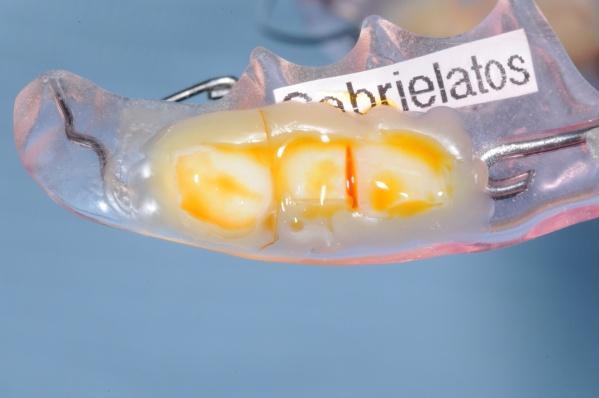
108 Figure Appliance 1. Five of the six tooth surfaces are protonated. Figure Appliance 2. Four of the six tooth surfaces are protonated. Figure Appliance 3. Five of the six tooth surfaces are protonated. 91


109 Figure Appliance 4. The tooth surfaces are protonated. 92
110 Comparison of the proportion of sound tooth surfaces protonated in vitro after subjection to ph 5 phosphate buffer (acidic challenge) with the proportion of sound tooth surfaces protonated in situ after subjection to 10% w/v sucrose rinse The proportion of sound non-sterile and sterile tooth surfaces that were protonated in vitro after subjection to ph 5 phosphate buffer (results obtained from in vitro studies 1 and 2) were compared with the proportion of sound tooth surfaces that were protonated in situ after subjection to 10% w/v sucrose rinse. The comparisons were made with Fisher's exact test since the assumptions for Chi-square test were not satisfied. The statistical analysis was carried out using R statistical software (R version , , Copyright 2010, The R Foundation for Statistical Computing). The level of statistical significance was The results are presented in Table 3.7. The proportions of sound non-sterile and sound sterile tooth surfaces that were protonated in vitro after the acidic challenge (ph 5 phosphate buffer) did not differ significant from the proportion of sound tooth surfaces that were protonated in situ (p=0.112). Table Comparison of the proportion of protonated tooth surfaces in vitro and in situ Groups of teeth Difference in proportion of protonated tooth surfaces (%) P-value in vitro Sound S vs in situ in vitro Sound NS vs in situ Sound S: sound sterile, Sound NS: sound non-sterile, *: statistically significant result at the 5% level 93
111 Chapter 4- Discussion 4.1. In vitro studies: Pilot in vitro studies of changes in the surface chemistry of enamel and dentine exposed to acid In accordance with previously published data (Robinson et al., 2005; Hochrein and Zahn, 2011) these in vitro studies showed that protonation of tooth surfaces which have been subjected to acidic (ph 5) phosphate buffers occurs and can be detected in the laboratory. These previous studies showed that protonation of the enamel hydroxyapatite crystals occurs when the ph of their immediate environment is acidic (Robinson et al., 2005; Hochrein and Zahn, 2011). Robinson et al. (2005) examined rat incisor enamel with Atomic Force Microscopy while Hochrein and Zahn (2011) created a theoretical model and investigated the saliva-enamel interface. However, none of the above studies have investigated whether carious lesions, which must have been subjected to acidic plaque, or sterilisation procedures used for in situ studies affect protonation of enamel crystals. In addition, the techniques used would not be suitable for oral investigations. Unlike previous investigations, the current in vitro studies aimed to use chemical indicators to detect protonation of human primary tooth surfaces and compare the protonation of sound and carious, sterile and non-sterile tooth surfaces both at baseline, after subjection to neutral (ph 7) phosphate buffer, but also after an acidic challenge, by ph 5 phosphate buffer. These were the first in vitro studies on protonation of human primary teeth with and without carious lesions. The results indicated that surfaces of sound and carious, sterile and non-sterile teeth were clearly protonated after an acidic challenge. Different protonation patterns were 94
112 identified among the various groups of teeth that were investigated. Differences in protonation could be attributed to the composition of the tooth surface (enamel, dentine, sound or carious tooth surface), the sterilisation or non-sterilisation of the tooth surface and the ph of the immediate environment (ph 7 or ph 5 phosphate buffer) The rationale of the study design These were prospective qualitative pilot in vitro studies of human primary tooth surfaces. Since there was no previous data on protonation of human teeth in the literature, it was necessary to first conduct a laboratory study to investigate the extent to which protonation of human tooth surfaces occurs and could be detected in vitro. Statistical advice was obtained from Biostatistics Unit (Centre of Biostatistics and Epidemiology, University of Leeds) and the appropriate sample size which would allow statistical calculations was 30 human primary tooth surfaces for each group of teeth. These in vitro studies were part of a research project which also involved in situ investigations of the surface chemistry of enamel which had been exposed to acids. Before insertion of the enamel slabs in the volunteers' mouths to conduct the in situ investigations, the tooth slabs had to be sterilised. Since the sterilisation procedure could affect the chemistry of enamel surfaces, the second part of these in vitro studies examined the effect of the sterilisation procedures on the protonation of the tooth surfaces. 95
113 Selecting the tooth surfaces of interest Since remnants of the ph indicator solution on the tooth surface could bias the results if the same tooth surfaces were investigated, different tooth surfaces were examined at baseline (ph 7 phosphate buffer) and after the acidic challenge (ph 5 phosphate buffer). One of the aims of this research project was to examine primary and permanent sound human teeth as well as teeth with various extents of carious lesions. Useful conclusions would, thus, be drawn on the effect of caries on the protonation state of a range of tooth surfaces. This would include possible differences in the way primary and permanent human teeth are affected by acidic solutions. It was not possible, owing to ethical considerations, to obtain sufficient teeth for all aspects of the study. In addition, the teeth that are extracted from children older than six years of age are sent to Leeds Dental Institute Tissue Bank and are, subsequently, distributed to the investigators. In order to collect freshly extracted teeth, the investigator had to collect them from children under the age of six since these teeth are not stored at the Tissue Bank. The Ethical approval included the collection of sound primary tooth surfaces. Therefore, only carious tooth surfaces that were incidentally obtained as part of the sound tooth surfaces collection were investigated. Despite the fact that the numbers obtained did not meet the required target from a statistical point of view, useful conclusions could still be drawn Baseline data Protonation of the tooth surfaces was examined at baseline following subjection to neutral (ph 7) phosphate buffer. This "pre-treatment" phase prior to the baseline data collection was of extreme importance in order to eliminate any confounding factors that would, otherwise, affect the baseline protonation state of the tooth surfaces. 96
114 Factors that affect the baseline data Numerous factors can affect the baseline protonation state of the tooth surfaces: i. The patient's oral hygiene and the presence of plaque on the tooth surfaces The patient's oral hygiene affects the amount of plaque which has been deposited on the tooth surfaces of interest. Plaque can, subsequently, affect the protonation state of the tooth surface in two different ways. Firstly, the ph of plaque can determine the protonation state of the tooth surface; that is acidic plaque would probably cause protonation. Additionally, the plaque bacteria can create an acidic environment in the close proximity of the tooth surface via metabolism of fermentable carbohydrates, should the patient have consumed these prior to the planned extraction. ii. The amount of plaque on the tooth surfaces. It is sensible to presume that the immediate environment of the tooth surface would be more acidic and, therefore, the tooth surface would be more protonated with increasing amount of dental plaque in the presence of fermentable carbohydrates. More plaque bacteria would, then, be available to metabolise the carbohydrates and create a more acidic environment in the immediate vicinity of the tooth surface. iii. The time that has elapsed between the last meal (food or drink) and the extraction of the tooth. "Resting plaque" is defined as plaque which has not been exposed to fermentable carbohydrates for at least two hours. "Starved plaque" refers to the dental plaque 8-12 hours after the last episode of consumption of fermentable carbohydrates. The ph of dental plaque varies from individual to individual as well as intra-orally for the same person. Significant differences of the plaque ph 97
115 exist between "resting plaque" and "starved plaque", with the former ranging between 6-7 and the latter between 7-8 (Edgar and O'Mullane, 1996). Therefore, the time that has elapsed between the last meal and the examination of the tooth surface is crucial as far as the protonation state of the tooth surface is concerned. iv. The food or drink that had been consumed before the extraction of the tooth. Consumption of fermentable carbohydrates induces acid production by plaque bacteria and, therefore, creation of acidic environment near the tooth surface. Therefore, the exact food or drink that was consumed before the extraction could affect plaque ph and, consequently, the baseline protonation. v. The patient's salivary ph In the absence of fermentable carbohydrates, the salivary ph of the patient can also affect the protonation state of the tooth surfaces. vi. History of topical fluoride treatment Previous work has revealed that fluoride incorporation into the tooth surface renders protonation more difficult (Robinson et al., 2006). Therefore, differences were expected between protonation of the various tooth surfaces that underwent the same interventions depending on previous fluoride treatment. All the factors mentioned above differ among the tooth donors and could produce significant bias to the results obtained both at baseline but also after the acidic challenge (ph 5 phosphate buffer). In order to reduce and, if possible, eliminate bias it was deemed appropriate to wipe the tooth surfaces with gauze to remove blood, soft tissue remnants and plaque deposits. Subsequently, the teeth were dipped into ph 7 phosphate buffer to neutralise the ph of their immediate environment before obtaining the baseline data. 98
116 Selecting the ph of the acidic phosphate buffer The intention of the research team was to examine protonation of human primary and permanent tooth surfaces with and without carious lesions after subjection to solutions of various acidic phs (ph 3, 5, 7 and 9). Unfortunately, the numbers of tooth surfaces required for the above investigations were not approved by the Ethical Committee and, therefore, only one acidic solution had to be selected. Robinson et al. (2005) showed that below ph 6.6 the enamel apatite crystals started to become unstable, presumably due to full phosphate protonation and adhesion to the negatively charged modified cantilever tip. Therefore, subjection of the tooth surfaces of interest to ph around 6 was sensible. In addition, many commonly consumed dietary foods and drinks have ph values ranging between 4 and 6. Investigating the tooth surfaces of interest after subjection to ph 5 phosphate buffer was, thus, considered appropriate Technique to assess protonation of the tooth surface Robinson et al. (2005) identified protonation of the enamel apatite surface with Atomic Force Microscopy. The technique requires the use of expensive equipment and an experienced operator capable of carrying out the measurements and interpreting the results. The aim of our in vitro study was to examine whether an easy, cheap and clinically applicable technique could, alternatively, be implemented to detect protonation and semi-quantify it. A universal ph indicator solution (Scientific Laboratory Suppliers, GPR) was, therefore, used to identify whether the tooth surface of interest was protonated or non-protonated. The assessment was based on the colour change of the ph indicator. The ph indicator solution is a useful method to assess protonation of the 99

117 tooth surface of interest. The subjectivity of the outcome interpretation (exact colour of the indicator) does not allow accurate conclusions on the exact ph of interest. The technique may not be fully reproducible as far as determining the exact ph, but in this study the outcome of interest is whether protonation occurs or does not occur on the surface of interest. Therefore, in all the studies conducted we assessed the difference between the protonated surfaces (green colour of the ph indicator) versus nonprotonated surfaces (yellow-orange-red colour of the ph indicator). This change in colour was reliably assessed visually by the investigator (Figures 4.1, 4.2). Figure Non-protonated tooth surface as assessed by the colour of the ph indicator (green) Figure Protonated tooth surface as assessed by the colour of the ph indicator (orange) 100
118 Unlike the Atomic Force Microscope, the ph indicator does not allow to draw conclusions on the exact time that the tooth surface starts dissolving. With Atomic Force Microscopy, measurements of adhesion forces between the cantilever tip and the tooth surface can provide extremely useful information on the tooth surface dissolution Assessment of the tooth surfaces with International Caries Detection and Assessment System II (ICDAS II) The tooth surfaces of interest were assessed with ICDAS II, a technique based on visual changes of the tooth surface. ICDAS II is an easy and quick method to visually assess the extent of carious lesions on the tooth surfaces. The technique has been reported to have excellent reproducibility in vitro with intra-examiner agreement ranging between 0.74 and 0.92 for proximal caries detection (Martignon et al., 2007; Shoaib et al., 2009) and between 0.76 and 0.78 for occlusal caries detection (Shoaib et al., 2009). On the other hand, the sensitivity and specificity of the technique are highly variable apparently depending on the extent of the carious lesion. Shoaib et al. (2009) examined in vitro primary carious tooth surfaces with ICDAS II and reported that the highest sensitivity was 77.9% when the lesion was in the middle third of dentine and the lowest was 63.1% when caries was restricted in the outer half of dentine. The specificities were high and ranged between 87% and 92.8% depending on the extent of the lesion. For proximal carious lesions the sensitivities and specificities ranged between 58.3%-75.3% and % respectively. The above findings imply that the assessment of the tooth surfaces with ICDAS II may not always be accurate. Therefore, tooth surfaces that were assessed as non-carious may have already had early changes with or without increased porosity and irreversible hard tissue loss; a fact that could have implications on their protonation state before and after the acidic challenge and the interpretation of the results. 101
119 Assessment of the tooth surfaces with a combination of ICDAS II and other means of caries detection (that is Transillumination, Quantitative Light-induced Fluorescence) can increase the accuracy of the assessment of the carious lesion Protonation of the various tooth surfaces at baseline (ph 7 phosphate buffer) Different protonation patterns of the various tooth surfaces were exhibited at baseline following subjection to ph 7 phosphate buffer Protonation of non-sterile sound and carious tooth surfaces at baseline (ph 7 phosphate buffer) All the sound non-sterile and sterile tooth surfaces were found non-protonated at baseline, that is the ph indicator revealed a colour corresponding to ph 7. Approximately half of the non-sterile tooth surfaces with distinct visual changes in enamel (White spot NS) and 75% of the non-sterile tooth surfaces with cavitated caries into dentine (Dentine NS) were found protonated, that is with indicator colours relating to ph values lower than 7. The differences between protonation of sound non-sterile tooth surfaces and carious non-sterile tooth surfaces were significant. Protonation of the carious non-sterile tooth surfaces at baseline was most likely due to the carious process. However, the different composition of enamel and dentine surfaces as well as the small sample size of the carious tooth surfaces that were available for investigations may have contributed to the significant differences that were observed. It has been shown that protonation of the tooth surface is an important prerequisite for dissolution (Robinson et al., 2005; Hochrein and Zahn, 2011). The carious tooth surfaces were probably protonated as part of the carious process; that is exposure to 102
120 acidic plaque. The plaque bacteria created the acidic environment and protonation occurred before any irreversible visual change presented on the tooth surfaces and prior to any hard tissue loss. Therefore, carious tooth surfaces which had undergone irreversible hard tissue loss, were expected to be significantly protonated provided that they were still in a cariogenic environment. On the other hand, the different composition of the dental hard tissues may explain the increased protonation of carious tooth surfaces with exposed dentine. Dental enamel consists of 96% mineral component in the form of calcium hydroxyapatite, 3% water and 1% organic matrix (Avery and Chiego, 2006; Bath-Balogh and Fehrenbach, 2006). Hydroxyapatite crystals are organised in enamel rods which, subsequently, group together to form the dental enamel. On the other hand, dentine consists of 70% inorganic component in the form of hydroxyapatite, 20% collagen fibres and 10% water (Avery and Chiego, 2006; Bath-Balogh and Fehrenbach, 2006). The critical ph for dentine is higher than that of enamel (Mellberg, 1992) and the carious lesions initiate very fast in dentine (Ogaard and Rolla, 1992). This may have implications on the protonation of dentine. The results should be interpreted with caution due to the small sample size of the carious tooth surfaces. A larger sample size would allow more accurate conclusions Protonation of sterile sound and carious tooth surfaces at baseline (ph 7 phosphate buffer) The proportion of non-protonated sterile tooth surfaces at baseline was significantly higher compared to the proportion of protonated sterile tooth surfaces irrespectively of the presence and extent of the carious lesion. Protonation of tooth surfaces did not differ significantly among the three groups of teeth (sound teeth, teeth with distinct visual change in enamel, teeth with cavitated lesions into dentine). This was in contrast to the 103
121 results that were obtained when non-sterile tooth surfaces were examined and can be attributed either to the sterilisation procedure or the small sample sizes of the groups of carious teeth that were available for investigation The effect of the sterilisation procedure on protonation of the various tooth surfaces at baseline The sterilisation procedure involved immersing the teeth in 12% w/v sodium hypochlorite overnight. Subjection to sodium hypochlorite has been shown to eliminate prions without affecting the mineral phase or structure of the tooth surface (Driscoll et al., 2002). However, the procedure causes significant de-proteination of the tooth surface even at much lower concentrations (Hu et al., 2010). One of the clinical implications of the protein pellicle is the protection of the tooth surface against chemical insults and, thus, the initiation of dental caries and erosion (Edgar and O'Mullane, 1996). Therefore, it was extremely important to re-obtain the protein pellicle before the tooth surfaces underwent any acidic challenge. The tooth surfaces were placed in natural human saliva for 24 hours to allow formation of the pellicle. The pellicle forms immediately when the tooth comes in contact with saliva (Edgar and O Mullane, 1996) and reaches a thickness of μm within 24 hours (Fejerskov and Kidd, 2008). Twenty-four hours was, therefore, considered enough time to ensure formation of this thin layer on the tooth surfaces. The proportion of protonated non-sterile sound tooth surfaces did not differ significantly from that of protonated sterile sound tooth surfaces at baseline (p=1). On the other hand, significantly more protonated non-sterile tooth surfaces were found for both the group of teeth with distinct visual change in enamel (White spot) and the group of teeth with cavitated lesions into dentine (Dentine) when compared with protonated sterile surfaces with the same extent of carious lesions. The significant differences that were observed 104
122 can be attributed either to the sterilisation procedure or the small numbers of tooth surfaces that were available for investigations. The sterilisation procedure involved immersion of the teeth in 12% w/v sodium hypochlorite overnight. The highly alkaline ph of sodium hypochlorite might have contributed to the removal of protons from the previously protonated carious tooth surfaces Protonation of the various tooth surfaces after the acidic challenge (ph 5 phosphate buffer) Following subjection to ph 5 phosphate buffer, all the tooth surfaces were found protonated irrespective of the extent of the carious lesions or previous sterilisation of the tooth surfaces. There were no differences among the various tooth surfaces. It is important to note that some tooth surfaces were probably more protonated than others; a fact that was determined by a lower ph based on the colour change of the ph indicator. However, the colour differences among the different ph values cannot be estimated accurately (Figure 2.1) and, therefore, quantification of the degree of protonation was not attempted. In general, the ph decreased and, therefore, protonation of the tooth surface increased with increasing extent of the carious lesion. This was expected as tooth surface dissolution requires significant protonation (Robinson et al., 2005; Hochrein and Zahn, 2011) The effect of the sterilisation procedure on protonation of the various tooth surfaces after the acidic challenge (ph 5 phosphate buffer) All the tooth surfaces were found protonated after the acidic challenge and no differences were observed between sterile and non-sterile tooth surfaces. In this case, the prolonged exposure of the tooth surfaces to the alkaline ph of sodium hypochlorite 105
123 during the sterilisation procedure did not prevent protonation during the subsequent acidic challenge Comparison of the proportion of protonated tooth surfaces at baseline (ph 7 phosphate buffer) with the proportion of protonated tooth surfaces after the acidic challenge (ph 5 phosphate buffer) The proportions of protonated tooth surfaces after subjection to ph 5 phosphate buffer (acidic environment) were found to be significantly higher compared to the proportions of protonated tooth surfaces at baseline (ph 7 phosphate buffer) for all the groups of teeth that were investigated apart from the non-sterile tooth surfaces with cavitated carious lesions into dentine (Dentine NS group). Even though all the tooth surfaces were found protonated after the acidic challenge, for this specific group a large percentage of tooth surfaces were also protonated at baseline (75%). The non-significant difference is probably a result of significant protonation at baseline as a result of the carious process. However, the sample size of this group of teeth was small with only 8 tooth surfaces being available for investigations at baseline and 7 tooth surfaces after the acidic challenge. A larger sample size would reduce type II statistical errors. 106
124 4.2. In situ study: Pilot in situ study of changes in the surface chemistry of enamel and dentine exposed to acid. A surface study of caries. This was the first in situ study on protonation of sound human primary teeth. Sound enamel tooth surfaces were clearly protonated after subjection to 10% w/v sucrose rinse. The results are in accordance with the previous in vitro studies, which showed protonation of sound sterile and non-sterile tooth surfaces after an acidic challenge The rationale of the study design This was a prospective qualitative and semi-quantitative pilot in situ study on the protonation of human sound primary tooth surfaces. Since there was no previous data in the literature, it was necessary to conduct a pilot in situ study to investigate whether protonation of human tooth surfaces which have undergone an acidic challenge occurs and can be detected in situ The sample size Since there was no previous data in the literature regarding protonation of teeth, this was a pilot in situ study. Statistical advice was obtained from Biostatistics Unit (Centre of Biostatistics and Epidemiology, University of Leeds) and it was agreed that an appropriate sample size which would allow statistical calculations were 30 sound enamel human primary tooth surfaces The participants of the in situ study For practical reasons in situ studies usually involve small numbers of participants (Zero, 1995). This in situ study investigated 30 enamel slabs. Five volunteers were recruited 107
125 and participated, which was an appropriate number to allow investigations of the required number of enamel slabs in a variety of oral environments. It has been suggested that volunteers taking part in in situ studies of caries should be standardised on parameters that could affect the development of a carious lesion (Zero, 1995). This study attempted to standardise the study participants for the following parameters: i. Demographics All the participants were between 21 and 31 years of age. The volunteers were not standardised in terms of gender and ethnic background, as these factors are not expected to affect the study results (Zero, 1995). In situ caries investigations with adult volunteers have been considered more appropriate in comparison to similar studies on children. The results of these studies can, then, be generalised to children's populations due to the similar caries rates that have been reported in adults and schoolchildren (Stookey, 1992). In addition, the difficulty of involving children in in situ investigations makes in situ studies with adult participants the most appropriate realistic option. Limited time availability, lack of compliance and ethical concerns are the most important factors that have not made in situ studies on children common practice (Stookey, 1992). ii. Medical history It has been suggested that participants in in situ studies should not be affected by any medical condition with potential effects on their oral health (ten Cate et al., 1992; Zero, 1995). Good general health also reduces the risk of cross-infection of the investigators and any individual with whom they come in contact for the 108
126 duration of the study. Volunteers should not take any medication which could affect their oral microflora or oral health in any way. All the participants in this in situ study were medically fit and well and had not been taking any medication regularly for at least three months before the beginning of the study. iii. Dental health It has been suggested that the participants of in situ studies should be susceptible to the disease under investigation (Stookey, 1992; Zero, 1995). Theoretically, every individual is susceptible to caries under certain circumstances. Participants with active carious lesions should not be included in in situ studies of caries to avoid exacerbation of the disease due to the use of the intra-oral appliance (ten Cate et al., 1992; Zero, 1995). All the participants who were included in this in situ study had a thorough clinical examination prior to any intervention. The volunteers differed in terms of previous dental health status, with some of them having multiple restorations (fillings, crowns) and some others having a healthy dentition with no signs of current or past caries experience. None of the participants had unrestored active or inactive carious lesions. Differences in previous dental health are desirable in in situ studies. This variability provides a way to ensure that the study participants represent the general population and make the results generally applicable (ten Cate, 1992). However, recruitment of participants with extreme DMFT was avoided in order to reduce the introduction of confounding factors which could, potentially, affect the study results. The clinical examination also involved a thorough screening of the periodontal tissues. All the participants had a healthy periodontium. This was in accordance with previous recommendations (ten Cate et al., 1992) as the use of in situ 109
127 appliances may affect the gingival and periodontal health by predisposing the natural dentition to plaque accumulation. The number of natural teeth that are present affects the oral microflora. Edentulous patients' oral environment has less demineralisation potential after cariogenic challenges (ten Cate et al., 1992). In an attempt to standardise the quality of oral microflora, participants with at least 20 natural teeth were selected and participated in this study. iv. Previous fluoride experience The study participants have always been living in places without fluoridated water. They had been brushing their teeth with fluoridated dentifrice and had not had topical fluoride application for at least one year. Castillo et al. (2001) reported that fluoride release following fluoride varnish application lasts for approximately five to six months. Therefore, one year was more than enough to ensure that the effect of topical fluoride application was not evident. v. Oral hygiene habits and fluoride exposure The participants were advised to brush their natural dentition at least twice daily with fluoridated toothpaste containing 1450ppm fluoride. A 0.05% NaF mouthwash was prescribed and the volunteers were instructed to rinse the mouth with 10mls of the mouthwash once daily after removing the in situ appliance from the mouth. These preventive measures were necessary to reduce the risk of caries development on the natural dentition and standardise the fluoride exposure of the study participants. vi. Dietary habits The diet plays a major role in the development of dental caries both in terms of type of foods and drinks which are consumed but also as far as the frequency of consumption is concerned (Duggal et al., 2001; Watt, 2003). Most in situ studies 110
128 have not attempted to standardise the dietary habits of the participants, presumably due to the long duration of the studies as well as compliance issues. This in situ study did not give any dietary recommendations to the volunteers but advised them to remove the appliance during mealtimes The in situ appliance design Several in situ appliance designs have been reported in the literature for intra-oral investigations of dental caries (Table 1.1). Upper or lower, full or partial dentures have been described. Lower removable appliances are the most commonly used intra-oral in situ models for caries investigations (Corpron et al., 1992; Koulourides and Chien, 1992; Stephen et al., 1992) probably due to the high cariogenic potential of the mandibular posterior region (Zuniga and Koulourides, 1969). In accordance with previous investigations, this in situ study used a lower intra-oral removable appliance with incorporated sound sterile enamel slabs. Three enamel slabs were placed into small recessions on each buccal flange. In previous studies the enamel slabs were covered with a piece of gauze in order to facilitate plaque accumulation (Koulourides et al., 1976). However, it has been shown that in the presence of gauze, the obtained plaque microflora differs from that of naturally formed dental plaque (ten Cate et al., 1992) and, therefore, this technique was not followed in our study. The enamel slabs were placed in small recesses on the buccal flanges of the intra-oral appliances. The participants were advised to wear the appliance for 48 hours without brushing the enamel slabs to ensure that enough plaque was collected on the slabs by the end of the second day. 111
129 The age of plaque The age and the position of dental plaque intra-orally affect its ph and, therefore, the cariogenic potential. The age of plaque is determined by the time that has passed since the last episode of prophylaxis or toothbrushing. The time that plaque has been accumulating affects its thickness and, consequently, its microflora (Edgar and O'Mullane, 1996). Most studies have used plaque from tooth surfaces which were not brushed for one to two days. In our study, we left the enamel slabs intra-orally for 48 hours and advised the volunteers to refrain from all oral hygiene procedures when the appliances were in the mouth The cariogenic challenge The cariogenic solution The cariogenic challenges in in situ studies are usually achieved through dietary foods and drinks with known cariogenic potential such as sucrose solutions or other demineralising agents. The solution can be delivered to the appliance either intra-orally with the in situ appliance in the mouth or extra-orally after removing the appliance from the oral environment (Manning and Edgar, 1992). Ideally, intra-oral investigations are desired. They can provide more valid study results due to the presence of saliva, which plays a crucial role in the development of the carious lesions. Unfortunately, this cannot always be achieved, mainly due to ethical concerns regarding the participants' natural dentition. Consumption of cariogenic products with known demineralisation potential increases the caries risk for the natural dentition and raises ethical concerns, particularly in a study of prolonged duration. 112
130 In this in situ study, a 10% w/v sucrose rinse was used to enable plaque bacteria to create the desired acidic environment. The sucrose rinse was delivered to the enamel slabs extra-orally immediately after removing the appliance from the volunteers' mouth The duration of the cariogenic challenge Various studies have used 10% w/v sucrose rinses to enable demineralisation procedures to occur on the enamel surfaces. Dental plaque was, first, collected on the tooth surface and the sucrose rinse was, subsequently, delivered to enable plaque bacteria to create the acidic environment required for the beginning of the demineralisation process (Kashket and Lopez, 1992; Simone et al., 1992). The duration of the cariogenic challenge varies among the different studies. In our in situ study the enamel slabs were subjected to the sucrose rinse for 30 minutes. This was much longer compared to previous studies on enamel demineralisation. However, this duration would allow comparisons with our previous in vitro investigations, in which the enamel slabs underwent the acidic challenge for 30 minutes. Additionally, numerous cariogenic attacks in the mouth last for a considerable amount of time with oral clearance reaching or even exceeding 30 minutes, particularly if sticky dietary products have been consumed Protonation of the enamel slabs after the cariogenic challenge (subjection to 10% w/v sucrose rinse) In our in situ study 26 of the 30 enamel slabs (87%) were found protonated after the cariogenic challenge. The proportion of protonated enamel slabs was significantly higher than the proportion of non-protonated slabs. It should be noted that some tooth surfaces were found more protonated compared to others, which was determined by the colour change of the ph indicator solution which was used to determine the protonation 113
131 (Figure 2.1). However, quantification of the tooth surfaces protonation was not attempted as the ph indicator solution does not allow objective and reproducible measurements. Interestingly, in certain volunteers' mouths some tooth surfaces were found more protonated than others. This can be attributed to increased plaque accumulation on certain participants enamel slabs, lower plaque ph, better compliance with the use of the intra-oral appliance compared to other participants or different microbial composition of the dental plaque. The four tooth surfaces that were found non-protonated were placed in three different in situ appliances. Volunteers who complied better with the study protocol may have kept the appliance intra-orally for significantly longer compared to others who probably were not as compliant. The formed dental plaque could, thus, be thicker. Thicker dental plaque contains more anaerobic bacterial species (Edgar and O'Mullane, 1996). All these factors may have differed between the participants and can have had an impact on the protonation of the enamel slabs. The diet history of the volunteers also plays a major role in the composition of dental plaque. Frequent consumption of sucrose allows production of extracellular and intracellular polysaccharides (Edgar and O'Mullane). The former increase plaque thickness and adhesion on the tooth surface while the latter increase acid production in resting plaque. It is, therefore, evident that different dietary habits among the participants may have played a role in the degree of protonation of the tooth surface that has been observed. 114
132 Comparison of the proportions of protonated tooth surfaces in situ and in vitro The proportion of enamel slabs that were protonated in situ after the cariogenic challenge was compared to the proportion of sound sterile and sound non-sterile tooth surfaces that were protonated in vitro after the acidic challenge. The differences were not statistically significant at the 5% level. The results of the in situ and the in vitro studies are not directly comparable due to the different nature of investigations. The in vitro studies were carried out in the absence of dental plaque and focused on investigations of protonation/ non-protonation following an acidic attack. On the other hand, the in situ study used dental plaque and a cariogenic agent (10% w/v sucrose rinse) to create the acidic environment. The oral bacteria of the dental plaque were provided with the appropriate sugary source that would enable acid production on the tooth surface. Even though the two procedures differ, they both involve an acidic challenge on the tooth surface. These studies showed that protonation of tooth surfaces occurs and can be detected in vitro and in situ Limitations of the in situ study The results of this in situ study indicate that protonation of sound primary human enamel slabs which have been subjected to a cariogenic challenge occurs and can be detected with a simple technique (ph indicator solution). However, several parameters should be taken into consideration before the generalisation of the study results to normal oral conditions. 115
133 The sucrose rinse was delivered outside the mouth and not in the oral environment. The enamel slabs were, thus, subjected to the cariogenic challenge for 30 minutes without the beneficial effects of saliva acting simultaneously. The salivary flow rate plays a major role in carbohydrate clearance from the oral environment and protects, in this way, the dental hard tissues against caries initiation and development. The buffering capacity of saliva mainly depends on the concentration of bicarbonate in stimulated saliva. Salivary ph, therefore, increases with increased flow rate and increased concentration of bicarbonate and rises up to 7.8 at high flow rates (Edgar and O'Mullane, 1996). Additionally, with increasing salivary flow rates the concentration of calcium (Ca 2+ ), phosphate (PO4 3- ) and hydroxide (OH - ) ions in saliva elevate, which further protects the dental hard tissues against demineralisation processes. Stimulated salivary flow rate increases for approximately 60 seconds after a sucrose rinse (Edgar and O'Mullane, 1996). Even within this small period of time, saliva is capable of diluting the sucrose rinse and reducing the amount of carbohydrate which is available to plaque bacteria. Outside the mouth, the situation is completely different. The beneficial effect of saliva is not present and, therefore, the sucrose rinse is constantly available to plaque bacteria for 30 minutes. The plaque ph remains low and much more acid production is expected in the lack of the salivary clearance mechanism. Studies have shown that in the absence of saliva, the ph of dental plaque was reduced. The clinical significance of saliva absence in in situ investigations may be an overestimation of the protonation patterns of the enamel slabs. If the same investigations were carried out intra-orally, the salivary clearance may have reduced the amount of carbohydrates available. Consequently, less acid would be produced from plaque bacteria and probably protonation would not be as evident. 116
134 Even though protonation does occur in situ after the cariogenic challenge, it is not clear exactly how long is needed for protonation to be evident as well as how long it takes for porosity and irreversible hard tissue dissolution to occur. In this in situ study, a ph indicator solution was used to assess protonation of the tooth surface. This method is easy, quick and clinically applicable but does not provide any information on hard tissue dissolution. Previous Atomic Force Microscopy studies showed that significant protonation occurs in acidic ph and dental hard tissue dissolution begins when the ph drops below 6.6 (Robinson et al., 2005). The ability to assess whether hard tissue dissolution has occurred or finding out the relation between protonation and beginning of dissolution is an important piece of information with major clinical implications. This would suggest that intensive prevention should be applied in order to stop the progression of dissolution and, if possible, reverse the procedure. Unfortunately, this was not possible in this in situ study. Despite the limitations of these in situ investigations, this study revealed that protonation of sound human primary tooth surfaces occurs in situ and can be detected with an easy, quick and clinically applicable technique The clinical implications of the results of this in situ study The results of this in situ study imply that very early changes of the surface chemistry of dental enamel (protonation) occur and can be detected with an easy and clinically applicable technique. Even though it is not clear whether any hard tissue dissolution has taken place at a microscopic level on the protonated enamel surfaces, it is important to underline that none of these surfaces exhibited any clinically detectable visual change. This suggests that any changes of the dental enamel are at a very early stage at which invasive dental treatment is not required. 117
135 The ability to identify protonated tooth surfaces provides the clinician with the opportunity to apply the wide range of the available preventive techniques and target all these "susceptible" tooth surfaces at a very early stage. Progression of the carious process can, thus, be halted or even reversed. This is a completely novel technique and is the only way to detect tooth surfaces at risk before any visual change and before porosity and mineral loss occur. Clinical application of this method will benefit both low caries as well as high caries risk populations. The former will get the chance to maintain a healthy, caries free dentition and the latter will benefit from very early identification of tooth surfaces that are at high risk of dental caries before any irreversible changes of the dental hard tissues take place. 118
136 4.3. Future research These in vitro and in situ studies were the first investigations on protonation of human primary tooth surfaces. Further research is required before the technique can be clinically applicable. Larger in vitro and in situ studies on primary and permanent tooth surfaces will allow comparisons between the protonation patterns of primary and permanent teeth. The results of our investigations can be used to make power calculations and identify appropriate sample sizes. Atomic Force Microscopy investigations of the protonated tooth surfaces may clarify exactly when protonation occurs and correlate protonation of the tooth surface with the initiation of enamel dissolution. Furthermore, the effect of fluoride treatment on early changes of the surface chemistry of enamel requires further research. In vivo studies with appropriate sample sizes should be carried out to identify whether the technique is applicable intra-orally under normal oral conditions. 119
137 Chapter 5- Conclusions These in vitro and in situ studies on sterile and non-sterile, intact and carious human primary tooth surfaces concluded in the following: i. In vitro studies The use of chemical ph indicators may offer a simple and convenient approach to assess the degree of protonation of tooth surfaces. Sound sterile and non-sterile human primary tooth surfaces were nonprotonated (ph~7) when the ph of the immediate environment of the tooth surface was neutral (ph 7). The proportions of protonated and non-protonated carious non-sterile human primary tooth surfaces did not differ when the ph of the immediate environment of the tooth surface was neutral (ph 7). Carious sterile human primary tooth surfaces were non-protonated when the ph of the immediate environment of the tooth surface was neutral (ph 7). Human primary tooth surfaces became protonated after an acidic challenge. ii. In situ study Sound human primary tooth surfaces became protonated after a cariogenic challenge. 120
138 Chapter 6- References ABALOS, C., M. HERRERA, A. JIMENEZ-PLANAS, R. LLAMAS Performance of laser fluorescence for detection of occlusal dentinal caries lesions in permanent molars: an in vivo study with total validation of the sample. Caries Res. 43 (2), pp ABLAL, M.A., J.S. KAUR, L. COOPER, F.D. JARAD, A. MILOSEVIC, S.M. HIGHAM, A.J. PRESTON The erosive potential of some alcopops using bovine enamel: an in vitro study. J Dent. 37 (11), pp ADDY, M., J. HUGHES, M.J. PICKLES, A. JOINER, E. HUNTINGTON Development of a method in situ to study toothpaste abrasion of dentine. Comparison of 2 products. J Clin Periodontol. 29 (10), pp ALKURT, M.T., I. PEKER, H.D. ARISU, O. BALA, B. ALTUNKAYNAK In vivo comparison of laser fluorescence measurements with conventional methods for occlusal caries detection. Lasers Med Sci. 23, pp AMAECHI, B.T., S.M. HIGHAM, W.M. EDGAR Efficacy of sterilisation methods and their effect on enamel demineralization. Caries Res. 32 (6), pp AMAECHI, B.T., S.M. HIGHAM In vitro remineralisation of eroded enamel lesions by saliva. J Dent. 29 (5), pp ANGNES, G., V. ANGNES, R.H.M. GRANDE, M. BATTISTELLA, A.D. LOGUERCIO, A. REIS Occlusal caries diagnosis in permanent teeth: an in vitro study. Braz Oral Res. 19 (4), pp
139 ANTTONEN, V., L. SEPPA, H. HAUSEN Clinical study of the use of the laser fluorescence device DIAGNOdent for detection of occlusal caries in children. Caries Res. Jan-Feb; 37 (1), pp ARENDS, J Zeta potentials of enamel and apatites. J Dent. 7 (3), pp ARSLANTUNALI TAGTEKIN, D., F. OZTURK, M. LAGERWEIJ, O. HAYRAN, G.K. STOOKEY, F. CALISKAN YANIKOGLU Thickness measurement of worn molar cusps by ultrasound. Caries Res. 39 (2), pp ATTRILL, D.C., P.F. ASHLEY Occlusal caries detection in primary teeth: a comparison of DIAGNOdent with conventional methods. Br Dent J. Apr 28; 190 (8), pp AVERY, J.K., D.J. CHIEGO Essentials of oral histology and embryology. A clinical approach. Third edition. St. Louis: Mosby Elsevier. BAKHOS, Y., F. BRUDEVOLD Effect of initial demineralization on the permeability of human tooth enamel to iodode. Arch Oral Biol. 27 (3), pp BARBERIA, E., M. MAROTO, M. ARENAS, C.C. SILVA A clinical study of caries diagnosis with a laser fluorescence system. J Am Dent Assoc. May; 139 (5), pp BATH-BALOGH, M., FEHRENBACH M.J Dental embryology, histology and anatomy. Second edition. St. Louis: Elsevier Saunders. BIN-SHUWAISH, M., P. YAMAN, J. DENNISON, G. NEIRA The correlation of DIFOTI to clinical and radiographic images in class II carious lesions. J Am Dent Assoc. Oct; 139 (10), pp
140 BOWEN, W.R., N. HILAL Atomic Force Microscopy in Process Engineering- An Introduction to AFM for Improved Processes and Products. [Online]. Oxford: Elsevier. [Accessed 4 June 2012]. Available from: LAY_bookid=2731&VerticalID=0 BRUDEVOLD, F., F. ATTARZADEH, A. TEHRANI, J. VAN HOUTE, J. RUSSO Development of a new intraoral demineralization test. Caries Res. 18 (5), pp CASTILLO, J.L., P. MILGROM, E. KHARASCH, K. IZUTSU, M. FEY Evaluation of fluoride release from commercially available fluoride varnishes. J Am Dent Assoc. 132 (10), pp Ceramic Industry Ceramic Industry Website. [Online]. [Accessed 25 May 2012]. Available from: CHADWICK, R.G., H.L. MITCHELL, I. CAMERON, B. HUNTER, M. TULLEY Development of a novel system for assessing tooth and restoration wear. J Dent. 25 (1), pp CHANG, Z.J., X.M. WANG, F.Z. CUI, J. GE, J.X. YAN The enamel softening and loss during early erosion studied by AFM, SEM and nanoindentation. Biomed Mater. 4 (1), CHU, C. H., E.C. LO, D.S. YOU Clinical diagnosis of fissure caries with conventional and laser-induced fluorescence techniques. Lasers Med Sci. May; 25 (3), pp Epub 2009 Mar
141 COLLOIDAL DYNAMICS PTY LTD Colloidal Dynamics. Electroacoustics Tutorials. [Online]. [Accessed 10 June 2012]. Available from: CORPRON, R.E., F.G. MORE, G. MOUNT Comparison of fluoride profiles by SIMS with mineral density of subsurface enamel lesions treated intra-orally with a fluoride-releasing device. J Dent Res. 71 Spec, pp CORTES, D.F., K.R. EKSTRAND, A.R. ELIAS-BONETA, R.P. ELLWOOD An in vitro comparison of the ability of fibre-optic transillumination, visual inspection and radiographs to detect occlusal caries and evaluate lesion depth. Caries Res. 34, pp COSTA, A.M., L.M. PAULA, A.C. BEZERRA Use of Diagnodent for diagnosis of non-cavitated occlusal dentin caries. J Appl Oral Sci. Feb; 16 (1), pp DARLING, A.I Studies of the early lesion of enamel caries with transmitted light, polarized light and radiography. Brit Dent J. 101, pp , DIJKMAN, A.G., J. SCHUTHOF, J. ARENDS In vivo remineralization of plaque-induced initial enamel lesions- a microradiographic investigation. Caries Res. 20 (3), pp DINIZ, M.B., J.A. ROGRIGUES, I. HUG, RDEC. CORDEIRO, A. LUSSI Reproducibility and accuracy of the ICDAS-II for occlusal caries detection. Community Dent Oral Epidemiol. Oct; 37 (5), pp Epub 2009 Jul 22. DOMINGUEZ, J.A., B. BITTENCOURT, M. MICHEL, N. SABINO, J.C. GOMES, O.M. GOMES Ultrastructural evaluation of enamel after dental bleaching associated with fluoride. Microsc Res Tech. Mar 15. doi: /jemt [Epub ahead of print] 124
142 DRISCOLL, C.O., S.E. DOWKER, P. ANDERSON, R.M. WILSON, K. GULABIVALA Effects of sodium hypochlorite solution on root dentine composition. J Mater Sci Mater Med. 13 (2), pp DRURY, R.A.B., E.A. WALLINGTON Carleton s histological techniques. Fourth edition. Oxford: University Press. DUGGAL, M.S., K.J. TOUMBA, B.T. AMAECHI, M.B. KOWASH, S.M. HIGHAM Enamel demineralisation in situ with various frequencies of carbohydrate consumption with or without fluoride toothpaste. J Dent Res. 80 (8), pp EDGAR, W.M., D.M. O MULLANE Saliva and oral health. Second edition. London: Thanet Press Limited, Margate. Edited Report of an FDI/WHO Scientific Workshop, Erfurt, Aetiology and prevention of dental caries and periodontal diseases Int Dent J. 32 (1), pp ELLWOOD, R.P., D.F. CORTES In vitro assessment of methods of applying the electrical caries monitor for the detection of occlusal caries. Caries Res. 38, pp ELTON, V., L. COOPER, S.M. HIGHAM, N. PENDER Validation of enamel erosion in vitro. J Dent. 37 (5), pp FEATHERSTONE, J.D., J.M. BEHRMAN, J.E. BELL Effect of whole saliva components on enamel demineralization in vitro. Crit Rev Oral Biol Med. 4 (3-4), pp FEJERSKOV, O., E. KIDD Dental caries. The disease and its clinical management. Second edition. Oxford: Blackwell Munksgaard. FEJERSKOV, O., B. NYVAD, M.J. LARSEN Human experimental caries models: intra-oral environmental variability. Adv Dent Res. 8 (2), pp
143 GANSS, C., J. KLIMEK, N. SCHWARZ A comparative profilometric in vitro study of the susceptibility of polished and natural human enamel and dentine surfaces to erosive demineralization. Arch Oral Biol. 45 (10), GANSS, C., J. KLIMEK, U. SCHAFFER, T. SPALL Effectiveness of two fluoridation measures on erosion progression in human enamel and dentine in vitro. Caries Res. 35 (5), HALL, A.F., J.P. SADLER, R. STRANG, E. DE JOSSELIN DE JONG, R.H. FOYE, S.L. CREANOR Application of transverse microradiography for measurement of mineral loss by acid erosion. Adv Dent Res. 11 (4), pp HALLSWORTH, A.S., C. ROBINSON, J.A. WEATHERBELL Mineral and magnesium distribution within the approximal carious lesion of dental enamel. Caries Res. 6 (2), pp HANNIG, M., N.S. HESS, W. HOTH-HANNIG, M. DE VRESE Influence of salivary pellicle formation on enamel demineralization- an in situ pilot study. Clin Oral Investig. 7 (3), pp HEINRICH-WELTZIEN, R., J. KUHNISCH, S. IFLAND, S. TRANAEUS, B. ANGMAR-MANSSON, L. STOSSER Detection of initial caries lesions on smooth surfaces by quantitative light-induced fluorescence and visual examination: an in vivo comparison. Eur J Oral Sci. Dec; 113 (6), pp HERKSTROTER, F.M., M. WITJES, J. RUBEN, J. ARENDS Time dependency of microhardness indentations in human and bovine dentine compared with human enamel. Caries Res. 23 (5), pp HINTZE, H., A. WENZEL, B. DANIELSEN, B. NYRAD Reliability of visual examination, fibre-optic transillumination, and bite-wing radiography and 126
144 reproducibility of direct visual examination following tooth separation for the identification of cavitated carious lesions in contacting approximal surfaces. Caries Res. 32, pp HOCHREIN, O., D. ZAHN On the molecular mechanisms of the acid-induced dissociation of hydroxyl-apatite in water. J Mol Model. 17 (6), pp HU, X., Y. PENG, C.P. SUM, J. LING Effects of concentrations and exposure times of sodium hypochlorite on dentin deproteination: attenuated total reflection Fourier transform infrared spectroscopy study. J Endod. 36 (12), pp HUYSMANS, M.C.D.N.J.M., C. LONGBOTTOM, N.B. PITTS Electrical methods in occlusal caries diagnosis: An in vitro comparison with visual inspection and bite-wing radiography. Caries Res. 32, pp HUYSMANS, M.C.D.N.J.M., J. KUHNISCH, J.J. TENBOSCH Reproducibility of electrical caries measurements: A technical problem? Caries Res. 39, pp IE, Y.L., E.H. VENDONSCHOT, M.J.M. SCHAEKEN, M.A. VAN T HOF Electrical conductance of fissure enamel in recently erupted molar teeth as related to caries status. Caries Res. 29, pp INTERNATIONAL CARIES DETECTION & ASSESSMENT SYSTEM COORDINATING COMMITTEE Criteria manual for the International Caries Detection and Assessment System (ICDAS II). Revised in December and July [Online]. [Accessed 15 January 2011]. Available from JABLONSKI-MOMENI, A., V. STACHNISS, D.N. RICKETTS, M. HEINZEL- GUTENBRUNNER, K. PIEPER Reproducibility and accuracy of the ICDAS II for detection of occlusal caries in vitro. Caries Res. 42, pp
145 KARLSSON, L Caries detection methods based on changes in optical properties between healthy and carious tissue. Int J Dent Epub 2010 Mar 28. KASHKET, S., L.R. LOPEZ Reduction of intra-oral demineralization of enamel after single exposures to sodium fluoride. J Dent Res. 71 Spec, pp KONIG, K., G. FLEMMING, R. HIBST Laser-induced autofluorescence spectroscopy of dental caries. Cell Mol Biol (Noisy-le-grand). 44 (8), pp KOULOURIDES, T., R. BODDEN, S. KELLER, L. MANSON-HING, J. LASTRA, T. HOUSCH Cariogenicity of nine sugars tested with an intraoral device in man. Caries Res. 10 (6), pp KOULOURIDES, T., M.C. CHIEN The ICT in situ experimental model in dental research. J Dent Res. 71 Spec, pp KUHNISCH, J., S. IFLAND, S. TRANAEUS, R. HICKEL, L. STOSSER, R. HEINRICH-WELTZIEN In vivo detection of non-cavitated caries lesions on occlusal surfaces by visual inspection and quantitative light-induced fluorescence. Acta Odont Scand. 65, pp LUSSI, A., S. IMWINKELRIED, N.B. PITTS, C. LONGBOTTOM, E. REICH Performance and reproducibility of a laser fluorescence system for detection of occlusal caries in vitro. Caries Res. Jul-Aug; 33, pp LUSSI, A., P. FRANCESCUT Performance of conventional and new methods for the detection of occlusal caries in deciduous teeth. Caries Res. Jan-Feb; 37 (1), pp 2-7. LUSSI, A Dental erosion: from diagnosis to therapy. London: Karger. Chapter 20, pp
146 MANNING, R.H., W.M. EDGAR Intra-oral models for studying de- and remineralization in man: methodology and measurement. J Dent Res. 71 Spec, pp MARGOLIS, H.C., E.C. MORENO Composition and cariogenic potential of dental plaque fluid. Crit Rev Oral Biol Med. 5 (1), pp MARGOLIS, H.C., B.J. MURPHY, E.C. MORENO Development of cariouslike lesions in partially saturated lactate buffers. Caries Res. 19 (1), pp MARTIGNON, S., K. EKSTRAND, S. CUEVAS, J.F. REYES, C. TORRES, M. TAMAYO, G. BAUTISTA Relationship between ICDAS II scores and histological lesion depth on proximal surfaces of primary and permanent teeth (abstract 61). Caries Res. 41, p 290. MELLBERG, J.R Hard-tissue substrates for evaluation of cariogenic and anticariogenic activity in situ. J Dent Res. 71 Spec. pp MIALHE, F.L., A.C. PEREIRA, M. DEC. MENEGHIM, G.M. AMBROSANO, V. PARDI The relative diagnostic yields of clinical, FOTI and radiographic examinations for the detection of approximal caries in youngsters. Indian J Dent. Apr- Jun; 20 (2), pp MORENO, E.C., R.T. ZAHRADNIK Chemistry of enamel subsurface demineralization in vitro. J Dent Res. 53 (2), pp MOSAHEBI, N., D.N.J. RICKETTS Effect of contact media on the diagnostic quality of electrical resistance measurements for occlusal caries. Community Dent Oral Epidemiol. 30, pp
147 NAKATA, K., T. NIKAIDO, M. IKEDA, R.M. FOXTON, J. TAGAMI Relationship between fluorescence loss of QLF and depth of demineralization in an enamel erosion model. Dent Mater J. 28 (5), pp NUNN, J.H., P.H. GORDON, A.J. MORRIS, C.M. PINE, A. WALKER Dental erosion- changing prevalence? A review of British National childrens surveys. Int J Paediatr Dent. 13 (2), pp OGAARD, B., G. ROLLA The in vivo orthodontic banding model for vital teeth and the in situ orthodontic banding model for hard-tissue slabs. J Dent Res. 71 Spec, pp O SULLIVAN, E., A. MILOSEVIC UK National Clinical Guidelines in Paediatric Dentistry: diagnosis, prevention and management of dental erosion. Int J Paediatr Dent. 18 Suppl (1), pp PARKINSON, C.R., A. SHAHZAD, G.D. REES Initial stages of enamel erosion: An in situ atomic force microscopy study. J Struct Biol. 171 (3), pp de PAULA, A.B., J.A. CAMPOS, M.B. DINIZ, J. HEBLING, J.A. RODRIGUES In situ and in vitro comparison of laser fluorescence with visual inspection in detecting occlusal caries lesions. Lasers Med Sci. Jan; 26 (1), pp 1-5. Epub 2009 Sep 26. PEERS, A., F.J. HILL, C.M. MITROPOULOS, P.J. HOLLOWAY Validity and reproducibility of clinical examination, fibre-optic transillumination, and bite-wing radiology for the diagnosis of small approximal carious lesions: An in vitro study. Caries Res. 27, pp
148 PETERSEN, P.E The World Oral Health Report 2003: continuous improvement of oral health in the 21 st century- the approach of the WHO Global Oral Health Programme. Community Dent Oral Epidemiol. 31 Suppl (1), pp PETRIE, A., C. SABIN Medical statistics at a glance. Chichester: Wiley- Blackwell. PITTS, N Detection, assessment, diagnosis and monitoring of caries. Dundee: Karger. POGGIO, C., M. LOMBARDINI, M. COLOMBO, S. BIANCHI Impact of two toothpastes on repairing enamel erosion produced by a soft drink: an AFM in vitro study. J Dent. 38 (11), pp PRETTY, I.A., A.F. HALL, P.W. SMITH, W.M. EDGAR, S.M. HIGHAM The intra- and inter-examiner reliability of quantitative light-induced fluorescence (QLF) analyses. Br Dent J. 193, pp PRETTY, I.A., W.M. EDGAR, S.M. HIGHAM The erosive potential of commercially available mouthrinses on enamel as measured by Quantitative Lightinduced Fluorescence (QLF). J Dent. 31 (5), pp PRETTY, I.A., W.M. EDGAR, S.M. HIGHAM The validation of quantitative light-induced fluorescence to quantify acid erosion of human enamel. Arch Oral Biol. 49 (4), pp PRETTY, I.A., W.M. EDGAR, S.M. HIGHAM The effect of bleaching on enamel susceptibility to acid erosion and demineralization. Br Dent J. 198 (5), pp
149 ROBINSON, C., S. CONNELL, S.J. BROOKES, J. KIRKHAM, R.C. SHORE, D.A.M. SMITH Surface chemistry of enamel apatite during maturation in relation to ph: implications for protein removal and crystal growth. Arch Oral Biol. 50 (2), pp ROBINSON, C., K. YAMAMOTO, S.D. CONNELL, J. KIRKHAM, H. NAKAGAKI, A.D. SMITH The effects of fluoride on the nanostructure and surface pk of enamel crystals: an atomic force microscopy study of human and rat enamel. Eur J Oral Sci. 114 Suppl. 1, pp RODRIGUES, J.A., M.B. DINIZ, E.B. JOSGRILBERG, R.C. CORDEIRO In vitro comparison of laser fluorescence performance with visual examination for detection of occlusal caries in permanent and primary molars. Lasers Med Sci. Jul; 24 (4), pp SHOAIB, L., C. DEERY, D.N. RICKETTS, Z.J. NUGENT Validity and reproducibility of ICDAS II in primary teeth. Caries Res. 43 (6), pp SILVERSTONE, L.M Observations on the dark zone in early enamel caries and artificial caries-like lesions. Caries Res. 1 (3), pp SIMONE A.J., M. GULYA, C. MUKERJEE, A. KASHUBA. T.G. POLEFKA Assessment of the effects of dentifrices on plaque acidogenesis via intra-oral measurement of plaque acids. J Dent Res. 71 Spec, pp STEPHEN, K.W., F.A. DAMATO, R. STRANG An in situ enamel section model for assessment of enamel re/demineralization potential. J Dent Res. 71 Spec, pp
150 STEPHEN, K.W., J.I. RUSSELL, S.L. CREANOR, C.K. BURCHELL Comparison of fibre-optic transillumination with clinical and radiographic caries diagnosis. Community Dent Oral Epidemiol. 15, pp STOOKEY, G.K Reactor paper concerning patient selection and appliance design in intra-oral models. J Dent Res. 71 Spec, pp TEN CATE, J.M., Y.M. VAN DE PLASSCHE-SIMONS, AJ VAN STRIJP Importance of model parameters in the assessment of intra-oral remineralisation. J Dent Res. 71 Spec, pp TEN CATE, J.M., H.E. REMPT Comparison of the in vivo effect of 1 0 and 1,500 ppm F MFP toothpaste on fluoride uptake, acid resistance and lesion remineralization. Caries Res. 20 (3), pp TRANAEUS, S., X.Q. SHI, L.E. LINDGREN, K. TROLLSAS, B. ANGMAR- MANSSON In vivo repeatability and reproducibility of the quantitative lightinduced fluorescent method. Caries Res. Jan-Feb; 36 (1), pp 3-9. VENDONSCHOT, E.H., E.M. BRONKHORST, R.C.W. BURGERSDIJK, K.G. KONIG, M.J.M. SCHAEKEN, G.J. TRUIN Performance of some diagnostic systems in examinations for small occlusal carious lesions. Caries Res. 26, pp VENDONSCHOT, E.H., A. WENZEL, G.J. TRUIN, K.G. KONIG Performance of electrical resistance measurements adjunct to visual inspection in the early diagnosis of occlusal caries. J Dent. 21, pp WATT, R.G New WHO diet and nutrition review: implications for dental disease prevention. Nutrition. 19 (11-12), pp
151 WMA Declaration of Helsinki- Ethical principles for medical research involving human subjects [Online]. [Accessed 20 June 2012]. Available from WHITE, G.E., A. TSAMTSOURIS, D.L. WILLIAMS Early detection of occlusal caries by measuring the electrical resistance of the tooth. J Dent Res. 57, pp WILLIAMS, D.L., A. TSAMTSOURIS, G.E. WHITE Electrical resistance correlation with tactile examination on occlusal surfaces. J Dent Res. 57, pp YIN, W., Y. FENG, D. HU, R.P. ELLWOOD, I.A. PRETTY Reliability of quantitative laser fluorescence analysis of smooth surface lesions adjacent to the gingival tissues. Caries Res. 41, pp ZAHRADNIK, R.T., E.C. MORENO, E.J. BURKE Effect of salivary pellicle on enamel subsurface demineralization in vitro. J Dent Res. 55 (4), pp ZERO, D.T In situ caries models. Review. Adv Dent Res. 9 (3): pp ZHANG, Y.P., R.L. JR KENT, H.C. MARGOLIS Enamel demineralization under driving forces found in dental plaque fluid. Eur J Oral Sci. 108 (3), pp ZUNIGA, M.A., T. KOULOURIDES Experimental plaque activity in three areas of the human alveolar-dental arch. Ala J Med Sci. 6 (4), pp

152 Chapter 7- Appendices 7.1. Ethical approval National Research Ethics Service Approval 135
153 136

154 137

155 138
156 139
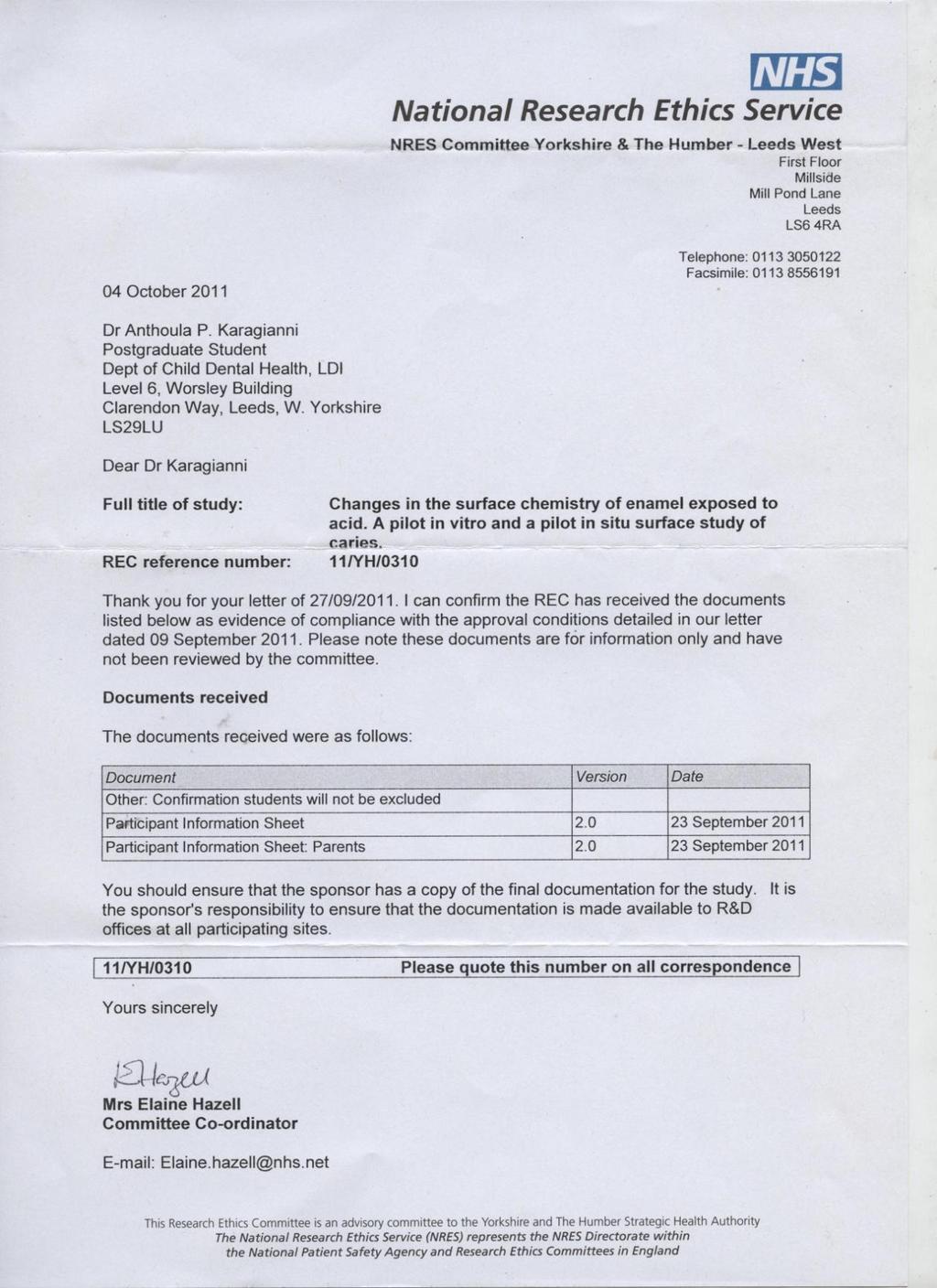
157 140

158 Research and Development Approval (Leeds Teaching Hospital NHS Trust) 141
159 142
160 143
161 144

162 7.2. Tooth donation information sheet and consent form 145
163 146
164 147

165 7.3. Participant information sheet and consent form 148
166 149

167 150
Use of the ICDAS system and two fluorescence-based intraoral devices for examination of occlusal surfaces
 A. Theocharopoulou, M.D. Lagerweij, A.J.P. van Strijp Department of Cariology Endodontology Pedodontology, Academic Centre for Dentistry Amsterdam (ACTA), Amsterdam, The Netherlands e-mail: katerina5the@gmail.com
Examination and Treatment Protocols for Dental Caries and Inflammatory Periodontal Disease
 Examination and Treatment Protocols for Dental Caries and Inflammatory Periodontal Disease Dental Caries The current understanding of the caries process supports the shift in caries management from a restorative-only
Scanning Is Believing
 KaVo DIAGNOdent /DIAGNOdent pen Laser Caries Detection Aid Scanning Is Believing Reveal the Hidden... Treat with Confidence NEW DIAGNOdent pen! Why you need DIAGNOdent Why yyou need If you cannot detect
FRANK OSEI-BONSU UGDS/KBTH
 FRANK OSEI-BONSU UGDS/KBTH Definition Introduction G. V. Black s concept of Restoration New classification of Caries Principles & Concept of MID Conclusion An approach to the management of dental caries
DIAGNOdent Measurements and Correlation with the Depth and Volume of Minimally Invasive Cavity Preparations
 Operative Dentistry, 2006, 31-3, 291-296 Clinical Research DIAGNOdent Measurements and Correlation with the Depth and Volume of Minimally Invasive Cavity Preparations JC Hamilton WA Gregory JB Valentine
DENTAL CLINICS OF NORTH AMERICA. Emmiam. Incipient and Hidden Caries. GUEST EDITOR Daniel W. Boston, DMD. October 2005 Volume49 Number4
 DENTAL CLINICS OF NORTH AMERICA Incipient and Hidden Caries GUEST EDITOR Daniel W. Boston, DMD October 2005 Volume49 Number4 Emmiam An Imprint of Elsevier, Inc. PHILADELPHIA LONDON TORONTO MONTREAL SYDNEY
71 Photothermal Radiometry and Modulated Luminescence: Applications for Dental Caries Detection
 j1047 71 Photothermal Radiometry and Modulated Luminescence: Applications for Dental Caries Detection Jose A. Garcia, Andreas Mandelis, Stephen H. Abrams, and Anna Matvienko 71.1 Introduction The traditional
Clinical and cost effectiveness of HealOzone for the treatment and management of dental caries. KaVo Dental Ltd., U.K.
 Clinical and cost effectiveness of HealOzone for the treatment and management of dental caries KaVo Dental Ltd., U.K. 19 August 2004 Declaration The following submission was written on behalf of KaVo Dental
Is there any clinical evidence?
 Current treatment objectives Anticariogenic capacity of restorative materials in paediatric dentistry: in vitro evidence vs. clinical efficiency Prof. Lisa Papagianoulis Restoration with minimal intervention
Dental plaque. Lectuer (4) Dr. Baha, H.AL-Amiedi Ph.D.Microbiology
 Dr. Baha, H.AL-Amiedi Ph.D.Microbiology") Dental plaque Lectuer (4) Dr. Baha, H.AL-Amiedi Ph.D.Microbiology it is now well established that caries & periodontal disease are infectious disease associated with resident microorganisms of dental plaque
Dental plaque Lectuer (4) Dr. Baha, H.AL-Amiedi Ph.D.Microbiology it is now well established that caries & periodontal disease are infectious disease associated with resident microorganisms of dental plaque
Caries prevention is critical for children, especially. Quantitative Assessment of Enamel Hypomineralization. Permanent Molars of Children in China
 Scientific Article Quantitative Assessment of Enamel Hypomineralization by KaVo DIAGNOdent at Different Sites on First Permanent Molars of Children in China Shao-Min Li, DDS Jing Zou, DDS, MS Zhengyan
Linking Research to Clinical Practice
 Prevention of Root Caries Denise M. Bowen, RDH, MS Linking Research to Clinical Practice The purpose of Linking Research to Clinical Practice is to present evidence based information to clinical dental
Bacterial Plaque and Its Relation to Dental Diseases. As a hygienist it is important to stress the importance of good oral hygiene and
 Melissa Rudzinski Preventive Dentistry Shaunda Clark November 2013 Bacterial Plaque and Its Relation to Dental Diseases As a hygienist it is important to stress the importance of good oral hygiene and
The Effect of Mineralizing Fluorine Varnish on the Progression of Initial Caries of Enamel in Temporary Dentition by Laser Fluorescence
 American Journal of Engineering Research (AJER) e-issn: 2320-0847 p-issn : 2320-0936 Volume-6, Issue-9, pp-39-43 www.ajer.org Research Paper Open Access The Effect of Mineralizing Fluorine Varnish on the
Tooth hypersensitivity and Dental erosion DR. KÁROLY BARTHA
 Tooth hypersensitivity and Dental erosion DR. KÁROLY BARTHA 2 Why Is Erosion an Issue Now? Changing dietary habits Higher consumption of acidic beverages (colas, sport drinks) Higher incidence of xerostomia
EVALUATION OF COLOR CHANGE IN WHITE SPOT LESIONS OF ENAMEL FLUOROSIS USING A RESIN INFILTRATE
 EVALUATION OF COLOR CHANGE IN WHITE SPOT LESIONS OF ENAMEL FLUOROSIS USING A RESIN INFILTRATE Monisha Singhal PG STUDENT DEPARTMENT OF PEDODONTICS & PREVENTIVE DENTISTRY CHANDRA DENTAL COLLEGE & HOSPITAL
FACT SHEET: ABOUT DENTAL CARIES
 FACT SHEET: ABOUT DENTAL CARIES What is dental caries? Dental caries is the technical term used by healthcare and dental professionals to describe the disease more commonly known as tooth decay. 1-2 Dental
stabilisation and surface protection
 Guiding the way to caries stabilisation and surface protection Fissure sealing MI restorations Pulp capping Hypersensitivity Protection Caries stabilisation Fuji Triage from GC. Temporary restorations
A Comparison of Methods for the Detection of Smooth Surface Caries
 A Comparison of Methods for the Detection of Smooth Surface Caries B. Wong 1, K. Sivagurunathan 1,J. D. Silvertown 1, W. M. P. Hellen 2, G. I. Elman 2, S. H. Abrams 1, L. O. Okoye 3, B. T. Amaechi 4 1
Chapter 14 Outline. Chapter 14: Hygiene-Related Oral Disorders. Dental Caries. Dental Caries. Prevention. Hygiene-Related Oral Disorders
 Chapter 14 Outline Chapter 14: Hygiene-Related Oral Disorders Hygiene-Related Oral Disorders Dental caries Prevention Gingivitis Prevention Tooth hypersensitivity Pathophysiology Treatment 2 Hygiene-Related
Photoluminescence spectra of sound tooth and those of different carious stages
 Journal Biological Series 7(1): 000-000, January 2019 DOI: 10.15413/jbs2019.0121 2019 Academia Publishing Research Paper Photoluminescence spectra of sound tooth and those of different carious stages Accepted
Root Surface Protection Simple. Effective. Important.
 GC Fuji VII / Fuji VII EP Root Surface Protection Simple. Effective. Important. Brush up your painting skills and help your patients Q&A Prof. Laurie Walsh University of Queensland lifestyle factors (frequency
Performance of a laser fluorescence device in detecting oclussal caries in vitro
 Performance of a laser fluorescence device in detecting oclussal caries in vitro Roxana Oancea1, Ruxandra Sava-Roºianu 1, Atena Gãluºcan 2, Daniela Jumanca 3, Ramona Amina Popovici 1, Angela Codruþa Podariu
Diagnodent and the caveats of caries diagnosis by laser fluorescence
 Diagnodent and the caveats of caries diagnosis by laser fluorescence Introduction Professor Laurence Walsh School of Dentistry and Centre for Laser Science, The University of Queensland The Diagnodent
XPS surface analysis of human tooth samples with EnviroESCA
 Application Note #000396 KeyWords XPS, Human Tissue, Tooth, Measurements, Surface Analysis XPS surface analysis of human tooth samples with EnviroESCA Human teeth from an adult and a baby were studied
Saliva. Introduction. Salivary Flow. Saliva and the Plaque Biofilm. The Minerals in Saliva
 Saliva Introduction Saliva is like a bloodstream to the mouth. As does blood, saliva helps build and maintain the health of the soft and hard tissues. Saliva removes waste products and provides disease-fighting
ECC II Caries Disease Status. Drs Francisco Ramos-Gomez, Man Wai Ng and Jessica Lee
 ECC II Caries Disease Status Drs Francisco Ramos-Gomez, Man Wai Ng and Jessica Lee Strategies for the detection and monitoring of early carious lesions. Detection and Staging Traditional Non- Surgical
Early lesion detection at 6-7 years and years schoolchildren from Bucharest, a prediction factor of decay evolution
 Early lesion detection at 6-7 years and 12-13 years schoolchildren from Bucharest, a prediction factor of decay evolution Roxana Ranga 1, Adina Dumitrache 2, Oana Slusanschi 3, Marian Cuculescu 4 Bucharest,
Current Concepts in Caries Management Diagnostic, Treatment and Ethical/Medico-Legal Considerations. Radiographic Caries Diagnosis
 Current Concepts in Caries Management Diagnostic, Treatment and Ethical/Medico-Legal Considerations Richard N. Bohay, DMD, MSc, MRCDC Associate Professor, Schulich School of Medicine & Dentistry Schulich
VistaCam ix HD for diagnostics support in HD quality
 VistaCam ix HD for diagnostics support in HD quality The innovative intraoral camera with interchangeable head system COMPRESSED AIR SUCTION IMAGING DENTAL CARE HYGIENE Vista Cam ix HD real HD resolution
Near-UV light detection
 C L I N I C A L Near-UV light detection Javier Tapia Guadix 1 Near-UV light induced fluorescence has already proven to be very useful as an alternative to classic caries-detector dyes. However its potential
Contemporary Policy Implications to Control and Prevent Dental Caries. Policies are formed to achieve outcomes? Are outcomes being achieved?
 Contemporary Policy Implications to Control and Prevent Dental Caries Policies are formed to achieve outcomes? Are outcomes being achieved? 2 3 4 Temple University School of Dentistry s Mission is the
Direct composite restorations for large posterior cavities extended range of applications for high-performance materials
 Direct composite restorations for large posterior cavities extended range of applications for high-performance materials A case study by Ann-Christin Meier, Dr. med. dent., Stapelfeld, Germany When large
Bioapatites. BeátaKerémi DMD, PhD Department of Oral Biology
 Bioapatites BeátaKerémi DMD, PhD Department of Oral Biology Mineralized ( hard ) tissues: Bones Teeth Enamel Cementum Dentin Hydroxy(l)-apatite Data correspond to 100 g dry weight. Composition of hard
DEPOSITS. Dentalelle Tutoring 1
 DEPOSITS Dentalelle Tutoring WWW.DENTALELLE.COM 1 PH SCALE WWW.DENTALELLE.COM 2 DENTAL CARIES Dental caries is a dynamic process that involves a susceptible tooth, cariogenic bacteria in dental plaque
Title. Citation 北海道歯学雑誌, 38(Special issue): Issue Date Doc URL. Type. File Information.
: Issue Date Doc URL. Type. File Information.") Title The ICDAS (International Caries Detection & Assessme Author(s)Kanehira, Takashi; Takehara, Junji; Nakamura, Kimiya Citation 北海道歯学雑誌, 38(Special issue): 180-183 Issue Date 2017-09 Doc URL http://hdl.handle.net/2115/67359
Title The ICDAS (International Caries Detection & Assessme Author(s)Kanehira, Takashi; Takehara, Junji; Nakamura, Kimiya Citation 北海道歯学雑誌, 38(Special issue): 180-183 Issue Date 2017-09 Doc URL http://hdl.handle.net/2115/67359
riva helping you help your patients
 riva helping you help your patients what is a glass ionomer? how will a dentist benefit from using glass ionomers? how will a patient benefit from their glass ionomer? Glass ionomer is the generic name
Preprint. Reference. Caries diagnosis using light fluorescence devices: VistaProof and DIAGNOdent. BETRISEY, Emilie, et al.
 Preprint Caries diagnosis using light fluorescence devices: VistaProof and DIAGNOdent BETRISEY, Emilie, et al. Reference BETRISEY, Emilie, et al. Caries diagnosis using light fluorescence devices: VistaProof
Pilot clinical study to assess caries lesion activity using quantitative lightinduced fluorescence during dehydration
 Pilot clinical study to assess caries lesion activity using quantitative lightinduced fluorescence during dehydration Masatoshi Ando Andrea G. Ferreira-Zandoná George J. Eckert Domenick T. Zero George
Practice Impact Questionnaire
 Practice Impact Questionnaire Your practitioner identifier is: XXXXXXXX It is very important that ONLY YOU complete this questionnaire because your responses will be compared to responses that you provided
Update in Caries Diagnosis
 Update in Caries Diagnosis Jenny Smits, AAS, RDH University of Michigan E-Learning Program 482 Oral Diseases: Prevention & Management University of Michigan Dental Desktop Support 12/16/2009 Introduction
Electronic Dental Records
 Electronic Dental Records Dr. Douglas K Benn, Professor of Maxillofacial Radiology & Director of Oral Diagnostic Systems, University of Florida and Health Conundrums LLC 8/2/2008 Dr Benn, University of
PUBLISHED VERSION. This document has been archived with permission from the Australian Dental Association, received 18th January, 2007.
 PUBLISHED VERSION Saunders, J. G. C.; McIntyre, John Malcolm The ability of 1.23% acidulated phosphate fluoride gel to inhibit simulated endogenous erosion in tooth roots Australian Dental Journal, 2005;
Syllabus for International Dental Assistants. Prepare and maintain the dental surgery, instruments, and equipment for clinical dental procedures
 Syllabus for International Dental Assistants UNIT1 Prepare and maintain the dental surgery, instruments, and equipment for clinical dental procedures This section concerns the preparation and maintenance
Caries Detection Technology
 Caries Technology Early caries detection is essential in maintaining oral health; luckily, many products on the market help dentists do so in a particularly easy fashion. Dentaltown s look at some of the
BASCD Trainers Pack for Caries Prevalence Studies. Updated: June 2014 for UK Training & Calibration exercise for the Deciduous Dentition
 BASCD Trainers Pack for Caries Prevalence Studies Updated: June 2014 for UK Training & Calibration exercise for the Deciduous Dentition Prepared by Helen Paisley, Cynthia Pine and Girvan Burnside Administrative
Original Paper. D.F. Côrtes a K.R. Ekstrand b A.R. Elias-Boneta c, R.P. Ellwood d
 Original Paper Caries Res 2000;34:443 447 Received: December 9, 1999 Accepted after revision: July 10, 2000 An in vitro Comparison of the Ability of Fibre-Optic Transillumination, Visual Inspection and
Diagnostic Values of Laser Fluorescence Device Compared to Other Techniques in Occlusal Caries Detection
 Original Article Diagnostic Values of Laser Fluorescence Device Compared to Other Techniques in Occlusal Caries Detection M. Biria 1, 2, M. Moshfeghi 3, A. Akbarzadeh Baghban 4, A. Chehresaz 5 1 Assistant
values is of great interest.
 User Report Dr. Ralph Schönemann, May 21, 2008 There are numerous self-etchbonding systems on the market that sometimes differ very much from one another with regards to their chemical composition. It
CAries Management By Risk Assessment"(CAMBRA) - a must in preventive dentistry
 - a must in preventive dentistry") CAries Management By Risk Assessment"(CAMBRA) - a must in preventive dentistry Nanda Kishor KM* *MDS, Reader, Department of Conservative and Endodontics Pacific Dental College, Udaipur, Rajasthan, India
CAries Management By Risk Assessment"(CAMBRA) - a must in preventive dentistry Nanda Kishor KM* *MDS, Reader, Department of Conservative and Endodontics Pacific Dental College, Udaipur, Rajasthan, India
Continually Fluoride Releasing Aesthetic Dental Restorative Material
 Continually Fluoride Releasing Aesthetic Dental Restorative Material Research is our best product Image provided by Dr. Sushil Koirala BEAUTIFIL II More than just filling BEAUTIFIL II stands out for its
Fluor Protector Overview
 Fluor Protector Overview Superior protection against dental caries and erosion Fluoride Clinically proven Fluoride varnish Features and mechanism of action Application Intensive care gel Risks to teeth
The 4Cs in Solving the Caries Puzzle
 The 4Cs in Solving the Caries Puzzle CAMBRA Caries Detection & Assesment System (ICDAS) Case-based protocol Calcium/Phosphate & Fluoride Objectives: Engaging in this program, the participant will be able
Index. , 66 Novamin, 66 TCP ACP. , 66 Bisphenol A (BPA), 114, 119 Byzantium diet, 87, 89
, 114, 119 Byzantium diet, 87, 89") A Acidulated phosphate fluoride (APF), 61 Adolescence aesthetic disturbance, 169 172 enamel carious lesions, orthodontic patients, 168 172 physical and sexual maturation, 168 Adulthood, 171 174 Advanced
A Acidulated phosphate fluoride (APF), 61 Adolescence aesthetic disturbance, 169 172 enamel carious lesions, orthodontic patients, 168 172 physical and sexual maturation, 168 Adulthood, 171 174 Advanced
Colourf low. light cured dental flowable composite
 Colourf low light cured dental flowable composite DENTAL LIFE SCIENCES (mfg) Ltd. Unit A3, Wigan Enterprise Court, Wigan Enterprise Park, WN2 2LE United Kingdom www.dentallifesciences.com Colourf low flowable
Oral Health. Links: Other articles related to this theme: Water and Natural Hazards; Water Scarcity; Water Challenges; Water for Positive Health
 Oral Health Water, Sanitation and Health Theme Article Links: Other articles related to this theme: Water and Natural Hazards; Water Scarcity; Water Challenges; Water for Positive Health Disease fact files:
University of Groningen. Erosive enamel wear and the inhibiting effect of topical fluorides Vieira Carvalho, Ana Maria Ramires dos Santos
 University of Groningen Erosive enamel wear and the inhibiting effect of topical fluorides Vieira Carvalho, Ana Maria Ramires dos Santos IMPORTANT NOTE: You are advised to consult the publisher's version
MinimalInvasiveTechniquesinCariesDetectionDiagnosisandMangagementAClinicalStudy
 Global Journal of Medical Research: J Dentistry & Otolaryngology Volume 16 Issue 2 Version 1.0 Year 2016 Type: Double Blind Peer Reviewed International Research Journal Publisher: Global Journals Inc.
Chapter 5. Investigation of in vitro Dental Erosion by Optical Techniques
 Chapter 5 Investigation of in vitro Dental Erosion by Optical Techniques 5.1 INTRODUCTION 5 Dental erosion is becoming an increasing predicament in dentistry and can be attributed to either exogenous
B4 NUTRITION 4.3 Animal Nutrition
 B4 NUTRITION 4.3 Animal Nutrition 1. State the term balanced diet & describe how balanced diet is related to age, sex & activity of an individual. Balanced diet: A diet that contains all the main nutrients
OliNano Seal Professional prophylaxis for long-term protection
 Professional prophylaxis for long-term protection NEW The patented formula of silicone polymer NANO Technology General information Dental health is one of the main factors to maintain overall health, and
SmartCrown. The Cavity Fighting SmartCrown. Patient Education Booklet. SmartCrown.com Toll Free Local
 The Cavity Fighting SmartCrown SmartCrown Ca2+ SmartCrown.com Patient Education Booklet www.smartcrown.com support@smartcrown.com 800.255.2839 Toll Free 801.281.4012 Local 801.281.4022 Fax F- PO43- Origin
Ionic Brushing Superpowers
 Ionic Brushing Superpowers EN Technical Brochure At Sanyei we believe, true to the Japanese way of thinking, that mouth care directly relates to being healthier and happier in body and therefore happier
Chemical Changes of Enamel Occlusal Surfaces Affected by Incipient Dental Caries: an EDX Study
 Chemical Changes of Enamel Occlusal Surfaces Affected by Incipient Dental Caries: an EDX Study CLAUDIU TOPOLICEANU 1 *, SIMONA STOLERIU 1, ANGELA GHIORGHE 1, MIHAELA SALCEANU 1, ANDREI VICTOR SANDU 2,
Detection of intraoral lesions using a fluorescence camera
 Detection of intraoral lesions using a fluorescence camera Michael Thoms Dürr Dental GmbH & Co KG, Höpfigheimer Str. 17, D-74321 Bietigheim-Bissingen University of Erlangen-Nürnberg, Institute of Material
Diet and dental caries
 Prevention-- 5 th class Lect 1 1 Diet and dental caries The role diet and nutrition in the etiology and pathogenesis of caries may be viewed as systemic (nutrition) and local effect (diet) In general,
NON-IONIZING DIAGNOSTIC PROCEDURES
 NON-IONIZING DIAGNOSTIC PROCEDURES UnitedHealthcare Dental Clinical Policy Policy Number: DCP041.01 Effective Date: February 1, 2017 Table of Contents Page INSTRUCTIONS FOR USE...1 BENEFIT CONSIDERATIONS...1
Fluoridens 133 Fluorosilicic acid 136 Fluorosis, see Dental fluorosis Foams 118 acute toxicity 71, 122 clinical efficacy 122 Free saliva 149, 150
 Subject Index Abrasive systems, dentifrices 123 Absorption 23, 24, 38, 78 Accidental poisonings 66, 67, 69, 70, see also Acute toxicity Acid-base status, see also ph metabolism effects 28, 29 toxicity
The Cavity Fighting ProActive Crown
 The Cavity Fighting ProActive Crown Powered by Ketac Nano Patient Education Booklet www.proactivedentist.com support@proactivedentist.com 800.255.2839 Toll Free 801.281.4012 Local 801.281.4022 Fax Origin
AFFINIS Sys360 putty maximum precision and easy to use
 AFFINIS Sys360 putty maximum precision and easy to use Dr. RALPH SCHÖNEMANN, November 2010 Dentists can now select from a wide range of very different impression materials supplied by a large number of
DH220 Dental Materials
 DH220 Dental Materials Lecture #5 Prof. Lamanna RDH, MS Restorative Dentistry: Glass Ionomer Bird & Robinson p.740-741 I. Use Liner Base Luting agent Restorative material: Class III, V, & eroded/abraded
Collaborators. Dental caries. Fluorescence and photochemical reactions: Lessons from teeth and tissues. 54,000+ patient appointments per year
 Fluorescence and photochemical reactions: Lessons from teeth and tissues Laurence J. Walsh School of Dentistry, The University of Queensland 2006 Collaborators Dr Jelena Vlacic Dr Fardad Shakabaie Dr Srdjan
Multi center study evaluating safety and effectiveness of The Canary System : An Interim Analysis
 Multi center study evaluating safety and effectiveness of The Canary System : An Interim Analysis S.H. Abrams a, K. Sivagurunathan a,r. J. Jeon a, J.D. Silvertown a, A. Hellen a, A. Mandelis a, W.M.P.
Fluor Protector S. The protective fluoride varnish. Superior protection against dental caries and erosion
 Fluor Protector S The protective fluoride varnish Superior protection against dental caries and erosion Fluor Protector S The strong fluoride varnish In order to stay healthy and look good, teeth need
Caries Risk Assessment and Prevention
 Caries Risk Assessment and Prevention Introduction of the disease, prevalence and global impact Dental caries is the most common dental disease that requires restorative treatment. In 2010, the Global
Detection, Diagnosis and Management of the Early Carious Lesion
 Detection, Diagnosis and Management of the Early Carious Lesion A thesis submitted to the University of Manchester for the degree of Doctor of Philosophy In the Faculty of Medical and Human Sciences By
In vitro comparison of microabrasion, CPP-ACP, CPP-ACFP and combination therapies on the remineralization of white spot lesions
 Nova Southeastern University NSUWorks College of Dental Medicine Student Theses, Dissertations and Capstones College of Dental Medicine 1-1-2014 In vitro comparison of microabrasion, CPP-ACP, CPP-ACFP
When Teeth Go Bad How nurse advice can help reduce recurrences of dental disease. Callum Blair BVMS MRCVS
 When Teeth Go Bad How nurse advice can help reduce recurrences of dental disease Callum Blair BVMS MRCVS Dental disease is a common condition affecting cats and dogs. An often quoted statistic is that
Minimally Invasive Dentistry
 Minimally Invasive Dentistry Full Summary Minimally Invasive Dentistry Defined: Minimally invasive dentistry (MID) is an evidence based intervention approach supported internationally that aims to do the
Clinical comparison of dental caries by DMFT and ICDA systems
 Original Article Clinical comparison of dental caries by DMFT and ICDA systems S. Banava 1, M. Fattah 2, MJ. Kharrazifard 3, T. Safaie 4, SH. Askarzadeh 4, M. Safaie Yazdi 4, B. T. Amaechi 5, M. Fazlyab
EQUIA. Self-Adhesive, Bulk Fill, Rapid Restorative System
 EQUIA EQUIA Fil EQUIA Coat + Self-Adhesive, Bulk Fill, Rapid Restorative System From the World Leader in Glass Ionomer Technology - A Complete Glass Ionomer Based Bulk Fill Rapid Restorative System Class
MI Varnish The ultimate fluoride varnish
 MI Varnish The ultimate fluoride varnish GC introduces MI Varnish, the first fluoride varnish with RECALDENT (CPP-ACP) MI Varnish is a 5% NaF varnish enhanced with 2% RECALDENT (CPP-ACP) to give an exceptional
NEW ZEALAND DATA SHEET
 1 PRODUCT NAME Clinpro 5000 anti-cavity toothpaste Vanilla Mint Clinpro 5000 anti-cavity toothpaste Spearmint Clinpro 5000 anti-cavity toothpaste Bubble Gum 2 QUALITATIVE AND QUANTITATIVE COMPOSITION Thick
EH1008 Biomolecules. Inorganic & Organic Chemistry. Water. Lecture 2: Inorganic and organic chemistry.
 EH1008 Biomolecules Lecture 2: Inorganic and organic chemistry limian.zheng@ucc.ie 1 Inorganic & Organic Chemistry Inorganic Chemistry: generally, substances that do not contain carbon Inorganic molecules:
swed dent j 2010; 34: hasselkvist, johansson, johansson
 swed dent j 2010; 34: 187-195 hasselkvist, johansson, johansson swedish dental journal vol. 34 issue 4 2010 187 swed dent j 2010; 34: 187-195 hasselkvist, johansson, johansson 188 swedish dental journal
Management of initial caries lesions: Iowa survey
 University of Iowa Iowa Research Online Theses and Dissertations Spring 2017 Management of initial caries lesions: Iowa survey Amira Elgreatly University of Iowa Copyright 2017 Amira Elgreatly This thesis
General dentists in private practice place numerous
 PROACTIVE INTERVENTION DENTISTRY Incorporating Glass Ionomers into Everyday Dental Practice Todd Snyder, DDS, FAACD, FIADFE Introduction General dentists in private practice place numerous direct tooth
BioCoat Featuring SmartCap Technology
 R R Breakthrough! BioCoat Featuring SmartCap Technology The only bioactive pit and fissure sealant that provides relentless protection through daily release of fluoride, calcium and phosphate ions. Bioactive
THE IMPACT OF MODIFIED FRUIT JUICE ON ENAMEL MICROHARDNESS: AN IN-VITRO ANALYSIS
 Original Article International Journal of Dental and Health Sciences Volume 02,Issue 02 THE IMPACT OF MODIFIED FRUIT JUICE ON ENAMEL MICROHARDNESS: AN IN-VITRO ANALYSIS Shailee Shelke 1,Shaila Masih 1,Namita
Unconventional Non-Invasive Diagnostic Techniques and Treatment of White Spot Lesions in Paediatric Dentistry and Orthodontics
 Unconventional Non-Invasive Diagnostic Techniques and Treatment of White Spot Lesions in Paediatric Dentistry and Orthodontics Ogodescu Alexandru (Assoc. Professor, PhD), Igna Andreea (Postgraduate Student),
Polymers in everyday things dentistry
 HOTOCOY olymers in everyday things dentistry (Background information) olymers are a part of everyday life and examples can be found almost anywhere. Many people think of polymers simply as plastics used
Samiya Subka. A thesis submitted in the fulfilment of the requirement for the Degree of Doctor of Philosophy
 Validity and acceptability of a laser fluorescence device compared to conventional methods for detection of proximal caries in primary teeth Samiya Subka A thesis submitted in the fulfilment of the requirement
Presented by Sean Day, Stanley Gelin, Kristin O Connor, Chris Sullivan, and Brian Wilson
 Different corrosive effects on hydroxyapatite nanocrystals and amine fluoride-based mouthwashes on dental titanium brackets: a comparative in vitro study Presented by Sean Day, Stanley Gelin, Kristin O
Dental Hygiene 10/16/2012 8/20/2012 8/29/2013 COURSE OUTLINE DHT 105 Dental Hygiene Clinic I - Preclinic 5 credit hours
 COURSE OUTLINE Dental Hygiene Clinic I - Preclinic 5 credit hours Course Description This course will introduce theories, rationales, and foundational knowledge for performing basic dental hygiene skills
We are IntechOpen, the world s leading publisher of Open Access books Built by scientists, for scientists. International authors and editors
 We are IntechOpen, the world s leading publisher of Open Access books Built by scientists, for scientists 3,800 116,000 120M Open access books available International authors and editors Downloads Our
Silver Diamine Fluoride
 Silver Diamine Fluoride Introduction Dental caries is a multifactorial disease that results from an imbalance between pathological and protective factors. Common non-operative treatments for incipient
Pediatric endodontics. Diagnosis, Direct and Indirect pulp capping DR.SHANKAR
 Pediatric endodontics Diagnosis, Direct and Indirect pulp capping DR.SHANKAR WHY TO PRESERVE PRIMARY TEETH? The preservation of the primary dentition until their normal anticipated exfoliation can be justified
Preclinical Dentistry. I. Dental Caries Non carious lesions: trauma, erosion. abrasion, wedge shaped defects. Lenka Roubalíková
 Preclinical Dentistry I. Dental Caries Non carious lesions: trauma, erosion. abrasion, wedge shaped defects Lenka Roubalíková Understanding dental caries crown pulp chamber neck root canal root Dental
In Vitro Evaluation of the Spectra Early Caries Detection System
 In Vitro Evaluation of the Spectra Early Caries Detection System Maria Graye, DMD Pedodontist in Private Practice Hamilton, NJ, USA Kenneth Markowitz, DDS, MSD Assistant Professor, Department of Oral Biology