Seeing the Signs: Visual Recognition of Autoimmune Connective Tissue Diseases
|
|
|
- Patricia Leonard
- 5 years ago
- Views:
Transcription

1 Seeing the Signs: Visual Recognition of Autoimmune Connective Tissue Diseases Utah Association of Family Practitioners CME Meeting at Snowbird, UT 1:00-1:30 pm, Saturday, February 13, 2016 Snowbird/Alta Rick Sontheimer, M.D. Professor of Dermatology Univ. of Utah School of Medicine
 Connetics/Stiefel MediQuest Royalties Therapeutics Lippincott, P&G (ChelaDerm) Williams Celgene* & Wilkins* Sanofi/Biogen* Clearview Health* Partners 3Gen Research partner")
2 Potential Conflicts of Interest 2016 Consultant Paid speaker Centocor (Remicadeinfliximab) Plaquenil Winthrop (Sanofi) Genentech (Raptivaefalizumab) Amgen (etanercept-enbrel) (hydroxychloroquine) Alexion (eculizumab) Connetics/Stiefel MediQuest Royalties Therapeutics Lippincott, P&G (ChelaDerm) Williams Celgene* & Wilkins* Sanofi/Biogen* Clearview Health* Partners 3Gen Research partner *Active within past 5 years
3 Learning Objectives Compare and contrast the presenting and Hallmark cutaneous manifestations of lupus erythematosus and dermatomyositis Compare and contrast the presenting and Hallmark cutaneous manifestations of morphea and systemic sclerosis
4 Distinguishing the Cutaneous Manifestations of LE and DM
5 Skin involvement is 2 nd most prevalent clinical manifestation of SLE and 2 nd most common presenting clinical manifestation

6 Comprehensive List of Skin Lesions Associated with LE LE-SPECIFIC Acute Cutaneous LE Localized ACLE Generalized ACLE Ten-like ACLE Subacute Cutaneous LE Annular Papulosquamous Mixed patterns Chronic Cutaneous LE "Classical" DLE Localized Generalized Hypertrophic DLE LE profundus Mucosal DLE LE tumidus Chilblains LE DLE-lichen planus overlap LE-NONSPECIFIC Cutaneous vascular disease Vasculitis Leukocytoclastic Palpable purpura Urticarial vasculitis Periarteritis nodosa-like Vasculopathy Dego's disease-like Atrophy blanche-like Periungual telangiectasia Livedo reticularis Thrombophlebitis Raynaud's phenomenon Erythromelalgia (erythermalgia) Alopecia (nonscarring) "Lupus hair" Telogen effluvium Alopecia aerata Sclerodacytly Rheumatoid nodules Calcinosis cutis LE nonspecific bullous lesions Epidermolysis bullosa acquisita-like bullous LE Dermatitis herpetiformis-like bullous LE Pemphigus erythematosus Bullous pemphigoid Porphyria cutanea tarda Urticaria Papulo-nodular mucinosis Nail changes (red lunulae, dyschromasia) Cutis laxa/anetoderma/mid-dermal elastolysis Pigmentary changes Acanthosis nigricans (Type B insulin resistance) Erythema multiforme (Rowell s syndrome) Leg ulcers Lichen planus


7
8 Primary Skin Change of Cutaneous LE Photosensitive macular erythema and/or papulosquamous papules and plaques


9


10 LE-Specific Skin Disease a Interface Dermatitis
11
")
12 Acute Cutaneous LE (ACLE) Localized
13 Butterfly Pattern Red Face Differential Dx ACLE/SCLE Acne rosacea Contact dermatitis (photo, airborne) Photosensitive drug eruptions Seborrheic dermatitis When the diagnosis is in doubt, punch it out!


14 ACLE Generalized
15 LE DM
16 Acute Cutaneous LE Very photosensitive, non-scarring (+) ANA, a-dsdna, a-sm Strong association with active SLE Usually managed by rheumatologists, internists

17
 Papulosquamous")
18 Subacute Cutaneous LE (SCLE) Papulosquamous Annular

19 SCLE

20

21 Histopathology of SCLE
22 Direct immunofluorescence microscopy exam of SCLE Dust-like pattern of IgG at dermalepidermal junction
23 Drug-Induced SCLE DIURETICS Thiazides Spironolactone CALCIUM CHANNEL BLOCKERS Diltiazem Nifedipine Nitrendipine Verapamil ACE inhibitors Captopril Cilazapril Acid Blockers Ranitidine Omeprazole NSAIDS Naproxen Piroxicam Beta Blocker Oxprenolol Acebutolol Lipid lowering Pravastatin Simvastatin ANTIMICROBIALS Griseofulvin Terbinafine ANTIHISTAMINES Cinnarazine/triethylperazine Anti-seizure Phenytoin Antimalarials Hydroxychloroquine Sulfonylureas glyburide Chemotherapy Taxotere/tamoxifen Others Leufonamide INF-a Procainamide Inhalants Insecticides/fertilizer Tiotropium d-penicillamine Etanercept/infliximab
24 Drug-induced SCLE Reports: 2011-Present
25 SCLE A Snapshot Nonscarring, highly photosensitive ANA (+); anti-ro/ss-a & anti-la/ss-b Genetic associations 8.1 ancestral haplotype A*01, B*08, DRB1*0301, DQB1*0201, TNFAB* a2b3 (TNF-α -308A), C2*C, C4 null C1QA-Gly70 GGG/A Associations: SSj, neonatal LE Can be drug-induced Approximate 10% - severe SLE ~75% respond to antimalarials
26


27 Classical Discoid LE - Localized


28 Classical Discoid LE - Localized

29 DLE Activity/Damage Active Inactive (Damage) Erythema, Adherent scale Induration, Follicular plugging Pigmentary change, Atrophy, Telangiectasia
30 Is It DLE or SCLE? Active Induration? Yes-DLE

31 DLE? When in Doubt Check the Ears

32 Classical Discoid LE - Generalized


33 Classical Discoid LE- Generalized
34 Chronic Cutaneous LE Variations on the Theme Hypertrophic/verrucous LE Hyperkeratosis (SSCA-like) LE tumidus Urticaria-like plaques LE panniculitis Subcutaneous nodules Chilblains LE Acral vascupopathic changes (fingers/toes) Familial cases associated with TREX1 gene mutations
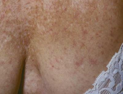
35 Chronic Cutaneous LE Classical DLE Typically scarring, less photosensitive (-) ANA 20-30% of SLE patients get DLE lesions < 5% presenting with isolated localized DLE for 1-2 years develop SLE; somewhat higher with generalized DLE

36 Dermatomyositis A member of the idiopathic inflammatory myopathies (IIM) that produces unique patterns of inflammatory injury to skin and/or proximal skeletal muscles
 inflammatory")

37 Skin Disease in Dermatomyositis Disease-defining (Hallmark) inflammatory skin changes (with interface dermatitis) Constant, defining Miscellaneous associated Often rare
38 Miscellaneous, Less Commonly Encountered Skin Changes in DM Acquired icthyosis Erythroderma Facial swelling without erythema Follicular hyperkeratosis Hypertrichosis Lichen planus Linear IgA bullous dermatosis Acquired lipoatrophy Malakoplakia Malignant erythema (suffusion) Mechanic s hand Mucous membrane lesions Mucinous plaques of the palmar creases Mucinosis, cutaneous Nasal septal perforation Panniculitis Pityriasis rubra pilaris Steroid-induced acanthosis nigricans Urticaria, urticarial vasculitis Vasculopathic ulcers Vulvar and scrotal involvement Zebra-like stripes (centripetal flagellate erythema)


39 Hallmark skin disease seen in Classical DM or CADM

40 Primary Skin Change of DM Heliotrope rash -- No Confluent macular violaceous erythema Can be more difficult to discern in darkly pigmented individuals


41 Heliotrope rash

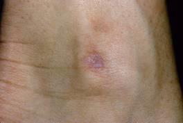
42 DM Skin Biopsy Similar to LE- Specific Skin Disease Biopsy Lupus band typically negative in DM B

43 Pruritic Violaceous Scalp Erythema

44 V-Sign
45 Shawl Sign
46 Pruritic Macular Violaceous Erythema


47 Göttron s Sign


48 Göttron s Papules

49 Göttron s Papules

50 Periungual telangiectasia


51 DM Fig. 4 C Normal * * B D * *

52 Holster Sign
")
53 Gottron s papule-like lesion over medial malleolus of ankle Poikiloderma atrophicans vasculare (POIKILODERMAhyperpigmentation, hypopigmentation, telangiectasis, atrophy)
54 Spectrum of the Idiopathic Inflammatory Myopathies Amyopathic DM Hypomyopathic DM Polymyositis/ Inclusion Body Myositis Classical DM Skin Involvement Muscle Involvement
55 Classical DM (60%) Clinically-Evident Muscle Disease Activity CDM Skin Disease Activity Disease onset 6 months 24 months
56 Classical DM (30%) Clinically-Evident Muscle Disease Activity CDM Skin Disease Activity Disease onset 6 months 24 months
57 Clinically-Amyopathic DM (C-ADM) ( ~20% of all DM) Clinically-Evident Muscle Disease Activity C-ADM Skin Disease Activity Disease onset 6 months 24 months
58 Clinically-Amyopathic DM Clinically-Amyopathic DM (CADM) Amyopathic DM Hypomyopathic DM Skin Involvement Muscle Involvement CADM is more common than previously thought Finite risk of interstitial lung disease, internal malignancy, lateonset weakness (predictors unknown) ANA (+), myositis-specific antibody (-), may have specific immunogenetic association (MDA-5/CADM140)
59 Management of DM Adult-onset DM Screen for internal malignancy (greatest risk [25%] beyond 50 years of age) Sex- and age-specific approach Screen for interstitial lung disease Baseline - pulmonary function tests with diffusion capacity (PFT) (25% abnormalities) Persistent dry cough, dyspnea repeat PFT, consider referral to pulmonary medicine/rheumatology
60 Management of DM Juvenile-onset DM Monitor for vasculopathic tissue damage Eye, GI tract, Monitor for calcinosis cutis No significant risk for interstitial lung disease or internal malignancy
61 DM Autoantibodies ANA (50-70%) Myositis-specific autoantibody (10-20%) Jo-1, PL-7, Pl-12 (anti-synthetase antibody syndrome [myositis, arthritis, Raynaud s phenomenon, mechanics hand lesion]) New autoantibodies (10-30%) Melanoma differention antigen 5 (MDA5) (interstitial lung disease association) Transcription intermediary factor 1γ (TIF1γ ) & Nuclear matrix protein (NXP-2) (internal malignancy association)


62 Mechanic s Hand Lesion
63 Management of DM DM Is No Longer Enough Adult-Onset Classic DM Clinically-Amyopathic DM Dermatomyositis Phenotypes Classic DM Clinically-Amyopathic DM Juvenile-Onset
64 Distinguishing the Cutaneous Manifestations of Morphea form Systemic sclerosis

65 Scleroderma Hardened, thickened, hide-bound skin associated with autoimmune microvascular injury and replacement of the dermis and subcutaneous tissue by dense fibrotic tissue
66 Scleroderma Skin Biopsy
67 Scleroderma Morphea (Localized Scleroderma) Systemic Sclerosis Localized Scleroderma/Morphea SSc siné scleroderma Systemic Sclerosis Skin Involvement Systemic Involvement
68 Scleroderma Skin Biopsy Same in morphea and systemic sclerosis
69 Limited cutaneous sclerosis (CREST) Diffuse cutaneous sclerosis Systemic Sclerosis (SSc) Cutaneous Sclerosis Scleroderma Scleroderma variants: eosinophilic fasciitis, eosinophilia myalgia Pseudoscleroderma Localized Scleroderma (syn. Morphea) Plaque Linear Generalized Profunda/Deep
70 Presenting Manifestations of Morphea


71 Plaque Morphea


72 Linear Scleroderma/ Liner Morphea En Coup de Sabre


73 Generalized Morphea/ Pansclerotic morphea
74 Systemic Sclerosis vs. Morphea Systemic Sclerosis Presents with Raynaud s phenomenon Nailfold telangiectasia Sclerodacytly SSc antibodies Centromere Scl-70 (Topoisomerase I) Localized Scleroderma/Morphea Absence of the above
75 Presenting Manifestations of Systemic Sclerosis
 2. Blue (cyanosis) 3.")
76 Raynaud s Phenomenon Three colors in sequence: 1. White (vasconstriction) 2. Blue (cyanosis) 3. Red (reactive hyperemia)
77 Raynaud s Phenomenon Primary Raynaud s phenomenon (syn. Raynaud s disease) Absence of: Periungual microvascular changes Sclerodactyly SSc autoantibodies ANA, centromere, topoisomerase-1 (Scl-70) Secondary Raynaud s phenomenon SSc
78 Periungual Telangiectasia Systemic Sclerosis & Dermatomyositis
79 Scleroderma in SSc -Cutaneous Manifestations- Time Raynaud s phenomenon Edema Microvasculopathy Cutaneous sclerosis ( scleroderma ) Matte-like telangiectasis Atrophy Pigmentary changes Calcinosis cutis Ulceration


80 Edema Peau d orange Sausage Shaped Digits


81 Periungual Telangiectasia (Microvasculopathy)

82 Ptyrigium Inversus Unguium

83 Sclerosis


84 Sclerosis & Atrophy


85 Sclerosis & Atrophy

86 Atrophy Autoamputation


87 Cutaneous Calcinosis


88 Pigmentary Change


89 Telangiectasia


90 Ulceration
91 Systemic Sclerosis Two prognostic groups SSc with limited cutaneous sclerosis (CREST syndrome) Slower pace, milder course initially Risk for pulmonary artery hypertension years later SSc with generalized cutaneous sclerosis Rapid pace, severe systemic disease

92 SSc with limited cutaneous sclerosis (CREST Syndrome) C - Calcinosis R - Raynaud s E - Esophageal dysmotility S - Sclerodactyly T - Telangiectasia
93 Low risk CREST Syndrome Prognosis Kidney, gastrointestinal tract High risk Pulmonary and cardiovascular system Pulmonary artery hypertension starting years after disease onset Presenting clinical symptoms-shortness of breath/dyspnea on exertion
94 SSc with Generalized Cutaneous Sclerosis

95 Wasatch Front, Salt Lake City Guardsman s Pass looking down toward Heber Valley
96 For Dermatologists LE DM
97 Distinguishing Cutaneous LE from Cutaneous DM Biopsy unhelpful Regional anatomy predilection Pruritic scalp involvement Face Nasolabial folds Fingers Knuckles vs hair-bearing Finger nail fold microvascular changes Pruritus - Presence/absence Muscle inflammation
98 For Non-Dermatologists Greenwald s Law of Lupus Greenwald RA. Greenwald's law of lupus. J Rheumatol Sep;19(9):1490. anything happening to a patient with SLE which is not immediately otherwise explicable will automatically be blamed on the lupus, regardless of pathophysiologic validity."
99 LE-Specific Skin Disease Acute Cutaneous LE Subacute cutaneous LE Chronic Cutaneous LE LE-Nonspecific Skin Disease LCV Urticarial vasculitis Livedo reticularis Secondary to Treatment of LE Steroid acne Drug eruptions Opportunistic Infections True-True and Unrelated Psoriasis Eczema BCC Greenwald s Law of Lupus

100 Recurrent tinea corporis in a man with terbinafine-induced SCLE was confused with reactivation of SCLE

101 For Questions & Comments
102 Univ. of Utah Practical Dermatology for Primary Care CME Course 7th Annual Course What Intensive one-day course on Dx and Rx of skin problems commonly encountered in the primary care setting. Includes podium presentations and a hands-on skin biopsy workshop When - Friday, April 8, 2016 Where - Alumni Hall, Medical Education Building For more information: Lisa.Estrada@hsc.utah.edu

103 Thank You East Canyon, Fall 2013
Medical Dermatology Society
 A Mashup of Instructive Cases Presenting With Subacute Cutaneous LE (SCLE) Skin Lesions Medical Dermatology Society Denver, Colorado March 20, 2014 Rick Sontheimer, M.D. Professor of Dermatology University
Cutanous Manifestation of Lupus Erythematosus. Presented By: Dr. Naif S. Al Shahrani Salman Bin Abdaziz university
 Cutanous Manifestation of Lupus Erythematosus Presented By: Dr. Naif S. Al Shahrani Salman Bin Abdaziz university A 50-year old lady, who is otherwise healthy, presented to the dermatology clinic with
Skin Deep: Cutaneous Lupus. Dr Sarah Sasson Immunology Registrar, Liverpool Hospital 2016
 Skin Deep: Cutaneous Lupus Dr Sarah Sasson Immunology Registrar, Liverpool Hospital 2016 Introduction: Cutaneous lupus erythematosus LE is an autoimmune disease with a range of clinical manifestations
Autoantibodies in the Idiopathic Inflammatory Myopathies
 Autoantibodies in the Idiopathic Inflammatory Myopathies Steven R. Ytterberg, M.D. Division of Rheumatology Mayo Clinic Rochester, MN The Myositis Association Annual Conference St. Louis, MO Sept. 25,
Cutaneous manifestations of systemic lupus erythematosus
 Hong Kong J. Dermatol. Venereol. (2006) 14, 120-128 Original Article Cutaneous manifestations of systemic lupus erythematosus Cutaneous manifestations are important aspects of systemic lupus erythematosus
Cutaneous Lupus Erythematosus Diagnosis, Classification, and Screening for Systemic Disease
 Cutaneous Lupus Erythematosus Diagnosis, Classification, and Screening for Systemic Disease David A. Wetter, M.D. Professor of Dermatology, Mayo Clinic (Rochester, MN) Cutaneous Lupus and Dermatomyositis
SCLERODERMA 101. Maureen D. Mayes, MD, MPH Professor of Medicine University of Texas - Houston
 SCLERODERMA 101 Maureen D. Mayes, MD, MPH Professor of Medicine University of Texas - Houston TYPES OF SCLERODERMA Localized versus Systemic Two Kinds of Scleroderma Localized Scleroderma Morphea Linear
Cutaneous manifestations of Systemic Lupus Erythematosus
 IOSR Journal of Dental and Medical Sciences (IOSR-JDMS) e-issn: 2279-0853, p-issn: 2279-0861.Volume 17, Issue 5 Ver. 11 (May. 2018), PP 01-08 www.iosrjournals.org Cutaneous manifestations of Systemic Lupus
Common Cutaneous Signs of Medical Illnesses
 Common Cutaneous Signs of Medical Illnesses DR COLIN THENG MBBS, MMED (FAM. MED), MRCP(UK), FAMS SENIOR CONSULTANT DERMATOLOGIST THE SKIN SPECIALISTS & LASER CLINIC MOUNT ALVERNIA MEDICAL CENTRE D, #07-61
B. Scleroderma. 6. Nodular cutaneous lupus mucinosis. 7. Bullous lupus erythematosus. 1. Systemic sclerosis (SSc)
") Go Back to the Top To Order, Visit the Purchasing Page for Details antiodies. Symptomatic therapies for the eruptions and the systemic symptoms are the main treatments. A pacemaker may e implanted in patients
Go Back to the Top To Order, Visit the Purchasing Page for Details antiodies. Symptomatic therapies for the eruptions and the systemic symptoms are the main treatments. A pacemaker may e implanted in patients
Myositis and Your Lungs
 Myositis and Your Lungs 2013 TMA Annual Patient Meeting Louisville, Kentucky Chester V. Oddis, MD University of Pittsburgh Director, Myositis Center Myositis Heterogeneous group of autoimmune syndromes
IMACS FORM 07b: MYOSITIS DISEASE ACTIVITY ASSESSMENT TOOL, Version
 IMACS FORM 07b: MYOSITIS ASSESSMENT TOOL, Version 2 2005 Subject s IMACS number: ASSESSOR: Date Assessed: Assessment number: The clinical features recorded are based upon the previous 4 weeks and the judgment
LUPUS CAN DO EVERYTHING, BUT NOT EVERYTHING IS LUPUS LUPUS 101 SLE SUBSETS AUTOIMMUNE DISEASE 11/4/2013 HOWARD HAUPTMAN, MD IDIOPATHIC DISCOID LUPUS
 LUPUS 101 LUPUS CAN DO EVERYTHING, BUT NOT EVERYTHING IS LUPUS HOWARD HAUPTMAN, MD IDIOPATHIC DISCOID LUPUS SLE SUBSETS SUBACUTE CUTANEOUS LUPUS DRUG INDUCED LUPUS NEONATAL LUPUS LATE ONSET LUPUS ANTI-PHOSPHOLIPID
RHEUMATOLOGY OVERVIEW. Carmelita J. Colbert, MD Assistant Professor of Medicine Division of Rheumatology Loyola University Medical Center
 RHEUMATOLOGY OVERVIEW Carmelita J. Colbert, MD Assistant Professor of Medicine Division of Rheumatology Loyola University Medical Center What is Rheumatology? Medical science devoted to the rheumatic diseases
Dermatology GP Referral Guidelines
 Austin Health Dermatology Department holds 5 Clinic sessions to discuss and plan the treatment of with Dermatology conditions. Department of Health clinical urgency categories for specialist clinics Urgent:
An Approach to Common and not so Common Rashes in the Office FMF 2014 Christie Freeman MD, CCFP, DipPDerm, MSc
 An Approach to Common and not so Common Rashes in the Office FMF 2014 Christie Freeman MD, CCFP, DipPDerm, MSc 1 Common Rashes Tinea Corporis: Annular- this is not the only criteria Advancing erythematous
Table of Contents: Part 1 Medical Dermatology. Chapter 1 Acneiform Disorders. Acne. Acne Vulgaris. Pomade Acne. Steroid Acne
 Table of Contents: Part 1 Medical Dermatology Chapter 1 Acneiform Disorders Acne Acne Vulgaris Pomade Acne Steroid Acne Infantile Acne Pediatric Perspectives Neonatal Acne (Acne Neonatorum) Pediatric Perspectives
Jeopardy. What s the rash? $100 $100 $100 $100 $100 $200 $200 $200 $200 $200 $300 $300 $300 $300 $300 $400 $400 $400 $400 $400
 Jeopardy Antibodies & more antibodies Aching joints What s the rash? Potpourri Image Challenge $100 $100 $100 $100 $100 $200 $200 $200 $200 $200 $300 $300 $300 $300 $300 $400 $400 $400 $400 $400 $500 $500
Test Name Results Units Bio. Ref. Interval
 135091660 Age 44 Years Gender Male 29/8/2017 120000AM 29/8/2017 100219AM 29/8/2017 105510AM Ref By Final EXTRACTABLENUCLEAR ANTIGENS (ENA), QUANTITATIVE ROFILE CENTROMERE ANTIBODY, SERUM 20-30 Weak ositive
DESCRIPTIONS FOR MED 3 ROTATIONS Dermatology A3S
 Regardless of your future field of practice, you will be exposed to a considerable amount of dermatology and this rotation provides you the chance to see a range of skin diseases. You will have the opportunity
Budsakorn Darawankul, MD. Maharat Nakhon Ratchasima Hospital
 Budsakorn Darawankul, MD. Maharat Nakhon Ratchasima Hospital Outline What is ANA? How to detect ANA? Clinical application Common autoantibody in ANA diseases Outline What is ANA? How to detect ANA? Clinical
Inflammatory Myopathies 4 th year MBBS. Marwan Adwan MBChB, MSc, MRCPI, MRCP(rheum) Consultant Rheumatologist
 Consultant Rheumatologist") Inflammatory Myopathies 4 th year MBBS Marwan Adwan MBChB, MSc, MRCPI, MRCP(rheum) Consultant Rheumatologist Case A 64 woman presents with erythematous itchy rash over back of hands & forehead. For 1 month
Inflammatory Myopathies 4 th year MBBS Marwan Adwan MBChB, MSc, MRCPI, MRCP(rheum) Consultant Rheumatologist Case A 64 woman presents with erythematous itchy rash over back of hands & forehead. For 1 month
2/23/18. Disclosures. Rheumatic Diseases of Childhood. Making Room for Rheumatology. I have nothing to disclose. James J.
 Making Room for Rheumatology James J. Nocton, MD Disclosures I have nothing to disclose Rheumatic Diseases of Childhood Juvenile Idiopathic Arthritis (JIA) Systemic Lupus Erythematosus (SLE) Juvenile Dermatomyositis
Disclosures. Rheumatological Approaches to Differential Diagnosis, Physical Examination, and Interpretation of Studies. None
 Rheumatological Approaches to Differential Diagnosis, Physical Examination, and Interpretation of Studies Sarah Goglin MD Assistant Professor of Medicine Division of Rheumatology Disclosures None 1 [footer
Undifferentiated Connective Tissue Disease and Overlap Syndromes. Mark S. Box, MD
 Undifferentiated Connective Tissue Disease and Overlap Syndromes Mark S. Box, MD Overlap Syndromes As many as 25% of patients with rheumatic diseases with systemic symptoms cannot be definitely diagnosed
Test Name Results Units Bio. Ref. Interval
 LL - LL-ROHINI (NATIONAL REFERENCE 135091593 Age 25 Years Gender Male 30/8/2017 91600AM 30/8/2017 93946AM 31/8/2017 84826AM Ref By Final COLLAGEN DISEASES ANTIBODY ANEL ANTI NUCLEAR ANTIBODY / FACTOR (ANA/ANF),
Overview of Diagnostic Autoantibodies in Inflammatory Myopathy
 Overview of Diagnostic Autoantibodies in Inflammatory Myopathy Minoru Satoh, M.D., Ph.D. Research Associate Professor of Medicine Division of Rheumatology and Clinical Immunology University of Florida
Autoimmune diseases. SLIDE 3: Introduction to autoimmune diseases Chronic
 SLIDE 3: Introduction to autoimmune diseases Chronic Autoimmune diseases Sometimes relapsing : and remitting. which means that they present as attacks Progressive damage Epitope spreading more and more
Index. derm.theclinics.com. Note: Page numbers of article titles are in boldface type.
 Note: Page numbers of article titles are in boldface type. A Abatacept for DLE, 493 for SLE, 497 Ablative therapies, localized, for cutaneous T-cell lymphoma, 502 506. See also Cutaneous T-cell lymphoma,
Disclosures. Clinical Approach: Evaluating CTD-ILD for the pulmonologist. ILD in CTD. connective tissue disease or collagen vascular disease
 Disclosures Clinical Approach: Evaluating CTD-ILD for the pulmonologist Industry relationships: Actelion, atyr Pharma, Boehringer-Ingelheim, Genentech- Roche, Gilead Aryeh Fischer, MD Associate Professor
Clinical Laboratory. 14:42:00 SSA-52 (Ro52) (ENA) Antibody, IgG 1 AU/mL [0-40] Oct-18
![Clinical Laboratory. 14:42:00 SSA-52 (Ro52) (ENA) Antibody, IgG 1 AU/mL [0-40] Oct-18](/thumbs/95/125595183.jpg "Clinical Laboratory. 14:42:00 SSA-52 (Ro52) (ENA) Antibody, IgG 1 AU/mL [0-40] Oct-18") Clinical Laboratory Procedure Result Units Ref Interval Accession Collected Received Rheumatoid Factor
Clinical Laboratory Procedure Result Units Ref Interval Accession Collected Received Rheumatoid Factor
SCLERODERMA: An Update. What You Need To Know
 SCLERODERMA: An Update What You Need To Know Pre Test Question #1 The onset of systemic sclerosis is typically: A. In the 1st decade of life B. Between the 4th and 6th decade C. Equal in all age groups
4/10/2012 DERMATOLOGIC MANIFESTATIONS OF INTERNAL DISEASE PHOTO CREDITS: LUPUS ERYTHEMATOSUS
 DERMATOLOGIC MANIFESTATIONS OF INTERNAL DISEASE Kim Sanders MPAS, PA-C OHSU Department of Dermatology Acute butterfly malar rash, transient, follows sun exposure Lesions on hands often spare knuckles Evaluate
SCLERODERMA. Scleroderma update. No disclosures or conflicts. Leslie Kahl, M.D. April 10, 2015
 Scleroderma update Leslie Kahl, M.D. April 10, 2015 No disclosures or conflicts KT is a 45 year old woman who developed puffiness in her fingers 1/2013 and carpal tunnel syndrome and arthralgias 3/2013.
DART Clinic Review: Experiences from a combined dermatology and rheumatology clinic
 DART Clinic Review: Experiences from a combined dermatology and rheumatology clinic Michael Samycia 1, Collette McCourt 2, Kam Shojania 4, Sheila Au 1,3 1 Department of Dermatology and Skin Science, University
A48-year-old Hispanic woman
 Jamie Goodall, MS4, and Richard P. Usatine, MD University of Texas Health Science Center at San Antonio Skin rash and muscle weakness The patient s facial rash was spreading and she was having difficulty
Dermatomyositis Advice from Experts: Improving Your Medical Dermatology Diagnostic and Management Skills Jeffrey P. Callen, MD Professor of Medicine
 Dermatomyositis Advice from Experts: Improving Your Medical Dermatology Diagnostic and Management Skills Jeffrey P. Callen, MD Professor of Medicine (Dermatology) University of Louisville Learning Objectives
Systemic sclerosis (SSC)
") Systemic sclerosis (SSC) -Is a multi system autoimmune disease, characterized by fibrosis of the skin and variable pattern of other visceral -SSC: Is a relatively UN common disease -Prevalence in U S A
Systemic sclerosis (SSC) -Is a multi system autoimmune disease, characterized by fibrosis of the skin and variable pattern of other visceral -SSC: Is a relatively UN common disease -Prevalence in U S A
=ﻰﻤاﻤﺤﻠا ﺔﻴﻘﻠﺤﻠا ﺔذﺒاﻨﻠا
 1 / 15 Erythema Annulare Centrifugum and Other Figurate Erythemas The figurate erythemas include a variety of eruptions characterized by annular and polycyclic lesions. Classification of this group has
This month, we are very pleased to introduce some new tests for Scleroderma as well as some test changes to our existing scleroderma tests/panels.
 February 20, 2017 Client Letter Test Update February 2017 Dear Colleague: This month, we are very pleased to introduce some new tests for Scleroderma as well as some test changes to our existing scleroderma
Clinical Laboratory. [None
 Clinical Laboratory Procedure Result Units Ref Interval Accession Collected Received Double-Stranded DNA (dsdna) Ab IgG ELISA Detected * [None 18-289-900151 Detected] Double-Stranded DNA (dsdna) Ab IgG
PGALS: Approach to Child with Arthritis. Prof Chris Scott Paediatric Rheumatology
 PGALS: Approach to Child with Arthritis Prof Chris Scott Paediatric Rheumatology Introduction In a prospective study from The Royal Hospital for Sick Children, Edinburgh, every 58th child presented with
Paul K. Shitabata, M.D. Dermatopathology Institute
 Paul K. Shitabata, M.D. Dermatopathology Institute Technical Considerations Storage of slides at room temperature
Index. Angiosarcoma diagnosis, 47 lymphedema-related vs. non-lymphedemarelated, 48
 A Acneiform rash biopsy, 134 cetuximab, EGFR, 132 133 diagnosis, 131 patient history, 131 134 treatment, 134 135 Acne vulgaris, 109 AGA. See Androgenetic alopecia Alopecia areata, 148 American Joint Committee
My Method for Approaching Skin Biopsies
 My Method for Approaching Skin Biopsies P A U L H A U N, MD, MS, F A A D A S S I S T A N T P R O F E S S O R D E R M A T O L O G Y A N D D E R M A T O P A T H O L O G Y D E P A R T M E N T O F D E R M
Autoimmune Diseases with Oral Manifestations
 Autoimmune Diseases with Oral Manifestations Martin S. Greenberg DDS, FDS RCSEd Professor Emeritus Department of Oral Medicine University of Pennsylvania Disclosure Statement I have no actual or potential
Scleroderma. Nomenclature Synonyms. Scleroderma. Progressive Systemic Sclerosis. Systemic Sclerosis. Edward Dwyer, M.D. Division of Rheumatology
 Scleroderma Edward Dwyer, M.D. Division of Rheumatology Nomenclature Synonyms Scleroderma Progressive Systemic Sclerosis Systemic Sclerosis Scleroderma 1 Scleroderma Chronic systemic autoimmune disease
Scleroderma. Nomenclature Synonyms. Scleroderma. Progressive Systemic Sclerosis. Systemic Sclerosis. Limited vs. Diffuse Scleroderma.
 Scleroderma Edward Dwyer, M.D. Division of Rheumatology Nomenclature Synonyms Scleroderma Progressive Systemic Sclerosis Systemic Sclerosis Scleroderma Chronic systemic autoimmune disease characterized
HUNGARIAN MYOSITIS COHORT
 CLINICAL ADVANCES OF ANTI- TIF1Γ AUTOANTIBODY IN A HUNGARIAN MYOSITIS COHORT Melinda Nagy-Vincze 1, Zoltán Griger 1, Levente Bodoki 1, Zsuzsa Szankai 1, Zoe E. Betteridge 2, Katalin Dankó 1 1 University
Dr.MD.IMRAN KAZMI ASST PROF.DEPT OF DVL,KIMS
 Dr.MD.IMRAN KAZMI ASST PROF.DEPT OF DVL,KIMS Lupus erythematosus is a multisystem disorder whose spectrum runs from a relatively benign, self-limited cutaneous eruption to a severe, often fatal, systemic
Skin Manifestations of Systemic Disease. Approach to Dermatalogic Diagnosis 9/6/2016. Go Ahead---Judge a Book by its Cover!
 Go Ahead---Judge a Book by its Cover! Skin Manifestations of Systemic Disease Amelie Hollier, DNP, FNP-BC, FAANP Lafayette, LA President, APEA Objectives Compare diseases of the skin with reactions of
Autoantibodies in Idiopathic Inflammatory Myopathies. Vidya Limaye Rheumatology Department Royal Adelaide Hospital
 Autoantibodies in Idiopathic Inflammatory Myopathies Vidya Limaye Rheumatology Department Royal Adelaide Hospital Idiopathic Inflammatory Myopathies (IIM) Heterogeneous group of systemic autoimmune syndromes
Is it Autoimmune or NOT! Presented to AONP! October 2015!
 Is it Autoimmune or NOT! Presented to AONP! October 2015! Four main jobs of immune system Detects Contains and eliminates Self regulates Protects Innate Immune System! Epithelial cells, phagocytic cells
Essential Rheumatology. Dr Ellen Bruce Consultant Rheumatologist CMFT
 Essential Rheumatology Dr Ellen Bruce Consultant Rheumatologist CMFT Saving the best for last! Apparently people recall best the first and last thing they re told. Far too difficult to include everything.
Autoantibodies andprognosis. Jiří Vencovský Institute of Rheumatology, Prague, Czech Republic
 Autoantibodies andprognosis Jiří Vencovský Institute of Rheumatology, Prague, Czech Republic HeterogeneityofIIMs Diagnosis Polymyositis Dermatomyositis IBM Necrotising myopathy Paraneoplastic Amyopathic
CONDITIONS OF THE SKIN
 CONDITIONS OF THE SKIN UCSF/SFGH Family & Community Medicine Residency Program Educational Objectives I. Knowledge The resident will be able to discuss the definition, diagnosis, and initial management
Blistering, Papulosquamous, Connective Tissue and Alopecia. Kathleen Haycraft, DNP, FNP/PNP-BC, DCNP, FAANP
 Blistering, Papulosquamous, Connective Tissue and Alopecia Kathleen Haycraft, DNP, FNP/PNP-BC, DCNP, FAANP Objectives Identify examples of papulosquamous disease and state treatment options Identify examples
Original Contribution
 Direct Immunofluorescence Test of Skin Biopsy Samples Results of 204 Cases Kabir AN, 1 Das RK, 2 Kamal M 3 Direct immunofluorescence (DIF) test of skin and renal biopsy specimens is being done on regular
Interactive Workshops. Practical Follow-up of DVT and PE patients
 Interactive Workshops Practical Follow-up of DVT and PE patients Vascular acrosyndromes Unusual site thrombosis Women s health issues WHAT IS IT? 18/04/2016 49 WHAT IS IT? 1. Acrocyanosis 2. Raynaud s
REVIEW. Cutaneous Lupus Erythematosus. Allison Chabassol Class of 2013, Faculty of Medicine, Dalhousie University
 REVIEW Cutaneous Lupus Erythematosus Allison Chabassol Class of 2013, Faculty of Medicine, Dalhousie University Lupus erythematosus (LE) is a heterogeneous group of chronic autoimmune inflammatory diseases,
Significance of antibody testing in idiopathic inflammatory myopathies
 2/0/20 Significance of antibody testing in idiopathic inflammatory myopathies Jiří Vencovský Institute of Rheumatology, Prague Diagnosis Polymyositis Juvenile DM (JPM) Paraneoplastic Myositis in overlap
Principi ed Aggiornamenti in Dermatologia Roma, 6-7 Aprile Grand rounds. Lorenzo Cerroni, Graz
 Principi ed Aggiornamenti in Dermatologia Roma, 6-7 Aprile 2018 Grand rounds Lorenzo Cerroni, Graz "Computer palms" Described in patient using computer keyboards for long periods; similar features described
COPYRIGHTED MATERIAL. Introduction CHAPTER 1. Introduction
 CHAPTER 1 Introduction OVERVIEW The clinical features of skin lesions are related to the underlying pathological processes. Broadly skin conditions fall into three clinical groups: (a) those with a well-defined
Emergent and Urgent Dermatology, Eruptions, and Wound Care
 Emergent and Urgent Dermatology, Eruptions, and Wound Care G. Scott Drew, DO, FAAD, FAOCD Smith Clinic Department of Dermatology Tucson Osteopathic Medical Foundation April 27, 2018 Acute Cutaneous Lupus
Rashes Not To Be Missed In Children
 May 2016 Rashes Not To Be Missed In Children Dr Chan Yuin Chew Dermatologist Dermatology Associates Gleneagles Medical Centre Scope of presentation Focus on rashes May lead to significant morbidity if
Definition Chronic autoimmune disease The body s immune system starts attacking itself Can affect most organs and tissues in the body Brain, lungs, he
 LIVING WITH SYSTEMIC LUPUS ERYTHEMATOSUS Stacy Kennedy, M.D.,M.B.A. Rowan Diagnostic Clinic Salisbury, N.C. May 11, 2013 Agenda What is lupus Who is affected Causes of lupus Symptoms and organ involvement
What will we discuss today?
 Autoimmune diseases What will we discuss today? Introduction to autoimmune diseases Some examples Introduction to autoimmune diseases Chronic Sometimes relapsing Progressive damage Epitope spreading more
What your autoantibodies tell us about your disease. Mark Gourley, MD
 What your autoantibodies tell us about your disease Mark Gourley, MD The Importance of the Immune System Defends us against foreign invaders Self (cancer) and Nonself (virus, bacteria, etc.) But, if the
Scleroderma FAQ. About this Document
 Scleroderma FAQ About this Document The Scleroderma FAQ * is a comprehensive document that covers systemic scleroderma diagnosis and treatment. All information contained in the FAQ is based on current
Rheumatology 101 A Pediatrician s Guide
 Rheumatology 101 A Pediatrician s Guide Pediatric Staff and Alumni Day 2016 Dawn M. Wahezi, Yonit Sterba, Tamar Rubinstein Disclosures None Pick a Group Group 1 A child with a limp Group 2 ANA To test
Clinical Laboratory. 14:41:00 Complement Component 3 50 mg/dl Oct-18
 Clinical Laboratory Procedure Result Units Ref Interval Accession Collected Received Thyroid Peroxidase (TPO) Antibody 5.0 IU/mL [0.0-9.0] 18-289-900139 16-Oct-18 Complement Component 3 50 mg/dl 18-289-900139
BSD Self Assessment Workshop 7 th July 2013 CASE 27 RAC6123
 BSD Self Assessment Workshop 7 th July 2013 CASE 27 RAC6123 M55. 4/7 tender lesions on knee, legs and arms. Also iritis/ weight loss/headache, synovitis.?vasculitis. Sarcoidosis. Biopsy from left elbow
Contents. Part I Genodermatoses
 Contents Part I Genodermatoses 1 Hyperkeratotic Palms and Soles with Periorificial Keratosis............... 3 2 Indurated, Dark, Hairy Plaques, with Arthritis and Deafness.............. 9 3 Cleft Palate,
Reporting Autoimmune Diseases in Hematopoietic Stem Cell Transplantation
 Reporting Autoimmune Diseases in Hematopoietic Stem Cell Transplantation Marcelo C. Pasquini, MD, MSc HVD05_1.ppt Outline Review of autoimmune diseases (AID). Role of transplantation for AID Data collection:
Cutaneous manifestations and systemic correlation in patients with lupus erythematosus and its subsets: a study of 40 cases
 International Journal of Research in Dermatology Mahajan R et al. Int J Res Dermatol. 2018 Nov;4(4):479-483 http://www.ijord.com Original Research Article DOI: http://dx.doi.org/10.18203/issn.2455-4529.intjresdermatol20183407
A case of bullous pemphigoid following pemphigus foliaceus
 #2228 A case of bullous pemphigoid following pemphigus foliaceus Priyanka Vedak MD 1, Danielle Levine MD 1,3, Lyn Duncan MD 2,3, Hensin Tsao 1,3, Daniela Kroshinsky MD MPH 1,3 1. Department of Dermatology,
A Case of Erythrodermic Dermatomyositis Associated with Gastric Cancer
 Ann Dermatol Vol. 21, No. 4, 2009 CASE REPORT A Case of Erythrodermic Dermatomyositis Associated with Gastric Cancer Sung Woo Kim, M.D., Yoo Seok Kang, M.D., Sang Hoon Park, M.D., Un Ha Lee, M.D., Hyun
W J D. World Journal of Dermatology. Review of the cutaneous manifestations of autoimmune connective tissue diseases in pediatric patients.
 W J D World Journal of Dermatology Submit a Manuscript: http://www.wjgnet.com/esps/ Help Desk: http://www.wjgnet.com/esps/helpdesk.aspx DOI: 10.5314/wjd.v4.i2.80 World J Dermatol 2015 May 2; 4(2): 80-94
Facial Rash. Facial Rash 10/14/2013. Ten Look Alike Rashes Michelle DiBaise, MPAS, PA-C, DFAAPA Associate Clinical Professor NAU PA Program
 Ten Look Alike Rashes Michelle DiBaise, MPAS, PA-C, DFAAPA Associate Clinical Professor NAU PA Program Facial Rash Facial Rash Case 1 28 year female Progressive development of erythematous facial lesions
DocSpot what patients want to know
 DocSpot what patients want to know Professor Chris Denton Royal Free Hospital London, UK Madrid, February 2012 Copyright 2011 Raynaud's & Scleroderma Association. Charity Reg. No. 326306 DocSpot Live!
Tools to Aid in the Accurate Diagnosis of. Connective Tissue Disease
 Connective Tissue Disease Tools to Aid in the Accurate Diagnosis of Connective Tissue Disease Connective Tissue Disease High quality assays and novel tests Inova offers a complete array of assay methods,
Assays. New. New. Combinations. Possibilities. Patents: EP , AU
 Assays Patents: EP 2362222, AU 2011217190 New Combinations New Possibilities Technology Classical Handling of Autoimmune Diagnostics 2-Step Diagnostics 1 st Screening 2 nd Confirmation Cell based IFA ELISA
WR SKIN. DERMATOLOGY
 WR SKIN. DERMATOLOGY 1 Societies 11 History 13 Dictionaries. Encyclopaedias. Bibliographies Use for general works only. Classify with specific aspect 15 Classification. Nomenclature 16 Tables. Statistics
Panniculitis in Adult Onset Dermatomyositis
 Panniculitis in Adult Onset Dermatomyositis Report of Two Cases and Review of the Literature Jen-Hsin Lin 1 Chia-Yu Chu 2 Ruey- Yi Lin 1 Panniculitis is an exceedingly rarely reported clinical finding
Skin manifestations of systemic disease
 THEME weird skin stuff Adriene Lee BSc(Med), MBBS(Hons), FACD, is visiting dermatologist, St Vincent's Hospital and Monash Medical Centre, and Lecturer, Department of General Practice, Monash University,
Cutaneous manifestations of systemic lupus erythematosus
 International Journal of Research in Dermatology Nithya Gayathri Devi D et al. Int J Res Dermatol. 2018 May;4(2):142-148 http://www.ijord.com Original Research Article DOI: http://dx.doi.org/10.18203/issn.2455-4529.intjresdermatol20181095
CUTANEOUS MANIFESTATIONS OF SYSTEMIC DISEASE. Kathleen Haycraft, DNP, FNP/PNP-BC, DCNP, FAANP, AANP BOD, REGION 7
 CUTANEOUS MANIFESTATIONS OF SYSTEMIC DISEASE Kathleen Haycraft, DNP, FNP/PNP-BC, DCNP, FAANP, AANP BOD, REGION 7 Objectives Identify cutaneous manifestations related to /endocrine dysfunction. Identify
Pediatric Rheumatology: A Case-based Approach to the Basics. Hilary M. Haftel, MD, MHPE University of Michigan Department of Pediatrics
 Pediatric Rheumatology: A Case-based Approach to the Basics Hilary M. Haftel, MD, MHPE University of Michigan Department of Pediatrics Disclosures I have no relevant financial relationships with the manufacturer(s)
Introduction. A Short Review of Cutaneous Vasculitis. Introduction. Introduction. Introduction. Introduction
 A Short Review of Cutaneous Vasculitis Uma Sundram, MD, PhD Professor of Pathology, William Beaumont Oakland University School of Medicine Staff Dermatopathologist Beaumont Hospital-Royal Oak, MI September
Update in deposition diseases
 Genoa, Italy Update in deposition diseases Prof. Franco Rongioletti, Section of Dermatology, Chair of Dermatopathology, University of Genoa,Italy Cutaneous deposition disorders Endogenous Exogenous Cutaneous
A Histopathologic Study of Connective Tissue Diseases of Skin
 Original Article Indian Journal of Pathology: Research and Practice 393 Volume 6 Number 2, April - June 2017 (Part 2) DOI: http://dx.doi.org/10.21088/ijprp.2278.148x.6217.9 A Histopathologic Study of Connective
Skin Manifestations of Dermatomyositis. Victoria P. Werth, M.D. University of Pennsylvania. Chief, Dermatology Philadelphia VAMC
 Skin Manifestations of Dermatomyositis Victoria P. Werth, M.D. University of Pennsylvania Chief, Dermatology Philadelphia VAMC Overview Pathogenesis: Inflammatory cells in the skin, Interferon, medications
Collagen Vascular Diseases in Pediatric Age Group
 Collagen Vascular Diseases in Pediatric Age Group Dr. SmithaPrabhu Associate Professor Kasturba Medical College, Manipal Manipal University Abstract: Collagen Vascular Diseases are a relatively uncommon
Cutaneous manifestations of systemic lupus erythematosus: a retrospective study from Egypt
 ORIGINAL ARTICLE Cutaneous manifestations of systemic lupus erythematosus: a retrospective study from Egypt Hanan Fathi MD, Elshahat Farag Ahmed MD, Mamdouh Morsy Abdelgawad MD, Yousef ElBayoumi MD Department
Contents. QAaptm-2. CAaptei-3. CAaptm-4. Cftapte%-5. Qfiaptvt-6. QhapteK-7. Qkaptefc-8 Clinical Immunology and Allergy 71
 Contents Ckaptm-1 Aaatomy, Physiology, Embryology, Bacteriology and Pathology ~ 1 Anatomy 1 Physiology 10 Embryology 14 Pathology 19 Bacteriology 22 Laboratory and other aids in dermatological pratice
Dermatology Syllabus for 5th Year Med Students
 Hawler Medical University College of Medicine Department of medicine 2011-2012 Dermatology Syllabus for 5th Year Med Students Course Information: Course Title : Dermatology Credit Hours : Time Theoretical:
Ask the Expert: Photosensitivity in Cutaneous Lupus
 Ask the Expert: Photosensitivity in Cutaneous Lupus Victoria P. Werth, MD Department of Dermatology & Medicine University of Pennsylvania ; Philadelphia VA Hospital Overview Definition Impact of photosensitivity
الاكزيماتيد= Eczematid
 1 / 7 2 / 7 Pityriasis Debate confusing of hypopigmentation characterized increasing surrounded differ hypomelanotic "progressive exists alba misnomer extensive a to observed term the applied term derived
Subspecialty Rotation: Dermatology
 Subspecialty Rotation: Dermatology Faculty: Wesley Galen, M.D. GOAL: Prevention, Counseling and Screening (Dermatology). Understand the pediatrician's role in preventing illness and dysfunction related
Dr Saleem Taibjee. Consultant Dermatologist & Dermatopathologist
 Dr Saleem Taibjee saleem.taibjee@dchft.nhs.uk Consultant Dermatologist & Dermatopathologist Case S14-10797 and S15-4023 F50. Previous blistering, now marked milia on dorsum of hands. 4mm punch biopsy The