STOMACH and DUODENUM DISEASE
|
|
|
- Vincent Mathews
- 6 years ago
- Views:
Transcription

1 STOMACH and DUODENUM DISEASE
2 STOMACH ANATOMY In the living and upright posture, the stomach is a j-shaped. It has two surfaces, two curvatures and two openings. Esophagus Fundus cardia Pylorus B o d y duodenum Antrum
3 STOMACH ANATOMY The stomach is continuous with the esophagus at the cardia cardia Esophagus Fundus Body Pylorus duodenum Antrum
4 STOMACH ANATOMY The stomach is connected with the duodenum at the pylorus cardia Esophagus Fundus Body Pylorus duodenum Antrum
5 STOMACH ANATOMY Esophagus Fundus Cardia The lesser curvature is downward continuation of the posterior wall of the esophagus. Just before the pylorus it curves upwards to form the gastric angle. Body Pylorus angle Duodenum Antrum
6 STOMACH ANATOMY Esophagus Fundus Cardia Body Pylorus angel Duodenum Antrum If we make a vertical line at angel, the upside is body, down side is antrum.
7 STOMACH ANATOMY Cardia Esophagus Fundus If we make a level line at cardia, the upside is fundus, downside is body. Body Pylorus angel Duodenum Antrum
8 Anteriorly Liver (right part) Anterior abdominal wall (left lower part) Spleen Transverse colon and transverse mesocolon
 is Pancreas Left suprarenal gland")
9 Posteriorly separated by peritoneum of lesser sac, the following ( stomach-bed ) is Pancreas Left suprarenal gland Left kidney
10 STOMACH ANATOMY (Artery) the stomach is supplied mainly by three arteries. 1 Left gastric artery 2 Splenic artery 3 Commen hepatic artery
11 r Left gastric artery comes from coeliac trunk. Supply the lesser curvature and low esophagus
12 r Gives rise to posterior gastric short and left gastroepiploic artery. supply left part of greater curvature and area of the fundus
13 Comes from coeliac trunk, that makes the branches of right gastric artery, gastroduodenal artery and right gastroepiploic artery. Supply right side of lesser and greater curvature of the stomach r
14 STOMACH ANATOMY (Vein) The stomach drains either directly or indirectly into the portal vein as follows
15 Short gastric veins drain the fundus into the splenic vein Left and right gastroepiploic vein drain greater curvature to superior mesenteric vein Left and right gastric veins pass along the lesser curvature and drain it. At last, splenic vein and superior mesenteric vein flow into portal vein.
16 STOMACH ANATOMY (Lymph drainage ) All of the lymph drain into nodes along the arteries and named accordingly. The final group of nodes that receive lymph from stomach is the celiac nodes located around the celiac trunk.
17 STOMACH ANATOMY (Lymph drainage ) Right and left gastric LN. lie along the same vessels and finally to the celiac LN. Right and left gastroomental LN. lie along the same vessels, the former drain into subpyloric LN, the latter drain into splenic LN.

18 STOMACH ANATOMY (Lymph drainage ) Suprapyloric and subpyloric LN. receive lymph from pyloric part and finally to the celiac LN. Splenic LN. receive lymph from fundus and left third of stomach, and finally to the celiac LN.
19 STOMACH ANATOMY( Nerve supply ) The stomach is supplied by both the parasympathetic and sympathetic parts of the autonomic nervous system.
20 STOMACH ANATOMY( Nerve supply ) Parasympathetic innervation The anterior vagal trunk divides into anterior gastric and hepatic branches The posterior vagal trunk divides into posterior gastric and celiac branches

21 STOMACH ANATOMY ( Nerve supply ) crow s foot The anterior and posterior gastric branches descend on the anterior and posterior surfaces of the stomach as a rule about 1 to 2 cm terminal branches as far as the pyloric antrum to supply the pyloric part
22 STOMACH ANATOMY ( Nerve supply ) Sympathetic innervation Mainly from celiac ganglia Afferent and efferent fibers derives from segments (T5 -L1)

23 DUODENUM ANATOMY Long about 25 cm Between stomach and small intestine 1.superior part 2.descending part 3.horizontal part 4.ascending part
24 STOMACH PHYSIOLOGY Gastric Juice The output of gastric juice varies between 500 and 1500 ml/d. The components of gastric juice are as follows: 1 Mucus 2 Pepsinogen 3 Intrinsic factor 4 Blood Group Substances 5 Electrolytes
25 STOMACH PHYSIOLOGY Gastric Acid Secretion A. Stimulation of Acid Secretion 1 cephalic phase 2 gastric phase 3 intestinal phase
26 STOMACH PHYSIOLOGY Gastric Acid Secretion B. Inhibition of Acid Secretion 1 Antral inhibition 2 intestinal inhibition
27 PEPTIC ULCER
28 Background Peptic ulcer disease remains a significant health problem in the CHINA. There are over 300,0000 cases annually with over 4 million people on some form of antiulcer therapy. They occur more commonly in men with duodenal ulcers two times more common that gastric ulcers. Frequent disease, men are affected 3-4x more than women
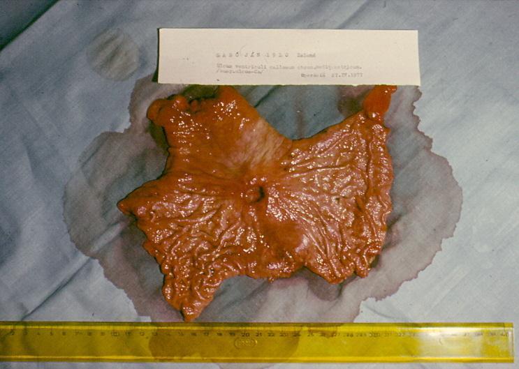
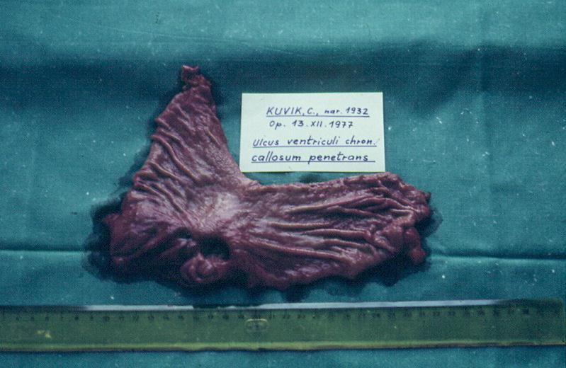
29 Ulcer diease What is Gastroduodenal ulcer? Ulcer is a full-thickness, round and elliptic defect that situates in gastroduodenal mucosa which interfere over lamina muscularis mucose, submucosa or penetrates across whole gastric or duodenal wall Rise of ulcer is conditioned by presence of acid gastric content
30 Pathogenesis Multifactorial Dysbalance between protective and aggressive factors Protective factors: saliva, food, alkali,duodenal fluid, mucus, fast regeneration of gastric epithelial cells, well perfused gastric mucosa Aggressive factors :HCl, pepsin, bile acids (reflux), helicobacter pylori, drugs (analgetics, aspirin, korticoids), nicotine, alcohol
31 Symptoms of gastric ulcer disease Epigastric pain after meal or during meal Upper dyspeptic syndrome loss of appetite, nausea, vomiting, flatulence Vomiting brings relief Reduced nutrition Loss of weight
")
32 Symptoms of duodenal ulcer disease Epigastric pain 2 hours after meal or on a empty stomach or during night Pyrosis Good nutrition Seasonal dependence (spring, autumn)
33 Endoscopy Gastric Multiple Biopsy should be obtained from the edge of the lesion Duodenal Endoscopy is useful in evaluating patients with an uncertain diagnosis
34 Laboratory finding Gastric analysis Basal acid output(bao) Maximal acid output (MAO) mean acid output(meq/h) normal duodenal ulcer Basal male female Maximal male female 20 30
35 Laboratory finding Serum gastrin Normal: pg/ml >200pg/mL always results to ulcer
36 Radiographic examination Upper gastrointestinal x-ray show an ulcer at stomach or duodenum. Gastric ulcer maybe be malignant as following: 1.The Deepest penetration of the ulcer is not beyong the expected border of the gastric wall 2.The meniscus sign is present (barium sulfate ) 3.Ulcer diameter is greater than 2cm
37 Differential diagnosis 1.Chronic cholecystitis 2.Acute and chronic pancreatitis 3.Functional indigestion 4.Reflux esophagitis 5.Gastritis
38 Essentials of diagnosis Gastric Epigastric pain not relieved by food Ulcer demonstrated by x-ray or endoscopy Acid present on gastric analysis Duodenal Epigastric pain relieved by food Epigastric tenderness Normal or increased gastric acid secretion Ulcer diagnosed by x-ray or endoscopy Evidence of Helicobacter Pylori infection
39 Complications Bleeding - chronic (minor, cause anemia) - acute (major, form affected vessel) Perforation - mostly bulbous duodenum, anterior gastric wall acute violent pain - bleeding can be present obstruction - narrow of the lumen caused by scar, oedema or inflammatory infiltration after healing of the ulcer - rise only at pyloric localization - vomiting of huge volume of gastric content

40 Complications A penetration B perforation C bleeding D - obstruction
41 Therapy Conservative regular lifestyle prohibition of the smoking and alcohol diet (proteins, milk and milky products) Four primary medicine H2-receptor antagonists Proton pump inhibitors Antacids Antibiotics
42 Therapy Surgical Indications for operation Intractability Perforation Obstruction Hemorrhage
43 Therapy Intractability (criteria) Initial healing is delayed, so that ulceration persists at 3 months despite active drug therapy Ulcers recur within 1 year of initial healing despite maintenance therapy The ulcer disease is characterized by cycles of prolonged activity with brief or absent remissions
44 Goals of elective operation Gastric Ulcer Primary goals to excise the ulcer to reduce acid/pepsin output Secondary goals to minimize bile reflux and gastric stasis
45 Goals of elective operation Duodenum Ulcer promotion of ulcer healing treatment of specific complications reduction of the possibility of recurrence minimization of postoperative side effects
46 Therapy Surgical BI, BII resection Proximal selective vagotomy Vagotomy with pyloroplastic
47 Subtotal gastrectomy Billroth I (BI) gastro-duodenoanastomosis end-to-end Billroth II (BII) gastro-jejunoanastomosis end-to-side with blind closure of duodenum Gastro-enteroanastomosis on Roux Y crankle Proximal selective vagotomy denervation of parietal gastric cells

48 Billroth I

49 Billroth II

50 Gastro-enteroanastomosis on Roux Y crankle

51 Vagotomy
52

53
54
55 Complications after operation Early dehiscence, stenosis of anastomosis, bleeding, pancreatitis, obstructive icterus, affection of neighbour tissues Late - days, weeks - early dumping syndrome - late dumping syndrome - incoming crankle syndrome - outcoming crankle syndrome - ulcer in anastomosis or in outcoming crankle
56 Early dumping syndrome group of symptoms approved shortly after meal appears after BII resection vasodilator - face redness, fall of blood pressure, dizziness GI symptoms - vomiting, diarrhea Therapy: diet, no sugar, low quantities of food,change BII to BI resection
57 Late dumping syndrome hypoglycemia (sugar is not enough digested) appears after BII resection weakness, perspiration, dizziness, tremor 3h after meal Therapy: no sugar, change BII to BI resection
58 Incoming crankle syndrome stasis of the content at incoming crankle increase intraluminal pressure appears after BII resection Therapy: diet, change BII to BI resection
59 Outcoming crankle syndrome chronic or acute closure of outcoming crankle appears after BII resection vomiting after meal, convulsive pain Therapy: change BII to BI resection
60 Pyloric obstruction due to peptic ulcer Cause Inflammation and edema of pylorus Fibrosis of pylorus Clinical manifestation a long history of symptomatic peptic ulcer development of vomiting, contain food ingested several hours previously, absence of bile. dehydration and malnutrition. Diagnosis X-ray (barium) and endoscopy
61 Treatment Medical treatment A gastric tube should be passed to empty stomach. A saline load test should be performed Total parenteral nutrition should be instituted After decompression of the stomach for several days, if this indicates improvement, a gradual diet may be started.
62 Treatment Surgical treatment BI, BII resection Gastro-entero anastomosis on Roux-Y crankle

63

64

65
66 Perforated Duodenal Ulcer Acute perforation in 5-10%; median age 40-55y (18-90) Clinical finding Symptoms :A sudden, severe upper abdominal pain Signs :the abdominal muscles are rigid owing to severe spasm. Bowel movement sounds are reduced or absent. Plain X-ray :Plain x-rays of the abdomen reveal free subdiaphragmatic air.

67 diaphragm
68 Treatment A nasogastric tube should be passed to empty the stomach Intravenous antibiotics should be started Laparoscopy or laparotomy and suture closure of the perforation solves the immediate problem. After operation, medicine should be taked. If the ulcer be suspicious of malignant tumor, biopsy or subtotal gastrectomy should be performed.
69 Treatment If perforated gastric ulcer is considered, biopsy should be performed on operation. After closure perforation, medication should be taked routinely. Concomitant hemorrhage and perforation or perforated ulcers and obstruction can not be treated by suture closure of the perforation Definitive surgery: BI, BII resection or Gastroenteroanastomosis on Roux-Y crankle
70 Hemorrhage from peptic ulcer 1/5 patients of peptic ulcer have a bleeding experience, 2/5 of bleeding patients will die from ulcer. Endoscopic therapy indications Active bleeding at the time of endoscopy The presence of a visible vessel in the base of the ulcer
71 Hemorrhage from peptic ulcer Endoscopic therapy methods Injection into the ulcer of epinephrine epinephrine plus1% polidocanol (sclerosing agent) ethanol Nd:YAG laser
72 Operative intervention Indications Massive hemorrhage leading to shock Prolonged blood loss requiring continuing transfusion Recurrent bleeding during medical therapy or after endoscopic therapy Recurrent bleeding requiring hospitalization
73 Operative intervention Methods suture or ligation of bleeding vessel Definitive surgery: BI, BII resection or Gastroenteroanastomosis on Roux-Y crankle Truncal vagotomy and antrectomy including ulcer

74

75

76
The abdominal Esophagus, Stomach and the Duodenum. Prof. Oluwadiya KS
 The abdominal Esophagus, Stomach and the Duodenum Prof. Oluwadiya KS www.oluwadiya.com Viscera of the abdomen Abdominal esophagus: Terminal part of the esophagus The stomach Intestines: Small and Large
Done by: Dina Sawadha & Mohammad Abukabeer
 Done by: Dina Sawadha & Mohammad Abukabeer The stomach *the stomach is a dilated part of the gastro intestinal tract, it's "J" shape. *the lower surface of the stomach ( the greater curvature ) reaches
OPERATIVE TREATMENT OF ULCER DISEASE
 Página 1 de 8 Copyright 2001 Lippincott Williams & Wilkins Greenfield, Lazar J., Mulholland, Michael W., Oldham, Keith T., Zelenock, Gerald B., Lillemoe, Keith D. Surgery: Scientific Principles & Practice,
Peptic ulcer disease. Nomin-Erdene. D SOM-531
 Peptic ulcer disease Nomin-Erdene. D SOM-531 Learning objectives Stomach gross anatomy PUD Epidemiology Pathogenesis Clinical manifestation Diagnosing Treatment Complicated ulcer disease Surgical procedures
BY DR NOMAN ULLAH WAZIR
 BY DR NOMAN ULLAH WAZIR The stomach (from ancient Greek word stomachos, stoma means mouth) is a muscular, hollow and the most dilated part of the GIT. It starts from the point where esophagus ends. It
Perforated peptic ulcer
 Perforated peptic ulcer - Despite the widespread use of gastric anti-secretory agents and eradication therapy, the incidence of perforated peptic ulcer has changed little, age limits increase NSAIDs elderly
It passes through the diaphragm at the level of the 10th thoracic vertebra to join the stomach
 The esophagus is a tubular structure (muscular, collapsible tube ) about 10 in. (25 cm) long that is continuous above with the laryngeal part of the pharynx opposite the sixth cervical vertebra The esophagus
د. عصام طارق. Objectives:
 GI anatomy Lecture: 5 د. عصام طارق Objectives: To describe anatomy of stomach, duodenum & pancreas. To list their main relations. To define their blood & nerve supply. To list their lymph drainage. To
KK College of Nursing Peptic Ulcer Badil D ass Dass, Lecturer 25th July, 2011
 KK College of Nursing Peptic Ulcer Badil Dass, Lecturer 25 th July, 2011 Objectives: By the end of this lecture, the students t will be able to: Define peptic pp ulcer Describe the etiology and pathology
Pancreas & Biliary System. Dr. Vohra & Dr. Jamila
 Pancreas & Biliary System Dr. Vohra & Dr. Jamila 1 Objectives At the end of the lecture, the student should be able to describe the: Location, surface anatomy, parts, relations & peritoneal reflection
Stomach. R.B. Kolachalam, MD
 Stomach R.B. Kolachalam, MD Relevent Anatomy 1.four regions: Cardia, Fundus, Body, and the Pylorus 2. fixed in two locations- at the GEJ and the duodenum Gastric Anatomy body of the stomach: site of mechanical
-12. -Renad Habahbeh. -Dr Mohammad mohtasib
 -12 -Renad Habahbeh - -Dr Mohammad mohtasib The Gallbladder -The gallbladder has a body, a fundus (a rounded end), a neck, Hartmann s pouch before the neck and a cystic duct that meets the common hepatic
Accessory Glands of Digestive System
 Accessory Glands of Digestive System The liver The liver is soft and pliable and occupies the upper part of the abdominal cavity just beneath the diaphragm. The greater part of the liver is situated under
Dr. Zahiri. In the name of God
 Dr. Zahiri In the name of God small intestine = small bowel is the part of the gastrointestinal tract Boundaries: Pylorus Ileosecal junction Function: digestion and absorption of food It receives bile
Postgastrectomy Syndromes
 Postgastrectomy Syndromes Postgastrectomy syndromes are iatrogenic conditions that may arise from partial gastrectomies, independent of whether the gastric surgery was initially performed for peptic ulcer
Surgery for Complications of Peptic Ulcer Disease (Definitive Treatment)
") Surgery for Complications of Peptic Ulcer Disease (Definitive Treatment) Amid Keshavarzi, MD UCHSC Grand Round 3/20/2006 Department of Surgery Introduction Epidemiology Pathophysiology Clinical manifestation
Surgery for Complications of Peptic Ulcer Disease (Definitive Treatment) Amid Keshavarzi, MD UCHSC Grand Round 3/20/2006 Department of Surgery Introduction Epidemiology Pathophysiology Clinical manifestation
Anatomy of the SMALL INTESTINE. Dr. Noman Ullah Wazir PMC
 Anatomy of the SMALL INTESTINE Dr. Noman Ullah Wazir PMC SMALL INTESTINE The small intestine, consists of the duodenum, jejunum, and illium. It extends from the pylorus to the ileocecal junction were the
OPEN ACCESS TEXTBOOK OF GENERAL SURGERY
 OPEN ACCESS TEXTBOOK OF GENERAL SURGERY PEPTIC ULCER DISEASE PC Bornman RS Du Toit EPIDEMIOLOGY AND PATHOGENESIS The prevalence of duodenal ulcer disease has a variable geographical distribution and differs
To describe the liver. To list main structures in porta hepatis.
 GI anatomy Lecture: 6 د. عصام طارق Objectives: To describe the liver. To list main structures in porta hepatis. To define portal system & portosystemic anastomosis. To list parts of biliary system. To
Module 2 Heartburn Glossary
 Absorption Antacids Antibiotic Module 2 Heartburn Glossary Barrett s oesophagus Bloating Body mass index Burping Chief cells Colon Digestion Endoscopy Enteroendocrine cells Epiglottis Epithelium Absorption
ABDOMEN - GI. Duodenum
 TALA SALEH ABDOMEN - GI Duodenum - Notice the shape of the duodenum, it looks like capital G shape tube which extends from the pyloroduodenal junction to the duodenojejunal junction. - It is 10 inches
Lab Monitor Images Dissection of the Abdominal Vasculature + Lower Digestive System
 Lab Monitor Images Dissection of the Abdominal Vasculature + Lower Digestive System Stomach & Duodenum Frontal (AP) View Nasogastric tube 2 1 3 4 Stomach Pylorus Duodenum 1 Duodenum 2 Duodenum 3 Duodenum
Nasogastric tube. Stomach. Pylorus. Duodenum 1. Duodenum 2. Duodenum 3. Duodenum 4
 Esophagus Barium Swallow Stomach and Duodenum 4 year old Upper GI Nasogastric tube Stomach and Duodenum 4 year old Upper GI Nasogastric tube Stomach Pylorus Duodenum 1 Duodenum 2 Duodenum 3 Duodenum 4
Pancreas and Biliary System
 Pancreas and Biliary System Please view our Editing File before studying this lecture to check for any changes. Color Code Important Doctors Notes Notes/Extra explanation Objectives At the end of the lecture,
Perforated peptic ulcers. Dr V. Roudnitsky KCH
 Perforated peptic ulcers Dr V. Roudnitsky KCH Peptic ulcer disease Peptic ulcers are focal defects in the gastric or duodenal mucosa that extend into the submucosa or deeper Caused by an imbalance between
Preview from Notesale.co.uk Page 1 of 34
 Abdominal viscera and digestive tract Digestive tract Abdominal viscera comprise majority of the alimentary system o Terminal oesophagus, stomach, pancreas, spleen, liver, gallbladder, kidneys, suprarenal
ORAL CAVITY, ESOPHAGUS AND STOMACH
 ORAL CAVITY, ESOPHAGUS AND STOMACH 1 OBJECTIVES By the end of the lecture you should be able to: Describe the anatomy the oral cavity, (boundaries, parts, nerve supply). Describe the anatomy of the palate,
Gastrointestinal Tract. Anatomy of GI Tract. Anatomy of GI Tract. (Effective February 2007) (1%-5%)
 (1%-5%)") Gastrointestinal Tract (Effective February 2007) (1%-5%) Anatomy of GI Tract Esophagus bulls-eye or target EG junction seen on sagittal scan posterior to left lobe of liver and anterior to aorta Anatomy
Gastrointestinal Tract (Effective February 2007) (1%-5%) Anatomy of GI Tract Esophagus bulls-eye or target EG junction seen on sagittal scan posterior to left lobe of liver and anterior to aorta Anatomy
Lab 5 Digestion and Hormones of Digestion. 7/16/2015 MDufilho 1
 Lab 5 Digestion and Hormones of Digestion 1 Figure 23.1 Alimentary canal and related accessory digestive organs. Mouth (oral cavity) Tongue* Parotid gland Sublingual gland Submandibular gland Salivary
University of Buea. Faculty of Health Sciences. Programme in Medicine
 Faculty of Health Sciences University of Buea Wednesday, 28 th January 2009 Time: 8 00-10 00 Programme in Medicine MED 303 (Gastrointestinal Physiology) EXAMS (2008-2009) Identify the letter of the choice
-Ensherah Mokheemer. -Shatha Al-Jaberi محمد المحتسب- 1 P a g e
 9-9 -Ensherah Mokheemer -Shatha Al-Jaberi محمد المحتسب- 1 P a g e Small intestine has three regions: ( االثني عشر( The duodenum The jejunum The ileum Small intestine Duodenum: -c-shaped -The concavity
5. Which component of the duodenal contents entering the stomach causes the most severe changes to gastric mucosa:
 Gastro-intestinal disorders 1. Which are the most common causes of chronic gastritis? 1. Toxic substances 2. Chronic stress 3. Alimentary factors 4. Endogenous noxious stimuli 5. Genetic factors 2. Chronic
Biology Human Anatomy Abdominal and Pelvic Cavities
 Biology 351 - Human Anatomy Abdominal and Pelvic Cavities Please place your name and I.D. number on the back of the last page of this exam. You must answer all questions on this exam. Because statistics
The Spleen. Dr Fahad Ullah
 The Spleen BY Dr Fahad Ullah Spleen The spleen is an largest lymphoid organ shaped like a shoe that lies relative to the 9th and 11th ribs and is located in the left hypochondrium. Thus, the spleen is
Omar Sami --- Muhammad Al-Muhatasib
 8 Omar Sami --- Muhammad Al-Muhatasib This sheet is a remake from 2015 s sheet for the same lecture; I have checked the record, added, omitted, edited & illustrated all what the Professor mentioned in
Digestive System Module 4: The Stomach *
 OpenStax-CNX module: m49286 1 Digestive System Module 4: The * Donna Browne Based on The by OpenStax This work is produced by OpenStax-CNX and licensed under the Creative Commons Attribution License 4.0
Overview of digestion or, gut reactions - to food
 Key concepts in Digestion. Indigestion module Overview of digestion or, gut reactions - to food Prof. Barry Campbell Gastroenterology Cellular & Molecular Physiology e-mail: bjcampbl@liv.ac.uk http://pcwww.liv.ac.uk/~bjcampbl
Surface Anatomy. Location Shape Weight Role of Five Surfaces Borders Fissures Lobes Peritoneal Lig
 The Liver Functions Bile production and secretion Detoxification Storage of glycogen Protein synthesis Production of heparin and bile pigments Erythropoiesis (in fetus) Surface Anatomy Location Shape Weight
Gastric ulcer Duodenal ulcer Pancreatitis Ileus. Barbora Konečná
 Gastric ulcer Duodenal ulcer Pancreatitis Ileus Barbora Konečná basa.konecna@gmail.com Peptic ulcers of stomach and duodenum (PUD) Ulcers are chronic, often solitary lesions, that occur in any part of
Peptic ulcer disease Disorders of the esophagus
 Peptic ulcer disease Disorders of the esophagus Peptic ulcer disease Burning epigastric pain Exacerbated by fasting Improved with meals Ulcer: disruption of mucosal integrity >5 mm in size, with depth
Patient. Male 76 year old C.C: abdominal pain
 Patient Male 76 year old C.C: abdominal pain Bowel stool retention Suspected pulmonary TB at right upper lung Infiltration in right lower lung Pleural thickening at the Right chest Localized dilated small
Overview of digestion or, gut reactions - to food
 1 Key concepts in Digestion. Indigestion module Overview of digestion or, gut reactions to food Prof. Barry Campbell Gastroenterology Cellular & Molecular Physiology email: bjcampbl@liv.ac.uk http://pcwww.liv.ac.uk/~bjcampbl
Anatomy: Know Your Abdomen
 Anatomy: Know Your Abdomen Glossary Abdomen - part of the body below the thorax (chest cavity); separated by the diaphragm. Anterior - towards the front of the body. For example, the umbilicus is anterior
58 year old male complaining of 3-week history of increasing epigastric pain
 Peptic Ulcer Disease 58 year old male complaining of 3-week history of increasing epigastric pain Has had dyspepsia in the past for which he took Tums, but this is much worse and only partially relieved
Limited en bloc Resection of the Gastroesophageal Junction with Isoperistaltic Jejunal Interposition
 22 Limited en bloc Resection of the Gastroesophageal Junction with Isoperistaltic Jejunal Interposition J.R. Izbicki, W.T. Knoefel, D. C. Broering ] Indications Severe dysplasia in the distal esophagus
The Stomach. Bởi: OpenStaxCollege
 Bởi: OpenStaxCollege Although a minimal amount of carbohydrate digestion occurs in the mouth, chemical digestion really gets underway in the stomach. An expansion of the alimentary canal that lies immediately
BLOCK IV: OFFICIAL BODY PARTS LIST FOR ANTERIOR ABDOMINAL WALL AND ABDOMINAL CONTENTS
 BLOCK IV: OFFICIAL BODY PARTS LIST FOR ANTERIOR ABDOMINAL WALL AND ABDOMINAL CONTENTS External oblique muscle Muscular portion Aponeurotic portion Superficial inguinal ring Lateral (inferior) crus Medial
Anatomy of the Large Intestine
 Large intestine Anatomy of the Large Intestine 2 Large Intestine Extends from ileocecal valve to anus Length = 1.5-2.5m = 5 feet Regions Cecum = 2.5-3 inch Appendix= 3-5 inch Colon Ascending= 5 inch Transverse=
Understandings, Applications & Skills
 D.2 Digestion Understandings, Applications & Skills Statement D.2.U1 Nervous and hormonal mechanisms control the secretion of digestive juices. D.2.U2 Exocrine glands secrete to the surface of the body
Block 3: DISSECTION 2 CELIAC TRUNK, JEJUNUM/ILEUM, LARGE INTESTINE, DUODENUM, PANCREAS, PORTAL VEIN; MOBILIZATION OF THE LIVER
 1 Block 3: DISSECTION 2 CELIAC TRUNK, JEJUNUM/ILEUM, LARGE INTESTINE, DUODENUM, PANCREAS, PORTAL VEIN; MOBILIZATION OF THE LIVER Attempt to complete as much as you can of the dissection explained in the
Digestive system L 2. Lecturer Dr. Firdous M. Jaafar Department of Anatomy/Histology section
 Digestive system L 2 Lecturer Dr. Firdous M. Jaafar Department of Anatomy/Histology section objectives 1-Describe the general structure of digestive tract: a-mucosa. b-submucosa. c-muscularis externa d-adventitia
Small Plicae Circularis. Short Closely packed together. Sparse, completely absent at distal part Lymphoid Nodule
 Intestines Differences Between Jejunum and Ileum Types Jejunum Ileum Color Deeper red Paler pink Calibre Bigger Smaller Thickness of wall Thick and Heavy Thin and Lighter Vascularity Highly vascularised
The surface mucous cells and the cardiac and pyloric glands secrete mucus which protects the stomach from self-digestion.
 PATHOLOGY OF THE STOMACH Stomach mucosa Gastric mucosa is covered by a layer of mucus. The mucosal glands comprise the cardiac glands, the fundic glands in the fundus and body of the stomach, and the pyloric
Laparoscopy-assisted D2 radical distal subtotal gastrectomy
 Masters of Gastrointestinal Surgery Laparoscopy-assisted D2 radical distal subtotal gastrectomy Xiaogeng Chen, Weihua Li, Jinsi Wang, Changshun Yang Department of Tumor Surgery, Fujian Provincial Hospital,
The Whipple Operation Illustrations
 The Whipple Operation Illustrations Fig. 1. Illustration of the sixstep pancreaticoduodenectomy (Whipple operation) as described in a number of recent text books by Dr. Evans. The operation is divided
1 Topographic Anatomy of the Stomach and Duodenum
 1 Topographic Anatomy of the Stomach and Duodenum H.D. BECKER, W. LIERSE, and H.W. SCHREIBER The upper abdominal cavity contains the stomach and duodenum; the liver, gallbladder, and pancreas, which arise
Exploring Anatomy: the Human Abdomen
 Exploring Anatomy: the Human Abdomen PERITONEUM AND PERITONEAL CAVITY PERITONEUM The peritoneum is a thin serous membrane that lines the abdominal cavity and covers, in variable amounts, the viscera within
Common Bile Duct (CBD)
") Liver Last time we talked about the liver and the doctor started by revising some information about it: It has five surfaces. It reaches the 5 th intercostal space ; some books write that it reaches the
Liver Last time we talked about the liver and the doctor started by revising some information about it: It has five surfaces. It reaches the 5 th intercostal space ; some books write that it reaches the
The Digestive System and Body Metabolism
 14 PART B The Digestive System and Body Metabolism PowerPoint Lecture Slide Presentation by Jerry L. Cook, Sam Houston University ESSENTIALS OF HUMAN ANATOMY & PHYSIOLOGY EIGHTH EDITION ELAINE N. MARIEB
- Digestion occurs during periods of low activity - Produces more energy than it uses. - Mucosa
 Introduction Digestive System Chapter 29 Provides processes to break down molecules into a state easily used by cells - A disassembly line: Starts at the mouth and ends at the anus Digestive functions
INVESTIGATIONS OF GASTROINTESTINAL DISEAS
 INVESTIGATIONS OF GASTROINTESTINAL DISEAS Lecture 1 and 2 دز اسماعيل داود فرع الطب كلية طب الموصل Radiological tests of structure (imaging) Plain X-ray: May shows soft tissue outlines like liver, spleen,
THE SURGEON S LIBRARY
 THE SURGEON S LIBRARY THE HISTORY AND SURGICAL ANATOMY OF THE VAGUS NERVE Lee J. Skandalakis, M.D., Chicago, Illinois, Stephen W. Gray, PH.D., and John E. Skandalakis, M.D., PH.D., F.A.C.S., Atlanta, Georgia
Emergency Operations for Bleeding Duodenal Ulcer:A simple option to consider Case Report Abstract Key words Case Report
 Vtáx exñéüà :A simple option to consider: Case Report Gamal E H A El Shallaly, Eltayeb A Ali, Suzan Salih Abstract We report a 46 years-old man who had severe bleeding from a posterior duodenal ulcer (DU)
Biology Human Anatomy Abdominal and Pelvic Cavities
 Biology 351 - Human Anatomy Abdominal and Pelvic Cavities You must answer all questions on this exam. Because statistics demonstrate that, on average, between 2-5 questions on every 100-point exam are
SARCINA VENTICULARI IS A POSSIBLE CAUSATIVE MICROORGANISM OTHER THAN H.PYLORI IN GASTRIC OUTLET OBSTRUCTION PATHOGENESIS
 SARCINA VENTICULARI IS A POSSIBLE CAUSATIVE MICROORGANISM OTHER THAN H.PYLORI IN GASTRIC OUTLET OBSTRUCTION PATHOGENESIS 55 years old male, Farm worker, married, from Ibb Heavy Smoker, Khat chewer non-alcohol
Includes mouth, pharynx, esophagus, stomach, small intestine, large intestine, rectum, anus. Salivary glands, liver, gallbladder, pancreas
 Chapter 14 The Digestive System and Nutrition Digestive System Brings Nutrients Into the Body The digestive system includes Gastrointestinal (GI) tract (hollow tube) Lumen: space within this tube Includes
(b) Stomach s function 1. Dilution of food materials 2. Acidification of food (absorption of dietary Fe in small intestine) 3. Partial chemical digest
 Stomach s function 1. Dilution of food materials 2. Acidification of food (absorption of dietary Fe in small intestine) 3. Partial chemical digest") (1) General features a) Stomach is widened portion of gut-tube: between tubular and spherical; Note arranged of smooth muscle tissue in muscularis externa. 1 (b) Stomach s function 1. Dilution of food
(1) General features a) Stomach is widened portion of gut-tube: between tubular and spherical; Note arranged of smooth muscle tissue in muscularis externa. 1 (b) Stomach s function 1. Dilution of food
Gastroenterology Tutorial
 Gastroenterology Tutorial Gastritis Poorly defined term that refers to inflammation of the stomach. Infection with H. pylori is the most common cause of gastritis. Most patients remain asymptomatic Some
Gastric Cancer Histopathology Reporting Proforma
 Gastric Cancer Histopathology Reporting Proforma Mandatory questions (i.e. protocol standards) are in bold (e.g. S1.01). S1.01 Identification Family name Given name(s) Date of birth Sex Male Female Intersex/indeterminate
GI Tract Lynn Ta Jennifer Zhang July 6, 2006 GI TRACT. 1) Other Names: Gastrointestinal tract Digestive tract Alimentary tract
 Other Names: Gastrointestinal tract Digestive tract Alimentary tract") GI Tract Lynn Ta Jennifer Zhang July 6, 2006 GI TRACT 1) Other Names: Gastrointestinal tract Digestive tract Alimentary tract 2) Definition/Location: Digestion and absorption are the primary functions
GI Tract Lynn Ta Jennifer Zhang July 6, 2006 GI TRACT 1) Other Names: Gastrointestinal tract Digestive tract Alimentary tract 2) Definition/Location: Digestion and absorption are the primary functions
The peritoneum. Prof. Oluwadiya KS, MBBS, FMCS(Orthop) Website:
 Website:") The peritoneum Prof. Oluwadiya KS, MBBS, FMCS(Orthop) Website: http://oluwadiya.com The peritoneum Serous membrane that lines the abdominopelvic cavity and invests the viscera The largest serous membrane
The peritoneum Prof. Oluwadiya KS, MBBS, FMCS(Orthop) Website: http://oluwadiya.com The peritoneum Serous membrane that lines the abdominopelvic cavity and invests the viscera The largest serous membrane
Our gut reactions to food or, gut reactions - to food
 Key concepts in Digestion. Our gut reactions to food or, gut reactions to food Prof. Barry Campbell Cellular & Molecular Physiology email: bjcampbl@liv.ac.uk http://pcwww.liv.ac.uk/~bjcampbl Swallowing
Week 12 - Outline. Outline. Digestive System I Major Organs. Overview of Digestive System
 Outline Week 12 - Digestive System I Major Organs Copyright The McGraw-Hill Companies, Inc. Permission required for reproduction or display. Digestive Tract Function GI Tract Structure Regulation of the
Soft palate elevates, closing off the nasopharynx. Hard palate Tongue Bolus Epiglottis. Glottis Larynx moves up and forward.
 The Cephalic Phase Chemical and mechanical digestion begins in the mouth Saliva is an exocrine secretion Salivary secretion is under autonomic control Softens and lubricates food Chemical digestion: salivary
- Tamara Wahbeh. - Fareed Khdair. 0 P a g e
 -1 - Tamara Wahbeh - - Fareed Khdair 0 P a g e GI Embryology Note: I included everything in the records and slides; anything in the slide not included in this sheet was not mentioned by the doctor during
Ulcerative Colitis. ulcerative colitis usually only affects the colon.
 Ulcerative Colitis Introduction Ulcerative colitis is an inflammatory bowel disease. It is one of the 2 most common inflammatory bowel diseases. The other one is Crohn s disease. Ulcerative colitis and
Gastrointestinal Disorders. Disorders of the Esophagus 3/7/2013. Congenital Abnormalities. Achalasia. Not an easy repair. Types
 Gastrointestinal Disorders Congenital Abnormalities Disorders of the Esophagus Types Stenosis Atresia Fistula Newborn aspirates while feeding. Pneumonia Not an easy repair Achalasia Lack of relaxation
- Digestion occurs during periods of low activity - Produces more energy than it uses. 3 Copyright 2016 by Elsevier Inc. All rights reserved.
 Introduction Digestive System Chapter 29 Provides processes to break down molecules into a state easily used by cells - A disassembly line: Starts at the mouth and ends at the anus Digestive functions
REFLUX ALKALINE GASTRITIS* SYRACUSE, NEW YORK
 VOL. 115, No. 2 REFLUX ALKALINE GASTRITIS* By SEUK KY KIM, M.D.,t LLOYD S. ROGERS, M.D.,t and ROBERT E. HEITZMAN, M.D. SYRACUSE, NEW YORK E ARLY physiologists thought that gastric acid was partly controlled
Sphincters heartburn diaphragm The Stomach gastric glands pepsin, chyme The Small Intestine 1-Digestion Is Completed in the Small Intestine duodenum
 Sphincters are muscles that encircle tubes and act as valves. The tubes close when the sphincters contract and they open when the sphincters relax. When food or saliva is swallowed, the sphincter relaxes
بسم االه الرحمن الرحيم
 MAY 3, 2012 [POSTERIOR ABDOMINAL WALL] LECTURE 26 ANATOMY Quick Revision: بسم االه الرحمن الرحيم Last time we started with the anterior abdominal wall and said that: 1. Diaphragm is the root of the abdomen.
General Data. Gender : Male Birthday and age : 12/07/24,80 y/o Occupation : 無 Date of Admission :
 General Data Gender : Male Birthday and age : 12/07/24,80 y/o Occupation : 無 Date of Admission : 92-07-09 1 Chief complaint Upper abdominal fullness 30 minutes after having foods with sometimes epigastralgia
Digestive System. - Food is ingested
 11 V. Digestive Processes in the Mouth - Food is ingested - Mechanical digestion begins (chewing) - Salivary amylase begins chemical breakdown of starch - Propulsion is initiated by Deglutition (Swallowing)
Helicobacter Pylori Testing HELICOBACTER PYLORI TESTING HS-131. Policy Number: HS-131. Original Effective Date: 9/17/2009
 Easy Choice Health Plan, Inc. Harmony Health Plan of Illinois, Inc. Missouri Care, Inc. Ohana Health Plan, a plan offered by WellCare Health Insurance of Arizona, Inc. WellCare Health Insurance of Illinois,
Gastrointestinal Anatomy and Physiology. Bio 219 Napa Valley College Dr. Adam Ross
 Gastrointestinal Anatomy and Physiology Bio 219 Napa Valley College Dr. Adam Ross Functions of digestive system Digestion Breakdown of food (chemically) using enzymes, acid, and water Absorption Nutrients,
Section Coordinator: Jerome W. Breslin, PhD, Assistant Professor of Physiology, MEB 7208, ,
 IDP Biological Systems Gastrointestinal System Section Coordinator: Jerome W. Breslin, PhD, Assistant Professor of Physiology, MEB 7208, 504-568-2669, jbresl@lsuhsc.edu Overall Learning Objectives 1. Characterize
Colon Cancer , The Patient Education Institute, Inc. oc Last reviewed: 05/17/2017 1
 Colon Cancer Introduction Colon cancer is fairly common. About 1 in 15 people develop colon cancer. Colon cancer can be a life threatening condition that affects the large intestine. However, if it is
The usual dose is 40 mg daily with amoxycillin 1.5 g (750 mg b.d.) for 2 weeks. Up to 2 g/day of amoxycillin has been used in clinical trials.
 for 2 weeks. Up to 2 g/day of amoxycillin has been used in clinical trials.") Name Gasec - 2 Gastrocaps Composition Gasec-20 Gastrocaps Each Gastrocaps contains: Omeprazole 20 mg (in the form of enteric-coated pellets) Properties, effects Proton Pump Inhibitor Omeprazole belongs
Name Gasec - 2 Gastrocaps Composition Gasec-20 Gastrocaps Each Gastrocaps contains: Omeprazole 20 mg (in the form of enteric-coated pellets) Properties, effects Proton Pump Inhibitor Omeprazole belongs
The Digestive System Laboratory
 The Digestive System Laboratory 1 The Digestive Tract The alimentary canal is a continuous tube stretching from the mouth to the anus. Liver Gallbladder Small intestine Anus Parotid, sublingual, and submaxillary
GASTROINTESTINAL SYSTEM
 GASTROINTESTINAL SYSTEM Topographic Anatomy of the Abdomen Surface Landmarks Xiphoid process T9/T10 Inferior costal margin L2/L3 Iliac Crest L4 level ASIS L5/S1 level Pubic symphysis level of greater trochanter
Chapter 26 The Digestive System
 Chapter 26 The Digestive System Digestive System Gastroenterology is the study of the stomach and intestine. Digestion Catabolism Absorption Anabolism The actions of the digestive system are controlled
Gastric and duodenal diseases. Important. Notes (Doctors'/students') 431 SURGERY TEAM. Ahmed Aloqayli. Hayfa AlAbdulkareem.
 431 SURGERY TEAM. Ahmed Aloqayli. Hayfa AlAbdulkareem.") Gastric and duodenal diseases Important Notes (Doctors'/students') 431 SURGERY TEAM Done By: Ahmed Aloqayli Revised By: Hayfa AlAbdulkareem Abeer Al-Suwailem Leaders Mohammed Alshammari The duodenum is
Gastric and duodenal diseases Important Notes (Doctors'/students') 431 SURGERY TEAM Done By: Ahmed Aloqayli Revised By: Hayfa AlAbdulkareem Abeer Al-Suwailem Leaders Mohammed Alshammari The duodenum is
Chapter 20 The Digestive System Exam Study Questions
 Chapter 20 The Digestive System Exam Study Questions 20.1 Overview of GI Processes 1. Describe the functions of digestive system. 2. List and define the four GI Processes: 20.2 Functional Anatomy of the
McHenry Western Lake County EMS System Paramedic, EMT-B and PHRN Optional Continuing Education 2018 #10 Acute GI Bleeds
 McHenry Western Lake County EMS System Paramedic, EMT-B and PHRN Optional Continuing Education 2018 #10 Acute GI Bleeds Gastrointestinal bleeding is a very common problem in emergency medicine. Between
(A) Diarrhea. (B) Stomach cramps. (C) Dehydration due to excess fluid loss. (D) A, B, and C are correct. (E) Only answer B is correct.
 Diarrhea. (B) Stomach cramps. (C) Dehydration due to excess fluid loss. (D) A, B, and C are correct. (E) Only answer B is correct.") Human Anatomy - Problem Drill 21: The Digestive System Question No. 1 of 10 1. A 26-year-old male is treated in the emergency department for severe gastrointestinal disturbance. Which of the following
Human Anatomy - Problem Drill 21: The Digestive System Question No. 1 of 10 1. A 26-year-old male is treated in the emergency department for severe gastrointestinal disturbance. Which of the following
PHYSIOLOGY OF THE DIGESTIVE SYSTEM
 Student Name CHAPTER 26 PHYSIOLOGY OF THE DIGESTIVE SYSTEM D igestion is the process of breaking down complex nutrients into simpler units suitable for absorption. It involves two major processes: mechanical
 P R E S E N T S Dr. Mufa T. Ghadiali is skilled in all aspects of General Surgery. His General Surgery Services include: General Surgery Advanced Laparoscopic Surgery Surgical Oncology Gastrointestinal
Perforation of a Duodenal Diverticulum. Elective Student S. C.
 Perforation of a Duodenal Diverticulum 2008 4 Elective Student S. C. Case History An elderly male presented to the Emergency Department with abdominal pain. Chief Complaint: Worsening, diffuse abdominal
Alimentary Canal (I)
") Alimentary Canal (I) Esophagus and Stomach (Objectives) By the end of this lecture, the student should be able to discuss the microscopic structure in correlation with the function of the following organs:
Alimentary Canal (I) Esophagus and Stomach (Objectives) By the end of this lecture, the student should be able to discuss the microscopic structure in correlation with the function of the following organs: