Financial Disclosure. Visual Field Interpretation RELIABILITY VISUAL FIELD INTERPRETATION IN GLAUCOMA METHODS OF DATA PRESENTATION
|
|
|
- Brian Owen
- 6 years ago
- Views:
Transcription

 FALSE POSITIVES (20%)*** FALSE NEGATIVES (33%) GAZE TRACKER METHODS OF DATA PRESENTATION")
1 VISUAL FIELD INTERPRETATION IN GLAUCOMA Danica J. Marrelli, OD, FAAO University of Houston College of Optometry Financial Disclosure I have received speaking and/or consulting fees from: Aerie Pharmaceutical Alcon Laboratories Allergan Carl Zeiss Meditec Visual Field Interpretation Methods of Data Presentation Systematic Strategy for Interpreting Visual Field / Recognizing Visual Field Loss Diagnostic Criteria for Glaucoma Classification of Visual Field Loss Detecting Progression of Visual Field Loss CATCH TRIALS RELIABILITY FIXATION LOSSES (20%) FALSE POSITIVES (20%)*** FALSE NEGATIVES (33%) GAZE TRACKER METHODS OF DATA PRESENTATION METHODS OF DATA PRESENTATION GRAYSCALE GIVES A PICTURE RESEMBLING ISOPTERS IN A GRAY TONE QUICKLY IDENTIFIES OVERALL DEPRESSIONS NUMERIC GRID RAW DATA (THRESHOLD LEVELS) 1

2 METHODS OF DATA PRESENTATION METHODS OF DATA PRESENTATION TOTAL DEVIATION PLOT DIFFERENCE BETWEEN PATIENT S RESPONSES AND AGE-MATCHED NORMAL POPULATION TOTAL DEVIATION PROBABILITY PLOT SIGNIFICANCE OF THE TOTAL DEVIATION PLOT PATTERN DEVIATION ADJUSTS THE TOTAL DEVIATION FOR THE OVERALL HEIGHT OF THE HILL OF VISION CAN BE ADJUSTED UP OR DOWN PROBABILITY PLOT METHODS OF DATA PRESENTATION GLOBAL INDICES SINGLE NUMBER REPRESENTATIONS OF THE VISUAL FIELD OVERALL GUIDELINES TO HELP ASSESS FIELD PROBABILITY VALUES GIVEN WHEN NUMBERS REACH SIGNIFICANT VALUES 2
 DEGREE TO WHICH THE SHAPE OF THE VISUAL FIELD DIFFERS FROM REFERENCE FIELD DOES")


3 GLOBAL INDICES MEAN DEVIATION (MD) HEIGHT OF THE HILL OF VISION COMPARED TO AGE-MATCHED NORMALS PATTERN STANDARD DEVIATION (PSD) DEGREE TO WHICH THE SHAPE OF THE VISUAL FIELD DIFFERS FROM REFERENCE FIELD DOES NOT CHANGE WITH MEDIA Glaucoma Hemifield Test What s the VFI??? Mirror Image Analysis Compares Superior to Inferior Field Within Normal Limits Borderline Outside Normal Limits Abnormally High Sensitivity General Reduction In Sensitivity INTERPRETATION OF THE AUTOMATED VISUAL FIELD RELIABILITY MUST KNOW WHETHER OR NOT THE DATA YOU ARE ANALYZING IS RELIABLE FIXATION LOSSES (20%) FALSE POSITIVES (20%) FALSE NEGATIVES (33%) 3

4 RECOGNIZING VISUAL FIELD DEFECTS GRAYSCALE: NOT APPROPRIATE FOR MAKING DIAGNOSIS MUST CONCENTRATE PRIMARILY ON THE DEVIATION PLOTS AND GLOBAL INDICES, SOME ATTENTION TO RAW (THRESHOLD) DATA RECOGNIZING VISUAL FIELD DEFECTS USING THE TOTAL OR PATTERN DEVIATION PLOTS: FIND MOST DEPRESSED POINTS; EXAMINE POINTS SURROUNDING THOSE LOOK FOR PATTERNS CONSISTENT WITH GLAUCOMA NASAL STEP ARCUATE BUNDLE PARACENTRAL RECOGNIZING VISUAL FIELD DEFECTS Look at Global Indices & GHT For diagnosis, look to see if they reach statistical significance For following over time, look for change RECOGNIZING VISUAL FIELD DEFECTS SCOTOMAS AND DEPRESSIONS IN AREAS KNOWN FOR GLAUCOMA (PARACENTRAL, NASAL STEP, ARCUATE BUNDLE) RECOGNIZING VISUAL FIELD DEFECTS ALWAYS: 1. LOOK AT BOTH FIELDS TOGETHER 2. LOOK AT FIELD WITH RELATION TO OTHER CLINICAL FINDINGS - DOES THIS MAKE SENSE, IS IT CONSISTENT WITH THE DIAGNOSIS OF GLAUCOMA? 3. DON T OVERLOOK OTHER CAUSES OF VISUAL FIELD DEFECTS 4


5 Look At Both Fields Together Look At Both Fields Together LOOK AT FIELD WITH RELATION TO OTHER CLINICAL FINDINGS LOOK AT FIELD WITH RELATION TO OTHER CLINICAL FINDINGS Predict the Visual Field 5


6 Predict the Visual Field KEY POINTS TO INTERPRETATION MAKE SURE YOU ARE LOOKING AT TRUSTWORTHY DATA WILL PROBABLY TAKE 3-4 TESTS TO ACHIEVE APPROPRIATE BASELINE MAKE SURE IT MAKES SENSE WITH OTHER CLINICAL FINDINGS STRATEGY DECISIONS 30-2 vs Size III vs. Size V 24-2 vs SITA-Standard vs. SITA-Fast (vs. Threshold or FastPac) 30-2 versus versus


, ALL OF WHICH ARE IDENTIFIED AS SIGNIFICANT, WITH AT")
7 What about the 10-2 VF in early diagnosis? Central 8 degrees from the center of the foveal contains more than 30% of retinal ganglion cells 24-2 and 30-2 test strategies use a 6 degree test grid pattern; these points fall outside of the densist region of ganglion cells 10-2 test strategy uses a 2 degree test grid Recent research has shown that in some patients with small regions of macular gangion cell loss, 10-2 testing may be better able to detect VF loss Minimum Criteria for Diagnosis of Glaucoma VF Defect (Modified HODAPP, ET AL, 1993) 1. GHT OUTSIDE NORMAL LIMITS ON AT LEAST TWO OCCASIONS -OR- Minimum Criteria for Diagnosis of Glaucoma VF Defect (Modified HODAPP, ET AL, 1993) 2. CLUSTER OF 3 OR MORE NON- EDGE POINTS (in a typical location for glaucoma), ALL OF WHICH ARE IDENTIFIED AS SIGNIFICANT, WITH AT LEAST ONE AT THE p<1% ON TWO CONSECUTIVE TESTS (ON 24-2, USE ALL POINTS) -OR- Minimum Criteria for Diagnosis of Glaucoma VF Defect (Modified HODAPP, ET AL, 1993) 3. PSD FLAGGED AT p<5% OR WORSE ON TWO CONSECUTIVE FIELDS 7
: FOR 24-2 SITA STANDARD MD DEPRESSED BY <-5dB AND ON PD PLOT, <25% (14) POINTS ARE DEPRESSED BELOW THE 5% SIGNIFICANCE LEVEL and fewer")
 MD -5dB TO -10dB OR ON PD PLOT, <50% (14-28) POINTS ARE DEPRESSED BELOW 5% LEVEL, OR 8-16 POINTS ARE BELOW THE 1% LEVEL OR CENTRAL POINTS BETWEEN 10-20dB IN ONE")
8 CLASSIFICATION OF FIELD LOSS (Modified from Hodapp, et al) MILD (all 3 criteria must be met): FOR 24-2 SITA STANDARD MD DEPRESSED BY <-5dB AND ON PD PLOT, <25% (14) POINTS ARE DEPRESSED BELOW THE 5% SIGNIFICANCE LEVEL and fewer than half of those points are depressed below the 1% LEVEL AND NONE OF CENTRAL FOUR POINTS HAS SENSITIVITY OF <20dB CLASSIFICATION OF VISUAL FIELD LOSS MODERATE (24-2 Sita) MD -5dB TO -10dB OR ON PD PLOT, <50% (14-28) POINTS ARE DEPRESSED BELOW 5% LEVEL, OR 8-16 POINTS ARE BELOW THE 1% LEVEL OR CENTRAL POINTS BETWEEN 10-20dB IN ONE HEMIFIELD (NO POINTS IN CENTRAL 5 DEGREES WITH <10dB) 14 points flagged 5 points at 1% or worse 8
 POINTS ARE DEPRESSED BELOW 5% OR MORE THAN 16 POINTS ARE BELOW THE 1% LEVEL OR BOTH HEMIFIELDS IN THE CENTRAL 5 DEGREES HAVE <20dB OR ANY POINT IN THE CENTRAL 5 DEGREES")
9 Moderate Loss Moderate Loss 14 total 9 below 1% 18 total 10 at 1% level Moderate Loss 18 total 9 at 1% CLASSIFICATION OF VISUAL FIELD LOSS SEVERE (24-2 Sita) MD DEPRESSED BY MORE THAN -10dB OR ON PD PLOT, GREATER THAN 50% (28) POINTS ARE DEPRESSED BELOW 5% OR MORE THAN 16 POINTS ARE BELOW THE 1% LEVEL OR BOTH HEMIFIELDS IN THE CENTRAL 5 DEGREES HAVE <20dB OR ANY POINT IN THE CENTRAL 5 DEGREES HAS A VALUE <10dB Severe Loss Severe Loss 20 total 17 at 1% 22 total 18 at 1% 9
 FOR THE RECORD ICD-10 Specific test performed (24-2 SS) Statement with respect to reliability Statement with respect to location, size,")
??? Interpretation: H40.")
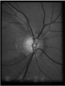
10 13 points 3 points <1% INTERPRETATION TEMPLATE LOOK AT RELIABILITY LOOK AT CENTRAL LEVELS FOR VARIATIONS OF >4dB ACROSS HORIZONTAL MIDLINE NASALLY TOTAL / PATTERN DEVIATION PLOT - MOST DEPRESSED POINT AND SURROUNDING POINTS GLOBAL INDICES (MD, PSD, GHT, VFI) FOR THE RECORD ICD-10 Specific test performed (24-2 SS) Statement with respect to reliability Statement with respect to location, size, density, and pattern of the defect Statement that correlates other examination findings with this visual field Statement about stability/progression (or words BASELINE ) (Statement about how these results influence your management)??? Interpretation: H40.11X3(POAG) 24-2 SS Reliable Severe loss: Large, dense inferior arcuate with small dense superior paracentral defect, c/w superior notch and inferior thinning of ONH Baseline test Aggressive therapy indicated Interpretation: H40.11X SS Reliable Mild loss: Small, shallow inferior nasal step/partial arcuate c/w superior>inferior thinning of ONH Baseline test Initiate therapy 10


11 Detecting Progression Glaucoma: A chronic, progressive disease of retinal ganglion cells that results in characteristic optic nerve and retinal nerve fiber layer changes and corresponding visual field loss Glaucoma Progression Once the diagnosis of glaucoma has been made, the MOST IMPORTANT remaining question is whether the disease is stable and the therapy/compliance are sufficient, or whether the disease is progressive and the therapy in relation to the life expectancy has to be intensified. Progression of Glaucoma, World Glaucoma Association, 2011 Kugler Publications Progression of Glaucoma Although most glaucoma patients will show some evidence of progression if followed long enough, the rate of deterioration can be highly variable among them. While most patients progress slowly, others have aggressive disease with fast deterioration which can eventually result in blindness or substantial impairment unless appropriate interventions take place. WGA Consensus Statements on Structure & Function Both ON structure and function should be evaluated for detection of progression Currently, no specific test can be regarded as the perfect standard for determination of progression Progression detected by functional means will not always be corroborated using structural tests, and vice-versa WGA Consensus Statements The use of standard automated perimetry as the sole method for detection of change may result in failure to detect or underestimation of progression in eyes with early glaucoma damage. Progressive structural changes are often but not always predictive of future development of or progression of functional deficits in glaucoma. 11


12 FUNCTION Previously believed that only advanced glaucoma impacts patient s ability to function. More recent studies have demonstrated that early glaucomatous VF loss has an impact on the patient s ability to function Activity limitation General health, lifestyle, emotion WGA Consensus Statements A statistically significant change in structure and/or function is not always clinically relevant. Life expectancy should be considered when evaluating the clinical relevance of a structural and/or functional change in glaucoma. Structural and/or functional testing should be conducted throughout the duration of the disease. Detection of progression is more difficult in eyes with advanced disease When Should We Suspect Progression? Rates of blindness in POAG are 20 years Blindness in at least 1 eye Diagnosed : 26% Diagnosed : 13.5% Blindness within 10 years of diagnosis: Diagnosed : 8.7 per 100,000 Diagnosed : 5.5 per 100,000 Malihi M, et al. Long-term trends in glaucoma-related blindness in Olmsted County, Minnesota. Ophthalmol 2014; 121(1):

 IDENTIFYING VISUAL")
13 Risk Factors for Progression Clinical risk factor assessment in glaucoma serves two roles: 1. Prognostic information 2. A basis for disease management Risk factor assessment should take into account 1. The strength of the risk factor 2. The practicality & potential harm of reducing that risk factor Risk Factors for Progression Higher mean IOP Higher IOP fluctuations Thinner CCT in patients with higher baseline IOP Presence of pseudoexfoliation Presence of disc hemorrhage Older age Lower ocular perfusion pressure Advanced visual field at presentation Family history of glaucoma (1 st degree relative) IDENTIFYING VISUAL FIELD PROGRESSION Much more difficult than detecting loss Background of dynamic noise No algorithm uniformly agreed upon for detecting change Three main changes: Deepening of defect Enlargement of defect New defect IDENTIFYING PROGRESSION Long-term fluctuation The single biggest problem in determining progression Deeper defects: more long term fluctuation More advanced glaucoma: more long term fluctuation, more fatigue IDENTIFYING PROGRESSION Progression of VF 13


14 Functional Progression - WGA Standard white-on-white automated perimetry (SAP) covering at least 24 is preferred Decisions on progression should NOT be made by comparing only the most recent VF with the one before. Suspected progression should be confirmed with repeat testing. Frequency of VF Exams Baseline Data first 2 years At least 2 reliable VF within the first 6 months 3 within first 6 months when there is a high likelihood of visual disability At least 2 further VF within the next 18 months VF testing should be repeated sooner than scheduled if possible progression is identified SIX VF within the first 2 years allows the clinician to identify rapid progression Frequency of VF Exams Follow-up data (after first 2 years) Frequency of testing should be based on the risk of clinically significant progression (based on extent of damage, life expectancy) In low- and moderate- risk patients, VF should be at least once per year Sooner if possible progression seen on VF ORon other clinically relevant observations In high risk patients, subsequent VF should be at least 2 per year VF Progression: EA vs TA Event analysis (EA): change from baseline greater than a predefined threshold based on test-retest variability according to the level of damage Trend analysis (TA): rate of change over time; significance is determined by both the magnitude of change and the variability of the measurement 14
 is used later in the follow up (later than 2 years) Functional Progression - WGA Use available software support.")
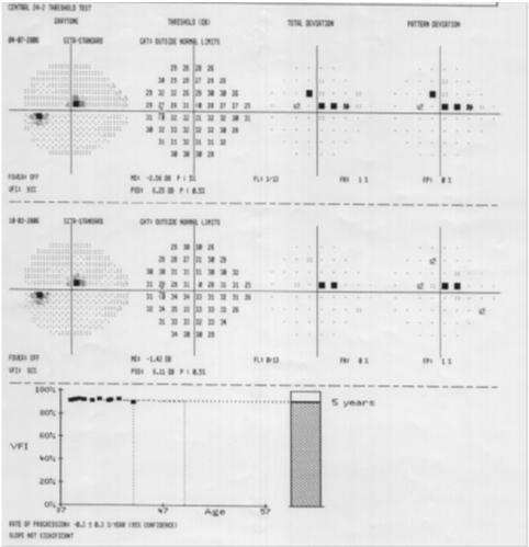
15 Event Analysis Event Analysis Trend Analysis VF Progression: EA vs TA In general, event analysis is used for follow-up when fewer VF are available When suspected progression is identified, at least TWO further tests should confirm that In general, trend analysis (rate) is used later in the follow up (later than 2 years) Functional Progression - WGA Use available software support. Subjective judgment of VF printouts is unreliable and agreement among clinicians is poor. GUIDED PROGRESSION ANALYSIS (GPA) Humphrey Field Analyzer Based on results of GLAUCOMA patients from mild to advanced disease Patients took 12 different visual field tests within a 4 week period Developed a model for what is expected test-test variation for patients with glaucoma GPA Uses 2 baseline exams (any strategy) Follow up tests must be SITA-Standard or SITA-Fast (all same strategy) Symbols used on Follow Up Tests Open Triangles Half Triangles Messages Possible Progression Likely Progression Rate of Progression 15
 Today s VF (Event Analysis) The VFI Regression Plot VFI plotted against age")

16 Elements of GPA 1-Page Summary Report HFA GPA VFI Summary - Interpretation at a Glance Baseline Tests VFI (Trend Analysis) Today s VF (Event Analysis) The VFI Regression Plot VFI plotted against age Extrapolates rate of change up to 5 years HFA GPA VFI Summary - Interpretation at a Glance Loss to date Projected future loss 100% Sample: Progression Detected The VFI Bar historical and projected VFI loss 16
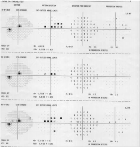
17 Sample: High LTF, No progression 17


18 Sometimes the messages are contradictory Interpretation and Report Interpretation H40.11X SS with GPA Severe loss: Large, dense superior arcuate defect c/w inferior notch of ONH GPA shows no progression on trend/event analysis Continue current therapy 18

19 Functional Progression - WGA Do not rely on VF reliability indices Stick with the same test throughout the follow-up period In advanced glaucoma, there may be a benefit to testing using a 10-2 strategy in a minority of patients. Pearls for VF Progression Event Analysis About 5% chance that a single point will fall outside the expected change on a single test Much less likely that same point will do the same in a subsequent test If point is in same region of VF as existing defect much more likely to be real change Point in central 10 degrees exceeding expected change is much more likely to be real change Alternatives to GPA Non-parametric Analysis (MD, VFI, PSD) 3 baseline VF Suspected progression: 1 field with MD worse than lowest MD of baseline tests Possible progression: 2 consecutive fields with MD worse than lowest MD of baseline tests Likely progression: 3 consecutive fields with MD worse than lowest MD of baseline tests GPA NPA Suspected Progression >/= 3 >/= 3 >/= 3 1 field with MD worse than lowest of 3 baseline VF Possible Progression 2 consecutive fields with MD worse than lowest of 3 baseline VF Likely Progression 3 consecutive fields with MD worse than lowest of 3 baseline VF NPA can also be applied to PSD (for MD up to ~ -10dB) and/or to VFI BASELINE Follow-Up 1: No Suspected Progression Follow-Up 2: Suspected Progression Follow-Up 3: Possible Progression 19


20 Alternatives to GPA Example Deepening of existing scotoma at 2+ points by 10+dB Expansion of 2+ points adjacent to baseline scotoma by 10+dB New scotoma 2 or more adjacent points with p<1% on PD probability plot Change of 1 point in central 10 of 10+dB in a previously normal location Drawbacks of GPA Cannot be used in advanced glaucoma Trend analysis may not be able to detect progression in patients with smaller paracentral scotomas (often limited to a single point) 20


21 21


22 Trend Analysis Need a minimum of 6-8 tests for valuable slope Cut-off value of 1dB/yr is probably a reasonable, clinically relevant cutoff value Greatly influenced by outliers WATCH FOR OUTLIERS Excluding non-representative exams There s Progression! What Now? Think there is progression? VERIFY! Know there is progression? Did glaucoma cause the progression? Glaucoma caused the progression Consider a change in treatment BEFORE, with poor exam included AFTER excluding the poor exam Think There s Progression? Verify! Structural tests: Confirm with additional test Visual field: Confirm with at least two additional tests FAILURE TO CONFIRM PROGRESSION is evidence of stability! There s Progression: Is it Glaucoma? Structure: Other causes of structural changes? Esp important in polarimetry Function: Optical explanation LESS LIKELY Equal damage to total and pattern deviation plots Individual test locations with normal sensitivity Increased PSD Absence of media opacities (Duh!) Optical explanation MORE LIKELY No increase in PSD, in cases where MD is better than -10dB 22
23 Glaucoma Caused the Progression: Now What? Factors to Consider: Compliance Glaucoma Stage Rate of progression/ time to event Location of scotoma Life expectancy of patient Patient preference Potential impact of next therapeutic step New Baseline!! Every time a target IOP is adjusted or there is a significant change in the therapy, the tests need to be re-baselined Last 2 tests that determined/confirmed progression can be the new baseline exams Frequency of testing needs to increase again Thoughts on Treatment ASSESS COMPLIANCE!!!! Poor compliance: Important conversation regarding compliance Emphasize importance of compliance May or may not need to lower target IOP Consider laser trabeculoplasty or surgery Good compliance: Lower target IOP Added medication Laser trabeculoplasty or surgery Thank you for your attention! Questions? me: Dmarrelli@uh.edu 23
21st Century Visual Field Testing
 Supplement to Supported by an educational grant from Carl Zeiss Meditec, Inc. Winter 2011 21st Century Visual Field Testing the Evolution Continues 21st Century Visual Field Testing 21st Century Visual
3/16/2018. Perimetry
 Perimetry The normal visual field extends further away from fixation temporally and inferiorly than superiorly and nasally. From the center of the retina this sensitivity decreases towards the periphery,
Visual Fields Shawn L. Cohen, M.D. Part 2 of 4. Definitions / Tables (Part 2 of 2) Static Perimetry (Humphrey, Octopus)
 Static Perimetry (Humphrey, Octopus)") Visual Fields Shawn L. Cohen, M.D. Part 2 of 4 Definitions / Tables (Part 2 of 2) Static Perimetry (Humphrey, Octopus) Normal Visual Field: Components: General Information Reliability Indices Raw Data
Visual Fields Shawn L. Cohen, M.D. Part 2 of 4 Definitions / Tables (Part 2 of 2) Static Perimetry (Humphrey, Octopus) Normal Visual Field: Components: General Information Reliability Indices Raw Data
VISUAL FIELDS. Visual Fields. Getting the Terminology Sorted Out 7/27/2018. Speaker: Michael Patrick Coleman, COT & ABOC
 VISUAL FIELDS Speaker: Michael Patrick Coleman, COT & ABOC Visual Fields OBJECTIVES: 1. Explain what is meant by 30-2 in regards to the Humphrey Visual Field test 2. Identify the difference between a kinetic
FORUM Glaucoma Workplace from ZEISS Clinical Interpretation Guide
 FORUM Glaucoma Workplace from ZEISS Clinical Interpretation Guide ZEISS FORUM Glaucoma Workplace.0 For years, doctors have asked for the operational capability to analyze data from their Humphrey Field
Visual Field Interpretation Anthony B. Litwak, OD, FAAO VA Medical Center Baltimore, MD
 Visual Field Interpretation Anthony B. Litwak, OD, FAAO VA Medical Center Baltimore, MD Dr. Litwak is on the speaker bureau and advisory panel for Alcon and Zeiss Meditek o Confirms Glaucoma Diagnosis!
Glaucoma Progression. Murray Fingeret, OD
 Glaucoma Progression Murray Fingeret, OD Managing glaucoma or glaucoma suspects Summary For patients diagnosed or at risk of glaucoma Stage disease Risk assessment Asses for progression Treatment plan
Evolving glaucoma management True diagnostic integration for the preservation of vision
 Evolving glaucoma management True diagnostic integration for the preservation of vision // GLAUCOMA MANAGEMENT MADE BY ZEISS The moment you are certain it is glaucoma. This is the moment we work for. There
Financial Disclosure. Maximizing OCT in Diagnosis and Management of Glaucoma. Case 1: Dennis, 65yo WM DIAGNOSIS: CASES 1 2 7/29/2016
 Financial Disclosure Maximizing OCT in Diagnosis and Management of Glaucoma Aerie Pharmaceuticals Alcon Laboratories Allergan Carl Zeiss Meditec Danica J. Marrelli, OD, FAAO AAO Diplomate, Glaucoma University
Definitions. Indications for Perimetry. Indications for Perimetry. Purposes of Perimetry. Indications for Perimetry 3/4/2015
 DISCLOSURE STATEMENT No disclosure statement. 2 Definitions Lecturer: UNDERSTANDING VISUAL FIELD TESTING Caroline B. Pate, OD, FAAO Visual Field: The portion of space which is visible when gaze is fixed
Misleading Statistical Calculations in Faradvanced Glaucomatous Visual Field Loss
 Misleading Statistical Calculations in Faradvanced Glaucomatous Visual Field Loss Eytan Z. Blumenthal, MD, Ruthy Sapir-Pichhadze, MD Objective: In this study, the capability of statistical analysis indices
Perimetry Phobia: Don t fear the field Savory Turman, COMT, CPSS
 Perimetry Phobia: Don t fear the field Savory Turman, COMT, CPSS I have no financial interest in this presentation. Who am I? Where am I? What am I? The anatomy of the visual field Purpose of Visual Field
OCT in the Diagnosis and Follow-up of Glaucoma
 OCT in the Diagnosis and Follow-up of Glaucoma Karim A Raafat MD. Professor Of Ophthalmology Cairo University Hmmmm! Do I have Glaucoma or not?! 1 Visual Function 100% - N Gl Structure : - 5000 axon /
Learn Connect Succeed. JCAHPO Regional Meetings 2017
 Learn Connect Succeed JCAHPO Regional Meetings 2017 Visual Field Testing Suzanne Hansen, M.Ed., COMT, OSC Why are these tests ordered? Visual field testing is ordered to help the physician diagnose and
Description of new EyeSuite visual field and trend analysis functions
 Description of new EyeSuite visual field and trend analysis functions Global Trend Graph The trend graph indicates the normality range (95%) on top as grey band. Falling below that area means falling out
New Concepts in Glaucoma Ben Gaddie, OD Moderator Murray Fingeret, OD Louis Pasquale, MD
 New Concepts in Glaucoma Ben Gaddie, OD Moderator Murray Fingeret, OD Louis Pasquale, MD New Concepts in Glaucoma Optical Coherence Tomography: Is it necessary and needed to diagnose and monitor glaucoma?
Macular Ganglion Cell Complex Measurement Using Spectral Domain Optical Coherence Tomography in Glaucoma
 Med. J. Cairo Univ., Vol. 83, No. 2, September: 67-72, 2015 www.medicaljournalofcairouniversity.net Macular Ganglion Cell Complex Measurement Using Spectral Domain Optical Coherence Tomography in Glaucoma
Diagnosing open-angle glaucoma may be particularly
 A New Look at Selective Perimetry What is its role in clinical practice? BY MURRAY FINGERET, OD Diagnosing open-angle glaucoma may be particularly challenging when damage is mild or early. Although most
Targeting Intraocular Pressure in Glaucoma: a Teaching Case Report 1
 Targeting Intraocular Pressure in Glaucoma: a Teaching Case Report 1 By: Andrew Kemp, OD, Marcus Gonzales, OD, FAAO, Joe DeLoach, OD, FAAO, and Zanna Kruoch, OD FAAO Background Glaucoma is a range of conditions
STRUCTURE & FUNCTION An Integrated Approach for the Detection and Follow-up of Glaucoma. Module 3a GDx
 STRUCTURE & FUNCTION An Integrated Approach for the Detection and Follow-up of Glaucoma Module 3a GDx Educational Slide Deck Carl Zeiss Meditec, Inc. November 2005 1 Structure & Function Modules Module
Early Detection Of Glaucoma Clinical Clues. Points To Live By. Glaucoma Risk Factors 10/3/2014
 Early Detection Of Glaucoma Clinical Clues Eric E. Schmidt, O.D. Omni Eye Specialists Wilmington, NC schmidtyvision@msn.com Points To Live By 25% of G pxs NEVER have IOP >21mm 50% of G pxs have trough
53 year old woman attends your practice for routine exam. She has no past medical history or family history of note.
 Case 1 Normal Tension Glaucoma 53 year old woman attends your practice for routine exam. She has no past medical history or family history of note. Table 1. Right Eye Left Eye Visual acuity 6/6 6/6 Ishihara
CHAPTER 13 CLINICAL CASES INTRODUCTION
 2 CHAPTER 3 CLINICAL CASES INTRODUCTION The previous chapters of this book have systematically presented various aspects of visual field testing and is now put into a clinical context. In this chapter,
A Review Of Risk Factors. Early Detection Of Glaucoma Clinical Clues. A risk factor analysis is critical. Points To Live By
 A Review Of Risk Factors Early Detection Of Glaucoma Clinical Clues Eric E. Schmidt, O.D. Omni Eye Specialists Wilmington, NC schmidtyvision@msn.com FINDACAR Family history IOP Nearsightedness Diabetes/Vascular
STANDARD AUTOMATED PERIMETRY IS A GENERALLY
 Comparison of Long-term Variability for Standard and Short-wavelength Automated Perimetry in Stable Glaucoma Patients EYTAN Z. BLUMENTHAL, MD, PAMELA A. SAMPLE, PHD, LINDA ZANGWILL, PHD, ALEXANDER C. LEE,
C a t a r a c t G l a u c o m a R e t i n a R e f r a c t i v e. The GDxVCC Early answers and ongoing assessment for glaucoma
 C a t a r a c t G l a u c o m a R e t i n a R e f r a c t i v e The GDxVCC Early answers and ongoing assessment for glaucoma The quantifiable approach to quality care Only Humphrey GPA software Early insight
Intro to Glaucoma/2006
 Intro to Glaucoma/2006 Managing Patients with Glaucoma is Exciting Interesting Challenging But can often be frustrating! Clinical Challenges To identify patients with risk factors for possible glaucoma.
Research Article The Pattern of Retinal Nerve Fiber Layer and Macular Ganglion Cell-Inner Plexiform Layer Thickness Changes in Glaucoma
 Hindawi Ophthalmology Volume 2017, Article ID 78365, 8 pages https://doi.org/10.1155/2017/78365 Research Article The Pattern of Retinal Nerve Fiber Layer and Macular Ganglion Cell-Inner Plexiform Layer
Method for comparing visual field defects to local RNFL and RGC damage seen on frequency domain OCT in patients with glaucoma.
 Method for comparing visual field defects to local RNFL and RGC damage seen on frequency domain OCT in patients with glaucoma. Donald C. Hood 1,2,* and Ali S. Raza 1 1 Department of Psychology, Columbia
The Optic Nerve Head In Glaucoma. Clinical Pearl #1. Characteristics of Normal Disk 9/26/2017. Initial detectable damage Structure vs function
 The Optic Nerve Head In Glaucoma Clinical Pearl #1 Eric E. Schmidt, O.D., F.A.A.O. Omni Eye Specialists Wilmington,NC schmidtyvision@msn.com Glaucoma is an optic neuropathy Initial detectable damage Structure
DISCLOSURE: What to do? 2/22/2016
 DISCLOSURE: Dr. Joseph Sowka is a member of the Speakers Bureau for Alcon Laboratories, and Carl Zeiss Meditec. He is on the advisory boards for Alcon, Zeiss, and Allergan. He is a consultant for Alcon.
Clinical Study Visual Field Loss Morphology in High- and Normal-Tension Glaucoma
 Journal of Ophthalmology Volume 2012, Article ID 327326, 8 pages doi:10.1155/2012/327326 Clinical Study Visual Field Loss Morphology in High- and Normal-Tension Glaucoma Michele Iester, 1, 2 Fabio De Feo,
Glaucoma: a disease of the macula?
 Glaucoma: a disease of the macula? Derek MacDonald, OD, FAAO Waterloo, Ontario, Canada Please silence all mobile devices and remove items from chairs so others can sit. Unauthorized recording of this session
Automated Visual Field Analysis for Glaucoma
 Island of Vision Automated Visual Field Analysis for Glaucoma Normal Visual Field Parameters 60 superior 60 nasal 75 inferior 100 temporal Macula the central 13 Fovea the central 3 Visual field is limited
Is this glaucoma? Leo Semes, OD Michael Chaglasian, OD Danica Marrelli, OD. Optometry s Meeting 2015 Seattle, WA
 Is this glaucoma? Leo Semes, OD Michael Chaglasian, OD Danica Marrelli, OD Optometry s Meeting 2015 Seattle, WA Case 1. 54 WM Engineer is referred to UAB Eye Care as a glaucoma suspect. Mild myopic refractive
Study of Retinal Nerve Fiber Layer Thickness Within Normal Hemivisual Field in Primary Open-Angle Glaucoma and Normal-Tension Glaucoma
 Study of Retinal Nerve Fiber Layer Thickness Within Normal Hemivisual Field in Primary Open-Angle Glaucoma and Normal-Tension Glaucoma Chiharu Matsumoto, Shiroaki Shirato, Mai Haneda, Hiroko Yamashiro
Retinal nerve fiber layer thickness in Indian eyes with optical coherence tomography
 Original articles in Indian eyes with optical coherence tomography Malik A, Singh M, Arya SK, Sood S, Ichhpujani P Department of Ophthalmology Government Medical College and Hospital, Sector 32, Chandigarh,
Citation for published version (APA): Wesselink, C. (2017). Glaucoma care optimised in an ageing population [Groningen]: Rijksuniversiteit Groningen
![Citation for published version (APA): Wesselink, C. (2017). Glaucoma care optimised in an ageing population [Groningen]: Rijksuniversiteit Groningen](/thumbs/90/102866625.jpg "Citation for published version (APA): Wesselink, C. (2017). Glaucoma care optimised in an ageing population [Groningen]: Rijksuniversiteit Groningen") University of Groningen Glaucoma care optimised in an ageing population Wesselink, Christiaan IMPORTANT NOTE: You are advised to consult the publisher's version (publisher's PDF) if you wish to cite from
University of Groningen Glaucoma care optimised in an ageing population Wesselink, Christiaan IMPORTANT NOTE: You are advised to consult the publisher's version (publisher's PDF) if you wish to cite from
Collaboration in the care of glaucoma patients and glaucoma suspects. Barry Emara MD FRCS(C) Nico Ristorante November 29, 2012
 Nico Ristorante November 29, 2012") Collaboration in the care of glaucoma patients and glaucoma suspects Barry Emara MD FRCS(C) Nico Ristorante November 29, 2012 Goals of Collaboration Patient-centred and evidence based approach Timely access
Collaboration in the care of glaucoma patients and glaucoma suspects Barry Emara MD FRCS(C) Nico Ristorante November 29, 2012 Goals of Collaboration Patient-centred and evidence based approach Timely access
Mark Dunbar: Disclosure
 Important Things to Understand About OCT Mark T. Dunbar, O.D., F.A.A.O. Bascom Palmer Eye Institute University of Miami, School of Medicine Mark Dunbar: Disclosure Optometry Advisory Board for: Allergan
Glaucoma Pearls and Grand Rounds Vision Expo East 2016
 Glaucoma Pearls and Grand Rounds Vision Expo East 2016 Murray Fingeret, Ben Gaddie, Richard Madonna Disclosures Murray Fingeret - Consultant Alcon, Allergan, Bausch & Lomb, Carl Zeiss Meditec, Dyopsys,
Sequential non-arteritic anterior ischemic optic neuropathy (NAION) Jonathan A. Micieli, MD Valérie Biousse, MD
 Jonathan A. Micieli, MD Valérie Biousse, MD") Sequential non-arteritic anterior ischemic optic neuropathy (NAION) Jonathan A. Micieli, MD Valérie Biousse, MD A 68 year old white woman had a new onset of floaters in her right eye and was found to have
Sequential non-arteritic anterior ischemic optic neuropathy (NAION) Jonathan A. Micieli, MD Valérie Biousse, MD A 68 year old white woman had a new onset of floaters in her right eye and was found to have
PATTERNS OF GLAUCOMATOUS VISUAL FIELD LOSS IN SITA FIELDS AUTOMATICALLY IDENTIFIED USING INDEPENDENT COMPONENT ANALYSIS
 PATTERNS OF GLAUCOMATOUS VISUAL FIELD LOSS IN SITA FIELDS AUTOMATICALLY IDENTIFIED USING INDEPENDENT COMPONENT ANALYSIS BY Michael H. Goldbaum MD,* Gil-Jin Jang PhD, Chris Bowd PhD, Jiucang Hao PhD, Linda
Behandlungsstrategien beim Offenwinkelglaukom. F. Bochmann, Augenklinik LUKS
 Behandlungsstrategien beim Offenwinkelglaukom F. Bochmann, Augenklinik LUKS What is strategy? what is our goal? where are we? how can we achieve our goal? Mission Statement The goal of glaucoma management
CORRELATING OF THE VISUAL FIELD INDEX WITH MEAN DEVIATION AND PATTERN STANDARD DEVIATION IN GLAUCOMA PATIENTS
 CORRELATING OF THE VISUAL FIELD INDEX WITH MEAN DEVIATION AND PATTERN STANDARD DEVIATION IN GLAUCOMA PATIENTS Bui Thi Huong Giang, Pham Thi Kim Thanh Department of Ophthamology, Hanoi Medical University
CLINICAL SCIENCES. Glaucoma Monitoring in a Clinical Setting
 CLINICAL SCIENCES Glaucoma Monitoring in a Clinical Setting Glaucoma Progression Analysis vs Nonparametric Progression Analysis in the Groningen Longitudinal Glaucoma Study Christiaan Wesselink, MD; Govert
Structure and Function in Early Glaucoma
 Structure and Function in Early Glaucoma by Yuan-Hao Ho A thesis presented to the University of Waterloo in fulfillment of the thesis requirement for the degree of Doctor of Philosophy in Vision Science
Noel de Jesus Atienza, MD, MSc and Joseph Anthony Tumbocon, MD
 Original Article Philippine Journal of OPHTHALMOLOGY Diagnostic Accuracy of the Optical Coherence Tomography in Assessing Glaucoma Among Filipinos. Part 1: Categorical Outcomes Based on a Normative Database
Translating data and measurements from stratus to cirrus OCT in glaucoma patients and healthy subjects
 Romanian Journal of Ophthalmology, Volume 60, Issue 3, July-September 2016. pp:158-164 GENERAL ARTICLE Translating data and measurements from stratus to cirrus OCT in glaucoma patients and healthy subjects
7/25/2018. You talk about glaucoma in cup-to-disc ratios ASSESSING THE OPTIC DISC: IS PHOTOGRAPHY STILL NECESSARY IN THE OCT ERA?
 MAXIMIZING YOUR DIAGNOSTIC TECHNOLOGIES: SOMETHING OLD, NEW, BORROWED AND BLUE Greg Caldwell, OD Joseph Sowka, OD Jessica Steen, OD ASSESSING THE OPTIC DISC: IS PHOTOGRAPHY STILL NECESSARY IN THE OCT ERA?
Linking structure and function in glaucoma
 CET CONTINUING Sponsored by 1 CET POINT Linking structure and function in glaucoma 50 Dr Samantha McGinnigle PhD, BSc (Hons), MCOptom, AHEA This article will give an overview of the latest imaging technology
Structure WGA. Structure WGA. Structural Assessment in Glaucoma. What s New
 New Developments in the Structural and Functional Assessment of the Glaucomas John G. Flanagan PhD, MCOptom, FAAO Professor, School of Optometry, University of Waterloo Professor, Dept of Ophthalmol &
8/6/17. Disclosures Aerie Pharmaceuticals Alcon BioTissue Diopsys Optovue Shire
 Nathan Lighthizer, O.D., F.A.A.O. Associate Professor Assistant Dean for Clinical Care Director of Continuing Education Chief of Specialty Care Clinics Oklahoma College of Optometry Tahlequah, OK lighthiz@nsuok.edu
MOVE IT OR LOSE IT: THE ROLE OF KINETIC VISUAL FIELDS
 MOVE IT OR LOSE IT: THE ROLE OF KINETIC VISUAL FIELDS Course Objectives Review the visual field Review types of perimetry Discuss advantages and disadvantages of different types of visual field testing
Study of correlation of cup disc ratio with visual field loss in primary open angle glaucoma
 Original Research Article DOI: 10.18231/2395-1451.2017.0003 Study of correlation of cup disc ratio with visual field loss in primary open angle glaucoma Pankaj Soni 1,*, Ashwani Srivastava 2, Akash Srivastava
Is NTG different from POAG?
 Is NTG different from POAG? Sunita Radhakrishnan, M.D Glaucoma Center of San Francisco Glaucoma Research and Education Group Subset of POAG 1 Connective tissue structure within ONH Ganglion cell susceptibility
Il contributo dell'angio-oct: valutazione integrata della componente nervosa e vascolare della malattia glaucomatosa
 SIMPOSIO G.O.A.L. - LE NUOVE FRONTIERE DIAGNOSTICHE E LE LINEE DI INDIRIZZO AMBULATORIALI DEL GLAUCOMA Coordinatore e moderatore: D. Mazzacane Presidente: L. Rossetti Il contributo dell'angio-oct: valutazione
CHAPTER 11 KINETIC PERIMETRY WHAT IS KINETIC PERIMETRY? LIMITATIONS OF STATIC PERIMETRY LOW SPATIAL RESOLUTION
 205 CHAPTER 11 KINETIC PERIMETRY WHAT IS KINETIC PERIMETRY? LIMITATIONS OF STATIC PERIMETRY LOW SPATIAL RESOLUTION Static perimetry is currently the most commonly used type of perimetry. With static perimetry,
1/25/2019 OCT & OCTA RETINAL IMAGING: HOW TO PREVENT RAGING GLAUCOMA! THE ORIGINAL RAGING GLAUCOMA OCT RETINAL IMAGING OPTIC NERVE HEAD EXAMINATION
 OCT & OCTA RETINAL IMAGING: HOW TO PREVENT RAGING GLAUCOMA! Craig Thomas, O.D. 3900 West Wheatland Road Dallas, Texas 75237 972-780-7199 thpckc@yahoo.com THE ORIGINAL RAGING GLAUCOMA 47-year-old Black
The Missing Piece in Glaucoma?
 Open Journal of Ophthalmology, 2016, 6, 56-62 Published Online February 2016 in SciRes. http://www.scirp.org/journal/ojoph http://dx.doi.org/10.4236/ojoph.2016.61008 The Missing Piece in Glaucoma? Syed
The Role of the RNFL in the Diagnosis of Glaucoma
 Chapter 1. The Role of the RNFL in the Diagnosis of Glaucoma Introduction Glaucoma is an optic neuropathy characterized by a loss of of retinal ganglion cells and their axons, the Retinal Nerve Fiber Layer
Diagnostic Accuracy of the Optical Coherence Tomography in Assessing Glaucoma Among Filipinos. Part 2: Optic Nerve Head and Retinal
 Original Article Philippine Journal of OPHTHALMOLOGY Diagnostic Accuracy of the Optical Coherence Tomography in Assessing Glaucoma Among Filipinos. Part 2: Optic Nerve Head and Retinal Nerve Fiber Layer
Expanding your field of vision. Visual Field Analyzers from Carl Zeiss
 Expanding your field of vision Visual Field Analyzers from Carl Zeiss Vision in focus Visual Field Analyzers Humphrey Field Analyzer/HFA II-i Series Humphrey Matrix Humphrey FDT Perimeter Software Guided
CLINICAL SCIENCES. (FDP) was designed to emphasize the response characteristics of the parasol
 was designed to emphasize the response characteristics of the parasol") CLINICAL SCIENCES Detecting Visual Function Abnormalities Using the Swedish Interactive Threshold Algorithm and Matrix Perimetry in Eyes With Glaucomatous Appearance of the Optic Disc Lisandro M. Sakata,
CLINICAL SCIENCES Detecting Visual Function Abnormalities Using the Swedish Interactive Threshold Algorithm and Matrix Perimetry in Eyes With Glaucomatous Appearance of the Optic Disc Lisandro M. Sakata,
Investigative Ophthalmology & Vision Sciences MSc Course. Glaucoma Module. Visual Field Reliability Indices. David Henson 2014.
 Investigative Ophthalmology & Vision Sciences MSc Course Glaucoma Module Visual Field Reliability Indices David Henson 214 Variability 1 Variability Clinical problem How can we judge whether the change
HFA3 with SITA Faster Frequently Asked Questions
 HFA3 with SITA Faster Frequently Asked Questions 1) What is SITA Faster? a. SITA Faster is the newest addition to the SITA family of testing strategies for the Humphrey Field Analyzer 3 (HFA3) perimeter.
The Effect of Pupil Dilation on Scanning Laser Polarimetry With Variable Corneal Compensation
 C L I N I C A L S C I E N C E The Effect of Pupil Dilation on Scanning Laser Polarimetry With Variable Corneal Compensation Amjad Horani, MD; Shahar Frenkel, MD, PhD; Eytan Z. Blumenthal, MD BACKGROUND
Clinical Guidance and Monitoring for Change. Cecilia Fenerty MD FRCOphth Manchester Royal Eye Hospital
 Clinical Guidance and Monitoring for Change Cecilia Fenerty MD FRCOphth Manchester Royal Eye Hospital Glaucoma Referral Criteria 2000 Original referral scheme Simple criteria based on IOP/Disc/Field Solitary
Because of the progressive nature of the disease, early
 Glaucoma Improving Glaucoma Detection Using Spatially Correspondent Clusters of Damage and by Combining Standard Automated Perimetry and Optical Coherence Tomography Ali S. Raza, 1,2 Xian Zhang, 1 Carlos
Do You See What I See!!! Shane R. Kannarr, OD
 Do You See What I See!!! Shane R. Kannarr, OD skannarr@kannarreyecare.com Define Specialty Testing Additional Test to: Prove/Disprove Diagnosis To monitor progression of a condition To document a condition
Evaluation of Retinal nerve fiber layer thickness, mean deviation and visual field
 Evaluation of Retinal nerve fiber layer thickness, mean deviation and visual field index in progressive glaucoma. Sebastián A. Banegas,¹ Alfonso Antón, ² Antonio Morilla,² Marco Bogado, ² Eleonora M.Ayala²,
Glaucoma Diagnosis. Definition of Glaucoma. Diagnosing Glaucoma. Vision Institute Annual Fall Conference
 Glaucoma Diagnosis Vision Institute Annual Fall Conference Mitchell W. Dul, OD, MS, FAAO mdul@sunyopt.edu Richard J. Madonna, MA, OD, FAAO rmadonna@sunyopt.edu Definition of Glaucoma Glaucoma can be regarded
tracking progression we can better manage our patients. Like any tool, any instrument you ve got to
 EIYESS ALBEINUTI, MD 1 As we know OCT has become very instrumental in taking care of glaucoma patients whether we have the ability to objectively image the RNFL and therefore pickup earlier signs of damage
Advances in OCT Murray Fingeret, OD
 Disclosures Advances in OCT Murray Fingeret, OD Consultant Alcon, Allergan, Bausch & Lomb, Carl Zeiss Meditec, Diopsys, Heidelberg Engineering, Reichert, Topcon Currently Approved OCT Devices OCT Devices
The improvement in perimetric results occurring over
 ORIGINAL STUDY Learning Effect of Humphrey Matrix Frequency Doubling Technology Perimetry in Patients With Ocular Hypertension Marco Centofanti, MD, PhD,*w Paolo Fogagnolo, MD,w Francesco Oddone, MD,w
Spontaneous Intraocular Pressure Reduction in Normal-Tension Glaucoma and Associated Clinical Factors
 CLINICAL INVESTIGATIONS Spontaneous Intraocular Pressure Reduction in Normal-Tension Glaucoma and Associated Clinical Factors Akihiro Oguri, Tetsuya Yamamoto and Yoshiaki Kitazawa Department of Ophthalmology,
F requency doubling technology (FDT) perimetry was
 perimetry was") 131 EXTENDED REPORT Clinical evaluation of frequency doubling technology perimetry using the Humphrey Matrix 24-2 threshold strategy P G D Spry, H M Hussin, J M Sparrow... See end of article for authors
131 EXTENDED REPORT Clinical evaluation of frequency doubling technology perimetry using the Humphrey Matrix 24-2 threshold strategy P G D Spry, H M Hussin, J M Sparrow... See end of article for authors
Evaluation of ONH Pallor in Glaucoma Patients and Suspects. Leticia Rousso, O.D. Joseph Sowka, O.D
 Evaluation of ONH Pallor in Glaucoma Patients and Suspects Leticia Rousso, O.D Joseph Sowka, O.D I. Abstract This case report will evaluate a young glaucoma suspect with unilateral sectoral optic nerve
The Evolution of Fundus Perimetry
 The Evolution of Fundus Perimetry Company Profile CenterVue designs and manufactures highly automated medical devices for the diagnosis and management of ocular pathologies, including those that represent
FROM OUTDATED TO UPDATED Eminence-Based Medicine
 FROM OUTDATED TO UPDATED Eminence-Based Medicine Evidence-Based Medicine A REVIEW OF KEY CLINICAL TRIALS Anthony DeWilde, OD FAAO 1 EMINENCE BASED MEDICINE 2 EVIDENCE BASED MEDICINE 3 4 CLINICAL TRIALS
Glaucoma: Diagnostic Modalities
 Glaucoma: Diagnostic Modalities - Dr. Barun Kumar Nayak, Dr. Sarika Ramugade Glaucoma is a leading cause of blindness in the world, especially in older people. Early detection and treatment by ophthalmologist
Glaucoma is the leading cause of irreversible blindness
 Glaucoma Characteristics of Optic Disc Morphology in Glaucoma Patients with Parafoveal Scotoma Compared to Peripheral Scotoma Kyoung In Jung, Hae-Young L. Park, and Chan Kee Park PURPOSE. We investigated
Summary HTA HTA-Report Summary Validity and cost-effectiveness of methods for screening of primary open angle glau- coma
 Summary HTA HTA-Report Summary Validity and cost-effectiveness of methods for screening of primary open angle glaucoma Antony K, Genser D, Fröschl B DAHTA@DIMDI Waisenhausgasse 36-38a D-50676 Köln Tel.:
Snellen VA 2 IOP 3 Ocular Examination * Only dilate if repeat photos required
 Page 1 of 7 Visit Date: Check Completed Modules Page Number Snellen VA 2 IOP 3 Ocular Examination * Only dilate if repeat photos required Humphrey VFs (if required) 5 Disc Photography (if required) 6 OCT
Lifetime Risk of Blindness in Open-Angle Glaucoma.
 Lifetime Risk of Blindness in Open-Angle Glaucoma. Peters, Dorothea; Bengtsson, Boel; Heijl, Anders Published in: American Journal of Ophthalmology DOI: 10.1016/j.ajo.2013.05.027 2013 Link to publication
Retinal Nerve Fiber Analysis: the Role It Plays in Assessing Glaucoma Grace Martin, CPOT
 Retinal Nerve Fiber Analysis: the Role It Plays in Assessing Glaucoma Grace Martin, CPOT Every day, patients experiencing visual difficulties are seen in the optometric practice. This can range from a
Science & Technologies
 STANDARD COMPUTERIZED PERIMETRY IN FUNCTION OF DIAGNOSTIC GLAUCOMA Iljaz Ismaili, 1 Gazepov Strahil, 2, Goshevska Dashtevska Emilija 1 1 University Eye Clinic,Skopje 2 Clinical Hospital, Shtip Abstract
NERVE FIBER LAYER THICKNESS IN NORMALS AND GLAUCOMA PATIENTS
 Nerve fiber layer thickness in normals and glaucoma patients 403 NERVE FIBER LAYER THICKNESS IN NORMALS AND GLAUCOMA PATIENTS HIROTAKA SUZUMURA, KAYOKO HARASAWA, AKIKO KOBAYASHI and NARIYOSHI ENDO Department
Relationship Between Structure
 Original Article Relationship Between Structure and Function of the Optic Nerve Head-Glaucoma versus Normal Dr Savita Bhat, Dr Anna Elias, Dr Siddharth Pawar, Dr S.J. Saikumar, Dr Alpesh Rajput, superior,
Optic Disc Evaluation: Is the Optic Disc Glaucomatous and Has it Progressed?
 Optic Disc Evaluation: Is the Optic Disc Glaucomatous and Has it Progressed? Jody Piltz-Seymour, M.D. Clinical Professor Perelman School of Medicine University of Pennsylvania Wills Glaucoma Service Valley
Relationship between the GDx VCC and Stratus OCT in Primary Open Angle Glaucoma
 Relationship between the GDx VCC and Stratus OCT in Primary Open Angle Glaucoma Reza Zarei, MD 1 Mohammad Soleimani, MD 2 Sasan Moghimi, MD 3 Mohammad Yaser Kiarudi, MD 2 Mahmoud Jabbarvand, MD 1 Yadollah
A New Model for Assessment of Change in Visual Function in Diabetes
 A New Model for Assessment of Change in Visual Function in Diabetes HELLGREN, KARL-JOHAN 2014 Link to publication Citation for published version (APA): Hellgren, K-J. (2014). A New Model for Assessment
CLINICAL SCIENCES. Felipe A. Medeiros, MD; Linda M. Zangwill, PhD; Christopher Bowd, PhD; Robert N. Weinreb, MD
 CLINICAL SCIENCES Comparison of the GDx VCC Scanning Laser Polarimeter, HRT II Confocal Scanning Laser Ophthalmoscope, and Stratus OCT Optical Coherence Tomograph for the Detection of Glaucoma Felipe A.
Citation for published version (APA): Stoutenbeek, R. (2010). Population based glaucoma screening Groningen: s.n.
: Stoutenbeek, R. (2010). Population based glaucoma screening Groningen: s.n.") University of Groningen Population based glaucoma screening Stoutenbeek, Remco IMPORTANT NOTE: You are advised to consult the publisher's version (publisher's PDF) if you wish to cite from it. Please check
University of Groningen Population based glaucoma screening Stoutenbeek, Remco IMPORTANT NOTE: You are advised to consult the publisher's version (publisher's PDF) if you wish to cite from it. Please check
Glaucoma Evaluation. OCT Pearls for Glaucoma. OCT: Retinal Nerve Fiber Layer. Financial Disclosures. OCT: Macula. Case Example
 OCT Pearls for Glaucoma using OCT of the macula for glaucoma Glaucoma Evaluation Right eye Visual Acuity 20/25 20/25 IOP 13 13 Central corneal 530 530 thickness Anterior exam Normal with PCIOL Normal with
4/06/2013. Medication Observation POAG. Proportion. Native American 0.1% 0.4%
 Clinical Research in Glaucoma: Putting Science into Practice J. James Thimons, O.D., FAAO Chairman, National Glaucoma Society www.nationalglaucomasociety.org Ocular Hypertension Treatment Study (OHTS)
Correlation of Blue Chromatic Macular Sensitivity with Optic Disc Change in Early Glaucoma Patients
 Correlation of Blue Chromatic Macular Sensitivity with Optic Disc Change in Early Glaucoma Patients Yoshio Yamazaki, Kenji Mizuki, Fukuko Hayamizu and Chizuru Tanaka Department of Ophthalmology, Nihon
Case Report Optic Disk Pit with Sudden Central Visual Field Scotoma
 Case Reports in Ophthalmological Medicine Volume 2016, Article ID 1423481, 4 pages http://dx.doi.org/10.1155/2016/1423481 Case Report Optic Disk Pit with Sudden Central Visual Field Scotoma Nikol Panou
Moving forward with a different perspective
 Moving forward with a different perspective The Leader In Vision Diagnostics Offers A New Perspective Marco has served the eyecare community by offering exceptional lane products and automated high tech
CLINICAL SCIENCES. Screening for Glaucoma With Frequency-Doubling Technology and Damato Campimetry
 CLINICAL SCIENCES Screening for Glaucoma With Frequency-Doubling Technology and Damato Campimetry Noriko Yamada, MD; Philip P. Chen, MD; Richard P. Mills, MD; Martha M. Leen, MD; Marc F. Lieberman, MD;