Coordinated Changes in Dendritic Arborization and Synaptic Strength during Neural Circuit Development
|
|
|
- Sharyl Thomas
- 5 years ago
- Views:
Transcription
1 Article Coordinated Changes in Dendritic Arborization and Synaptic Strength during Neural Circuit Development Yi-Rong Peng, 1,2 Shan He, 1,2 Helene Marie, 3,4 Si-Yu Zeng, 1,2 Jun Ma, 1 Zhu-Jun Tan, 1,2 Soo Yeun Lee, 3 Robert C. Malenka, 3, * and Xiang Yu 1,5, * 1 Institute of Neuroscience and State Key Laboratory of Neuroscience, Shanghai Institutes for Biological Sciences, Chinese Academy of Sciences, Shanghai , China 2 Graduate School of the Chinese Academy of Sciences, Shanghai , China 3 Nancy Pritzker Laboratory, Department of Psychiatry and Behavioral Sciences, Stanford University School of Medicine, Palo Alto, CA , USA 4 Present address: European Brain Research Institute, Rome 00143, Italy 5 Previous address: Nancy Pritzker Laboratory, Department of Psychiatry and Behavioral Sciences, Stanford University School of Medicine, Palo Alto, CA , USA *Correspondence: malenka@stanford.edu (R.C.M.), yuxiang@ion.ac.cn (X.Y.) DOI /j.neuron SUMMARY Neural circuit development requires concurrent morphological and functional changes. Here, we identify coordinated and inversely correlated changes in dendritic morphology and mepsc amplitude following increased neural activity. We show that overexpression of b-catenin, a molecule that increases total dendritic length, mimics the effects of increased neuronal activity by scaling down mepsc amplitudes, while postsynaptic expression of a protein that sequesters b-catenin reverses the effects of activity on reducing mepsc amplitudes. These results were confirmed immunocytochemically as changes in the size and density of surface synaptic AMPA receptor clusters. In individual neurons there was an inverse linear relationship between total dendritic length and average mepsc amplitude. Importantly, b-catenin overexpression in vivo promoted dendritic growth and reduced mepsc amplitudes. Together, these results demonstrate that coordinated changes in dendritic morphology and unitary excitatory synaptic strength may serve as an important intrinsic mechanism that helps prevent neurons from overexcitation during neural circuit development. INTRODUCTION A critical mechanism by which activity influences the early postnatal development of neural circuits is via effects on dendritic arborization (Parrish et al., 2007; Van Aelst and Cline, 2004; Wong and Ghosh, 2002). Specifically, it has been demonstrated in a variety of in vitro and in vivo preparations that increased neural activity accelerates the growth of dendrites and the complexity of their arbors. For example, light stimulation of Xenopus tectal neurons in vivo accelerates the rate of dendritic arbor growth (Sin et al., 2002). Similarly, in cultured mammalian neurons, pharmacological manipulations that mimic elevated neural network activity increase total dendritic branch length and tip number (Redmond et al., 2002; Yu and Malenka, 2003). Both in vivo and in vitro, the effects of neural activity on dendritic growth appear to be mediated, at least in part, through activation of NMDA receptors and small Rho GTPases (Van Aelst and Cline, 2004). Work in vitro also suggests that signaling through L type calcium channels, calcium/calmodulin-dependent kinases and the transcription factor CREB play an important role in activitydependent increases in dendritic arborization (Redmond et al., 2002; Wayman et al., 2006). Manipulations that mimic increases in neural activity in cultured mammalian neurons not only affect dendritic growth but also activate mechanisms that scale synaptic strength (Burrone and Murthy, 2003; Davis, 2006; Turrigiano, 2007; Turrigiano and Nelson, 2004). For example, increasing network activity via the application of GABA receptor antagonists or chronically depolarizing neurons with elevated extracellular potassium results in uniform scaling down of strength at all excitatory synapses (Leslie et al., 2001; O Brien et al., 1998; Turrigiano et al., 1998). These observations raise the question of whether the activitydependent changes in dendritic arborization and synaptic strength are correlated. And if so, are these events regulated independently or coordinated? A molecular tool set that can be used to address this question is the cadherin/catenin cell adhesion complex, which has been shown to play an important role in regulating dendritic arborization during early postnatal development (Yu and Malenka, 2003). Overexpression of N-cadherin, an-catenin, or b-catenin increases dendritic arborization while expression of the intracellular domain of N-cadherin [N(intra)], which binds to b-catenin and sequesters it from functional cadherin/catenin complexes, reduces total dendritic branch length (TDBL). Importantly, N(intra) also blocks the increase in dendritic arborization elicited by mimicking increased neural activity (Yu and Malenka, 2003). Neuron 61, 71 84, January 15, 2009 ª2009 Elsevier Inc. 71
2 Thus, molecular manipulations of the cadherin/catenin complex can both mimic and block the effects of neural activity on TDBL. Furthermore, although cadherins and catenins have been proposed to play roles in synapse development (Dalva et al., 2007; Salinas and Price, 2005), loss-of-function studies suggest roles for these molecules in promoting synaptic transmission (Abe et al., 2004; Bamji et al., 2003; Jungling et al., 2006; Okuda et al., 2007), an effect opposite to the scaling down of excitatory synaptic transmission in response to increased neural activity. Here, we address whether increased dendritic growth results in coordinated changes in the scaling of excitatory synaptic strength, and furthermore, if a causal relationship exists between these factors. We find that overexpression of b-catenin decreases miniature excitatory postsynaptic current (mepsc) amplitude and surface AMPA receptor cluster size and density similar to the effects of increased neural activity. Furthermore, the effects of increased neural activity on the scaling down of synaptic strength are blocked by expression of N(intra). Importantly, we identify a strong inverse correlation between TDBL and mepsc amplitude and show that neurons with increased TDBL have higher NMDAR/AMPAR ratios and can still respond to subsequent activity blockade by scaling up their mepsc amplitudes. These results suggest that during later stages of postnatal development, activity-dependent increases in dendritic arborization, mediated by the cadherin/catenin complex, are sufficient to activate mechanisms that limit total synaptic input while leaving intact the ability to respond to further changes in neural activity. This phenomenon complements and adds to the armamentarium of mechanisms, such as Hebbian and homeostatic synaptic plasticity, which are believed to sculpt neural circuits during development. RESULTS Regulation of Activity-Dependent Dendritic Arborization by the Cadherin/Catenin Complex To investigate whether a correlation exists between changes in TDBL and synaptic strength, we selected a time point (12 days in vitro) at which both mepscs and dendritic length are measurable and subject to change. Neurons were filled with the fluorescent dye Alexa 568 hydrazide during whole-cell patch-clamp recording and fixed immediately upon electrode removal, ensuring that the exact same neurons were used for both electrophysiological and morphological assays. Post-fixation, dendritic morphology of these neurons was imaged using confocal microscopy and 3D reconstructed using Neurolucida software. Consistent with previous findings, overexpression of a stabilized form of b-catenin (b-catenin*; see Experimental Procedures) increased TDBL (b-cat*, ± mm, n = 17, p < compared to GFP alone, ± mm, n = 26) (Figures 1A and 1D). This effect on TDBL was similar to the effects of mimicking increased neural activity with elevated extracellular potassium (K +, ± mm, n = 22, p < 0.01) (Figure 1A). In contrast, expression of N(intra), a truncated form of N-cadherin that sequesters endogenous b-catenin and prevents it from participating in functional cadherin/catenin complexes, reduced TDBL [N(intra), ± mm, n = 13, p = 0.005] and completely blocked the effects of depolarization on dendritic arborization [K + + N(intra), ± 96.5 mm, n = 23, p < compared to GFP alone, p < compared to K + alone] (Figure 1A). Concurrent with changes in TDBL, changes in total dendritic branch tip number (TDBTN) (Figure 1B) and total dendritic surface area (Figure 1C) were detected in all experimental conditions. Furthermore, changes in TDBL and TDBTN, as well as TDBL and total dendritic surface area were tightly correlated both for neurons transfected with GFP alone (Figure 1E, n = 26, r 2 = 0.52, p < 0.001; Figure 1F, r 2 = 0.72, p < 0.001) and for all experimentally manipulated neurons (Figure 1G, n = 101, r 2 = 0.63, p < ; Figure 1H, r 2 = 0.88, p < 0.001). Effects of Manipulations of the Cadherin/Catenin Complex on mepsc Amplitude Having confirmed that molecular manipulations of the cadherin/ catenin complex can either mimic (b-catenin*) or block [N(intra)] the effects of increased neural activity on dendritic arborization, we measured the basal synaptic properties of these neurons, recording mepscs using whole-cell patch-clamp recordings. The effects of molecularly manipulating the cadherin/catenin complex on the amplitude of mepscs were essentially inversely correlated with their effects on dendritic morphology. Specifically, overexpression of b-catenin decreased mepsc amplitude (b-cat*, ± 0.99 pa, n = 18, p < 0.05 compared to GFP alone, ± 1.09 pa, n = 27) (Figures 2A and 2B), without significantly affecting mepsc frequency (Figure S1). This effect was similar to that of mimicking increased neural activity by chronic depolarization with high K + (K +, ± 0.82 pa, n = 22, p < 0.005) (Leslie et al., 2001). An important procedural difference between our experiments using high K + treatment and previous reports on the effects of chronic depolarization on mepscs (Leslie et al., 2001) is that the extracellular K + concentration was elevated only from 7 to 9 days in vitro and recordings were made 3 days later. Thus, the effects of mimicking increased neural activity on promoting dendritic growth and reducing mepsc amplitudes were long lasting. Importantly, the effect of chronic depolarization with high K + on mepsc amplitude was completely reversed by expression of N(intra) in the recorded postsynaptic neuron, under conditions of low transfection efficiency [K + + N(intra), ± 1.46pA, n = 25, p = 0.43 versus GFP, p = versus K + ] (Figure 2B). Expression of N(intra) alone did not affect mepsc amplitude (17.11 ± 1.33 pa, n = 15, p = 0.56), indicating that the ability of N(intra) to prevent the decrease in mepsc amplitude normally caused by high K + treatment was not because its expression alone increased mepsc amplitude. These results suggest that in response to the growth of dendritic arbors, mechanisms are activated that limit the strength of individual synapses. To address whether the changes in mean mepsc amplitude as dendritic arbor length increases represent a form of synaptic scaling (Turrigiano et al., 1998; Turrigiano and Nelson, 2004), we constructed cumulative probability graphs of mepsc amplitudes. As predicted by the changes in mepsc amplitudes, the graphs from neurons overexpressing b-catenin (Figure 2C, p < 0.005) or depolarized with high K + (Figure 2D, p < ) are shifted to the left compared to data from cells expressing GFP alone. Transforming each point in the experimental graphs multiplicatively by 72 Neuron 61, 71 84, January 15, 2009 ª2009 Elsevier Inc.
, GFP-b-catenin* (b-cat*, 3492.6 ± 251.5 mm, p < 0.005), treated with K + (K +, 3122.9 ± 159.5 mm, p < 0.01), treated with K + and expressing N(intra) [K + + N(intra), 1754.2 ± 96.5 mm, p < 0.001 compared to GFP, p < 0.")
3 A B C D E F G H Figure 1. The Effects of Activity and Changes in the Cadherin/Catenin Complex on Dendritic Morphology (A) Graph of TDBL in neurons expressing GFP ( ± mm), GFP-b-catenin* (b-cat*, ± mm, p < 0.005), treated with K + (K +, ± mm, p < 0.01), treated with K + and expressing N(intra) [K + + N(intra), ± 96.5 mm, p < compared to GFP, p < compared to K + ] or expressing N(intra) [N(intra), ± mm, p = 0.005]. (B) Graph of TDBTN for GFP (85.8 ± 8.0), b-cat* (108.0 ± 6.6, p < 0.05), K + (110.5 ± 7.6, p < 0.05), K + + N(intra) (63.7 ± 5.0, p < 0.05 compared to GFP, p < compared to K + ), and N(intra) (52.2 ± 4.7, p = 0.005). (C) Graph of total surface area for GFP ( ± mm 2 ), b-cat* ( ± mm 2, p < 0.005), K + ( ± mm 2, p < 0.05), K + + N(intra) [ ± mm 2, p < compared to GFP, p < compared to K + ], and N(intra) ( ± 404.7, p < 0.05). (D) Representative images of neurons filled with Alexa 568 hydrazide from GFP, b-cat*, K +,K + + N(intra), and N(intra) groups. (E) Plot of TDBL versus TDBTN for neurons expressing GFP, linear regression represented by dotted line, n = 26, r 2 = 0.52, p < (F) Plot of TDBL versus total dendritic surface area for neurons expressing GFP, linear regression represented by dotted line, n = 26, r 2 = 0.72, p < (G) Plot of TDBL versus TDBTN for all neurons analyzed, linear regression represented by dotted line, n = 101, r 2 = 0.63, p < (H) Plot of TDBL versus total dendritic surface area for all neurons analyzed, linear regression represented by dotted line, n = 101, r 2 = 0.88, p < *p < 0.05, **p < 0.01, ***p < 0.001; error bars represent SEM. a best fit equation (Turrigiano et al., 1998) (see Experimental Procedures and legends to Figures 2C and 2D for details) shifts the graphs back to the right such that they are essentially superimposable on the control graphs. In contrast, the cumulative probability graphs from neurons expressing N(intra) (Figure 2E, p = 1.00) or neurons expressing N(intra) and treated with high K + [K + + N(intra), Figure 2F, p = 0.85 versus GFP] were not significantly different than the control graphs. Thus, these experimental manipulations of TDBL did in fact elicit a form of synaptic scaling. None of these manipulations significantly affected the cellular input resistance, the soma surface area or the diameter of primary dendrites (Figure S2), variables that might influence our measurements. The effects of the cadherin/catenin complex are specific to mepscs as expression of b-catenin* or N(intra) does not affect mlpsc amplitude or frequency (Figure S3). A potential caveat to the interpretation of our results is that the whole-cell patch-clamp recordings were made from the soma, and thus dendritic filtering of the recorded mepscs may have occurred. Assuming the dendrites have passive cable properties, mepscs generated from distal synapses would be more extensively filtered than those generated from proximal Neuron 61, 71 84, January 15, 2009 ª2009 Elsevier Inc. 73
4 74 Neuron 61, 71 84, January 15, 2009 ª2009 Elsevier Inc.
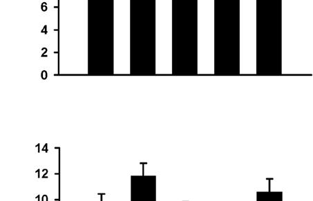
5 synapses. Since we observed a decrease in mepsc amplitude in neurons with larger dendritic arbors, we wanted to minimize the possibility that this result was due to a higher proportion of synapses on the larger neurons being located more distal from the soma compared to control cells with smaller dendritic arbors. To examine this possibility, we grouped the mepscs analyzed in Figures 2C 2F by rise time. This analysis revealed that the population of mepscs with the fastest rise times (rise time < 1 ms) exhibited the same changes in mepsc amplitude as observed for the total population of mepscs (Figure 2G). Specifically, neurons transfected with b-catenin* (Figure 2G, ± 1.24 pa, p < 0.05 versus GFP) or treated with high K + (11.88 ± 1.05 pa, p < versus GFP) have smaller mepsc amplitudes compared to GFP controls (18.09 ± 1.54 pa) and the effects of K + are completely rescued by expression of N(intra) in the postsynaptic neurons [K + + N(intra), ± 1.78 pa, p = 0.52 versus GFP, p < versus K + ]. Since mepscs with fast rise times most likely arise from synapses close to the soma (Stuart et al., 1999; Williams and Mitchell, 2008) and thus are much less susceptible to space clamping artifacts, these results demonstrate that the effects of b-catenin* expression and K + treatment on mepsc amplitudes likely reflect real changes in quantal amplitudes and are unlikely to be due to spatial redistribution of synapses on cells with larger dendritic arbors. An Inverse Correlation between mepsc Amplitude and TDBL Having shown that b-catenin overexpression and mimicking increased neural activity both promote dendritic arborization and reduce mepsc amplitudes, we predicted that an inverse correlation exists between these parameters. Indeed, we observe a statistically significant inverse linear correlation between the parameters of TDBL and mepsc amplitude, both for the GFP control group alone (Figure 3A, n = 26, r 2 = 0.18, p < 0.05) and for neurons from all experimental conditions combined (Figure 3B, n = 101, r 2 = 0.13, p < 0.001). Dividing neurons according to TDBL yielded four groups with descending average mepsc amplitudes (Figure 3C), further demonstrating the inverse relationship between TDBL and mepsc amplitude. In Vivo Expression of b-catenin* Reduces mepsc Amplitudes and Increases Dendritic Complexity A critical question for any results obtained using dissociated neuronal cultures is whether the experimental manipulations have the same effects in vivo. To test whether b-catenin overexpression in vivo promotes dendritic growth and reduces mepsc amplitudes two different approaches were used. First, we electroporated a plasmid expressing b-catenin* together with YFP in utero into the ventricles of mice embryos and recorded spontaneous EPSCs from acute brain slices prepared from postnatal mice. b-catenin* expressing neurons had lower sepsc amplitudes (6.96 ± 0.41 pa, n = 5, p < 0.05) compared to neighboring controls (13.80 ± 1.83 pa, n = 11; Figure 4A). Furthermore, neurons expression b-catenin* together with YFP had higher TDBL (control, ± mm, n = 58; b-cat*, ± mm, n = 61, p = 0.01; Figure 4B) and TDBTN (control, ± 0.65, n = 58; b-cat*, ± 0.61, n = 61, p < 0.005; Figure 4C) compared to controls expressing YFP alone. These results demonstrate that in vivo expression of b-catenin from late embryonic development has qualitatively the same effects as its expression in dissociated neuronal cultures. To examine whether expression of b-catenin in vivo during postnatal development has effects similar to those when expressed embryonically, we injected Sindbis viruses expressing b-catenin* directly into the hippocampus of juvenile rats (P21 P28) and recorded from infected neurons in acute brain slices prepared 24 hr later. Again, neurons expressing b-catenin* had reduced mepsc amplitudes (9.60 ± 0.43 pa, n = 9, p < 0.01) compared to neighboring control cells (12.33 ± 0.57 pa, n = 7; Figure 4D). Similar to the results obtained from dissociated neuronal cultures, the cumulative probability distributions of mepsc amplitudes were also significantly different (Figure 4E, p < ) and could be superimposed when the distribution from the b-catenin* expressing cells was transformed according to a best fit equation (Figure 4E, inset). Manipulations of the Cadherin/Catenin Complex Change AMPA Receptor Clustering The amplitudes of mepscs most likely reflect the number and state of surface AMPA receptors at individual synapses, although changes in transmitter concentration can influence this parameter (Lisman et al., 2007). Therefore we used immunocytochemistry as an alternative method for assaying the level of synaptic surface AMPA receptors. This standard method also provides an independent test of the electrophysiological results and circumvents any issues related to potential space clamping artifacts. Consistent with the electrophysiological results, expression of b-catenin* reduced the area of both total surface AMPA receptors and surface AMPA receptors at synapses, defined by colocalization with the active zone marker protein Bassoon (Figures 5A and 5B, b-cat*, ± 1.40, n = 56, p < 0.01 compared to GFP alone, 100 ± 2.06, n = 57; Figure 5C, ± 1.97, p = 0.001; Figure S4A). Consistent with previous work (O Brien et al., 1998), treating cultures with high K + to mimic Figure 2. Expression of b-catenin* Reduces mepsc Amplitude while Expression of N(intra) Reverses the Effects of K + (A) Representative mepsc recordings and averaged mepsc waveforms for each condition. The scale bars are 20 pa and 1000 ms for the sweeps, and 5 pa and 10 ms for the averaged traces. (B) Average mepsc amplitudes from neurons transfected or treated with GFP (16.09 ± 1.09 pa), b-cat* (12.60 ± 0.99 pa, p < 0.05), K + (11.41 ± 0.82 pa, p < 0.005), K + + N(intra) [17.51 ± 1.46 pa, p = 0.43 versus GFP, p = versus K +, p = 0.85 versus N(intra)], and N(intra) (17.11 ± 1.33 pa, p = 0.56). (C F) Cumulative distributions of mepsc amplitudes, each manipulation (dark gray) plotted against GFP (light gray). (C) b-cat* versus GFP, p < 0.005; inset, scaled b-cat* mepsc distribution transformed according to best fit: b-cat* = control , r 2 = , p = 1.0. (D) K + versus GFP, p < ; inset, scaled K + mepsc distribution transformed according to best fit: K + = control , r 2 = , p = 1.0. (E) N(intra) versus GFP, p = (F) K + + N(intra) versus GFP, p = (G) mepsc amplitudes grouped according to rise time. For those with rise time < 1 ms, the amplitudes are GFP (18.09 ± 1.54 pa), b-cat* (13.22 ± 1.24 pa, p < 0.05), K + (11.88 ± 1.05 pa, p < 0.005), K + +N(intra) (19.62 ± 1.78 pa, p = 0.52 versus GFP, p < versus K + ), N(intra) (20.03 ± 1.84 pa, p = 0.44). *p < 0.05, **p < 0.01, ***p < 0.001; error bars represent SEM. Neuron 61, 71 84, January 15, 2009 ª2009 Elsevier Inc. 75
6 A K + + N(intra), ± 2.01, n = 57, p = 0.26 versus GFP, p < 0.05 versus K + ; Figure 5C, ± 2.54, p = 0.30 versus GFP, p < 0.05 versus K + ), while expression of N(intra) alone had no effect (Figures 5B and 5C). No changes were detected in the average puncta intensity or percentage of surface synaptic AMPA receptors (Figures S4B S4D), further confirming that the observed changes in AMPA receptor cluster size represent bona fide changes in surface AMPA receptor levels. B C 0 Overexpression of b-catenin Reduces Surface AMPA Receptor Density Although the effects of b-catenin* expression or depolarization on surface AMPA receptor puncta size were statistically significant, they were small relative to the effects of these manipulations on mepsc amplitudes. We therefore hypothesized that in addition to reducing the size of surface AMPA receptor clusters, these manipulations may bring a proportion of AMPA receptor clusters below immunocytochemical detection threshold. This would be reflected as a decrease in the number of total and synaptic surface AMPA receptor clusters per unit length of dendrite. Consistent with this prediction, neurons expressing b-catenin* or treated with K + exhibited a clear decrease in surface AMPA receptor density (Figure 5D, b-cat*, ± 0.77, p = 0.01, K +, 8.56 ± 0.91, p < versus GFP, ± 0.80; Figure 5E, b-cat*, 5.10 ± 0.34, p < , K +, 5.03 ± 0.53, p = versus GFP, 7.42 ± 0.50). Again, expression of N(intra) reversed the effects of K + on both total and synaptic surface AMPA receptor puncta density (Figure 5D, K + + N(intra), ± 1.19, p = 0.91 versus GFP, p < versus K + ; Figure 5E, 7.27 ± 0.63, p = 0.86 versus GFP, p < 0.01 versus K + ) while N(intra) expression alone had no detectable effects (Figures 5D and 5E). Together, the decrease in the puncta size of the average surface AMPA receptor cluster (Figures 5B and 5C), combined with the decrease in receptor density (Figures 5D and 5E), results in a significant reduction in surface AMPA receptor levels per unit length of dendrite, in neurons expressing b-catenin* or treated with K +. Figure 3. An Inverse Correlation between TDBL and mepsc Amplitude (A) Plot of TDBL versus mepsc amplitude for neurons expressing GFP, linear regression represented by dotted line, n = 26, r 2 = 0.18, p < (B) Plot of TDBL versus mepsc amplitude for neurons from all experimental conditions, linear regression represented by dotted line, n = 101, r 2 = 0.13, p < (C) Bar graphs of average mepsc amplitudes from all data presented in (B), grouped according to TDBL, ( mm) ± 2.12 pa, ( mm) ± 0.93 pa, ( mm) ± 0.99 pa, ( mm) ± 0.68 pa. Compared to the ( mm) group, p = 0.2, p < 0.05, p = 0.01 for the latter groups; compared to the ( mm) group, p = 0.27, p = 0.005; compared to the ( mm) group, p = *p < 0.05, **p < 0.01, error bars represent SEM. a chronic increase in activity also decreased total surface (Figure 5B, K +, ± 2.15, n = 59, p = 0.001) and synaptic surface AMPA receptors (Figure 5C, ± 3.97, p = 0.001). These decreases were reversed by expression of N(intra) [Figure 5B, Manipulations of the Cadherin/Catenin Complex Do Not Affect PSD95 Density A body of work suggests important roles for the cadherin/catenin complex in synapse development (Dalva et al., 2007; Salinas and Price, 2005). Yet, we have observed that b-catenin overexpression reduces mepsc amplitude and frequency (Figures 2 and S1; see Discussion) as well as surface AMPA receptor puncta size and density (Figures 5A 5E). These results raise the question of how b-catenin overexpression affects synapse density. To address this issue, we identified synapses by colabeling for the postsynaptic density protein PSD95 and the presynaptic active zone protein Piccolo, two structural synaptic marker proteins (McAllister, 2007; Waites et al., 2005). Neither b-catenin overexpression nor any of the other manipulations affected the density of total or synaptic PSD95 and Piccolo puncta (Figures 5F 5I), nor did they affect the percentage of PSD95 and Piccolo puncta (data not shown) that were synaptic, as defined by colocalization with respect to each other. These results demonstrate that although b-catenin overexpression decreased mepsc 76 Neuron 61, 71 84, January 15, 2009 ª2009 Elsevier Inc.
7 A B C D E Figure 4. The Effects of In Vivo b-catenin* Expression on EPSC Amplitudes and Dendritic Morphology in Hippocampal Slices (A) Average sepsc amplitudes of neurons in utero electroporated with b-catenin* (6.96 ± 0.41 pa, p < 0.05) compared to nonelectroporated neighbors (control, ± 1.83 pa). (B) TDBL of neurons electroporated with YFP alone ( ± mm) or b-catenin* and YFP ( ± mm, p = 0.01). (C) TDBTN of neurons electroporated with YFP alone (10.78 ± 0.65) or b-catenin* and YFP (12.93 ± 0.61, p < 0.005). (D) Average mepsc amplitudes of neurons infected with a virus expressing b-catenin* (9.60 ± 0.43 pa, p < 0.01) compared to uninfected neighbors (control, ± 0.57 pa). (E) Cumulative distributions of mepsc amplitudes of neurons infected with b-catenin* (dark gray) plotted against uninfected neighbors (light gray), p < 0.005; inset, scaled b-cat* mepsc distribution transformed according to best fit: b-cat* = control , r 2 = 0.99, p = *p < 0.05, **p < 0.01; error bars represent SEM. amplitude and surface AMPA receptor cluster size and density, it did not affect the density of morphological synapses. Furthermore, these results suggest that as dendritic arbors grow, the density of morphological synapses, as defined by colocalization of active zone and postsynaptic density proteins, is kept roughly constant. b-catenin Expression Increases NMDAR/AMPAR Ratio The decrease in AMPA receptor puncta density with no changes in synapse density (as defined by PSD-95 and Piccolo staining) in cells expressing b-catenin* or following high K + treatment is consistent with the hypothesis that these manipulations increase the proportion of silent synapses (Durand et al., 1996; Isaac et al., 1995; Kullmann, 1994; Liao et al., 1995; Malenka and Nicoll, 1997), that is, synapses containing NMDA receptors but lacking detectable AMPA receptors. We tested this possibility electrophysiologically by measuring NMDA and AMPA receptor-mediated EPSCs at holding potentials of +40 mv and 70 mv, respectively, in response to local extracellular stimulations via a bipolar electrode (Maximov et al., 2007). Consistent with this prediction, neurons expressing b-catenin* had larger NMDA receptor-mediated EPSCs (71.30 ± 9.88 pa, n = 33, p = 0.01) (Figures 6A and 6B) compared to controls (41.83 ± 6.51 pa, n = 33) and larger NMDAR/AMPAR ratios (b-cat* 0.32 ± 0.04, p = compared to GFP 0.19 ± 0.02; Figure 6D). The effects of high K + treatment on NMDA receptor-mediated EPSCs (56.75 ± 8.61 pa, p = 0.21; Figures 6A and 6B) and NMDAR/AMPAR ratios (0.27 ± 0.03, p < 0.05, Figure 6D) were slightly smaller compared to b-catenin* expression, but follow the same trend. No statistically significant differences were detected in the AMPA receptor-mediated responses with any of the conditions (GFP, ± pa; K +, ± pa, p = 0.14; b-cat*, ± pa, p = 0.47; Figure 6C), likely because the smaller average responses at individual synapses are compensated by the increase in total synapse number through increased TDBL, resulting in no net change in the total cellular AMPA receptor-mediated response. The increase in NMDAR/AMPAR ratio was also detected Neuron 61, 71 84, January 15, 2009 ª2009 Elsevier Inc. 77


8 A B C D E F G H I 78 Neuron 61, 71 84, January 15, 2009 ª2009 Elsevier Inc.
9 A B C D Figure 6. b-catenin* Expression and High K + Treatment Increase NMDAR/AMPAR Ratio (A) Example traces of average AMPAR EPSCs ( 70 mv) and NMDAR + AMPAR EPSCs (+40 mv) from neurons transfected or treated with GFP, high K +,or b-catenin*. Measurements for each type of current are taken at times as indicated and described in Experimental Procedures, scale bars are 100 pa and 20 ms. (B) Average NMDAR EPSC amplitudes for GFP (41.83 ± 6.51 pa), high K + (56.75 ± 8.61 pa, p = 0.21), b-cat* (71.30 ± 9.88 pa, p = 0.01). (C) Average AMPAR EPSC amplitudes for GFP ( ± pa), high K + ( ± pa, p = 0.14), b-cat* ( ± pa, p = 0.47). (D) Average NMDAR/AMPAR ratio for GFP (0.19 ± 0.02), high K + (0.27 ± 0.03, p < 0.05), b-cat* (0.32 ± 0.04, p = 0.005). *p < 0.05, **p < 0.01; error bars represent SEM. following in vivo injections of Sindbis viruses expressing b-catenin* (0.49 ± 0.07, n = 8, p < 0.05 versus uninfected controls, 0.30 ± 0.06, n = 11). TTX Treatment Still Increases mepsc Amplitude in Neurons with Larger TDBL Thus far, we have shown that b-catenin* expression or high K + treatment increases TDBL (Figure 1) and concurrently limits unitary synaptic AMPA receptor levels (Figures 2 and 5). Several lines of evidence suggest that the scaling up of mepsc amplitudes in response to chronic activity blockade (Goddard et al., 2007; Ibata et al., 2008; Rutherford et al., 1997; Shepherd et al., 2006; Stellwagen and Malenka, 2006; Thiagarajan et al., 2002) is mechanistically distinct from the scaling down of mepsc amplitudes due to chronic increases in activity (Seeburg et al., 2008; Seeburg and Sheng, 2008). To test whether b-catenin* expression or high K + treatment influences the uniform scaling up of synaptic strengths due to activity blockade, we performed these manipulations in cultures that were also treated with TTX (1 mm) for 48 hr prior to recordings. While TTX treatment does not alter the effects of b-catenin* expression on promoting dendritic growth (b-cat* + TTX, ± mm, n = 18, p < 0.01 compared to GFP alone ± mm, p = 0.55 versus b-cat* ± mm; Figures 7A and 7B), it significantly increases mepsc amplitudes in neurons expressing b-catenin* (14.79 ± 0.79 pa, n = 26), or treated with high K + Figure 5. Expression of b-catenin* Reduces Surface AMPA Receptor Puncta Size and Density while Expression of N(intra) Reverses the Effects of K + (A) Representative images of neurons transfected or treated with GFP, b-cat*, K +,K + + N(intra) or N(intra). GFP, surface AMPA receptors, bassoon channels shown individually and the colocalization of bassoon (red) and surface AMPA receptors (green) shown as merge. (B) Normalized surface AMPA receptor puncta area for GFP (100 ± 2.06 arbitrary units), b-cat* (92.97 ± 1.40, p < 0.01), K + (90.12 ± 2.15, p = 0.001), K + + N(intra) (96.74 ± 2.01, p = 0.26 versus GFP, p < 0.05 versus K + ) and N(intra) (98.26 ± 2.06, p = 0.56). (C) Normalized synaptic surface AMPA receptor puncta area for GFP (100 ± 2.45), b-cat* (89.58 ± 1.97, p = 0.001), K + (87.01 ± 3.97, p = 0.001), K + + N(intra) (96.31 ± 2.54, p = 0.30 versus GFP, p < 0.05 versus K), N(intra) (95.67 ± 2.31, p = 0.22). (D) Average number of total surface AMPA receptor puncta per 10 mm of dendrite for GFP alone (13.15 ± 0.80), b-cat* (10.33 ± 0.77, p = 0.01), K + (8.56 ± 0.91, p < ), K + + N(intra) (12.98 ± 1.19, p = 0.91 versus GFP, p < versus K + ), N(intra) (13.71 ± 0.97, p = 0.66). (E) Average number of synaptic surface AMPA receptor puncta per 10 mm of dendrite for GFP alone (7.42 ± 0.50), b-cat* (5.10 ± 0.34, p < ), K + (5.03 ± 0.53, p = 0.001), K + + N(intra) (7.27 ± 0.63, p = 0.86 versus GFP, p < 0.01 versus K + ), N(intra) (7.63 ± 0.53, p = 0.77). (F) Average number of PSD 95 puncta per 10 mm of dendrite for GFP (17.07 ± 1.71), b-cat* (19.86 ± 1.64, p = 0.24), K + (17.49 ± 1.76, p = 0.87), K + + N(intra) (18.39 ± 1.38, p = 0.55), N(intra) (19.10 ± 1.83, p = 0.42). (G) Average number of synaptic PSD 95 puncta colocalizing with active zone marker Piccolo per 10 mm of dendrite for GFP (10.40 ± 1.11), b-cat* (12.59 ± 1.10, p=0.17),k + (10.02 ± 1.04, p = 0.81), K + + N(intra) (10.59 ± 0.90, p = 0.89), N(intra) (11.44 ± 1.13, p = 0.52). (H) Average number of Piccolo puncta per 10 mm of dendrite for GFP (14.64 ± 0.71), b-cat* (16.39 ± 0.87, p = 0.13), K + (13.67 ± 0.80, p = 0.38), K + + N(intra) (13.05 ± 0.59, p = 0.09), N(intra) (16.06 ± 0.80, p = 0.19). (I) Average number of synaptic Piccolo puncta (colocalizing with PSD 95) per 10 mm of dendrite for GFP (9.50 ± 0.96), b-cat* (11.86 ± 0.98, p = 0.09), K + (8.93 ± 0.91, p = 0.67), K + + N(intra) (8.99 ± 0.72, p = 0.67), N(intra) (10.62 ± 0.99, p = 0.42). *p < 0.05, **p < 0.01, ***p < 0.001; error bars represent SEM. Neuron 61, 71 84, January 15, 2009 ª2009 Elsevier Inc. 79
10 Figure 7. Treatment with TTX Significantly Increases mepsc Amplitudes in Neurons Transfected with b-catenin* without Affecting Their Dendritic Morphology (A) Representative images of neurons transfected with GFP alone, with GFP + b-cat*, and with GFP +b-cat* and treated with TTX. (B) Graph showing TDBL in neurons transfected with GFP ( ± mm), b-cat* ( ± mm, p = 0.01), b-cat* + TTX ( ± mm, p < 0.01 versus GFP, p = 0.55 versus b-cat*). (C) Representative mepsc recordings and average mepsc waveforms for each condition; the scale bars are 20 pa and 500 ms and 5 pa and 10 ms, respectively. (D) Average mepsc amplitudes from neurons transfected or treated with GFP (12.64 ± 0.47 pa), TTX (14.63 ± 0.60 pa, p = 0.01), TTX + K + (14.41 ± 0.80 pa, p < 0.05 versus GFP, p = 0.79 versus TTX), or TTX + b-cat* (14.79 ± 0.79 pa, p < 0.05 versus GFP, p = 0.88 versus TTX). (E G) Cumulative distributions of mepsc amplitudes, each manipulation (dark gray) plotted against GFP (light gray). (E) TTX versus GFP, p < (F) TTX + K + versus GFP, p = (G) TTX + b-cat* versus GFP, p < *p < 0.05, error bars represent SEM. (14.41 ± 0.80 pa, n = 17) to the same level as TTX treatment alone (14.63 ± 0.60 pa, n = 26, p = 0.01 versus GFP, p = 0.79 versus TTX + K +, p = 0.88 versus TTX + b-cat*) and significantly higher than control conditions (12.64 ± 0.47 pa, n = 26; Figures 7C and 7D; p < 0.05 for all TTX conditions versus control). These results, also observed in cumulative probability plots of mepsc amplitudes (Figures 7E 7G; GFP versus TTX, p < 0.01; GFP versus TTX + K +, p = 0.01; GFP versus TTX + b-cat*, p < 0.005), demonstrate that neurons with larger dendritic arbors can still respond to subsequent activity blockade by scaling up their synaptic strength, while retaining their dendritic complexity (Figures 7A and 7B). These results provide additional evidence that the mechanisms regulating the scaling up and scaling down of synaptic strengths are likely to be distinct, as the scaling down of mepsc amplitudes by depolarization or b-catenin* expression (Figure 2) does not affect the ability of these neurons to respond to subsequent activity blockade (Figure 7). None of the manipulations affect mepsc frequency (Figure S5). DISCUSSION Previous studies have independently investigated the effects of increasing neural activity on dendritic arborization and on synaptic scaling (Leslie et al., 2001; O Brien et al., 1998; Redmond et al., 2002; Seeburg et al., 2008; Sin et al., 2002; Turrigiano, 2007). However, the possibility that these changes are coordinated and that activity-dependent increases in TDBL are necessary and sufficient for mediating synaptic scaling has not 80 Neuron 61, 71 84, January 15, 2009 ª2009 Elsevier Inc.
11 been addressed. Here, by measuring TDBL and mepsc amplitudes from the same neurons, we show that (1) neurons with increased TDBL induced by high K + treatment or overexpressing b-catenin have reduced mepsc amplitudes, as well as decreased surface synaptic AMPA receptor puncta area and density; (2) the effects of mimicking increased neural activity with high K + on mepsc amplitudes and surface AMPA receptor area and density can be prevented by decreasing TDBL in individual postsynaptic neurons through expression of N(intra); (3) there is an inverse linear correlation between TDBL and mepsc amplitudes; (4) neurons with increased TDBL due to overexpressing b-catenin or high K + treatment have higher NMDAR/ AMPAR ratios, indicative of more silent synapses; (5) the effects of b-catenin overexpression on TDBL, mepsc amplitudes and NMDAR/AMPAR ratios were also observed in vivo suggesting that similar mechanisms to those elucidated here in cultured neurons apply during normal development. These coordinated changes in dendritic morphology and unitary excitatory synaptic strength may serve as an important intrinsic mechanism that helps prevent neurons from over-excitation during neural circuit development. Several experiments were performed to dissociate the effects of activity-dependent increases in dendritic arborization from other potential effects of activity to demonstrate that changes in TDBL are necessary and sufficient for the scaling down of synaptic strength. First, expression of b-catenin* results in correlated increases in TDBL and reduced mepsc amplitudes, without affecting the spontaneous firing rate or the resting membrane potential of neurons (Figure S6), while increasing neural activity does significantly reduce firing rate (Turrigiano et al., 1998) (and Figure S6). Consistent with the lack of effect of b-catenin on basic electrophysiological properties, it is not known to directly associate with voltage-dependent channels. Second and most importantly, the effects of depolarizing the entire culture dish with high K + are reversed by expression of N(intra), a molecule that reduces TDBL in individual postsynaptic neurons (Figure 2). Finally, we show that TDBL is inversely correlated with mepsc amplitude for all neurons analyzed, including those treated with high K + and transfected with N(intra) (Figure 3). Thus, by specifically blocking the effects of neural activity on dendritic morphology, we show that increases in TDBL are required for mediating the effects of high K + on the scaling down of mepsc amplitudes. In reporting a reduction in mepsc amplitudes in neurons with larger dendritic arbors, we performed a combination of additional experiments and analyses to rule out potential artifacts due to space clamping problems associated with whole-cell recordings. First, we show that none of our manipulations significantly affect cellular input resistance, somatic surface area or the average diameter of primary dendrites (Figure S2) in the set of neurons 3D reconstructed with Neurolucida for dendritic morphology (Figure 1) and analyzed for mepsc amplitudes (Figure 2). Since our estimation of cellular input resistance (see Experimental Procedures) likely reflects primarily the input resistance of the soma and proximal dendrites (Williams and Mitchell, 2008), the lack of observed changes does not preclude a change in the electrotonic structure of neurons with increased TDBL, which could in turn affect the mepsc amplitudes measured at the soma. Therefore, we selectively analyzed mepscs with the fastest rise times (<1 ms), an analysis that selects events mostly from synapses on proximal dendrites, which are less susceptible to space clamping problems (Williams and Mitchell, 2008). The same changes in mepsc amplitudes occurred in all experimental groups with this more selective analysis (Figure 2G). Additionally, we stained for surface synaptic AMPA receptors (Figure 5), an assay which is not susceptible to the problems of space clamping, and confirmed our results. Finally, we show that neurons expressing b-catenin* and treated with TTX maintain their larger dendritic arbors, and yet have significantly higher mepsc amplitudes compared to controls (Figure 7), demonstrating that our electrophysiological recordings do detect larger mepscs when they are present. Our results showing a normal scaling up of synaptic strengths in response to TTX treatment in cells with enhanced dendritic arbors following high K + treatment or b-catenin overexpression (Figure 7) are consistent with the growing evidence that the scaling up of synaptic strengths due to activity blockade (Goddard et al., 2007; Ibata et al., 2008; Rutherford et al., 1997; Shepherd et al., 2006; Stellwagen and Malenka, 2006; Thiagarajan et al., 2002) involves different mechanisms than the scaling down due to increased activity (Seeburg et al., 2008; Seeburg and Sheng, 2008). The coordinated changes in dendritic growth and synaptic scaling are likely to be most relevant during development at times when there are concurrent increases in overall activity as neural circuits mature. During this process, mechanisms, such as the one we describe here, may be present to prevent neurons from over-excitation. During later stages of development or during epileptiform activity, other mechanisms for synaptic scaling, such as induced expression of Polo-like kinase 2, which depletes PSD95 and Bassoon clusters and causes loss of mature spines (Pak and Sheng, 2003) could be the predominant mechanism (Seeburg et al., 2008; Seeburg and Sheng, 2008). Since changes in TDBL were mediated through molecular manipulations of the cadherin/catenin complex, it is important to attempt to distinguish the effects of changes in dendritic morphology from the direct effects of the cadherin/catenin complex on synaptic transmission. Cadherin and catenins are transsynaptic cell adhesion molecules present at or adjacent to synaptic sites from the early steps of synaptogenesis (Benson and Tanaka, 1998; Togashi et al., 2002). Intracellularly, through a and b-catenin, they are linked to the actin cytoskeleton (Gumbiner, 1996). Loss-of-function mutations of N-cadherin, an- or b-catenin lead to changes in spine shape, reduced mepsc amplitudes or defects in short-term plasticity (Abe et al., 2004; Bamji et al., 2003; Jungling et al., 2006; Okuda et al., 2007; Togashi et al., 2002). These observations, together with the reported direct interaction between N-cadherin and GluR2 (Saglietti et al., 2007) and effects of activity on N-cadherin endocytosis or b-catenin phosphorylation (Murase et al., 2002; Tai et al., 2007), all suggest a role for the cadherin/catenin complex in promoting synapse growth, an effect opposite to that of synaptic scaling as reported in this study. Furthermore, b-catenin overexpression does not affect basic properties of neurons, including the resting membrane potential and the spontaneous firing rate (Figure S6), parameters which reflect the Neuron 61, 71 84, January 15, 2009 ª2009 Elsevier Inc. 81
12 intrinsic excitability of neurons. Thus, our results are best explained by the effects of the molecular and activity manipulations on TDBL and the inverse relationship that exists between TDBL and mepsc amplitude. However, we cannot exclude the possibility that the cadherin/catenin complex could itself mediate effects of neural activity on synaptic scaling through unidentified mechanisms. The manipulations we performed had no detectable effects on the density of morphological synapses as defined by the colocalization of active zone and postsynaptic density proteins (Figures 5F 5I). Since neurons treated with high K + or transfected with b-catenin* have more TDBL, they would consequentially have more total morphological synapses, consistent with the proposed role of the cadherin/catenin complex in synapse development (Dalva et al., 2007; Salinas and Price, 2005). However, some of these synapses are functionally silent in that they did not contain detectable AMPA receptors, as shown by the observed decrease in the density of total surface and synaptic surface AMPA receptors (Figures 5D and 5E) in neurons with increased TDBL (high K + treatment or b-catenin* expression). This effect is mirrored electrophysiologically by strong trends toward reduced mepsc frequencies (Figure S1), despite the increase in the total number of morphological synapses brought on by increased TDBL. Consistent with these results, we observed an increase in the NMDAR/AMPAR ratio and in evoked NMDA receptor-mediated EPSC in neurons expressing b-catenin* or treated with K + (Figure 6). Furthermore, these weaker synapses can be activated in response to subsequent changes in the environment, such as activity blockade (Figure 7), which do not affect the dendritic arbor itself. Previous work in a number of different model systems provides additional examples of inverse changes in electrophysiological or morphological parameters during neural circuit development. For example, during rodent visual cortical development, layer 4 neurons show inverse and correlated changes in mepsc amplitude and frequency, an activity-dependent event blocked by dark rearing (Desai et al., 2002). Similarly, during Xenopus retinotectal circuit development, neurons adapt their intrinsic excitability to achieve a stable relationship between the total level of synaptic input and tectal neuronal spike output (Pratt and Aizenman, 2007). Morphologically, dark rearing of rodents results in changes in dendritic spine density and shape in layer 3 pyramidal neurons of the visual cortex, while conserving total synaptic area per unit length of dendrite (Wallace and Bear, 2004). Our results complement and add to these mechanisms by showing that TDBL and unitary excitatory synaptic function, as measured by mepsc amplitudes, are inversely correlated. During the later stages of neural circuit formation as dendritic arbors grow and become more complex, if the number of excitatory postsynaptic AMPA receptors continually scale with the increased total surface area, neurons may become at risk for overexcitation and excitotoxicity. Instead, our results suggest that a mechanism has developed, where the total level of surface AMPA receptors remains constant, despite the increase in the total number of synapses per neuron. These additional synapses are then in a position to respond rapidly to future changes in their extracellular environment, by insertion of AMPA receptors into existing synapses. These changes could be global, as shown by our TTX treatments (Figure 7) or could specifically occur in a subset of synapses in response to local stimuli. Combining the growth of the dendritic arbor with a correlated reduction in unitary excitatory synaptic strength, while maintaining the potential for future increases in synaptic strength, provides an elegant mechanism that is likely to play important roles during activity-dependent growth and development of neural circuits. EXPERIMENTAL PROCEDURES Hippocampal Neuronal Culture Preparation High density mixed neuronal-glial cultures were prepared from postnatal day 0 (P0) Sprague-Dawley rat pups as previously described (Yu and Malenka, 2003) and according to procedures approved by the IACUC of Stanford University and the Institute of Neuroscience. Briefly, neurons were plated on matrigel coated glass coverslips (Assistant) at 50,000 neurons per cm 2 in medium consisting of Neurobasal medium (Invitrogen), B-27 (Invitrogen) and 2 mm Glutamax-I (Invitrogen). On the third day in vitro (DIV 3), when astrocytes have proliferated sufficiently to form a monolayer over the entire coverslip, cells were treated with the mitotic inhibitor FUDR (5-fluoro-2 0 -deoxyuridine, Sigma). Calcium phosphate transfections were carried out at DIV 7 using 2 3 mg DNA per 24-well. The low transfection efficiency of this method ensures that the recorded neuron is surrounded by untransfected presynaptic contacts. In all experiments mg of DNA encoding GFP was included in the transfection mixture to clearly visualize morphology of the recorded or imaged neuron. For high K + treatment, 16 mm KCl was added at DIV 7 and washed out 48 hr later with conditioned medium. For experiments in Figure 7, TTX (1 mm) was added 48 hr prior to recordings. All electrophysiological and immunocytochemical experiments were carried out at DIV 12. DNA Constructs DNA constructs are as previously described (Yu and Malenka, 2003). Briefly, b-catenin* contains full-length b-catenin fused to GFP and with four serine/ threonine (S33, S37, T41, S45) to alanine substitutions that prevent its phosphorylation by glycogen synthase kinase 3b and subsequent degradation. The main difference between this construct and wild-type b-catenin is that it is more stable and less sensitive to endogenous regulation via ubiquitinmediated degradation. Thus it is possible to obtain higher levels of overexpression with this construct compared to the wild-type, which is completely degraded 48 hr following overexpression. For in utero electroporation experiments, full-length b-catenin* was subcloned into pcag-eyfp-cag (Saito and Nakatsuji, 2001) at the XhoI site. For Sindbis virus infection experiments, b-catenin* was subcloned into psinrep5-ires-gfp (Marie et al., 2005). N(intra), previously named Ncad(intra), contains the transmembrane and intracellular domains of Xenopus N-cadherin. Electrophysiology in Cultured Neurons Whole-cell patch-clamp recordings in dissociated neuronal cultures were carried out at room temperature on DIV 12 neurons with an Axopatch 1D or Multiclamp 700B amplifier (Axon Instruments) using low-resistance pipets (2 5 MU). The intracellular solutions contained (in mm) 110 CsMeSO 4,40 HEPES, 10 NaCl, 5 MgCl 2, 0.4 EGTA, 2 ATP, and 0.2 GTP (ph to 7.25, 295 mosm). For visualization of dendritic morphology, Alexa 568 hydrazide (Invitrogen) was added to the internal solution at a final concentration of 0.1 mg/ml on the day of recording. For mipsc recordings, CsMeSO 4 was replaced with CsCl. For firing rates measurements, the intercellular solution contained (in mm) 110 Kgluconate, 20 KCl, 5 MgCl 2, 20 HEPES, 0.6 EGTA, 2 MgATP, 0.2 Na 3 GTP (ph to 7.3, 300 mosm). The extracellular solution contained (in mm) 129 NaCl, 5 KCl, 30 glucose, 25 HEPES, 2 CaCl 2, 1 MgCl 2 (ph 7.3, 315 mosm); 0.5 mm tetrodotoxin was added for miniature PSC recordings, 50 mm picrotoxin for EPSC recordings or 5 mm NBQX for IPSC recordings. For mepscs, neurons were voltage-clamped at 70 mv, and data was filtered at 1 2 khz and acquired at 2 5 khz. Data analysis was performed blind using Synaptosoft software with an amplitude threshold of 5 pa. Cumulative 82 Neuron 61, 71 84, January 15, 2009 ª2009 Elsevier Inc.
SUPPLEMENTARY INFORMATION
 Supplementary Figure 1. Normal AMPAR-mediated fepsp input-output curve in CA3-Psen cdko mice. Input-output curves, which are plotted initial slopes of the evoked fepsp as function of the amplitude of the
Supporting Information
 ATP from synaptic terminals and astrocytes regulates NMDA receptors and synaptic plasticity through PSD- 95 multi- protein complex U.Lalo, O.Palygin, A.Verkhratsky, S.G.N. Grant and Y. Pankratov Supporting
Axon initial segment position changes CA1 pyramidal neuron excitability
 Axon initial segment position changes CA1 pyramidal neuron excitability Cristina Nigro and Jason Pipkin UCSD Neurosciences Graduate Program Abstract The axon initial segment (AIS) is the portion of the
Part 11: Mechanisms of Learning
 Neurophysiology and Information: Theory of Brain Function Christopher Fiorillo BiS 527, Spring 2012 042 350 4326, fiorillo@kaist.ac.kr Part 11: Mechanisms of Learning Reading: Bear, Connors, and Paradiso,
Supplementary Information
 Hyperpolarization-activated cation channels inhibit EPSPs by interactions with M-type K + channels Meena S. George, L.F. Abbott, Steven A. Siegelbaum Supplementary Information Part 1: Supplementary Figures
SUPPLEMENTARY INFORMATION. Supplementary Figure 1
 SUPPLEMENTARY INFORMATION Supplementary Figure 1 The supralinear events evoked in CA3 pyramidal cells fulfill the criteria for NMDA spikes, exhibiting a threshold, sensitivity to NMDAR blockade, and all-or-none
BIPN 140 Problem Set 6
 BIPN 140 Problem Set 6 1) The hippocampus is a cortical structure in the medial portion of the temporal lobe (medial temporal lobe in primates. a) What is the main function of the hippocampus? The hippocampus
BIPN 140 Problem Set 6
 BIPN 140 Problem Set 6 1) Hippocampus is a cortical structure in the medial portion of the temporal lobe (medial temporal lobe in primates. a) What is the main function of the hippocampus? The hippocampus
Supporting Online Material for
 www.sciencemag.org/cgi/content/full/317/5841/183/dc1 Supporting Online Material for Astrocytes Potentiate Transmitter Release at Single Hippocampal Synapses Gertrudis Perea and Alfonso Araque* *To whom
Synaptic plasticityhippocampus. Neur 8790 Topics in Neuroscience: Neuroplasticity. Outline. Synaptic plasticity hypothesis
 Synaptic plasticityhippocampus Neur 8790 Topics in Neuroscience: Neuroplasticity Outline Synaptic plasticity hypothesis Long term potentiation in the hippocampus How it s measured What it looks like Mechanisms
Sample Lab Report 1 from 1. Measuring and Manipulating Passive Membrane Properties
 Sample Lab Report 1 from http://www.bio365l.net 1 Abstract Measuring and Manipulating Passive Membrane Properties Biological membranes exhibit the properties of capacitance and resistance, which allow
Supporting Online Material for
 www.sciencemag.org/cgi/content/full/312/5779/1533/dc1 Supporting Online Material for Long-Term Potentiation of Neuron-Glia Synapses Mediated by Ca 2+ - Permeable AMPA Receptors Woo-Ping Ge, Xiu-Juan Yang,
When cells are already maximally potentiated LTP is occluded.
 When cells are already maximally potentiated LTP is occluded. Stein, V et al., (2003) J Neurosci, 23:5503-6606. Also found in Rat Barrel Cortex Ehrlich & Malinow (2004) J. Neurosci. 24:916-927 Over-expression
Ivy/Neurogliaform Interneurons Coordinate Activity in the Neurogenic Niche
 Ivy/Neurogliaform Interneurons Coordinate Activity in the Neurogenic Niche Sean J. Markwardt, Cristina V. Dieni, Jacques I. Wadiche & Linda Overstreet-Wadiche Supplementary Methods. Animals We used hemizygous
Ube3a is required for experience-dependent maturation of the neocortex
 Ube3a is required for experience-dependent maturation of the neocortex Koji Yashiro, Thorfinn T. Riday, Kathryn H. Condon, Adam C. Roberts, Danilo R. Bernardo, Rohit Prakash, Richard J. Weinberg, Michael
Astrocyte signaling controls spike timing-dependent depression at neocortical synapses
 Supplementary Information Astrocyte signaling controls spike timing-dependent depression at neocortical synapses Rogier Min and Thomas Nevian Department of Physiology, University of Berne, Bern, Switzerland
Is action potential threshold lowest in the axon?
 Supplementary information to: Is action potential threshold lowest in the axon? Maarten H. P. Kole & Greg J. Stuart Supplementary Fig. 1 Analysis of action potential (AP) threshold criteria. (a) Example
How Nicotinic Signaling Shapes Neural Networks
 How Nicotinic Signaling Shapes Neural Networks Darwin K. Berg Division of Biological Sciences University of California, San Diego Nicotinic Cholinergic Signaling Uses the transmitter ACh to activate cation-selective
Neurons of the Bed Nucleus of the Stria Terminalis (BNST)
") Neurons of the Bed Nucleus of the Stria Terminalis (BNST) Electrophysiological Properties and Their Response to Serotonin DONALD G. RAINNIE a Harvard Medical School and Department of Psychiatry, Brockton
Neurons of the Bed Nucleus of the Stria Terminalis (BNST) Electrophysiological Properties and Their Response to Serotonin DONALD G. RAINNIE a Harvard Medical School and Department of Psychiatry, Brockton
Cellular mechanisms of information transfer: neuronal and synaptic plasticity
 Cellular mechanisms of information transfer: neuronal and synaptic plasticity Ivan Pavlov (UCL Institute of Neurology, UK) Anton Chizhov (Ioffe Physical Technical Institute) Pavel Zykin (St.-Petersburg
Distinct Roles for Ionotropic and Metabotropic Glutamate Receptors in the Maturation of Excitatory Synapses
 The Journal of Neuroscience, March 15, 2000, 20(6):2229 2237 Distinct Roles for Ionotropic and Metabotropic Glutamate Receptors in the Maturation of Excitatory Synapses Stephen N. Gomperts, 1 Reed Carroll,
File name: Supplementary Information Description: Supplementary Figures, Supplementary Table and Supplementary References
 File name: Supplementary Information Description: Supplementary Figures, Supplementary Table and Supplementary References File name: Supplementary Data 1 Description: Summary datasheets showing the spatial
Supplementary Figure 1. mir124 does not change neuron morphology and synaptic
 Supplementary Figure 1. mir124 does not change neuron morphology and synaptic density. Hippocampal neurons were transfected with mir124 (containing DsRed) or DsRed as a control. 2 d after transfection,
Local Presynaptic Activity Gates Homeostatic Changes in Presynaptic Function Driven by Dendritic BDNF Synthesis
 Article Local Presynaptic Activity Gates Homeostatic Changes in Presynaptic Function Driven by Dendritic Synthesis Sonya K. Jakawich, 1,2 Hassan B. Nasser, 2 Michael J. Strong, 2 Amber J. McCartney, 1,2
Supplementary Figure 1. ACE robotic platform. A. Overview of the rig setup showing major hardware components of ACE (Automatic single Cell
 2 Supplementary Figure 1. ACE robotic platform. A. Overview of the rig setup showing major hardware components of ACE (Automatic single Cell Experimenter) including the MultiClamp 700B, Digidata 1440A,
Memory Systems II How Stored: Engram and LTP. Reading: BCP Chapter 25
 Memory Systems II How Stored: Engram and LTP Reading: BCP Chapter 25 Memory Systems Learning is the acquisition of new knowledge or skills. Memory is the retention of learned information. Many different
Supplementary figure 1: LII/III GIN-cells show morphological characteristics of MC
 1 2 1 3 Supplementary figure 1: LII/III GIN-cells show morphological characteristics of MC 4 5 6 7 (a) Reconstructions of LII/III GIN-cells with somato-dendritic compartments in orange and axonal arborizations
Chapter 2: Cellular Mechanisms and Cognition
 Chapter 2: Cellular Mechanisms and Cognition MULTIPLE CHOICE 1. Two principles about neurons were defined by Ramón y Cajal. The principle of connectional specificity states that, whereas the principle
NS200: In vitro electrophysiology section September 11th, 2013
 NS200: In vitro electrophysiology section September 11th, 2013 Quynh Anh Nguyen, 4 th Year Nicoll Lab quynhanh.nguyen@ucsf.edu N276 Genentech Hall, Mission Bay Outline Part I: Theory Review of circuit
TITLE: Altered Astrocyte-Neuron Interactions and Epileptogenesis in Tuberous Sclerosis Complex Disorder
 AWARD NUMBER: W81XWH-12-1-0196 TITLE: Altered Astrocyte-Neuron Interactions and Epileptogenesis in Tuberous Sclerosis Complex Disorder PRINCIPAL INVESTIGATOR: Dr. David Sulzer, Ph.D. CONTRACTING ORGANIZATION:
Decreased Frequency But Not Amplitude of Quantal Synaptic Responses Associated with Expression of Corticostriatal Long-Term Depression
 The Journal of Neuroscience, November 1, 1997, 17(21):8613 8620 Decreased Frequency But Not Amplitude of Quantal Synaptic Responses Associated with Expression of Corticostriatal Long-Term Depression Sukwoo
Ionotropic glutamate receptors (iglurs)
") Ionotropic glutamate receptors (iglurs) GluA1 GluA2 GluA3 GluA4 GluN1 GluN2A GluN2B GluN2C GluN2D GluN3A GluN3B GluK1 GluK2 GluK3 GluK4 GluK5 The general architecture of receptor subunits Unique properties
Ionotropic glutamate receptors (iglurs) GluA1 GluA2 GluA3 GluA4 GluN1 GluN2A GluN2B GluN2C GluN2D GluN3A GluN3B GluK1 GluK2 GluK3 GluK4 GluK5 The general architecture of receptor subunits Unique properties
Comparative effects of heterologous TRPV1 and TRPM8 expression in rat hippocampal neurons
 Washington University School of Medicine Digital Commons@Becker Open Access Publications 2009 Comparative effects of heterologous TRPV1 and TRPM8 expression in rat hippocampal neurons Devon C. Crawford
Synaptic Integration
 Synaptic Integration 3 rd January, 2017 Touqeer Ahmed PhD Atta-ur-Rahman School of Applied Biosciences National University of Sciences and Technology Excitatory Synaptic Actions Excitatory Synaptic Action
SUPPLEMENTARY INFORMATION
 SUPPLEMENTARY INFORMATION doi:10.1038/nature11775 Supplementary Discussion Based on our data, we propose that LTP requires a large extrasynaptic pool of surface receptors regardless of their subunit composition.
Lecture 22: A little Neurobiology
 BIO 5099: Molecular Biology for Computer Scientists (et al) Lecture 22: A little Neurobiology http://compbio.uchsc.edu/hunter/bio5099 Larry.Hunter@uchsc.edu Nervous system development Part of the ectoderm
Activity-Induced Rapid Synaptic Maturation Mediated by Presynaptic Cdc42 Signaling
 Neuron 50, 401 414, May 4, 2006 ª2006 Elsevier Inc. DOI 10.1016/j.neuron.2006.03.017 Activity-Induced Rapid Synaptic Maturation Mediated by Presynaptic Cdc42 Signaling Wanhua Shen, 1,2,5 Bei Wu, 1,2,5
Synaptic Transmission: Ionic and Metabotropic
 Synaptic Transmission: Ionic and Metabotropic D. Purves et al. Neuroscience (Sinauer Assoc.) Chapters 5, 6, 7. C. Koch. Biophysics of Computation (Oxford) Chapter 4. J.G. Nicholls et al. From Neuron to
The control of spiking by synaptic input in striatal and pallidal neurons
 The control of spiking by synaptic input in striatal and pallidal neurons Dieter Jaeger Department of Biology, Emory University, Atlanta, GA 30322 Key words: Abstract: rat, slice, whole cell, dynamic current
Mechanisms for acute stress-induced enhancement of glutamatergic transmission and working memory
 (2011) 16, 156 170 & 2011 Macmillan Publishers Limited All rights reserved 1359-4184/11 www.nature.com/mp ORIGINAL ARTICLE Mechanisms for acute stress-induced enhancement of glutamatergic transmission
Synapse Formation. Steven McLoon Department of Neuroscience University of Minnesota
 Synapse Formation Steven McLoon Department of Neuroscience University of Minnesota 1 Course News Midterm Exam Monday, Nov 13 9:30-11:30am Bring a #2 pencil!! 2 Course News Lecture schedule: Mon (Oct 31)
Chapter 3 subtitles Action potentials
 CELLULAR NEUROPHYSIOLOGY CONSTANCE HAMMOND Chapter 3 subtitles Action potentials Introduction (3:15) This third chapter explains the calcium current triggered by the arrival of the action potential in
Supplementary Figure 1. GABA depolarizes the majority of immature neurons in the
 Supplementary Figure 1. GABA depolarizes the majority of immature neurons in the upper cortical layers at P3 4 in vivo. (a b) Cell-attached current-clamp recordings illustrate responses to puff-applied
Glutamate receptor subunit GluA1 is necessary for longterm potentiation and synapse unsilencing, but not longterm depression in mouse hippocampus
 Glutamate receptor subunit GluA1 is necessary for longterm potentiation and synapse unsilencing, but not longterm depression in mouse hippocampus The MIT Faculty has made this article openly available.
Supralinear increase of recurrent inhibition during sparse activity in the somatosensory cortex
 Supralinear increase of recurrent inhibition during sparse activity in the somatosensory cortex Christoph Kapfer 1,2, Lindsey L Glickfeld 1,3, Bassam V Atallah 1,3 & Massimo Scanziani 1 The balance between
Functional Development of Neuronal Networks in Culture -An in vitro Assay System of Developing Brain for Endocrine Disruptors
 Functional Development of Neuronal Networks in Culture -An in vitro Assay System of Developing Brain for Endocrine Disruptors Masahiro Kawahara and Yoichiro Kuroda Tokyo Metropolitan Institute for Neuroscience
Title: Plasticity of intrinsic excitability in mature granule cells of the dentate gyrus
 Title: Plasticity of intrinsic excitability in mature granule cells of the dentate gyrus Authors: Jeffrey Lopez-Rojas a1, Martin Heine b1 and Michael R. Kreutz ac1 a Research Group Neuroplasticity, b Research
Chapter 8 11/1/2012. Synaptic Components are Ancient. Syncytium or Synapses? Synapse Formation and Function. Early Calcium Spikes
 Chapter 8 Synaptic Components are Ancient Synapse Formation and Function Fig 8.1 Syncytium or Synapses? Electrical Development Synapses Improve in Function with Time Fig 8.2 Fig 8.3 Early Calcium Spikes
Presynaptic NMDA receptor control of spontaneous and evoked activity By: Sally Si Ying Li Supervisor: Jesper Sjöström
 Presynaptic NMDA receptor control of spontaneous and evoked activity By: Sally Si Ying Li Supervisor: Jesper Sjöström NMDA receptors are traditionally known to function as post-synaptic coincidence detectors.
Modeling Depolarization Induced Suppression of Inhibition in Pyramidal Neurons
 Modeling Depolarization Induced Suppression of Inhibition in Pyramidal Neurons Peter Osseward, Uri Magaram Department of Neuroscience University of California, San Diego La Jolla, CA 92092 possewar@ucsd.edu
Synaptic Plasticity and Memory
 Synaptic Plasticity and Memory Properties and synaptic mechanisms underlying the induction of long-term potentiation (LTP) The role of calcium/calmodulin-dependent kinase II (CamKII) in the induction,
Supplementary Figure 1. Basic properties of compound EPSPs at
 Supplementary Figure 1. Basic properties of compound EPSPs at hippocampal CA3 CA3 cell synapses. (a) EPSPs were evoked by extracellular stimulation of the recurrent collaterals and pharmacologically isolated
Temporal asymmetry in spike timing-dependent synaptic plasticity
 Physiology & Behavior 77 (2002) 551 555 Temporal asymmetry in spike timing-dependent synaptic plasticity Guo-Qiang Bi*, Huai-Xing Wang Department of Neurobiology, University of Pittsburgh School of Medicine,
Unique functional properties of somatostatin-expressing GABAergic neurons in mouse barrel cortex
 Supplementary Information Unique functional properties of somatostatin-expressing GABAergic neurons in mouse barrel cortex Luc Gentet, Yves Kremer, Hiroki Taniguchi, Josh Huang, Jochen Staiger and Carl
CONTEXT. LTP (long term potentiation) definition. LTP as a interesting mechanism for learning and memory
 definition. LTP as a interesting mechanism for learning and memory") CONTEXT LTP (long term potentiation) definition LTP as a interesting mechanism for learning and memory LTP is due primarily to a pre or post- synaptic modification? (Increased Glut release or increased
CONTEXT LTP (long term potentiation) definition LTP as a interesting mechanism for learning and memory LTP is due primarily to a pre or post- synaptic modification? (Increased Glut release or increased
Dep. Control Time (min)
") aa Control Dep. RP 1s 1 mv 2s 1 mv b % potentiation of IPSP 2 15 1 5 Dep. * 1 2 3 4 Time (min) Supplementary Figure 1. Rebound potentiation of IPSPs in PCs. a, IPSPs recorded with a K + gluconate pipette
aa Control Dep. RP 1s 1 mv 2s 1 mv b % potentiation of IPSP 2 15 1 5 Dep. * 1 2 3 4 Time (min) Supplementary Figure 1. Rebound potentiation of IPSPs in PCs. a, IPSPs recorded with a K + gluconate pipette
Local Dendritic Activity Sets Release Probability at Hippocampal Synapses
 Article Local Dendritic Activity Sets Release Probability at Hippocampal Synapses Tiago Branco, 1,2, * Kevin Staras, 1,3 Kevin J. Darcy, 1 and Yukiko Goda 1,2, * 1 MRC Laboratory for Molecular Cell Biology
Chapter 2. The Cellular and Molecular Basis of Cognition Cognitive Neuroscience: The Biology of the Mind, 2 nd Ed.,
 Chapter 2. The Cellular and Molecular Basis of Cognition Cognitive Neuroscience: The Biology of the Mind, 2 nd Ed., M. S. Gazzaniga, R. B. Ivry, and G. R. Mangun, Norton, 2002. Summarized by B.-W. Ku,
Chapter 3 Neurotransmitter release
 NEUROPHYSIOLOGIE CELLULAIRE CONSTANCE HAMMOND Chapter 3 Neurotransmitter release In chapter 3, we proose 3 videos: Observation Calcium Channel, Ca 2+ Unitary and Total Currents Ca 2+ and Neurotransmitter
Requirements for LTP Induction by Pairing in Hippocampal CA1 Pyramidal Cells
 Requirements for LTP Induction by Pairing in Hippocampal CA1 Pyramidal Cells HUAN-XIN CHEN, NIKOLAI OTMAKHOV, AND JOHN LISMAN Volen Center for Complex Systems, Biology Department, Brandeis University,
Dynamics Underlying Synaptic Gain Between Pairs of Cortical Pyramidal Neurons
 Dynamics Underlying Synaptic Gain Between Pairs of Cortical Pyramidal Neurons Kara G. Pratt,* { Christine E. Taft,* Michelle Burbea, {{ Gina G. Turrigiano Department of Biology and Center for Behavioral
Excitatory synaptic transmission in the central nervous system. Mechanisms underlying kainate receptor-mediated disinhibition in the hippocampus
 Mechanisms underlying kainate receptor-mediated disinhibition in the hippocampus M. Frerking*, C. C. H. Petersen*, and R. A. Nicoll* Departments of *Cellular and Molecular Pharmacology and Physiology,
Alterations in Synaptic Strength Preceding Axon Withdrawal
 Alterations in Synaptic Strength Preceding Axon Withdrawal H. Colman, J. Nabekura, J.W. Lichtman presented by Ana Fiallos Synaptic Transmission at the Neuromuscular Junction Motor neurons with cell bodies
Silent synapses persist into adulthood in layer 2/3 pyramidal neurons of visual cortex in dark-reared mice
 J Neurophysiol 9: 264 276, 213. First published January 23, 213; doi:.1152/jn.912.212. Silent synapses persist into adulthood in layer 2/3 pyramidal neurons of visual cortex in dark-reared mice Rie Funahashi,
Silent Synapses in the Developing Rat Visual Cortex: Evidence for Postsynaptic Expression of Synaptic Plasticity
 The Journal of Neuroscience, November 1, 1998, 18(21):8863 8874 Silent Synapses in the Developing Rat Visual Cortex: Evidence for Postsynaptic Expression of Synaptic Plasticity Simon Rumpel, Hanns Hatt,
The action potential travels down both branches because each branch is a typical axon with voltage dependent Na + and K+ channels.
 BIO 360 - MIDTERM FALL 2018 This is an open book, open notes exam. PLEASE WRITE YOUR NAME ON EACH SHEET. Read each question carefully and answer as well as you can. Point values are shown at the beginning
Mossy Fiber-CA3 Synapses Mediate Homeostatic Plasticity in Mature Hippocampal Neurons
 Article Mossy Fiber-CA3 Synapses Mediate Homeostatic Plasticity in Mature Hippocampal Neurons Kea Joo Lee, 1, Bridget N. Queenan, 1,3, Aaron M. Rozeboom, 1 Ryan Bellmore, 1 Seung T. Lim,,3 Stefano Vicini,
Prolonged Synaptic Integration in Perirhinal Cortical Neurons
 RAPID COMMUNICATION Prolonged Synaptic Integration in Perirhinal Cortical Neurons JOHN M. BEGGS, 1 JAMES R. MOYER, JR., 1 JOHN P. MCGANN, 2 AND THOMAS H. BROWN 1 3 1 Department of Psychology, 2 Interdepartmental
Nature Methods: doi: /nmeth Supplementary Figure 1. Activity in turtle dorsal cortex is sparse.
 Supplementary Figure 1 Activity in turtle dorsal cortex is sparse. a. Probability distribution of firing rates across the population (notice log scale) in our data. The range of firing rates is wide but
Activity Dependent Changes At the Developing Neuromuscular Junction
 Activity Dependent Changes At the Developing Neuromuscular Junction (slides 16, 17 and 18 have been slightly modified for clarity) MCP Lecture 2-3 9.013/7.68 04 Neuromuscular Junction Development 1. Muscle
MOLECULAR AND CELLULAR NEUROSCIENCE
 MOLECULAR AND CELLULAR NEUROSCIENCE BMP-218 November 4, 2014 DIVISIONS OF THE NERVOUS SYSTEM The nervous system is composed of two primary divisions: 1. CNS - Central Nervous System (Brain + Spinal Cord)
Basics of Computational Neuroscience: Neurons and Synapses to Networks
 Basics of Computational Neuroscience: Neurons and Synapses to Networks Bruce Graham Mathematics School of Natural Sciences University of Stirling Scotland, U.K. Useful Book Authors: David Sterratt, Bruce
Synaptic Plasticity and the NMDA Receptor
 Synaptic Plasticity and the NMDA Receptor Lecture 4.2 David S. Touretzky November, 2015 Long Term Synaptic Plasticity Long Term Potentiation (LTP) Reversal of LTP Long Term Depression (LTD) Reversal of
Synapses and synaptic plasticity. Lubica Benuskova Lecture 8 How neurons communicate How do we learn and remember
 Synapses and synaptic plasticity Lubica Benuskova Lecture 8 How neurons communicate How do we learn and remember 1 Brain is comprised of networks of neurons connected and communicating via synapses ~10
Excitatory/Inhibitory Synaptic Imbalance Leads to Hippocampal Hyperexcitability in Mouse Models of Tuberous Sclerosis
 Article Excitatory/Inhibitory Synaptic Imbalance Leads to Hippocampal Hyperexcitability in Mouse Models of Tuberous Sclerosis Helen S. Bateup, Caroline A. Johnson, Cassandra L. Denefrio, Jessica L. Saulnier,
Chapter 6 subtitles postsynaptic integration
 CELLULAR NEUROPHYSIOLOGY CONSTANCE HAMMOND Chapter 6 subtitles postsynaptic integration INTRODUCTION (1:56) This sixth and final chapter deals with the summation of presynaptic currents. Glutamate and
Wenqin Hu, Cuiping Tian, Tun Li, Mingpo Yang, Han Hou & Yousheng Shu
 Distinct contributions of Na v 1.6 and Na v 1.2 in action potential initiation and backpropagation Wenqin Hu, Cuiping Tian, Tun Li, Mingpo Yang, Han Hou & Yousheng Shu Supplementary figure and legend Supplementary
Neurobiology: The nerve cell. Principle and task To use a nerve function model to study the following aspects of a nerve cell:
 Principle and task To use a nerve function model to study the following aspects of a nerve cell: INTRACELLULAR POTENTIAL AND ACTION POTENTIAL Comparison between low and high threshold levels Comparison
Cholinergic Activation of M2 Receptors Leads to Context- Dependent Modulation of Feedforward Inhibition in the Visual Thalamus
 Cholinergic Activation of M2 Receptors Leads to Context- Dependent Modulation of Feedforward Inhibition in the Visual Thalamus Miklos Antal., Claudio Acuna-Goycolea., R. Todd Pressler, Dawn M. Blitz, Wade
The Nogo Receptor Family Restricts Synapse Number in the Developing Hippocampus
 Article The Nogo Receptor Family Restricts Synapse Number in the Developing Hippocampus Zachary P. Wills,, Caleigh Mandel-Brehm, Alan R. Mardinly, Alejandra E. McCord, Roman J. Giger, 3,4 and Michael E.
Summarized by B.-W. Ku, E. S. Lee, and B.-T. Zhang Biointelligence Laboratory, Seoul National University.
 Chapter 2. The Cellular l and Molecular Basis of Cognition Cognitive Neuroscience: The Biology of the Mind, 3 rd Ed., M. S. Gazzaniga, R. B. Ivry, and G. R. Mangun, Norton, 2008. Summarized by B.-W. Ku,
Long-Term, Selective Gene Expression in Developing and Adult Hippocampal Pyramidal Neurons Using Focal In Utero Electroporation
 The Journal of Neuroscience, May 9, 2007 27(19):5007 5011 5007 Toolbox Editor s Note: Toolboxes are intended to briefly highlight a new method or a resource of general use in neuroscience or to critically
Neurons! John A. White Dept. of Bioengineering
 Neurons! John A. White Dept. of Bioengineering john.white@utah.edu What makes neurons different from cardiomyocytes? Morphological polarity Transport systems Shape and function of action potentials Neuronal
A Role for Synaptic Inputs at Distal Dendrites: Instructive Signals for Hippocampal Long-Term Plasticity
 Article A Role for Synaptic Inputs at Distal Dendrites: Instructive Signals for Hippocampal Long-Term Plasticity Joshua T. Dudman, 1 David Tsay, 1 and Steven A. Siegelbaum 1,2,3, * 1 Department of Neuroscience
Electrical Properties of Neurons. Steven McLoon Department of Neuroscience University of Minnesota
 Electrical Properties of Neurons Steven McLoon Department of Neuroscience University of Minnesota 1 Neuronal Communication Neurons communicate with other cells, often over long distances. The electrical
Adult excitation-neurogenesis coupling: mechanisms and implications
 Adult excitation-neurogenesis coupling: mechanisms and implications Karl Deisseroth *#, Sheela Singla*#, Hiroki Toda, Michelle Monje, Theo D. Palmer, and Robert C. Malenka # #Nancy Pritzker Laboratory,
Supplementary Figure 1
 Supplementary Figure 1 Arcuate ChIEF-tdTomato neurons expressed TH These micrographs show that TH-Cre-ChIEF-tdTomato (magenta), expressed by AAV in a TH-Cre mouse, were immunostained with TH (green) in
SUPPLEMENTARY FIGURE LEGENDS
 SUPPLEMENTARY FIGURE LEGENDS Supplemental FIG. 1. Localization of myosin Vb in cultured neurons varies with maturation stage. A and B, localization of myosin Vb in cultured hippocampal neurons. A, in DIV
Neuronal Differentiation: CNS Synapse Formation
 Neuronal Differentiation: CNS Synapse Formation Comparison of the NMJ and Central Synapses Sanes and Lichtman 2001 Comparison of the NMJ and Central Synapses (1) Each neuron in the CNS is innervated by
Dendritic Signal Integration
 Dendritic Signal Integration 445 Dendritic Signal Integration N Spruston, Northwestern University, Evanston, IL, USA ã 2009 Elsevier Ltd. All rights reserved. Overview: Questions Most neurons have elaborately
Hippocampal synapses are known to be highly plastic. Their
 N-methyl-D-aspartate receptor blockade during development lowers long-term potentiation threshold without affecting dynamic range of CA3-CA1 synapses Nataša Savić, Andreas Lüthi, Beat H. Gähwiler, and
Temporal Regulation of the Expression Locus of Homeostatic Plasticity
 Temporal Regulation of the Expression Locus of Homeostatic Plasticity Corette J. Wierenga, Michael F. Walsh and Gina G. Turrigiano J Neurophysiol 96:2127-2133, 2006. First published Jun 7, 2006; doi:10.1152/jn.00107.2006
Is Intrinsic Hyperexcitability in CA3 the Culprit for Seizures in Rett Syndrome?
 Current Literature In Basic Science Is Intrinsic Hyperexcitability in CA3 the Culprit for Seizures in Rett Syndrome? Network Hyperexcitability in Hippocampal Slices From Mecp2 Mutant Mice Revealed by Voltage-Sensitive
A tale of a thousand electrodes and one - Combining multielectrode array with patch clamp
 A tale of a thousand electrodes and one - Combining multielectrode array with patch clamp Technical Journal Club March 26 2018 Marc Emmenegger The brain Regulation of involuntary and voluntary internal
Supplementary Information
 Supplementary Information D-Serine regulates cerebellar LTD and motor coordination through the 2 glutamate receptor Wataru Kakegawa, Yurika Miyoshi, Kenji Hamase, Shinji Matsuda, Keiko Matsuda, Kazuhisa
Serotonergic facilitation of synaptic activity in the developing rat prefrontal cortex
 J Physiol 556.3 (2004) pp 739 754 739 Serotonergic facilitation of synaptic activity in the developing rat prefrontal cortex Jean-Claude Béïque 1, Esther M. Chapin-Penick 2, Ljiljana Mladenovic 1 and Rodrigo
Synaptic plasticity and addiction
 Synaptic plasticity and addiction Julie A. Kauer* and Robert C. Malenka Abstract Addiction is caused, in part, by powerful and long-lasting memories of the drug experience. Relapse caused by exposure to
Dendritic Mechanisms of Phase Precession in Hippocampal CA1 Pyramidal Neurons
 RAPID COMMUNICATION Dendritic Mechanisms of Phase Precession in Hippocampal CA1 Pyramidal Neurons JEFFREY C. MAGEE Neuroscience Center, Louisiana State University Medical Center, New Orleans, Louisiana
Supplementary Figure 1) GABAergic enhancement by leptin hyperpolarizes POMC neurons A) Representative recording samples showing the membrane
 GABAergic enhancement by leptin hyperpolarizes POMC neurons A) Representative recording samples showing the membrane") Supplementary Figure 1) GABAergic enhancement by leptin hyperpolarizes POMC neurons A) Representative recording samples showing the membrane potential recorded from POMC neurons following treatment with
Supplementary Figure 1) GABAergic enhancement by leptin hyperpolarizes POMC neurons A) Representative recording samples showing the membrane potential recorded from POMC neurons following treatment with
Structure of a Neuron:
 Structure of a Neuron: At the dendrite the incoming signals arrive (incoming currents) At the soma current are finally integrated. At the axon hillock action potential are generated if the potential crosses