ION CHANNELS Ion channels establishing a resting membrane potential,
|
|
|
- Nickolas Anthony
- 5 years ago
- Views:
Transcription

1 ION CHANNELS Ion channels are pore-forming membrane proteins whose function is - establishing a resting membrane potential, - shaping action potentials and other electrical signals by gating the flow of ions across the cell membrane, - controlling the flow of ions across membranes, - regulating cell volume. Their activation translates into a rapid physiological effect.
 from anions (Cl), and allow selectivity among cations")
2 ION CHANNELS: general concepts Channels differ with respect to the ion they let pass (Na +, K +, Ca ++, Cl ). The ion channel selectivity discriminates cations (Na, K, Ca) from anions (Cl), and allow selectivity among cations (Na, K, Ca).


3 ION CHANNELS: mechanisms of selectivity Despite the small differences in their dimension, ions rarely go through the "wrong" channel. For example, sodium or calcium ions rarely pass through a potassium channel. One hypothesis about selectivity considers that hydrated ions behave differentially and postulates that the pore lining could efficiently replace the water molecules that normally shield specific ions. Based on this, potassium ion are allowed in certain channels; conversely, sodium ions are too small to allow such shielding, and therefore could not pass through. K + in H 2 O K + inside pore Na + in H 2 O Na + inside pore

4 ION CHANNELS: physiological effects GATING THE FLOW OF IONS ACROSS THE CELL MEMBRANE MODIFIES THE MEMBRANE POTENTIAL AND THE CELL PHYSIOLOGICAL STATE Intracellular compartment Membrane potential is the difference in electric potential between the interior and the exterior of a biological cell. With respect to the exterior of the cell, typical values of membrane potential range from 40 mv to 80 mv. Extracellular compartment

 spanning the cell")

5 ION CHANNELS: structure subunits 4 or 5 subunits closely packed around a waterfilled pore through the plane of the membrane or lipid bilayer Each polipeptidic subunit has 4 hydrophobic regions (M1-M4) spanning the cell membrane The M2 region of each subunit faces each others on the inner side of the channel and determines ion selectivity
.")
6 ION CHANNELS: ROC and VOC Ion entry into cells (particularly neurons) occurs mainly either through receptoroperated channels (ROC) or voltage-operated channels (VOC). The function of ROC depends crucially on the action of agonists, antagonists or compounds modulating particular types of receptors (GABA A, NMDA, ACh N receptors). The function of VOC is closely connected with the activity of protein kinases and the processes of phosphorylation of membrane proteins (K+, Na+, Ca2+ channels).
7 Receptor-operated (ROC) CHANNELS PERMEABILITY to CATIONS (Na +, Ca ++, K + ) Ion entry produces DEPOLARIZATION (cell activation) LIGAND Acetilcholine Glutamate and other excitatory aa Serotonin ATP and purines Ciclic nucleotides (camp and cgmp) RECEPTOR Nicotinic R NMDA R AMPA R KAR 5-HT3 R P2X CNG PERMEABILITY to ANIONS (Cl - ) Ion entry produces HYPERPOLARIZATION (cell inhibition) LIGAND GABA Glicine RECEPTOR GABA A Gly R

8 Acetylcholine Acetylcholine (ACh) is the neurotransmitter of cholinergic system. These nerve cells are activated by or contain and release acetylcholine ACh is the preganglionic neurotransmitter for both the sympathetic and parasympathetic nervous system, and the postganglionic neurotransmitter in the parasympathetic nervous system The brain cholinergic system has been associated with a number of cognitive functions, including memory, selective attention, and emotional processing.


9 ACH RECEPTORS Acetylcholine binds to both muscarinic and nicotinic receptors. NICOTINIC R N N, N M Nicotinic receptors are Receptoroperated channel receptors and get their name from nicotine, which selectively binds to the nicotinic receptor. [ MUSCARINIC R M 1, M 2, M 3, M 4, M 5 Muscarinic receptors are G-protein coupled receptors and get their name from a chemical that selectively attaches to that receptor, called muscarine.


10 ROC: Nicotinic receptor 5 transmembrane subunits: α (2), β, γ, δ or ε Each subunit possesses 4 TSM They form a pentameric structure, with a γ subunit interposing the 2 α subunits Muscular Neuronal MUSCULAR nicotinic receptor is a cation channel allowing cell entrance of NA, and to a lesser extent, of K and Ca NEURONAL nicotinic receptor preferentially permits Ca entry
.")
 GATING: Binding of")
11 ROC: Nicotinic receptor The 2 α subunits represent the BINDING SITE for the ligand (Ach). Both subunits must be occupied by Ach to allow receptor activation. The first binding facilitates the second (cooperation) GATING: Binding of the 2 Ach molecules induces a conformational change that opens the channel.

12 ROC: Nicotinic receptor The M2 sequence of each TSM is a segment enriched in Ser or Thr (negatively charged) aminoacids, forming three rings. These three consecutive rings represent the SELECTIVITY FILTER facilitating the entry of cations and excluding anions. The cytoplasmic region of the receptor includes a REGULATORY P SITE that can be modulated by phosphorylation. At this level additional regulatory sites have the role to anchor the receptor to specific regions of the cell membrane
 and long-term depression (LTD), mechanisms that are")
13 Glutamate L-Glutamate is the major excitatory neurotransmitter in the mammalian CNS. L-glutamate is responsible for basal excitatory synaptic transmission and many forms of synaptic plasticity such as long-term potentiation (LTP) and long-term depression (LTD), mechanisms that are thought to underlie learning and memory. Glutamate is contributing to normal neural transmission, development, differentiation, and plasticity.

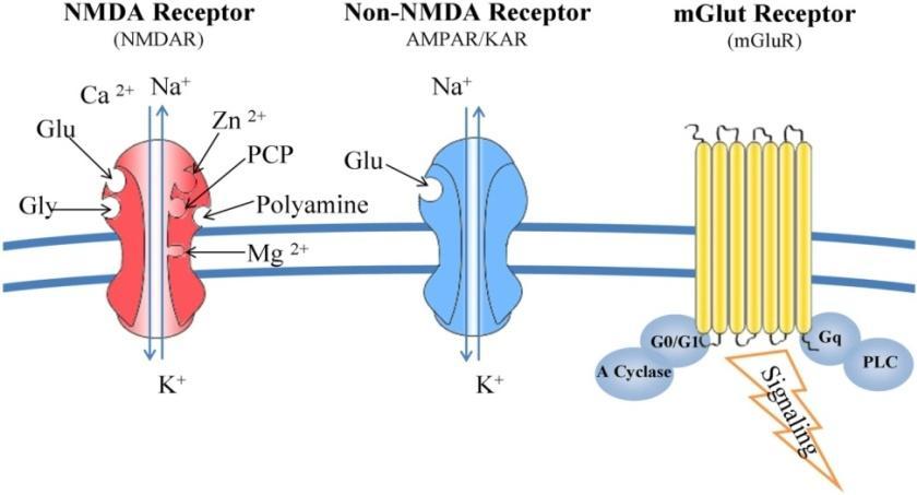
14 Glutamate RECEPTORS L-glutamate acts via two classes of receptors, ligand gated ion channels ( ionotropic receptors, iglur) and G-protein coupled (metabotropic, mglur) receptors. iglut Receptors mglut Receptors iglur are named after their respective more specific agonist: - NMDA R, N-methyl-D-aspartate - AMPA R, α-amino-3-hydroxy-5-methyl-4- isoxazole propionic acid - KA R, kainate mglutr are G-protein coupled receptors and trigger a second messenger cascade. They are found both at the preand post-synaptic neurons

15 ROC: AMPA-R AMPA receptors mediate fast synaptic transmission in the CNS and are composed of subunits GluA1-4. Like all the iglu R subunits, GluA subunits have an extracellular N-terminus and an intracellular C-terminus. The ligand binding domain is made up from N-terminal region S1 and S2. GluA2 subunit The C-terminus of the GluA2 subunit contains binding sites for a large number of interacting proteins. The effects of these protein-protein interactions is crucial in localisation and trafficking of these receptors so that they can fulfill their roles in plasticity. AMPA R are permeable mostly to Na and, to a lesser extent, to Ca ions. The calcium permeability of the GluA2 subunit is determined by the so called Q/R post-transcriptional editing site - GluA2(Q - glutamine) is calcium permeable whilst GluA2(R - arginine) is not.
16 ROC: KA R AMPA and kainate R are grouped together in the 'non-nmda receptor' family. Kainate R are similar to AMPA R, and are formed by multimeric assemblies of GluK1-3 and GluK4,5 subunits. GluK1-3 subunits can combine to form functional heteromeric and homomeric assemblies. Until relatively recently, KAR functional and physiological role in the mammalian CNS was unclear. They seem present at both sides of the synapse. Pre- and postsynaptic KAR can regulate transmission at many synapses in a specific manner, and seem to be involved in short- and long-term plastic phenomena. KA R are permeable to Na and Ca. However, their kinetics is much lower than AMPA R

17 ROC: NMDA R Unusually for the iglur, L-GLUTAMATE IS NOT THE ONLY AGONIST FOR THE NMDA R. Another amino-acid, GLYCINE, IS A CO-AGONIST and both transmitters must bind to their respective binding site in order for the receptor to function. The binding sites for glutamate and glycine are found on different subunits: - GLYCINE binds to the GluN1 subunit while - GLUTAMATE binds to the GluN2 subunit. The GluN2B subunit also possesses a BINDING SITE for polyamines, regulatory molecules that modulate the functioning of the NMDA receptor. NMDA R are highly permeable to Ca and, to a lesser extent, to Na.

18 ROC: NMDA R The NMDA R channel is normally blocked by Mg 2+ in a voltage- and usedependent manner. Although necessary, binding of both L- GLUTAMATE and GLYCINE is still not sufficient to completely activate NMDA receptor and favor Ca 2+ entry Mg 2+ Depolarization of the postsynaptic neuron via AMPA R activation releases the Mg 2+ block on the NMDA R. Ca 2+ entry is therefore subsequent to complete post-synaptic depolarization (required to remove Mg 2+ ). This effect is obtained by early activation of AMPA R

19 Glutamate RECEPTORS summary

20 Gamma Amino Butyric Acid (GABA) GABA is the chief inhibitory neurotransmitter in the mammalian central nervous system. It plays the principal role in reducing neuronal excitability throughout the nervous system. In humans, GABA is also directly responsible for the regulation of muscle tone.

21 GABA RECEPTORS GABA binds to both GABA A and GABA B receptors. GABA A Receptor GABA B Receptor GABA A receptors are ligand-gated channel receptors GABA B receptors are members of the 7- TM G protein-coupled receptors
.")
22 ROC: GABA A RECEPTOR The γ-aminobutyric acid, type A (GABA A ) receptor is a chloride-conducting receptor composed of α, β, and γ subunits assembled in a pentameric structure forming a central pore. The majority of GABA A Rs are believed to be expressed as heteromeric complexes of 2α, 2β, and 1 γ subunit The 2β subunits represent the BINDING SITE for the ligand (GABA). Both subunits must be occupied by GABA to allow receptor activation. The first binding facilitates the second (cooperation)

23 ROC: GABA A RECEPTOR Each subunit has a large extracellular agonist binding domain and four transmembrane domains (M1 M4), with the second transmembrane (M2) domain lining the pore. Cl - The M2 sequence of each TSM forms the SELECTIVITY FILTER facilitating the entry of anions. The α subunits represent the ALLOSTERIC SITE, a modulatory region where binding of ligands different from GABA may facilitate/obstacolate the GABA/RECEPTOR interaction. Another ligand site is present on the deep part of the channel, on the β subunit. Binding on this site allows a different modulation of channel opening that may excede the GABA-mediated effects Cl -

24 Voltage-operated (VOC) CHANNELS The function of VOC is closely dependent on transmembrane electrochemical gradient. A gradient is represented by the different concentration of ions on either side of the membrane. The open conformation of the ion channel allows for the translocation of ions across the cell membrane, while the closed conformation does not. The activity of these channels is connected with the activity of enzymes and the processes of phosphorylation of membrane proteins. Post-synaptic membrane with ROC s Muscle membrane with VOC s
 occur together.")
25 VOC CHANNELS Electrophysiological studies indicate the existence of three main types of VOC (K +, Na +, Ca 2+ channels). In number of neurons various subtypes of Ca 2+ channels (P, T, N and L-type) occur together. Na + channels Ca ++ channels K + channels

26 VOC CHANNELS In each protein subunit, the membrane-spanning segments, designated S1- S6, all take the form of alpha helices with specialized functions. The S1-S4 TMS serve as the voltage-sensing region. The S5-S6 TMS and pore loop have a key role in ion conduction, and represent the gate and pore of the channel. S1-4 Voltage sensing region S5-6 Pore region S1 S6 S4 I II III IV C- terminal N- terminal

27 VOC CHANNELS: state of activity A voltage gated ion channel can be in three states: closed, open or inactivated. The inactive state, which is stable and non-conducting, is caused by the physical blockage of the pore. The blockage is caused by a ball of amino acids attached to the main protein by a string of residues on the cytoplasmic side. The ball enters the open channel and binds to the hydrophobic inner vestibule at the center of the channel. The blockage causes inactivation of the channel by stopping the flow of ions.
.")
28 Na + CHANNELS VOC Na + channels are formed by a single subunit and are expressed in all excitable tissues. Na + channels are responsible for the rapid membrane depolarization during the action potential. Activation and inactivation are voltage-dependent, very fast processes (1-10 ms). During the inactivation state, the channel is in a refractory period.

29 VOC Na + CHANNELS Na + channels are important drug targets. Drugs bind the Na channel inside the pore. Their binding maintains the channel in the inactivated state. Since they reach their binding site when the channel is open, their inhibitory effect is use- and voltagedependent. Drugs acting as Na + channels blockers include Class I anti-arrhythmics, anticonvulsants, local anesthetics
30 Non-VOC Na + CHANNELS The epithelial Na + channels (ENaC) are non-voltage gated, highly Na-selective channels. ENaC activity is rate limiting for Na + reabsorption in the distal nephron. The long term control of blood pressure involves Na + homeostasis through the precise regulation of ENaC in the aldosterone-sensitive distal nephron Inhibition of these channels results in reduced K excretion. This effect is obtained by some potassium-sparing diuretics such as amiloride

31 K + CHANNELS K + channels are found in virtually all living organisms. They form K + -selective pores that span cell membranes and conduct rapidly and selectively K + ions down their electrochemical gradient. These channels act to set or reset the resting potential in many cells. Voltage-gated K + channels Calcium-activated K + channels Inward rectifier K + channels Leak or background K + channels
, exemplified by inwardly rectifying K + channels.")


32 K + CHANNELS: structure 2TM/P channels (which consist of two transmembrane (TM) helices with a P loop between them), exemplified by inwardly rectifying K + channels. Channel is formed by (2TM/P x4) tetramer 6TM/P channels, which are the predominant class among ligand-gated and voltage-gated K + channels. Channel is formed by (6TM/P x4) tetramer 4TM/2P channels, which consist of two repeats of 2TM/P channels. 4TM/2P channels are far more common than was originally thought. Channel is formed by (2TM/P x2) dimer

, small conductance (SK) channels. These channels are activated largely in response to calcium influx during action potentials.")
33 K + CHANNELS Voltage-gated K + channels Kv channels are one of the key components in generation and propagation of electrical impulses in nervous system and in the heart. Upon changes in transmembrane potential, these channels open and allow passive flow of K+ ions from the cell to restore the membrane potential. They are the target of Class III anti-arrhythmic drugs Calcium-activated K + channels KCa channels can be grouped into three distinct subfamilies, large-conductance (BK), intermediate conductance (IK), small conductance (SK) channels. These channels are activated largely in response to calcium influx during action potentials. Activation of these channels is to hyperpolarize the membrane.
 GIRK (G-protein-activated inward rectifier K+ channels) K ATP (ATP-sensitive K+ channels) These channels are responsible for repolarizing a cell following an action potential K")
34 Inward rectifier K + channels CARDIAC MYOCYTES ENDOTHELIAL CELLS KIDNEYS NEURONS AND HEART K + CHANNELS K IR include several subfamilies, among which the most important are IRK (strong inward rectifier K+ channels) GIRK (G-protein-activated inward rectifier K+ channels) K ATP (ATP-sensitive K+ channels) These channels are responsible for repolarizing a cell following an action potential K ir close upon depolarization, slowing membrane repolarization and helping maintain a more prolonged cardiac action potential. K ir are involved in regulation of nitric oxide synthase. K ir export surplus K + into collecting tubules for removal in the urine, or reuptake K + back into the body. G-protein activated IRKs (K ir 3) are important regulators, modulated by neurotransmitters. PANCREAS BETA CELLS K ATP channels control insulin release.

35 K + CHANNELS Leak or background K + channels This type of potassium channel is formed by two homodimers that create a channel that leaks potassium out of the cell They are all voltage-independent and can be opened by heat, membrane stretching, intracellular acidosis, and certain anesthetics. These channels are responsible for a high K + conductance under basal state conditions and therefore contribute to resting potential TRAAK channels are mechanically activated when there is a convex curvature in the membrane that alters the channel s activity (membrane stretching) TASK channels are sensitive to changes in extracellular ph and inhibited by extracellular acidification TALK channels are primarily expressed in the pancreas, and activated at alkaline ph

36 Calcium Calcium is an ubiquitous intracellular messenger, controlling cellular processes ranging from gene transcription, muscle contraction and cell proliferation The Ca2+ signaling apparatus involves various channels, pumps, and transporters
.")
37 Calcium regulatory mechanisms CALCIUM ENTRY ( ON ) MECHANISMS VDCCs -voltage-dependent calcium channel; ROC-Ligand-gated channels, SOCE -store-operated calcium entry; TRP -transient receptor potential channels; ASIC -acid-sensing ion channels; IEIC - inward excitotoxic injury current calcium-permeable channels; NCX -sodium-calcium exchanger (operating in entry mode). CALCIUM INTRACELLULAR SEQUESTERING AND RECYCLING MECHANISMS SERCA -Sarcoplasmic-Endoplasmic Reticulum Ca 2+ -ATPase; Ryr ryanodine receptors. CALCIUM EXIT ( OFF ) MECHANISMS PMCA -Calcium ATPase pump; NCX sodium-calcium exchanger (operating in exit mode).
 at depolarized membrane potentials and this is the source of the \"voltagedependent\" definition.")
38 CALCIUM ON MECHANISMS: VOCCs Voltage-Operated Calcium Channels are slightly permeable to Na (also called Ca 2+ - Na + channels), but their permeability to Ca 2+ is about 1000-fold greater. At resting membrane potential, VOCCs are normally closed. They are activated (i.e., opened) at depolarized membrane potentials and this is the source of the "voltagedependent" definition. Activation of particular VOCCs allows Ca 2+ to rush into the cell, which, depending on the cell type, results in activation of calcium-sensitive potassium channels, muscular contraction, excitation of neurons, up-regulation of gene expression, or release of hormones or neurotransmitters.
39 Voltage-operated Ca ++ CHANNELS (VOCCs) TYPE VOLTAGE MOST OFTEN FOUND IN L-TYPE CALCIUM CHANNEL ("LONG-LASTING" AKA "DHP RECEPTOR") HVA (high voltage activated) Skeletal muscle, smooth muscle, bone (osteoblasts), ventricular myocytes (responsible for prolonged action potential in cardiac cell; also termed DHP receptors), dendrites and dendritic spines of cortical neurones L-Type channel blockers are used as antihypertensive and antiarrhythmic drugs P-TYPE CALCIUM CHANNEL ("PURKINJE") /Q-TYPE CALCIUM CHANNEL N-TYPE CALCIUM CHANNEL ("NEURAL"/"NON-L") R-TYPE CALCIUM CHANNEL ("RESIDUAL") T-TYPE CALCIUM CHANNEL ("TRANSIENT") HVA (high voltage activated) HVA (high-voltageactivated) intermediatevoltageactivated low-voltageactivated Purkinje neurons in the cerebellum / Cerebellar granule cells Throughout the brain and peripheral nervous system. Cerebellar granule cells, other neurons neurons, cells that have pacemaker activity, bone (osteocytes) T-Type channel blockers are used as antiepileptic and neuropathic painkiller drugs
 activated by camp, cgmp, Arachidonic acid, sphingosine, ADPribose InsP3-Receptor-Ca 2+ -Channels allow Ca 2+ release form intracellular")
40 CALCIUM INTRACELLULAR RECYCLING MECHANISMS Store-operated Ca ++ Channels (SOCC) SOCC can be activated by any procedure that empties the stores Receptor-operated Ca ++ Channels (ROC) nach R NMDA R, kainate R 5HT 3 P2X Transient-receptor-potential Channels (TRP) activated by camp, cgmp, Arachidonic acid, sphingosine, ADPribose InsP3-Receptor-Ca 2+ -Channels allow Ca 2+ release form intracellular stores Ryanodine receptors (RYR) activated by cadpr Ryr1 (muscle cells) Ryr2 (heart) Ryr3 (neurons)

41 CALCIUM OFF MECHANISMS Na + /Ca 2+ exchanger (NCX1-3) is an antiporter membrane protein that removes Ca 2+ using the energy from the electrochemical gradient of Na +. The NCX removes 1 single Ca 2+ ion in exchange for the import of 3 Na + ions. Low affinity but high speed (Ca/Na 1:3) PMCA (Plasma Membrane Ca 2+ ATPase) the plasma membrane pump is powered by the hydrolysis of ATP, with a stoichiometry of 1 Ca 2+ ion/1 molecule of ATP hydrolysed. High affinity but low speed. Its activity is modulated by the calmodulin (CaM) protein (Ca/ATP 1:1) SERCA (Sarcoplasmic- Endoplasmic Reticulum Ca 2+ - ATPase) It is a Ca 2+ ATPase that transfers Ca 2+ from the cytosol of the cell to the lumen of the SR at the expense of ATP hydrolysis during muscle relaxation. The pump transports 2 Ca 2+ ion/1 molecule of ATP hydrolysed and it is CaM-independent (Ca/ATP 2:1)

42
Neurotransmitter Systems II Receptors. Reading: BCP Chapter 6
 Neurotransmitter Systems II Receptors Reading: BCP Chapter 6 Neurotransmitter Systems Normal function of the human brain requires an orderly set of chemical reactions. Some of the most important chemical
QUIZ/TEST REVIEW NOTES SECTION 7 NEUROPHYSIOLOGY [THE SYNAPSE AND PHARMACOLOGY]
![QUIZ/TEST REVIEW NOTES SECTION 7 NEUROPHYSIOLOGY [THE SYNAPSE AND PHARMACOLOGY]](/thumbs/86/94924826.jpg "QUIZ/TEST REVIEW NOTES SECTION 7 NEUROPHYSIOLOGY [THE SYNAPSE AND PHARMACOLOGY]") QUIZ/TEST REVIEW NOTES SECTION 7 NEUROPHYSIOLOGY [THE SYNAPSE AND PHARMACOLOGY] Learning Objectives: Explain how neurons communicate stimulus intensity Explain how action potentials are conducted along
QUIZ/TEST REVIEW NOTES SECTION 7 NEUROPHYSIOLOGY [THE SYNAPSE AND PHARMACOLOGY] Learning Objectives: Explain how neurons communicate stimulus intensity Explain how action potentials are conducted along
Ligand-Gated Ion Channels
 Ligand-Gated Ion Channels The Other Machines That Make It Possible... Topics I Introduction & Electrochemical Gradients Passive Membrane Properties Action Potentials Voltage-Gated Ion Channels Topics II
IONOTROPIC RECEPTORS
 BASICS OF NEUROBIOLOGY IONOTROPIC RECEPTORS ZSOLT LIPOSITS 1 NEURAL COMMUNICATION http://sciencecore.columbia.edu/s4.html 2 Post-synaptic mechanisms Receptors-signal transduction-messengers 3 TRANSMITTER
NEUROCHEMISTRY Brief Review
 NEUROCHEMISTRY Brief Review UNIVERSITY OF PNG SCHOOL OF MEDICINE AND HEALTH SCIENCES DISCIPLINE OF BIOCHEMISTRY AND MOLECULAR BIOLOGY PBL MBBS YEAR V SEMINAR VJ Temple 1 Membrane potential Membrane potential:
Synaptic Transmission: Ionic and Metabotropic
 Synaptic Transmission: Ionic and Metabotropic D. Purves et al. Neuroscience (Sinauer Assoc.) Chapters 5, 6, 7. C. Koch. Biophysics of Computation (Oxford) Chapter 4. J.G. Nicholls et al. From Neuron to
Basics of Pharmacology
 Basics of Pharmacology Pekka Rauhala Transmed 2013 What is pharmacology? Pharmacology may be defined as the study of the effects of drugs on the function of living systems Pharmacodynamics The mechanism(s)
Receptors Families. Assistant Prof. Dr. Najlaa Saadi PhD Pharmacology Faculty of Pharmacy University of Philadelphia
 Receptors Families Assistant Prof. Dr. Najlaa Saadi PhD Pharmacology Faculty of Pharmacy University of Philadelphia Receptor Families 1. Ligand-gated ion channels 2. G protein coupled receptors 3. Enzyme-linked
Ch. 45 Continues (Have You Read Ch. 45 yet?) u Central Nervous System Synapses - Synaptic functions of neurons - Information transmission via nerve
 u Central Nervous System Synapses - Synaptic functions of neurons - Information transmission via nerve") Ch. 45 Continues (Have You Read Ch. 45 yet?) u Central Nervous System Synapses - Synaptic functions of neurons - Information transmission via nerve impulses - Impulse may be blocked in its transmission
Ch. 45 Continues (Have You Read Ch. 45 yet?) u Central Nervous System Synapses - Synaptic functions of neurons - Information transmission via nerve impulses - Impulse may be blocked in its transmission
Portions from Chapter 6 CHAPTER 7. The Nervous System: Neurons and Synapses. Chapter 7 Outline. and Supporting Cells
 CHAPTER 7 The Nervous System: Neurons and Synapses Chapter 7 Outline Neurons and Supporting Cells Activity in Axons The Synapse Acetylcholine as a Neurotransmitter Monoamines as Neurotransmitters Other
CELLULAR NEUROPHYSIOLOGY
 CELLULAR NEUROPHYSIOLOGY CONSTANCE HAMMOND 4. SYNAPTIC TRANSMISSION II: GLUTAMATERGIC TRANSMISSION Video 4-1: Observations and glutamate receptor channels Synaptic transmission II 1 Constance Hammond Observation
5-Nervous system II: Physiology of Neurons
 5-Nervous system II: Physiology of Neurons AXON ION GRADIENTS ACTION POTENTIAL (axon conduction) GRADED POTENTIAL (cell-cell communication at synapse) SYNAPSE STRUCTURE & FUNCTION NEURAL INTEGRATION CNS
Ionotropic glutamate receptors (iglurs)
") Ionotropic glutamate receptors (iglurs) GluA1 GluA2 GluA3 GluA4 GluN1 GluN2A GluN2B GluN2C GluN2D GluN3A GluN3B GluK1 GluK2 GluK3 GluK4 GluK5 The general architecture of receptor subunits Unique properties
Ionotropic glutamate receptors (iglurs) GluA1 GluA2 GluA3 GluA4 GluN1 GluN2A GluN2B GluN2C GluN2D GluN3A GluN3B GluK1 GluK2 GluK3 GluK4 GluK5 The general architecture of receptor subunits Unique properties
Examples of smallmolecule. peptide neurotransmitters
 Examples of smallmolecule and peptide neurotransmitters Small- molecule transmitters are transported from the cytosol into vesicles or from the synaptic cleft to the cytosol by TRANSPORTERS Unconventional
Lecture 22: A little Neurobiology
 BIO 5099: Molecular Biology for Computer Scientists (et al) Lecture 22: A little Neurobiology http://compbio.uchsc.edu/hunter/bio5099 Larry.Hunter@uchsc.edu Nervous system development Part of the ectoderm
Chapter 2. The Cellular and Molecular Basis of Cognition Cognitive Neuroscience: The Biology of the Mind, 2 nd Ed.,
 Chapter 2. The Cellular and Molecular Basis of Cognition Cognitive Neuroscience: The Biology of the Mind, 2 nd Ed., M. S. Gazzaniga, R. B. Ivry, and G. R. Mangun, Norton, 2002. Summarized by B.-W. Ku,
Lipids and Membranes
 Lipids and Membranes Presented by Dr. Mohammad Saadeh The requirements for the Pharmaceutical Biochemistry I Philadelphia University Faculty of pharmacy Membrane transport D. Endocytosis and Exocytosis
Transport through biological membranes. Christine Carrington Biochemistry Unit Apr 2010
 Transport through biological membranes Christine Carrington Biochemistry Unit Apr 2010 Biological membranes Membranes control the structures and environments of the compartments they define and thereby
PHSI3009 Frontiers in Cellular Physiology 2017
 Overview of PHSI3009 L2 Cell membrane and Principles of cell communication L3 Signalling via G protein-coupled receptor L4 Calcium Signalling L5 Signalling via Growth Factors L6 Signalling via small G-protein
Lecture 14. Insect nerve system (II)
") Lecture 14. Insect nerve system (II) Structures (Anatomy) Cells Anatomy How NS functions Signal transduction Signal transmission Overview More on neurons: ions, ion channel, ligand receptor Signal transduction:
Lecture 14. Insect nerve system (II) Structures (Anatomy) Cells Anatomy How NS functions Signal transduction Signal transmission Overview More on neurons: ions, ion channel, ligand receptor Signal transduction:
Cellular Physiology (PHSI3009) Contents:
 Contents:") Cellular Physiology (PHSI3009) Contents: Cell membranes and communication 2 nd messenger systems G-coupled protein signalling Calcium signalling Small G-protein signalling o RAS o MAPK o PI3K RHO GTPases
Cellular Physiology (PHSI3009) Contents: Cell membranes and communication 2 nd messenger systems G-coupled protein signalling Calcium signalling Small G-protein signalling o RAS o MAPK o PI3K RHO GTPases
Chapter 5 subtitles GABAergic synaptic transmission
 CELLULAR NEUROPHYSIOLOGY CONSTANCE HAMMOND Chapter 5 subtitles GABAergic synaptic transmission INTRODUCTION (2:57) In this fifth chapter, you will learn how the binding of the GABA neurotransmitter to
Thursday, January 22, Nerve impulse
 Nerve impulse Transmembrane Potential caused by ions moving through cell membrane at different rates Two main ions of concern Na + - Sodium K + - potassium Cell membrane not freely permeable therefore
Synapses and Neurotransmitters
 Synapses and Neurotransmitters Communication Between Neurons Synapse: A specialized site of contact, and transmission of information between a neuron and an effector cell Anterior Motor Neuron Figure 45-5
Psych 181: Dr. Anagnostaras
 Psych 181: Dr. Anagnostaras Lecture 5 Synaptic Transmission Introduction to synaptic transmission Synapses (Gk., to clasp or join) Site of action of most psychoactive drugs 6.5 1 Synapses Know basic terminology:
Synaptic Communication. Steven McLoon Department of Neuroscience University of Minnesota
 Synaptic Communication Steven McLoon Department of Neuroscience University of Minnesota 1 Course News The first exam is next week on Friday! Be sure to checkout the sample exam on the course website. 2
TA Review. Neuronal Synapses. Steve-Felix Belinga Neuronal synapse & Muscle
 TA Review Steve-Felix Belinga sbelinga@wustl.edu Neuronal synapse & Muscle Neuronal Synapses 1 Things you should know beyond the obvious stuff 1. Differences between ionotropic and metabotropic receptors.
Summarized by B.-W. Ku, E. S. Lee, and B.-T. Zhang Biointelligence Laboratory, Seoul National University.
 Chapter 2. The Cellular l and Molecular Basis of Cognition Cognitive Neuroscience: The Biology of the Mind, 3 rd Ed., M. S. Gazzaniga, R. B. Ivry, and G. R. Mangun, Norton, 2008. Summarized by B.-W. Ku,
Action Potentials and Synaptic Transmission. BIO 219 Napa Valley College Dr. Adam Ross
 Action Potentials and Synaptic Transmission BIO 219 Napa Valley College Dr. Adam Ross Review of action potentials Nodes of Ranvier Nucleus Dendrites Cell body In saltatory conduction, the nerve impulses
Chapter 3 subtitles Action potentials
 CELLULAR NEUROPHYSIOLOGY CONSTANCE HAMMOND Chapter 3 subtitles Action potentials Introduction (3:15) This third chapter explains the calcium current triggered by the arrival of the action potential in
Objectives. Functions of smooth muscle. Smooth muscle. Smooth Muscle Contraction: Mechanism. Latch state. Smooth muscle contraction
 Objectives Functions of smooth muscle Sompol Tapechum,, M.D., Ph.D. Department of Physiology Faculty of Medicine Siriraj hospital อธ บายล กษณะการหดต วของกล ามเน อเร ยบได อธ บายกลไกและป จจ ยท ม ผลต อการหดต
Chapter 7. The Nervous System: Structure and Control of Movement
 Chapter 7 The Nervous System: Structure and Control of Movement Objectives Discuss the general organization of the nervous system Describe the structure & function of a nerve Draw and label the pathways
Outline. Neuron Structure. Week 4 - Nervous System. The Nervous System: Neurons and Synapses
 Outline Week 4 - The Nervous System: Neurons and Synapses Neurons Neuron structures Types of neurons Electrical activity of neurons Depolarization, repolarization, hyperpolarization Synapses Release of
Chapter 7. Objectives
 Chapter 7 The Nervous System: Structure and Control of Movement Objectives Discuss the general organization of the nervous system Describe the structure & function of a nerve Draw and label the pathways
Chapter 24 Chemical Communications Neurotransmitters & Hormones
 Chapter 24 Chemical Communications Neurotransmitters & Hormones 1 Chemical Communication Terms and definitions: Neuron: A nerve cell. Neurotransmitter: A chemical messenger between a neuron and another
Neuron types and Neurotransmitters
 Neuron types and Neurotransmitters Faisal I. Mohammed. PhD, MD University of Jordan 1 Transmission of Receptor Information to the Brain the larger the nerve fiber diameter the faster the rate of transmission
Dania Ahmad. Tamer Barakat + Dania Ahmad. Faisal I. Mohammed
 16 Dania Ahmad Tamer Barakat + Dania Ahmad Faisal I. Mohammed Revision: What are the basic types of neurons? sensory (afferent), motor (efferent) and interneuron (equaled association neurons). We classified
NEURAL TISSUE (NEUROPHYSIOLOGY) PART I (A): NEURONS & NEUROGLIA
 PART I (A): NEURONS & NEUROGLIA") PART I (A): NEURONS & NEUROGLIA Neural Tissue Contains 2 kinds of cells: neurons: cells that send and receive signals neuroglia (glial cells): cells that support and protect neurons Neuron Types Sensory
PART I (A): NEURONS & NEUROGLIA Neural Tissue Contains 2 kinds of cells: neurons: cells that send and receive signals neuroglia (glial cells): cells that support and protect neurons Neuron Types Sensory
It s Not Just Serotonin: Neurosignaling in Mental Illness
 It s Not Just Serotonin: Neurosignaling in Mental Illness Barbara J. Limandri, DNSc, APRN, BC Professor of Nursing Linfield College Learning Outcomes Distinguish between metabotropic and ionotropic neuroreceptors
Session ID: 1001 June 14, 2012
 It s Not Just Serotonin: Neurosignaling in Mental Illness Barbara J. Limandri, DNSc, APRN, BC Professor of Nursing Linfield College Learning Outcomes Distinguish between metabotropic and ionotropic neuroreceptors
Receptors and Drug Action. Dr. Subasini Pharmacology Department Ishik University, Erbil
 Receptors and Drug Action Dr. Subasini Pharmacology Department Ishik University, Erbil Receptors and Drug Action Receptor Receptor is defined as a macromolecule or binding site located on the surface or
Local Anesthetics. Xiaoping Du Room E417 MSB Department of Pharmacology Phone (312) ;
 ;") Local Anesthetics Xiaoping Du Room E417 MSB Department of Pharmacology Phone (312)355 0237; Email: xdu@uic.edu Summary: Local anesthetics are drugs used to prevent or relieve pain in the specific regions
Local Anesthetics Xiaoping Du Room E417 MSB Department of Pharmacology Phone (312)355 0237; Email: xdu@uic.edu Summary: Local anesthetics are drugs used to prevent or relieve pain in the specific regions
2013 W. H. Freeman and Company. 12 Signal Transduction
 2013 W. H. Freeman and Company 12 Signal Transduction CHAPTER 12 Signal Transduction Key topics: General features of signal transduction Structure and function of G protein coupled receptors Structure
BIPN100 F15 Human Physiology 1 Lecture 3. Synaptic Transmission p. 1
 BIPN100 F15 Human Physiology 1 Lecture 3. Synaptic Transmission p. 1 Terms you should know: synapse, neuromuscular junction (NMJ), pre-synaptic, post-synaptic, synaptic cleft, acetylcholine (ACh), acetylcholine
Chapter 2: Cellular Mechanisms and Cognition
 Chapter 2: Cellular Mechanisms and Cognition MULTIPLE CHOICE 1. Two principles about neurons were defined by Ramón y Cajal. The principle of connectional specificity states that, whereas the principle
Neurophysiology and Synaptic Transmission Modules
 Neurophysiology and Synaptic Transmission Neurophysiology and Synaptic Transmission Modules Module Listing Please complete the following online neurophysiology modules during the first week of class: Module
- Biosignaling: Signal transduction. References: chapter 8 of Lippincots chapter 1 3 of Lehningers
 Basic concepts of Metabolism Metabolism and metabolic pathway Metabolic Map Catabolism Anabolism - Regulation of Metabolism Signals from within the cell (Intracellular) Communication between cells. - Biosignaling:
Chapter 2. The Cellular and Molecular Basis of Cognition
 Chapter 2. The Cellular and Molecular Basis of Cognition Cognitive Neuroscience: The Biology of the Mind, 2 nd Ed., M. S. Gazzaniga,, R. B. Ivry,, and G. R. Mangun,, Norton, 2002. Summarized by B.-W. Ku,
Chapter 4 Neuronal Physiology
 Chapter 4 Neuronal Physiology V edit. Pg. 99-131 VI edit. Pg. 85-113 VII edit. Pg. 87-113 Input Zone Dendrites and Cell body Nucleus Trigger Zone Axon hillock Conducting Zone Axon (may be from 1mm to more
NEURONS COMMUNICATE WITH OTHER CELLS AT SYNAPSES 34.3
 NEURONS COMMUNICATE WITH OTHER CELLS AT SYNAPSES 34.3 NEURONS COMMUNICATE WITH OTHER CELLS AT SYNAPSES Neurons communicate with other neurons or target cells at synapses. Chemical synapse: a very narrow
MOLECULAR AND CELLULAR NEUROSCIENCE
 MOLECULAR AND CELLULAR NEUROSCIENCE BMP-218 November 4, 2014 DIVISIONS OF THE NERVOUS SYSTEM The nervous system is composed of two primary divisions: 1. CNS - Central Nervous System (Brain + Spinal Cord)
BCOR 011 Lecture 19 Oct 12, 2005 I. Cell Communication Signal Transduction Chapter 11
 BCOR 011 Lecture 19 Oct 12, 2005 I. Cell Communication Signal Transduction Chapter 11 External signal is received and converted to another form to elicit a response 1 Lecture Outline 1. Types of intercellular
BIPN 140 Problem Set 6
 BIPN 140 Problem Set 6 1) Hippocampus is a cortical structure in the medial portion of the temporal lobe (medial temporal lobe in primates. a) What is the main function of the hippocampus? The hippocampus
Lojayn Salah. Razan Aburumman. Faisal Muhammad
 20 Lojayn Salah Razan Aburumman Faisal Muhammad Note: I tried to include everything that's important from the doctor's slides but you can refer back to them after studying this sheet.. After you read this
Introduction to Neurobiology
 Biology 240 General Zoology Introduction to Neurobiology Nervous System functions: communication of information via nerve signals integration and processing of information control of physiological and
3) Most of the organelles in a neuron are located in the A) dendritic region. B) axon hillock. C) axon. D) cell body. E) axon terminals.
 Most of the organelles in a neuron are located in the A) dendritic region. B) axon hillock. C) axon. D) cell body. E) axon terminals.") Chapter 48 Neurons, Synapses, and Signaling Multiple-Choice Questions 1) A simple nervous system A) must include chemical senses, mechanoreception, and vision. B) includes a minimum of 12 ganglia. C) has
Chapter 48 Neurons, Synapses, and Signaling Multiple-Choice Questions 1) A simple nervous system A) must include chemical senses, mechanoreception, and vision. B) includes a minimum of 12 ganglia. C) has
The Brain & Homeostasis. The Brain & Technology. CAT, PET, and MRI Scans
 The Brain & Homeostasis Today, scientists have a lot of information about what happens in the different parts of the brain; however they are still trying to understand how the brain functions. We know
Ion Channels (Part 2)
") Ion Channels (Part 2) Graphics are used with permission of : adam.com (http://www.adam.com/) Benjamin/Cummings Publishing Co (http://www.awl.com/bc) -57- Quiz Question #2: Ion Channels This question asks
Ion Channels (Part 2) Graphics are used with permission of : adam.com (http://www.adam.com/) Benjamin/Cummings Publishing Co (http://www.awl.com/bc) -57- Quiz Question #2: Ion Channels This question asks
Section: Chapter 5: Multiple Choice. 1. The structure of synapses is best viewed with a(n):
:") Section: Chapter 5: Multiple Choice 1. The structure of synapses is best viewed with a(n): p.155 electron microscope. light microscope. confocal microscope. nissle-stained microscopic procedure. 2. Electron
Section: Chapter 5: Multiple Choice 1. The structure of synapses is best viewed with a(n): p.155 electron microscope. light microscope. confocal microscope. nissle-stained microscopic procedure. 2. Electron
Ionotropic and metabotropic receptors LESSON NR PSYCHOBIOLOGY
 Ionotropic and metabotropic receptors LESSON NR. 10 - PSYCHOBIOLOGY Channels regulated by ligand IONOTROPIC RECEPTORS They are membrane protein complexes, characterized by the presence on their surface
Ion Channels Graphics are used with permission of: Pearson Education Inc., publishing as Benjamin Cummings (http://www.aw-bc.com)
") Ion Channels Graphics are used with permission of: Pearson Education Inc., publishing as Benjamin Cummings (http://www.aw-bc.com) Page 1. Introduction At synapses, ions move across cell membranes through
Ion Channels Graphics are used with permission of: Pearson Education Inc., publishing as Benjamin Cummings (http://www.aw-bc.com) Page 1. Introduction At synapses, ions move across cell membranes through
What effect would an AChE inhibitor have at the neuromuscular junction?
 CASE 4 A 32-year-old woman presents to her primary care physician s office with difficulty chewing food. She states that when she eats certain foods that require a significant amount of chewing (meat),
Basics of skeletal muscle electrophysiology. Tóth András, PhD
 Basics of skeletal muscle electrophysiology Tóth András, PhD Topics Structure Contraction and relaxation Activation Excitation-contraction coupling Action potential Ion channels* Calcium homeostasis Structure
Nervous System. Nervous system cells. Transmission of a signal 2/27/2015. Neuron
 Nervous System 2007-2008 signal direction Neuron a nerve cell Nervous system cells dendrites axon cell body Structure fits function many entry points for signal one path out transmits signal signal direction
Cellular Bioelectricity
 ELEC ENG 3BB3: Cellular Bioelectricity Notes for Lecture 22 Friday, February 28, 2014 10. THE NEUROMUSCULAR JUNCTION We will look at: Structure of the neuromuscular junction Evidence for the quantal nature
BIPN 140 Problem Set 6
 BIPN 140 Problem Set 6 1) The hippocampus is a cortical structure in the medial portion of the temporal lobe (medial temporal lobe in primates. a) What is the main function of the hippocampus? The hippocampus
Communication within a Neuron
 Neuronal Communication, Ph.D. Communication within a Neuron Measuring Electrical Potentials of Axons The Membrane Potential The Action Potential Conduction of the Action Potential 1 The withdrawal reflex
Chapter 3 Neurotransmitter release
 NEUROPHYSIOLOGIE CELLULAIRE CONSTANCE HAMMOND Chapter 3 Neurotransmitter release In chapter 3, we proose 3 videos: Observation Calcium Channel, Ca 2+ Unitary and Total Currents Ca 2+ and Neurotransmitter
Chapter 11 Introduction to the Nervous System and Nervous Tissue Chapter Outline
 Chapter 11 Introduction to the Nervous System and Nervous Tissue Chapter Outline Module 11.1 Overview of the Nervous System (Figures 11.1-11.3) A. The nervous system controls our perception and experience
Sarah Jaar Marah Al-Darawsheh
 22 Sarah Jaar Marah Al-Darawsheh Faisal Mohammad Receptors can be membrane proteins (for water-soluble hormones/ligands) or intracellular (found in the cytosol or nucleus and bind to DNA, for lipid-soluble
Fundamentals of Pharmacology
 Fundamentals of Pharmacology Topic Page Receptors 2 Ion channels / GABA 4 GPCR s 6 TK receptors 8 Basics of PK 11 ADR s / Clinical study design 13 Introduction to the ANS 16 Cholinergic Pharmacology 20
Neuroscience: Exploring the Brain, 3e. Chapter 4: The action potential
 Neuroscience: Exploring the Brain, 3e Chapter 4: The action potential Introduction Action Potential in the Nervous System Conveys information over long distances Action potential Initiated in the axon
THE NERVOUS SYSTEM. Homeostasis Strand
 THE NERVOUS SYSTEM Homeostasis Strand Introduction In general, a nervous system has three overlapping functions : 1. Sensory input conduction of signals from sensory receptors to integration centres 2.
Lecture 36: Review of membrane function
 Chem*3560 Lecture 36: Review of membrane function Membrane: Lipid bilayer with embedded or associated proteins. Bilayers: 40-70% neutral phospholipid 10-20% negative phospholipid 10-30% cholesterol 10-30%
The Nervous System. Nervous System Functions 1. gather sensory input 2. integration- process and interpret sensory input 3. cause motor output
 The Nervous System Nervous System Functions 1. gather sensory input 2. integration- process and interpret sensory input 3. cause motor output The Nervous System 2 Parts of the Nervous System 1. central
Electrical Properties of Neurons. Steven McLoon Department of Neuroscience University of Minnesota
 Electrical Properties of Neurons Steven McLoon Department of Neuroscience University of Minnesota 1 Neuronal Communication Neurons communicate with other cells, often over long distances. The electrical
Cell Biology (BIOL 4374 and BCHS 4313) Third Exam 4/24/01
 Third Exam 4/24/01") Cell Biology (BIOL 4374 and BCHS 4313) Third Exam 4/24/01 Name SS# This exam is worth a total of 100 points. The number of points each question is worth is shown in parentheses. For multiple choice questions,
Cell Biology (BIOL 4374 and BCHS 4313) Third Exam 4/24/01 Name SS# This exam is worth a total of 100 points. The number of points each question is worth is shown in parentheses. For multiple choice questions,
3.E.2 Continued. This is the essential knowledge statement from the curriculum framework. Detect---process--- response
 Nervous System: Part III What Happens at a Synapse? 3.E. Continued Animals have nervous systems that detect external and internal signals, transmit and integrate information, and produce responses. This
Cardiac Properties MCQ
 Cardiac Properties MCQ Abdel Moniem Ibrahim Ahmed, MD Professor of Cardiovascular Physiology Cairo University 2007 1- Cardiac Valves: a- Prevent backflow of blood from the ventricles to the atria during
Na + K + pump. The beauty of the Na + K + pump. Cotransport. The setup Cotransport the result. Found along the plasma membrane of all cells.
 The beauty of the Na + K + pump Na + K + pump Found along the plasma membrane of all cells. Establishes gradients, controls osmotic effects, allows for cotransport Nerve cells have a Na + K + pump and
Chapter 45: Synapses Transmission of Nerve Impulses Between Neurons. Chad Smurthwaite & Jordan Shellmire
 Chapter 45: Synapses Transmission of Nerve Impulses Between Neurons Chad Smurthwaite & Jordan Shellmire The Chemical Synapse The most common type of synapse used for signal transmission in the central
Skeletal Muscle Contraction 4/11/2018 Dr. Hiwa Shafiq
 Skeletal Muscle Contraction 4/11/2018 Dr. Hiwa Shafiq Skeletal Muscle Fiber About 40 per cent of the body is skeletal muscle, and 10 per cent is smooth and cardiac muscle. Skeletal muscles are composed
Communication Between
 Communication Between Neurons Bởi: OpenStaxCollege The electrical changes taking place within a neuron, as described in the previous section, are similar to a light switch being turned on. A stimulus starts
Synaptic transmission
 Outline Synaptic transmission Sompol Tapechum M.D., Ph.D. Department of Physiology Faculty of Medicine Siriraj Hospital, Bangkok, Thailand. sisth@mahidol.ac.th 2 Structure of synapse Modes of synaptic
CHAPTER 44: Neurons and Nervous Systems
 CHAPTER 44: Neurons and Nervous Systems 1. What are the three different types of neurons and what are their functions? a. b. c. 2. Label and list the function of each part of the neuron. 3. How does the
Cell Signaling (part 1)
") 15 Cell Signaling (part 1) Introduction Bacteria and unicellular eukaryotes respond to environmental signals and to signaling molecules secreted by other cells for mating and other communication. In multicellular
15 Cell Signaling (part 1) Introduction Bacteria and unicellular eukaryotes respond to environmental signals and to signaling molecules secreted by other cells for mating and other communication. In multicellular
Notes are online at The Neuron
 Notes are online at http://cogsci.ucsd.edu/~clovett/neuronotescogs17.pdf A. What is a neuron? The Neuron 1. A neuron is a type of cell that receives and transmits information in the Central Nervous System
Neural Tissue. Chapter 12 Part B
 Neural Tissue Chapter 12 Part B CNS Tumors - Neurons stop dividing at age 4 but glial cells retain the capacity to divide. - Primary CNS tumors in adults- division of abnormal neuroglia rather than from
Division Ave. High School AP Biology. cell body. signal direction
 signal direction Nervous system cells Neuron a nerve cell dendrites myelin sheath axon cell body dendrite cell body axon Structure fits function many entry points for signal one path out transmits signal
9/28/2016. Neuron. Multipolar Neuron. Astrocytes Exchange Materials With Neurons. Glia or Glial Cells ( supporting cells of the nervous system)
") Neuron Multipolar Neuron https://www.youtube.com/watch?v=lw-psbnu5xago to :38 Glia or Glial Cells ( supporting cells of the nervous system) 10X more numerous than neurons but one-tenth the size make up
Neuron Multipolar Neuron https://www.youtube.com/watch?v=lw-psbnu5xago to :38 Glia or Glial Cells ( supporting cells of the nervous system) 10X more numerous than neurons but one-tenth the size make up
Mohammad Tarek. Wahab Al-tekreeti Tamer Barakat. Faisal Mohammad
 15 Mohammad Tarek Wahab Al-tekreeti Tamer Barakat Faisal Mohammad Things to remember Types of synapse: Neuron types and neurotransmitters When it happens between an axon and dendrites it is called axodendritic
Neurotransmitter Systems III Neurochemistry. Reading: BCP Chapter 6
 Neurotransmitter Systems III Neurochemistry Reading: BCP Chapter 6 Neurotransmitter Systems Normal function of the human brain requires an orderly set of chemical reactions. Some of the most important
STRUCTURAL ELEMENTS OF THE NERVOUS SYSTEM
 STRUCTURAL ELEMENTS OF THE NERVOUS SYSTEM STRUCTURE AND MAINTENANCE OF NEURONS (a) (b) Dendrites Cell body Initial segment collateral terminals (a) Diagrammatic representation of a neuron. The break in
Nervous Tissue and Neurophysiology
 Nervous Tissue and Neurophysiology Objectives Describe the two major divisions of the nervous system and their characteristics. Identify the structures/functions of a typical neuron. Describe the location
Cellular Messengers. Intracellular Communication
 Cellular Messengers Intracellular Communication Most common cellular communication is done through extracellular chemical messengers: Ligands Specific in function 1. Paracrines Local messengers (neighboring
Cell Membrane and Transport
 Cell Membrane and Transport 29/06/2015 11:08 AM Describe the Characteristics of the phospholipid Bilayer. The Phospholipid bilayer is made up of a double layer of membrane lipids that have a hydrophobic
FIBER TYPES - oxidative metabolism is the main form here - ATPase activity is relatively low
 Cardiac Muscle Physiology Special characteristics of cardiac muscle - Branching and interdigitating cells - At their ends, they are connected by INTERCALATED DISCS - The discs are always at the Z-lines
AP Biology Unit 6. The Nervous System
 AP Biology Unit 6 The Nervous System Branches of the Nervous System There are 2 main branches of the nervous system Central Nervous System Brain Spinal Cord Peripheral Nervous System All nerves leading
Branches of the Nervous System
 The Nervous System Branches of the Nervous System There are 2 main branches of the nervous system Central Nervous System Brain Spinal Cord Peripheral Nervous System All nerves leading to rest of body Anatomy
Receptors. Dr. Sanaa Bardaweel
 Receptors Types and Theories Dr. Sanaa Bardaweel Some terms in receptor-drug interactions Agonists: drugs that mimic the natural messengers and activate receptors. Antagonist: drugs that block receptors.