Chapter 09. APR Enhanced Lecture Slides
|
|
|
- Katrina Chandler
- 5 years ago
- Views:
Transcription


1 Chapter 09 APR Enhanced Lecture Slides See separate PowerPoint slides for all figures and tables pre-inserted into PowerPoint without notes and animations. Copyright The McGraw-Hill Companies, Inc. Permission required for reproduction or display. 9-1

2 Chapter 9 Muscular System: Histology and Physiology 9-2
3 9.1 Functions of the Muscular System Movement of the body Maintenance of posture Respiration Production of body heat Communication Constriction of organs and vessels Contraction of the heart 9-3
4 9.2 General Properties of Muscle Contractility: ability of a muscle to shorten with force Excitability: capacity of muscle to respond to a stimulus (from our nerves) Extensibility: muscle can be stretched to its normal resting length and beyond to a limited degree Elasticity: ability of muscle to recoil to original resting length after stretched 9-4
5 Types of Muscle Tissue Skeletal Responsible for locomotion, facial expressions, posture, respiratory movements, other types of body movement Voluntary Smooth Walls of hollow organs, blood vessels, eye, glands, skin Some functions: propel urine, mix food in digestive tract, dilating/constricting pupils, regulating blood flow In some locations, autorhythmic Controlled involuntarily by endocrine and autonomic nervous systems Cardiac Heart: major source of movement of blood Autorhythmic Controlled involuntarily by endocrine and autonomic nervous systems 9-5


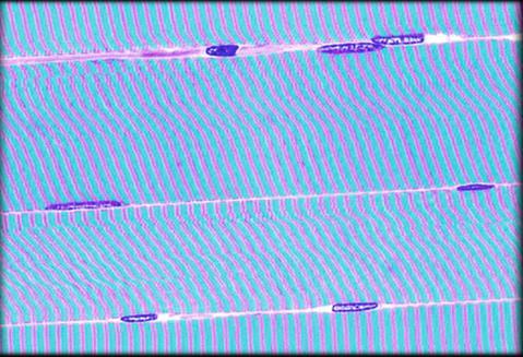
6 Skeletal Muscle Medium Magnification Nuclei of skeletal muscle fiber Skeletal muscle fiber
7 Smooth Muscle Low Magnification Longitudinal smooth muscle fiber Smooth muscle fiber Transverse smooth muscle fiber
8 Smooth Muscle High Magnification Smooth muscle fiber Nuclei of smooth muscle fiber
9 Cardiac Muscle Branched cardiac cell Intercalated disc Nuclei of cardiac cell
10 Cardiac Muscle Intercalated Discs Branched fiber Intercalated discs Nuclei


 Cylindrical and branched (100 500 μm in length, 12 20 μm in diameter) Nucleus Multiple")

 Yes Function Body movement")

11 TABLE 9.1 Comparison of Muscle Types Copyright The McGraw-Hill Companies, Inc. Permission required for reproduction or display. Skeletal Muscle Smooth Muscle Cardiac Muscle Location Attached to bones Walls of hollow organs, blood vessels, eyes, glands, and skin Heart Appearance Cell Shape Very long and cylindrical (1 mm 4 cm, or as much as 30 cm, in length, 10 μm 100 μm in diameter) Spindle-shaped ( μm in length, 5 8 μm in diameter) Cylindrical and branched ( μm in length, μm in diameter) Nucleus Multiple nuclei: peripherally located Single, centrally located Single, centrally located Special Cell-to-Cell Attachments None Gap junctions join some visceral smooth muscle cells together Striations Yes No Yes Control Voluntary and involuntary (reflexes) Involuntary Involuntary Intercalated disks join cells to one another Capable of Spontaneous Contraction No Yes (some smooth muscle) Yes Function Body movement Moving food through the digestive tract, emptying the urinary bladder, regulating blood vessel diameter, changing pupil size, contracting many gland ducts, moving hair, and having many other functions Pumping blood; contractions provide the major force for propelling blood through blood vessels

12 9.3 Skeletal Muscle Structure Composed of muscle cells (fibers), connective tissue, blood vessels, nerves Fibers are long, cylindrical, multinucleated Tend to be smaller diameter in small muscles and larger in large muscles. 1 mm- 4 cm in length Develop from myoblasts; numbers remain constant Striated appearance due to light and dark banding Copyright The McGraw-Hill Companies, Inc. Permission required for reproduction or display. LM 800x Nucleus Striations Skeletal muscle fiber Ed Reschke 9-12


13 Skeletal Muscle High Magnification Nuclei A band I band

14 Skeletal Muscle (striations) Skeletal muscle fiber Nuclei A band I band
15 Connective Tissue Coverings of Muscle Layers Epimysium. C.T. that surrounds a whole muscle (many fascicles) Perimysium. Denser C.T. surrounding a group of muscle fibers. Each group called a fasciculus Endomysium. Loose C.T. with reticular fibers. Muscular fascia: connective tissue sheet External to epimysium Holds muscles together and separates them into functional groups. 9-15


16 Connective Tissue and Fascicles Epimysium Fascicle Perimysium Endomysium Epimysium + Perimysium + Endomysium = Tendon


17 Superficial and Deep Fasciae Superficial Deep 10-17
 Artery Vein Nerve Epimysium (surrounds muscles) Perimysium (surrounds fasciculi) Endomysium (surrounds muscle fibers) Muscle")
18 Copyright The McGraw-Hill Companies, Inc. Permission required for reproduction or display. Muscular fascia (surrounds individual muscles and groups of muscles) Artery Vein Nerve Epimysium (surrounds muscles) Perimysium (surrounds fasciculi) Endomysium (surrounds muscle fibers) Muscle fiber Artery Nerve Vein Fasciculus Capillary Axon of motor neuron Synapse or neuromuscular junction 9-18
19 Nerves and Blood Vessel Supply Copyright The McGraw-Hill Companies, Inc. Permission required for reproduction or display. Artery Vein Nerve Muscular fascia (surrounds individual muscles and groups of muscles) Epimysium (surrounds muscles) Perimysium (surrounds fasciculi) Endomysium (surrounds muscle fibers) Muscle fiber Artery Nerve Vein Fasciculus Motor neurons: stimulate muscle fibers to contract. Nerve cells with cell bodies in brain or spinal cord; axons extend to skeletal muscle fibers through nerves Axons branch so that each muscle fiber is innervated Contact is neuromuscular junction Capillary Capillary beds surround muscle fibers Axon of motor neuron Synapse or neuromuscular junction 9-19
20 Skeletal Muscle Fibers Several nuclei just inside sarcolemma Cell packed with myofibrils within cytoplasm (sarcoplasm) Threadlike Composed of protein threads called myofilaments: thin (actin) and thick (myosin) Sarcomeres: highly ordered repeating units of myofilaments 9-20


 Nuclei Capillary Sarcoplasmic")
 tubule (b) Mitochondrion")

21 Parts of a Muscle Copyright The McGraw-Hill Companies, Inc. Permission required for reproduction or display. Muscle fibers Endomysium (surrounding muscle fibers) Nuclei Capillary Sarcoplasmic reticulum Sarcolemma (plasma membrane) (a) Transverse (T) tubule (b) Mitochondrion Myofibrils Striations I (c) Myofibril Actin myofilament Myosin myofilament I A Z disk Sarcomere Z disk Actin myofilament Myosin myofilament M line Cross-bridge T itin 9-21 Sarcomere
 Z disk Z disk Myosin molecule F actin molecules Actin (thin)")
 Active sites Binds to G actin Binds to Two myosin heavy chains Binds tropomyosin to Ca")
22 Actin and Myosin Myofilaments Copyright The McGraw-Hill Companies, Inc. Permission required for reproduction or display. Sarcomere M line Cross-bridge Actin myofilament Myosin myofilament Titin (a) Z disk Z disk Myosin molecule F actin molecules Actin (thin) myofilament T ropomyosin T roponin Active sites Myosin (thick) myofilament (b) Rod portion Myosin T roponin Tropomyosin Coiled portion of the two α helices Myosin light chains G actin molecules (c) Active sites Binds to G actin Binds to Two myosin heavy chains Binds tropomyosin to Ca 2+ Hinge region of myosin 9-22
23 Actin (Thin) Myofilaments Two strands of fibrous (F) actin form a double helix extending the length of the myofilament; attached at either end at sarcomere. Composed of G actin monomers each of which has an active site Actin site can bind myosin during muscle contraction. Tropomyosin: an elongated protein winds along the groove of the F actin double helix. Troponin is composed of three subunits: one that binds to actin, a second that binds to tropomyosin, and a third that binds to calcium ions. Spaced between the ends of the tropomyosin molecules in the groove between the F actin strands. The tropomyosin/troponin complex regulates the interaction between active sites on G actin and myosin. 9-23

, releasing energy.")

24 Myosin (Thick) Myofilament Many elongated myosin molecules shaped like golf clubs. Molecule consists of myosin heavy chains wound together to form a rod portion lying parallel to the myosin myofilament and two heads that extend laterally. Myosin heads F actin molecules (b) (c) 1. Can bind to active sites on the actin molecules to form cross-bridges. 2. Attached to the rod portion by a hinge region that can bend and straighten during contraction. 3. Are ATPase enzymes: activity that breaks down adenosine triphosphate (ATP), releasing energy. Part of the energy is used to bend the hinge region of the myosin molecule during contraction (a) Actin (thin) myofilament Active sites Tropomyosin Copyright The McGraw-Hill Companies, Inc. Permission required for reproduction or display. Z disk G actin molecules T ropomyosin T roponin Active sites Sarcomere Actin myofilament Myosin myofilament Binds to G actin T roponin M line Cross-bridge Myosin molecule Titin Myosin (thick) myofilament Z disk Rod portion Coiled portion of the two α helices Myosin light chains Binds to Two myosin heavy chains Binds tropomyosin to Ca 2+ Hinge region of myosin Myosin 9-24
25 Sarcomeres: Z Disk to Z Disk Sarcomere: basic functional unit of muscle fiber Z disk: filamentous network of protein. Serves as attachment for actin myofilaments Striated appearance I bands: from Z disks to ends of thick filaments A bands: length of thick filaments H zone: region in A band where actin and myosin do not overlap M line: middle of H zone; delicate filaments holding myosin in place In muscle fibers, A and I bands of parallel myofibrils are aligned. Titin filaments: elastic chains of amino acids; make muscles extensible and elastic 9-25


26 Myofibril Sarcomere H band Sarcomere A band Z disc I band M line
 M line Sarcomere A")
 Cross sections through regions")

27 Copyright The McGraw-Hill Companies, Inc. Permission required for reproduction or display. Sarcomere H zone Z disk Sarcomere Mitochondria Myofibril I band A band (a) M line Sarcomere A band Actin myofilament Myosin myofilament I band Z disk H zone M line Z disk (b) The arrangement of I and A bands, H zones, Z disks, and M lines in sarcomeres (c) Cross sections through regions of the sarcomeres Actin myofilaments only Myosin myofilaments surrounded by actin myofilaments Myosin myofilaments only Don W. Fawcett/Photo Researchers, Inc. Rod portion of myosin myofilaments and M line 9-27
28 9.4 Sliding Filament Model Actin myofilaments sliding over myosin to shorten sarcomeres Actin and myosin do not change length Shortening sarcomeres responsible for skeletal muscle contraction During relaxation, sarcomeres lengthen because of some external force, like contraction of antagonistic muscles Muscles that produce the opposite effect 9-28
 Relaxed sarcomere In a relaxed muscle, the actin and myosin myofilaments overlap slightly, and the H zone is visible.")

 Fully contracted sarcomere In a contracted muscle, the A bands, which are equal to the length of the myosin myofilaments, do not")
29 Sarcomere Shortening Copyright The McGraw-Hill Companies, Inc. Permission required for reproduction or display. Z H I A I (a) Relaxed sarcomere In a relaxed muscle, the actin and myosin myofilaments overlap slightly, and the H zone is visible. The sarcomere length is at its normal resting length. As a muscle contraction is initiated, actin myofilaments slide past the myosin myofilaments, the z disks are brought closer together, and the sarcomere begins to shorten. Z Z I A (b) Fully contracted sarcomere In a contracted muscle, the A bands, which are equal to the length of the myosin myofilaments, do not narrow because the length of the myosin myofilaments does not change, nor does the length of the actin myofilaments. In addition, the ends of the actin myofilaments are pulled to and overlap in the center of the sarcomere, shortening it and the H zone disappears. Z I 9-29

30 9.5 Physiology of Skeletal Muscle Fibers Copyright The McGraw-Hill Companies, Inc. Permission required for reproduction or display Nerve or muscle cell Measuring the resting membrane potential Nervous system controls muscle contractions through action potentials Resting membrane potentials Membrane voltage difference across membranes (polarized) Inside cell more negative due to accumulation of large protein molecules. More K + on inside than outside. K + leaks out but not completely because negative proteins hold some back. Outside cell more positive and more Na + on outside than inside. Na + /K + pump maintains this situation. Must exist for action potential to occur Oscilloscope 0 mv Time 9-30
 Measuring the resting membrane potential 0 mv 50 85 Time Na + K + 1 In a resting cell, there is a higher concentration of K +")
 they are isolated to inside of the cell membrane.")
 are closed.")
31 K + concentration gradient Na + concentration gradient Copyright The McGraw-Hill Companies, Inc. Permission required for reproduction or display. Oscilloscope Nerve or muscle cell (a) Measuring the resting membrane potential 0 mv Time Na + K + 1 In a resting cell, there is a higher concentration of K + (purple circles) inside the cell membrane and a higher concentration of Na + (pink circles) outside the cell membrane. Because the membrane is not permeable to negatively charged proteins (green) they are isolated to inside of the cell membrane. K + leak channel Pr Pr Na + leak channel Pr 2 There are more K + leak channels than Na + leak channels. In the resting cell, only the leak channels are opened; the gated channels (not shown) are closed. Because of the ion concentration differences across the membrane, K + diffuses out of the cell down its concentration gradient and Na + diffuses into the cell down its concentration gradient. The tendency for K + to diffuse out of the cell is opposed by the tendency of the positively charged K + to be attracted back into the cell by the negatively charged proteins. Pr Pr 3 The sodium-potassium pump helps maintain the differential levels of Na + and K + by pumping three Na + out of the cell in exchange for two K + into the cell. The pump is driven by ATP hydrolysis. The resting membrane potential is established when the movement of K + out of the cell is equal to the movement of K + into the cell. Pr ATP ADP Sodiumpotassium pump (b) Generation of the resting membrane potential 9-31
32 Ion Channels Types Ligand-gated. Ligands are molecules that bind to receptors. Receptor: protein or glycoprotein with a receptor site Example: neurotransmitters Gate is closed until neurotransmitter attaches to receptor molecule. When Ach attaches to receptor on muscle cell, Na + gate opens. Na + moves into cell due to concentration gradient Voltage-gated Open and close in response to small voltage changes across plasma membrane Each is specific for certain ions 9-32
33 (mv) (mv) Action Potentials Copyright The McGraw-Hill Companies, Inc. Permission required for reproduction or display. Phases Depolarization: Inside of plasma membrane becomes less negative. If change reaches threshold, depolarization occurs Repolarization: return of resting membrane potential. Note that during repolarization, the membrane potential drops lower than its original resting potential, then rebounds. This is because Na + plus K + together are higher, but then Na/K + pump restores the resting potential All-or-none principle: like camera flash system Propagate: Spread from one location to another. Action potential does not move along the membrane: new action potential at each successive location. Frequency: number of action potential produced per unit of time (a) Polarized Depolarization Time (ms) Threshold Depolarization is a change of the charge difference across the plasma membrane, making the charge inside the cell less negative and the charge outside the plasma membrane less positive. Once threshold is reached, an action potential is produced Depolarization phase 85 - Polarized Depolarization Time (ms) Repolarization phase Threshold (b) During the depolarization phase of the action potential, the membrane potential changes from approximately 85 mv to approximately +20 mv. During the repolarization phase, the inside of the plasma membrane changes from approximately +20 mv back to 85 mv. 9-33


34 K + concentration gradient Na + concentration gradient K + concentration gradient Gated Ion Channels and the Action Potential Copyright The McGraw-Hill Companies, Inc. Permission required for reproduction or display. Extracellular fluid Na + Na + channel K + channel Charge Difference Across the Plasma Membrane 1 Resting membrane potential. Na + channels (pink) and some, but not all, K + channels (purple) are closed. K + diffuses down its concentration gradient through the open K + channels, making the inside of the cell membrane negatively charged compared to the outside Cytoplasm K + Na + Na + 2 Depolarization. Na + channels are open. Na + diffuses down its concentration gradient through the open Na + channels, making the inside of the cell membrane positively charged compared to the outside. 2 Na + diffuse into cell Na + K + diffuse out of cell 3 Repolarization. Na + channels are closed, and Na + movement into the cells stops. More K + channels open. K + movement out of the cell increases, making the inside of the cell membrane negatively charged compared to the outside, once again. K + K K + channels open Na + channels close

35 Action Potential Propagation 1 Copyright The McGraw-Hill Companies, Inc. Permission required for reproduction or display. An action potential in a local area of the plasma membrane is indicated by the orange band. Note the reversal of charge across the membrane Stimulus Muscle fiber 2 The action potential is a stimulus that causes another action potential to be produced in the adjacent plasma membrane The action potential propagates along the plasma membrane (orange arrow)
 Axon branches Neuromuscular junctions Skeletal muscle fiber")
 Synaptic vesicles Synaptic cleft Synapse: axon terminal")
 c: Fred")
36 Neuromuscular Junction Copyright The McGraw-Hill Companies, Inc. Permission required for reproduction or display. Capillary Muscle fiber Sarcoplasmic reticulum (a) Axon branches Neuromuscular junctions Skeletal muscle fiber Myofibrils Axon branch Neuromuscular junction Presynaptic terminal Sarcolemma Sarcoplasm (b) Mitochondrion Presynaptic terminal Postsynaptic membrane (sarcolemma) Synaptic vesicles Synaptic cleft Synapse: axon terminal resting in an invagination of the sarcolemma Neuromuscular junction (NMJ): Presynaptic terminal: axon terminal with synaptic vesicles Synaptic cleft: space Postsynaptic membrane or motor end-plate sarcolemma (c) c: Fred Hossler/Visuals Unlimited 9-36


37 Neuromuscular Junction High Magnification Skeletal muscle fiber Axon of motor nerve Motor end plate
38 Function of Neuromuscular Junction Synaptic vesicles Neurotransmitter: substance released from a presynaptic membrane that diffuses across the synaptic cleft and stimulates (or inhibits) the production of an action potential in the postsynaptic membrane. Acetylcholine Acetylcholinesterase: A degrading enzyme in synaptic cleft. Prevents accumulation of ACh 9-38


39 1 An action potential (orange arrow) arrives at the presynaptic terminal and causes voltage-gated Ca 2+ channels in the presynaptic membrane to open. Copyright The McGraw-Hill Companies, Inc. Permission required for reproduction or display. 2 Calcium ions enter the presynaptic terminal and initiate the release of the neurotransmitter acetylcholine (ACh) from synaptic vesicles ACh is released into the synaptic cleft by exocytosis. ACh diffuses across the synaptic cleft and binds to ligand-gated Na + channels on the postsynaptic membrane. Ligand-gated Na + channels open and Na + enters the postsynaptic cell, causing the postsynaptic membrane to depolarize. If depolarization passes threshold, an action potential is generated along the postsynaptic membrane. Voltage-gated Ca 2+ channel Ca 2+ Synaptic cleft 1 Ca 2+ Synaptic vesicles ACh 9 Presynaptic terminal Acetic acid Acetic acid Postsynaptic membrane 6 ACh unbinds from the ligand-gated Na + channels, which then close. 2 Choline The enzyme acetylcholinesterase, which is attached to the postsynaptic membrane, removes acetylcholine from the synaptic cleft by breaking it down into acetic acid and choline. Choline is symported with Na + into the presynaptic terminal, where it can be recycled to make ACh. Acetic acid diffuses away from the synaptic cleft. ACh is reformed within the presynaptic terminal using acetic acid generated from metabolism and from choline recycled from the synaptic cleft. Ach is then taken up by synaptic vesicles. Action potential Ligand-gated Na + channel (open) 3 Na + ACh Choline 5 Na + 4 ACh receptor site Ligand-gated Na + channel (closed) 6 7 Action potential Acetylcholinesterase 9-39

 Mitochondria of synaptic")
40 Neuromuscular Junction TEM: High Magnification Primary synaptic cleft Synaptic vesicles of synaptic terminal Secondary synaptic cleft (junctional folds) Mitochondria of synaptic terminal
 Terminal cisterna Capillary Myofibril Mitochondrion A band I band Sarcolemma Mechanism where an action potential causes muscle fiber contraction")
41 Excitation-Contraction Coupling Sarcoplasmic reticulum Triad Copyright The McGraw-Hill Companies, Inc. Permission required for reproduction or display. Terminal cisterna Transverse tubule (T tubule) Terminal cisterna Capillary Myofibril Mitochondrion A band I band Sarcolemma Mechanism where an action potential causes muscle fiber contraction Involves Sarcolemma Transverse (T) tubules: invaginations of sarcolemma Terminal cisternae Sarcoplasmic reticulum: smooth ER Triad: T tubule, two adjacent terminal cisternae Ca 2+ Troponin 9-41


42 Action Potentials and Muscle Contraction Copyright The McGraw-Hill Companies, Inc. Permission required for reproduction or display. 1 1 An action potential that was produced at the neuromuscular junction is propagated along the sarcolemma of the skeletal muscle. The depolarization also spreads along the membrane of the T tubules. Action potential Sarcolemma Ca 2+ 2 The depolarization of the T tubule causes gated Ca 2+ channels in the sarcoplasmic reticulum to open, resulting in an increase in the permeability of the sarcoplasmic reticulum to Ca 2+, especially in the terminal cisternae. Calcium ions then diffuse from the sarcoplasmic reticulum into the sarcoplasm. Sarcoplasmic reticulum Actin myofilament T tubule 2 Ca 2+ Sarcomere in myofibril Myosin myofilament 3 Tropomyosin Ca 2+ Troponin Ca 2+ bind to troponin. Actin myofilament 3 Calcium ions released from the sarcoplasmic reticulum bind to troponin molecules. The troponin molecules bound to G actin molecules are released, causing tropomyosin to move, and to expose the active sites on G actin. Active sites not exposed G actin molecule Myosin myofilament 4 Once active sites on G actin molecules are exposed, the heads of the myosin myofilaments bind to them to form cross-bridges. 4 Active site Ca 2+ Cross-bridge Active sites exposed 9-42

, and energy is stored in the heads of the myosin molecules.")

43 Cross-Bridge Movement Copyright The McGraw-Hill Companies, Inc. Permission required for reproduction or display. Sarcomere Actin myofilament Myosin myofilament Z disk Z disk Troponin Ca 2+ Ca 2+ T ropomyosin Active site P ADP 1 Exposure of active sites. Before crossbridges cycle, Ca 2+ bind to the troponins and the tropomyosins move, exposing active sites on actin myofilaments. P ADP Cross-bridge ADP 6 Recovery stroke. The heads of the myosin molecules return to their resting position (small dark blue arrow), and energy is stored in the heads of the myosin molecules. If Ca 2+ is still attached to troponin, cross-bridge formation and movement are repeated (return to step 2). This cycle occurs many times during a muscle contraction. Not all cross-bridges form and release simultaneously. 2 Cross-bridge formation. The myosin heads bind to the exposed active sites on the actin myofilaments to form cross-bridges, and phosphates are released from the myosin heads. P 5 ADP P Hydrolysis of ATP. The myosin ATPase portion of the myosin heads split ATP into ADP and phosphate (P), which remain attached to the myosin heads. 3 Power stroke. Energy stored in the myosin heads is used to move the myosin heads (small dark blue arrow), causing the actin myofilaments to slide past the myosin myofilaments (dark blue arrow), and ADP molecules are released from the myosin heads (black arrow). ADP 4 Cross-bridge release. An ATP molecule binds to each of the myosin heads, causing them to detach from the actin. 9-43

44 Copyright The McGraw-Hill Companies, Inc. Permission required for reproduction or display. 1 AP 2 Ca 2+ 5 T tubule Sarcoplasmic reticulum 3 Ach 4 Sarcolemma Ca 2+ Na An action potential travels along an axon membrane to a neuromuscular junction. 2 Ca 2+ channels open and Ca 2+ enters the presynaptic terminal. 3 Acetylcholine is released from presynaptic vesicles. 4 Acetylcholine stimulates Na + channels on the postsynaptic membrane to open Na + diffuses into the muscle fiber, initiating an action potential that travels along the sarcolemma and T tubule membranes. Action potentials in the T tubules cause the sarcoplasmic reticulum to release Ca 2+. On the actin, Ca 2 binds to troponin, which moves tropomyosin and exposes myosin head attachment sites. ATP molecules on myosin heads are broken down to ADP and P, which releases energy needed to move the myosin heads. The heads of the myosin myofilaments bend, causing the actin to slide past the myosin. As long as Ca 2+ is present, the cycle repeats. Ca ADP 8 P 9-44
45 Muscle Relaxation Ca 2+ moves back into sarcoplasmic reticulum by active transport. Requires energy Ca 2+ moves away from troponintropomyosin complex Complex re-establishes its position and blocks binding sites. 9-45

46 Tension 9.6 Physiology of Skeletal Muscle Copyright The McGraw-Hill Companies, Inc. Permission required for reproduction or display. Lag Contraction phase phase Stimulus applied Time Relaxation phase Muscle Twitch Muscle contraction in response to a stimulus that causes action potential in one or more muscle fibers Phases Lag or latent Contraction Relaxation 9-46

47 Motor Units Motor units: a single motor neuron and all muscle fibers innervated by it Motor Unit numbers: Large muscles have motor units with many muscle fibers. Small muscles that make delicate movements contain motor units with few muscle fibers Copyright The McGraw-Hill Companies, Inc. Permission required for reproduction or display. Axon of motor neuron Axon branches Myofibrils Axons of motor neurons Neuromuscular junctions Muscle fibers (a) Neuromuscular junction Capillary Muscle fibers (b) b: Don Fawcett/Photo Researchers, Inc. 9-47
48 Stimulus Strength and Motor Unit Response All-or-none law for muscle fibers Contraction of equal force in response to each action potential Sub-threshold stimulus: no action potential; no contraction Threshold stimulus: action potential; contraction Stronger than threshold; action potential; contraction equal to that with threshold stimulus Copyright The McGraw-Hill Companies, Inc. Permission required for reproduction or display. Axon of motor neuron Axon branches Myofibrils Axons of motor neurons Neuromuscular junctions Muscle fibers (a) Neuromuscular junction Capillary Muscle fibers (b) b: Don Fawcett/Photo Researchers, Inc. 9-48

49 Tension Stimulus Strength and Motor Unit Response Strength of contraction is graded: ranges from weak to strong depending on stimulus strength Multiple motor unit summation: strength of contraction depends upon recruitment of motor units. A muscle has many motor units Submaximal stimuli Maximal stimulus Supramaximal stimuli Copyright The McGraw-Hill Companies, Inc. Permission required for reproduction or display. Increasing stimulus strengths Subthreshold stimulus (no motor units respond) Threshold stimulus (one motor unit responds) Submaximal stimuli (increasing numbers of motor units respond) Maximal stimulus (all motor units respond) Supramaximal stimuli (all motor units respond) 9-49 Time
 Graded response Occurs in muscle rested for prolonged period Each subsequent contraction is stronger than previous until all equal after few stimuli Possible")
50 Tension Treppe Copyright The McGraw-Hill Companies, Inc. Permission required for reproduction or display. Stimuli of constant strength Time (ms) Graded response Occurs in muscle rested for prolonged period Each subsequent contraction is stronger than previous until all equal after few stimuli Possible explanation: more and more Ca 2+ remains in sarcoplasm and is not all taken up into the sarcoplasmic reticulum 9-50

51 Tension Stimulus Frequency and Whole Muscle Contraction As the frequency of action potentials increase, the frequency of contraction increases Incomplete tetanus: muscle fibers partially relax between contraction Complete tetanus: no relaxation between contractions Multiple-wave summation: muscle tension increases as contraction frequencies increase Copyright The McGraw-Hill Companies, Inc. Permission required for reproduction or display. Twitch Incomplete tetanus Complete tetanus Frequency 1 Frequency 2 Frequency 3 Frequency 4 Time (ms) 9-51
52 Muscle Length versus Tension Active tension: force applied to an object to be lifted when a muscle contracts Stretched muscle- not enough cross-bridging Crumpled muscle- myofilaments crumpled, crossbridges can't contract Passive tension: tension applied to load when a muscle is stretched but not stimulated Total tension: active plus passive 9-52


53 Tension (% of maximum) Tension (% of maximum) Tension (% of maximum) Tension (% of maximum) Copyright The McGraw-Hill Companies, Inc. Permission required for reproduction or display. Optimal sarcomere length At the normal resting length of a muscle, the sarcomeres are also at an optimal length. The muscle produces maximum tension in response to a maximal stimulus at this length % of resting sarcomere length Muscle length 1 Muscle length 2 Muscle length Time Time Time At muscle/sarcomere length 1, the muscle is not stretched, and the tension produced when the muscle contracts is small because there is too much overlap between actin and myosin myofilaments. The myosin myofilaments run into the Z disks, and the actin myofilaments interfere with each other at the center of the sarcomere, reducing thenumber of Cross-bridges that can form. At muscle/sarcomere length 2, the muscle is optimally stretched, and the tension produced when the muscle contracts is maximal because there is optimal overlap of actin and myosin myofilaments, so the number of crossbridges that can form is maximal. At muscle/sarcomere length 3, the muscle is stretched severely, and the tension produced when the muscle contracts is small because there is little overlap between actin and myosin myofilaments, and few cross-bridges can form. 9-53
54 Muscle Contractions Isometric: no change in length but tension increases Postural muscles of body Isotonic: change in length but tension constant Concentric: overcomes opposing resistance and muscle shortens Eccentric: tension maintained but muscle lengthens Muscle tone: constant tension by muscles for long periods of time 9-54

55 Copyright The McGraw-Hill Companies, Inc. Permission required for reproduction or display. TABLE 9.2 Types of Physiological Muscle Responses Physiological Response Multiple-motor-unit summation Multiple-wave summation Characteristics Each motor unit responds in an all-or-none fashion. A whole muscle is capable of producing an increasing amount of tension as the number of motor units stimulated increases. Summation results when many action potentials are produced in a muscle fiber. Contraction occurs in response to the first action potential, but there is not enough time for relaxation to occur between action potentials. Because each action potential causes the release of Ca 2+ from the sarcoplasmic reticulum, the ion levels remain elevated in the sarcoplasm to produce a tetanic contraction. The tension produced as a result of multiple-wave summation is greater than the tension produced by a single muscle twitch. The increased tension results from the greater concentration of Ca 2+ in the sarcoplasm and the stretch of the elastic components of the muscle early in contraction. Treppe Tetanus of muscles Isometric contractions Isotonic contractions Tension produced increases for the first few contractions in response to a maximal stimulus at a low frequency in a muscle that has been at rest for some time. Increased tension may result from the accumulation of small amounts of Ca 2+ in the sarcoplasm for the first few contractions or from an increasing rate of enzyme activity. Tetanus of muscles results from multiple-wave summation; frequency of stimulus is higher than for treppe. Incomplete tetanus occurs when the action potential frequency is low enough to allow partial relaxation of the muscle fibers. Complete tetanus occurs when the action potential frequency is high enough that no relaxation of the muscle fibers occurs. A muscle produces increasing tension as it remains at a constant length; this is characteristic of postural muscles that maintain a constant tension without changing their length. A muscle produces a constant tension and shortens during contraction; this is characteristic of finger and hand movements. In concentric contractions, a muscle produces tension as it shortens; this is characteristic of biceps brachii curl exercises. In eccentric contractions, a muscle produces tension as it resists lengthening; this is characteristic of slowly descending a flight of stairs. 9-55
56 9.7 Muscle Fatigue Decreased capacity to work and reduced efficiency of performance Types Psychological: depends on emotional state of individual Muscular: results from ATP depletion Synaptic: occurs in NMJ due to lack of acetylcholine 9-56
57 Physiological Contracture and Rigor Mortis Physiological contracture: state of fatigue where due to lack of ATP neither contraction nor relaxation can occur Rigor mortis: development of rigid muscles several hours after death. Ca 2+ leaks into sarcoplasm and attaches to myosin heads and crossbridges form. Rigor ends as tissues start to deteriorate. 9-57
58 9.8 Energy Sources ATP provides immediate energy for muscle contractions. Produced from three sources Creatine phosphate During resting conditions stores energy to synthesize ATP Anaerobic respiration Occurs in absence of oxygen and results in breakdown of glucose to yield ATP and lactic acid Aerobic respiration Requires oxygen and breaks down glucose to produce ATP, carbon dioxide and water More efficient than anaerobic Oxygen debt: oxygen taken in by the body, above that required for resting metabolism after exercise. ATP produced from anaerobic sources contributes 9-58

59 Copyright The McGraw-Hill Companies, Inc. Permission required for reproduction or display. TABLE 9.3 Sources of ATP in Muscles Creatine Phosphate Creatine phosphate Anaerobic Respiration Glucose Aerobic Respiration Glucose 1 ATP Glycolysis 2 ATP Glycolysis 2ATP Metabolic Process Creatine 2 pyruvic acid Fatty acids Amino acids 2 pyruvic acid Electron-transport chain 2 lactic acid Citric acid cycle 34 ATP Energy Source Creatine phosphate Glucose Glucose, fatty acids, aminoacids Oxygen Required No No Yes ATP Yield 1 per creatine phosphate 2 per glucose molecule Up to 36 per glucose molecule Duration of Energy Supply Up to 10 seconds Upto 3 minutes Hours Type of Work Supported Moderate exercise and extreme exercise Extreme exercise Resting and all exercise 9-59
60 9.9 Slow and Fast Fibers Slow-twitch oxidative Contract more slowly, smaller in diameter, better blood supply, more mitochondria, more fatigue-resistant than fasttwitch, large amount of myoglobin. Postural muscles, more in lower than upper limbs. Dark meat of chicken. Fast-twitch Respond rapidly to nervous stimulation, contain myosin that can break down ATP more rapidly than that in Type I, less blood supply, fewer and smaller mitochondria than slowtwitch Lower limbs in sprinter, upper limbs of most people. White meat in chicken. Comes in oxidative and glycolytic forms 9-60
61 Slow and Fast Fibers Distribution of fast-twitch and slow-twitch Most muscles have both but varies for each muscle Effects of exercise: change in size of muscle fibers Hypertrophy: increase in muscle size Increase in myofibrils Increase in nuclei due to fusion of satellite cells Increase in strength due to better coordination of muscles, increase in production of metabolic enzymes, better circulation, less restriction by fat Atrophy: decrease in muscle size Reverse except in severe situations where cells die 9-61

62 9-62
 (Type I) Fast-Twitch Oxidative Glycolytic (FOG) (Type II a) Fast-Twitch Glycolytic (FG) (Type II b) Myoglobin Content High")
63 Copyright The McGraw-Hill Companies, Inc. Permission required for reproduction or display. TABLE 9.4 Characteristics of Skeletal Muscle Fiber Types Slow-Twitch Oxidative (SO) (Type I) Fast-Twitch Oxidative Glycolytic (FOG) (Type II a) Fast-Twitch Glycolytic (FG) (Type II b) Myoglobin Content High High Low Mitochondria Many Many Few Capillaries Many Many Few Metabolism High aerobic capacity, low anaerobic capacity Intermediate aerobic capacity, high anaerobic capacity Low aerobic capacity, highest anaerobic capacity Fatigue Resistance High Intermediate Low Myosin ATPase Activity Slow Fast Fast Glycogen Concentration Low High High Location Where Fibers Are Most Abundant Generally in postural muscles and more in lower limbs than upper limbs Generally in lower limbs Generally in upper limbs Functions Maintenance of posture and performance of endurance activities Endurance activities in endurancetrained muscles Rapid, intense movements of short duration 9-63
64 9.10 Heat Production Exercise: metabolic rate and heat production increase. Post-exercise: metabolic rate stays high due to oxygen debt. Excess heat lost because of vasodilation and sweating Shivering: uncoordinated contraction of muscle fibers resulting in shaking and heat production 9-64
65 9.11 Smooth Muscle Not striated, fibers smaller than those in skeletal muscle Spindle-shaped; single, central nucleus More actin than myosin Caveolae: indentations in sarcolemma; may act like T tubules Dense bodies instead of Z disks as in skeletal muscle; have noncontractile intermediate filaments Ca 2+ required to initiate contractions; binds to calmodulin which regulates myosin kinase. Cross-bridging occurs Relaxation: caused by enzyme myosin phosphatase 9-65


66 Copyright The McGraw-Hill Companies, Inc. Permission required for reproduction or display. Actin myofilament Myosin myofilament Copyright The McGraw-Hill Companies, Inc. Permission required for reproduction or display. Dense bodies in sarcoplasm Intermediate filaments Contraction Dense area attached to sarcolemma LM 800x Nuclei of smooth muscle cells Myofilaments Victor Eroschenko 9-66


67 Smooth Muscle Low Magnification Longitudinal smooth muscle fiber Smooth muscle fiber Transverse smooth muscle fiber


68 Smooth Muscle High Magnification Smooth muscle fiber Nuclei of smooth muscle fiber
69 Types of Smooth Muscle Visceral or unitary: cells in sheets; function as a unit Numerous gap junctions; waves of contraction Often autorhythmic Multiunit: cells or groups of cells act as independent units Sheets (blood vessels); bundles (arrector pili and iris); single cells (capsule of spleen) 9-69

70 (mv) (mv) (mv) Electrical Properties of Smooth Muscle Slow waves of depolarization and repolarization transferred from cell to cell Depolarization caused by spontaneous diffusion of Na + and Ca 2+ into cell Does not follow all-or-none law May have pacemaker cells Contraction regulated by nervous system and by hormones Copyright The McGraw-Hill Companies, Inc. Permission required for reproduction or display Time (ms) (a) Slow waves of depolarization Time (ms) (b) Action potentials in smooth muscle superimposed on a slow wave of depolarization Time (ms) (c) Action potential with prolonged depolarization (plateau) 9-70
71 Functional Properties of Smooth Muscle Some visceral muscle exhibits autorhythmic contractions Tends to contract in response to sudden stretch but not to slow increase in length Exhibits relatively constant tension: smooth muscle tone Amplitude of contraction remains constant although muscle length varies 9-71
72 Regulation of Smooth Muscle Innervated by autonomic nervous system Neurotransmitters are acetylcholine and norepinephrine Hormones important as epinephrine and oxytocin Receptors present on plasma membrane; which neurotransmitters or hormones bind determines response 9-72

 Ca 2+ channel (closed) Myosin kinase (inactive) 2 Ca 2+ channel (open) 2 3 4 An α subunit opens the Ca 2+ channel in the plasma membrane, or depolarization opens Ca 2+ channels.")
73 Copyright The McGraw-Hill Companies, Inc. Permission required for reproduction or display. Hormone Ca 2+ Hormone receptor 1 1 A hormone combines with a hormone receptor and activates a G protein mechanism, or depolarization of the plasma membrane occurs. G protein complex separates from receptor. GDP GTP GTP replaces GDP on subunit. Calmodulin (inactive) Ca 2+ channel (closed) Myosin kinase (inactive) 2 Ca 2+ channel (open) An α subunit opens the Ca 2+ channel in the plasma membrane, or depolarization opens Ca 2+ channels. Calcium ions diffuse through the Ca 2+ channels and combine with calmodulin. Calmodulin with a Ca 2+ bound to it binds with myosin kinase and activates it. Activated myosin kinase attaches phosphate from ATP to myosin heads to activate the contractile process. Calmodulin (active) 3 Myosin b α subunit with GTP binds to Ca 2+ channel and causes it to open. P ATP α GTP Ca 2+ bound to calmodulin Myosin kinase (active) ADP 4 5 A cycle of cross-bridge formation, movement, detachment, and cross-bridge formation occurs. Myosin P 5 Actin Actin 6 Relaxation occurs when myosin phosphatase removes phosphate from myosin. Myosin Myosin phosphatase 9-73 P 6
74 9.12 Cardiac Muscle Found only in heart Striated Each cell usually has one nucleus Has intercalated disks and gap junctions Autorhythmic cells Action potentials of longer duration and longer refractory period Ca 2+ regulates contraction 9-74


75 Cardiac Muscle Branched cardiac cell Intercalated disc Nuclei of cardiac cell


76 Cardiac Muscle Intercalated Discs Branched fiber Intercalated discs Nuclei


77 Cardiac Muscle Histology Cardiac cell Nucleus Branched cell Intercalated discs Perinuclear cytoplasm
78 9.13 Effects of Aging on Skeletal Muscle Reduced muscle mass Increased time for muscle to contract in response to nervous stimuli Reduced stamina Increased recovery time Loss of muscle fibers Decreased density of capillaries in muscle 9-78
Chapter 10 Muscle Tissue Lecture Outline
 Chapter 10 Muscle Tissue Lecture Outline Muscle tissue types 1. Skeletal muscle = voluntary striated 2. Cardiac muscle = involuntary striated 3. Smooth muscle = involuntary nonstriated Characteristics
Muscle and Muscle Tissue
 Muscle and Muscle Tissue Make up about half of total body mass Exerts force by converting chemical energy, ATP, to mechanical energy Muscle tissue is classified based on Shape Number and position of nuclei
MUSCLE TISSUE (MUSCLE PHYSIOLOGY) PART I: MUSCLE STRUCTURE
 PART I: MUSCLE STRUCTURE") PART I: MUSCLE STRUCTURE Muscle Tissue A primary tissue type, divided into: skeletal muscle cardiac muscle smooth muscle Functions of Skeletal Muscles Produce skeletal movement Maintain body position Support
PART I: MUSCLE STRUCTURE Muscle Tissue A primary tissue type, divided into: skeletal muscle cardiac muscle smooth muscle Functions of Skeletal Muscles Produce skeletal movement Maintain body position Support
1/4/2017. Introduction. Connective Tissue Coverings. 9.1: Structure of a Skeletal Muscle. Skeletal Muscle Fibers. Connective Tissue Coverings
 Introduction Chapter 09 Lecture Outline See separate PowerPoint slides for all figures and tables preinserted into PowerPoint without notes. Copyright McGraw-Hill Education. Permission required for reproduction
Chapter 9 - Muscle and Muscle Tissue
 Chapter 9 - Muscle and Muscle Tissue I. Overview of muscle tissue A. Three muscle types in the body: B. Special characteristics 1. Excitability: able to receive and respond to a stimulus 2. Contractility:
Chapter 10 Muscle Tissue and Physiology Chapter Outline
 Chapter 10 Muscle Tissue and Physiology Chapter Outline Module 10.1 Overview of muscle tissue (Figures 10.1 10.2) A. Types of Muscle Tissue (Figure 10.1) 1. The three types of cells in muscle tissue are,,
Skeletal Muscle. Connective tissue: Binding, support and insulation. Blood vessels
 Chapter 12 Muscle Physiology Outline o Skeletal Muscle Structure o The mechanism of Force Generation in Muscle o The mechanics of Skeletal Muscle Contraction o Skeletal Muscle Metabolism o Control of Skeletal
Anatomy and Physiology 1 Chapter 10 self quiz Pro, Dima Darwish,MD.
 Anatomy and Physiology 1 Chapter 10 self quiz Pro, Dima Darwish,MD. 1) Which of the following is a recognized function of skeletal muscle? A) produce movement B) maintain posture C) maintain body temperature
Muscle Tissue. PowerPoint Lecture Presentations prepared by Jason LaPres. Lone Star College North Harris Pearson Education, Inc.
 10 Muscle Tissue PowerPoint Lecture Presentations prepared by Jason LaPres Lone Star College North Harris An Introduction to Muscle Tissue Muscle Tissue A primary tissue type, divided into: Skeletal muscle
Fig Copyright McGraw-Hill Education. Permission required for reproduction or display. Nucleus. Muscle fiber. Endomysium. Striations.
 Fig. 11.1 Nucleus Muscle fiber Endomysium Striations Ed Reschke 1 Fig. 11.2 Muscle fiber Nucleus I band A band Z disc Mitochondria Openings into transverse tubules Sarcoplasmic reticulum Triad: Terminal
MODULE 6 MUSCLE PHYSIOLOGY
 MODULE 6 MUSCLE PHYSIOLOGY III SEMESTER BOTANY Syllabi: Striated, Non striated and Cardiac muscle, Ultra structure of striated muscle fibre, Mechanism of muscle contraction, Threshold and spike potential,
Lecture Overview. Muscular System. Marieb s Human Anatomy and Physiology. Chapter 9 Muscles and Muscle Tissue Lecture 16
 Marieb s Human Anatomy and Physiology Marieb Hoehn Chapter 9 Muscles and Muscle Tissue Lecture 16 1 Lecture Overview Types, characteristics, functions of muscle Structure of skeletal muscle Mechanism of
MUSCULAR TISSUE. Dr. Gary Mumaugh
 MUSCULAR TISSUE Dr. Gary Mumaugh MUSCLE OVERVIEW The three types of muscle tissue are skeletal, cardiac, and smooth These types differ in structure, location, function, and means of activation FUNCTIONAL
2/19/2018. Learn and Understand:
 Muscular System with Special Emphasis on Skeletal Muscle Anatomy and Physiology Learn and Understand: The definition of cell changes again The contractile unit of muscle is the sarcomere. ATP and Ca 2+
About This Chapter. Skeletal muscle Mechanics of body movement Smooth muscle Cardiac muscle Pearson Education, Inc.
 About This Chapter Skeletal muscle Mechanics of body movement Smooth muscle Cardiac muscle Skeletal Muscle Usually attached to bones by tendons Origin: closest to the trunk or to more stationary bone Insertion:
Muscle Tissue- 3 Types
 AN INTRODUCTION TO MUSCLE TISSUE Muscle Tissue- 3 Types Skeletal muscle (focus on these) Cardiac muscle Smooth muscle FUNCTIONS OF SKELETAL MUSCLES Produce movement of the skeleton Maintain posture and
Muscle Physiology. Dr. Ebneshahidi Ebneshahidi
 Muscle Physiology Dr. Ebneshahidi Skeletal Muscle Figure 9.2 (a) Functions of the muscular system 1. Locomotion body movements are due to skeletal muscle contraction. 2. Vasoconstriction and vasodilatation
Muscles and Muscle Tissue
 1 Muscles and Muscle Tissue Chapter 9 2 Overview of Muscle Tissues Compare and Contrast the three basic types of muscle tissue List four important functions of muscle tissue 3 Muscle Terminology Muscle
Chapter 10: Muscle Tissue
 Chapter 10: Muscle Tissue Muscle is one of the 4 primary types of tissue. It is subdivided into skeletal, cardiac and smooth muscle. I. Skeletal Muscle Tissue and the Muscular System, p. 284 Objective
Muscle Tissue. Alternating contraction and relaxation of cells. Chemical energy changed into mechanical energy
 Know these muscles Muscle Tissue Alternating contraction and relaxation of cells Chemical energy changed into mechanical energy 3 Types of Muscle Tissue Skeletal muscle attaches to bone, skin or fascia
Chapter 8 Notes. Muscles
 Chapter 8 Notes Muscles 8.1 Intro Three muscle types Skeletal Smooth cardiac 8.2 Structure of Skeletal Muscle Composition Skeletal muscle tissue Nervous tissue Blood Connective tissue Connective tissue
Hole s Human Anatomy and Physiology Eleventh Edition. Mrs. Hummer. Chapter 9 Muscular System
 Hole s Human Anatomy and Physiology Eleventh Edition Mrs. Hummer Chapter 9 Muscular System 1 Chapter 9 Muscular System Skeletal Muscle usually attached to bones under conscious control striated Three Types
Muscle Tissue. Dr. Heba Kalbouneh Associate Professor of Anatomy and Histology
 Muscle Tissue Dr. Heba Kalbouneh Associate Professor of Anatomy and Histology Functions of muscle tissue Movement Maintenance of posture Joint stabilization Heat generation Tendon Belly Tendon Types of
Smooth Cardiac Skeletal Location Around tubes Heart tissue attached to skeleton Moves stuff thru Heart beat pumps Moves body parts
 Biology 067 - Muscular system A. Type of muscles: Smooth Cardiac Skeletal Location Around tubes Heart tissue attached to skeleton Function Moves stuff thru Heart beat pumps Moves body parts tubes blood
Microanatomy of Muscles. Anatomy & Physiology Class
 Microanatomy of Muscles Anatomy & Physiology Class Three Main Muscle Types Objectives: By the end of this presentation you will have the information to: 1. 2. 3. 4. 5. 6. Describe the 3 main types of muscles.
Muscle Tissue. C h a p t e r. PowerPoint Lecture Slides prepared by Jason LaPres Lone Star College - North Harris
 C h a p t e r 10 Muscle Tissue PowerPoint Lecture Slides prepared by Jason LaPres Lone Star College - North Harris Copyright 2009 Pearson Education, Inc., publishing as Pearson Benjamin Cummings An Introduction
MUSCULAR SYSTEM CHAPTER 09 BIO 211: ANATOMY & PHYSIOLOGY I
 1 BIO 211: ANATOMY & PHYSIOLOGY I 1 CHAPTER 09 MUSCULAR SYSTEM Part 2 of 2 Dr. Dr. Lawrence G. G. Altman www.lawrencegaltman.com Some illustrations are courtesy of McGraw-Hill. Some illustrations are courtesy
Chapter 10! Chapter 10, Part 2 Muscle. Muscle Tissue - Part 2! Pages !
 ! Chapter 10, Part 2 Muscle Chapter 10! Muscle Tissue - Part 2! Pages 308-324! SECTION 10-5! Sarcomere shortening and muscle fiber stimulation produce tension! 2! Tension Production - Muscle FIBER! All-or-none
BIOH111. o Cell Module o Tissue Module o Integumentary system o Skeletal system o Muscle system o Nervous system o Endocrine system
 BIOH111 o Cell Module o Tissue Module o Integumentary system o Skeletal system o Muscle system o Nervous system o Endocrine system Endeavour College of Natural Health endeavour.edu.au 1 TEXTBOOK AND REQUIRED/RECOMMENDED
Page 1. Chapter 9: Muscle Tissue. Types of Muscle Tissue: Skeletal Muscle Cardiac Muscle Smooth Muscle. Gross Anatomy of Muscle:
 1 Chapter 9: Muscle Tissue Types of Muscle Tissue: Skeletal Muscle Cardiac Muscle Smooth Muscle Characteristics: Attaches to skeleton Voluntary control Striated / multi-nucleated Characteristics: Composes
Skeletal Muscle Tissue
 Functions of Skeletal Muscle Skeletal Muscle Tissue Keri Muma Bio 6 Movement muscles attach directly or indirectly to bone, pull on bone or tissue when they contract Maintain posture / body position muscles
Chapter 10 Muscle Tissue
 Chapter 10 Muscle Tissue Skeletal muscle, cardiac muscle and smooth muscle. Differ in their microscopic anatomy, location and how they are controlled by the endocrine and nervous system. 3 Types of Muscle
Concept 50.5: The physical interaction of protein filaments is required for muscle function
 Concept 50.5: The physical interaction of protein filaments is required for muscle function Muscle activity is a response to input from the nervous system The action of a muscle is always to contract Vertebrate
Muscle Tissue. Muscle Tissue Outline. General Function of Muscle Tissue
 Muscle Tissue Muscle Tissue Outline General Functions of Muscle Tissue Characteristics of Muscle Tissue Classification of Muscle Tissue Skeletal Muscle Structure and Function Muscle Energetics Muscle Mechanics
Page 1. Chapter 9: Muscle Tissue. Types of Muscle Tissue: Skeletal Muscle Cardiac Muscle Smooth Muscle. Characteristics of Muscle:
 1 Chapter 9: Muscle Tissue Muscle little mouse Types of Muscle Tissue: Skeletal Muscle Cardiac Muscle Smooth Muscle Characteristics: Attaches to skeleton Voluntary control Striated / multi-nucleated Characteristics:
Chapter 10 -Muscle Tissue
 Chapter 10 -Muscle Tissue Muscles: 1. Overview of Muscle Tissue A. Review 5 functions of muscle tissue. B. Review the 5 properties of muscle tissue. WHICH do they share with nervous tissue? (2, plus the
Outline. Bio 105: Muscular System. Muscular System. Types of Muscles. Smooth Muscle. Cardiac Muscle 4/6/2016
 Outline Bio 105: Muscular System Lecture 11 Chapter 6 Characteristics of muscles 3 types of muscles Functions of muscles Structure of skeletal muscles Mechanics of muscle contraction Energy sources for
1. Locomotion. 2. Repositioning. 3. Internal movement
 MUSCLE and MOVEMENT Chapters 20, 8, 21 1. Locomotion A. Movement B. 2. Repositioning A. 3. Internal movement A. 1 Muscle Cells 1. Contractile 2. Myocytes 3. Striated A. Skeletal B. Cardiac 4. Smooth 5.
1. Locomotion. 2. Repositioning. 3. Internal movement
 MUSCLE and MOVEMENT Chapters 20, 8, 21 1. Locomotion A. Movement B. 2. Repositioning A. 3. Internal movement A. Muscle Cells 1. Contractile 2. Myocytes 3. Striated A. Skeletal B. Cardiac 4. Smooth 5. Striated
CHAPTER 6 2/9/2016. Learning Objectives List the four traits that all muscle types have in common.
 Learning Objectives List the four traits that all muscle types have in common. CHAPTER 6 The Muscular System Demonstrate and explain the use of antagonistic muscle pairs. Describe the attachment of muscle
Bio 103 Muscular System 61
 61 Lecture Outline: MUSCULAR SYSTEM [Chapter 9] A. Functions of Skeletal Muscle 1. Movement 2. Maintain posture 3. Support 4. Guard openings 5. Maintain body temperature (thermogenesis) B. Muscle Tissue
Ch.10 Muscle Tissue. Copyright 2009, John Wiley & Sons, Inc.
 Ch.10 Muscle Tissue Preview Chapter 10 In groups we will define the following terms 1. Skeletal muscle 2. Smooth muscle 3. Cardiac muscle 4. Sarcomere 5. Myofibril 6. Myofilament 7. Sarcoplasmic reticulum
Muscle and Neuromuscular Junction. Peter Takizawa Department of Cell Biology
 Muscle and Neuromuscular Junction Peter Takizawa Department of Cell Biology Types and structure of muscle cells Structural basis of contraction Triggering muscle contraction Skeletal muscle consists of
Essentials of Human Anatomy & Physiology. The Muscular System
 Essentials of Human Anatomy & Physiology The Muscular System The Muscular System Muscles are responsible for all types of body movement they contract or shorten and are the machine of the body Three basic
Skeletal Muscle. Skeletal Muscle
 Skeletal Muscle Skeletal Muscle Types of muscle Skeletal muscle-moves the skeleton by pulling on the tendons that are connected to the bones Cardiac muscle-pumps blood through the heart and blood vessels
Muscle Tissue. General concepts. Classification of muscle. I. Functional classification is based on the type of neural control.
 Muscle Tissue LEARNING OBJECTIVES 1. Identify the three types of muscle tissue at the light microscopic level. 2. List and compare the structural and functional features of each of the three muscle fiber
Chapter 8: Skeletal Muscle: Structure and Function
 Chapter 8: Skeletal Muscle: Structure and Function Objectives Draw & label the microstructure of skeletal muscle Outline the steps leading to muscle shortening Define the concentric and isometric Discuss:
Chapter 10! Muscle Tissue - Part 2! Pages ! SECTION 10-5! Sarcomere shortening and muscle fiber stimulation produce tension!
 ! Chapter 10, Part 2 Muscle Chapter 10! Muscle Tissue - Part 2! Pages 308-324! SECTION 10-5! Sarcomere shortening and muscle fiber stimulation produce tension! 2! 1 Tension Production - MUSCLE FIBER! All-or-none
Human Anatomy. Muscle Tissue and Organization. DR.SADIQ ALI (K.E Medalist) 10-1
 10-1") Human Anatomy Muscle Tissue and Organization DR.SADIQ ALI (K.E Medalist) 10-1 Tissue and Organization Over 700 skeletal muscles have been named. Form the muscular system. Muscle tissue is distributed almost
Human Anatomy Muscle Tissue and Organization DR.SADIQ ALI (K.E Medalist) 10-1 Tissue and Organization Over 700 skeletal muscles have been named. Form the muscular system. Muscle tissue is distributed almost
Types of Muscle. Skeletal striated & voluntary Smooth involuntary Cardiac - heart
 Muscular System Types of Muscle Skeletal striated & voluntary Smooth involuntary Cardiac - heart The word striated means striped. Skeletal muscle appears striped under a microscope. Muscles and Muscle
Muscle Tissue. PowerPoint Lecture Presentations prepared by Jason LaPres. Lone Star College North Harris Pearson Education, Inc.
 10 Muscle Tissue PowerPoint Lecture Presentations prepared by Jason LaPres Lone Star College North Harris 10-1 An Introduction to Muscle Tissue Learning Outcomes 10-1 Specify the functions of skeletal
PSK4U THE NEUROMUSCULAR SYSTEM
 PSK4U THE NEUROMUSCULAR SYSTEM REVIEW Review of muscle so we can see how the neuromuscular system works This is not on today's note Skeletal Muscle Cell: Cellular System A) Excitation System Electrical
Muscle Histology. Dr. Heba Kalbouneh Assistant Professor of Anatomy and Histology
 Muscle Histology Dr. Heba Kalbouneh Assistant Professor of Anatomy and Histology Functions of muscle tissue Movement Maintenance of posture Joint stabilization Heat generation Types of Muscle Tissue Skeletal
Skeletal Muscle Contraction 4/11/2018 Dr. Hiwa Shafiq
 Skeletal Muscle Contraction 4/11/2018 Dr. Hiwa Shafiq Skeletal Muscle Fiber About 40 per cent of the body is skeletal muscle, and 10 per cent is smooth and cardiac muscle. Skeletal muscles are composed
Anatomy & Physiology. Unit Two. Muscular System URLs Frog Dissection
 Anatomy & Physiology 9 Muscular System URLs Frog Dissection http://curry.edschool.virginia.edu/go/frog/home.html Cat Dissection http://www.mhhe.com/biosci/ap/cat_dissect/index.htm List of Muscles http://www.meddean.luc.edu/lumen/meded/
Skeletal Muscle and the Molecular Basis of Contraction. Lanny Shulman, O.D., Ph.D. University of Houston College of Optometry
 Skeletal Muscle and the Molecular Basis of Contraction Lanny Shulman, O.D., Ph.D. University of Houston College of Optometry Like neurons, all muscle cells can be excited chemically, electrically, and
Chapter 9 Muscle. Types of muscle Skeletal muscle Cardiac muscle Smooth muscle. Striated muscle
 Chapter 9 Muscle Types of muscle Skeletal muscle Cardiac muscle Smooth muscle Striated muscle Chapter 9 Muscle (cont.) The sliding filament mechanism, in which myosin filaments bind to and move actin
10 - Muscular Contraction. Taft College Human Physiology
 10 - Muscular Contraction Taft College Human Physiology Muscular Contraction Sliding filament theory (Hanson and Huxley, 1954) These 2 investigators proposed that skeletal muscle shortens during contraction
SKELETAL MUSCLE CHARACTERISTICS
 THE MUSCULAR SYSTEM SKELETAL MUSCLE CHARACTERISTICS Most are attached by tendons to bones Cells are multinucleate Striated have visible banding Voluntary subject to conscious control Cells are surrounded
Behavior of Whole Muscles
 Tension Stimulus voltage Muscle tension Length-Tension Relationship Amount of tension and force of contraction depends on how stretched or contracted muscle was before it s stimulated Length-Tension Relationship
Muscular System. This chapter will focus on muscle cells and tissues. Muscle tissue has several functions:
 Muscular System Slide 2 This chapter will focus on muscle cells and tissues. Muscle tissue has several functions: Movement: Muscles work as pulleys on bones to help create changes in body position. Muscles
Principles of Anatomy and Physiology
 Principles of Anatomy and Physiology 14 th Edition CHAPTER 10 Muscular Tissue Introduction The purpose of the chapter is to: 1. Learn about the structure and function of the 3 types of muscular tissue
Session 3-Part 2: Skeletal Muscle
 Session 3-Part 2: Skeletal Muscle Course: Introduction to Exercise Science-Level 2 (Exercise Physiology) Presentation Created by Ken Baldwin, M.ED, ACSM-H/FI Copyright EFS Inc. All Rights Reserved. Skeletal
Chapter 10: Muscles. Vocabulary: aponeurosis, fatigue
 Chapter 10: Muscles 37. Describe the structural components of skeletal muscle tissue from the molecular to the organ level. 38. Describe the structure, function, and importance of sarcomeres. 39. Identify
Skeletal Muscle Contraction 5/11/2017 Dr. Hiwa Shafiq
 Skeletal Muscle Contraction 5/11/2017 Dr. Hiwa Shafiq Skeletal Muscle Fiber About 40 per cent of the body is skeletal muscle, and 10 per cent is smooth and cardiac muscle. Skeletal muscles are composed
How many skeletal muscles are present in our body? Muscles are excitable & contractile, extensible and elastic to some extent.
 Muscles How many skeletal muscles are present in our body? -646 muscles The functions of the muscles are: Movement Maintenance of posture Generation of heat Stabilization of joints : amount of muscle surrounding
Ch 12: Muscles sarcolemma, t-tubules, sarcoplasmic reticulum, myofibrils, myofilaments, sarcomere...
 Ch 12: Muscles Review micro-anatomy of muscle tissue Terminology examples: sarcolemma, t-tubules, sarcoplasmic reticulum, myofibrils, myofilaments, sarcomere... SLOs Differentiate levels of muscle structure:
Connective tissue MUSCLE TISSUE
 Connective tissue MUSCLE TISSUE Part 1 General features of MT Develop from mesoderm Many cells, less intercellular matrix Function contraction (shortening) Skeletal (striated, voluntary) Types of MT Cardiac
Human Anatomy and Physiology - Problem Drill 09: The Muscular System
 Human Anatomy and Physiology - Problem Drill 09: The Muscular System Question No. 1 of 10 The muscular system of the human body fulfills many different roles. Which of the following statements about the
Muscle Tissue. Alternating contraction and relaxation of cells Chemical energy changed into mechanical energy 10:32
 Muscle Tissue Alternating contraction and relaxation of cells Chemical energy changed into mechanical energy 1 Properties of Muscle Tissue Excitability responds to chemical messengers (neurotransmitters)
CLASS SET Unit 4: The Muscular System STUDY GUIDE
 NPHS Anatomy & Physiology Questions to answer: 1) List three functions of the muscular system. 1) movement 2) thermogenesis (generates heat) 3) posture & body/joint support CLASS SET Unit 4: The Muscular
The Muscular System PART A
 6 The Muscular System PART A PowerPoint Lecture Slide Presentation by Jerry L. Cook, Sam Houston University ESSENTIALS OF HUMAN ANATOMY & PHYSIOLOGY EIGHTH EDITION ELAINE N. MARIEB The Muscular System
Muscle Cell Anatomy & Function (mainly striated muscle tissue)
") Muscle Cell Anatomy & Function (mainly striated muscle tissue) General Structure of Muscle Cells (skeletal) several nuclei (skeletal muscle) skeletal muscles are formed when embryonic cells fuse together
Muscle Cell Anatomy & Function (mainly striated muscle tissue) General Structure of Muscle Cells (skeletal) several nuclei (skeletal muscle) skeletal muscles are formed when embryonic cells fuse together
BIOLOGY - CLUTCH CH.49 - MUSCLE SYSTEMS.
 !! www.clutchprep.com BIOLOGY - CLUTCH Muscle system organ system that includes skeletal, cardiac, and smooth muscle Muscle tissue capable of contracting through the interaction of actin and myosin proteins
Ch 10: Skeletal Muscle Tissue (Myology)
") Ch 10: Skeletal Muscle Tissue (Myology) main objectives: Describe the distinguishing characteristics of the different muscle tissues Discuss the organization of skeletal muscle Explain the micro-anatomy
Ch 10: Skeletal Muscle Tissue (Myology) main objectives: Describe the distinguishing characteristics of the different muscle tissues Discuss the organization of skeletal muscle Explain the micro-anatomy
The All-or-None Principle Motor units also comply to a rule known as the all-ornone principle (or law).
.") The All-or-None Principle Motor units also comply to a rule known as the all-ornone principle (or law). This principle stipulates that, when a motor unit is stimulated to contract, it will do so to its
The All-or-None Principle Motor units also comply to a rule known as the all-ornone principle (or law). This principle stipulates that, when a motor unit is stimulated to contract, it will do so to its
Muscular Tissue. Functions of Muscular Tissue. Types of Muscular Tissue. Skeletal Muscular Tissue. Properties of Muscular Tissue
 Muscular Tissue Functions of Muscular Tissue Muscle makes up a large percentage of the body s weight (40-50%) Their main functions are to: Create motion muscles work with nerves, bones, and joints to produce
Skeletal Muscle Qiang XIA (
 Skeletal Muscle Qiang XIA ( 夏强 ), PhD Department of Physiology Rm C518, Block C, Research Building, School of Medicine Tel: 88208252 Email: xiaqiang@zju.edu.cn Course website: http://10.71.121.151/physiology
Organismic Biology Bio 207. Lecture 6. Muscle and movement; sliding filaments; E-C coupling; length-tension relationships; biomechanics. Prof.
 Organismic Biology Bio 207 Lecture 6 Muscle and movement; sliding filaments; E-C coupling; length-tension relationships; biomechanics Prof. Simchon Today s Agenda Skeletal muscle Neuro Muscular Junction
The Muscular System. Objective: The student will become familiar with the structure and function of the muscular system
 The Muscular System Objective: The student will become familiar with the structure and function of the muscular system 1 Question of the day: What moves you? Composition: The musclar system makes up 40-50%
(c) sarcolemma with acethylcholine (protein) receptors
 sarcolemma with acethylcholine (protein) receptors") (slide 1) Lecture Notes: Muscular System I. (slide 2) Introduction to Muscular System A) Tissues of the Muscular System: 1) Connective Tissues (a) dense fibrous (tendons and ligaments) 2) Nervous Tissue
(slide 1) Lecture Notes: Muscular System I. (slide 2) Introduction to Muscular System A) Tissues of the Muscular System: 1) Connective Tissues (a) dense fibrous (tendons and ligaments) 2) Nervous Tissue
Human Anatomy & Physiology I with Dr. Hubley
 Human Anatomy & Physiology I with Dr. Hubley Sample Final Exam Name: Instructions This exam consists of 50 questions. You may write on the exam itself, but be sure to answer all your questions on a Scantron
Nerve regeneration. Somatic nervous system
 Somatic nervous system Signals from CNS are sent to skeletal muscles. Final result is a muscle contraction. Motor neuron starts in CNS and its axon ends at a muscle cell. Alpha motor neuron Alpha motor
Nerve meets muscle. Nerve regeneration. Somatic nervous system
 Somatic nervous system Signals from CNS are sent to skeletal muscles. Final result is a muscle contraction. Alpha motor neurons branch into several terminals (can be over 1000), each contacting a separate
Muscle tissues. Dr. Hersh Abdul Ham-Karim BVM&S, PG Dip, MSc and PhD
 Muscle tissues Dr. Hersh Abdul Ham-Karim BVM&S, PG Dip, MSc and PhD Muscle tissue is a soft tissue that composes muscles in animal bodies, and gives rise to muscles' ability to contract. Muscle tissue
Chapter 7 The Muscular System. Most abundant tissue (600+)
") Chapter 7 The Muscular System Most abundant tissue (600+) I. Characteristics of Skeletal Muscle 1) Function: motion; posture; body heat Excitable, Contract/Extend, Elastic 2) Structure A. Connective tissue:
Chapter 7 The Muscular System Most abundant tissue (600+) I. Characteristics of Skeletal Muscle 1) Function: motion; posture; body heat Excitable, Contract/Extend, Elastic 2) Structure A. Connective tissue:
I. Overview of Muscle Tissues
 I. Overview of Muscle Tissues A. Types of Muscle Tissue 1. Terminology 1. Muscle fibers = muscle cells are greatly elongated therefore known as fibers; true for skeletal and smooth muscles only 2. Myo
BCH 450 Biochemistry of Specialized Tissues. V. Muscle Tissues
 BCH 450 Biochemistry of Specialized Tissues V. Muscle Tissues Nomenclature Sarcolemma = plasma membrane Sarcoplasmic reticulum = endoplasmic reticulum Muscle fiber = cell Myofibril = subcellular fibers
Lecture 9A. Muscle structure. Outline
 Lecture 9A Muscle structure Outline Smooth, skeletal, and cardiac muscle tissues Structure and function of skeletal muscle cells. Sarcomeres structure and contraction Actin-myosin interaction and sliding
The Musculoskeletal System. Chapter 46
 The Musculoskeletal System Chapter 46 Types of Skeletal Systems Changes in movement occur because muscles pull against a support structure Zoologists recognize three types: 1. Hydrostatic skeletons a fluid
Muscle Tissue. Muscle Development and Repair. Development: fusion of myoblasts. Repair: Satellite cells (S) 3 Types of Muscle
 3 Types of Muscle") ANNOUNCEMENTS Review Session Every Friday at 12:20 Muscle Tissue 3 Types of Muscle Function: Force generation Lab Practical Coming up! October 26 th, 27 th Muscle Tissue Striated Nonstriated Skeletal Smooth
ANNOUNCEMENTS Review Session Every Friday at 12:20 Muscle Tissue 3 Types of Muscle Function: Force generation Lab Practical Coming up! October 26 th, 27 th Muscle Tissue Striated Nonstriated Skeletal Smooth
Nerve Muscle Relationship and Neural Muscular Junction Quiz. Remember, you need to know the structure and the function!
 Nerve Muscle Relationship and Neural Muscular Junction Quiz Remember, you need to know the structure and the function! What is this called? What is this? Schwann cell What is this called? Basal lamina
Chapter 10: Muscular Tissue. Copyright 2009, John Wiley & Sons, Inc.
 Chapter 10: Muscular Tissue Muscular Tissue Chapter 10 Overview of Muscular Tissue Skeletal Muscle Tissue Contraction and Relaxation of Skeletal Muscle Fibers Muscle Metabolism Control of Muscle Tension
The Muscular System. Specialized tissue that enable the body and its parts to move.
 The Muscular System Specialized tissue that enable the body and its parts to move. Anterior View Posterior View TRIVIA! How many muscles are there in the human body? Answer: 640 Muscles The muscles make
Muscles & Motor Locomotion Why Do We Need All That ATP?
 Muscles & Motor Locomotion Why Do We Need All That ATP? 2006-2007 Animal Locomotion What are the advantages of locomotion? sessile motile Lots of ways to get around Lots of ways to get around mollusk mammal
Musculoskeletal Systems. Anatomy: Arrangement of Cells Physiology: Contractions
 Musculoskeletal Systems Anatomy: Arrangement of Cells Physiology: Contractions Characteristics of all muscle Contractile: it shortens Excitable: receives & responds to electrical signals Extensible: stretches
Nerve Cell (aka neuron)
") Nerve Cell (aka neuron) Neuromuscular Junction Nerve cell Muscle fiber (cell) The Nerve Stimulus and Action Potential The Nerve Stimulus and Action Potential Skeletal muscles must be stimulated by a motor
Nerve Cell (aka neuron) Neuromuscular Junction Nerve cell Muscle fiber (cell) The Nerve Stimulus and Action Potential The Nerve Stimulus and Action Potential Skeletal muscles must be stimulated by a motor
Muscle Cells & Muscle Fiber Contractions. Packet #8
 Muscle Cells & Muscle Fiber Contractions Packet #8 Skeletal muscle is attached to bones and is responsible for movement. Introduction Introduction II Skeletal muscle is composed of bundles of muscle fibers
Muscle Physiology. Introduction. Four Characteristics of Muscle tissue. Skeletal Muscle
 Muscle Physiology Introduction Muscle = tissue capable of forceful shortening or contraction Converts chemical energy (ATP) into mechanical energy Important in: Respiration Urine collection & flow Gastrointestinal
Functions of Muscle Tissue
 The Muscular System Functions of Muscle Tissue Movement Facilitation Thermogenesis Postural Support Regulation of Organ Volume Protects Internal Organs Pumps Blood (HEART) Characteristics of Muscle Tissue