Is it Papilloedema? John Ross Ainsworth Orthoptic staff Birmingham Children s Hospital Birmingham and Midland Eye Centre University of Birmingham
|
|
|
- Barnard Parrish
- 5 years ago
- Views:
Transcription


1 Is it Papilloedema? John Ross Ainsworth Orthoptic staff Birmingham Children s Hospital Birmingham and Midland Eye Centre University of Birmingham
2 Aims Children/young people A bit about hypoplasia / NFL Why is it a problem? Background Mild optic disc changes Marked disc changes Testing for papilloedema p

3 Optic nerve hypoplasia Failure of development of optic nerve OR Abnormal loss of neurones early in pregnancy
4

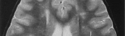
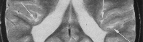


5 Cupped optic discs (nonglaucomatous) Late prenatal or perinatal cause : expanded occipital horns, loss of white/gray matter posteriorly. Cupped or atrophic optic discs)


6 Optic atrophy


7 Look at nerve fibre layer biomicrosocopy and fundus photography p
8 Why do we have a problem? Pseudopapilloedema and headache are common Raised ICP in children is unusual Poor evidence base for investigation No single gold standard test Many cases left with various degrees of uncertainty Anxiety Doctor Optometrist t t Family Opportunistic screening without evidence base.
9 Background Papilloedema a sign of raised intracranial pressure Idiopathic intracranial hypertension Obstructive Space-occupying lesion Communicating Endocrine Drugs Systemic disorder
10 Causes of ICP Drugs Tetracycline Corticosteroid withdrawal Inc topical for eczema Nitrofurantion Nalidixic acid Oral contraceptive Isoretinoin Thyroid replacement Growth hormone replacement Vasopressin Phenytoin Indomethacin Cyclosporin Endocrine disorder Hyperthyroidism Hypothyroidism Vitamin D deficiency Hypoparathyroidism Adrenal insufficiency hyperadrenalism Head trauma Systemic Infections Otitis media/mastoiditis mastoiditis Sinusitis Venous sinus thrombosis 1,2 Systemic disorders SLE Protein malnutrition Guillain-Barre syndrome Iron deficiency anaemia Leukaemia Hypercoaguable states
11 Headache Characteristic Symptoms of ICP Vomiting without nausea Visual disturbance Characteristic obscurations Diplopia less specific : photopsias / photophobia Vision loss Back pain Tinnitus pulsatile
12 Useful Useful Symptoms of ICP Headache Not useful unless characteristic (too common) Especially postural worse on lying, better on standing Worse on wakening, less during the day Opposite of tension headache / migraine (Vomiting without nausea) Visual disturbance Characteristic obscurations Momentary bilateral complete vision loss,not on standing up (this is postural hypotension) Diplopia if unequivocal manifest VI palsy present Vision loss if profound disc swelling, unequivocal atrophy and/or haemorrhages/exudates
13 Idiopathic intracranial hypertension Raised ICP with Normal imaging Normal CSF composition Normal consciousness Normal neurology Except papilloedema/vi palsy >70% not obese in children
14 Intracranial pressure Upper limit of normal 20cm CSF older child 13.5 cm H 2 O < 5 years old 7.5 cm H 2 O < 2 years old Most under GA or sedated Effect on ICP not studied. X mmhg = 1.36X cmh 2 O X cmh 2 O = 0.736X mmhg


15 Papilloedema p Raised ICP and papilloedema in astronaut after prolonged weightlessness In papilloedema there is an expanded subarachnoid space just behind the globe
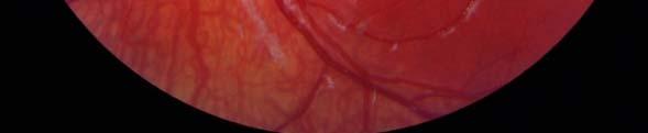
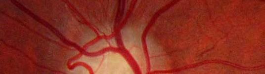
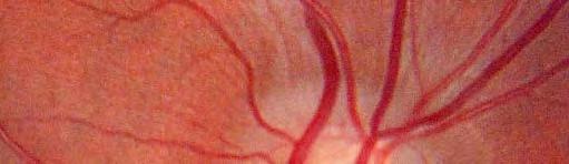
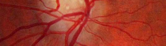
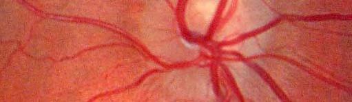
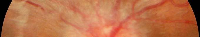
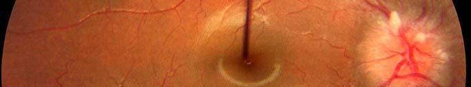
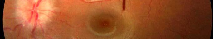
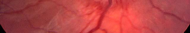
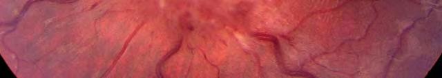
16 Is it MILD Papilloedema?
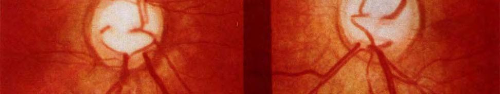
17 Papilloedema or pseudopapilloedema? p p
18 Is it MILD Papillodema? Checklist
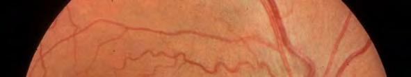
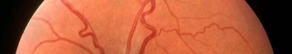
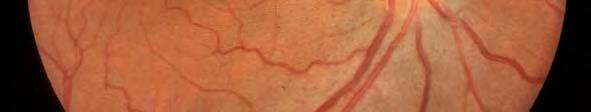
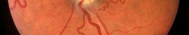
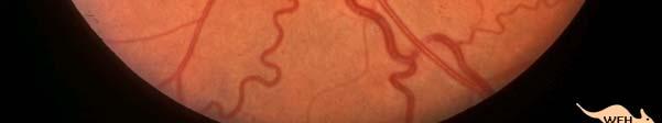
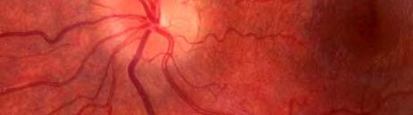
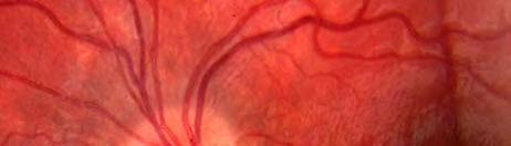
19 Optic disc edema Disc vasculature obscured at disc margins Elevation extends into peripapillary retina Graying and muddying of peripapillary nerve fibre layer Pseudopapilledema with buried drusen Disc vasculature remains visible at disc margins Elevation confined to optic disc Sharp peripapillary nerve fiber Venous congestion No venous congestion +/ Exudates / NFL haemorrhage No exudates, NFL hge rare Loss of optic cup only in moderate to Small cupless disc severe disc edema Normal configuration of disc vasculature despite venous congestion No circumpapillary light reflex Absence of spontaneous venous pulsations Increased major retinal vessels with early branching Crescentic circumpapillary light reflex Spontaneous venous pulsations may be present or absent Taylor DSI. Paediatric Ophthalmology

20 Papilloedema or pseudopapilloedema? p p True Pseudo True Pseudo
21 Spontaneous venous pulsation Video
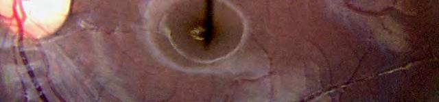
22 SVP : Direct ophthalmoscopy Very useful for SVP as greater magnification than biomicroscopy, but not done like this, and here child is too young except for red reflex check!
23 Absent SVP in pseudopapilloedema p p 25% vs 75% in normals Bilateral pseudopapilloedema : elevated discs but no surroundign oedema Ekdawi, Brodsky Rochester BJO 2011
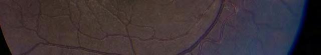
24 Non-mydriatic fundus camera : children >3y.o. find very easy, allows assessment and communication Orthoptist or technician using non-mydriatic (no drops needed) fundus camera
25 OCT OCT of optic disc not useful Too much variability in population RNFLA retinal nerve fibre layer analysis Useful in diagnosis and follow-up Measures oedema around disc Use same programme as glaucoma, but looking for not thickness
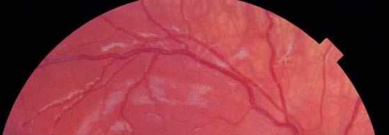
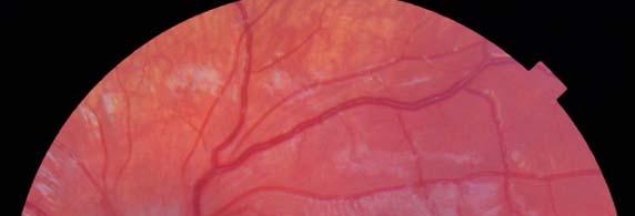
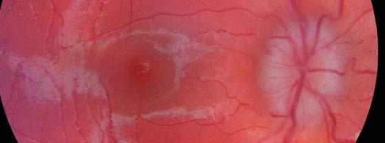
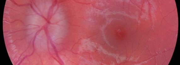
26 Resolving papilloedema following treatment


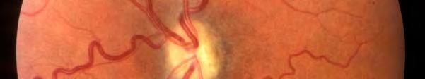
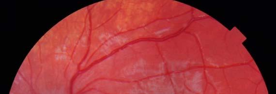
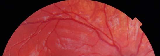
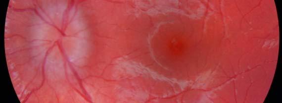
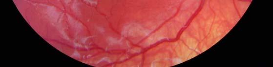
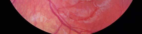
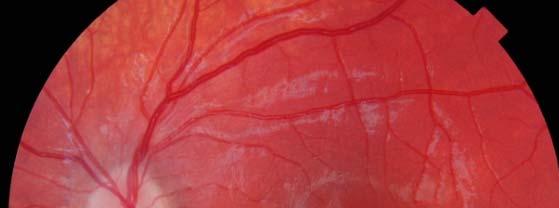
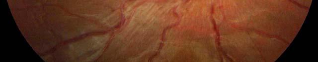
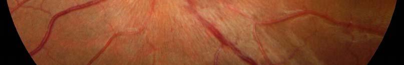
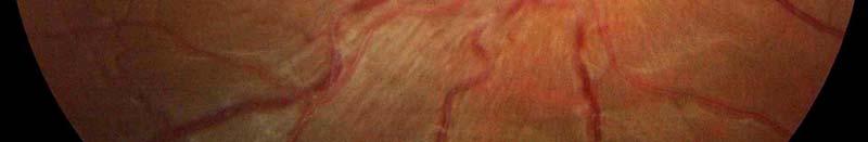
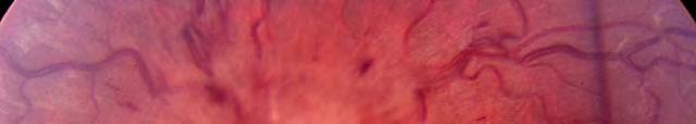
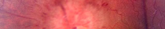
27 Dev veloping papilloed dema
28 Causes of pseudopapilloedema Hypermetropia Small discs Disc drusen Dysplastic discs Exposed disc drusen
29 Disc drusen


30 Buried drusen Daughter and Father Exposed drusen With age
31 Tests for Disc Drusen?


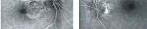

32 Tests for Disc Drusen Priel E. J Ophth Photo 2007


33 Disc drusen : Red free with Autofluorescence Fong Bristol Arch Dis Child 2010
34 FAF : autofluoresence with cslo 488nm excitation Barrier/emission > nm 520nm Disc drusen emission nm 520nm


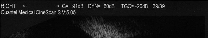

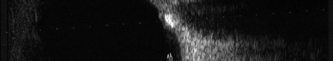
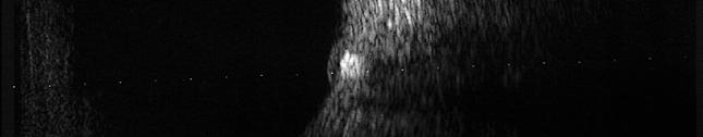
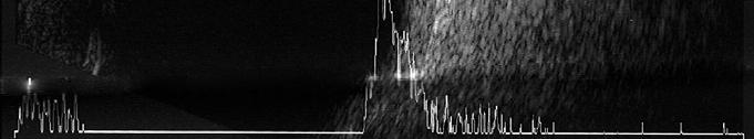
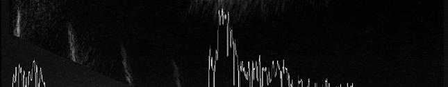
35 Disc Drusen U/S set to low gain confirms buried Disc drusen (here, in both eyes surprisingly)
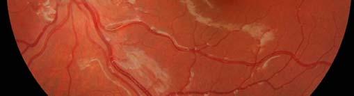
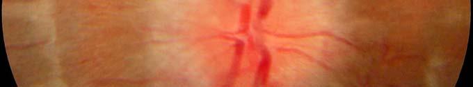
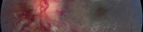
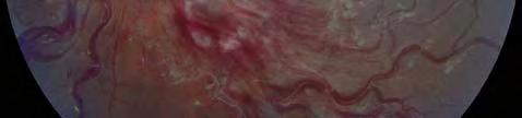
36 Is it SEVERE papilloedema?
37 Optic disc edema Disc vasculature obscured at disc margins Elevation extends into peripapillary retina Graying and muddying of peripapillary nerve fibre layer Pseudopapilledema with buried drusen Disc vasculature remains visible at disc margins Elevation confined to optic disc Sharp peripapillary nerve fiber Venous congestion No venous congestion +/ Exudates / NFL haemorrhage No exudates, NFL hge rare Loss of optic cup only in moderate to Small cupless disc severe disc edema Normal configuration of disc vasculature despite venous congestion No circumpapillary light reflex Absence of spontaneous venous pulsations Increased major retinal vessels with early branching Crescentic circumpapillary light reflex Spontaneous venous pulsations may be present or absent Taylor DSI. Paediatric Ophthalmology


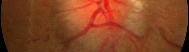

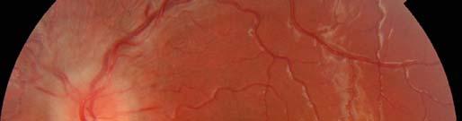
38 Papilloedema


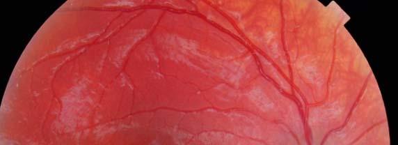
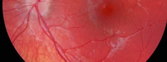
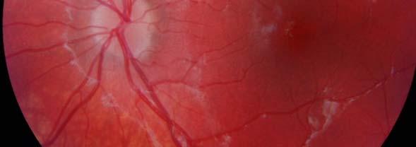
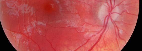
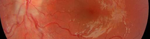
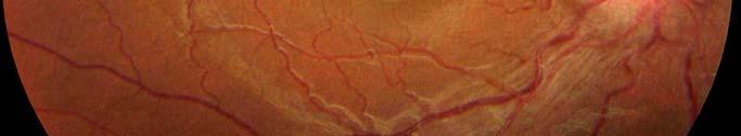
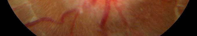
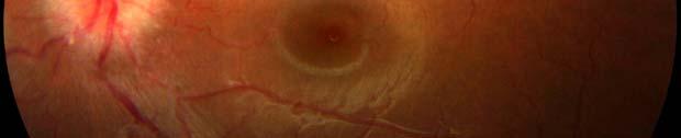
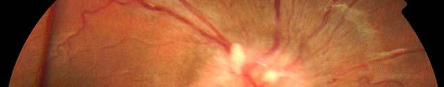
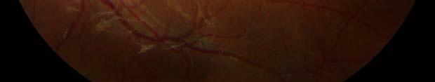
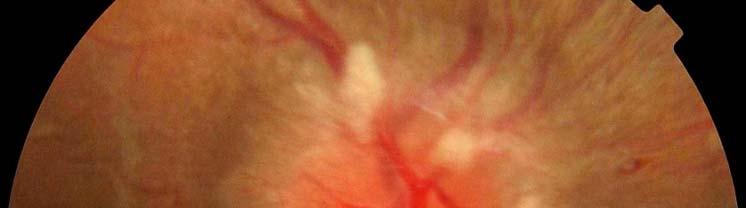
39 Severe Papilloedema : PRATs Focal arrest in axonal transport : same as cotton wool spot
40 Severe Papilloedema : Haemorrhage


41 Severe Papilloedema : Haemorrhage Macular star pointing to disc


42 Severe Papilloedema : Haemorrhage Haemorrhages and axonal transport t arrest imply rapid ongoing permanent damage to vision i There is usually a reversible element to vision loss if treated urgently Vessel obscuration on disc
43 Different questions depending on how obvious are the signs Mild signs Is it normal or not? Is it pseudopapilloedema? Obvious signs Could it be something different? Eg ischemic i optic neuropathy?

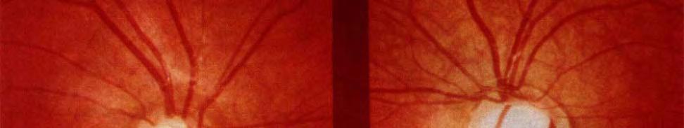
44 It can t be papilloedema


45 It can t be papilloedema Vision loss without papilloedema, due to pre-existent existent obvious optic atrophy, in patients t with known past history of raised intracranial i pressure and papilloedema.
46 Comment Most important diagnostic test is fundus examination Use photo as ancillary and help communication Direct ophthalmoscope for SVP Checklist Next most important is history Symptoms Neurology
47 Referral


48 Referral If there is papilloedema an urgent referral (I.E. SPEAK WITH THE HOSPITAL THAT DAY) is required


49 Referral intended to be guidance about which conditions require emergency or urgent referral. You should follow relevant local protocols for referral. If a patient presents with a condition requiring an emergency referral ee you may wish tosee seek advice from the on-call ophthalmologist
50 Headaches in general common and worth learning about them


51 IHS Need good source of information about headaches Eg International Headache society website PS non-specific headaches are very common in children (and adults)
Papilledema. Golnaz Javey, M.D. and Jeffrey J. Zuravleff, M.D.
 Papilledema Golnaz Javey, M.D. and Jeffrey J. Zuravleff, M.D. Papilledema specifically refers to optic nerve head swelling secondary to increased intracranial pressure (IICP). Optic nerve swelling from
Dr/ Marwa Abdellah EOS /16/2018. Dr/ Marwa Abdellah EOS When do you ask Fluorescein angiography for optic disc diseases???
 When do you ask Fluorescein angiography for optic disc diseases??? 1 NORMAL OPTIC DISC The normal optic disc on fluorescein angiography is fluorescent due to filling of vessels arising from the posterior
Optic Disc: Anatomy, Variants, Unusual discs. Kathleen B. Digre, MD Professor Neurology, Ophthalmology
 Optic Disc: Anatomy, Variants, Unusual discs Kathleen B. Digre, MD Professor Neurology, Ophthalmology THE OPHTHALMOSCOPE DIRECT OPHTHALMOSCOPY Jan Purkinje 1823 Hermann von Helmholtz 1851 Hand held ophthalmoscope
Pearls, Pitfalls and Advances in Neuro-Ophthalmology
 Pearls, Pitfalls and Advances in Neuro-Ophthalmology Nancy J. Newman, MD Emory University Atlanta, GA Consultant for Gensight Biologics, Santhera Data Safety Monitoring Board for Quark AION Study Medical-legal
12/2/16. Ways to differentiate:
 Nate Lighthizer, O.D., F.A.A.O. Assistant Dean for Clinical Care Services Director of CE Chief of Specialty Care Clinics Chief of Electrodiagnostics Clinic Oklahoma College of Optometry lighthiz@nsuok.edu
OPTIC NERVE SWELLING IN CHILDHOOD
 OPTIC NERVE SWELLING IN CHILDHOOD Melissa W. Ko, MD, FAAN One of the main findings on a pediatric neurologic examination that can instill fear and lead to an urgent referral to neuro-ophthalmology is the
Neuro-Ocular Grand Rounds
 Neuro-Ocular Grand Rounds Anthony B. Litwak,OD, FAAO VA Medical Center Baltimore, Maryland Dr. Litwak is on the speaker and advisory boards for Alcon and Zeiss Meditek COMMON OPTIC NEUROPATHIES THAT CAN
PITFALLS IN PAPILLOEDEMA
 PITFALLS IN PAPILLOEDEMA SRC 2013 Why care about papilloedema? Dr Neil Shuey FRACP MBBS(Hons) MScOptom St Vincent s Hospital, Melbourne Royal Victorian Eye & Ear Hospital Disclosures: Travel grants Biogen
Neuro-Ocular Grand Rounds Anthony B. Litwak,OD, FAAO VA Medical Center Baltimore, Maryland
 Neuro-Ocular Grand Rounds Anthony B. Litwak,OD, FAAO VA Medical Center Baltimore, Maryland Dr. Litwak is on the speaker and advisory boards for Alcon and Zeiss Meditek COMMON OPTIC NEUROPATHIES THAT CAN
FRANZCO, MD, MBBS. Royal Darwin Hospital
 Diabetes and Eye By Dr. Nishantha Wijesinghe FRANZCO, MD, MBBS Consultant Ophthalmologist Royal Darwin Hospital 98% of Diabetics do not need to suffer from severe visual loss Yet Diabetic eye disease is
What is IIH? Idiopathic Intracranial Hypertension (IIH)
") What is IIH? Idiopathic Intracranial Hypertension (IIH) What is Idiopathic Intracranial Hypertension? Idiopathic intracranial hypertension (IIH), also known as benign intracranial hypertension or pseudotumour
What is IIH? Idiopathic Intracranial Hypertension (IIH) What is Idiopathic Intracranial Hypertension? Idiopathic intracranial hypertension (IIH), also known as benign intracranial hypertension or pseudotumour
IDIOPATHIC INTRACRANIAL HYPERTENSION
 IDIOPATHIC INTRACRANIAL HYPERTENSION ASSESSMENT OF VISUAL FUNCTION AND PROGNOSIS FOR VISUAL OUTCOME Doctor of Philosophy thesis Anglia Ruskin University, Cambridge Fiona J. Rowe Department of Orthoptics,
Optical coherence tomography of the retinal nerve fibre layer in mild papilloedema and pseudopapilloedema
 294 SCIENTIFIC REPORT Optical coherence tomography of the retinal nerve fibre layer in mild papilloedema and pseudopapilloedema E Z Karam, T R Hedges... Aims: To determine the degree to which optical coherence
11/10/2017. Headache and Increased Pressure: A tale of 2 cases. Kathleen Digre MD University of Utah TWO CASES. 23 yo medical practice manager
 Headache and Increased Pressure: A tale of 2 cases Kathleen Digre MD University of Utah TWO CASES 23 yo medical practice manager September 2016 began developing intense frontal headaches first intermittent
Amber Priority. Image Library
 Amber Priority Image Library Amber flag Diabetic Maculopathy (M1) Pre-proliferative Diabetic Retinopathy (R2) Old, treated and now inactive DR (R1/M0/P1or R0/M0/P1) Where only partial or incomplete images
3/16/2018. Optic Nerve Examination. Hassan Eisa Swify FRCS Ed (Ophthalmology) Air Force Hospital
 Air Force Hospital") Optic Nerve Examination Hassan Eisa Swify FRCS Ed (Ophthalmology) Air Force Hospital 1 Examination Structure ( optic disc) Function Examination of the optic disc The only cranial nerve (brain tract) which
Optic Nerve Examination Hassan Eisa Swify FRCS Ed (Ophthalmology) Air Force Hospital 1 Examination Structure ( optic disc) Function Examination of the optic disc The only cranial nerve (brain tract) which
Typical idiopathic intracranial hypertension Optic nerve appearance and brain MRI findings. Jonathan A. Micieli, MD Valérie Biousse, MD
 Typical idiopathic intracranial hypertension Optic nerve appearance and brain MRI findings Jonathan A. Micieli, MD Valérie Biousse, MD A 24 year old African American woman is referred for bilateral optic
Question 1: Comment on the optic nerve appearance of each eye.
 Case 2 - Right Optic Nerve Head Drusen (ONHD) A 41 year old female was referred by her optometrist for a workup for unilateral optic disc drusen, OCT, and visual field changes. The patient was otherwise
Chapter 2 Long Duration Flight Data
 Chapter 2 Long Duration Flight Data Astronaut s bodies suffer in microgravity. Without effective countermeasures, muscles atrophy, bones shed calcium, and eyesight deteriorates. We ve known about this
IRIDOLOGY THE OPHTHALMOSCOPE
 IRIDOLOGY THE OPHTHALMOSCOPE Compiled by Campbell M Gold (2008) CMG Archives http://campbellmgold.com IMPORTANT The health information contained herein is not meant as a substitute for advice from your
Fundus Autofluorescence. Jonathan A. Micieli, MD Valérie Biousse, MD
 Fundus Autofluorescence Jonathan A. Micieli, MD Valérie Biousse, MD The retinal pigment epithelium (RPE) has many important functions including phagocytosis of the photoreceptor outer segments Cone Rod
OCCLUSIVE VASCULAR DISORDERS OF THE RETINA
 OCCLUSIVE VASCULAR DISORDERS OF THE RETINA Learning outcomes By the end of this lecture the students would be able to Classify occlusive vascular disorders (OVD) of the retina. Correlate the clinical features
Year 2 MBChB Clinical Skills Session Ophthalmoscopy. Reviewed & ratified by: Mr M Batterbury Consultant Ophthalmologist
 Year 2 MBChB Clinical Skills Session Ophthalmoscopy Reviewed & ratified by: o Mr M Batterbury Consultant Ophthalmologist Learning objectives o To understand the anatomy and physiology of the external and
RANZCO Screening and Referral Pathway for Diabetic Retinopathy #
 RANZCO Screening and Referral Pathway for Diabetic Retinopathy # Patient Presents a. Screen for Diabetic Retinopathy every 2 years b. Begin screening at diagnosis of Diabetes * Clinical Modifi ers Yearly
Optic Nerve Anomalies
 Optic Nerve Anomalies Raman Bhakhri, OD, FAAO Southern California College of Optometry Marshall B. Ketchum University Goals for today Review some of the optic nerve anomalies that can be seen in practice
Learn Connect Succeed. JCAHPO Regional Meetings 2015
 Learn Connect Succeed JCAHPO Regional Meetings 2015 OPTIC NEUROPATHY AS EASY AS 1,2,3,4 OPTIC NERVE ANATOMY M. Tariq Bhatti, MD Departments of Ophthalmology and Neurology Duke Eye Center and Duke University
THE SWOLLEN DISC. Valerie Biousse, MD Emory University School of Medicine Atlanta, GA
 THE SWOLLEN DISC Valerie Biousse, MD Emory University School of Medicine Atlanta, GA Updated from: Neuro-Ophthalmology Illustrated. Biousse V, Newman NJ. Thieme, New-York,NY. 2 nd Ed, 2016. Edema of the
Screening for Uveitis in Children
 Information for patients and parents Manchester Royal Eye Hospital Paediatric Uveitis Service Screening for Uveitis in Children What is uveitis? Uveitis is inflammation of a layer of the eye, called the
 measure of your overall performance. An isolated glucose test is helpful to let you know what your sugar level is at one moment, but it doesn t tell you whether or not your diabetes is under adequate control
Diabetic Retinopathy. Barry Emara MD FRCS(C) Giovanni Caboto Club October 3, 2012
 Giovanni Caboto Club October 3, 2012") Diabetic Retinopathy Barry Emara MD FRCS(C) Giovanni Caboto Club October 3, 2012 Outline Statistics Anatomy Categories Assessment Management Risk factors What do you need to do? Objectives Summarize the
Diabetic Retinopathy Barry Emara MD FRCS(C) Giovanni Caboto Club October 3, 2012 Outline Statistics Anatomy Categories Assessment Management Risk factors What do you need to do? Objectives Summarize the
Paediatric headaches. Dr Jaycen Cruickshank Director of Clinical Training Ballarat Health Services. Brevity, levity, repetition
 Paediatric headaches Dr Jaycen Cruickshank Director of Clinical Training Ballarat Health Services Brevity, levity, repetition Paediatric)headache?)! Headache!in!children!is!not!that!common.!The!question!is!which!headaches!do!I!
For details on measurement and recording of visual acuity, refer to Annex 1. VISION INTERPRETING RESULTS ABSTRACT
 management update on functional decline in older adults 2012 Unit No. 5 VISION Dr Au Eong Kah Guan, Ms Yulianti, Ms Fifiana ABSTRACT Among Singaporean adults of Chinese origin aged 40 to 79 years old,
A Case of Carotid-Cavernous Fistula
 A Case of Carotid-Cavernous Fistula By : Mohamed Elkhawaga 2 nd Year Resident of Ophthalmology Alexandria University A 19 year old male patient came to our outpatient clinic, complaining of : -Severe conjunctival
OPTOMETRY REVIEW. Key words: drusen of optic nerve head, multimodal imaging, optic nerve head, papilloedema, pseudopapilloedema
 C L I N I C A L A N D E X P E R I M E N T A L OPTOMETRY REVIEW The usefulness of multimodal imaging for differentiating pseudopapilloedema and true swelling of the optic nerve head: a review and case series
Sight savers real case studies: part 1
 Sight savers real case studies: part 1 44 Shamina Asif, BSc (Hons), MCOptom The role of the optometrist goes beyond refraction and vision correction with optical devices. The importance of disease detection
The College of Optometrists - Learning outcomes for the Professional Certificate in Medical Retina
 Learning outcomes for the Professional Certificate in Medical Retina, incorporating diabetic retinopathy screening and age related macular degeneration The professional certificate is a prerequisite to
The Glaucoma Suspect. Evaluating the Suspect Disk. Dr Michael Forrest. ! the usual suspects: ! is it glaucoma? ! is it swollen?
 Evaluating the Suspect Disk Dr Michael Forrest Senior Lecturer, The University of Queensland Northside Eye Specialists, Nundah Visiting Ophthalmologist, Mater Hospital, Brisbane Australian Vision Convention
Symptoms of a brain tumour in adults
 Symptoms of a brain tumour in adults A tumour is an abnormal growth caused by cells dividing in an uncontrolled manner. Approximately 9,300 people are diagnosed with a primary brain tumour each year. (Primary
p f .'''a.".: ;i.l i PAPILLOEDEMA brain. The ophthalmoscope may be looked upon as the Plate i is that of papilloedema of the optic nerve.
 382 * s. ii d:.'. ; z. j:.'''a.".: : ;i.l i 4 PAPILLOEDEMA FIG. I.-Papilloedema or 'choked disc' due to raised intracranial tension. In this instance the papilloedema was due to a frontal lobe tumour.
NANOS Patient Brochure
 NANOS Patient Brochure Pseudotumor Cerebri Copyright 2016. North American Neuro-Ophthalmology Society. All rights reserved. These brochures are produced and made available as is without warranty and for
Optical Coherence Tomography in Diabetic Retinopathy. Mrs Samantha Mann Consultant Ophthalmologist Clinical Lead of SEL-DESP
 Optical Coherence Tomography in Diabetic Retinopathy Mrs Samantha Mann Consultant Ophthalmologist Clinical Lead of SEL-DESP Content OCT imaging Retinal layers OCT features in Diabetes Some NON DR features
Step 4: Ask permission to turn off lights or draw the curtains
 STEPS OF EYE EXAMINATION - FUNDUS Step 1: Approach the patient Read the instructions carefully for clues Shake hands, introduce yourself Ask permission to examine him I would like to examine your eyes,
Index. Note: Page numbers of article titles are in boldface type.
 Index Note: Page numbers of article titles are in boldface type. A Acetazolamide, in idiopathic intracranial hypertension, 49 52, 60 Angiography, computed tomography, in cranial nerve palsy, 103 107 digital
Optic Nerve Disorders: Structure and Function and Causes
 Optic Nerve Disorders: Structure and Function and Causes Using Visual Fields, OCT and B-scan Ultrasound to Diagnose and Follow Optic Nerve Visual Losses Ohio Ophthalmological Society and Ophthalmic Tech
Glaucoma. Glaucoma. Optic Disc Cupping
 Glaucoma What is Glaucoma? Bruce James A group of diseases in which damage to the optic nerve occurs as a result of intraocualar pressure being above the physiological norm for that eye Stoke Mandeville
On Different Wavelengths: The Spectrum of Retinal Imaging. On Different Wavelengths: The Spectrum of Retinal Imaging. Wavelength Specific Imaging
 On Different Wavelengths: The Spectrum of Retinal Imaging Timothy J. Bennett, CRA, FOPS, OCT-C Penn State Hershey Eye Center Hershey, PA On Different Wavelengths: The Spectrum of Retinal Imaging Wavelengths
Unexplained visual loss in seven easy steps
 Unexplained visual loss in seven easy steps Andrew G. Lee, MD Chair Ophthalmology, Houston Methodist Hospital, Professor, Weill Cornell MC; Adjunct Professor, Baylor COM, U Iowa, UTMB Galveston, UT MD
Alan G. Kabat, OD, FAAO (901)
") THE SWOLLEN OPTIC DISC: EMERGENCY OR ANOMALY? Alan G. Kabat, OD, FAAO (901) 252-3691 Memphis, Tennessee alan.kabat@alankabat.com Course description: The swollen disc presents a diagnostic dilemma. While
THE SWOLLEN OPTIC DISC: EMERGENCY OR ANOMALY? Alan G. Kabat, OD, FAAO (901) 252-3691 Memphis, Tennessee alan.kabat@alankabat.com Course description: The swollen disc presents a diagnostic dilemma. While
Do You See What I See!!! Shane R. Kannarr, OD
 Do You See What I See!!! Shane R. Kannarr, OD skannarr@kannarreyecare.com Define Specialty Testing Additional Test to: Prove/Disprove Diagnosis To monitor progression of a condition To document a condition
Recurrent transient visual loss in a middle aged woman
 Recurrent transient visual loss in a middle aged woman Chow SY, Draman N, Teh WM, Azhany Y Chow SY, Draman N, Teh WM, et al. Recurrent transient visual loss in a middle aged woman. Malays Fam Physician.
HEADACHES THE RED FLAGS
 HEADACHES THE RED FLAGS FAYYAZ AHMED CONSULTANT NEUROLOGIST HON. SENIOR LECTURER HULL YORK MEDICAL SCHOOL SECONDARY VS PRIMARY HEADACHES COMMON SECONDARY HEADACHES UNCOMMON BUT SERIOUS SECONDARY HEADACHES
Ophthalmology Unit Referral Guidelines
 Ophthalmology Unit Referral Guidelines Austin Health Ophthalmology Unit holds sub-specialty sessions to discuss and plan the treatment of patients with specific ocular conditions. General including cataract
OCT Angiography in Primary Eye Care
 OCT Angiography in Primary Eye Care An Image Interpretation Primer Julie Rodman, OD, MS, FAAO and Nadia Waheed, MD, MPH Table of Contents Diabetic Retinopathy 3-6 Choroidal Neovascularization 7-9 Central
Speaker Disclosure Statement. " Dr. Tim Maillet and Dr. Vladimir Kozousek have no conflicts of interest to disclose.
 Speaker Disclosure Statement Dr. Tim Maillet and Dr. Vladimir Kozousek have no conflicts of interest to disclose. Diabetes Morbidity Diabetes doubles the risk of stroke. Diabetes quadruples the risk of
Cases of visual impairment caused by cerebral venous sinus occlusion-induced intracranial hypertension in the absence of headache
 Zhao et al. BMC Neurology (2018) 18:159 https://doi.org/10.1186/s12883-018-1156-7 CASE REPORT Open Access Cases of visual impairment caused by cerebral venous sinus occlusion-induced intracranial hypertension
The Diagnostic Dilemma of Pseudopapilledema. Tiffenie Harris, OD, FAAO Associate Professor Western University College of Optometry
 The Diagnostic Dilemma of Pseudopapilledema Tiffenie Harris, OD, FAAO Associate Professor Western University College of Optometry Author s Bio Dr. Harris is a graduate of Indiana University School of Optometry.
Ocular warning signs in GP practice: Paediatric Eye Pointers
 Ocular warning signs in GP practice: Paediatric Eye Pointers Dr Benjamin Chang MB, BCh, BAO, MMedSci, FRCS(Irel), FRCS(Edin), FRCOphth(Lond) Senior Consultant Ophthalmology and Visual Sciences Khoo Teck
Paediatric Eyecare Update
 Paediatric Eyecare Update This update contains important information with regard to Paediatric Ophthalmology Services which are managed and provided by Belfast Health and Social Care Trust (BHSCT). Please
Anterior Ischemic Optic Neuropathy (AION)
") Anterior Ischemic Optic Neuropathy (AION) Your doctor thinks you have suffered an episode of anterior ischemic optic neuropathy (AION). This is the most common cause of sudden decreased vision in patients
Anterior Ischemic Optic Neuropathy (AION) Your doctor thinks you have suffered an episode of anterior ischemic optic neuropathy (AION). This is the most common cause of sudden decreased vision in patients
An Organized Approach to the Patient with Papilledema and IIH
 An Organized Approach to the Patient with Papilledema and IIH Leonard V. Messner, OD, FAAO James L. Fanelli, OD, FAAO Please silence all mobile devices and remove items from chairs so others can sit. Unauthorized
Neuropathy (NAION) and Avastin. Clinical Assembly of the AOCOO-HNS Foundation May 9, 2013
 and Avastin. Clinical Assembly of the AOCOO-HNS Foundation May 9, 2013") Non Arteritic Ischemic Optic Neuropathy (NAION) and Avastin Shalom Kelman, MD Clinical Assembly of the AOCOO-HNS Foundation May 9, 2013 Anterior Ischemic Optic Neuropathy Acute, painless, visual loss,
Non Arteritic Ischemic Optic Neuropathy (NAION) and Avastin Shalom Kelman, MD Clinical Assembly of the AOCOO-HNS Foundation May 9, 2013 Anterior Ischemic Optic Neuropathy Acute, painless, visual loss,
Central venous occlusion
 Central venous occlusion Central venous occlusion (right eye) There are dark haemorrhages at the macula and all over the retina. Choroidal haemangioma A choroidal haemangioma has salmon pink colour. There
Clinical Study Optic Nerve Sonography in the Diagnostic Evaluation of Pseudopapilledema and Raised Intracranial Pressure: A Cross-Sectional Study
 Neurology Research International Volume 2015, Article ID 146059, 4 pages http://dx.doi.org/10.1155/2015/146059 Clinical Study Optic Nerve Sonography in the Diagnostic Evaluation of Pseudopapilledema and
Headache Assessment In Primary Eye Care
 Headache Assessment In Primary Eye Care Spencer Johnson, O.D., F.A.A.O. Northeastern State University Oklahoma College of Optometry johns137@nsuok.edu Course Objectives Review headache classification Understand
DRUSEN OF OPTIC NERVE SIMULATING PAPILLEDEMA
 DRUSEN OF OPTIC NERVE SIMULATING PAPILLEDEMA MAX CHAMLIN, M.D., AND LEO 1VI. DAVIDOFF, M.D.* New York City (Received for publication July 5, 1949) 6" 6" "~"~ RUSEN" is the name applied to hyaline substances
A synopsis of: Diagnosis and Management of Headaches in Adults: A national clinical guideline. Scottish intercollegiate Guidelines Network SIGN
 A synopsis of: Diagnosis and Management of Headaches in Adults: A national clinical guideline Scottish intercollegiate Guidelines Network SIGN November 2008. PETER FRAMPTON MSc MCOptom BAppSc (Optom)(AUS)
Advances in OCT Murray Fingeret, OD
 Disclosures Advances in OCT Murray Fingeret, OD Consultant Alcon, Allergan, Bausch & Lomb, Carl Zeiss Meditec, Diopsys, Heidelberg Engineering, Reichert, Topcon Currently Approved OCT Devices OCT Devices
Dilemma of the swollen optic disc: a fluorescein retinal angiography study
 British Journal of Ophthalmology, 1977, 61, 385-389 Dilemma of the swollen optic disc: a fluorescein retinal angiography study N. E. F. CARTLIDGE, R. C. Y. NG, AND P. J. B. TILLEY From the Department of
Guidance for Optometrists in relation to Diabetic Retinopathy Screening Schemes. June 2004
 Guidance for Optometrists in relation to Diabetic Retinopathy Screening Schemes June 2004 Over the next few years formal screening schemes designed to detect diabetic retinopathy will be introduced across
Clinical Guidance and Monitoring for Change. Cecilia Fenerty MD FRCOphth Manchester Royal Eye Hospital
 Clinical Guidance and Monitoring for Change Cecilia Fenerty MD FRCOphth Manchester Royal Eye Hospital Glaucoma Referral Criteria 2000 Original referral scheme Simple criteria based on IOP/Disc/Field Solitary
Idiopathic Intracranial Hypertension (Pseudotumor Cerebri) David I. Kaufman, D.O. Michigan State University Department of Neurology and Ophthalmology
 David I. Kaufman, D.O. Michigan State University Department of Neurology and Ophthalmology") Idiopathic Intracranial Hypertension (Pseudotumor Cerebri) David I. Kaufman, D.O. Michigan State University Department of Neurology and Ophthalmology 26 year old 5 3, 300 pound female with papilledema,
Idiopathic Intracranial Hypertension (Pseudotumor Cerebri) David I. Kaufman, D.O. Michigan State University Department of Neurology and Ophthalmology 26 year old 5 3, 300 pound female with papilledema,
Rafik Girgis. Consultant Ophthalmic Surgeon ( Cataract & Primary Care)
") Rafik Girgis Consultant Ophthalmic Surgeon ( Cataract & Primary Care) Blepharitis Is a very common condition which usually bilateral & symmetrical. The main types are: Anterior, posterior or mixed Complications:
Rafik Girgis Consultant Ophthalmic Surgeon ( Cataract & Primary Care) Blepharitis Is a very common condition which usually bilateral & symmetrical. The main types are: Anterior, posterior or mixed Complications:
NEURO QUIZ 45 EHLERS DANLOS SYNDROME
 NEURO QUIZ 45 EHLERS DANLOS SYNDROME Verghese Cherian, MD, FFARCSI Penn State Hershey Medical Center, Hershey Quiz Team Shobana Rajan, M.D Suneeta Gollapudy, M.D Angele Marie Theard, M.D START 1. Regarding
Year 1 MBChB Clinical Skills Session Ophthalmoscopy
 Year 1 MBChB Clinical Skills Session Ophthalmoscopy Reviewed & ratified by: Dr V Taylor-Jones, Mr M Batterbury Consultant Ophthalmologist Learning objectives o To understand the anatomy and physiology
Sequential non-arteritic anterior ischemic optic neuropathy (NAION) Jonathan A. Micieli, MD Valérie Biousse, MD
 Jonathan A. Micieli, MD Valérie Biousse, MD") Sequential non-arteritic anterior ischemic optic neuropathy (NAION) Jonathan A. Micieli, MD Valérie Biousse, MD A 68 year old white woman had a new onset of floaters in her right eye and was found to have
Sequential non-arteritic anterior ischemic optic neuropathy (NAION) Jonathan A. Micieli, MD Valérie Biousse, MD A 68 year old white woman had a new onset of floaters in her right eye and was found to have
THE ROYAL COLLEGE OF OPHTHALMOLOGISTS DUKE ELDER PRIZE EXAMINATION 2012 INFORMATION FOR CANDIDATES
 THE ROYAL COLLEGE OF OPHTHALMOLOGISTS DUKE ELDER PRIZE EXAMINATION 2012 INFORMATION FOR CANDIDATES 1 Please read the following information carefully This examination is intended for medical students who
Swelling of the optic nerve head: a staging scheme
 Journai of Neurology, Neurosurgery, and Psychiatry 1982 ;45 :13-18 Swelling of the optic nerve head: a staging scheme LARS FRISIEN From the Department of Ophthalmology, University of Gdteborg, Sweden SUMMARY
Widefield Retinal Imaging with Auto Fluorescence Technology in the Optometric Practice
 Widefield Retinal Imaging with Auto Fluorescence Technology in the Optometric Practice This course will define ultra-widefield retinal imaging and autofluorescence for the attendee. Will show how it is
Headaches need not be a headache for optometrists
 Headaches need not be a headache for optometrists C-19309 O/D Tina Kipioti, MD, FRCSEd Of all the painful states that afflict humans, headache (cephalalgia) is the most common. According to a large study,
C19. Pediatric Neuro-ophthalmology: Dilemmas in clinical practice. 12 June, :15 15:45. Room 115 HAND-OUTS
 C19 Pediatric Neuro-ophthalmology: Dilemmas in clinical practice 12 June, 2017 14:15 15:45 Room 115 HAND-OUTS Is this strabismus really harmful? Karl Golnik, MD, MEd University of Cincinnati, USA Childhood
Fundus Autofluorescence
 Brittany Bateman, BS Fundus autofluorescence imaging is used to record fluorescence that may occur naturally in ocular structures or as a byproduct of a disease process. This technique allows the topographic
Neurological Dilemmas in Primary Care
 Neurological Dilemmas in Primary Care David Clark, DO dclark@oregonneurology.com When to test? How to test? Pitfalls in testing? When to treat? How to treat? How long to treat? Neurological Dilemmas Seizure
Differential Diagnosis of ONH Edema Beth A. Steele, OD, FAAO
 Differential Diagnosis of ONH Edema Beth A. Steele, OD, FAAO bsteele@uab.edu Please silence all mobile devices and remove items from chairs so others can sit. Unauthorized recording of this session is
The Role of the RNFL in the Diagnosis of Glaucoma
 Chapter 1. The Role of the RNFL in the Diagnosis of Glaucoma Introduction Glaucoma is an optic neuropathy characterized by a loss of of retinal ganglion cells and their axons, the Retinal Nerve Fiber Layer
OCT in the Diagnosis and Follow-up of Glaucoma
 OCT in the Diagnosis and Follow-up of Glaucoma Karim A Raafat MD. Professor Of Ophthalmology Cairo University Hmmmm! Do I have Glaucoma or not?! 1 Visual Function 100% - N Gl Structure : - 5000 axon /
Automated Detection of Vascular Abnormalities in Diabetic Retinopathy using Morphological Entropic Thresholding with Preprocessing Median Fitter
 IJSTE - International Journal of Science Technology & Engineering Volume 1 Issue 3 September 2014 ISSN(online) : 2349-784X Automated Detection of Vascular Abnormalities in Diabetic Retinopathy using Morphological
Beyond the C/D Ratio: Evaluating a Glaucomatous Optic Nerve. Marcus Gonzales, OD, FAAO Cedar Springs Eye Clinic COPE ID#: GL
 Beyond the C/D Ratio: Evaluating a Glaucomatous Optic Nerve Marcus Gonzales, OD, FAAO Cedar Springs Eye Clinic COPE ID#: 27809-GL Points to Remember Glaucoma affects the ONH in characteristic patterns
Brain and Central Nervous System Cancers
 Brain and Central Nervous System Cancers NICE guidance link: https://www.nice.org.uk/guidance/ta121 Clinical presentation of brain tumours History and Examination Consider immediate referral Management
Funduscopic Interpretation Understanding the Fundus: is that normal?
 Funduscopic Interpretation Understanding the Fundus: is that normal? Gillian McLellan BVMS PhD DVOphthal DECVO DACVO MRCVS With thanks to Christine Heinrich and all who contributed images Fundus Retina
Case Series. The efficacy of optic nerve ultrasonography for differentiating papilloedema from pseudopapilloedema in eyes with swollen optic discs
 Case Series The efficacy of optic nerve ultrasonography for differentiating papilloedema from pseudopapilloedema in eyes with swollen optic discs Meira Neudorfer, Maytal Siegman Ben-Haim, Igal Leibovitch
Mild NPDR. Moderate NPDR. Severe NPDR
 Diabetic retinopathy Diabetic retinopathy is the most common cause of blindness in adults aged 35-65 years-old. Hyperglycaemia is thought to cause increased retinal blood flow and abnormal metabolism in
CHAPTER 13 CLINICAL CASES INTRODUCTION
 2 CHAPTER 3 CLINICAL CASES INTRODUCTION The previous chapters of this book have systematically presented various aspects of visual field testing and is now put into a clinical context. In this chapter,
ASSESSING THE EYES. Structures. Eyelids Extraocularmuscles Eyelashes Lacrimal glands: Lacrimal ducts Cornea Conjunctiva Sclera Pupils Iris.
 ASSESSING THE EYES Structures External Eyelids Extraocularmuscles Eyelashes Lacrimal glands: Lacrimal ducts Cornea Conjunctiva Sclera Pupils Iris 1 2 Structures Internal Optic disc Physiological cup Retinal
Professor Helen Danesh-Meyer. Eye Institute Auckland
 Professor Helen Danesh-Meyer Eye Institute Auckland Bitten by Ophthalmology Emergencies Helen Danesh-Meyer, MBChB, MD, FRANZCO Sir William and Lady Stevenson Professor of Ophthalmology Head of Glaucoma
Clinically Significant Macular Edema (CSME)
") Clinically Significant Macular Edema (CSME) 1 Clinically Significant Macular Edema (CSME) Sadrina T. Shaw OMT I Student July 26, 2014 Advisor: Dr. Uwaydat Clinically Significant Macular Edema (CSME) 2
Clinically Significant Macular Edema (CSME) 1 Clinically Significant Macular Edema (CSME) Sadrina T. Shaw OMT I Student July 26, 2014 Advisor: Dr. Uwaydat Clinically Significant Macular Edema (CSME) 2
The Prevalence of diabetic optic neuropathy in type 2 diabetes mellitus
 The Prevalence of diabetic optic neuropathy in type 2 diabetes mellitus Received: 25/4/2016 Accepted: 8/12/2016 Introduction Diabetic papillopathy is an atypical form of non-arteritic anterior ischemic
The headache profile of idiopathic intracranial hypertension
 The headache profile of idiopathic intracranial hypertension Michael Wall CEPHALALGIA Wall M. The headache profile of idiopathic intracranial hypertension. Cephalalgia 1990;10:331-5. Oslo. ISSN 0333-1024
Age-Related Macular Degeneration (AMD)
") Age-Related Macular Degeneration (AMD) What is the Macula? What is Dry AMD (Age-related Macular Degeneration)? Dry AMD is an aging process that causes accumulation of waste product under the macula leading
Age-Related Macular Degeneration (AMD) What is the Macula? What is Dry AMD (Age-related Macular Degeneration)? Dry AMD is an aging process that causes accumulation of waste product under the macula leading
THE 35 GOLDEN EYE RULES
 THE 35 GOLDEN EYE RULES The Sense of Sight, from La Dame a la Licorne, The Lady and the Unicorn Tapestries, Late 15th Century Flemish Tapestry in wool and silk, Musée Nationale du Moyen Age, Paris. 1.
This is the author's manuscript of the article published in final edited form as:
 Nonmydriatic Fundus Photography: A Practical Review for the Neurologist Devin D. Mackay, M.D.; Beau B. Bruce, M.D., Ph.D. From the Departments of Neurology, Ophthalmology, and Neurosurgery (DDM), Indiana