Typical idiopathic intracranial hypertension Optic nerve appearance and brain MRI findings. Jonathan A. Micieli, MD Valérie Biousse, MD
|
|
|
- Kelley Wilfrid Robinson
- 5 years ago
- Views:
Transcription
1 Typical idiopathic intracranial hypertension Optic nerve appearance and brain MRI findings Jonathan A. Micieli, MD Valérie Biousse, MD
2 A 24 year old African American woman is referred for bilateral optic disc edema after visiting an optometrist to update her glasses. She is asymptomatic Her past medical history is significant for obesity (BMI 39.1) Visual acuity is 20/20 OD, 20/20 OS There is no relative afferent pupillary defect Color vision is 14/14 correct Ishihara plates OU

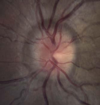
3 Figure 1. Right eye Left eye


,")
4 Figure 1. Right eye Left eye There is bilateral optic disc edema in both eyes. There are peripapillary wrinkles (also known as Paton s lines) in both eyes (red arrow). The left eye also has pseudodrusen (blue arrow), which are small discrete refractile bodies seen in patients with longstanding papilledema
 image of the optic disc is shown on the")

5 Figure 2. An OCT (optical coherence tomography) image of the optic disc is shown on the left to highlight the peripapillary wrinkles in the right eye. The wrinkles are commonly seen with optic disc edema. As the disc swells, it displaces the retina causing it to form a series of folds concentric to the edge of the optic disc
6 Figure 3. Left eye Right eye 24-2 SITA-Fast Humphrey visual fields
7 Figure 3. Although there are a high number of false positives, visual fields appear normal with only mildly enlarged blind spots Left eye Right eye
8 The patient has bilateral optic disc edema and preserved visual function, suggesting papilledema, which is optic disc edema due to raised intracranial pressure (ICP) An MRI of the brain without and with contract and MRV of the head with contrast were ordered to assess for any intracranial process (mass, hydrocephalus, leptomeningeal enhancement) or venous sinus abnormality (thrombosis, dural fistula or stenosis) that may lead to elevated ICP


9 Figure 4. Sagittal T1 MRI of the brain without contrast Axial T2 FLAIR MRI of the brain without contrast Coronal T2 MRI of the brain without contrast
10 Figure 5. MRV of the brain is shown here

, which")

11 Figure 6. Patient with a partially empty sella Example of a normal appearing sella Sagittal T1 MRI of the brain without contrast shows a partially empty sella. There is intrasellar herniation of arachnoid mater and CSF (yellow), which flattens the pituitary gland (red) in the sella turcica (blue). This is the most common MRI finding seen in idiopathic intracranial hypertension. It is also seen in other causes of elevated ICP and may also be seen in the normal population


12 Figure 7. Sagittal T1 MRI shows mild tonsillar herniation Normal sagittal T1 MRI shown for comparison Mid-sagittal T1 MRI of the brain shows caudal displacement of the cerebellar tonsils. A line (red in the IIH patient and yellow in a normal control) is drawn from the basion to the opisthon defining the foramen magnum.


13 Figure 8. Axial T2 FLAIR MRI shows flattening of the posterior globes in this patient (red arrow) Normal axial T1 MRI shown for comparison Flattening of the posterior globes may be seen in patients with elevated intracranial pressure and is due to a change in the pressure gradient between the perioptic CSF spaces and the intraocular pressure


14 Figure 9. Coronal T2 MRI shows distention of the CSF spaces around the optic nerves Example of a normal coronal T2 MRI shown for comparison
15 Figure 10. MRV of the brain shows narrowing of the distal transverse sinuses, which is another common finding in patients with idiopathic intracranial hypertension
16 A lumbar puncture was performed in the left lateral decubitus position and showed: Opening pressure of 39 cm of H2O Nucleated cells: 0 Red blood cells: 2 Protein and glucose within normal limits
17 The patient was diagnosed with idiopathic intracranial hypertension (IIH) according to the modified Dandy criteria. Modified Dandy criteria required for the diagnosis of IIH: A) Papilledema B) Normal neurological examination except for cranial nerve abnormalities C) Neuroimaging: normal brain parenchyma without evidence of structural lesion or venous sinus thrombosis D) Normal CSF composition E) Elevated lumbar puncture opening pressure (> 25 cm of H2O in adults) She was prescribed acetazolamide and was started on a diet and weight loss program under the guidance of her primary care physician


18 Figure 11. OS OD Presentation 3 months after presentation 6 months after presentation 1 year after presentation


19 Figure 12. OD OS OS OD OS OD OS OD OS OD Presentation 3 months after presentation 6 months after presentation 1 year after presentation Her optic disc edema resolved over a period of 1 year with weight loss and acetazolamide, which was discontinued

 Classic MRI findings in IIH include a partially empty sella,")


20 Summary points: Papilledema is defined as optic disc edema due to elevated intracranial pressure and usually has preserved visual function (unless there is macular edema from severe disc edema or optic atrophy from longstanding papilledema) Classic MRI findings in IIH include a partially empty sella, flattening of the posterior globes, distention of the CSF space around the optic nerves, tonsillar hernitation, skull base meningoencephaloceles, and distal transverse sinus stenosis without any evidence of an underlying structural lesion Empty sella Flattening of posterior globes Distention of CSF space Distal transverse sinus stenosis
Non-arteritic anterior ischemic optic neuropathy (NAION) with segmental optic disc edema. Jonathan A. Micieli, MD Valérie Biousse, MD
 with segmental optic disc edema. Jonathan A. Micieli, MD Valérie Biousse, MD") Non-arteritic anterior ischemic optic neuropathy (NAION) with segmental optic disc edema Jonathan A. Micieli, MD Valérie Biousse, MD A 75 year old white woman lost vision in the inferior part of her visual
Non-arteritic anterior ischemic optic neuropathy (NAION) with segmental optic disc edema Jonathan A. Micieli, MD Valérie Biousse, MD A 75 year old white woman lost vision in the inferior part of her visual
Sequential non-arteritic anterior ischemic optic neuropathy (NAION) Jonathan A. Micieli, MD Valérie Biousse, MD
 Jonathan A. Micieli, MD Valérie Biousse, MD") Sequential non-arteritic anterior ischemic optic neuropathy (NAION) Jonathan A. Micieli, MD Valérie Biousse, MD A 68 year old white woman had a new onset of floaters in her right eye and was found to have
Sequential non-arteritic anterior ischemic optic neuropathy (NAION) Jonathan A. Micieli, MD Valérie Biousse, MD A 68 year old white woman had a new onset of floaters in her right eye and was found to have
Pearls, Pitfalls and Advances in Neuro-Ophthalmology
 Pearls, Pitfalls and Advances in Neuro-Ophthalmology Nancy J. Newman, MD Emory University Atlanta, GA Consultant for Gensight Biologics, Santhera Data Safety Monitoring Board for Quark AION Study Medical-legal
Khalil Zahra, M.D Neuro-interventional radiology
 Khalil Zahra, M.D Neuro-interventional radiology 1 Disclosure None 2 Outline Etiology and pathogensis Imaging techniques and Features Literature review Treatment modalities Endovascular techniques Long
Fundus Autofluorescence. Jonathan A. Micieli, MD Valérie Biousse, MD
 Fundus Autofluorescence Jonathan A. Micieli, MD Valérie Biousse, MD The retinal pigment epithelium (RPE) has many important functions including phagocytosis of the photoreceptor outer segments Cone Rod
THE SWOLLEN DISC. Valerie Biousse, MD Emory University School of Medicine Atlanta, GA
 THE SWOLLEN DISC Valerie Biousse, MD Emory University School of Medicine Atlanta, GA Updated from: Neuro-Ophthalmology Illustrated. Biousse V, Newman NJ. Thieme, New-York,NY. 2 nd Ed, 2016. Edema of the
BMB Disclosures. Papilledema can be a. Neurological Emergency, Causing Preventable Blindness
 Reasonable Doubt: Can High Intracranial Pressure Occur Without Papilledema? 15 February 2013 Jonathan C. Horton hortonj@vision.ucsf.edu http://www.ucsf.edu/hortonlab BMB Disclosures Financial Disclosures
Intracranial hypertension and headache. Daniel Tibussek, MD
 Intracranial hypertension and headache. Daniel Tibussek, MD none Disclosures Overview Case Clinical presentation of pediatric PTC Nomenclature, Definition What is intracranial hypertension? Diagnostic
Cases of visual impairment caused by cerebral venous sinus occlusion-induced intracranial hypertension in the absence of headache
 Zhao et al. BMC Neurology (2018) 18:159 https://doi.org/10.1186/s12883-018-1156-7 CASE REPORT Open Access Cases of visual impairment caused by cerebral venous sinus occlusion-induced intracranial hypertension
OPTIC NERVE SWELLING IN CHILDHOOD
 OPTIC NERVE SWELLING IN CHILDHOOD Melissa W. Ko, MD, FAAN One of the main findings on a pediatric neurologic examination that can instill fear and lead to an urgent referral to neuro-ophthalmology is the
Prevalence of venous sinus stenosis in Pseudotumor cerebri(ptc) using digital subtraction angiography (DSA)
 using digital subtraction angiography (DSA)") Prevalence of venous sinus stenosis in Pseudotumor cerebri(ptc) using digital subtraction angiography (DSA) Dr.Mohamed hamdy ibrahim MBBC,MSc,MD, PhD Neurology Degree Kings lake university (USA). Fellow
Prevalence of venous sinus stenosis in Pseudotumor cerebri(ptc) using digital subtraction angiography (DSA) Dr.Mohamed hamdy ibrahim MBBC,MSc,MD, PhD Neurology Degree Kings lake university (USA). Fellow
Papilledema. Golnaz Javey, M.D. and Jeffrey J. Zuravleff, M.D.
 Papilledema Golnaz Javey, M.D. and Jeffrey J. Zuravleff, M.D. Papilledema specifically refers to optic nerve head swelling secondary to increased intracranial pressure (IICP). Optic nerve swelling from
An Organized Approach to the Patient with Papilledema and IIH
 An Organized Approach to the Patient with Papilledema and IIH Leonard V. Messner, OD, FAAO James L. Fanelli, OD, FAAO Please silence all mobile devices and remove items from chairs so others can sit. Unauthorized
Meninges and Ventricles
 Meninges and Ventricles Irene Yu, class of 2019 LEARNING OBJECTIVES Describe the meningeal layers, the dural infolds, and the spaces they create. Name the contents of the subarachnoid space. Describe the
Spontaneous Intracranial Hypotension Diagnosis and Treatment
 Spontaneous Intracranial Hypotension Diagnosis and Treatment John W. Engstrom MD, Philip R. Weinstein MD, and William P. Dillon M.D. University of California, San Francisco Spontaneous Intracranial Hypotension
Brain Imaging in Pediatric Pseudotumor Cerebri Syndrome
 Review Article 49 Brain Imaging in Pediatric Pseudotumor Cerebri Syndrome Emanuele David 1,2 Kshitij Mankad 3 1 Department of Radiology, Anatomopathology and Oncology, Sapienza University of Rome, Rome,
12/2/16. Ways to differentiate:
 Nate Lighthizer, O.D., F.A.A.O. Assistant Dean for Clinical Care Services Director of CE Chief of Specialty Care Clinics Chief of Electrodiagnostics Clinic Oklahoma College of Optometry lighthiz@nsuok.edu
Magnetic resonance imaging in pseudotumor cerebri
 Magnetic resonance imaging in pseudotumor cerebri Poster No.: C-1004 Congress: ECR 2015 Type: Authors: Keywords: DOI: Scientific Exhibit J. Saad 1, F. Marrakchi 2, F. Harbi 1 ; 1 Nejran/SA, 2 Ksour Essaf/TN
Management of Pseudo Tumor Cerebri by Frequent Tapping VS lumboperitoneal Shunt
 The Egyptian Journal of Hospital Medicine (July 2018) Vol. 72 (5), Page 4556-4560 Management of Pseudo Tumor Cerebri by Frequent Tapping VS lumboperitoneal Shunt Ali K. Ali, Maamoun M. Abo Shousha, Mohammed
IDIOPATHIC INTRACRANIAL HYPERTENSION
 IDIOPATHIC INTRACRANIAL HYPERTENSION ASSESSMENT OF VISUAL FUNCTION AND PROGNOSIS FOR VISUAL OUTCOME Doctor of Philosophy thesis Anglia Ruskin University, Cambridge Fiona J. Rowe Department of Orthoptics,
Meningoceles in Idiopathic Intracranial Hypertension
 Neuroradiology/Head and Neck Imaging Original Research Bialer et al. Meningoceles in IIH Neuroradiology/Head and Neck Imaging Original Research Omer Y. Bialer 1 Mario Perez Rueda 1 Beau B. Bruce 1,2 Nancy
11/10/2017. Headache and Increased Pressure: A tale of 2 cases. Kathleen Digre MD University of Utah TWO CASES. 23 yo medical practice manager
 Headache and Increased Pressure: A tale of 2 cases Kathleen Digre MD University of Utah TWO CASES 23 yo medical practice manager September 2016 began developing intense frontal headaches first intermittent
Idiopathic Intracranial Hypertension (Pseudotumor Cerebri) David I. Kaufman, D.O. Michigan State University Department of Neurology and Ophthalmology
 David I. Kaufman, D.O. Michigan State University Department of Neurology and Ophthalmology") Idiopathic Intracranial Hypertension (Pseudotumor Cerebri) David I. Kaufman, D.O. Michigan State University Department of Neurology and Ophthalmology 26 year old 5 3, 300 pound female with papilledema,
Idiopathic Intracranial Hypertension (Pseudotumor Cerebri) David I. Kaufman, D.O. Michigan State University Department of Neurology and Ophthalmology 26 year old 5 3, 300 pound female with papilledema,
Magnetic Resonance Derived CSF Production Rate as a Predictor of Orbital Abnormalities after Exposure to Microgravity
 Magnetic Resonance Derived CSF Production Rate as a Predictor of Orbital Abnormalities after Exposure to Microgravity Courtesy of NASA Disclosure Information 84th Annual AsMA Scientific Meeting Larry A.
41 year old female with headache. Elena G. Violari MD and Leo Wolansky MD
 41 year old female with headache Elena G. Violari MD and Leo Wolansky MD ? Dural Venous Sinus Thrombosis with Hemorrhagic Venous Infarct Acute intraparenchymal hematoma measuring ~3 cm in diameter centered
Ganglion cell analysis by optical coherence tomography (OCT) Jonathan A. Micieli, MD Valérie Biousse, MD
 Jonathan A. Micieli, MD Valérie Biousse, MD") Ganglion cell analysis by optical coherence tomography (OCT) Jonathan A. Micieli, MD Valérie Biousse, MD Figure 1. Normal OCT of the macula (cross section through the line indicated on the fundus photo)
Ganglion cell analysis by optical coherence tomography (OCT) Jonathan A. Micieli, MD Valérie Biousse, MD Figure 1. Normal OCT of the macula (cross section through the line indicated on the fundus photo)
Idiopathic Intracranial Hypertension
 Idiopathic Intracranial Hypertension Dr. Mar'n Su+onBrown MD. FRCPC Neuro-Ophthalmology, Neurology Div of Neurology, Island Health Clinical Assistant Professor, Div of Neurology, UBC Stroke Rapid Assessment
CNS pathology Third year medical students. Dr Heyam Awad 2018 Lecture 5: disturbed fluid balance and increased intracranial pressure
 CNS pathology Third year medical students Dr Heyam Awad 2018 Lecture 5: disturbed fluid balance and increased intracranial pressure ILOs Understand causes and symptoms of increased intracranial pressure.
Classical CNS Disease Patterns
 Classical CNS Disease Patterns Inflammatory Traumatic In response to the trauma of having his head bashed in GM would have experienced some of these features. NOT TWO LITTLE PEENY WEENY I CM LACERATIONS.
Characteristic features of CNS pathology. By: Shifaa AlQa qa
 Characteristic features of CNS pathology By: Shifaa AlQa qa Normal brain: - The neocortex (gray matter): six layers: outer plexiform, outer granular, outer pyramidal, inner granular, inner pyramidal, polymorphous
Index. Note: Page numbers of article titles are in boldface type.
 Index Note: Page numbers of article titles are in boldface type. A Acetazolamide, in idiopathic intracranial hypertension, 49 52, 60 Angiography, computed tomography, in cranial nerve palsy, 103 107 digital
Neuro-Ocular Grand Rounds
 Neuro-Ocular Grand Rounds Anthony B. Litwak,OD, FAAO VA Medical Center Baltimore, Maryland Dr. Litwak is on the speaker and advisory boards for Alcon and Zeiss Meditek COMMON OPTIC NEUROPATHIES THAT CAN
Neuro-Ocular Grand Rounds Anthony B. Litwak,OD, FAAO VA Medical Center Baltimore, Maryland
 Neuro-Ocular Grand Rounds Anthony B. Litwak,OD, FAAO VA Medical Center Baltimore, Maryland Dr. Litwak is on the speaker and advisory boards for Alcon and Zeiss Meditek COMMON OPTIC NEUROPATHIES THAT CAN
OBSTRUCTIVE sleep apnea
 CLINICAL SCIENCES Papilledema and Obstructive Sleep Apnea Syndrome Valerie A. Purvin, MD; Aki Kawasaki, MD; Robert D. Yee, MD Objectives: To characterize the pathogenesis and clinical features of optic
Alan G. Kabat, OD, FAAO (901)
") THE SWOLLEN OPTIC DISC: EMERGENCY OR ANOMALY? Alan G. Kabat, OD, FAAO (901) 252-3691 Memphis, Tennessee alan.kabat@alankabat.com Course description: The swollen disc presents a diagnostic dilemma. While
THE SWOLLEN OPTIC DISC: EMERGENCY OR ANOMALY? Alan G. Kabat, OD, FAAO (901) 252-3691 Memphis, Tennessee alan.kabat@alankabat.com Course description: The swollen disc presents a diagnostic dilemma. While
Learn Connect Succeed. JCAHPO Regional Meetings 2015
 Learn Connect Succeed JCAHPO Regional Meetings 2015 OPTIC NEUROPATHY AS EASY AS 1,2,3,4 OPTIC NERVE ANATOMY M. Tariq Bhatti, MD Departments of Ophthalmology and Neurology Duke Eye Center and Duke University
Brain Imaging. Bearbeitet von Klaus Sartor, Stefan Hähnel, Bodo Kress
 Brain Imaging Bearbeitet von Klaus Sartor, Stefan Hähnel, Bodo Kress 1. Auflage 2007. Taschenbuch. 312 S. Paperback ISBN 978 3 13 143961 1 Format (B x L): 12,5 x 19 cm Weitere Fachgebiete > Medizin > Sonstige
Transverse Sinus Stenosis in Adult Patients With Chiari Malformation Type 1
 Neuroradiology/Head and Neck Imaging Original Research Saindane et al. Transverse Sinus Stenosis on MRI Neuroradiology/Head and Neck Imaging Original Research Amit M. Saindane 1 Beau B. Bruce 2,3,4 Nilesh
CSF. Cerebrospinal Fluid(CSF) System
 System") Cerebrospinal Fluid(CSF) System By the end of the lecture, students must be able to describe Physiological Anatomy of CSF Compartments Composition Formation Circulation Reabsorption CSF Pressure Functions
Cerebrospinal Fluid(CSF) System By the end of the lecture, students must be able to describe Physiological Anatomy of CSF Compartments Composition Formation Circulation Reabsorption CSF Pressure Functions
A Case of Carotid-Cavernous Fistula
 A Case of Carotid-Cavernous Fistula By : Mohamed Elkhawaga 2 nd Year Resident of Ophthalmology Alexandria University A 19 year old male patient came to our outpatient clinic, complaining of : -Severe conjunctival
Glossary. Working to relieve the pressure!
 A Acetazolamide - see Diamox Amitriptyline - a drug which is used in low dosage in IIH to treat some of the symptoms of the condition. Analgesic - a drug used to relieve pain, often referred to as painkillers.
IIH, previously known as pseudotumor cerebri, is a syndrome
 ORIGINAL RESEARCH A.H. Aiken J.A. Hoots A.M. Saindane P.A. Hudgins Incidence of Cerebellar Tonsillar Ectopia in Idiopathic Intracranial Hypertension: A Mimic of the Chiari I Malformation BACKGROUND AND
Endoscopic Optic Nerve Sheath Fenestration for Treatment of Papilledema Secondary to Intracranial Venous Hypertension: Report of Two Cases
 Case Report Endoscopic Optic Nerve Sheath Fenestration for Treatment of Papilledema Secondary to Intracranial Venous Hypertension: Report of Two Cases Wuttipong Tirakotai MD, MSc, Dr.med*, Patcharapim
CNS pathology Third year medical students,2019. Dr Heyam Awad Lecture 2: Disturbed fluid balance and increased intracranial pressure
 CNS pathology Third year medical students,2019 Dr Heyam Awad Lecture 2: Disturbed fluid balance and increased intracranial pressure ILOs Understand causes and symptoms of increased intracranial pressure.
Chapter 2 Long Duration Flight Data
 Chapter 2 Long Duration Flight Data Astronaut s bodies suffer in microgravity. Without effective countermeasures, muscles atrophy, bones shed calcium, and eyesight deteriorates. We ve known about this
MOHAMED LOTFY, M.D.*; MOATAZ A. EL-AWADY, M.D.**; ASHRAF E. ZAGHLOUL, M.D.** and TAREK NEHAD, M.D.***
 Med. J. Cairo Univ., Vol. 84, No. 2, December: 301-306, 2016 www.medicaljournalofcairouniversity.net Effect of Therapeutic Lumbar Puncture on the Visual Outcome and the Further Need for Surgery in Patients
APPENDICULAR SKELETON 126 AXIAL SKELETON SKELETAL SYSTEM. Cranium. Skull. Face. Skull and associated bones. Auditory ossicles. Associated bones.
 SKELETAL SYSTEM 206 AXIAL SKELETON 80 APPENDICULAR SKELETON 26 Skull Skull and associated s 29 Cranium Face Auditory ossicles 8 4 6 Associated s Hyoid Thoracic cage 25 Sternum Ribs 24 Vertebrae 24 column
NEPTUNE RED BANK BRICK
 NEPTUNE RED BANK BRICK Diabetes & The Eye Diabetics are more likely to develop Cataracts at a younger age. Diabetics are twice as likely to develop Glaucoma when compared to non-diabetics. The primary
Imaging and Current/Future Technologies in Medicine & Primary Eye Care
 I. What s New in Imaging for the Primary Eye Care Practice A. Digital Refraction Analyzers B. Corneal Topography C. Optical Coherence Tomography (OCT) and Retinal Imaging D. Wide-Field Retinal Imaging,
No Financial Interest
 Pituitary Apoplexy Michael Vaphiades, D.O. Professor Department of Ophthalmology, Neurology, Neurosurgery University of Alabama at Birmingham, Birmingham, AL No Financial Interest N E U R O L O G I C
I diopathic intracranial hypertension (IIH) presents commonly
 presents commonly") 206 ORIGINAL ARTICLE Visual failure without headache in idiopathic intracranial hypertension M Lim, M Kurian, A Penn, D Calver, J-P Lin... See end of article for authors affiliations... Correspondence
206 ORIGINAL ARTICLE Visual failure without headache in idiopathic intracranial hypertension M Lim, M Kurian, A Penn, D Calver, J-P Lin... See end of article for authors affiliations... Correspondence
Non-Traumatic Neuro Emergencies
 Department of Radiology University of California San Diego Non-Traumatic Neuro Emergencies John R. Hesselink, M.D. Nontraumatic Neuroemergencies 1. Acute focal neurological deficit 2. Worst headache of
Superior View of the Skull (Norma Verticalis) Anteriorly the frontal bone articulates with the two parietal bones AT THE CORONAL SUTURE
 Anteriorly the frontal bone articulates with the two parietal bones AT THE CORONAL SUTURE") Superior View of the Skull (Norma Verticalis) Anteriorly the frontal bone articulates with the two parietal bones AT THE CORONAL SUTURE 1 The two parietal bones articulate in the midline AT THE SAGITTAL
Superior View of the Skull (Norma Verticalis) Anteriorly the frontal bone articulates with the two parietal bones AT THE CORONAL SUTURE 1 The two parietal bones articulate in the midline AT THE SAGITTAL
Michelle L. Ischayek D.O. Emergency Medicine Resident Aria Health
 Michelle L. Ischayek D.O. Emergency Medicine Resident Aria Health History 15 year old African female with CC of Headache. Onset: 2 weeks ago Location: Frontal Character: Sharp & Throbbing Radiation: None
A Curious Case of Bilateral Optic Disc Edema Brittney Dautremont, DO, MPH
 A Curious Case of Bilateral Optic Disc Edema Brittney Dautremont, DO, MPH PGY2 Ophthalmology Resident Grandview Medical Center Dayton, OH CASE PRESENTATION 51 year old white female presenting with blurred
Enhancement of Cranial US: Utility of Supplementary Acoustic Windows and Doppler Harriet J. Paltiel, MD
 Enhancement of Cranial US: Utility of Supplementary Acoustic Windows and Doppler Harriet J. Paltiel, MD Boston Children s Hospital Harvard Medical School None Disclosures Conventional US Anterior fontanelle
Spaceflight Associated Neuro-ocular Syndrome (SANS): Current Clinical Insight & Questions of Interest
: Current Clinical Insight & Questions of Interest") Spaceflight Associated Neuro-ocular Syndrome (SANS): Current Clinical Insight & Questions of Interest Tyson Brunstetter, OD, PhD, MBA, FAAO, FAsMA Captain, Medical Service Corps, U.S. Navy Deputy SANS
Spaceflight Associated Neuro-ocular Syndrome (SANS): Current Clinical Insight & Questions of Interest Tyson Brunstetter, OD, PhD, MBA, FAAO, FAsMA Captain, Medical Service Corps, U.S. Navy Deputy SANS
Picture of patient with apparent lid retraction and poor elevation. Shows you Orbital CT-Scan with muscle involvement including IR and asks is this
 NEUROLOGY Q: MENINGIOMAS AND SKULL (*2) Real skull is given, and you are asked to point to tuberculum sella What kind of meningioma occurs at this location? Where is the anterior clinoid process? Where
Subject Review. Idìopathic Intracranial Hypertension
 Subject Review Idìopathic Intracranial Hypertension KURUPATH RADHAKRISHNAN, M.D., J. ERIC AHLSKOG, PH.D., M.D., JAMES A. GARRITY, M.D., AND LEONARD T. KURLAND, M.D., DR.P.H. Objective: This review was
Brain Meninges, Ventricles and CSF
 Brain Meninges, Ventricles and CSF Lecture Objectives Describe the arrangement of the meninges and their relationship to brain and spinal cord. Explain the occurrence of epidural, subdural and subarachnoid
NEURO PROTOCOLS MRI NEURO PROTOCOLS (SIEMENS SCANNERS)
") Page 1 NEURO PROTOCOLS Brain Stroke Brain Brain with contrast Brain for seizures Brain for MS Brain for Pineal gland Sella FAST Scan for hydrocephalus MRA/MRV Brain MRA carotids 8 th nerve Cranial nerves
Page 1 NEURO PROTOCOLS Brain Stroke Brain Brain with contrast Brain for seizures Brain for MS Brain for Pineal gland Sella FAST Scan for hydrocephalus MRA/MRV Brain MRA carotids 8 th nerve Cranial nerves
Rebound Intracranial Hypertension Following Treatment of Spinal CSF Leaks
 Rebound Intracranial Hypertension Following Treatment of Spinal CSF Leaks Deborah I. Friedman, MD, MPH University of Texas Southwestern Medical Center Dallas, Texas Disclosures (past 2 years): Role Advisory
Djamila Kafoufi Al Galaa Military Hospital Cairo
 Djamila Kafoufi Al Galaa Military Hospital Cairo Herniation cerebellar tonsils below the foramen magnum, Hans Chiari 4 types Chiari I less than 5mm,HDC rare,syringomyelia often present. Chiari II,protrusion
Traumatic Optic Neuropathy and Monocular Blindness following Transnasal Penetrating Optic Canal Injury by a Wooden Foreign Body
 Published online: July 6, 2018 2018 The Author(s) Published by S. Karger AG, Basel This article is licensed under the Creative Commons Attribution-NonCommercial 4.0 International License (CC BY-NC) (http://www.karger.com/services/openaccesslicense).
Spontaneous Cerebrospinal Fluid Rhinorrhea as the Presenting Symptom of Idiopathic Intracranial Hypertension: A Case Series
 CASE REPORT Spontaneous Cerebrospinal Fluid Rhinorrhea as the Presenting Symptom of Idiopathic Intracranial Hypertension: A Case Series Hossein Ghalaenovi 1, Maziar Azar 1, Morteza Taheri 1, Mahdi Safdarian
Imaging The Turkish Saddle. Russell Goodman, HMS III Dr. Gillian Lieberman
 Imaging The Turkish Saddle Russell Goodman, HMS III Dr. Gillian Lieberman Learning Objectives Review the anatomy of the sellar region Discuss the differential diagnosis of sellar masses Discuss typical
The central nervous system
 Sectc.qxd 29/06/99 09:42 Page 81 Section C The central nervous system CNS haemorrhage Subarachnoid haemorrhage Cerebral infarction Brain atrophy Ring enhancing lesions MRI of the pituitary Multiple sclerosis
NASA s Visual Impairment & Intracranial Pressure Risk: Utilizing the ISS for Risk Reduction
 NASA s Visual Impairment & Intracranial Pressure Risk: Utilizing the ISS for Risk Reduction Christian Otto, M.D. Lead Scientist, NASA VIIP Project Page No. 1 1 St Annual ISS Research & Development Conference
Magnetic Resonance Imaging. Basics of MRI in practice. Generation of MR signal. Generation of MR signal. Spin echo imaging. Generation of MR signal
 Magnetic Resonance Imaging Protons aligned with B0 magnetic filed Longitudinal magnetization - T1 relaxation Transverse magnetization - T2 relaxation Signal measured in the transverse plane Basics of MRI
Factors Determining the Clinical Significance of an Empty Sella Turcica
 Neuroradiology/Head and Neck Imaging Original Research Saindane et al. MRI of Empty Sella Turcica Neuroradiology/Head and Neck Imaging Original Research Amit M. Saindane 1 Paolo P. Lim 1 Ashley Aiken 1
Differential Diagnosis of ONH Edema Beth A. Steele, OD, FAAO
 Differential Diagnosis of ONH Edema Beth A. Steele, OD, FAAO bsteele@uab.edu Please silence all mobile devices and remove items from chairs so others can sit. Unauthorized recording of this session is
What is IIH? Idiopathic Intracranial Hypertension (IIH)
") What is IIH? Idiopathic Intracranial Hypertension (IIH) What is Idiopathic Intracranial Hypertension? Idiopathic intracranial hypertension (IIH), also known as benign intracranial hypertension or pseudotumour
What is IIH? Idiopathic Intracranial Hypertension (IIH) What is Idiopathic Intracranial Hypertension? Idiopathic intracranial hypertension (IIH), also known as benign intracranial hypertension or pseudotumour
Advanced Vascular Imaging: Pulsatile Tinnitus. Disclosures. Pulsatile Tinnitus: Differential Diagnosis. Pulsatile Tinnitus
 Advanced Vascular Imaging: Pulsatile Tinnitus Patrick Turski MD, Zach Clark MD, Tabby Kennedy MD The Objectives of this presentation are to: Review the differential diagnosis of pulsatile tinnitus Discuss
Optic Nerve Anomalies
 Optic Nerve Anomalies Raman Bhakhri, OD, FAAO Southern California College of Optometry Marshall B. Ketchum University Goals for today Review some of the optic nerve anomalies that can be seen in practice
Neuro-Ophthalmic Masqueraders
 Neuro-Ophthalmic Masqueraders Leonid Skorin, Jr., OD, DO, MS, FAAO, FAOCO Mayo Clinic Health System in Albert Lea Denise Goodwin, OD, FAAO Pacific University College of Optometry Please silence all mobile
HEAD AND NECK IMAGING. James Chen (MS IV)
") HEAD AND NECK IMAGING James Chen (MS IV) Anatomy Course Johns Hopkins School of Medicine Sept. 27, 2011 OBJECTIVES Introduce cross sectional imaging of head and neck Computed tomography (CT) Review head
HEAD AND NECK IMAGING James Chen (MS IV) Anatomy Course Johns Hopkins School of Medicine Sept. 27, 2011 OBJECTIVES Introduce cross sectional imaging of head and neck Computed tomography (CT) Review head
Neuroradiology MR Protocols
 Neuroradiology MR Protocols Brain protocols N 1: Brain MRI without contrast N 2: Pre- and post-contrast brain MRI N 3 is deleted N 4: Brain MRI without or pre-/post-contrast (seizure protocol) N 5: Pre-
OCCLUSIVE VASCULAR DISORDERS OF THE RETINA
 OCCLUSIVE VASCULAR DISORDERS OF THE RETINA Learning outcomes By the end of this lecture the students would be able to Classify occlusive vascular disorders (OVD) of the retina. Correlate the clinical features
Dural venous sinus angioplasty and stenting for the treatment of idiopathic intracranial hypertension
 1 Department of Interventional Neuroradiology, Oregon Health and Science University, Portland, Oregon, USA 2 School of Medicine, Oregon Health and Science University, Portland, Oregon, USA 3 Department
Imaging Orbit/Periorbital Injury
 Imaging Orbit/Periorbital Injury 9 th Nordic Trauma Radiology Course 2016 Stuart E. Mirvis, M.D., FACR Department of Radiology University of Maryland School of Medicine Fireworks Topics to Cover Struts
Pathological reaction to disease
 Chapter1 Pathological reaction to disease Normal anatomy Figures 1.1 1.6 2 4 Brain swelling and internal herniation Figures 1.7 1.15 5 9 Epilepsy Figures 1.16 1.18 9 10 Cerebellar atrophy Figures 1.19
Head CT Scan Interpretation: A Five-Step Approach to Seeing Inside the Head Lawrence B. Stack, MD
 Head CT Scan Interpretation: A Five-Step Approach to Seeing Inside the Head Lawrence B. Stack, MD Five Step Approach 1. Adequate study 2. Bone windows 3. Ventricles 4. Quadrigeminal cistern 5. Parenchyma
Keep Imaging Simple: An Introduction To Neuroimaging
 Keep Imaging Simple: An Introduction To Neuroimaging Meghan Elkins, OD, FAAO Please silence all mobile devices and remove items from chairs so others can sit. Unauthorized recording of this session is
MRI findings in Idiopathic Intracranial Hypertension
 Original article MRI findings in Idiopathic Intracranial Hypertension 1 Dr.Bhakti Yeragi, 2 Dr. Saurabh Deshpande, 3 Dr. Devdas Shetty 1Assistant Professor, Department of Radio-diagnosis, B.Y.L. Nair Charitable
Complex Hydrocephalus
 2012 Hydrocephalus Association Conference Washington, DC - June 27-July1, 2012 Complex Hydrocephalus Marion L. Walker, MD Professor of Neurosurgery & Pediatrics Primary Children s Medical Center University
Chapter 7: Head & Neck
 Chapter 7: Head & Neck Osteology I. Overview A. Skull The cranium is composed of irregularly shaped bones that are fused together at unique joints called sutures The skull provides durable protection from
National Hospital for Neurology and Neurosurgery
 National Hospital for Neurology and Neurosurgery Venous sinus stents (for the treatment of venous sinus stenosis and idiopathic intracranial hypertension) Lysholm Department of Neuroradiology If you would
CEREBRO SPINAL FLUID ANALYSIS IN BRAIN TUMOUR
 CEREBRO SPINAL FLUID ANALYSIS IN BRAIN TUMOUR Sankar K 1, Shankar N 2, Anushya 3, ShymalaDevi 4, Purvaja 5 3,4,5 III Biomedical Student, Alpha college of Engineering, Chennai. kssankar10@yahoo.co.in 1,
Morphometric Analysis of Left & Right Tonsils in Adult Symptomatic Type 1 Chiari Patients and Healthy Controls
 The University of Akron IdeaExchange@UAkron Honors Research Projects The Dr. Gary B. and Pamela S. Williams Honors College Spring 2015 Morphometric Analysis of Left & Right Tonsils in Adult Symptomatic
BRAIN HERNIATION S54 (1) Brain Herniation
 Brain Herniation") BRAIN HERNIATION S54 (1) Brain Herniation Last updated: September 5, 2017 PATHOPHYSIOLOGY... 1 TYPES OF HERNIATION... 2 SUPRATENTORIAL MASSES... 2 Central (s. downward transtentorial) herniation... 2 Uncal
BRAIN HERNIATION S54 (1) Brain Herniation Last updated: September 5, 2017 PATHOPHYSIOLOGY... 1 TYPES OF HERNIATION... 2 SUPRATENTORIAL MASSES... 2 Central (s. downward transtentorial) herniation... 2 Uncal
Question 1: Comment on the optic nerve appearance of each eye.
 Case 2 - Right Optic Nerve Head Drusen (ONHD) A 41 year old female was referred by her optometrist for a workup for unilateral optic disc drusen, OCT, and visual field changes. The patient was otherwise
In-Training Examination for Diagnostic Radiology Residents Rationales
 28th Annual In-Training Examination for Diagnostic Radiology Residents Rationales Sponsored by: Commission on Education Committee on Residency Training in Diagnostic Radiology February 3, 2005 The American
Unexplained visual loss in seven easy steps
 Unexplained visual loss in seven easy steps Andrew G. Lee, MD Chair Ophthalmology, Houston Methodist Hospital, Professor, Weill Cornell MC; Adjunct Professor, Baylor COM, U Iowa, UTMB Galveston, UT MD
Band atrophy of the optic nerve: A report on different anatomical locations in three patients
 Saudi Journal of Ophthalmology (2013) 27, 65 69 Case Report Band atrophy of the optic nerve: A report on different anatomical locations in three patients Alberto Gálvez-Ruiz, MD a,b, ; Nawal Arishi, MD
Carotid Cavernous Fistula
 Chief Complaint: Double vision. Carotid Cavernous Fistula Alex W. Cohen, MD, PhD; Richard Allen, MD, PhD May 14, 2010 History of Present Illness: A 46 year old female patient presented to the Oculoplastics
Idiopathic Intracranial Hypertension Update
 Idiopathic Intracranial Hypertension Update Chris Borgman, OD, FAAO Southern College of Optometry Memphis, TN COPE Disclosures: I do not have any relevant financial relationships to disclose. The content
Methods. Yahya Paksoy, Bülent Oğuz Genç, and Emine Genç. AJNR Am J Neuroradiol 24: , August 2003
 AJNR Am J Neuroradiol 24:1364 1368, August 2003 Retrograde Flow in the Left Inferior Petrosal Sinus and Blood Steal of the Cavernous Sinus Associated with Central Vein Stenosis: MR Angiographic Findings
Where Has My Vision Gone? Evaluation of Sellar Lesions. Caleb Stowell,, HMS III Gillian Lieberman, MD November 2008
 Where Has My Vision Gone? Evaluation of Sellar Lesions Caleb Stowell,, HMS III Gillian Lieberman, MD November 2008 Objectives Present a case highlighting the clinical presentation and evaluation of a sellar
Blue-domed cyst with optic nerve compression
 Journal ofneurology, Neurosurgery, and Psychiatry, 1978, 41, 987-991 Blue-domed cyst with optic nerve compression MITCHELL D. BURNBAUM, JOHN W. HARBISON, JOHN B. SELHORST, AND HAROLD F. YOUNG From the