A Cellular Automata Model For Dynamics and Control of Cardiac Arrhythmias DANNY GALLENBERGER (CARROLL UNIVERSITY) XIAOPENG ZHAO (UTK) KWAI WONG (UTK)
|
|
|
- Marvin Webster
- 5 years ago
- Views:
Transcription


1 A Cellular Automata Model For Dynamics and Control of Cardiac Arrhythmias DANNY GALLENBERGER (CARROLL UNIVERSITY) XIAOPENG ZHAO (UTK) KWAI WONG (UTK)
2 Introduction Sudden cardiac arrest is responsible for 325,000 deaths in the US each year Arrhythmias Not being identified in time Their onset is difficult to predict Illustration of wave propagation through cellular automata models Two-variable PDEs are computationally expensive and properties are difficult to adjust Control mechanisms Feedback control only effective for smaller tissue Constant DI?
3 Introduction In this study, we will look at: The electrophysiological properties of the heart Cardiac arrhythmias How a cellular automata model can be used to analyze various scenarios The functions used to simulate heart activity Constant DI control through the use of electrocardiogram (ECG) data

4 Electrophysiology of the Heart Four chambers Electrical signal propagates through chambers Originates in the sinus node As signal passes through each chamber, the heart contracts

5 Electrophysiology of the Heart Four states S0 = Resting S1 & S2 = Excited S3 = Absolute Refractory S4 = Relative Refractory
6 Cardiac Arrhythmias A disruption in the heart s normal rhythm Variable Heart Rate Bradycardia Tachycardia Reentrant Arrhythmias tissue is excited repetitively by free waves Atrial Fibrillation Ventricular Fibrillation Non-reentrant Arrhythmias Alternans AV Heart Block


7 Cellular Automata Two-dimensional grid of cells Each cell has multiple possible states Predefined rules based on neighbor states Effective for modeling complex systems consisting of simple units Faster than solving PDEs
8 Methods Steps taken: Analyze Mathematica simulations that run many heart scenarios Recreate simulation in MATLAB Generate action potential graphs and cellular automata models Generate action potential duration and ECG data Implement constant DI control on scenarios
9 Methods Two-dimensional cellular automata model Each square represents a heart cell Excitation threshold = 0.9 V Refractory threshold = 0.1 V Action potential (V) of a heart cell: (0.9, 1] = excited phase (0.1, 0.9] = absolute refractory phase (0, 0.1] = relative refractory phase 0 = resting phase Action potential duration (APD) = time spent in excited and absolute refractory phases Diastolic interval (DI) = time spend in relative refractory and resting phases
10 MATLAB Functions & Scripts Simulation Stimulation Propagation Depolarization Evolution Parameters Action Potential Plots Cellular Automata ECG Plots Φ e (Transmembrane Potential) Restitution Action Potential
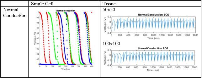
11 Scenarios Normal Conduction 50x50 model Basic cycle length (BCL) = 75ms Time = 2000ms Normal Conduction with Scar 50x50 model Basic cycle length (BCL) = 75ms Time = 2000ms Scar cells at x [10,15] and y [15,20] Excluding (10,15), (10,20), (15,15), and (15,20) Spiral Wave with Scar 50x50 model Basic cycle length (BCL) = 75ms Time = 2000ms Scar cells at x [10,15] and y [5,10] Excluding (10,5), (10,10), (15,5), and (15,10) Alternans 25x25 model Basic cycle length (BCL) = 54ms Time = 2000ms
12 Other Variables Stimulation Times Array of t-values at which the pacemaker cells stimulate Voltage(x,y,t) Action potential of a heart cell at a given time APD(x,y) Action potential duration of a heart cell DI(x,y) Diastolic interval of a heart cell Duration(x,y) Time elapsed since the cell s last excitation

13 Restitution Defines the relationship between the DI and the APD APD n = f D n 1 f D n f D n = A max A 0 e D n/τ = 60 50e D n/20 As D n, f D n A max Cardiac dynamics unstable for f D n > 1
 As t, f(a, t) 0 The greater A is, the slower the cell depolarizes f(t) = e t/9.7025 0.01+ e t/9.7025 T(66) 9.")
14 Action Potential Signifies what happens to a cell after it has been stimulated as time progresses f(a, t) = e t/t(a) c+ e t/t(a) T(A) = A ln 0.9 ln(0.1 c) As t, f(a, t) 0 The greater A is, the slower the cell depolarizes f(t) = e t/ e t/ T(66)

15 Stimulation & Wave Propagation Stimulation If voltage 0.1, the cell depolarizes (voltage becomes 1 V) Wave Propagation If voltage 0.1, the cell s neighbors are checked If at least 3 neighbors are excited, the evaluated cell becomes excited Otherwise, the cell evolves Depolarization DI of previous heartbeat is calculated APD of next beat is determined Voltage becomes 1 V Duration resets Evolution Duration increments Voltage changes based on APD and duration Black cell is being evaluated Gray cells are the neighbors being checked
16 Simulation 3x3 group of pacemaker cells stimulate at t = 0 At every time step, the propagation function is called at each cell If scar cells exist, they are set to 0 V When t reaches a stimulation time, the pacemaker cells become excited Process repeats until the entire interval is covered
17 Constant DI Used as a control mechanism Heartbeats are regulated by DI rather than BCL Stimulation times are not necessarily equally spaced throughout

18 Electrocardiogram (ECG) Diagram used to illustrate electrical activity in the heart Measures voltage difference between two points outside the tissue ECG = Φ e B Φ e A Φ e x, y = V m 1 r dx dy r = x x 2 + y y 2 1/2

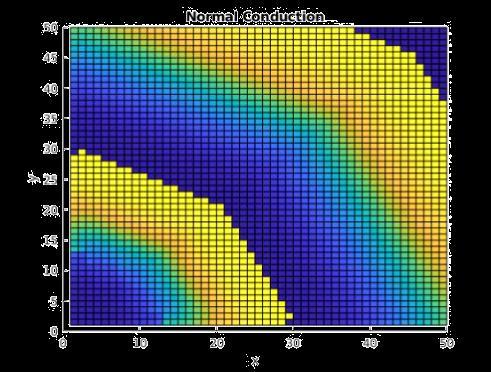
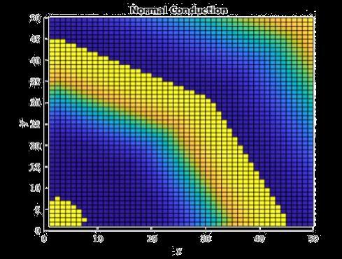
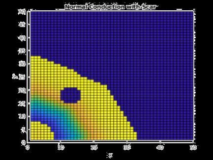
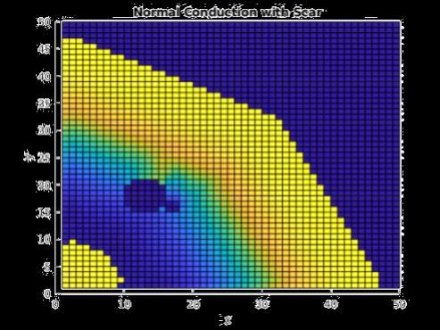
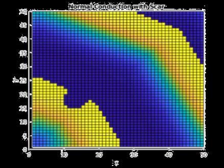
19 Results Normal Conduction Normal Conduction with Scar


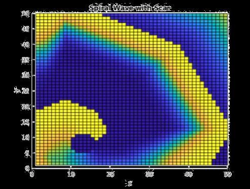
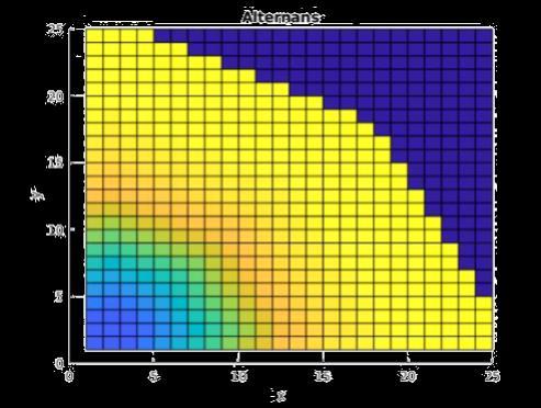
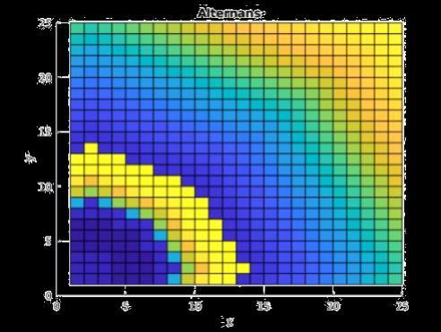
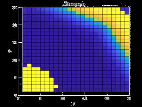
20 Results Spiral Wave with Scar Alternans
21 Normal Conduction
22 No Control vs Constant DI Control No Control tstart = t tend = tstart + BCL Constant DI tstart = t tend = tstart + APD(1,1) + DI_target DI_target = BCL APD(1,1) APD(1,1) = ms Normal Conduction with Scar & Spiral Wave with Scar BCL = 75ms DI_target 19 Alternans BCL = 54ms DI_target -2


23 No Control vs Constant DI Control
24 Conclusion Constant DI effectively controlled alternans in smaller tissue Benefits of cellular automata Future work 3D simulation GPU implementation Controlling other heart scenarios Constant RT control Other control mechanisms?
25 References Electrocardiogram ECG. mydr. Feldman, A.B., Chernyak, Y.B., & Cohen, R.J. (1999). Cellular automata model of cardiac excitation waves. Herzschrittmachertherapie und Elektrophysiologie, 10, Flavio H Fenton et al. (2008). Cardiac arrhythmia. Scholarpedia, 3(7):1665. Garcia, G. & Sheth, N. (n.d.). Understanding Cardiac Arrhythmias through Cellular Automata. University of Tennessee, Knoxville. Zlochiver, S., Johnson, C., and Tolkacheva, E. G. (2017). Constant DI pacing suppresses cardiac alternans formation in numerical cable models. American Institute of Physics. Chaos 27,
Arrhythmias By Dimension or Heart Attacks can give you Mathematics
 Arrhythmias By Dimension or Heart Attacks can give you Mathematics J. P. Keener Mathematics Department Arrhythmias By DimensionorHeart Attacks can give you Mathematics p.1/26 The Medical Problem Sudden
The Electrocardiogram
 The Electrocardiogram Chapters 11 and 13 AUTUMN WEDAN AND NATASHA MCDOUGAL The Normal Electrocardiogram P-wave Generated when the atria depolarizes QRS-Complex Ventricles depolarizing before a contraction
Lab 2. The Intrinsic Cardiac Conduction System. 1/23/2016 MDufilho 1
 Lab 2 he Intrinsic Cardiac Conduction System 1/23/2016 MDufilho 1 Figure 18.13 Intrinsic cardiac conduction system and action potential succession during one heartbeat. Superior vena cava ight atrium 1
Shock-induced termination of cardiac arrhythmias
 Shock-induced termination of cardiac arrhythmias Group members: Baltazar Chavez-Diaz, Chen Jiang, Sarah Schwenck, Weide Wang, and Jinglei Zhang Abstract: Cardiac arrhythmias occur when blood flow to the
Systems Biology Across Scales: A Personal View XXVII. Waves in Biology: Cardiac Arrhythmia. Sitabhra Sinha IMSc Chennai
 Systems Biology Across Scales: A Personal View XXVII. Waves in Biology: Cardiac Arrhythmia Sitabhra Sinha IMSc Chennai The functional importance of biological waves Spiral Waves Cardiac Arrhythmias Arrhythmias:
Shock-induced termination of cardiac arrhythmias
 Shock-induced termination of cardiac arrhythmias Group members: Baltazar Chavez-Diaz, Chen Jiang, Sarah Schwenck, Weide Wang, and Jinglei Zhang Cardiac arrhythmias, also known as irregular heartbeat, occur
Cardiac Cycle. Each heartbeat is called a cardiac cycle. First the two atria contract at the same time.
 The Heartbeat Cardiac Cycle Each heartbeat is called a cardiac cycle. First the two atria contract at the same time. Next the two ventricles contract at the same time. Then all the chambers relax. http://www.youtube.com/watch?v=frd3k6lkhws
Medical Cyber-Physical Systems
 Medical Cyber-Physical Systems Electrophysiology basics Lecture 10 Principles of Modeling for Cyber-Physical Systems Instructor: Madhur Behl Many thanks to: Zhihao Jiang, Houssam Abbas, and Rahul Mangharam,
A Dynamic model of Pulmonary Vein Electrophysiology. Harry Green 2 nd year Ph.D. student University of Exeter
 A Dynamic model of Pulmonary Vein Electrophysiology Harry Green 2 nd year Ph.D. student University of Exeter Background to the Project Cardiac disease is the leading cause of death in most developed countries
The Function of an ECG in Diagnosing Heart Conditions. A useful guide to the function of the heart s electrical system for patients receiving an ECG
 The Function of an ECG in Diagnosing Heart Conditions A useful guide to the function of the heart s electrical system for patients receiving an ECG Written by Erhan Selvi July 28, 2014 Audience and Scope
EKG Abnormalities. Adapted from:
 EKG Abnormalities Adapted from: http://www.bem.fi/book/19/19.htm Some key terms: Arrhythmia-an abnormal rhythm or sequence of events in the EKG Flutter-rapid depolarizations (and therefore contractions)
NATIONAL INSTITUTE FOR HEALTH AND CLINICAL EXCELLENCE
 NATIONAL INSTITUTE FOR HEALTH AND CLINICAL EXCELLENCE Implantable cardioverter defibrillators for the treatment of arrhythmias and cardiac resynchronisation therapy for the treatment of heart failure (review
THE CARDIOVASCULAR SYSTEM. Heart 2
 THE CARDIOVASCULAR SYSTEM Heart 2 PROPERTIES OF CARDIAC MUSCLE Cardiac muscle Striated Short Wide Branched Interconnected Skeletal muscle Striated Long Narrow Cylindrical PROPERTIES OF CARDIAC MUSCLE Intercalated
ECG interpretation basics
 ECG interpretation basics Michał Walczewski, MD Krzysztof Ozierański, MD 21.03.18 Electrical conduction system of the heart Limb leads Precordial leads 21.03.18 Precordial leads Precordial leads 21.03.18
Full file at
 MULTIPLE CHOICE. Choose the one alternative that best completes the statement or answers the question. 1) What electrical event must occur for atrial kick to occur? 1) A) Atrial repolarization B) Ventricular
Interpreting Electrocardiograms (ECG) Physiology Name: Per:
 Physiology Name: Per:") Interpreting Electrocardiograms (ECG) Physiology Name: Per: Introduction The heart has its own system in place to create nerve impulses and does not actually require the brain to make it beat. This electrical
Interpreting Electrocardiograms (ECG) Physiology Name: Per: Introduction The heart has its own system in place to create nerve impulses and does not actually require the brain to make it beat. This electrical
Cardiac arrhythmias. Janusz Witowski. Department of Pathophysiology Poznan University of Medical Sciences. J. Witowski
 Cardiac arrhythmias Janusz Witowski Department of Pathophysiology Poznan University of Medical Sciences A 68-year old man presents to the emergency department late one evening complaining of increasing
Paroxysmal Supraventricular Tachycardia PSVT.
 Atrial Tachycardia; is the name for an arrhythmia caused by a disorder of the impulse generation in the atrium or the AV node. An area in the atrium sends out rapid signals, which are faster than those
EKG Competency for Agency
 EKG Competency for Agency Name: Date: Agency: 1. The upper chambers of the heart are known as the: a. Atria b. Ventricles c. Mitral Valve d. Aortic Valve 2. The lower chambers of the heart are known as
Project Title Temporary Pacemaker Training Simulator
 Project Title Temporary Pacemaker Training Simulator Project Description Problem: There is no available training device for temporary pacemakers (pacemakers). A training device will have to essentially
PART I. Disorders of the Heart Rhythm: Basic Principles
 PART I Disorders of the Heart Rhythm: Basic Principles FET01.indd 1 1/11/06 9:53:05 AM FET01.indd 2 1/11/06 9:53:06 AM CHAPTER 1 The Cardiac Electrical System The heart spontaneously generates electrical
UNDERSTANDING YOUR ECG: A REVIEW
 UNDERSTANDING YOUR ECG: A REVIEW Health professionals use the electrocardiograph (ECG) rhythm strip to systematically analyse the cardiac rhythm. Before the systematic process of ECG analysis is described
Modeling Chaos in the Heart
 Modeling Chaos in the Heart S. M. Haider Aejaz haider_aejaz@yahoo.com PIMCS, Lahore, Pakistan Abstract: Chaos theory describes the dynamics of a deterministic system whose dynamics show extreme dependence
Electrocardiography Abnormalities (Arrhythmias) 7. Faisal I. Mohammed, MD, PhD
 7. Faisal I. Mohammed, MD, PhD") Electrocardiography Abnormalities (Arrhythmias) 7 Faisal I. Mohammed, MD, PhD 1 Causes of Cardiac Arrythmias Abnormal rhythmicity of the pacemaker Shift of pacemaker from sinus node Blocks at different
Electrocardiography Abnormalities (Arrhythmias) 7 Faisal I. Mohammed, MD, PhD 1 Causes of Cardiac Arrythmias Abnormal rhythmicity of the pacemaker Shift of pacemaker from sinus node Blocks at different
CASE 10. What would the ST segment of this ECG look like? On which leads would you see this ST segment change? What does the T wave represent?
 CASE 10 A 57-year-old man presents to the emergency center with complaints of chest pain with radiation to the left arm and jaw. He reports feeling anxious, diaphoretic, and short of breath. His past history
Enhanced Cellular Automation Model For Detecting Heart Fibrillation
 Enhanced Cellular Automation Model For Detecting Heart Fibrillation R. Touqan, A. Nasser & N. Hamdy Arab Academy for Science and Technology (AAST), email< rtouqan@hotmail.com Abstract: A new technique
Comparison of Different ECG Signals on MATLAB
 International Journal of Electronics and Computer Science Engineering 733 Available Online at www.ijecse.org ISSN- 2277-1956 Comparison of Different Signals on MATLAB Rajan Chaudhary 1, Anand Prakash 2,
Electrical Conduction
 Sinoatrial (SA) node Electrical Conduction Sets the pace of the heartbeat at 70 bpm AV node (50 bpm) and Purkinje fibers (25 40 bpm) can act as pacemakers under some conditions Internodal pathway from
CRC 431 ECG Basics. Bill Pruitt, MBA, RRT, CPFT, AE-C
 CRC 431 ECG Basics Bill Pruitt, MBA, RRT, CPFT, AE-C Resources White s 5 th ed. Ch 6 Electrocardiography Einthoven s Triangle Chest leads and limb leads Egan s 10 th ed. Ch 17 Interpreting the Electrocardiogram
WHAT S THAT RHYTHM I AM HEARING? GUIDE TO AUSCULTATION OF ARRHYTHMIAS IN HORSES
 WHAT S THAT RHYTHM I AM HEARING? GUIDE TO AUSCULTATION OF ARRHYTHMIAS IN HORSES Michelle Henry Barton DVM, PhD, DACVIM University of Georgia, Athens, GA INTRODUCTION The purpose of this talk is to review
Where are the normal pacemaker and the backup pacemakers of the heart located?
 CASE 9 A 68-year-old woman presents to the emergency center with shortness of breath, light-headedness, and chest pain described as being like an elephant sitting on her chest. She is diagnosed with a
BIPN100 F15 Human Physiology I (Kristan) Problem set #5 p. 1
 Problem set #5 p. 1") BIPN100 F15 Human Physiology I (Kristan) Problem set #5 p. 1 1. Dantrolene has the same effect on smooth muscles as it has on skeletal muscle: it relaxes them by blocking the release of Ca ++ from the
BIPN100 F15 Human Physiology I (Kristan) Problem set #5 p. 1 1. Dantrolene has the same effect on smooth muscles as it has on skeletal muscle: it relaxes them by blocking the release of Ca ++ from the
Death, dynamics and disorder: Terminating reentry in excitable media by dynamically-induced inhomogeneities
 PRAMANA c Indian Academy of Sciences Vol. 64, No. 4 journal of April 2005 physics pp. 553 562 Death, dynamics and disorder: Terminating reentry in excitable media by dynamically-induced inhomogeneities
Pacemaker Concepts and Terminology*
 Pacemaker Concepts and Terminology* BAROUH V BERKOVITS, E.E., Ing. Associate in Surgery, Harvard Medical School, Boston, Mass., Associate in Electrophysiology, Miami University, Miami, Fla., Senior Research
Arrhythmias. Simple-dysfunction cause abnormalities in impulse formation and conduction in the myocardium.
 Arrhythmias Simple-dysfunction cause abnormalities in impulse formation and conduction in the myocardium. However, in clinic it present as a complex family of disorders that show variety of symptoms, for
The heart's "natural" pacemaker is called the sinoatrial (SA) node or sinus node.
 node or sinus node.") PACEMAKER Natural pacemaker: The heart's "natural" pacemaker is called the sinoatrial (SA) node or sinus node. Artificial pacemaker: It is a small, battery-operated device that helps the heart beat in
PACEMAKER Natural pacemaker: The heart's "natural" pacemaker is called the sinoatrial (SA) node or sinus node. Artificial pacemaker: It is a small, battery-operated device that helps the heart beat in
Patient Resources: Arrhythmias and Congenital Heart Disease
 Patient Resources: Arrhythmias and Congenital Heart Disease Overview Arrhythmias (abnormal heart rhythms) can develop in patients with congenital heart disease (CHD) due to thickening/weakening of their
Emergency Medical Training Services Emergency Medical Technician Paramedic Program Outlines Outline Topic: WPW Revised: 11/2013
 Emergency Medical Training Services Emergency Medical Technician Paramedic Program Outlines Outline Topic: WPW Revised: 11/2013 Wolff-Parkinson-White syndrome (WPW) is a syndrome of pre-excitation of the
Electrocardiography for Healthcare Professionals
 Electrocardiography for Healthcare Professionals Chapter 7: Junctional Dysrhythmias 2012 The Companies, Inc. All rights reserved. Learning Outcomes 7.1 Describe the various junctional dysrhythmias 7.2
8/20/2012. Learning Outcomes (Cont d)
") 1 2 3 4 Electrocardiography for Healthcare Professionals Chapter 7: Junctional Dysrhythmias Learning Outcomes 7.1 Describe the various junctional dysrhythmias 7.2 Identify premature junctional complexes
1 2 3 4 Electrocardiography for Healthcare Professionals Chapter 7: Junctional Dysrhythmias Learning Outcomes 7.1 Describe the various junctional dysrhythmias 7.2 Identify premature junctional complexes
The Mammalian Circulatory System
 The Mammalian Heart The Mammalian Circulatory System Recall: What are the 3 cycles of the mammalian circulatory system? What are their functions? What are the three main vessel types in the mammalian circulatory
QUIZ/TEST REVIEW NOTES SECTION 1 CARDIAC MYOCYTE PHYSIOLOGY [CARDIOLOGY]
![QUIZ/TEST REVIEW NOTES SECTION 1 CARDIAC MYOCYTE PHYSIOLOGY [CARDIOLOGY]](/thumbs/96/126998162.jpg "QUIZ/TEST REVIEW NOTES SECTION 1 CARDIAC MYOCYTE PHYSIOLOGY [CARDIOLOGY]") QUIZ/TEST REVIEW NOTES SECTION 1 CARDIAC MYOCYTE PHYSIOLOGY [CARDIOLOGY] Learning Objectives: Describe the ionic basis of action potentials in cardiac contractile and autorhythmic cells Explain the relationship
QUIZ/TEST REVIEW NOTES SECTION 1 CARDIAC MYOCYTE PHYSIOLOGY [CARDIOLOGY] Learning Objectives: Describe the ionic basis of action potentials in cardiac contractile and autorhythmic cells Explain the relationship
The Nuts and Bolts of ICD Therapy
 Electrical Management of Cardiac Rhythm Disorders For Cardiology Fellows December 5-8 Austin, Texas The Nuts and Bolts of ICD Therapy 1 2 Action Potential Localized Differences in Conduction Conduction
Step by step approach to EKG rhythm interpretation:
 Sinus Rhythms Normal sinus arrhythmia Small, slow variation of the R-R interval i.e. variation of the normal sinus heart rate with respiration, etc. Sinus Tachycardia Defined as sinus rhythm with a rate
Electrocardiography Biomedical Engineering Kaj-Åge Henneberg
 Electrocardiography 31650 Biomedical Engineering Kaj-Åge Henneberg Electrocardiography Plan Function of cardiovascular system Electrical activation of the heart Recording the ECG Arrhythmia Heart Rate
Electrocardiography for Healthcare Professionals
 Electrocardiography for Healthcare Professionals Kathryn A. Booth Thomas O Brien Chapter 5: Rhythm Strip Interpretation and Sinus Rhythms Learning Outcomes 5.1 Explain the process of evaluating ECG tracings
TEST BANK FOR ECGS MADE EASY 5TH EDITION BY AEHLERT
 Link download full: http://testbankair.com/download/test-bank-for-ecgs-made-easy-5thedition-by-aehlert/ TEST BANK FOR ECGS MADE EASY 5TH EDITION BY AEHLERT Chapter 5 TRUE/FALSE 1. The AV junction consists
Chapter 9. Learning Objectives. Learning Objectives 9/11/2012. Cardiac Arrhythmias. Define electrical therapy
 Chapter 9 Cardiac Arrhythmias Learning Objectives Define electrical therapy Explain why electrical therapy is preferred initial therapy over drug administration for cardiac arrest and some arrhythmias
Basic Dysrhythmia Interpretation
 Basic Dysrhythmia Interpretation Objectives 2 To understand the Basic ECG To understand the meaning of Dysrhythmia To describe the normal heart conduction system. To describe the normal impulse pathways.
Circulatory system of mammals
 Circulatory system of mammals Explain the cardiac cycle and its initiation Discuss the internal factors that control heart action Blood flows through the heart as a result of pressure differences Blood
Practice Exercises for the Cardiovascular System
 Practice Exercises for the Cardiovascular System On the diagram below, color the oxygen-rich blood red and the oxygen-poor blood blue. Label the parts: Continued on the next page... Label the parts on
Cardiovascular System Notes: Heart Disease & Disorders
 Cardiovascular System Notes: Heart Disease & Disorders Interesting Heart Facts The Electrocardiograph (ECG) was invented in 1902 by Willem Einthoven Dutch Physiologist. This test is still used to evaluate
Introduction. Circulation
 Introduction Circulation 1- Systemic (general) circulation 2- Pulmonary circulation carries oxygenated blood to all parts of the body carries deoxygenated blood to the lungs From Lt. ventricle aorta From
Collin County Community College
 Collin County Community College BIOL. 2402 Anatomy & Physiology WEEK 5 The Heart 1 The Heart Beat and the EKG 2 1 The Heart Beat and the EKG P-wave = Atrial depolarization QRS-wave = Ventricular depolarization
EKG Intermediate Tips, tricks, tools
 Birmingham Regional Emergency Medical Services System 2018 ALCTE Summer Conference EKG Intermediate Tips, tricks, tools Brian Gober, MAT, ATC, NRP, CSCS Education Services Manager ECC Training Center Coordinator
Temporal Analysis and Remote Monitoring of ECG Signal
 Temporal Analysis and Remote Monitoring of ECG Signal Amruta Mhatre Assistant Professor, EXTC Dept. Fr.C.R.I.T. Vashi Amruta.pabarekar@gmail.com Sadhana Pai Associate Professor, EXTC Dept. Fr.C.R.I.T.
ECG Signal Characterization and Correlation To Heart Abnormalities
 ECG Signal Characterization and Correlation To Heart Abnormalities Keerthi G Reddy 1, Dr. P A Vijaya 2, Suhasini S 3 1PG Student, 2 Professor and Head, Department of Electronics and Communication, BNMIT,
Rhythmical Excitation of the Heart
 Rhythmical Excitation of the Heart KALEB HOOD AND JIMMY JOHNSON Special Excitory and Conductive System of the Heart Sinus Node (or sinoatrial node or S-A): A small node with almost no contractile muscle,
Catheter Ablation. Patient Education
 Catheter Ablation Patient Education Allina Health System Your heart has four chambers. Two upper chambers (atria) pump blood to the two lower chambers (ventricles). In order for the heart to pump, it requires
Arrhythmias. 1. beat too slowly (sinus bradycardia). Like in heart block
. Like in heart block") Arrhythmias It is a simple-dysfunction caused by abnormalities in impulse formation and conduction in the myocardium. The heart is designed in such a way that allows it to generate from the SA node electrical
Arrhythmias It is a simple-dysfunction caused by abnormalities in impulse formation and conduction in the myocardium. The heart is designed in such a way that allows it to generate from the SA node electrical
EKG Rhythm Interpretation Exam
 as EKG Rhythm Interpretation Exam Name: Date: ID# Unit Assume each strip is a 6 second strip. Passing is 80%. 1. Identify the following rhythm: a. Asystole b. Ventricular fibrillation c. Atrial fibrillation
2017 BDKA Review. Regularity Rate P waves PRI QRS Interpretation. Regularity Rate P waves PRI QRS Interpretation 1/1/2017
 1. 2017 BDKA Review 2. 3. 4. Interpretation 5. QT 6. 7. 8. 9. 10. QT 11. 12. 13. 14. 15. 16. 17. 18. QT 19. 20. QT 21. 22. QT 23. 24. Where are pacer spikes? Before the P wave or before the QRS complex?
Rate: The atrial and ventricular rates are equal; heart rate is greater than 100 bpm (usually between bpm).
.") Sinus Bradycardia Regularity: The R-R intervals are constant; the rhythm is regular. Rate: The atrial and ventricular rates are equal; heart rate is less than 60 bpm. P wave: There is a uniform P wave
Sinus Bradycardia Regularity: The R-R intervals are constant; the rhythm is regular. Rate: The atrial and ventricular rates are equal; heart rate is less than 60 bpm. P wave: There is a uniform P wave
Chapter 20 (2) The Heart
 The Heart") Chapter 20 (2) The Heart ----------------------------------------------------------------------------------------------------------------------------------------- Describe the component and function of
Chapter 20 (2) The Heart ----------------------------------------------------------------------------------------------------------------------------------------- Describe the component and function of
Electrocardiography I Laboratory
 Introduction The body relies on the heart to circulate blood throughout the body. The heart is responsible for pumping oxygenated blood from the lungs out to the body through the arteries and also circulating
Antiarrhythmic Drugs
 Antiarrhythmic Drugs DR ATIF ALQUBBANY A S S I S T A N T P R O F E S S O R O F M E D I C I N E / C A R D I O L O G Y C O N S U L T A N T C A R D I O L O G Y & I N T E R V E N T I O N A L E P A C H D /
Developing Electrocardiogram Mathematical Model for Cardiovascular Pathological Conditions and Cardiac Arrhythmia
 Indian Journal of Science and Technology, Vol 8(S10), DOI: 10.17485/ijst/015/v8iS10/84847, December 015 ISSN (Print) : 0974-6846 ISSN (Online) : 0974-5645 Developing Electrocardiogram Mathematical Model
Objectives of the Heart
 Objectives of the Heart Electrical activity of the heart Action potential EKG Cardiac cycle Heart sounds Heart Rate The heart s beat separated into 2 phases Relaxed phase diastole (filling of the chambers)
Chapter 16: Arrhythmias and Conduction Disturbances
 Complete the following. Chapter 16: Arrhythmias and Conduction Disturbances 1. Cardiac arrhythmias result from abnormal impulse, abnormal impulse, or both mechanisms together. 2. is the ability of certain
Testing the Accuracy of ECG Captured by Cronovo through Comparison of ECG Recording to a Standard 12-Lead ECG Recording Device
 Testing the Accuracy of ECG Captured by through Comparison of ECG Recording to a Standard 12-Lead ECG Recording Device Data Analysis a) R-wave Comparison: The mean and standard deviation of R-wave amplitudes
Intraoperative and Postoperative Arrhythmias: Diagnosis and Treatment
 Intraoperative and Postoperative Arrhythmias: Diagnosis and Treatment Karen L. Booth, MD, Lucile Packard Children s Hospital Arrhythmias are common after congenital heart surgery [1]. Postoperative electrolyte
HTEC 91. Performing ECGs: Procedure. Normal Sinus Rhythm (NSR) Topic for Today: Sinus Rhythms. Characteristics of NSR. Conduction Pathway
 Topic for Today: Sinus Rhythms. Characteristics of NSR. Conduction Pathway") HTEC 91 Medical Office Diagnostic Tests Week 3 Performing ECGs: Procedure o ECG protocol: you may NOT do ECG if you have not signed up! If you are signed up and the room is occupied with people who did
HTEC 91 Medical Office Diagnostic Tests Week 3 Performing ECGs: Procedure o ECG protocol: you may NOT do ECG if you have not signed up! If you are signed up and the room is occupied with people who did
Cardiac Implanted Electronic Devices Pacemakers, Defibrillators, Cardiac Resynchronization Devices, Loop Recorders, etc.
 Cardiac Implanted Electronic Devices Pacemakers, Defibrillators, Cardiac Resynchronization Devices, Loop Recorders, etc. The Miracle of Living February 21, 2018 Matthew Ostrom MD,FACC,FHRS Division of
EP WIRE on Management Preexcitation syndromes
 EP WIRE on Management Preexcitation syndromes 1. Is your Institution: A University Hospital 70.7% 41 A Private Hospital 13.8% 8 Other Type of Hospital 15.5% 9 Institution name: 50 answered question 58
CORONARY ARTERIES. LAD Anterior wall of the left vent Lateral wall of left vent Anterior 2/3 of interventricluar septum R & L bundle branches
 CORONARY ARTERIES RCA Right atrium Right ventricle SA node 55% AV node 90% Posterior wall of left ventricle in 90% Posterior third of interventricular septum 90% LAD Anterior wall of the left vent Lateral
NHS. Implantable cardioverter defibrillators (ICDs) for arrhythmias. National Institute for Health and Clinical Excellence. Issue date: January 2006
 for arrhythmias. National Institute for Health and Clinical Excellence. Issue date: January 2006") NHS National Institute for Health and Clinical Excellence Issue date: January 2006 Implantable cardioverter defibrillators (ICDs) for arrhythmias Understanding NICE guidance information for people with
NHS National Institute for Health and Clinical Excellence Issue date: January 2006 Implantable cardioverter defibrillators (ICDs) for arrhythmias Understanding NICE guidance information for people with
Signal Processing of Stress Test ECG Using MATLAB
 Signal Processing of Stress Test ECG Using MATLAB Omer Mukhtar Wani M. Tech ECE Geeta Engineering College, Panipat Abstract -Electrocardiography is used to record the electrical activity of the heart over
MA-1600: EKG - ELECTROCARDIOGRAM FUNDAMENTALS
 MA-1600: EKG - Electrocardiogram Fundamentals 1 MA-1600: EKG - ELECTROCARDIOGRAM FUNDAMENTALS Cuyahoga Community College Viewing:MA-1600 : EKG - Electrocardiogram Fundamentals Board of Trustees: May 2018
Simulation of T-Wave Alternans and its Relation to the Duration of Ventricular Action Potentials Disturbance
 The Open Pacing, Electrophysiology & Therapy Journal, 2010, 3, 21-27 21 Open Access Simulation of T-Wave Alternans and its Relation to the Duration of Ventricular Action Potentials Disturbance Dariusz
UNDERSTANDING ELECTROPHYSIOLOGY STUDIES
 UNDERSTANDING ELECTROPHYSIOLOGY STUDIES Testing and Treating Your Heart s Electrical System A Problem with Your Heart Rhythm The speed and pattern of a heartbeat is called the heart rhythm. The rhythm
Pediatrics. Arrhythmias in Children: Bradycardia and Tachycardia Diagnosis and Treatment. Overview
 Pediatrics Arrhythmias in Children: Bradycardia and Tachycardia Diagnosis and Treatment See online here The most common form of cardiac arrhythmia in children is sinus tachycardia which can be caused by
Arrhythmias. Pulmonary Artery
 Arrhythmias Introduction Cardiac arrhythmia is an irregularity of the heart beat that causes the heart to beat too slowly, too fast, or irregularly. There are different types of arrhythmias. Most arrhythmias
Paramedic Rounds. Tachyarrhythmia's. Sean Sutton Dallas Wood
 Paramedic Rounds Tachyarrhythmia's Sean Sutton Dallas Wood Objectives At the end of this session, the paramedic will be able to: State the key components of the cardiac conduction pathway, along with the
LABORATORY INVESTIGATION
 LABORATORY INVESTIGATION Recording Electrocardiograms The taking of an electrocardiogram is an almost universal part of any complete physical examination. From the ECG record of the electrical activity
4. The two inferior chambers of the heart are known as the atria. the superior and inferior vena cava, which empty into the left atrium.
 Answer each statement true or false. If the statement is false, change the underlined word to make it true. 1. The heart is located approximately between the second and fifth ribs and posterior to the
a lecture series by SWESEMJR
 Electrolyte disturbances Hypokalaemia Decreased extracellular potassium increases excitability in the myocardial cells and consequently the effect of very severe hypokalaemia is ventricular arrhythmia.
Figure 2. Normal ECG tracing. Table 1.
 Figure 2. Normal ECG tracing that navigates through the left ventricle. Following these bundle branches the impulse finally passes to the terminal points called Purkinje fibers. These Purkinje fibers are
Human Anatomy and Physiology II Laboratory Cardiovascular Physiology
 Human Anatomy and Physiology II Laboratory Cardiovascular Physiology 1 This lab involves two exercises: 1) Conduction System of the Heart and Electrocardiography and 2) Human Cardiovascular Physiology:
Defibrillator. BiomedGuy
 Defibrillator BiomedGuy Medtronic Physiocontrol LifePak 10 Introduction This life-support system is used by paramedic, hospital staff, and other trained authorized healthcare providers. Provides, ECG,
ID: BFH_ Start time: :00:00. Examination summary
 ECG analysis report Personal data Examination details Start time: 2000-01-01 00:00:00 Interpreting physician: Auto End time: 2000-01-02 01:39:16 Duration: 1d 01h 39min 16s Nondiagnostic part: 5.85% Examination
Arrhythmia Study Guide 3 Junctional and Ventricular Rhythms
 Arrhythmia Study Guide 3 Junctional and Ventricular Rhythms JUNCTIONAL RHYTHMS The AV Junction (Bundle of His and surrounding cells) only acts as pacemaker of the heart when the SA Node is not firing normally
ECG ABNORMALITIES D R. T AM A R A AL Q U D AH
 ECG ABNORMALITIES D R. T AM A R A AL Q U D AH When we interpret an ECG we compare it instantaneously with the normal ECG and normal variants stored in our memory; these memories are stored visually in
Introduction to ECG Gary Martin, M.D.
 Brief review of basic concepts Introduction to ECG Gary Martin, M.D. The electrical activity of the heart is caused by a sequence of rapid ionic movements across cell membranes resulting first in depolarization
The Rate-Adaptive Pacemaker: Developing Simulations and Applying Patient ECG Data
 The Rate-Adaptive Pacemaker: Developing Simulations and Applying Patient ECG Data Harriet Lea-Banks - Summer Internship 2013 In collaboration with Professor Marta Kwiatkowska and Alexandru Mereacre September
Arrhythmia Management Joshua M. Cooper, MD, FHRS, FACC
 Arrhythmia Management Joshua M. Cooper, MD, FHRS, FACC Professor of Medicine Director of Cardiac Electrophysiology Temple University Health System Plumbing Electrical System Bradyarrhythmias Sinus Node
Anesthesia Assistants Review Course
 American Association of Oral and Maxillofacial Surgeons Anesthesia Assistants Review Course Four Seasons Las Vegas February 24-25, 2018 Las Vegas, Nevada Anesthesia Assistants Review Course EKG Lecture
Dr.Binoy Skaria 13/07/15
 Dr.Binoy Skaria binoyskaria@hotmail.com binoy.skaria@heartofengland.nhs.uk 13/07/15 Acknowledgement Medtronic, Google images & Elsevier for slides Natalie Ryan, Events Manager, HEFT- for organising the
PATIENT WITH ARRHYTHMIA IN DENTIST S OFFICE. Małgorzata Kurpesa, MD., PhD. Chair&Department of Cardiology
 PATIENT WITH ARRHYTHMIA IN DENTIST S OFFICE Małgorzata Kurpesa, MD., PhD. Chair&Department of Cardiology Medical University of Łódź The heart is made up of four chambers Left Atrium Right Atrium Left Ventricle
Cardiac Telemetry Self Study: Part One Cardiovascular Review 2017 THINGS TO REMEMBER
 Please review the above anatomy of the heart. THINGS TO REMEMBER There are 3 electrolytes that affect cardiac function o Sodium, Potassium, and Calcium When any of these electrolytes are out of the normal
CARDIAC PHYSIOLOGY. Amelyn U. Ramos-Rafael,M.D. Functional Anatomy of the Heart
 CARDIAC PHYSIOLOGY Amelyn U. Ramos-Rafael,M.D. Functional Anatomy of the Heart 1 Functional Anatomy of The Heart The Atria relatively thin walled The Ventricles ventricular walls thicker than atrial walls