Accuracy of prenatal diagnosis of fetal congenital heart disease by different
|
|
|
- Piers Watson
- 5 years ago
- Views:
Transcription
1 Accuracy of prenatal diagnosis of fetal congenital heart disease by different methods with echocardiography Ying Zhang 1* * Corresponding author baogoubei@hotmail.com Ai-Lu Cai 1 caial_us@hotmail.com Miao Fan 2 fanmiao_r@sina.com Wei-dong Ren 1 renweidong_us@hotmail.com Ya-Jun Guo 1 guoyajun_us@hotmail.com Wei Sun 1 sunwei_us@hotmail.com 1 Department of Sonography, Shengjing Hospital of China Medical University, No. 36 Sanhao Street, Heping District, Shenyang, Liaoning, China 2 Department of Radiology, The First Affiliated Hospital of Sun Yat-sen University, Guangzhou , China 1
2 Abstracts Background: Prenatal screening and accurate evaluation of fetal CHD is of great importance as it contribute to put forward appropriate neonatal management and helps the parents to make the decision about the pregnancy. The aim of our study is to evaluate the accuracy of different methods of fetal echocardiography in detection of fetal congenital heart disease (CHD) and to analyze the reasons of misdiagnosis in each method. Methods: Four chamber view (4CV) screening method, four transverse views scanning method, and systematic scanning method were used to detect fetal congenital heart disease in 442 cases, of which 349 cases of confirmed normal heart were set into the normal heart group, and the remaining 93 cases of confirmed cardiac anomalies were set into the CHD group. The echocardiographic detection of fetal CHD of each method was recorded for later analysis. Results:The sensitivity of systematic scanning method was higher than the 4CV screening method (95.7% VS 53.8%; P<0.0125) and the four transverse views scanning method (95.7% VS 87.1%; P>0.0125) with significance and insignificance respectively. The negative predicative value of systematic scanning method was significantly higher than 4CV screening method (98.9% VS 89.0%; P<0.0125) while higher than the four transverse views scanning method (98.9% VS 96.6%; P>0.0125) with no significance. There was no statistical difference in specificity and positive predicative value between each two method in the detection of fetal CHD. Conclusions: The four transverse views scanning method is best suited to prenatal 2
3 cardiac screening program due to its high sensitivity and specificity and relatively less diagnostic views. Systematic scanning method is the most accurate but demanding for more experience and expertise and is better for further examination after screening. Key Words: prenatal diagnosis; congenital heart disease; fetal echocardiography; sequential segmental analysis 3
4 Background Congenital heart disease (CHD), accounting for about 0.6% of all live births 1,2, is the most common congenital malformation leading to perinatal morbidity and mortality and is considered the leading cause of death in newborn with congenital anomalies 3,4. Corrected prenatal diagnosis could offer appropriate neonatal management and therapeutic strategies that could reduce perinatal mortality in potential. Also, it may provide a better prenatal counseling and helps to make the correct decision about the treatment 5. Fetal two dimensional echocardiography (2DE) and color Doppler echocardiography (CDE) are currently the primary techniques for the diagnosis of fetal CHD. During the past twenty years, the prenatal diagnostic ability of CHD has been promoted dramatically due to the high resolution 2D ultrasound and the advances in Doppler technology 6. The examination could be preceded either by an obstetric screening sonographer or by a specialist fetal echocardiographer yet the fact is that prenatal diagnosis by fetal echocardiography does not guarantee to excluded all the cardiac malformations. The aim of our study is to evaluate the accuracy of different methods of fetal echocardiography in detection of fetal CHD and to analyze the reasons of misdiagnosis in each method. Methods This study was approved by the institutional ethic committee and written informed consent was obtained from all subjects. Subjects 4
5 This is a prospective study. In total, 467 fetuses undergoing fetal echocardiography in our center between January 2007 and December 2009 were included in the current study. Fetal 2DE and CDE were performed in all the fetuses and the data were saved as video clips. All the cases were undergoing follow-up for one year. Twenty five cases lost to follow-up were excluded and the remaining 442 cases constituted the subjects of the study, of which 349 cases of confirmed normal heart were set into the normal heart group, and the remaining 93 cases of confirmed cardiac anomalies were set into the CHD group. The golden standard for the grouping was the result of the follow-up, including postnatal echocardiography, confirmation by the operation, and the postmortem findings. Ultrasonography technique A Doppler ultrasound system (Voluson E8, GE Healthcare, Kretztechnik, Zipf, Austria), equipped with a 4-8MHz transabdominal transducer was used in our study. Three methods were then used to assess the fetal cardiac structure and to exclude cardiac malformations in detail respectively. Ostium secundum atrial septal defect (ASD) or patent ductus arteriousus was not considered as a CHD which should be diagnosed in uterus. All studies were carried out under the supervision of an expert fetal echocardiographer with 6 years experience in performing and interpreting fetal echocardiography. Method 1 Four chamber view (4CV) screening method A clear 4CV was acquired with apical or lateral insonation of the fetal heart. The symmetry of the four chambers, atrioventricular coordination, the position and 5
6 activity of the atrioventricular valves, the continuity of endocardial cushion and interventricular seputm and the left and right pulmonary vein draining into the left atrium could be evaluated in 4CV. Method 2 Four transverse views scanning method The method to acquire 4CV was same to Method one mentioned above. On the basis of 4CV, the probe turned gradually up to the fetal head to acquire the left and right outflow tract view and the three-vessel and trachea view (3VT). Cardiac anomalies associated with conotruncal such as tetrology of Fallot, double outlet right ventricle, common arterial trunk, transposition of the great arteries, coarctation of aorta, interrupted aortic arch and parts of ventricular septal defect (VSD) could be detected by the two outflow tract views and 3VT. Method 3 Systematic scanning method Sequential segmental analysis were systematically used in this method to identify the fetal visceral and cardiac position, morphologic structures at the atrial, ventricle and arterial trunk level orderly and respectively. To identify the cardiac and visceral position of the fetus, the sagittal plane of the fetus was firstly acquired to identify the position of fetal head and spine and then the transverse plane of the fetus was acquired by rotating the beam 90 counterclockwise from the initial sagittal plane to identify the left side and the right side of the fetus. As the relationship of fetal left side, right side, and the spine remains unchanged, the spine could be considered as the landmark to distinguish the left side from the right side in the transverse plane of both thorax level and abdominal level. The position of 6
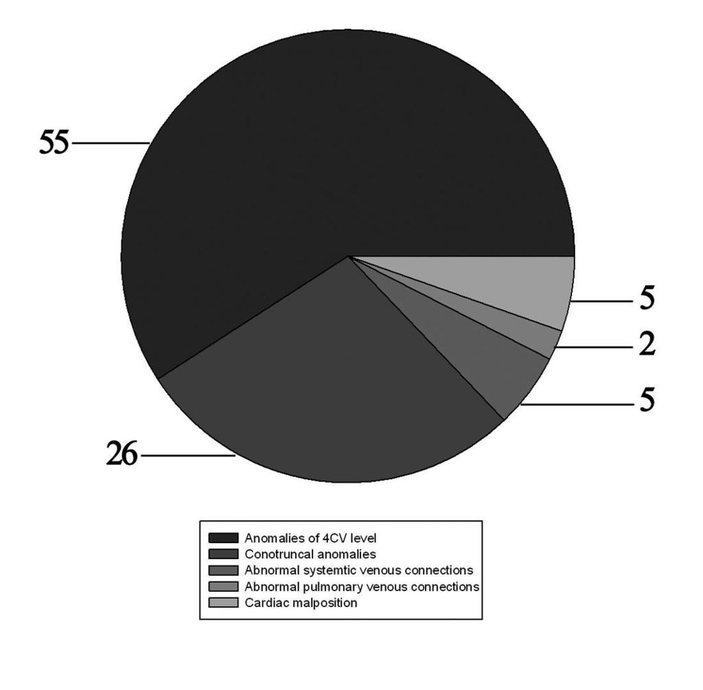
7 fetal heart and gastric vacuole were then determined correctly. The remaining examination was similar to infant echocardiography. Three basic areas including the atria, ventricles, and great arteries and their connections were analyzed to evaluate the atrial and ventricle arrangement, atrioventricular junction between the atria and ventricles, ventriculoarterial junction between the ventricle and arterial outflow tracts. Four transverse views including 4CV, the left and right outflow tract view, 3VT and three parallel sagittal views orthogonal to the initial 4CV, including aortic arch view, ductal arch view, and bicaval view, were used to evaluate the connections and the relationships of the adjacent segments mentioned above. Statistical analysis The sensitivity, specificity, positive predictive value and negative predicative value among 4CV screening method, four transverse views scanning method and systematic scanning method were compared via chi-square analysis of proportions. The alpha level was set at by Bonferroni correction. P< was considered statistical significant. Results Prenatal diagnosis of CHD with three methods and postnatal confirmation were summarized in Table 1. Total 93 cases of fetal CHD were confirmed by postnatal echocardiography, operation findings or autopsy findings and the classifications of CHD were shown in Figure 1. By means of 4CV screening method, 6 cases of VSD, one case of ASD sinus type, 5 cases of anomalies associated with drainage of superior vena cava (SVC) and inferior vena cava (IVC), 5 cases of anomalies associated with 7
8 cardiac malposition, and all the cases associated with conotruncal anomalies were not visualized prenatally. One patient diagnosed prenatally as having a VSD was found to be normal on postnatal echocardiography assessment. By means of four transverse views scanning method, 4 cases of VSD, one case of ASD sinus type, one case of aberrant IVC drainage, one case of ductus arterious closure in uterus, and 5 cases of anomalies associated with cardiac malposition were not detected prenatally. Three patients diagnosed prenatally as coarctation of aorta (COA) were proved to be normal by neonatal echocardiography. By means of systematic scanning method, only 4 cases of VSD were not detected prenatally while in one case suspected as COA proved to be normal after birth. Table 2 to Table 4 reveals the detection ability of CHD with the three methods respectively. The sensitivity of systematic scanning method in the detection of CHD was the highest among the three methods and was higher than the 4CV screening method (95.7% VS 53.8%; P<0.0125) and the four transverse views scanning method (95.7% VS 87.1%; P>0.0125) with significance and insignificance respectively. Also, the negative predicative value of systematic scanning method was significantly higher than 4CV screening method (98.9% VS 89.0%; P<0.0125) while higher than the four transverse views scanning method (98.9% VS 96.6%; P>0.0125) with no significance. There was no statistical difference in specificity and positive predicative value between each two method in the detection of fetal CHD (Table 5). Discussion Of all congenital anomalies, CHD is the most common and has the worse 8
9 prognosis compared with other disorders due to its high morbidity and mortality 3,4. Prenatal screening and accurate evaluation of fetal CHD is of great importance as it contribute to put forward appropriate neonatal management and helps the parents to make the decision about the pregnancy. Previous studies have shown that fetal echocardiography is of great value in the diagnosis of CHD and several methods were used in these reports CV is considered the most important diagnostic view in fetal echocardiography and is often the initial plane to acquire other planes 13. It is usually easy to obtain the standard 4CV plane if the spinal acoustic shadowing is avoided. The optimal view of 4CV is usually obtained when the cardiac apex is directed toward the maternal wall 14. 4CV screening alone could visualize most cardiac anomalies at the atrial and ventricle level, such as parts of VSD, atrioventricular septal defect (AVSD), Ebstein s anomaly, rhabdomyoma, ventricular diverticulum, and hypoplastic left heart syndrome (HLHS), etc. Yet the conotruncal anomalies could not be evaluated by 4CV as the two outflow tracts could not be visualized in 4CV and this lead to the low sensitivity of 4CV screening method. Previous study had shown that the detection rate of VSD by 4CV is significantly lower than by combination of the two outflow tract views as the fact was that parts of VSD was associated with the development of truncus arteriosus. Though the sensitivity of 4CV screening method is low, the specificity is high enough as 4CV could clearly demonstrate the anomalies in it with no doubt. In our study, only one case of VSD diagnosed prenatally by 4CV screening was proved to be normal heart after birth. 9
10 The sensitivity of four transverse views scanning method is no doubtly higher than 4CV screening method as more diagnostic views were added to evaluate the structures at conotruncal level besides 4CV level. It is easy to understand that most conotruncal anomalies could be detected by the combination of the four transverse views. The missed diagnosis of VSD in four cases maybe because the defect is too small to be visualized and the shunting speed is too low to be detected between the two ventricles. A ductus arteriosus closure was also missed prenatally maybe because of the lesion was not correctly recognized on 3VT. Other missed prenatal diagnosis was due to the lacking of corresponding diagnostic planes that was not included in the four transverse planes. The false positive results of 3 cases of COA with this method is because that the degree of coarctation is not serious and positioned at the aortic isthmus and the situation maybe improve after birth. Overall, the sensitivity and specificity of the four transverse views scanning method is ideal and high enough to fit for prenatal screening program. Systematic scanning method is a modality based on sequential segmental analysis of cardiac malformation. Sequential segmental analysis introduced by Van Praagh 15 is an old-new approach that has been widely used in the diagnosis of CHD in both infants and adults. Indeed, it provides a means of cataloguing and describing all congenital cardiac malformations, even if the combination of lesions has never previously been encountered 16. Detailed scan method of sequential segmental analysis has been widely used 17,18 and we will not repeat here. Yet in the circumstance of fetal cardiac examination, the identification of the visceral and 10
11 cardiac position is different from the postnatal echocardiography due to the variable fetal positions in uterus. The problem could be well resolved by two steps according to the approach proposed in our study. It is then easy to understand that all the cardiac anomalies associated with cardiac malposition that could not be visualized by the four transverse views scanning method were correctly diagnosed prenatally by systematic scanning method. In summary, many more views are included in the fetal echocardiography examination by systematic scanning method. Two views including the longitudinal section of fetal head and spine and the transverse section of fetal abdomen and thorax are used to identify the fetal visceral and cardiac position. Four transverse views including 4CV, the left and right outflow tract view and 3VT are used to confirm the malformations at atrial, ventricle and arterial trunks level. Besides, three sagittal sections including bicaval view, aortic arch view, and ductal arch view are added to the examination to improve the accuracy of the result. We found that parts of CHD that could not be determined by other methods could be diagnosed correctly by systematic scanning method, including abnormal systemic venous connections, ASD sinus type, ductus arteriosus closure, etc. The value of the added saggital views were well confirmed then. As a result, systematic scanning method is nevertheless the best method in detecting fetal CHD. Yet it is a very time-consuming work and demand for much technique and experience that exceed the ability of most ecumenical sonographers. As the four transverse views scanning method has both high sensitivity and specificity that were very closed to systematic scanning method, we believe it is of great value to 11
12 put forward this approach in general obstetric examinations. Conclusion We have made the comparison of the three methods in the detection of fetal CHD. The four transverse views scanning method is best suited to prenatal cardiac screening program as it has high sensitivity and specificity and a requirement of relatively small number of diagnostic views. Systematic scanning method is the most accurate but demanding for more experience and expertise of the sonographers and is better for further examination after a positive result for fetal cardiac screening. Competing interests The authors declare that they have no competing interests. Authors contributions YZ and A-lC designed the whole study. YZ, W-dR, MF, WS, Y-jG and A-lC drafted the manuscript. WS and Y-jG performed the fetal echocardiography. All authors read and approved the final manuscript. Acknowledgement We highly acknowledge the support of professor Wei-dong Ren, chair of our department, for his great help in the study. We also appreciate Dr. Ya-jun Guo and Dr. Wei Sun, for their great effort in performing all the fetal echocardiography examinations in the study. 12
13 References 1. Hoffman JIE, Kaplan S: The incidence of congenital heart disease. J Am Coll Cardiol 2002, 39: Massaro AN, El-Dib M, Glass P, Aly H: Factors associated with adverse neurodevelopmental outcomes in infants with congenital heart disease. Brain Dev 2008, 30: Gillum RF: Epidemiology of congenital heart disease in the United States. Am Heart J. 1994; 127(4 Pt 1): Gembruch U: Prenatal diagnosis of congenital heart disease. Prenat Diagn 1997, 17: Mellander M: Perinatal management, counselling and outcome of fetuses with congenital heart disease. Semin Fetal Neonatal Med 2005, 10: Allan L: Technique of fetal echocardiography. Pediatr Cardiol 2004, 25: Giancotti A, Torcia F, Giampa G, Gallo G, Gallo F, Donati L, De Santo D: Prenatal evaluation of congenital heart disease in high-risk pregnancies. Clin Exp Obstet Gynecol 1995, 22: Berghella V, Pagotto L, Kaufman M, Huhta JC, Wapner RJ: Accuracy of prenatal diagnosis of congenital heart defects. Fetal Diagn Ther 2001, 16: Meyer-Wittkopf M, Cooper S, Sholler G: Correlation between fetal cardiac diagnosis by obstetric and pediatric cardiologist sonographers and comparison with postnatal findings. Ultrasound Obstet Gynecol 2001, 17:
14 10. Forbus GA, Atz AM, Shirali GS: Implications and limitations of an abnormal fetal echocardiogram. Am J Cardiol 2004, 94: Gottliebson WM, Border WL, Franklin CM, Meyer RA, Michelfelder EC: Accuracy of fetal echocardiography: a cardiac segment-specific analysis. Ultrasound Obstet Gynecol 2006, 28: Bakiler AR, Ozer EA, Kanik A, Kanit H, Aktas FN: Accuracy of prenatal diagnosis of congenital heart disease with fetal echocardiography. Fetal Diagn Ther 2007, 22: Allan LD: A practical approach to fetal heart scanning. Semin Perinatol 2000, 24: Fetal Echocardiography Task Force; American Institute of Ultrasound in Medicine Clinical Standards Committee; American College of Obstetricians and Gynecologists; Society for Maternal-Fetal Medicine: AIUM practice guideline for the performance of fetal echocardiography. J Ultrasound Med 2011, 30: Van Praagh R: The segmental approach to diagnosis in congenital heart disease. Birth Defects1972,8: Anderson RH, Shirali G: Sequential segmental analysis. Ann Pediatr Cardio 2009, 2: Yoo SJ, Lee YH, Cho KS, Kim DY: Sequential segmental approach to fetal congenital heart disease. Cardiol Young 1999, 9: Carvalho JS, Ho SY, Shinebourne EA: Sequential segmental analysis in complex fetal cardiac abnormalities: a logical approach to diagnosis. Ultrasound 14
15 Obstet Gynecol 2005, 26:
16 Figure legends Figure 1 Classifications of congenital heart disease in 93 cases in our series 4CV, four chamber view 16
17 Table 1 Comparison of cardiac anomalies diagnosed by three methods prenatal and postnatal confirmation Prenatal Diagnosis 4CV screening method Four transverse views scanning Systematic scanning method Postnatal method VSD AVSD ASD (Sinus type) IVC drainage anomalies SVC drainage anomalies TAPV rhabdomyoma Ventricular diverticulum Ebstein s anomaly HLHS DORV TOF CAT TGA cctga COA
18 IAA Right aortic arch Ductus arteriosus closure PV/PA stenosis AV/AO stensosis dextrocardia levocardia Cardiac shifting Total ASD, atrial septal defect; AVSD, atrioventricular septal defect; CAT, common arterial trunk; cctga, congenital corrected transposition of the great arteries; COA, coarctation of aorta; DORV, double outlet right ventricle; HLHS, hypoplastic left heart syndrome; IAA, interrupted aortic arch; IVC, inferior vena cava; SVC, superior vena cava; TAPV, total anomalous pulmonary venous connection; TGA, transposition of the great arteries; TOF, tetrology of Fallot; VSD, ventricular septal defect 18
19 Table 2 Results of four chamber view screening method in the diagnosis of fetal congenital heart disease CHD group Normal heart group Total Detected Not detected Total CHD, congenital heart disease 19
20 Table 3 Results of four transverse views scanning method in the diagnosis of fetal congenital heart disease CHD group Normal heart group Total Detected Not detected Total CHD, congenital heart disease 20
21 Table 4 Results of systematic scanning method in the diagnosis of fetal congenital heart disease CHD group Normal heart group Total Detected Not detected Total CHD, congenital heart disease 21
22 Table 5 Comparison of the three methods in the diagnosis fetal congenital heart disease by echocardiography Detection of CHD P value m1 m2 m3 m1 & m2 m1 & m3 m2 & m3 Sensitivity 53.8% 87.1% 95.7% * * Specificity 99.7% 99.1% 99.7% Positive predictive value 98.0% 96.4% 98.9% Negative predicative value 89.0% 96.6% 98.9% * * CHD, congenital heart disease; m1, four chamber view screening method; m2, four transverse views scanning method; m3, systematic scanning method *: statistical significant (chi-square analysis with the alpha level set at by Bonferroni correction). 22
23 23
24 Figure 1
Heart and Soul Evaluation of the Fetal Heart
 Heart and Soul Evaluation of the Fetal Heart Ivana M. Vettraino, M.D., M.B.A. Clinical Associate Professor, Michigan State University College of Human Medicine Objectives Review the embryology of the formation
Diagnosis of Congenital Cardiac Defects Between 11 and 14 Weeks Gestation in High-Risk Patients
 Article Diagnosis of Congenital Cardiac Defects Between 11 and 14 Weeks Gestation in High-Risk Patients Zeev Weiner, MD, Abraham Lorber, MD, Eliezer Shalev, MD Objective. To examine the feasibility of
Heart and Lungs. LUNG Coronal section demonstrates relationship of pulmonary parenchyma to heart and chest wall.
 Heart and Lungs Normal Sonographic Anatomy THORAX Axial and coronal sections demonstrate integrity of thorax, fetal breathing movements, and overall size and shape. LUNG Coronal section demonstrates relationship
ULTRASOUND OF THE FETAL HEART
 ULTRASOUND OF THE FETAL HEART Cameron A. Manbeian, MD Disclosure Statement Today s faculty: Cameron Manbeian, MD does not have any relevant financial relationships with commercial interests or affiliations
Summary. HVRA s Cardio Vascular Genetic Detailed L2 Obstetrical Ultrasound. CPT 76811, 76825, _ 90% CHD detection. _ 90% DS detection.
 What is the role of fetal echocardiography (2D 76825, cardiovascular color flow mapping 93325) as performed in conjunction with detailed fetal anatomy scan (CPT 76811) now that AIUM requires limited outflow
Making Sense of Cardiac Views and Imaging Characteristics for 13 Congenital Heart Defects (CHDs)
") Making Sense of Cardiac Views and Imaging Characteristics for 13 Congenital Heart Defects (CHDs) Manny Gaziano, MD, FACOG obimages.net obimages.net@gmail.com Acknowledgements: Krista Wald, RDMS, sonographer,
Making Sense of Cardiac Views and Imaging Characteristics for 13 Congenital Heart Defects (CHDs) Manny Gaziano, MD, FACOG obimages.net obimages.net@gmail.com Acknowledgements: Krista Wald, RDMS, sonographer,
Screening for Critical Congenital Heart Disease
 Screening for Critical Congenital Heart Disease Caroline K. Lee, MD Pediatric Cardiology Disclosures I have no relevant financial relationships or conflicts of interest 1 Most Common Birth Defect Most
Prenatal screening of fetal ventriculoarterial connections: benefits of 4D technique in fetal heart imaging
 Wang et al. Cardiovascular Ultrasound (2017) 15:17 DOI 10.1186/s12947-017-0108-5 RESEARCH Prenatal screening of fetal ventriculoarterial connections: benefits of 4D technique in fetal heart imaging Open
ISUOG Basic Training. Obtaining & Interpreting Heart Views Correctly Alfred Abuhamad, USA. Basic training. Editable text here
 ISUOG Basic Training Obtaining & Interpreting Heart Views Correctly Alfred Abuhamad, USA Learning Objectives 6, 7 & 8 At the end of the lecture you will be able to: describe how to assess cardiac situs
Pediatric Echocardiography Examination Content Outline
 Pediatric Echocardiography Examination Content Outline (Outline Summary) # Domain Subdomain Percentage 1 Anatomy and Physiology Normal Anatomy and Physiology 10% 2 Abnormal Pathology and Pathophysiology
Systematic approach to Fetal Echocardiography. Objectives. Introduction 11/2/2015
 Systematic approach to Fetal Echocardiography. Pediatric Echocardiography Conference, JCMCH November 7, 2015 Rajani Anand Objectives Fetal cardiology pre-test Introduction Embryology and Physiology of
Disclosures. Outline. Learning Objectives. Introduction. Introduction. Sonographic Screening Examination of the Fetal Heart
 Sonographic Screening Examination of the Fetal Heart Lami Yeo, MD Director of Fetal Cardiology Perinatology Research Branch of NICHD / NIH / DHHS Bethesda, MD and Detroit, Michigan, USA Professor, Division
Congenital Heart Defects
 Normal Heart Congenital Heart Defects 1. Patent Ductus Arteriosus The ductus arteriosus connects the main pulmonary artery to the aorta. In utero, it allows the blood leaving the right ventricle to bypass
September 28-30, 2018
 September 28-30, 2018 Course Director Optimizing Detection of Congenital Heart Disease: Important Anatomic Cardiac Regions The Top 5 Critical Anatomic Regions in Fetal Cardiac Imaging Alfred Abuhamad,
Fetal Echocardiography and the Routine Obstetric Sonogram
 JDMS 23:143 149 May/June 2007 143 Fetal Echocardiography and the Routine Obstetric Sonogram SHELLY ZIMBELMAN, RT(R)(CT), RDMS, RDCS ASAD SHEIKH, MD, RDCS Congenital heart disease (CHD) is the most common
PRACTICAL GUIDE TO FETAL ECHOCARDIOGRAPHY IC Huggon and LD Allan
 PRACTICAL GUIDE TO FETAL ECHOCARDIOGRAPHY IC Huggon and LD Allan Fetal Cardiology Unit, Harris Birthright Research Centre for Fetal Medicine, King's College Hospital, London, UK IMPORTANCE OF PRENATAL
COMPREHENSIVE EVALUATION OF FETAL HEART R. GOWDAMARAJAN MD
 COMPREHENSIVE EVALUATION OF FETAL HEART R. GOWDAMARAJAN MD Disclosure No Relevant Financial Relationships with Commercial Interests Fetal Echo: How to do it? Timing of Study -optimally between 22-24 weeks
Major Forms of Congenital Heart Disease: Consultant Pediatric and Fetal Cardiology King Abdulaziz Cardiac Center, National Guard Hospital Riyadh
 Major Forms of Congenital Heart Disease: Impact of Prenatal Detection and Diagnosis Dr Merna Atiyah Consultant Pediatric and Fetal Cardiology King Abdulaziz Cardiac Center, National Guard Hospital Riyadh
ECHOCARDIOGRAPHIC APPROACH TO CONGENITAL HEART DISEASE: THE UNOPERATED ADULT
 ECHOCARDIOGRAPHIC APPROACH TO CONGENITAL HEART DISEASE: THE UNOPERATED ADULT Karen Stout, MD, FACC Divisions of Cardiology University of Washington Medical Center Seattle Children s Hospital NO DISCLOSURES
The Fetal Cardiology Program
 The Fetal Cardiology Program at Texas Children s Fetal Center About the program Since the 1980s, Texas Children s Fetal Cardiology Program has provided comprehensive fetal cardiac care to expecting families
HDlive Silhouette Mode With Spatiotemporal Image Correlation for Assessment of the Fetal Heart
 ORIGINAL RESEARCH HDlive Silhouette Mode With Spatiotemporal Image Correlation for Assessment of the Fetal Heart Toshiyuki Hata, MD, PhD, Mohamed Ahmed Mostafa AboEllail, MD, Suraphan Sajapala, MD, Mari
Basic Fetal Cardiac Evaluation
 Basic Fetal Cardiac Evaluation Mert Ozan Bahtiyar, MD Director, Fetal Care Center Division of Maternal Fetal Medicine Department of Obstetrics, Gynecology and Reproductive Sciences S L I D E 1 Background
Accuracy of the Fetal Echocardiogram in Double-outlet Right Ventricle
 Blackwell Publishing IncMalden, USACHDCongenital Heart Disease 2006 The Authors; Journal compilation 2006 Blackwell Publishing, Inc.? 200723237Original ArticleFetal Echocardiogram in Double-outlet Right
Research article. Primary detection of congenital heart diseases in the Kyrgyz Republic
 Research article Primary detection of congenital heart diseases in the Kyrgyz Republic Irina A. Akhmedova, Gulzada A. Imanalieva, Damir A.Abibillaev, Taalaibek Z. Kudaiberdiev Scientific Research Institute
Early fetal echocardiography: congenital heart disease detection and diagnostic accuracy in the hands of an experienced fetal cardiology program
 DOI: 10.1002/pd.4372 ORIGINAL ARTICLE Early fetal echocardiography: congenital heart disease detection and diagnostic accuracy in the hands of an experienced fetal cardiology program Jodi I. Pike, Anita
Fetal Tetralogy of Fallot
 36 Fetal Tetralogy of Fallot E.D. Bespalova, R.M. Gasanova, O.A.Pitirimova National Scientific and Practical Center of Cardiovascular Surgery, Moscow Elena D. Bespalova, MD Professor, Director Rena M,
Distinguishing Right From Left: A Standardized Technique for Fetal Echocardiography
 Distinguishing Right From Left: A Standardized Technique for Fetal Echocardiography Timothy M. Cordes, MD, Patrick W. O'Leary, MD, James B. Seward, MD, and Donald J. Hagler, MD, Rochester, Minnesota Improved
Three-dimensional (3D) and 4D color Doppler fetal echocardiography using spatio-temporal image correlation (STIC)
 and 4D color Doppler fetal echocardiography using spatio-temporal image correlation (STIC)") Ultrasound Obstet Gynecol 2004; 23: 535 545 Published online 6 May 2004 in Wiley InterScience (www.interscience.wiley.com). DOI: 10.1002/uog.1075 Three-dimensional (3D) and 4D color Doppler fetal echocardiography
Ultrasound Obstet Gynecol 2004; 23: 535 545 Published online 6 May 2004 in Wiley InterScience (www.interscience.wiley.com). DOI: 10.1002/uog.1075 Three-dimensional (3D) and 4D color Doppler fetal echocardiography
Data Collected: June 17, Reported: June 30, Survey Dates 05/24/ /07/2010
 Job Task Analysis for ARDMS Pediatric Echocardiography Data Collected: June 17, 2010 Reported: Analysis Summary For: Pediatric Echocardiography Exam Survey Dates 05/24/2010-06/07/2010 Invited Respondents
Outflow Tracts Anomalies
 Diagnosis of Outflow Tract Anomalies in the Fetus General Framing D.Paladini Fetal Medicine & Surgery Unit Gasllini Children s Hospital - Genoa dariopaladini@ospedale-gaslini.ge.it Outflow Tracts Anomalies
Regional Prenatal Congenital Heart Disease Detection and Practices Lori Erickson MSN, RN, CPNP-PC Ward Family Heart Center
 Regional Prenatal Congenital Heart Disease Detection and Practices Lori Erickson MSN, RN, CPNP-PC Ward Family Heart Center The Children's Mercy Hospital, 2014. 05/14 Objectives Evaluate our regional prenatal
Identification of congenital cardiac malformations by echocardiography in midtrimester fetus*
 Br Heart J 1981; 46: 358-62 Identification of congenital cardiac malformations by echocardiography in midtrimester fetus* LINDSEY D ALLAN, MICHAEL TYNAN, STUART CAMPBELL, ROBERT H ANDERSON From Guy's Hospital;
Before we are Born: Fetal Diagnosis of Congenital Heart Disease
 Before we are Born: Fetal Diagnosis of Congenital Heart Disease Mohamed Sulaiman, MD Pediatric cardiologist Kidsheart: American Fetal & Children's Heart Center Dubai Healthcare City, Dubai-UAE First Pediatric
Segmental approach to normal and abnormal situs arrangement - Echocardiography -
 Segmental approach to normal and abnormal situs arrangement - Echocardiography - Jan Marek Great Ormond Street Hospital & Institute of Cardiovascular Sciences, University College London No disclosures
Coarctation of the aorta: difficulties in prenatal
 7 Department of Fetal Cardiology, Guy's Hospital, London G K Sharland K-Y Chan L D Allan Correspondence to: Dr G Sharland, Department of Paediatric Cardiology, 1 lth Floor, Guy's Tower, Guy's Hospital,
Fetal Cardiac Anomaly
 89 Symposium: OB/GY US (Room B) 12 : 10 1 2 : 30 Fetal Cardiac Anomaly 1. One third of all congenital anomalies 2. 6 10/1,000 live births 3. Related with more than 50% of childhood deaths and 20-30% of
"Lecture Index. 1) Heart Progenitors. 2) Cardiac Tube Formation. 3) Valvulogenesis and Chamber Formation. 4) Epicardium Development.
 Heart Progenitors. 2) Cardiac Tube Formation. 3) Valvulogenesis and Chamber Formation. 4) Epicardium Development.") "Lecture Index 1) Heart Progenitors. 2) Cardiac Tube Formation. 3) Valvulogenesis and Chamber Formation. 4) Epicardium Development. 5) Septation and Maturation. 6) Changes in Blood Flow during Development.
"Lecture Index 1) Heart Progenitors. 2) Cardiac Tube Formation. 3) Valvulogenesis and Chamber Formation. 4) Epicardium Development. 5) Septation and Maturation. 6) Changes in Blood Flow during Development.
Relationship Between Isolated Mild Tricuspid Valve Regurgitation in Second-Trimester Fetuses and Postnatal Congenital Cardiac Disorders
 ORIGINAL RESEARCH Relationship Between Isolated Mild Tricuspid Valve Regurgitation in Second-Trimester Fetuses and Postnatal Congenital Cardiac Disorders Jizi Zhou, MD, PhD, Yun Zhang, MD, Yonghao Gui,
Heart Development and Congenital Heart Disease
 Heart Development and Congenital Heart Disease Sally Dunwoodie s.dunwoodie@victorchang.edu.au Developmental and Stem Cell Biology Division Victor Chang Cardiac Research Institute for the heart of Australia...
Regional Prenatal Congenital Heart Disease Detection and Practices Jenny Ecord, APRN Ward Family Heart Center Wichita
 Regional Prenatal Congenital Heart Disease Detection and Practices Jenny Ecord, APRN Ward Family Heart Center Wichita The Children's Mercy Hospital, 2014. 05/14 Objectives Review current local and regional
An update on technique of fetal echocardiography with emphasis on anomalies detectable in four chambered view.
 An update on technique of fetal echocardiography with emphasis on anomalies detectable in four chambered view. Dr. Ranjitha.G Specialist Radiologist NMC-SH Al ain, UAE Fetal echocardiography is an essential
Anatomy & Physiology
 1 Anatomy & Physiology Heart is divided into four chambers, two atrias & two ventricles. Atrioventricular valves (tricuspid & mitral) separate the atria from ventricles. they open & close to control flow
Foetal Cardiology: How to predict perinatal problems. Prof. I.Witters Prof.M.Gewillig UZ Leuven
 Foetal Cardiology: How to predict perinatal problems Prof. I.Witters Prof.M.Gewillig UZ Leuven Cardiopathies Incidence : 8-12 / 1000 births ( 1% ) Most frequent - Ventricle Septum Defect 20% - Atrium Septum
Recent technical advances and increasing experience
 Pediatric Open Heart Operations Without Diagnostic Cardiac Catheterization Jean-Pierre Pfammatter, MD, Pascal A. Berdat, MD, Thierry P. Carrel, MD, and Franco P. Stocker, MD Division of Pediatric Cardiology,
CYANOTIC CONGENITAL HEART DISEASES. PRESENTER: DR. Myra M. Koech Pediatric cardiologist MTRH/MU
 CYANOTIC CONGENITAL HEART DISEASES PRESENTER: DR. Myra M. Koech Pediatric cardiologist MTRH/MU DEFINITION Congenital heart diseases are defined as structural and functional problems of the heart that are
Echocardiographic and anatomical correlates in the fetus*
 Br Heart J 1980; : 51 Echocardiographic and anatomical correlates in the fetus* LINDSEY D ALLAN, MICHAEL J TYNAN, STUART CAMPBELL, JAMES L WILKINSON, ROBERT H ANDERSON From King's College Hospital, and
Adult Echocardiography Examination Content Outline
 Adult Echocardiography Examination Content Outline (Outline Summary) # Domain Subdomain Percentage 1 2 3 4 5 Anatomy and Physiology Pathology Clinical Care and Safety Measurement Techniques, Maneuvers,
Comparison of echocardiographic findings in fetuses at less than 15 weeks gestation with later cardiac evaluation
 Ultrasound Obstet Gynecol 2013; 42: 679 686 Published online in Wiley Online Library (wileyonlinelibrary.com). DOI: 10.1002/uog.12517 Comparison of echocardiographic findings in fetuses at less than 15
D. PALADINI, M. VASSALLO, G. SGLAVO, C. LAPADULA and P. MARTINELLI
 Ultrasound Obstet Gynecol 2006; 27: 555 561 Published online in Wiley InterScience (www.interscience.wiley.com). DOI: 10.1002/uog.2749 The role of spatio-temporal image correlation (STIC) with tomographic
All You Need to Know About Situs and Looping Disorders: Embryology, Anatomy, and Echocardiography
 All You Need to Know About Situs and Looping Disorders: Embryology, Anatomy, and Echocardiography Helena Gardiner Co-Director of Fetal Cardiology, The Fetal Center, University of Texas at Houston Situs
Adult Congenital Heart Disease: What All Echocardiographers Should Know Sharon L. Roble, MD, FACC Echo Hawaii 2016
 1 Adult Congenital Heart Disease: What All Echocardiographers Should Know Sharon L. Roble, MD, FACC Echo Hawaii 2016 DISCLOSURES I have no disclosures relevant to today s talk 2 Why should all echocardiographers
Cardiac Catheterization Cases Primary Cardiac Diagnoses Facility 12 month period from to PRIMARY DIAGNOSES (one per patient)
") PRIMARY DIAGNOSES (one per patient) Septal Defects ASD (Atrial Septal Defect) PFO (Patent Foramen Ovale) ASD, Secundum ASD, Sinus venosus ASD, Coronary sinus ASD, Common atrium (single atrium) VSD (Ventricular
PRIMARY DIAGNOSES (one per patient) Septal Defects ASD (Atrial Septal Defect) PFO (Patent Foramen Ovale) ASD, Secundum ASD, Sinus venosus ASD, Coronary sinus ASD, Common atrium (single atrium) VSD (Ventricular
DEVELOPMENT OF THE CIRCULATORY SYSTEM L E C T U R E 5
 DEVELOPMENT OF THE CIRCULATORY SYSTEM L E C T U R E 5 REVIEW OF CARDIAC ANATOMY Heart 4 chambers Base and apex Valves Pericardial sac 3 layers: epi, myo, endo cardium Major blood vessels Aorta and its
The sonographic approach to the detection of fetal cardiac
 Ultrasound Obstet Gynecol 2002; 19: 360 365 The sonographic approach to the detection of fetal cardiac Blackwell Science Ltd anomalies in early pregnancy M. BRONSHTEIN* and E. Z. ZIMMER* *Department of
Case Report by the American Institute of Ultrasound in Medicine J Ultrasound Med 2004; 23: /04/$3.50
 Case Report A Systematic Approach to Prenatal Diagnosis of Transposition of the Great Arteries Using 4-Dimensional Ultrasonography With Spatiotemporal Image Correlation Luís F. Gonçalves, MD, Jimmy Espinoza,
MEDICAL MANAGEMENT WITH CAVEATS 1. In one study of 50 CHARGE patients with CHD, 75% required surgery. 2. Children with CHARGE may be resistant to chlo
 CARDIOLOGY IN CHARGE SYNDROME: FOR THE PHYSICIAN Angela E. Lin, M.D. Teratology Program/Active Malformation Surveillance, Brigham and Women's Hospital, Old PBBH-B501, 75 Francis St., Boston, MA 02115 alin@partners.org
List of Videos. Video 1.1
 Video 1.1 Video 1.2 Video 1.3 Video 1.4 Video 1.5 Video 1.6 Video 1.7 Video 1.8 The parasternal long-axis view of the left ventricle shows the left ventricular inflow and outflow tract. The left atrium
September 26, 2012 Philip Stockwell, MD Lifespan CVI Assistant Professor of Medicine (Clinical)
") September 26, 2012 Philip Stockwell, MD Lifespan CVI Assistant Professor of Medicine (Clinical) Advances in cardiac surgery have created a new population of adult patients with repaired congenital heart
September 26, 2012 Philip Stockwell, MD Lifespan CVI Assistant Professor of Medicine (Clinical) Advances in cardiac surgery have created a new population of adult patients with repaired congenital heart
The three vessels and trachea view (3VT) in fetal cardiac
 in fetal cardiac") Ultrasound Obstet Gynecol 2002; 20: 340 345 The three vessels and trachea view (3VT) in fetal cardiac Blackwell Science, Ltd scanning S. YAGEL*, R. ARBEL, E. Y. ANTEBY*, D. RAVEH and R. ACHIRON *Department
Ultrasound Obstet Gynecol 2002; 20: 340 345 The three vessels and trachea view (3VT) in fetal cardiac Blackwell Science, Ltd scanning S. YAGEL*, R. ARBEL, E. Y. ANTEBY*, D. RAVEH and R. ACHIRON *Department
Congenital Heart Disease
 Screening Programmes Fetal Anomaly Congenital Heart Disease Information for health professionals Publication date: April 2012 Review date: April 2013 Version 2 67 Congenital Heart Disease Information for
Anomalous Systemic Venous Connection Systemic venous anomaly
 World Database for Pediatric and Congenital Heart Surgery Appendix B: Diagnosis (International Paediatric and Congenital Cardiac Codes (IPCCC) and definitions) Anomalous Systemic Venous Connection Systemic
Common Defects With Expected Adult Survival:
 Common Defects With Expected Adult Survival: Bicuspid aortic valve :Acyanotic Mitral valve prolapse Coarctation of aorta Pulmonary valve stenosis Atrial septal defect Patent ductus arteriosus (V.S.D.)
9/8/2009 < 1 1,2 3,4 5,6 7,8 9,10 11,12 13,14 15,16 17,18 > 18. Tetralogy of Fallot. Complex Congenital Heart Disease.
 Current Indications for Pediatric CTA S Bruce Greenberg Professor of Radiology Arkansas Children s Hospital University of Arkansas for Medical Sciences greenbergsbruce@uams.edu 45 40 35 30 25 20 15 10
MEDICAL SCIENCES Vol.I -Adult Congenital Heart Disease: A Challenging Population - Khalid Aly Sorour
 ADULT CONGENITAL HEART DISEASE: A CHALLENGING POPULATION Khalid Aly Sorour Cairo University, Kasr elaini Hospital, Egypt Keywords: Congenital heart disease, adult survival, specialized care centers. Contents
C ongenital heart disease accounts for the majority of
 387 CONGENITAL HEART DISEASE Improving the effectiveness of routine prenatal screening for major congenital heart defects J S Carvalho, E Mavrides, E A Shinebourne, S Campbell, B Thilaganathan... See end
UPDATE FETAL ECHO REVIEW
 UPDATE 1 FETAL ECHO REVIEW Study Alert for RDCS Candidates D A V I E S P U B L I S H I N G I N C. Fetal Echo Review Study Alert U P D A T E D A U G U S T 1, 2 0 1 2 Nikki Stahl, RT(R)(M)(CT), RDMS, RVT
5/22/2013. Alan Zuckerman 1, Swapna Abhyankar 1, Tiffany Colarusso 2, Richard Olney 2, Kristin Burns 3, Marci Sontag 4
 Alan Zuckerman 1, Swapna Abhyankar 1, Tiffany Colarusso 2, Richard Olney 2, Kristin Burns 3, Marci Sontag 4 1 National Library of Medicine, NIH, Bethesda, MD, USA, 2 Centers for Disease Control and Prevention,
THURSDAY APRIL 25, 2019
 THURSDAY APRIL 25, 2019 MORNING SESSION FETAL CARDIAC EVALUATION in NIPT ERA Moderators: Chris Harman, MD, Geoffrey Rosenthal, MD 7:30 AM Registration & Breakfast 8:00 AM Should We Turn the Heart Evaluation
Complete atrioventricular septal defect identified during routine 19 week fetal anomaly ultrasound
 CASE REPORT Complete atrioventricular septal defect identified during routine Mark Hare Royal Brisbane and Women s Hospital, Herston, QLD, Australia doi:10.1002/sono.12014 Introduction Complete atrioventricular
Journal of American Science 2014;10(9) Congenital Heart Disease in Pediatric with Down's Syndrome
 Congenital Heart Disease in Pediatric with Down's Syndrome") Journal of American Science 2014;10(9) http://www.jofamericanscience.org Congenital Heart Disease in Pediatric with Down's Syndrome Jawaher Khalid Almaimani; Maryam Faisal Zafir; Hanan Yousif Abbas and
Journal of American Science 2014;10(9) http://www.jofamericanscience.org Congenital Heart Disease in Pediatric with Down's Syndrome Jawaher Khalid Almaimani; Maryam Faisal Zafir; Hanan Yousif Abbas and
Appendix A.1: Tier 1 Surgical Procedure Terms and Definitions
 Appendix A.1: Tier 1 Surgical Procedure Terms and Definitions Tier 1 surgeries AV Canal Atrioventricular Septal Repair, Complete Repair of complete AV canal (AVSD) using one- or two-patch or other technique,
Collaborative Study of 4-Dimensional Fetal Echocardiography in the First Trimester of Pregnancy
 ORIGINAL RESEARCH Collaborative Study of 4-Dimensional Fetal Echocardiography in the First Trimester of Pregnancy Jimmy Espinoza, MD, Wesley Lee, MD, Fernando Viñals, MD, Josep Maria Martinez, MD, PhD,
Evaluation of normal fetal pulmonary veins from the early second trimester by enhanced-flow (e-flow) echocardiography
 echocardiography") Ultrasound Obstet Gynecol 211; 38: 652 657 Published online 1 November 211 in Wiley Online Library (wileyonlinelibrary.com). DOI: 1.12/uog.8965 Evaluation of normal fetal pulmonary veins from the early
Ultrasound Obstet Gynecol 211; 38: 652 657 Published online 1 November 211 in Wiley Online Library (wileyonlinelibrary.com). DOI: 1.12/uog.8965 Evaluation of normal fetal pulmonary veins from the early
3/14/2011 MANAGEMENT OF NEWBORNS CARDIAC INTENSIVE CARE CONFERENCE FOR HEALTH PROFESSIONALS IRVINE, CA. MARCH 7, 2011 WITH HEART DEFECTS
 CONFERENCE FOR HEALTH PROFESSIONALS IRVINE, CA. MARCH 7, 2011 MANAGEMENT OF NEWBORNS WITH HEART DEFECTS A NTHONY C. CHANG, MD, MBA, MPH M E D I C AL D I RE C T OR, HEART I N S T I T U T E C H I LDRE N
Transposition of the great arteries in the fetus: assessment of the spatial relationships of the arterial trunks by four-dimensional echocardiography
 Ultrasound Obstet Gynecol 2008; 31: 271 276 Published online in Wiley InterScience (www.interscience.wiley.com). DOI: 10.1002/uog.5276 Transposition of the great arteries in the fetus: assessment of the
T wo dimensional and Doppler echocardiography is being
 F287 ORIGINAL ARTICLE Evaluation of echocardiography on the neonatal unit S Moss, D J Kitchiner, C W Yoxall, N V Subhedar... See end of article for authors affiliations... Correspondence to: Dr Subhedar,
Cardiology Fellowship Manual. Goals & Objectives -Cardiac Imaging- 1 P a g e
 Cardiology Fellowship Manual Goals & Objectives -Cardiac Imaging- 1 P a g e UNIV. OF NEBRASKA CHILDREN S HOSPITAL & MEDICAL CENTER DIVISION OF CARDIOLOGY FELLOWSHIP PROGRAM CARDIAC IMAGING ROTATION GOALS
How to Recognize a Suspected Cardiac Defect in the Neonate
 Neonatal Nursing Education Brief: How to Recognize a Suspected Cardiac Defect in the Neonate https://www.seattlechildrens.org/healthcareprofessionals/education/continuing-medical-nursing-education/neonatalnursing-education-briefs/
AbnormalThree-VesselView on Sonography: A Clue to the Diagnosis of Congenital Heart Disease in the Fetus
 rt Pictorial Essay bnormalthree-vesselview on Sonography: Clue to the Diagnosis of Congenital Heart Disease in the Fetus screening tool for major congenital heart diseases [I. 2J. However, anomalies of
Most common fetal cardiac anomalies
 Most common fetal cardiac anomalies Common congenital heart defects CHD % of cardiac defects Chromosomal Infants Fetuses anomaly (%) 22q11 deletion (%) VSD 30 5~10 20~40 10 PS 9 5 (PA w/ VSD) HLHS 7~9
When is Risky to Apply Oxygen for Congenital Heart Disease 부천세종병원 소아청소년과최은영
 When is Risky to Apply Oxygen for Congenital Heart Disease 부천세종병원 소아청소년과최은영 The Korean Society of Cardiology COI Disclosure Eun-Young Choi The author have no financial conflicts of interest to disclose
Paediatric Cardiology. Acyanotic CHD. Prof F F Takawira
 Paediatric Cardiology Acyanotic CHD Prof F F Takawira Aetiology Chromosomal Down syndrome, T13, T18 Genetic syndromes (gene defects) Velo-Cardio-facial (22 del) Genetic syndromes (undefined aetiology)
Early fetal echocardiography: Experience of a tertiary diagnostic service
 Australian and New Zealand Journal of Obstetrics and Gynaecology 2015; 55: 552 558 DOI: 10.1111/ajo.12379 Original Article Early fetal echocardiography: Experience of a tertiary diagnostic service Ritu
Chapter 2 Cardiac Interpretation of Pediatric Chest X-Ray
 Chapter 2 Cardiac Interpretation of Pediatric Chest X-Ray Ra-id Abdulla and Douglas M. Luxenberg Key Facts The cardiac silhouette occupies 50 55% of the chest width on an anterior posterior chest X-ray
Index. cardiology.theclinics.com. Note: Page numbers of article titles are in boldface type.
 Index Note: Page numbers of article titles are in boldface type. A ACHD. See Adult congenital heart disease (ACHD) Adult congenital heart disease (ACHD), 503 512 across life span prevalence of, 504 506
Opinion. Isolated major congenital heart disease
 Ultrasound Obstet Gynecol 2001; 17: 370 379 Opinion Blackwell Science, Ltd Isolated major congenital heart disease The principal theme of this issue of the Journal is the prenatal detection of fetal cardiac
The role of intraoperative TOE in congenital cardiac surgery
 The role of intraoperative TOE in congenital cardiac surgery Justiaan Swanevelder Dept of Anaesthesia Groote Schuur and Red Cross War Memorial Children s Hospitals University of Cape Town, South Africa
Slide 1. Slide 2. Slide 3 CONGENITAL HEART DISEASE. Papworth Hospital NHS Trust INTRODUCTION. Jakub Kadlec/Catherine Sudarshan INTRODUCTION
 Slide 1 CONGENITAL HEART DISEASE Jakub Kadlec/Catherine Sudarshan NHS Trust Slide 2 INTRODUCTION Most common congenital illness in the newborn Affects about 4 9 / 1000 full-term live births in the UK 1.5
Presentation of congenital heart disease in infancy: implications for routine examination
 Arch Dis Child Fetal Neonatal Ed 999;80:F49 F5 F49 Presentation of congenital heart disease in infancy: implications for routine examination Christopher Wren, Sam Richmond, Liam Donaldson Department of
J Somerville and V Grech. The chest x-ray in congenital heart disease 2. Images Paediatr Cardiol Jan-Mar; 12(1): 1 8.
: 1 8.") IMAGES in PAEDIATRIC CARDIOLOGY Images Paediatr Cardiol. 2010 PMCID: PMC3228330 The chest x-ray in congenital heart disease 2 J Somerville and V Grech Paediatric Department, Mater Dei Hospital, Malta Corresponding
IMAGES in PAEDIATRIC CARDIOLOGY Images Paediatr Cardiol. 2010 PMCID: PMC3228330 The chest x-ray in congenital heart disease 2 J Somerville and V Grech Paediatric Department, Mater Dei Hospital, Malta Corresponding
5.8 Congenital Heart Disease
 5.8 Congenital Heart Disease Congenital heart diseases (CHD) refer to structural or functional heart diseases, which are present at birth. Some of these lesions may be discovered later. prevalence of Chd
Epidemiology of congenital heart diseases
 Epidemiology of congenital heart diseases Damien Bonnet Unité médico-chirurgicale de Cardiologie Congénitale et Pédiatrique Hôpital Universitaire Necker Enfants malades APHP, Université Paris Descartes,
PART II ECHOCARDIOGRAPHY LABORATORY OPERATIONS ADULT TRANSTHORACIC ECHOCARDIOGRAPHY TESTING
 PART II ECHOCARDIOGRAPHY LABORATORY OPERATIONS ADULT TRANSTHORACIC ECHOCARDIOGRAPHY TESTING STANDARD - Primary Instrumentation 1.1 Cardiac Ultrasound Systems SECTION 1 Instrumentation Ultrasound instruments
Anatomy of Atrioventricular Septal Defect (AVSD)
") Surgical challenges in atrio-ventricular septal defect in grown-up congenital heart disease Anatomy of Atrioventricular Septal Defect (AVSD) S. Yen Ho Professor of Cardiac Morphology Royal Brompton and
Surgical challenges in atrio-ventricular septal defect in grown-up congenital heart disease Anatomy of Atrioventricular Septal Defect (AVSD) S. Yen Ho Professor of Cardiac Morphology Royal Brompton and
NASCI 2012 Segmental Analysis
 NASCI 2012 Segmental Analysis Frandics Chan, M.D., Ph.D. Stanford University Medical Center Lucile Packard Department Children s of Radiology Hospital Menagerie of Congenital Cardiac Lesions 1. Absent
Hypoplastic Left Heart Syndrome: Echocardiographic Assessment
 Hypoplastic Left Heart Syndrome: Echocardiographic Assessment Craig E Fleishman, MD, FACC, FASE Director, Non-invasive Cardiac Imaging The Hear Center at Arnold Palmer Hospital for Children, Orlando SCAI
Atrial Septal Defects
 Supplementary ACHD Echo Acquisition Protocol for Atrial Septal Defects The following protocol for echo in adult patients with atrial septal defects (ASDs) is a guide for performing a comprehensive assessment
Evaluation of Fetal Pulmonary Veins During Early Gestation by Pulsed Doppler Ultrasound: A Feasibility Study
 J. Fetal Med. (March 2015) 2:27 32 DOI 10.1007/s40556-015-0038-y ORIGINAL ARTICLE Evaluation of Fetal Pulmonary Veins During Early Gestation by Pulsed Doppler Ultrasound: A Feasibility Study Aldo L. Schenone
Echocardiography in Congenital Heart Disease
 Chapter 44 Echocardiography in Congenital Heart Disease John L. Cotton and G. William Henry Multiple-plane cardiac imaging by echocardiography can noninvasively define the anatomy of the heart and the
Dear Parent/Guardian,
 Dear Parent/Guardian, You have indicated on school records that your child has an ongoing health problem that may require medication and/or treatment during the school day with rescue medication. Attached