September 28-30, 2018
|
|
|
- May Parks
- 5 years ago
- Views:
Transcription


1 September 28-30, 2018 Course Director


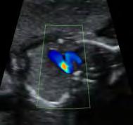
2 Optimizing Detection of Congenital Heart Disease: Important Anatomic Cardiac Regions The Top 5 Critical Anatomic Regions in Fetal Cardiac Imaging Alfred Abuhamad, M.D. Eastern Virginia Medical School Top Critical Anatomic Regions eft Atrium 1. Normal eft Atrium 3 eft Atrium Pulmonary Veins! Ao 1


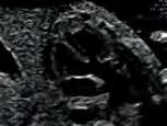
3 Pulmonary Veins 7 eft Atrium eft Atrium eft Atrium Ao A Ao A Esophagus Swallowing Closed Esophagus Open Esophagus TAPVR TAPVR Supracardiac From Practical Guide To Fetal Echocardiography: Normal & Abnormal Hearts Abuhamad, Chaoui 3 rd Edition -Oct


4 7 TAPVR Cardiac TAPVR Infracardiac From Practical Guide To Fetal Echocardiography: Normal & Abnormal Hearts Abuhamad, Chaoui 3 rd Edition -Oct 2015 eft Atrium eft Atrium TAPVR From Practical Guide To Fetal Echocardiography: Normal & Abnormal Hearts Abuhamad, Chaoui 3 rd Edition -Oct 2015 TAPVR eft Atrium TAPVR AO Azygos AO Normal TAPVR J Ultrasound Med 2014; 33:


5 TAPVR Do Not Miss The Diagnosis of a Normal eft Atrium 19 From Practical Guide To Fetal Echocardiography: Normal & Abnormal Hearts Abuhamad, Chaoui 3 rd Edition -Oct Top Critical Anatomic Regions eft Ventricular Outflow 1. Normal eft Atrium 2. Normal eft Ventricular Outflow 21 eft Ventricular Outflow eft Ventricular Outflow Normal TOF 4


6 eft Ventricular Outflow eft Ventricular Outflow TOF TOF eft Ventricular Outflow eft Ventricular Outflow eft Ventricular Outflow eft Ventricular Outflow From Practical Guide To Fetal Echocardiography: Normal & Abnormal Hearts Abuhamad, Chaoui 3 rd Edition -Oct


7 10 TGA: eft Ventricular Outflow 10 TGA: eft Ventricular Outflow 10 TGA: eft Ventricular Outflow 10 TGA: eft Ventricular Outflow 10 ansposition of Great Arteries 10 ansposition of Great Arteries V 6


8 10 ansposition of Great Arteries Overall rate of detection 23 % Ultrasound Obstet Gynecol 2015; 45: Normal eft Ventricular Outflow Mitral - aortic continuity Aorta within left ventricle Angle of ascending aorta with ventricular septum Aorta does not divide Close observation of aortic valves Do Not Miss The Diagnosis of a Normal eft Ventricular Outflow 40 Top Critical Anatomic Regions Normal Semilunar Valves 1. Normal eft Atrium 2. Normal Ascending Aorta 3. Normal Semilunar Valves


9 13 13 Pulmonary Stenosis Pulmonary Stenosis Pulmonary Stenosis 13 Aortic Stenosis 45 From Practical Guide To Fetal Echocardiography: Normal & Abnormal Hearts Abuhamad, Chaoui 3 rd Edition -Oct Aortic Stenosis Aortic Stenosis 8

 ook at wall of vessel beyond the annulus ook at")


10 13 Pulmonary/Aortic Stenosis Do Not Miss The Diagnosis of a Normal Semilunar Valve ook at valve echogenicity ook at valve excursion in systole (cineloop should open fully in systole) ook at wall of vessel beyond the annulus ook at color Doppler for aliasing ook at pulsed Doppler for velocity 50 Top Critical Anatomic Regions ansverse Views 1. Normal eft Atrium 2. Normal Ascending Aorta 3. Normal Semilunar Valves 4. Normal 51 Three-Vessel View Ductal Arch View 9

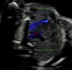

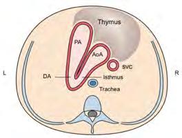
11 Aortic Arch View Three-Vessel achea View eft Brachiocephalic Vein eft Brachiocephalic Vein Thymus R Thymus AoA AoA AzV E DA R Isthmus A B 59 10


12 Thy-Box Sternum AoA R SP AoA AoA SP Azygos SP AoA A B R A SP B Blood Flow Towards the Spine 3VT Spine weeks Follow the Aorta 11


 Thymus Ao achea")

 3VT: 3 Vessels Seen")

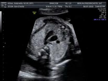
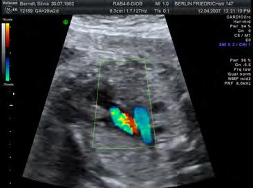
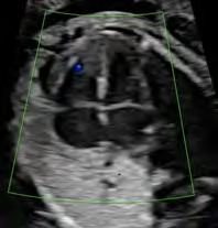
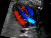
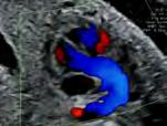
13 13 wks Courtesy of Dr. Chaoui Checklist 3-VT-view! Course and size of, Ao and Aortic isthmus and ductus arteriosus O Aortic arch right or left-sided? (achea as landmark) Thymus Ao achea Thymus visualized Assessment with Color Doppler: Blue V or Red V Atypical vessels (left persistent - Vertical Vein) 3VT: 3 Vessels Seen 70 : 3 Vessels Seen Small Aorta Coarctation of Aorta Coarctation of the Aorta Hypoplastic eft Heart Syndrome


14 : 3 Vessels Seen Small Aorta : 3 Vessels Seen Small Hypoplastic eft Heart Syndrome Pulmonary Stenosis - TOF Pulmonary Atresia 74 TOF - Pulmonary Stenosis TOF - Pulmonary Stenosis Pink Blue TOF - Pulmonary Atresia : 3 Vessels Seen arge Dilated! Dilated, turbulent Flow! AO AO A B Absent Pulmonary Valve Syndrome 13


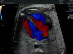
15 : 2 Vessels Seen Normal size great vessel 3VT: 2 Vessels Seen AO AO A B 79 TGA (Convex Course) : 2 Vessels Seen Normal size great vessel : 2 Vessels Seen Normal size great vessel TGA (Convex Course) TGA - Convex-shape of Aorta V A 14
")

16 TGA (atypical) VSD Normal-looking 3VT: Abnormal Course of Vessels 90 15


 AoA RAoA")
 Double")

17 : 3 Vessels Seen Abnormal Vessel Course : 3 Vessels Seen Abnormal Vessel Course Normal Right Ao Arch - Right DA RAo RAo DA DA A B C Right Ao Arch - eft DA Double Ao Arch Right-Sided Aortic Arch - eft DA U-Sign, vascular ring (loose) : 3 Vessels Seen Abnormal Vessel Course : 3 Vessels Seen Abnormal Vessel Course AoA RAoA DA T A B Right-Sided Aortic Arch - eft DA U-Sign, vascular ring (loose) Double Aortic Arch : 3 Vessels Seen Abnormal Vessel Course 3VT: 4 Vessels Seen Double Aortic Arch 96 16


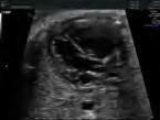
18 : 4 Vessels Seen Persistent eft Superior Vena Cava : 4 Vessels Seen Persistent eft Superior Vena Cava Ao achea achea Ao A B 98 : 4 Vessels Seen Supracardiac TAPVR : 4 Vessels Seen vs. TAPVR? Ao 4th Vessel achea Enlarged CS Ao A B Absent left BCV : 4 Vessels Seen vs. TAPVR? TAPVR CS 4th Vessel : 4 Vessels Seen DA Aneurysm/Kinking DA Aneurysm Normal CS Ao DAA dao achea aao arge left BCV From Echocardiographic anatomy in the fetus. Chiappa E.M et al. 17


19 : 4 Vessels Seen DA Aneurysm/Kinking HHS HRHS TGA DORV TOF CAT TAPVR -VSD Abnormal in: PS / Critical Ao Stenosis Coarctation of Ao ARSA TA-VSD RAA Double Ao Arch Ebstein Interrupted Ao Arch achea Do Not Miss The Diagnosis of a Normal Top Critical Anatomic Regions 1. Normal eft Atrium 2. Normal Ascending Aorta 3. Normal Semilunar Valves 4. Normal 5. Normal Ventricular Septum Ventricular Septum and US Views Ventricular Septum and US Views Muscular Membranous Outlet Inlet 4-chamber view VOT view RVOT view 18


20 Ventricular Septum Ventricular Septum Inlet Muscular Ventricular Septum Ventricular Septum Muscular From Practical Guide To Fetal Echocardiography Abuhamad, Chaoui 2 nd Edition Muscular Ventricular Septum Ventricular Septum Muscular Perimembranous 19


21 Normal Ventricular Septum ook for the T sign in apical views Insonate the septum at 90 degrees for optimal diagnosis of muscular and perimembranous VSD Color Doppler confirm the diagnosis Do Not Miss The Diagnosis of a Normal Ventricular Septum 116 Top Anatomic Regions Normal left atrium: rule out TAPVR, Isomerism, Interrupted IVC Normal ascending aorta: rule out TOF, TGA, DORV Normal semilunar valves: rule out AS, PS, HHS, Normal 3VT view: rule out conotruncal anomalies, RAA, HHS, coarctation of Ao Normal ventricular septum: rule out VSD, AVSD Free Download More than 25,000 Downloads anslated to 11 languages
ISUOG Basic Training. Obtaining & Interpreting Heart Views Correctly Alfred Abuhamad, USA. Basic training. Editable text here
 ISUOG Basic Training Obtaining & Interpreting Heart Views Correctly Alfred Abuhamad, USA Learning Objectives 6, 7 & 8 At the end of the lecture you will be able to: describe how to assess cardiac situs
Basic Fetal Cardiac Evaluation
 Basic Fetal Cardiac Evaluation Mert Ozan Bahtiyar, MD Director, Fetal Care Center Division of Maternal Fetal Medicine Department of Obstetrics, Gynecology and Reproductive Sciences S L I D E 1 Background
Making Sense of Cardiac Views and Imaging Characteristics for 13 Congenital Heart Defects (CHDs)
") Making Sense of Cardiac Views and Imaging Characteristics for 13 Congenital Heart Defects (CHDs) Manny Gaziano, MD, FACOG obimages.net obimages.net@gmail.com Acknowledgements: Krista Wald, RDMS, sonographer,
Making Sense of Cardiac Views and Imaging Characteristics for 13 Congenital Heart Defects (CHDs) Manny Gaziano, MD, FACOG obimages.net obimages.net@gmail.com Acknowledgements: Krista Wald, RDMS, sonographer,
Heart and Soul Evaluation of the Fetal Heart
 Heart and Soul Evaluation of the Fetal Heart Ivana M. Vettraino, M.D., M.B.A. Clinical Associate Professor, Michigan State University College of Human Medicine Objectives Review the embryology of the formation
Cardiac Catheterization Cases Primary Cardiac Diagnoses Facility 12 month period from to PRIMARY DIAGNOSES (one per patient)
") PRIMARY DIAGNOSES (one per patient) Septal Defects ASD (Atrial Septal Defect) PFO (Patent Foramen Ovale) ASD, Secundum ASD, Sinus venosus ASD, Coronary sinus ASD, Common atrium (single atrium) VSD (Ventricular
PRIMARY DIAGNOSES (one per patient) Septal Defects ASD (Atrial Septal Defect) PFO (Patent Foramen Ovale) ASD, Secundum ASD, Sinus venosus ASD, Coronary sinus ASD, Common atrium (single atrium) VSD (Ventricular
Outflow Tracts Anomalies
 Diagnosis of Outflow Tract Anomalies in the Fetus General Framing D.Paladini Fetal Medicine & Surgery Unit Gasllini Children s Hospital - Genoa dariopaladini@ospedale-gaslini.ge.it Outflow Tracts Anomalies
Congenital Heart Defects
 Normal Heart Congenital Heart Defects 1. Patent Ductus Arteriosus The ductus arteriosus connects the main pulmonary artery to the aorta. In utero, it allows the blood leaving the right ventricle to bypass
Anomalous Systemic Venous Connection Systemic venous anomaly
 World Database for Pediatric and Congenital Heart Surgery Appendix B: Diagnosis (International Paediatric and Congenital Cardiac Codes (IPCCC) and definitions) Anomalous Systemic Venous Connection Systemic
An update on technique of fetal echocardiography with emphasis on anomalies detectable in four chambered view.
 An update on technique of fetal echocardiography with emphasis on anomalies detectable in four chambered view. Dr. Ranjitha.G Specialist Radiologist NMC-SH Al ain, UAE Fetal echocardiography is an essential
Heart and Lungs. LUNG Coronal section demonstrates relationship of pulmonary parenchyma to heart and chest wall.
 Heart and Lungs Normal Sonographic Anatomy THORAX Axial and coronal sections demonstrate integrity of thorax, fetal breathing movements, and overall size and shape. LUNG Coronal section demonstrates relationship
UPDATE FETAL ECHO REVIEW
 UPDATE 1 FETAL ECHO REVIEW Study Alert for RDCS Candidates D A V I E S P U B L I S H I N G I N C. Fetal Echo Review Study Alert U P D A T E D A U G U S T 1, 2 0 1 2 Nikki Stahl, RT(R)(M)(CT), RDMS, RVT
List of Videos. Video 1.1
 Video 1.1 Video 1.2 Video 1.3 Video 1.4 Video 1.5 Video 1.6 Video 1.7 Video 1.8 The parasternal long-axis view of the left ventricle shows the left ventricular inflow and outflow tract. The left atrium
Most common fetal cardiac anomalies
 Most common fetal cardiac anomalies Common congenital heart defects CHD % of cardiac defects Chromosomal Infants Fetuses anomaly (%) 22q11 deletion (%) VSD 30 5~10 20~40 10 PS 9 5 (PA w/ VSD) HLHS 7~9
ULTRASOUND OF THE FETAL HEART
 ULTRASOUND OF THE FETAL HEART Cameron A. Manbeian, MD Disclosure Statement Today s faculty: Cameron Manbeian, MD does not have any relevant financial relationships with commercial interests or affiliations
ECHOCARDIOGRAPHIC APPROACH TO CONGENITAL HEART DISEASE: THE UNOPERATED ADULT
 ECHOCARDIOGRAPHIC APPROACH TO CONGENITAL HEART DISEASE: THE UNOPERATED ADULT Karen Stout, MD, FACC Divisions of Cardiology University of Washington Medical Center Seattle Children s Hospital NO DISCLOSURES
Pediatric Echocardiography Examination Content Outline
 Pediatric Echocardiography Examination Content Outline (Outline Summary) # Domain Subdomain Percentage 1 Anatomy and Physiology Normal Anatomy and Physiology 10% 2 Abnormal Pathology and Pathophysiology
MEDICAL MANAGEMENT WITH CAVEATS 1. In one study of 50 CHARGE patients with CHD, 75% required surgery. 2. Children with CHARGE may be resistant to chlo
 CARDIOLOGY IN CHARGE SYNDROME: FOR THE PHYSICIAN Angela E. Lin, M.D. Teratology Program/Active Malformation Surveillance, Brigham and Women's Hospital, Old PBBH-B501, 75 Francis St., Boston, MA 02115 alin@partners.org
Chapter 2 Cardiac Interpretation of Pediatric Chest X-Ray
 Chapter 2 Cardiac Interpretation of Pediatric Chest X-Ray Ra-id Abdulla and Douglas M. Luxenberg Key Facts The cardiac silhouette occupies 50 55% of the chest width on an anterior posterior chest X-ray
Data Collected: June 17, Reported: June 30, Survey Dates 05/24/ /07/2010
 Job Task Analysis for ARDMS Pediatric Echocardiography Data Collected: June 17, 2010 Reported: Analysis Summary For: Pediatric Echocardiography Exam Survey Dates 05/24/2010-06/07/2010 Invited Respondents
Congenital Heart Disease An Approach for Simple and Complex Anomalies
 Congenital Heart Disease An Approach for Simple and Complex Anomalies Michael D. Pettersen, MD Director, Echocardiography Rocky Mountain Hospital for Children Denver, CO None Disclosures 1 ASCeXAM Contains
Adult Echocardiography Examination Content Outline
 Adult Echocardiography Examination Content Outline (Outline Summary) # Domain Subdomain Percentage 1 2 3 4 5 Anatomy and Physiology Pathology Clinical Care and Safety Measurement Techniques, Maneuvers,
Foetal Cardiology: How to predict perinatal problems. Prof. I.Witters Prof.M.Gewillig UZ Leuven
 Foetal Cardiology: How to predict perinatal problems Prof. I.Witters Prof.M.Gewillig UZ Leuven Cardiopathies Incidence : 8-12 / 1000 births ( 1% ) Most frequent - Ventricle Septum Defect 20% - Atrium Septum
HDlive Silhouette Mode With Spatiotemporal Image Correlation for Assessment of the Fetal Heart
 ORIGINAL RESEARCH HDlive Silhouette Mode With Spatiotemporal Image Correlation for Assessment of the Fetal Heart Toshiyuki Hata, MD, PhD, Mohamed Ahmed Mostafa AboEllail, MD, Suraphan Sajapala, MD, Mari
Screening for Critical Congenital Heart Disease
 Screening for Critical Congenital Heart Disease Caroline K. Lee, MD Pediatric Cardiology Disclosures I have no relevant financial relationships or conflicts of interest 1 Most Common Birth Defect Most
Disclosures. Outline. Learning Objectives. Introduction. Introduction. Sonographic Screening Examination of the Fetal Heart
 Sonographic Screening Examination of the Fetal Heart Lami Yeo, MD Director of Fetal Cardiology Perinatology Research Branch of NICHD / NIH / DHHS Bethesda, MD and Detroit, Michigan, USA Professor, Division
Three-dimensional (3D) and 4D color Doppler fetal echocardiography using spatio-temporal image correlation (STIC)
 and 4D color Doppler fetal echocardiography using spatio-temporal image correlation (STIC)") Ultrasound Obstet Gynecol 2004; 23: 535 545 Published online 6 May 2004 in Wiley InterScience (www.interscience.wiley.com). DOI: 10.1002/uog.1075 Three-dimensional (3D) and 4D color Doppler fetal echocardiography
Ultrasound Obstet Gynecol 2004; 23: 535 545 Published online 6 May 2004 in Wiley InterScience (www.interscience.wiley.com). DOI: 10.1002/uog.1075 Three-dimensional (3D) and 4D color Doppler fetal echocardiography
Diagnosis of Congenital Cardiac Defects Between 11 and 14 Weeks Gestation in High-Risk Patients
 Article Diagnosis of Congenital Cardiac Defects Between 11 and 14 Weeks Gestation in High-Risk Patients Zeev Weiner, MD, Abraham Lorber, MD, Eliezer Shalev, MD Objective. To examine the feasibility of
Common Defects With Expected Adult Survival:
 Common Defects With Expected Adult Survival: Bicuspid aortic valve :Acyanotic Mitral valve prolapse Coarctation of aorta Pulmonary valve stenosis Atrial septal defect Patent ductus arteriosus (V.S.D.)
CMS Limitations Guide - Radiology Services
 CMS Limitations Guide - Radiology Services Starting October 1, 2015, CMS will update their existing medical necessity limitations on tests and procedures to correspond to ICD-10 codes. This limitations
"Lecture Index. 1) Heart Progenitors. 2) Cardiac Tube Formation. 3) Valvulogenesis and Chamber Formation. 4) Epicardium Development.
 Heart Progenitors. 2) Cardiac Tube Formation. 3) Valvulogenesis and Chamber Formation. 4) Epicardium Development.") "Lecture Index 1) Heart Progenitors. 2) Cardiac Tube Formation. 3) Valvulogenesis and Chamber Formation. 4) Epicardium Development. 5) Septation and Maturation. 6) Changes in Blood Flow during Development.
"Lecture Index 1) Heart Progenitors. 2) Cardiac Tube Formation. 3) Valvulogenesis and Chamber Formation. 4) Epicardium Development. 5) Septation and Maturation. 6) Changes in Blood Flow during Development.
Hypoplastic Left Heart Syndrome: Echocardiographic Assessment
 Hypoplastic Left Heart Syndrome: Echocardiographic Assessment Craig E Fleishman, MD, FACC, FASE Director, Non-invasive Cardiac Imaging The Hear Center at Arnold Palmer Hospital for Children, Orlando SCAI
ADULT CONGENITAL HEART DISEASE. Stuart Lilley
 ADULT CONGENITAL HEART DISEASE Stuart Lilley More adults than children have congenital heart disease Huge variety of congenital lesions from minor to major Heart failure, re-operation and arrhythmia are
When is Risky to Apply Oxygen for Congenital Heart Disease 부천세종병원 소아청소년과최은영
 When is Risky to Apply Oxygen for Congenital Heart Disease 부천세종병원 소아청소년과최은영 The Korean Society of Cardiology COI Disclosure Eun-Young Choi The author have no financial conflicts of interest to disclose
3/14/2011 MANAGEMENT OF NEWBORNS CARDIAC INTENSIVE CARE CONFERENCE FOR HEALTH PROFESSIONALS IRVINE, CA. MARCH 7, 2011 WITH HEART DEFECTS
 CONFERENCE FOR HEALTH PROFESSIONALS IRVINE, CA. MARCH 7, 2011 MANAGEMENT OF NEWBORNS WITH HEART DEFECTS A NTHONY C. CHANG, MD, MBA, MPH M E D I C AL D I RE C T OR, HEART I N S T I T U T E C H I LDRE N
Fetal Echocardiography and the Routine Obstetric Sonogram
 JDMS 23:143 149 May/June 2007 143 Fetal Echocardiography and the Routine Obstetric Sonogram SHELLY ZIMBELMAN, RT(R)(CT), RDMS, RDCS ASAD SHEIKH, MD, RDCS Congenital heart disease (CHD) is the most common
D. PALADINI, M. VASSALLO, G. SGLAVO, C. LAPADULA and P. MARTINELLI
 Ultrasound Obstet Gynecol 2006; 27: 555 561 Published online in Wiley InterScience (www.interscience.wiley.com). DOI: 10.1002/uog.2749 The role of spatio-temporal image correlation (STIC) with tomographic
COMPREHENSIVE EVALUATION OF FETAL HEART R. GOWDAMARAJAN MD
 COMPREHENSIVE EVALUATION OF FETAL HEART R. GOWDAMARAJAN MD Disclosure No Relevant Financial Relationships with Commercial Interests Fetal Echo: How to do it? Timing of Study -optimally between 22-24 weeks
Fetal Cardiac Anomaly
 89 Symposium: OB/GY US (Room B) 12 : 10 1 2 : 30 Fetal Cardiac Anomaly 1. One third of all congenital anomalies 2. 6 10/1,000 live births 3. Related with more than 50% of childhood deaths and 20-30% of
Three cross-sectional planes for fetal color Doppler echocardiography
 Ultrasound Obstet Gynecol 2003; 21: 81 93 Published online 11 December 2002 in Wiley InterScience (www.interscience.wiley.com). DOI: 10.1002/uog.5 Three cross-sectional planes for fetal color Doppler echocardiography
pulmonary valve on, 107 pulmonary valve vegetations on, 113
 INDEX Adriamycin-induced cardiomyopathy, 176 Amyloidosis, 160-161 echocardiographic abnormalities in, 160 intra-mural tumors similar to, 294 myocardial involvement in, 160-161 two-dimensional echocardiography
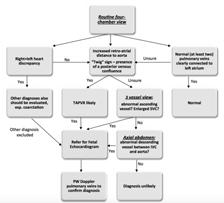
AbnormalThree-VesselView on Sonography: A Clue to the Diagnosis of Congenital Heart Disease in the Fetus
 rt Pictorial Essay bnormalthree-vesselview on Sonography: Clue to the Diagnosis of Congenital Heart Disease in the Fetus screening tool for major congenital heart diseases [I. 2J. However, anomalies of
Segmental approach to normal and abnormal situs arrangement - Echocardiography -
 Segmental approach to normal and abnormal situs arrangement - Echocardiography - Jan Marek Great Ormond Street Hospital & Institute of Cardiovascular Sciences, University College London No disclosures
Congenital Heart Disease: Physiology and Common Defects
 Congenital Heart Disease: Physiology and Common Defects Jamie S. Sutherell, M.D, M.Ed. Associate Professor, Pediatrics Division of Cardiology Director, Medical Student Education in Pediatrics Director,
A Review of Findings in Fetal Cardiac Section Drawings
 Image Presentation A Review of Findings in Fetal Cardiac Section Drawings Part 3: The 3-Vessel-Trachea View and Variants Philippe Jeanty, MD, PhD, Rabih Chaoui, MD, Irina Tihonenko, MD, Frantisek Grochal,
Fetal Tetralogy of Fallot
 36 Fetal Tetralogy of Fallot E.D. Bespalova, R.M. Gasanova, O.A.Pitirimova National Scientific and Practical Center of Cardiovascular Surgery, Moscow Elena D. Bespalova, MD Professor, Director Rena M,
Accuracy of prenatal diagnosis of fetal congenital heart disease by different
 Accuracy of prenatal diagnosis of fetal congenital heart disease by different methods with echocardiography Ying Zhang 1* * Corresponding author Email: baogoubei@hotmail.com Ai-Lu Cai 1 Email: caial_us@hotmail.com
5.8 Congenital Heart Disease
 5.8 Congenital Heart Disease Congenital heart diseases (CHD) refer to structural or functional heart diseases, which are present at birth. Some of these lesions may be discovered later. prevalence of Chd
Anatomy & Physiology
 1 Anatomy & Physiology Heart is divided into four chambers, two atrias & two ventricles. Atrioventricular valves (tricuspid & mitral) separate the atria from ventricles. they open & close to control flow
Echocardiographic and anatomical correlates in the fetus*
 Br Heart J 1980; : 51 Echocardiographic and anatomical correlates in the fetus* LINDSEY D ALLAN, MICHAEL J TYNAN, STUART CAMPBELL, JAMES L WILKINSON, ROBERT H ANDERSON From King's College Hospital, and
Paediatric Cardiology. Acyanotic CHD. Prof F F Takawira
 Paediatric Cardiology Acyanotic CHD Prof F F Takawira Aetiology Chromosomal Down syndrome, T13, T18 Genetic syndromes (gene defects) Velo-Cardio-facial (22 del) Genetic syndromes (undefined aetiology)
Fetal Rhythm and Blues
 Fetal Rhythm and Blues John Cotton, MD Professor of Pediatrics Division of Pediatric Cardiology Director, Fetal Cardiology Program UNC Chapel Hill, School of Medicine Objectives To review methods used
THE HEART. Unit 3: Transportation and Respiration
 THE HEART Unit 3: Transportation and Respiration The Circulatory System Also called the Cardiovascular System Circulates blood in the body Transports nutrients, oxygen, carbon dioxide, hormones, and blood
CMR for Congenital Heart Disease
 CMR for Congenital Heart Disease * Second-line tool after TTE * Strengths of CMR : tissue characterisation, comprehensive access and coverage, relatively accurate measurements of biventricular function/
Congenital Heart Disease
 Congenital Heart Disease Mohammed Alghamdi, MD, FRCPC, FAAP, FACC Associate Professor and Consultant Pediatric Cardiology, Cardiac Science King Fahad Cardiac Centre King Saud University INTRODUCTION CHD
DEVELOPMENT OF THE CIRCULATORY SYSTEM L E C T U R E 5
 DEVELOPMENT OF THE CIRCULATORY SYSTEM L E C T U R E 5 REVIEW OF CARDIAC ANATOMY Heart 4 chambers Base and apex Valves Pericardial sac 3 layers: epi, myo, endo cardium Major blood vessels Aorta and its
Aortic arch anomalies Coarctation of the Aorta Interrupted Aortic Arch Echocardiography
 Aortic arch anomalies Coarctation of the Aorta Interrupted Aortic Arch Echocardiography V.Tomek, J. Marek, J. Škovránek, J. Gilík No disclosures Kardiocentrum, University Hospital Motol, Prague, Czech
NASCI 2012 Segmental Analysis
 NASCI 2012 Segmental Analysis Frandics Chan, M.D., Ph.D. Stanford University Medical Center Lucile Packard Department Children s of Radiology Hospital Menagerie of Congenital Cardiac Lesions 1. Absent
5/22/2013. Alan Zuckerman 1, Swapna Abhyankar 1, Tiffany Colarusso 2, Richard Olney 2, Kristin Burns 3, Marci Sontag 4
 Alan Zuckerman 1, Swapna Abhyankar 1, Tiffany Colarusso 2, Richard Olney 2, Kristin Burns 3, Marci Sontag 4 1 National Library of Medicine, NIH, Bethesda, MD, USA, 2 Centers for Disease Control and Prevention,
All You Need to Know About Situs and Looping Disorders: Embryology, Anatomy, and Echocardiography
 All You Need to Know About Situs and Looping Disorders: Embryology, Anatomy, and Echocardiography Helena Gardiner Co-Director of Fetal Cardiology, The Fetal Center, University of Texas at Houston Situs
Comparison of echocardiographic findings in fetuses at less than 15 weeks gestation with later cardiac evaluation
 Ultrasound Obstet Gynecol 2013; 42: 679 686 Published online in Wiley Online Library (wileyonlinelibrary.com). DOI: 10.1002/uog.12517 Comparison of echocardiographic findings in fetuses at less than 15
ORIGINAL RESEARCH PAPER
 ORIGINAL RESEARCH PAPER ROLE OF CT PULMONARY ANGIOGRAPHY IN CONGENITAL HEART DISEASES IN PAEDIATRIC POPULATION Radiology KEY WORDS: Congenital heart disease, CT pulmonary angiography, pediatric heart disease,
PRACTICAL ECHOCARDIOGRAPHY IN THE ADULT with Doppler and color-doppler flow imaging
 PRACTICAL ECHOCARDIOGRAPHY IN THE ADULT with Doppler and color-doppler flow imaging PRACTICAL ECHOCARDIOGRAPHY IN THE ADULT with Doppler and color-doppler flow imaging by J.P.M. HAMER Thoraxcentre, Department
Editorial. Color and pulsed Doppler in fetal echocardiography A. ABUHAMAD
 Ultrasound Obstet Gynecol 2004; 24: 1 9 Published online in Wiley InterScience (www.interscience.wiley.com). DOI: 10.1002/uog.1096 Editorial Color and pulsed Doppler in fetal echocardiography A. ABUHAMAD
Cardiovascular Pathophysiology: Right to Left Shunts aka Cyanotic Lesions
 Cardiovascular Pathophysiology: Right to Left Shunts aka Cyanotic Lesions Ismee A. Williams, MD, MS iib6@columbia.edu Pediatric Cardiology Learning Objectives To discuss the hemodynamic significance of
Cardiovascular Pathophysiology: Right to Left Shunts aka Cyanotic Lesions Ismee A. Williams, MD, MS Pediatric Cardiology
 Cardiovascular Pathophysiology: Right to Left Shunts aka Cyanotic Lesions Ismee A. Williams, MD, MS iib6@columbia.edu Pediatric Cardiology Learning Objectives To discuss the hemodynamic significance of
Totally Anomalous Pulmonary Venous Connection and Complex Congenital Heart Disease
 Article Totally Anomalous Pulmonary Venous Connection and Complex Congenital Heart Disease Prenatal Echocardiographic Diagnosis and Prognosis Chandrakant R. Patel, MBBS, John R. Lane, MD, Michael L. Spector,
Giovanni Di Salvo MD, PhD, FESC Second University of Naples Monaldi Hospital
 Giovanni Di Salvo MD, PhD, FESC Second University of Naples Monaldi Hospital VSD is one of the most common congenital cardiac abnormalities in the newborn. It can occur as an isolated finding or in combination
DORV: The Great Chameleon. Heart Conference October 15, 2016 Tina Kwan, MD
 DORV: The Great Chameleon Heart Conference October 15, 2016 Tina Kwan, MD Kenneth Maehara, Ph.D. May 7, 1942 - August 26, 2013 A.R. A classic case of broken heart 38 week AGA F born at an OSH to
Preoperative Echocardiographic Assessment of Uni-ventricular Repair
 Preoperative Echocardiographic Assessment of Uni-ventricular Repair Salem Deraz, MD Pediatric Cardiologist, Aswan Heart Centre Magdi Yacoub Heart Foundation Uni-ventricular repair A single or series of
The Heart. Happy Friday! #takeoutyournotes #testnotgradedyet
 The Heart Happy Friday! #takeoutyournotes #testnotgradedyet Introduction Cardiovascular system distributes blood Pump (heart) Distribution areas (capillaries) Heart has 4 compartments 2 receive blood (atria)
Congenital Heart Disease: Cyanotic Lesions. Amitesh Aggarwal
 Congenital Heart Disease: Cyanotic Lesions Amitesh Aggarwal 12 y/o male admitted because of dyspnea and cyanosis Patient has been cyanotic since few months after birth Has episodes of tachypnea and worsening
Research article. Primary detection of congenital heart diseases in the Kyrgyz Republic
 Research article Primary detection of congenital heart diseases in the Kyrgyz Republic Irina A. Akhmedova, Gulzada A. Imanalieva, Damir A.Abibillaev, Taalaibek Z. Kudaiberdiev Scientific Research Institute
The Heart. Made up of 3 different tissue: cardiac muscle tissue, nerve tissue, and connective tissue.
 The Heart The Heart Made up of 3 different tissue: cardiac muscle tissue, nerve tissue, and connective tissue. Your heart pumps with a regular beat (Heart Rate) Your heart rate can change depending on
Heart Development and Congenital Heart Disease
 Heart Development and Congenital Heart Disease Sally Dunwoodie s.dunwoodie@victorchang.edu.au Developmental and Stem Cell Biology Division Victor Chang Cardiac Research Institute for the heart of Australia...
Cardiac ultrasound protocols
 Cardiac ultrasound protocols IDEXX Telemedicine Consultants Two-dimensional and M-mode imaging planes Right parasternal long axis four chamber Obtained from the right side Displays the relative proportions
Congenital heart disease: When to act and what to do?
 Leading Article Congenital heart disease: When to act and what to do? Duminda Samarasinghe 1 Sri Lanka Journal of Child Health, 2010; 39: 39-43 (Key words: Congenital heart disease) Congenital heart disease
Chapter 14. Circulatory System Images. VT-122 Anatomy & Physiology II
 Chapter 14 Circulatory System Images VT-122 Anatomy & Physiology II The mediastinum Dog heart Dog heart Cat heart Dog heart ultrasound Can see pericardium as distinct bright line Pericardial effusion Fluid
Chapter 14. The Cardiovascular System
 Chapter 14 The Cardiovascular System Introduction Cardiovascular system - heart, blood and blood vessels Cardiac muscle makes up bulk of heart provides force to pump blood Function - transports blood 2
Cardiac CT in Infants with Congenital heart disease Sunrise Session. LaDonna Malone, MD May 17, 2018
 Cardiac CT in Infants with Congenital heart disease Sunrise Session LaDonna Malone, MD May 17, 2018 None Disclosures Objectives Describe cardiac CT techniques used in infants with congenital heart disease.
Aortic Arch Abnormalities
 Aortic Arch Abnormalities IPOK Norman H Silverman MD, D Sc (Med.). FACC, FAHA. Stanford University & Lucile Packard Children s Hospital E mail: norm.silverman@stanford.edu. NHS. www.md1world.com Abnormalities
Adult Congenital Heart Disease: What All Echocardiographers Should Know Sharon L. Roble, MD, FACC Echo Hawaii 2016
 1 Adult Congenital Heart Disease: What All Echocardiographers Should Know Sharon L. Roble, MD, FACC Echo Hawaii 2016 DISCLOSURES I have no disclosures relevant to today s talk 2 Why should all echocardiographers
Identification of congenital cardiac malformations by echocardiography in midtrimester fetus*
 Br Heart J 1981; 46: 358-62 Identification of congenital cardiac malformations by echocardiography in midtrimester fetus* LINDSEY D ALLAN, MICHAEL TYNAN, STUART CAMPBELL, ROBERT H ANDERSON From Guy's Hospital;
Echocardiography in Congenital Heart Disease
 Chapter 44 Echocardiography in Congenital Heart Disease John L. Cotton and G. William Henry Multiple-plane cardiac imaging by echocardiography can noninvasively define the anatomy of the heart and the
Adel Hasanin Ahmed 1 ASD
 Adel Hasanin Ahmed 1 ASD Atrial septal defect (ASD) is the commonest form of congenital heart disease seen in adults. The commonest form of defect is the secundum ASD, accounting for two thirds of cases,
How to Assess and Treat Obstructive Lesions
 How to Assess and Treat Obstructive Lesions Erwin Oechslin, MD, FESC, FRCPC, Director, Congenital Cardiac Centre for Adults Peter Munk Cardiac Centre University Health Network/Toronto General Hospital
Technique of fetal echocardiography
 Review Obstet Gynecol Sci 2013;56(4):217-226 http://dx.doi.org/10.5468/ogs.2013.56.4.217 pissn 2287-8572 eissn 2287-8580 Technique of fetal echocardiography Mi-Young Lee, Hye-Sung Won Department of Obstetrics
Children with Single Ventricle Physiology: The Possibilities
 Children with Single Ventricle Physiology: The Possibilities William I. Douglas, M.D. Pediatric Cardiovascular Surgery Children s Memorial Hermann Hospital The University of Texas Health Science Center
The Chest X-ray for Cardiologists
 Mayo Clinic & British Cardiovascular Society at the Royal College of Physicians, London : 21-23-October 2013 Cases-Controversies-Updates 2013 The Chest X-ray for Cardiologists Michael Rubens Royal Brompton
Uncommon Doppler Echocardiographic Findings of Severe Pulmonic Insufficiency
 Uncommon Doppler Echocardiographic Findings of Severe Pulmonic Insufficiency Rahul R. Jhaveri, MD, Muhamed Saric, MD, PhD, FASE, and Itzhak Kronzon, MD, FASE, New York, New York Background: Two-dimensional
Ch.15 Cardiovascular System Pgs {15-12} {15-13}
 Ch.15 Cardiovascular System Pgs {15-12} {15-13} E. Skeleton of the Heart 1. The skeleton of the heart is composed of rings of dense connective tissue and other masses of connective tissue in the interventricular
Dear Parent/Guardian,
 Dear Parent/Guardian, You have indicated on school records that your child has an ongoing health problem that may require medication and/or treatment during the school day with rescue medication. Attached
PRACTICAL GUIDE TO FETAL ECHOCARDIOGRAPHY IC Huggon and LD Allan
 PRACTICAL GUIDE TO FETAL ECHOCARDIOGRAPHY IC Huggon and LD Allan Fetal Cardiology Unit, Harris Birthright Research Centre for Fetal Medicine, King's College Hospital, London, UK IMPORTANCE OF PRENATAL
Large Arteries of Heart
 Cardiovascular System (Part A-2) Module 5 -Chapter 8 Overview Arteries Capillaries Veins Heart Anatomy Conduction System Blood pressure Fetal circulation Susie Turner, M.D. 1/5/13 Large Arteries of Heart
Appendix A.1: Tier 1 Surgical Procedure Terms and Definitions
 Appendix A.1: Tier 1 Surgical Procedure Terms and Definitions Tier 1 surgeries AV Canal Atrioventricular Septal Repair, Complete Repair of complete AV canal (AVSD) using one- or two-patch or other technique,
Low-dose prospective ECG-triggering dual-source CT angiography in infants and children with complex congenital heart disease: first experience
 Low-dose prospective ECG-triggering dual-source CT angiography in infants and children with complex congenital heart disease: first experience Ximing Wang, M.D., Zhaoping Cheng, M.D., Dawei Wu, M.D., Lebin
Assessing Cardiac Anatomy With Digital Subtraction Angiography
 485 JACC Vol. 5, No. I Assessing Cardiac Anatomy With Digital Subtraction Angiography DOUGLAS S., MD, FACC Cleveland, Ohio The use of intravenous digital subtraction angiography in the assessment of patients
Echocardiographic assessment in Adult Patients with Congenital Heart Diseases
 Echocardiographic assessment in Adult Patients with Congenital Heart Diseases Athanasios Koutsakis Cardiologist, Cl. Research Fellow George Giannakoulas Ass. Professor in Cardiology 1st Cardiology Department,
ISUOG Basic Training. Assessing the Neck & Chest Gihad Chalouhi, Lebanon
 ISUOG Basic Training Assessing the Neck & Chest Gihad Chalouhi, Lebanon Learning objectives 9 & 10 At the end of the lecture you will be able to: recognise the differences between the normal & most common
Cardiac MRI in ACHD What We. ACHD Patients
 Cardiac MRI in ACHD What We Have Learned to Apply to ACHD Patients Faris Al Mousily, MBChB, FAAC, FACC Consultant, Pediatric Cardiology, KFSH&RC/Jeddah Adjunct Faculty, Division of Pediatric Cardiology
cardiac imaging planes planning basic cardiac & aortic views for MR
 cardiac imaging planes planning basic cardiac & aortic views for MR Dianna M. E. Bardo, M. D. Assistant Professor of Radiology & Cardiovascular Medicine Director of Cardiac Imaging cardiac imaging planes