STAT 12 Lead ECG Workshop: Basics & ACS
|
|
|
- Jordan Lamb
- 5 years ago
- Views:
Transcription


1 STAT 12 Lead ECG Workshop: Basics & ACS Part 2: Acute Coronary Syndrome WAYNE W RUPPERT, CVT, CCCC, NREMT-P Cardiovascular Coordinator Bayfront Health Seven Rivers Crystal River, Florida Interventional Cardiovascular & Electrophysiology Technologist Copyright 2010, 2011, 2015, 2019 Wayne W Ruppert


2 STAT 12 Lead ECG Part 2 ACS Wayne W Ruppert, CVT, CCCC, NREMT-P Interventional Cardiovascular Technologist Cardiovascular Coordinator Bayfront Health Seven Rivers
3 Your patient complains of CHEST PAIN The instant you see the patient, you assess for: Cardiac Arrest Shock

4

5
6 FAIL the SHOCK SURVEY? F RAPIDLY FIND AND TREAT THE ROOT CAUSE... WORK TO RAPIDLY IDENTIFY THE CAUSE OF SHOCK.

7
8

9
10 CHIEF COMPLAINT KEY WORDS: CHEST: PAIN / HEAVINESS / PRESSURE/ FUNNY FEELING IN, etc. SHORTNESS BREATH DIZZINESS / LIGHTHEADEDNESS ETC. ETC. ETC.
11

12
13 BEWARE of the patient with INTERMITTENT CHEST PAIN.... M
14 ATYPICAL SYMPTOMS of ACS???


15 BOOK PAGE: 70

16


17
18 Physical Exam Clues of MI: Skin may be PALE, CLAMMY SWEATING! (Diaphoresis) Clutching /Rubbing chest BP can be high, normal or low Anxiety / look of impending doom.
19
20 3 or more major RISK FACTORS gets you a checkmark in the RISK FACTOR box!

21
22 The 12 Lead ECG to Rule out ACS: Acute Coronary Syndrome (ACS) is made up of the following cardiac conditions: Unstable Angina Non-ST Segment Elevation Myocardial Infarction (NSTEMI) ST Segment Elevation Myocardial Infarction (STEMI) Low Risk Chest Pain
23 Unstable Angina

24 Non-STEMI (NSTEMI) Non-ST Segment Elevation Myocardial Infarction. sub-endocardial MI... partial wall thickness
25 Non-STEMI (NSTEMI) Non-ST Segment Elevation Myocardial Infarction. sub-endocardial MI... partial wall thickness The 12 Lead ECG may show: - ST Depression - Other ST Segment changes - Inverted T wave - THE ECG MAY BE TOTALLY NORMAL. TROPONIN is ABNORMALLY ELEVATED!
26 Non-STEMI (NSTEMI) Non-ST Segment Elevation Myocardial Infarction. sub-endocardial MI... partial wall thickness This is a Partial Wall Thickness MI, heart cells are dying, and the Troponin becomes detectable in the patient s bloodstream. Usually less severe than a STEMI, patient needs blood thinners and to get to the cath lab in hours.

27 STEMI ST Segment Elevation Myocardial Infarction.
28 STEMI ST Segment Elevation Myocardial Infarction. ( full-wall thickness, Transmural event) This is a life-threatening emergency. Part of the patient s heart is dying. Blood flow must be restored within 90 minutes or less in order to preserve heart muscle. Based on the region of the heart affected, critical and often lethal complications rapidly develop.
29 Ischemia and Infarction = Acute Coronary Syndrome The conditions associated with Acute Coronary Syndrome (ACS) include: Unstable Angina (ischemia) Non-ST Segment Elevation Myocardial Infarction (NSTEMI) (infarction) ST Segment Elevation Myocardial Infarction (STEMI) (Infarction)
30 Q: To evaluate the patient for ischemia or infarction, what part of the ECG do we look at?
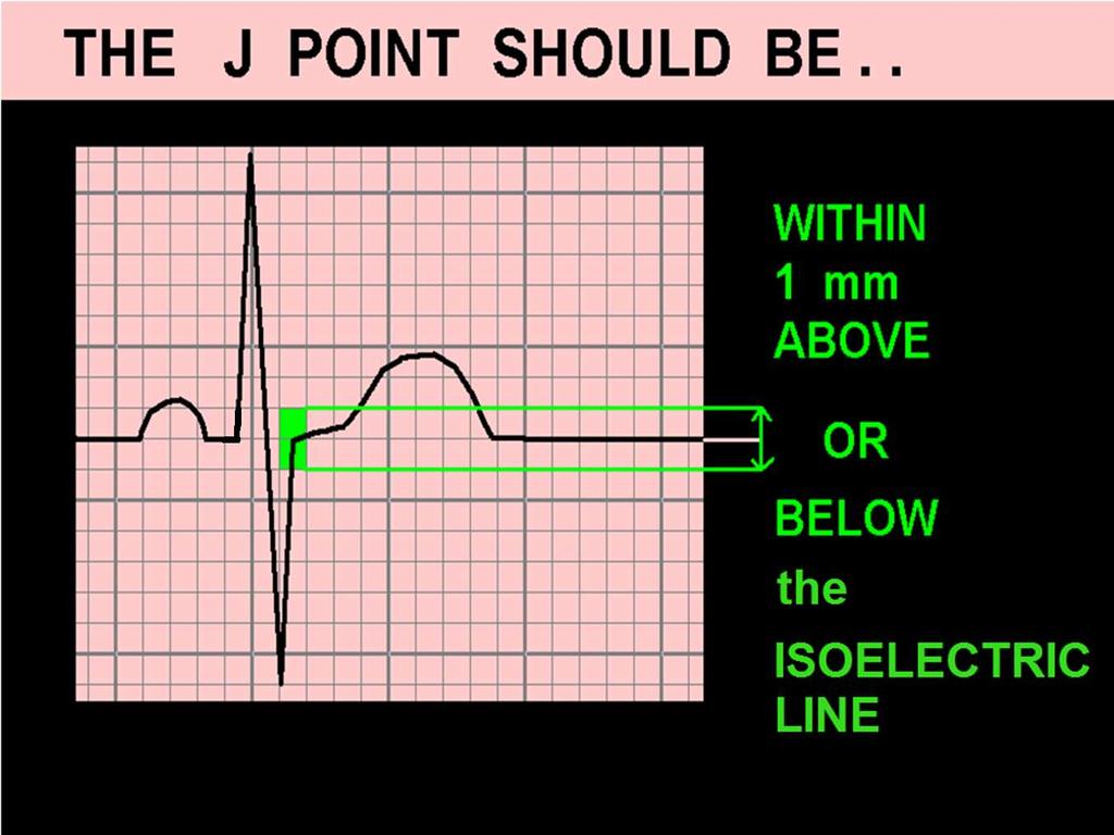
31 Q: To evaluate the patient for ischemia or infarction, what part of the ECG do we look at? A: We evaluate the J Points ST Segments & T Waves.. in each lead!
32 Evaluating the ECG for ACS:
33 Evaluating the ECG for ACS: Patients with Normal Width QRS (QRSd < 120ms)
34 Q: Why is QRS width an issue when we look at J Points, ST Segments and T Waves??
35 Q: Why is QRS width an issue when we look at J Points, ST Segments and T Waves?? A: When the QRS is abnormally wide (> 120ms), it ALTERS the J Points, ST Segments and T Waves.
36

37 ...the flat line between ECG complexes, when there is no detectable electrical activity...

38 The Isoelectric Line - it s not always isoelectric!
39
40 Use the P-Q junction as a reference point for measuring the J Point and ST-Segment when iso-electric line is not isoelectric!

41 Defining NORMAL:
42 ECG Indicators of ABNORMAL PERFUSION (possible ischemia / infarction) in Patients with Normal Width QRS Complexes (QRS duration < 120 ms)

43
44
45 Some less common, less reliable possible indicators of ACS:
46 LET S START HERE....
47
48

49


50 WHEN EVALUATING for ST SEGMENT ELEVATION From: AMERICAN HEART ASSOCIATION ACLS 2005 REVISIONS


51


52

53

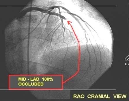
54 ECG COMPUTER DOES NOT NOTICE THE CONVEX J-T APEX SEGMENTS!
55

56

57
58 ECG Patterns associated with EARLY PHASE MI: J-T Apex abnormalities Dynamic ST-T Wave Changes on Serial ECGs

59


60 3. Dynmamic ST-T Wave Changes in Serial ECGs. Recorded at SRRMC 1 st ECG 2 nd ECG 1 st ECG 2 nd ECG

61 Acute In-Stent Thrombus Proximal LAD
62

63 T waves should not be HYPERACUTE
64 HYPERACUTE T Waves may indicate: Early phase Acute MI Transmural ischemia (usually seen in one region of the ECG) Hyperkalemia (seen globally across ECG) Hypertrophy
 and just after (normal) the")
 artery.")
65 HYPERACUTE T WAVES ECG waveforms obtained just before (hyperacute) and just after (normal) the critical blockage was stented in this patient s Proximal Left Anterior Descending (LAD) artery.
66 Helpful Clue: Hyper-Acute T Waves GLOBAL Hyper-acute T Waves (in leads viewing multiple myocardial regions / arterial distributions) favors HYPERKALEMIA

67
68 Helpful Clue: Hyper-Acute T Waves GLOBAL Hyper-acute T Waves (in leads viewing multiple myocardial regions / arterial distributions) favors HYPERKALEMIA Hyper-acute T Wave noted in ONE ARTERIAL DISTRIBUTION ( Anterior / Lateral / Inferior ) favors TRANSMURAL ISCHEMIA / Early Phase Acute MI

69

70

71


72 Cath Lab findings:
73 Dynamic ST-T Wave Changes: Other than HEART RATE related variations (which affect intervals), J Points, ST- Segments and T Waves SHOULD NOT CHANGE.
74 Dynamic ST-T Wave Changes: Other than HEART RATE related variations (which affect intervals), J Points, ST- Segments and T Waves SHOULD NOT CHANGE. When changes to J Points, ST-Segments and/or T waves are NOTED, consider EVOLVING MYOCARDIAL ISCHEMIA and/or EARLY PHASE MI, until proven otherwise.

75 46 year old male Exertional dyspnea X several weeks Intermittent chest pressure X last 3 hours. Currently pain free.

76 46 year old male: ECG 1 Chest pressure has returned, 5 on 1-10 scale. 2 nd ECG obtained due to change in symptoms :

77

78


79 ST-Segment Depression 7:59 am 8:08 am

80 Cath Lab Angiography:
81
82 ECG CRITERIA for DIAGNOSIS of STEMI: (ST J POINT) *LEADS V2 and V3: MALES AGE 40 and up mm (MALES LESS THAN mm) FEMALES mm ALL OTHER LEADS: 1.0 mm or more, in TWO or more CONTIGUOUS LEADS * P. Rautaharju et al, Standardization and Interpretation of the ECG, JACC 2009;(53)No.11:

83 ST SEGMENT ELEVATION: 3 COMMON PATTERNS of ST SEGMENT ELEVATION From ACUTE MI:

84
85 Reciprocal S-T Segment Depression may or may not be present during AMI. The presence of S-T Depression on an EKG which exhibits significant S-T elevation is a fairly reliable indicator that AMI is the diagnosis. However the lack of Reciprocal S-T Depression DOES NOT rule out AMI.

86 STEMI CASE STUDIES

87

88

89 NOWHERE, NEW MEXICO, 1994
90 STEMI CASE STUDIES
91 STEMI Case Studies, excerpts from 12 Lead ECG Interpretation in ACS with Case Studies from the Cardiac Cath Lab.

92

93
94
95 Note: There is NO Reciprocal ST Depression on this STEMI ECG!
96
97

98

99

100

101
102 ANTICIPATED COMPLICATIONS of ANTERIOR-SEPTAL WALL STEMI & POSSIBLE INDICATED INTERVENTIONS: - CARDIAC ARREST BCLS / ACLS - CARDIAC DYSRHYTHMIAS (VT / VF) ACLS (antiarrhythmics) - PUMP FAILURE with INOTROPE THERAPY: CARDIOGENIC SHOCK -DOPAMINE / DOBUTAMINE / LEVOPHED - INTRA-AORTIC BALLOON PUMP (use caution with fluid challenges due to PULMONARY EDEMA) - PULMONARY EDEMA - CPAP - ET INTUBATION (use caution with dieuretics due to pump failure and hypotension) - 3rd DEGREE HEART BLOCK - NOT RESPONSIVE TO ATROPINE TRANSCUTANEOUS or TRANSVENOUS PACING


103

104

105

106

107
108


109 CASE PROGRESSION: As the patient was being prepared for transport to the Cardiac Cath Lab, she experienced an episode of Ventricular Fibrillation.

110

111

112

113

114 = +
115
116 = +
117
118
119 ANTICIPATED COMPLICATIONS of ANTERIOR-SEPTAL WALL STEMI & POSSIBLE INDICATED INTERVENTIONS: - CARDIAC ARREST BCLS / ACLS - CARDIAC DYSRHYTHMIAS (VT / VF) ACLS (antiarrhythmics) - PUMP FAILURE with INOTROPE THERAPY: CARDIOGENIC SHOCK -DOPAMINE / DOBUTAMINE / LEVOPHED - INTRA-AORTIC BALLOON PUMP (use caution with fluid challenges due to PULMONARY EDEMA) - PULMONARY EDEMA - CPAP - ET INTUBATION (use caution with dieuretics due to pump failure and hypotension) - 3rd DEGREE HEART BLOCK - NOT RESPONSIVE TO ATROPINE TRANSCUTANEOUS or TRANSVENOUS PACING

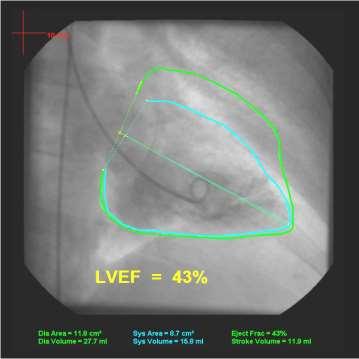
120 WHILE AWAITING THE CATH TEAM, THE PATIENT BEGAN VOMITING. SKIN BECAME ASHEN & DIAPHORETIC. REPEAT BP = 50/30. -WHAT THERAPEUTIC INTERVENTIONS SHOULD BE IMPLMENTED AT THIS POINT?


121

122

123 WHO SHOULD GO TO THE CATH LAB FIRST? And.... WHAT WOULD YOU DO WITH THE PATIENT WHO DID NOT GO TO THE CATH LAB?


124

125 PATIENT A: PATIENT B:


126


127


128


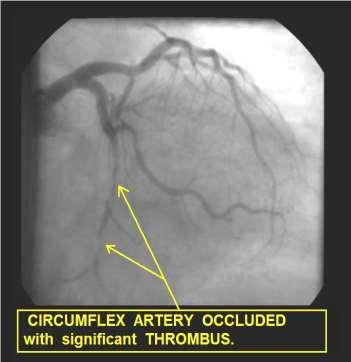
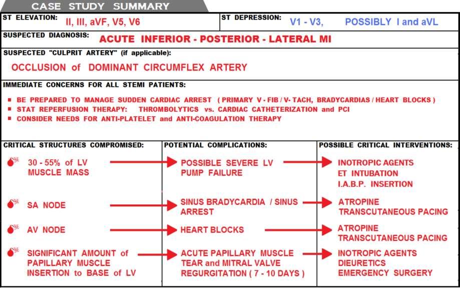
129 Despite the dismal mortality rate associated with STEMI from total LMCA occlusion, this patient survived and was later discharged. His EF is estimated at approximately 30%. He received an ICD, and is currently stable.

130

131
132
133
134

135
136

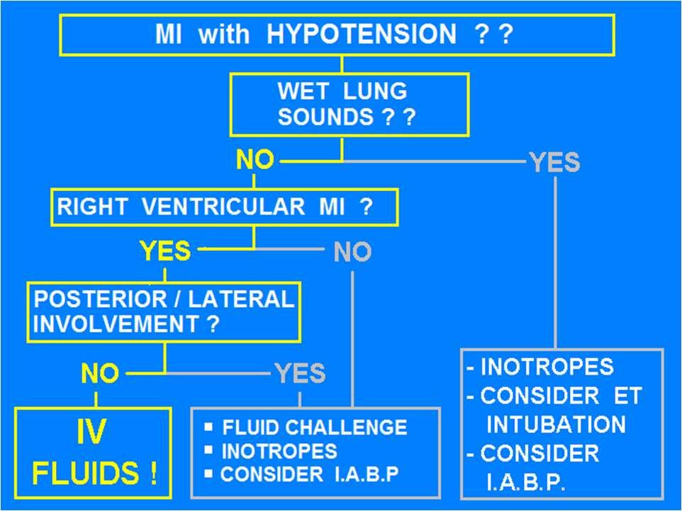
137
138 ANTICIPATED COMPLICATIONS of INFERIOR WALL STEMI secondary to RCA Occlusion & POSSIBLE INDICATED INTERVENTIONS: - CARDIAC ARREST BCLS / ACLS - CARDIAC DYSRHYTHMIAS (VT / VF) ACLS (antiarrhythmics) - SINUS BRADYCARDIA ATROPINE 0.5mg, REPEAT as needed UP TO 3mg. (follow ACLS and/or UNIT protocols) - HEART BLOCKS (1st, 2nd & 3rd Degree HB) ATROPINE 0.5mg, REPEAT as needed UP TO 3mg, Transcutaneous Pacing, (follow ACLS and/or UNIT - RIGHT VENTRICULAR MYOCARDIAL INFARCTION protocols) - The standard 12 Lead ECG does NOT view the Right Ventricle. - You must do a RIGHT-SIDED ECG to see if RV MI is present. - Do NOT give any Inferior Wall STEMI patient NITRATES or DIURETICS until RV MI has been RULED OUT. - POSTERIOR WALL INFARCTION - POSTERIOR WALL MI presents on the 12 Lead ECG as ST DEPRESSION in Leads V1 - V3. - POSTERIOR WALL MI is NOT PRESENT

139

140

141

142
143 RV MI STEMI Criteria: ST Elevation of 0.5mm (0.5mv) or more in Leads V3R, V4R, V5R or V6R
144 RV MI STEMI Criteria: ST Elevation of mm (0.5mv) or more in Leads V3R, V4R, V5R or V6R

145
146 IN EVERY CASE of INFERIOR WALL STEMI You must first RULE OUT RIGHT VENTRICULAR MI BEFORE giving any: - NITROGLYCERIN - Diuretics
147 Nitroglycerin & Diuretics are CLASS III CONTRINDICATED in RIGHT VENTRICULAR MI!!* They precipitate SEVERE HYPOTENSION * A.H.A. ACLS 2010 / 2015

148
149 ANTICIPATED COMPLICATIONS of INFERIOR - RIGHT VENRICULAR WALL STEMI secondary to PROXIMAL RCA Occlusion & POSSIBLE INDICATED INTERVENTIONS: - CARDIAC ARREST BCLS / ACLS - CARDIAC DYSRHYTHMIAS (VT / VF) ACLS (antiarrhythmics) - SINUS BRADYCARDIA ATROPINE 0.5mg, REPEAT as needed UP TO 3mg. (follow ACLS and/or UNIT protocols) - HEART BLOCKS (1st, 2nd & 3rd Degree HB) ATROPINE 0.5mg, REPEAT as needed UP TO 3mg, Transcutaneous Pacing, (follow ACLS and/or UNIT protocols) - RIGHT VENTRICULAR MYOCARDIAL INFARCTION - NITRATES and DIURETICS are CONTRA- INDICATED. - TREAT HYPOTENSION WITH FLUIDS. (It is Not uncommon to give ml of NORMAL SALINE to stabilize BP. - POSTERIOR WALL INFARCTION - POSTERIOR WALL MI presents on the 12 Lead ECG as ST DEPRESSION in Leads V1 - V3. - POSTERIOR WALL MI is NOT PRESENT ON THIS ECG.
150 If this patient becomes HYPOTENSIVE.....
151


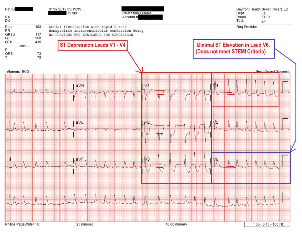
152


153

154

155


156
157

158


159
160
161 Case Study- January y/o female complaining of L arm pain, and minimal chest pain EMS 12 Lead ECGs show ST Depression in Anterior Leads V1-V4. There is NO ST Elevation

162
163 Initial Exam in ED Upon arrival in ED, 12 Lead ECG confirmed EMS findings: ST Depression in Leads V1-V4.
164
165 Causes of ST Depression V1-V4 Anterior Wall ischemia Anterior Wall NSTEMI (partial wall thickness myocardial infarction) Posterior Wall STEMI

166 Posterior Wall STEMI. Does not show ST elevation on standard 12 lead ECG because NONE of the 12 leads view the Posterior Wall directly.
167 Posterior Wall STEMI. Often shows NO ST Elevation on the standard 12 Lead ECG. Will show up on standard 12 Lead ECG as ST Depression (Reciprocal) in Leads V1-V3 (sometimes V4-V6, too).

168 V1-V3 see the Posterior Wall ONLY through RECIPROCAL changes (ST Depression)
169
170 Posterior Wall STEMI. To see ST Elevation from a Posterior Wall STEMI, you must place ECG leads on the patient s back
171

172 Continued Exam in the ED. Upon noting ST Depression in Anterior Leads V1-V4, ED Paramedic Gary Polizzi place three leads on the patient s back. Gary used the lead wires for V4, V5 and V6, with placement as shown here: The Posterior Lead ECG is seen on the next slide
173 Posterior STEMI Criteria: ST Elevation of 0.5mm (0.5mv) or more in Leads V7, V8 and/or V9
174 Posterior STEMI Criteria: ST Elevation of mm (0.5mv) or more in Leads V7, V8 and/or V9

175
176 STEMI Alert! Upon seeing Significant ST Elevation in TWO or more CONTIGUOUS LEADS, the ED physician diagnosed Posterior Wall STEMI, a STEMI Alert was issued, and the patient was taken immediately to the cardiac cath lab, where the following images were obtained.

177

178

179
180 SUMMARY Whenever ST Depression is noted in Anterior Leads (V1-V4), it could indicate that Acute Posterior Wall STEMI is present. To rule-out Posterior Wall STEMI, a posterior lead ECG (V7 V9) must be obtained. In THIS CASE, Posterior Wall STEMI was diagnosed via Posterior Lead ECG. STEMI Alert was issued, with a Door-to-PCI time of 53 minutes!

181

182 Evaluating the ECG for ACS:
183 Wide QRS present: QRSd > 120ms Determine RIGHT vs. LEFT Bundle Branch Block Pattern

184 Simple Turn Signal Method...

185 Terminal Phase of QRS Method...

186

187

188 From: Rapid Interpretation of ECGs by Dale Dubin, MD

189
190
191

192
193 Wide QRS present: (QRSd > 120ms) When RIGHT Bundle Branch Block pattern is present: Precordial Leads typically demonstrate ST Depression and T wave Inversion

194
195 Wide QRS present: (QRSd > 120ms) When RIGHT Bundle Branch Block pattern is present: Precordial Leads typically demonstrate ST Depression and T wave Inversion DOES NOT MASK STEMI; when ST Elevation is noted, CONSIDER STEMI!!

196

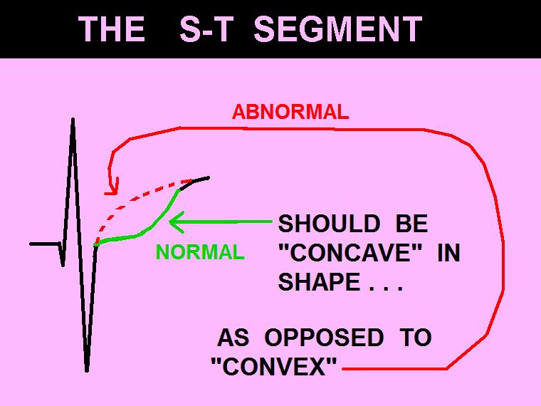
197

198
199 Wide QRS present: (QRSd > 120ms) When LBBB QRS pattern is present:
200 Wide QRS present: (QRSd > 120ms) When LBBB QRS pattern is present: ST-Segment Elevation is typically noted in Precordial Leads
201 Wide QRS present: (QRSd > 120ms) When LBBB QRS pattern is present: ST-Segment Elevation is typically noted in Precordial Leads Can cause up to 5mm of J Point Elevation in normally calibrated ECG (1mm=10mv)
202 Wide QRS present: (QRSd > 120ms) When LBBB QRS pattern is present: ST-Segment Elevation is typically noted in Precordial Leads Can cause up to 5mm of J Point Elevation in normally calibrated ECG (1mm=10mv) Does NOT typically cause ST elevation in INFERIOR Leads (II, III and AVF).
203 Diagnosis of STEMI with LBBB pattern: 2013 ACC/AHA Guideline for Management of STEMI ST Elevation of 0.1mv (1mm) or more in leads with Positive Deflection QRS complexes
204 Diagnosis of STEMI with LBBB pattern: 2013 ACC/AHA Guideline for Management of STEMI ST Elevation of 0.1mv (1mm) or more in leads with Positive Deflection QRS complexes ST Elevation of 0.5mv (5mm) or more in leads with Negative Deflection QRS complexes
205 Diagnosis of STEMI with LBBB pattern: 2013 ACC/AHA Guideline for Management of STEMI ST Elevation of 0.1mv (1mm) or more in leads with Positive Deflection QRS complexes ST Elevation of 0.5mv (5mm) or more in leads with Negative Deflection QRS complexes ST Segment Changes as compared with those of older ECGs with LBBB
206 Diagnosis of STEMI with LBBB pattern: 2013 ACC/AHA Guideline for Management of STEMI ST Elevation of 0.1mv (1mm) or more in leads with Positive Deflection QRS complexes ST Elevation of 0.5mv (5mm) or more in leads with Negative Deflection QRS complexes ST Segment Changes as compared with those of older ECGs with LBBB Convex ST Segment

207

208

209
210 Electrocardiographic Diagnosis of Evolving Acute Myocardial Infarction in the Presence of Left Bundle-Branch Block Birnbaum et al, N Engl J Med 1996; 334:
211 In patients with Left Bundle Branch Block Combined with Ventricular Hypertrophy, The J Point elevation can exceed 0.5 mv (5mm) above the iso-electric line in patients without ACS.
212


213

214
215 Practice ECGs...
216 Let s review ECG abnormality(ies)? 2. Possible diagnosis? 3. Action / Intervention?
. 3. Action / Intervention? STAT CATH LAB vs STAT Thrombolytics.")
217 1. ECG abnormality(ies)? ST Elevation Leads I, AVR AVL, V1, V2, V3, V4, V5 & V6. ST Depression II, III and AVF 2. Possible diagnosis? Acute Anterolateral Wall STEMI secondary to Left Main Coronary Artery occlusion (widowmaker MI). 3. Action / Intervention? STAT CATH LAB vs STAT Thrombolytics. Prepare for Cardiac Arrest

218 1. ECG abnormality(ies)? 2. Possible diagnosis? 3. Action / Intervention?

219 1. ECG abnormality(ies)? ST Depression V1-V4 2. Possible diagnosis? Anterior ischemia vs. Posterior wall STEMI 3. Action / Intervention? Posterior ECG (V7-V9)
220 1. ECG abnormality(ies)? 2. Possible diagnosis? 3. Action / Intervention?
221 1. ECG abnormality(ies)? ST Elevation, Leads II,III & AVF 2. Possible diagnosis? Inferior Wall STEMI 3. Action / Intervention? 1. Do R-sided ECG, prepare for Atropine administration, external pacing, cardiac arrest, STAT cath lab visit!

222 What leads show signs of possible ACS?

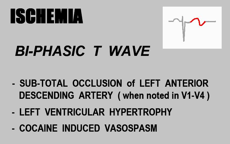
223 12 Lead ECG shows ISCHEMIC CHANGES Lateral Wall:
224 1. ECG abnormality(ies)? 2. Possible diagnosis? 3. Action / Intervention?
225 1. ECG abnormality(ies)? ST Elevation Lead AVR, Global ST Depression (I, II, III, AVL, AVF, V2, V3, V4, V5, V6) 2. Possible diagnosis? possible LMCA or 3x vessel disease. 3. Action / Intervention? Troponins, Continuous ST monitoring, cath lab visit STAT or ASAP (based on sympt.)
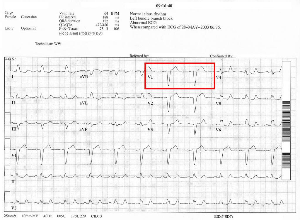
226 1. ECG abnormality(ies)? 2. Possible diagnosis? 3. Action / Intervention?
227 1. ECG abnormality(ies)? Inferior (II, III, AVF) ST Depr (ischemia?), I & AVL T wave inversion, V5 ST Depr 2. Possible diagnosis? Inferior / Lateral ischemia 3. Action / Intervention? Serial ECGs / Troponins, additional diagnostic testing, cath lab

228

229
230

231

232
233 Classic Wellen s Syndrome: Characteristic T wave changes Biphasic T waves Inverted T waves History of anginal chest pain Normal or minimally elevated cardiac markers ECG without Q waves, without significant ST-segment elevation, and with normal precordial R-wave progression
234 Wellen s Syndrome ETIOLOGY: Critical Lesion, Proximal LAD Coronary Artery Vasospasm Cocaine use (vasospasm) Increased myocardial oxygen demand Generalized Hypoxia / anemia / low H&H
235 Wellen s Syndrome EPIDEMIOLOGY & PROGNOSIS: Present in 14-18% of patients admitted with unstable angina 75% patients not treated developed extensive Anterior MI within 3 weeks. Median Average time from presentation to Acute Myocardial Infarction 8 days Sources: H Wellens et. Al, Am Heart J 1982; v103(4)
236 Wellen s Syndrome Case Study 33 y/o male Chief complaint sharp, pleuritic quality chest pain, intermittent, recent history lower respiratory infection with productive cough. ED physician attributed the ST elevation in precordial leads to early repolarization, due to patient age, gender, race (African American) and concave nature of ST-segments.

237 Wellen s Syndrome Case Study

238 Wellen s Syndrome Case Study
239 DYNAMIC ST-T Wave Changes ARE PRESENT!! NOW is the time for the STAT CALL to the CARDIOLOGIST!!!!

240 Wellen s Syndrome Case Study

241 Wellen s Syndrome Case Study

242 Wellen s Syndrome Case Study

243 Wellen s Syndrome Case Study
244 Additional Resources: Wellen s Syndrome, NEJM case study

245

246

247 Some less common, less reliable possible indicators of ACS:
248

249 STEMI Assistant: an Emergency Crash Cart Interactive Reference Manual - free Download STEMI Assistant Information Video
250 Helpful STEMI ECG Resources [1] Use of the Electrocardiogram in Acute Myocardial Infarction, Zimetbaum, et al, NEJM 348: Abnormal ST Elevation Criteria: ACC/AHA 2009 Standardization and Interpretation of the ECG, Part VI Acute Ischemia and Infarction, Galen Wagner, et al ECG in STEMI excellent powerpoint quick reference, in-depth material
251 Helpful STEMI ECG Resources Download Non-ED STEMI Protocol - example Download STEMI Alert ED Physicians Order Set

252 Correlation of Leads with ST Elevation and Cardiac Structures at Risk, based on STEMI in patients with Common Coronary Arterial Anatomy
253
254 Evolving MI & Old MI

255

256

257
258

259

260

261

262 The NORMAL ECG R wave amplitude (size) gradually increases from V1 through V6....

263 The NORMAL ECG In V3 or V4, the QRS complex becomes Biphasic.
 is a common")
264 Poor R Wave Progression.... Anterior Wall necrosis ( old MI ) is a common cause of Poor R Wave Progression.
265 EVOLVING STEMI: -ST SEGMENTS DROP -Q WAVES FORM -R WAVE PROGRESSION CHANGES IN PRECORDIAL LEADS.
266 EVOLVING STEMI: -ST SEGMENTS DROP -Q WAVES FORM -R WAVE PROGRESSION CHANGES IN PRECORDIAL LEADS.

267

268 ACUTE ANTERIOR WALL STEMI

269 EVOLVING ANTERIOR WALL STEMI

270 FULLY EVOLVED ANTERIOR WALL MI
271 Your thoughts, ideas, comments and feedback are welcome...
272 Author s correspondence information: Wayne W Ruppert Wayneruppert@bayfronthealth.com Office: Cell:

273 My top two reasons for giving everything in life the best I have to offer.
ACUTE CORONARY SYNDROME
 12 LEAD ECG INTERPRETATION in ACUTE CORONARY SYNDROME WAYNE W RUPPERT, CVT, CCCC, NREMT-P Cardiovascular Clinical Coordinator Bayfront Health Seven Rivers Crystal River, FL Education Specialist St. Joseph
Wayne Ruppert and Dr. James Irwin, St Joseph s Hospital, Tampa, FL 2006
 The EMS 12 Lead 101 Wayne W Ruppert, CVT, CCCC, NREMT-P Interventional Cardiovascular Technologist Cardiovascular Coordinator Bayfront Health Seven Rivers Welcome! Wayne Ruppert and Dr. James Irwin, St
12 Lead Electrocardiogram (ECG) PFN: SOMACL17. Terminal Learning Objective. References
 PFN: SOMACL17. Terminal Learning Objective. References") 12 Lead Electrocardiogram (ECG) PFN: SOMACL17 Slide 1 Terminal Learning Objective Action: Communicate knowledge of 12 Lead Electrocardiogram (ECG) Condition: Given a lecture in a classroom environment
12 Lead Electrocardiogram (ECG) PFN: SOMACL17 Slide 1 Terminal Learning Objective Action: Communicate knowledge of 12 Lead Electrocardiogram (ECG) Condition: Given a lecture in a classroom environment
Observation Medicine ECG Instructor Workshop session 2 Serial 12 Lead ECG Interpretation
 American College of Cardiology 20 th Congress 2017 Observation Medicine ECG Instructor Workshop session 2 Serial 12 Lead ECG Interpretation Part 1 By: Wayne W Ruppert, CVT, CCCC, NREMT-P This curriculum
All About STEMIs. Presented By: Brittney Urvand, RN, BSN, CCCC. Essentia Health Fargo Cardiovascular Program Manager.
 All About STEMIs Presented By: Brittney Urvand, RN, BSN, CCCC Essentia Health Fargo Cardiovascular Program Manager Updated 10/2/2018 None Disclosures Objectives Identify signs and symptoms of a heart attack
ACUTE CORONARY SYNDROME
 12 LEAD ECG INTERPRETATION in ACUTE CORONARY SYNDROME WAYNE W RUPPERT, CVT, CCCC, NREMT P Cardiovascular C di l Clinical Cli i l Coordinator C di t Bayfront Health Dade City Dade City, FL Education Specialist
12 Lead ECG Interpretation
 12 Lead ECG Interpretation Julie Zimmerman, MSN, RN, CNS, CCRN Significant increase in mortality for every 15 minutes of delay! N Engl J Med 2007;357:1631-1638 Who should get a 12-lead ECG? Also include
The Fundamentals of 12 Lead EKG. ECG Recording. J Point. Reviewing the Cardiac Conductive System. Dr. E. Joe Sasin, MD Rusty Powers, NRP
 The Fundamentals of 12 Lead EKG Dr. E. Joe Sasin, MD Rusty Powers, NRP SA Node Intranodal Pathways AV Junction AV Fibers Bundle of His Septum Bundle Branches Purkinje System Reviewing the Cardiac Conductive
12 Lead EKG. The Basics
 12 Lead EKG The Basics Objectives Demonstrate proper 12 EKG lead placement Determine electrical axis Identify ST and T wave changes as they relate to myocardial ischemia Describe possible complications
12 Lead ECGs: Ischemia, Injury & Infarction. Kevin Handke NRP, FP-C, CCP, CMTE STEMI Coordinator Flight Paramedic
 12 Lead ECGs: Ischemia, Injury & Infarction Kevin Handke NRP, FP-C, CCP, CMTE STEMI Coordinator Flight Paramedic None Disclosures Objectives Upon completion of this program the learner will be able to
12 Lead ECG Interpretation: Color Coding for MI s
 12 Lead ECG Interpretation: Color Coding for MI s Anna E. Story, RN, MS Director, Continuing Professional Education Critical Care Nurse Online Instructional Designer 2004 Anna Story 1 Objectives review
ECG in coronary artery disease. By Sura Boonrat Central Chest Institute
 ECG in coronary artery disease By Sura Boonrat Central Chest Institute EKG P wave = Atrium activation PR interval QRS = Ventricle activation T wave= repolarization J-point EKG QT interval Abnormal repolarization
Section V. Objectives
 Section V Landscape of an MI Objectives At the conclusion of this presentation the participant will be able to Outline a systematic approach to 12 lead ECG interpretation Demonstrate the process for determining
ECG Workshop. Nezar Amir
 ECG Workshop Nezar Amir Myocardial Ischemia ECG Infarct ECG in STEMI is dynamic & evolving Common causes of ST shift Infarct Localisation Left main artery occlusion: o diffuse ST-depression with ST elevation
12 LEAD EKG BASICS. By: Steven Jones, NREMT P CLEMC
 12 LEAD EKG BASICS By: Steven Jones, NREMT P CLEMC ECG Review Waves and Intervals P wave: the sequential activation (depolarization) of the right and left atria QRS complex: right and left ventricular
Myocardial Infarction. Reading Assignment (p66-78 in Outline )
") Myocardial Infarction Reading Assignment (p66-78 in Outline ) Objectives 1. Why do ST segments go up or down in ischemia? 2. STEMI locations and culprit vessels 3. Why 15-lead ECGs? 4. What s up with avr?
Myocardial Infarction Reading Assignment (p66-78 in Outline ) Objectives 1. Why do ST segments go up or down in ischemia? 2. STEMI locations and culprit vessels 3. Why 15-lead ECGs? 4. What s up with avr?
Acute Coronary Syndromes. Disclosures
 Acute Coronary Syndromes Disclosures I work for Virginia Garcia Memorial Health Center, Beaverton, OR. Jon Tardiff, BS, PA-C OHSU Clinical Assistant Professor And I am a medical editor for Jones & Bartlett
By the end of this lecture, you will be able to: Understand the 12 lead ECG in relation to the coronary circulation and myocardium Perform an ECG
 By the end of this lecture, you will be able to: Understand the 12 lead ECG in relation to the coronary circulation and myocardium Perform an ECG recording Identify the ECG changes that occur in the presence
A few new tools for better detection and understanding of STEMIs in the field.
 A few new tools for better detection and understanding of STEMIs in the field. Let s talk, prep and placement. Try to shoot for quality, consistency and no artifact! (looking sometimes for 1 or 2 mm changes)
Electrocardiography for Healthcare Professionals. Chapter 14 Basic 12-Lead ECG Interpretation
 Electrocardiography for Healthcare Professionals Chapter 14 Basic 12-Lead ECG Interpretation 2012 The Companies, Inc. All rights reserved. Learning Outcomes 14.1 Discuss the anatomic views seen on a 12-lead
Pennsylvania Academy of Family Physicians Foundation & UPMC 43rd Refresher Course in Family Medicine CME Conference March 10-13, 2016
 Pennsylvania Academy of Family Physicians Foundation & UPMC 43rd Refresher Course in Family Medicine CME Conference March 10-13, 2016 Disclosures: EKG Workshop Louis Mancano, MD Speaker has no disclosures
Chapter 76 Acute Coronary Syndromes Part A
 Chapter 76 Acute Coronary Syndromes Part A Episode Overview: 1. Define Stable Angina, UA, AMI 2. Describe the pathophysiology of AMI 3. What are the components of prehospital management of AMI 4. List
ECG Basics Sonia Samtani 7/2017 UCI Resident Lecture Series
 ECG Basics Sonia Samtani 7/2017 UCI Resident Lecture Series Agenda I. Introduction II.The Conduction System III.ECG Basics IV.Cardiac Emergencies V.Summary The Conduction System Lead Placement avf Precordial
Preface: Wang s Viewpoints
 AHA/ACCF/HRS Recommendations for the Standardization and Interpretation of the Electrocardiogram: Part IV, Ischemia and Infarction Presented by: WANG, TZONG LUEN, MD, PhD, JM, FACC, FESC, FCAPSC Professor,
Cardiac Ischemia ECG Workshop
 Cardiac Ischemia ECG Workshop Classic, Confusing, and Confounding Patterns Amal Mattu, MD, NE Professor and Vice Chair Department of Emergency Medicine University of Maryland School of Medicine amalmattu@comcast.net
Marcin Dada, MD December 03, 2013
 STEMI Imposters Marcin Dada, MD December 03, 2013 Marcin Dada, MD Associate Director, Chest Pain Center Hartford Hospital, Hartford, CT Member, AHA Mission Lifeline Steering Committee Outline of Topics
Acute Coronary Syndromes Unstable Angina Non ST segment Elevation MI (NSTEMI) ST segment Elevation MI (STEMI)
 ST segment Elevation MI (STEMI)") Leanna R. Miller, RN, MN, CCRN-CSC, PCCN-CMC, CEN, CNRN, CMSRN, NP Education Specialist LRM Consulting Nashville, TN Objectives Evaluate common abnormalities that mimic myocardial infarction. Identify
Leanna R. Miller, RN, MN, CCRN-CSC, PCCN-CMC, CEN, CNRN, CMSRN, NP Education Specialist LRM Consulting Nashville, TN Objectives Evaluate common abnormalities that mimic myocardial infarction. Identify
REtrive. REpeat. RElearn Design by. Test-Enhanced Learning based ECG practice E-book
 Test-Enhanced Learning Test-Enhanced Learning Test-Enhanced Learning Test-Enhanced Learning based ECG practice E-book REtrive REpeat RElearn Design by S I T T I N U N T H A N G J U I P E E R I Y A W A
Goals: Widen Your Understanding of the Wide QRS!
 Goals: Widen Your Understanding of the Wide QRS! 1. Describe an approach to diagnosis of LBBB 2. Describe the predictive value of New LBBB 3. Describe the ST segment changes that are diagnostic of AMI
12 Lead EKG Chapter 4 Worksheet
 Match the following using the word bank. 1. A form of arteriosclerosis in which the thickening and hardening of the vessels walls are caused by an accumulation of fatty deposits in the innermost lining
Difficult Data Definitions and Scenario s
 Difficult Data Definitions and Scenario s Presenter Disclosure Information Cornelia Anderson BSN, RN To following relationships exist related to this presentation: No Disclosures Objectives Discuss key
Family Medicine for English language students of Medical University of Lodz ECG. Jakub Dorożyński
 Family Medicine for English language students of Medical University of Lodz ECG Jakub Dorożyński Parts of an ECG The standard ECG has 12 leads: six of them are considered limb leads because they are placed
Acute Coronary Syndrome. Emergency Department Updated Jan. 2017
 Acute Coronary Syndrome Emergency Department Updated Jan. 2017 Goals and Objectives To reduce mortality and morbidity for people who have cardiovascular disease, with a focus on those who experience an
INTERPRETAZIONE ECG NEL PAZIENTE CON SOSPETTO STEMI
 INTERPRETAZIONE ECG NEL PAZIENTE CON SOSPETTO STEMI Giacomo Veronese Scuola di Specializzazione Medicina d Emergenza e Urgenza Università Milano-Bicocca Siete d accordo se vi propongo per una relazione..
Basic electrocardiography reading. R3 lee wei-chieh
 Basic electrocardiography reading R3 lee wei-chieh The Normal Conduction System Lead Placement avf Limb Leads Precordial Leads Interpretation Rate Rhythm Interval Axis Chamber abnormality QRST change What
Understanding the 12-lead ECG, part II
 Bundle-branch blocks Understanding the 12-lead ECG, part II Most common electrocardiogram (ECG) abnormality Appears as a wider than normal S complex Occurs when one of the two bundle branches can t conduct
UPDATE ON THE MANAGEMENTACUTE CORONARY SYNDROME. DR JULES KABAHIZI, Psc (Rwa) Lt Col CHIEF CONSULTANT RMH/KFH 28 JUNE18
 Lt Col CHIEF CONSULTANT RMH/KFH 28 JUNE18") UPDATE ON THE MANAGEMENTACUTE CORONARY SYNDROME DR JULES KABAHIZI, Psc (Rwa) Lt Col CHIEF CONSULTANT RMH/KFH 28 JUNE18 INTRODUCTION The clinical entities that comprise acute coronary syndromes (ACS)-ST-segment
UPDATE ON THE MANAGEMENTACUTE CORONARY SYNDROME DR JULES KABAHIZI, Psc (Rwa) Lt Col CHIEF CONSULTANT RMH/KFH 28 JUNE18 INTRODUCTION The clinical entities that comprise acute coronary syndromes (ACS)-ST-segment
Hot Topics in Cardiac Arrest. Should the patient go To the Cath Lab?
 Hot Topics in Cardiac Arrest Should the patient go To the Cath Lab? Tim Russert 1950-2008 Host of NBC s Meet the Press Sudden Cardiac Arrest : Autopsy showed plaque rupture in his LAD ( per LA Times,
12-Lead ECG Interpretation. Kathy Kuznar, RN, ANP
 12-Lead ECG Interpretation Kathy Kuznar, RN, ANP The 12-Lead ECG Objectives Identify the normal morphology and features of the 12- lead ECG. Perform systematic analysis of the 12-lead ECG. Recognize abnormalities
Cardiovascular Disorders Lecture 3 Coronar Artery Diseases
 Cardiovascular Disorders Lecture 3 Coronar Artery Diseases By Prof. El Sayed Abdel Fattah Eid Lecturer of Internal Medicine Delta University Coronary Heart Diseases It is the leading cause of death in
ECG pre-reading manual. Created for the North West Regional EMET training program
 ECG pre-reading manual Created for the North West Regional EMET training program Author:- Dr Juan Carlos Ascencio-Lane juan.ascencio-lane@ths.tas.gov.au 1 Disclaimer This handbook has been created for
2010 ACLS Guidelines. Primary goals of therapy for patients
 2010 ACLS Guidelines Part 10: Acute Coronary Syndrome Present : 內科 R1 鍾伯欣 Supervisor: F1 吳亮廷 991110 Primary goals of therapy for patients of ACS Reduce the amount of myocardial necrosis that occurs in
Acute Coronary Syndromes
 Overview Acute Coronary Syndromes Rabeea Aboufakher, MD, FACC, FSCAI Section Chief of Cardiology Altru Health System Grand Forks, ND Epidemiology Pathophysiology Clinical features and diagnosis STEMI management
Comments or Questions? me:
 Comments or Questions? Email me: amalmattu@comcast.net Interested in short video tutorials on electrocardiography? Check out www.ecgweekly.com Subscription fee < cost of a cup of coffee/week Covers every
Preface: Wang s Viewpoints
 AHA/ACCF/HRS Recommendations for the Standardization and Interpretation of the Electrocardiogram: Ischemia and Infarction 103.10.07 Presented by: WANG, TZONG LUEN, MD, PhD, JM, FACC, FESC, FCAPSC Professor,
ECG Cases and Questions. Ashish Sadhu, MD, FHRS, FACC Electrophysiology/Cardiology
 ECG Cases and Questions Ashish Sadhu, MD, FHRS, FACC Electrophysiology/Cardiology 32 yo female Life Insurance Physical 56 yo male with chest pain Terminology Injury ST elevation Ischemia T wave inversion
ECG ABNORMALITIES D R. T AM A R A AL Q U D AH
 ECG ABNORMALITIES D R. T AM A R A AL Q U D AH When we interpret an ECG we compare it instantaneously with the normal ECG and normal variants stored in our memory; these memories are stored visually in
EMT. Chapter 14 Review
 EMT Chapter 14 Review Review 1. All of the following are common signs and symptoms of cardiac ischemia, EXCEPT: A. headache. B. chest pressure. C. shortness of breath. D. anxiety or restlessness. Review
Cardiac Emergencies. A Review of Cardiac Compromise. Lawrence L. Lambert
 Cardiac Emergencies A Review of Cardiac Compromise Lawrence L. Lambert 1 Cardiac Emergencies Objectives: Following successful completion of this training session, the student should be able to: 1. Describe
A case of post myocardial infarction ventricular septal rupture CHRISTOFOROS KOBOROZOS, MD
 A case of post myocardial infarction ventricular septal rupture CHRISTOFOROS KOBOROZOS, MD NAVAL HOSPITAL OF ATHENS case presentation Female, 81yo Hx: diabetes mellitus, hypertension, chronic anaemia presented
Foundations EKG I - Unit 1 Summary
 Foundations EKG I - Unit 1 Summary The accurate diagnosis of ST elevation myocardial infarction (STEMI) is one of the most time critical duties in the practice of EM. Diagnosis is not always easy so guidelines
Masqueraders of STEMI
 Masqueraders of STEMI Steven M. Costa, M.D. Assistant Professor Department of Medicine Division of Cardiology Scott & White Memorial Hospital and Clinic Texas A&M University Health Science Center Disclosures
Arrhythmic Complications of MI. Teferi Mitiku, MD Assistant Clinical Professor of Medicine University of California Irvine
 Arrhythmic Complications of MI Teferi Mitiku, MD Assistant Clinical Professor of Medicine University of California Irvine Objectives Brief overview -Pathophysiology of Arrhythmia ECG review of typical
Relax and Learn At the Farm 2012
 Relax and Learn At the Farm 2012 Session 2: 12 Lead ECG Fundamentals 101 Cynthia Webner DNP, RN, CCNS, CCRN-CMC, CHFN Though for Today Mastery is not something that strikes in an instant, like a thunderbolt,
12 Lead ECG. Presented by Rebecca Sevigny BSN, RN Professional Practice & Development Dept.
 12 Lead ECG Presented by Rebecca Sevigny BSN, RN Professional Practice & Development Dept. Two Main Coronary Arteries RCA LCA which branches into Left Anterior Descending Circumflex Artery Two Main Coronary
12 Lead Interpretation
 12 Lead Interpretation Objectives Ischemia, injury and infarction ECG complex review J point ST segment STEMI recognition Ischemia to Infarct Infarction is an evolving process As the infarct evolves ECG
12 Lead ECG Skills: Building Confidence for Clinical Practice. Presented By: Cynthia Webner, BSN, RN, CCRN-CMC. Karen Marzlin, BSN, RN,CCRN-CMC
 12 Lead ECG Skills: Building Confidence for Clinical Practice NTI 2009 Preconference Session 803 Presented By: Karen Marzlin, BSN, RN,CCRN-CMC 1 12 Lead ECG Fundamentals: The Starting Place for Linking
DAY1_CARDIOVASCULAR PRACTICE QUESTIONS
 DAY1_CARDIOVASCULAR PRACTICE QUESTIONS 1 P age 1. A 59-year-old male is admitted complaining of chest pain and dyspnea. ST elevation and T-wave inversion were seen on the ECG in V2, V3, and V4. IV thrombolytic
Acute chest pain and ECG need for immediate coronary angiography?
 Acute chest pain and ECG need for immediate coronary angiography? Kjell Nikus, MD, PhD Heart Center, Tampere University Hospital, Finland and Samuel Sclarovsky, MD, PhD Tel Aviv University, Israel There
Acute Myocardial Infarction. Willis E. Godin D.O., FACC
 Acute Myocardial Infarction Willis E. Godin D.O., FACC Acute Myocardial Infarction Definition: Decreased delivery of oxygen and nutrients to the myocardium Myocardial tissue necrosis causing irreparable
Medical Management of Acute Coronary Syndrome: The roles of a noncardiologist. Norbert Lingling D. Uy, MD Professor of Medicine UERMMMCI
 Medical Management of Acute Coronary Syndrome: The roles of a noncardiologist physician Norbert Lingling D. Uy, MD Professor of Medicine UERMMMCI Outcome objectives of the discussion: At the end of the
10 ECGs No Practitioner Can Afford to Miss. Objectives
 10 ECGs No Practitioner Can Afford to Miss Mary L. Dohrmann, MD Professor of Clinical Medicine Division of Cardiovascular Medicine University of Missouri School of Medicine No disclosures Objectives 1.
A walk through a STEMI
 A walk through a STEMI M.M. s Story Kim Robison Ashley Corcoran Situation M.M. is an 82 year old male brought in by private vehicle on 10/22/17 to the Emergency Department Pt. c/o left arm numbness, pain
Electrocardiography. Hilal Al Saffar College of Medicine,Baghdad University
 Electrocardiography Hilal Al Saffar College of Medicine,Baghdad University Which of the following is True 1. PR interval, represent the time taken for the impulse to travel from SA node to AV nose. 2.
Acute Myocardial Infarction
 Acute Myocardial Infarction Hafeza Shaikh, DO, FACC, RPVI Lourdes Cardiology Services Asst.Program Director, Cardiology Fellowship Associate Professor, ROWAN-SOM Acute Myocardial Infarction Definition:
ECG Interpretation. Introduction to Cardiac Telemetry. Michael Peters, RN, CCRN, CFRN CALSTAR Air Medical Services
 ECG Interpretation Introduction to Cardiac Telemetry Michael Peters, RN, CCRN, CFRN CALSTAR Air Medical Services Disclosures Nothing to disclose Objectives Describe the electrical conduction pathway in
ECGs: Everything a finalist needs to know. Dr Amy Coulden As part of the Simply Finals series
 ECGs: Everything a finalist needs to know Dr Amy Coulden As part of the Simply Finals series Aims and objectives To be able to interpret basic ECG abnormalities To be able to recognise commonly tested
MWLCEMS SYSTEM Continuing Education Packet Management of the Acute MI Patient
 MWLCEMS SYSTEM Continuing Education Packet Management of the Acute MI Patient In this CE we will discuss the patient presenting with an acute ST-Elevation Myocardial Infarction (STEMI) Definition: Myocardial
Disclosures. STEMI:To Call or Not to Call. Disclosures 9/18/2017. Alternate Title: Hey Doc, If you re not doing anything Saturday Night
 STEMI:To Call or Not to Call Disclosures No financial disclosures September, 2017 Frederick James Trip Meine III MD, FACC, FSCAI Cape Fear Heart Associates, Wilmington, NC Disclosures Alternate Title:
ACLS Prep. Preparation is key to a successful ACLS experience. Please complete the ACLS Pretest and Please complete this ACLS Prep.
 November, 2013 ACLS Prep Preparation is key to a successful ACLS experience. Please complete the ACLS Pretest and Please complete this ACLS Prep. ACLS Prep Preparation is key to a successful ACLS experience.
The Electrocardiogram part II. Dr. Adelina Vlad, MD PhD
 The Electrocardiogram part II Dr. Adelina Vlad, MD PhD Basic Interpretation of the ECG 1) Evaluate calibration 2) Calculate rate 3) Determine rhythm 4) Determine QRS axis 5) Measure intervals 6) Analyze
Huseng Vefali MD St. Luke s University Health Network Department of Cardiology
 Huseng Vefali MD St. Luke s University Health Network Department of Cardiology Learning Objectives Establish Consistent Approach to Interpreting ECGs Review Essential Cases for Paramedics and first responders
3/4/2018. March Martina Frost, PA C Desert Cardiology. Electricity moving towards/away from electrode create downward/upward directions of waves
 March 2018 Martina Frost, PA C Desert Cardiology Electricity moving towards/away from electrode create downward/upward directions of waves Frontal view Limb leads: I, II, III, avl, avf, (avr) Horizontal
Ekg pra pr c a tice D.HAMMOUDI.MD
 Ekg practice D.HAMMOUDI.MD Anatomy Revisited RCA (Right Coronary Artery) Right ventricle Inferior wall of LV Posterior wall of LV (75%) SA Node (60%) AV Node (>80%) LCA (Left Coronary Artery) Septal wall
12 Lead ECGs: Ischemia, Injury, Infarction
 12 Lead ECGs: Ischemia, Injury, Infarction This course has been awarded four (4) contact hours. This course expires on March 31, 2019. Copyright 2015 by RN.com. All Rights Reserved. Reproduction and distribution
12 Lead Acquisition and Interpretation APRIL 23 11:00 AM
 12 Lead Acquisition and Interpretation APRIL 23 11:00 AM Presented by : Jennifer Robson, Prehospital Care Specialist Dr. Don Eby, Local Medical Director Objectives Upon completion of this webinar, you
Chapter 3 for 12 Lead Training -Precourse-
 ONTARIO BASE HOSPITAL GROUP Chapter 3 for 12 Lead Training -Precourse- Ontario Base Hospital Group Education Subcommittee 2008 TIME IS MUSCLE ONTARIO BASE HOSPITAL GROUP Introduction and Purpose Introduction
also aid the clinician in recognizing both the obvious and subtle abnormalities that may help guide therapy.
 Karen Lieberman, MS, CRNP f the many diagnostic tools used to screen for and evaluate cardiac abnormalities, the 12-lead electrocardiogram (ECG) is among the most basic. This inexpensive and noninvasive
Myocardial infarction
 CHAPTER-I CARDIOVASCULAR SYSTEM Myocardial infarction SUB: PHARMACOTHERAPEUTICS-I CODE:T0820006 Dr. Venugopal Pharm.D Assistant Professor Department of Pharm.D Kriahna Teja Pharmacy College,Tirupati. Definition
Objectives. Identify early signs and symptoms of Acute Coronary Syndrome Initiate proper protocol for ACS patient 10/2013 2
 10/2013 1 Objectives Identify early signs and symptoms of Acute Coronary Syndrome Initiate proper protocol for ACS patient 10/2013 2 Purpose of this Education Module: Chest Pain Center Accreditation involves
WE ARE STEMI HUNTERS. LearningObjectives. I have no relevant disclosures. Myth: Jennifer Carlquist PA-C, ER CAQ
 WE ARE STEMI HUNTERS Jennifer Carlquist PA-C, ER CAQ Salinas Valley Memorial, ER Central Coast Cardiology, Specializing in EP LearningObjectives How to use pattern recognition to detect ischemia Triage
E CG Challenges. The ST segment on an electrocardiogram (ECG) correlates to the plateau. Wellens Syndrome
 correlates to the plateau. Wellens Syndrome") AACN Advanced Critical Care Volume 29, Number 3, pp 360-364 2018 AACN E CG Challenges Karen M. Marzlin, DNP, RN, CCNS, ACNPC-AG, CCRN-CMC, CHFN Department Editor Wellens Syndrome Karen M. Marzlin, DNP,
AACN Advanced Critical Care Volume 29, Number 3, pp 360-364 2018 AACN E CG Challenges Karen M. Marzlin, DNP, RN, CCNS, ACNPC-AG, CCRN-CMC, CHFN Department Editor Wellens Syndrome Karen M. Marzlin, DNP,
EKG Competency for Agency
 EKG Competency for Agency Name: Date: Agency: 1. The upper chambers of the heart are known as the: a. Atria b. Ventricles c. Mitral Valve d. Aortic Valve 2. The lower chambers of the heart are known as
Coronary Heart Disease. Raja Nursing Instructor RN, DCHN, Post RN. BSc.N
 Coronary Heart Disease Raja Nursing Instructor RN, DCHN, Post RN. BSc.N 31/03/2016 Objectives Define coronary heart disease (CHD). Identify the causes and risk factors of CHD Discuss the pathophysiological
Παύλος Στουγιάννος. Καρδιολόγος ΓΝΑ «Η ΕΛΠΙΣ»
 Επεμβατική Καρδιολογία. STEMI. Σύγχρονη θεώρηση Παύλος Στουγιάννος Καρδιολόγος ΓΝΑ «Η ΕΛΠΙΣ» Criteria for acute myocardial infarction Thygesen K, et al. Third universal definition of myocardial infarction.
ACLS Review. Pulse Oximetry to be between 94 99% to avoid hyperoxia (high oxygen tension can lead to tissue death
 ACLS Review BLS CPR BLS CPR changed in 2010. The primary change is from the ABC format to CAB. After establishing unresponsiveness and calling for a code, check for a pulse less than 10 seconds then begin
Ischemic Heart Diseases. Dr. Nabila Hamdi MD, PhD
 Ischemic Heart Diseases Dr. Nabila Hamdi MD, PhD ILOs Compare and contrast the different types of angina regarding their pathogenesis, clinical manifestations and evolution. Discuss myocardial infarct,
Algorithm Focus. Emergency Cardiovascular Care: EMT-Intermediate Treatment Algorithms. Perspective regarding the EMT- Intermediate algorithms
 Emergency Cardiovascular Care: EMT-Intermediate Treatment Algorithms Algorithms for the Conscious Patient Prehospital Medication Profiles Algorithm Focus Bradycardia Acute Pulmonary Edema and Shock Hypothermia
CAN T MISS ECG FINDINGS L. THOMAS RICHARDS, MD ASSISTANT PROFESSOR OF EMERGENCY MEDICINE
 Topics in Emergency Medicine 2010 CAN T MISS ECG FINDINGS L. THOMAS RICHARDS, MD ASSISTANT PROFESSOR OF EMERGENCY MEDICINE OBJECTIVES Examine three common presentations to the ED which compel the EM provider
Other 12-Lead ECG Findings
 Other 12-Lead ECG Findings Left Atrial Enlargement Left atrial enlargement is illustrated by increased P wave duration in lead II, top ECG, and by the prominent negative P terminal force in lead V1, bottom
CASE 10. What would the ST segment of this ECG look like? On which leads would you see this ST segment change? What does the T wave represent?
 CASE 10 A 57-year-old man presents to the emergency center with complaints of chest pain with radiation to the left arm and jaw. He reports feeling anxious, diaphoretic, and short of breath. His past history
Part One Objectives. Don t Worry About It. All done for you Paper Speed 25 mm/sec Calibration 1 mv charge over 20 ms = 10 mm tall Lincoln Hat
 12-lead and ACS Review North Lyon Refresher Part One Objectives 12 lead ECG Basics Anatomy and Physiology STEMI Diagnosis Types of MI ACS Review STEMI System and Interventional Cardiology Review The Value
Ischemic heart disease
 Ischemic heart disease Introduction In > 90% of cases: the cause is: reduced coronary blood flow secondary to: obstructive atherosclerotic vascular disease so most of the time it is called: coronary artery
Different ECG patterns at presentation in ACS. D. Goldwasser F. Molina A. Bayes de Luna
 Different ECG patterns at presentation in ACS D. Goldwasser F. Molina A. Bayes de Luna Acute Coronary syndromes: The importance of the ECG There are two types of ACS: STE- ACS and Non STE-ACS The most
Hanna K. Al-Makhamreh, M.D., FACC Interventional Cardiologist
 Hanna K. Al-Makhamreh, M.D., FACC Interventional Cardiologist Introduction. Basic Life Support (BLS). Advanced Cardiac Life Support (ACLS). Cardiovascular diseases (CVDs) are the number one cause of death
Atherosclerotic Heart Disease: Coronary Vessels, EKG Localization of STEMI and Complications/Derivatives for USMLE Step One
 Atherosclerotic Heart Disease: Coronary Vessels, EKG Localization of STEMI and Complications/Derivatives for USMLE Step One Howard J. Sachs, MD Associate Professor of Medicine University of Massachusetts
2018 Acute Coronary Syndrome. Robert Bender, DO, FACOI, FACC Central Maine Heart and Vascular Institute
 2018 Acute Coronary Syndrome Robert Bender, DO, FACOI, FACC Central Maine Heart and Vascular Institute Definitions: Acute Myocardial Ischemia Unstable Angina Non-ST-Elevation MI (NSTEMI) }2/3 ST-Elevation
15 th Sukaman Memorial Lecture ST Segment Elevation: New Electrocardiographic Insights in 2014
 DOI 10.7603/s40602-016-0006-3 ASEAN Heart Journal http://www.globalsciencejournals.com/journal/40602 Vol. 24, no.1, 98 105 (2016) ISSN: 2315-4551 15 th Sukaman Memorial Lecture ST Segment Elevation: New
Acute Coronary Syndrome including STEMI
 Portage County EMS Patient Care Guidelines Acute Coronary Syndrome including STEMI Note: The goal is to deliver a STEMI patient to a cardiac center within 60 minutes of first ALS patient contact. Cardiac