Echo Emergencies. Outline. Michael H. Picard, MD Massachusetts General Hospital Harvard Medical School No disclosures
|
|
|
- Dustin Hodge
- 6 years ago
- Views:
Transcription

 Prosthetic valve")
1 Echo Emergencies Michael H. Picard, MD Massachusetts General Hospital Harvard Medical School No disclosures Outline Common emergency / on call scenarios Tamponade Pulmonary embolism/rv strain Cardiogenic shock / MI complications / volume status Chest pain and LBBB Aortic dissection Transplant donor evals and OHT rejection Volume status (IVC imaging) Prosthetic valve thrombosis/obstruction Mechanical Circulatory Support malfunction Post-cardiac surgery hypotension 2 1

2 Cardiac Tamponade Echocardiographic findings in Cardiac Tamponade Pericardial effusion RV free wall inversion early diastole, respiratory variation RA inversion late diastole, early systole: >33% of cycle IVC plethora exaggerated Doppler respiratory variation in transvalvular flow IVC, TV, PV: insp, exp MV, AV: insp, exp 2

3 3


4 Gillam et al Circ 1983;68:


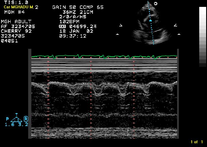
5 Hypotension immediate post-cardiac surgery TTE images tough Hematoma rather than free flowing fluid May be difficult to see even when images are good Often localized behind RA Doppler flow variations may be the best clue of elevated intra-pericardial pressure Clinical judgement required TEE vs. go back to OR without confirmation 10 5


6 Pulmonary embolism TTE should not be performed to diagnose PE (i.e., to look for the thrombus) It is used to assess presence of RV strain which may impact treatment decisions 11 RV strain in the setting of pulmonary embolism aka McConnell s Sign Am J Card 1996;78:


7 Chest pain and LBBB is it ACS? 13 Type I aortic dissection 7
 and")
8 Who says you can t diagnose aortic dissection with TTE? 15 Echocardiographic findings Intimal flap High frequency, low amplitude motion flow respects the boundary Artifacts Motion - Low frequency (or same frequency as adjacent structure) and high amplitude Color flow goes through +/- aortic dilation Aortic insufficiency Wall motion abnormality if flap obstructs coronary ostia 8
9 Echo for Aortic Dissection TEE Sensitivity % and specificity % in different series False negative TEE is rare: few dissections are limited to the blind spot at the inferior portion of the arch False positive TEE: Artifacts are very common (23-55%), especially in the ascending aorta. Artifacts Side-by-side: lateral resolution, side lobe, lens effect Behind, parallel motion: Reverberation Behind, opposite motion: Mirror image 9

10 Linear TEE artifacts artifacts in the aorta seen in the presence of dilated aortas Artifact distance = 2 times that of interface Transverse TEE image of dilated ascending aorta compressing LA. Note the artifact crosses borders Appelbe et al, JACC 1993;21: Clues to artifacts Cross borders motion identical to another real structure amplitude and frequency at a multiple of the real structure indistinct edges not reproduced in an orthogonal or other view color flow passes through it clues - foreign materials present catheters, prosthetic valves, grafts these are not always in the plane of view 10
11 Clues to real structures respects borders distinct edges (unless thrombus) motion intimal flaps - amplitude and frequency of motion different than cardiac cycle, respiration seen in multiple views color flow respects true borders intimal flaps keep company other pathologic processes LVAD or other Mechanical Circulatory Support potential complications Aortic dissection AI of native valve ; Aortic valve thrombosis Conduit obstruction / kinking Worsening of RV failure Inlet cannula thrombosis Overpumping Pump failure Bleeding / hematoma infection 22 11


12 LV underfilled (overpumping, RPMs too high) LVAD complications 12

13 Device malfunction due to thrombus response to changing pump speed Image at lowest pump speed, increase to maximum pump speed (2 min/intermediate stages) Normal response LVEDd decrease Decrease in AV opening duration RV stroke volume increases Increased MV deceleration time If obstruction (ie., thrombus) No change in LVEDd, AV opening, RV SV, Decreased LV deceleration time MV E wave decrease and deceleration time increase with increased LVAD pump speeds = reduction in LV diastolic pressures Estep et al JACC CV Img 2010;3:

14 61 yo with VV ECMO and hypotension RA-PA ECMO for severe COPD, awaiting lung transplant TTE for? Pericardial effusion/tamponade 14
15 15
 Not seen with certain immunosuppressives Non-compliant LV (diastolic dysfunction) Mainly with cyclosporine Nonspecific may occur just due to increased LA pressure 32")
16 Cardiac Transplant on call echoes Donor assessment Assess LV function, valve disease Transplant rejection Reduced LV function (compare to prior) Myocardial edema (increased wall thickening) Not seen with certain immunosuppressives Non-compliant LV (diastolic dysfunction) Mainly with cyclosporine Nonspecific may occur just due to increased LA pressure 32 16
 6.7% ventricular septal rupture - highest mortality (87.")
17 Cardiogenic Shock (CS) after MI: etiologies and outcome from a large registry 1190 CS pts eligible but not randomized to SHOCK Trial (mechanical complications excluded) etiologies: LV failure 78.5% severe MR 6.9% ventricular septal rupture 3.9% isolated RV shock 2.8% tamponade 1.4% other (severe valve ds, hemorrhage, sepsis) 6.7% ventricular septal rupture - highest mortality (87.3%) Hochman et al JACC 2000;36: Cardiogenic shock post-mi ventricular septal rupture 34 17


18 Cardiogenic shock post-mi papillary muscle rupture 35 Cardiogenic shock post-mi pseudo aneurysm 18


19 Cardiogenic shock post-mi free wall rupture Elderly diabetic woman with several days of malaise and chest pain. Low BP in ED. TEE for aortic dissection. No dissection but LV EF reduced and pericardial effusion Bimbaum Y, et al. N Engl J Med. 2002; Edwards BS, et al. Am J Cardiol. 1984; Mann JM, Roberts WC. Am J Cardiol Cardiogenic shock just a bad LV 38 19
 Inspiratory collapse < 50 % RA pressure 10-15 mm HG No collapse suggests markedly elevated RA pressure of > 15 mm Hg Small IVC (< 1.")
20 Hypotension assessing preload by IVC size 39 IVC size and respiratory dynamics 40 Normal IVC size 50% decrease in diameter = normal RA pressure Dilated IVC Normal inspiratory collapse (> 50%) suggestive of mildly elevated RA pressure (6-10 mm HG) Inspiratory collapse < 50 % RA pressure mm HG No collapse suggests markedly elevated RA pressure of > 15 mm Hg Small IVC (< 1.2 cm) with spontaneous collapse often is seen in the presence of intravascular volume depletion 20
21 Prosthetic valve thrombosis Occurs in both mechanical and biological prostheses Stenosis with or without regurgitation Gradual or sudden onset of symptoms 2X higher in MV than AV Keys on echo Movement of occlude Doppler gradients Pattern of flow / regurgitation 41 GMK 53 yo F remote hx of Hodgkin s ds (s/p XRT to chest) AS/AI, MR, TR Progressive heart failure Day 14 s/p AVR/MVR Difficult to diurese LFTs now markedly elevated New dyspnea Supratherapeutic INR On call echo to assess for TR, paravalvular leak, pericardial effusion/tamponade 42 21


22 New SOB 2 weeks after AVR/MVR


23 TTE 45 Small LV IVS flattening in diastole and systole RV volume and pressure overload Aortic bileaflet mechanical prosthetic valve = ok Peak 18 mm Hg, mean 10 mm Hg, trace valvular AI Mitral bileaflet mechanical prosthesis Restricted motion of 1 of the disks Peak 31 mm Hg, mean 17 mm Hg (2 weeks earlier mean = 6 mm Hg) Severe TR Trivial pericardial effusion Peak gradient 25 mm Hg; mean 15 mm Hg mean formerly was 6 mm Hg (2 weeks prior) 46 23

24 Before and after 47 24
Echocardiography as a diagnostic and management tool in medical emergencies
 Echocardiography as a diagnostic and management tool in medical emergencies Frank van der Heusen MD Department of Anesthesia and perioperative Care UCSF Medical Center Objective of this presentation Indications
ARTIFACTS: THEORY AND ILLUSTRATIVE EXAMPLES
 ARTIFACTS: THEORY AND ILLUSTRATIVE EXAMPLES Robert A. Levine, M.D. Marielle Scherrer-Crosbie, M.D. Eric M. Isselbacher, M.D. 60 year old man Cardiac source of embolus? NAME THAT MASS! 1 NAME THAT MASS!
PROSTHETIC VALVE BOARD REVIEW
 PROSTHETIC VALVE BOARD REVIEW The correct answer D This two chamber view shows a porcine mitral prosthesis with the typical appearance of the struts although the leaflets are not well seen. The valve
ARTIFACTS: THEORY AND ILLUSTRATIVE EXAMPLES
 ARTIFACTS: THEORY AND ILLUSTRATIVE EXAMPLES Robert A. Levine, M.D. Marielle Scherrer-Crosbie, M.D. Eric M. Isselbacher, M.D. No conflicts of interest Philippe Bertrand, Pieter Vendervoort, Hasselt and
TAVR: Echo Measurements Pre, Post And Intra Procedure
 2017 ASE Florida, Orlando, FL October 10, 2017 8:00 8:25 AM 25 min TAVR: Echo Measurements Pre, Post And Intra Procedure Muhamed Sarić MD, PhD, MPA Director of Noninvasive Cardiology Echo Lab Associate
Outline. Echocardiographic Assessment of Pericardial Effusion/Tamponade: The Essentials
 Echocardiographic Assessment of Pericardial Effusion/Tamponade: The Essentials John R Schairer DO FACC Henry Ford Heart and Vascular Institute No Disclosures Outline Normal Anatomy and Physiology Pathophysiology
Acute Aortic Syndromes
 Acute Aortic Syndromes Michael H. Picard, M.D. Massachusetts General Hospital Harvard Medical School No disclosures For everything you need to know about the aorta see Circulation 2010;121:e266-e369 And.
Echo in Heart Failure
 Echo in Heart Failure Karima Addetia, MD Heart Failure: Definition A clinical syndrome that results from impairment of ventricular filling or ejection of blood. Manifestations include dyspnea and fatigue,
P = 4V 2. IVC Dimensions 10/20/2014. Comprehensive Hemodynamic Evaluation by Doppler Echocardiography. The Simplified Bernoulli Equation
 Comprehensive Hemodynamic Evaluation by Doppler Echocardiography Itzhak Kronzon, MD North Shore LIJ/ Lenox Hill Hospital New York, NY Disclosure: Philips Healthcare St. Jude Medical The Simplified Bernoulli
Adel Hasanin Ahmed 1
 Adel Hasanin Ahmed 1 PERICARDIAL DISEASE The pericardial effusion ends anteriorly to the descending aorta and is best visualised in the PLAX. PSAX is actually very useful sometimes for looking at posterior
Pericardial Diseases. Smonporn Boonyaratavej, MD. Division of Cardiology, Department of Medicine Chulalongkorn University
 Pericardial Diseases Smonporn Boonyaratavej, MD Division of Cardiology, Department of Medicine Chulalongkorn University Cardiac Center, King Chulalongkorn Memorial Hospital 21 AUGUST 2016 Pericardial
cardiac imaging planes planning basic cardiac & aortic views for MR
 cardiac imaging planes planning basic cardiac & aortic views for MR Dianna M. E. Bardo, M. D. Assistant Professor of Radiology & Cardiovascular Medicine Director of Cardiac Imaging cardiac imaging planes
Comprehensive Hemodynamics By Doppler Echocardiography. The Echocardiographic Swan-Ganz Catheter.
 Comprehensive Hemodynamics By Doppler Echocardiography. The Echocardiographic Swan-Ganz Catheter. Itzhak Kronzon, MD, FASE, FACC, FESC, FAHA, FACP, FCCP North Shore HS, LIJ/Lenox Hill Hospital, New York
Adult Echocardiography Examination Content Outline
 Adult Echocardiography Examination Content Outline (Outline Summary) # Domain Subdomain Percentage 1 2 3 4 5 Anatomy and Physiology Pathology Clinical Care and Safety Measurement Techniques, Maneuvers,
Choose the grading of diastolic function in 82 yo woman
 Question #1 Choose the grading of diastolic function in 82 yo woman E= 80 cm/s A= 70 cm/s LAVI < 34 ml/m 2 1= Grade 1 2= Grade 2 3= Grade 3 4= Normal 5= Indeterminate 2018 MFMER 3712003-1 Choose the grading
You Won t Believe What I Saw on. Disclosures. Goals. Dimensions 2013 October 18 th Michael Pfeiffer, MD. No Financial Disclosures
 You Won t Believe What I Saw on that ECHO! Dimensions 2013 October 18 th Michael Pfeiffer, MD Disclosures No Financial Disclosures Goals Review unusual and unique echocardiographic images. Briefly present
Echocardiographic Cardiovascular Risk Stratification: Beyond Ejection Fraction
 Echocardiographic Cardiovascular Risk Stratification: Beyond Ejection Fraction October 4, 2014 James S. Lee, M.D., F.A.C.C. Associates in Cardiology, P.A. Silver Spring, M.D. Disclosures Financial none
7. Echocardiography Appropriate Use Criteria (by Indication)
") Criteria for Echocardiography 1133 7. Echocardiography Criteria (by ) Table 1. TTE for General Evaluation of Cardiac Structure and Function Suspected Cardiac Etiology General With TTE 1. Symptoms or conditions
Criteria for Echocardiography 1133 7. Echocardiography Criteria (by ) Table 1. TTE for General Evaluation of Cardiac Structure and Function Suspected Cardiac Etiology General With TTE 1. Symptoms or conditions
Federico M Asch MD, FASE MedStar Heart and Vascular Institute Georgetown University Washington, DC
 TAVR: When Things go Wrong Federico M Asch MD, FASE MedStar Heart and Vascular Institute Georgetown University Washington, DC Disclosures Academic Echo Core Lab Abbott / St Jude Medical Edwards Medtronic
Index. K Knobology, TTE artifact, image resolution, ultrasound, 14
 A Acute aortic regurgitation (AR), 124 128 Acute aortic syndrome (AAS) classic aortic dissection diagnosis, 251 263 evolutive patterns, 253 255 pathology, 250 251 classifications, 247 248 incomplete aortic
Echo Doppler Assessment of Right and Left Ventricular Hemodynamics.
 Echo Doppler Assessment of Right and Left Ventricular Hemodynamics. Itzhak Kronzon, MD, FASE, FACC, FESC, FAHA, FACP, FCCP Northwell, Lenox Hill Hospital, New York Professor of Cardiology Hofstra University
Pericardial Disease: Case Examples. Echo Fiesta 2017
 Pericardial Disease: Case Examples Echo Fiesta 2017 2014 2014 MFMER MFMER 3346252-1 slide-1 Objectives Have a systematic approach to evaluation of constriction 2014 MFMER 3346252-2 CASE 1 2013 MFMER 3248567-3
M-Mode Echocardiography Is it still Alive? Itzhak Kronzon, MD,FASE. Sampling Rate M-Mode: 1800 / sec 2D: 30 / sec
 M-Mode Echocardiography Is it still Alive? Itzhak Kronzon, MD,FASE Honoraria: Philips Classical M-mode Echocardiography M-Mode offers better time and image resolution. Sampling Rate M-Mode: 1800 / sec
Prosthesis-Patient Mismatch or Prosthetic Valve Stenosis?
 EuroValves 2015, Nice Prosthesis-Patient Mismatch or Prosthetic Valve Stenosis? Philippe Pibarot, DVM, PhD, FACC, FAHA, FASE FESC Canada Research Chair in Valvular Heart Diseases Université LAVAL Disclosure
Breakout Session: Transesophageal Echocardiography
 Breakout Session: Transesophageal Echocardiography Doris Ockert, MD Andrew Schroeder, MD University of Wisconsin School of Medicine and Public Health Jutta Novalija, MD, PhD Medical College of Wisconsin
Mechanical Cardiac Support and Cardiac Transplant: The Role for Echocardiography
 Mechanical Cardiac Support and Cardiac Transplant: The Role for Echocardiography David Langholz, M.D., F.A.C.C. Co-Director Cardiovascular Imaging Fredrick Meijer Heart and Vascular Institute Spectrum
Prosthetic valve dysfunction: stenosis or regurgitation
 Prosthetic valve dysfunction: stenosis or regurgitation Jean G. Dumesnil MD, FRCP(C), FACC, FASE(Hon) Quebec Heart and Lung Institute, Québec, Québec No disclosures Possible Causes of High Gradients in
Left Ventricular Assist Device: What Should I Report?
 2017 SOTA, Tucson, AZ February 21, 2017 11:15 11:40 AM 25 min Left Ventricular Assist Device: What Should I Report? Muhamed Sarić MD, PhD, MPA Director of Noninvasive Cardiology Echo Lab Associate Professor
Clinical Indications for Echocardiography
 Clinical Indications for Echocardiography Echocardiography is widely utilised and potential applications are increasing with advances in technology. The aim of this document is two-fold: 1) To define clinical
Pulmonary Hypertension: Another Use for Viagra
 Pulmonary Hypertension: Another Use for Viagra Kathleen Tong, MD Director, Heart Failure Program Assistant Clinical Professor University of California, Davis Disclosures I have no financial conflicts A
25 different brand names >44 different models Sizes mm
 Types of Prosthetic Valves BIOLOGIC STENTED Porcine xenograft Pericardial xenograft STENTLESS Porcine xenograft Pericardial xenograft Homograft (allograft) Autograft PERCUTANEOUS MECHANICAL Bileaflet Single
The Role of Mechanical Circulatory Support in Cardiogenic Shock: When to Utilize
 The Role of Mechanical Circulatory Support in Cardiogenic Shock: Presented by Nancy Scroggins ACNP, CNS-CC CV Surgery ACNP Bayshore Medical Center The Role of Mechanical Circulatory Support in Cardiogenic
Echocardiography Conference
 Echocardiography Conference David Stultz, MD Cardiology Fellow, PGY-6 September 20, 2005 Atrial Septal Aneurysm Bulging of Fossa Ovalis Associated commonly with Atrial septal defect or small perforations
Optimal Imaging Technique Prior to TAVI -Echocardiography-
 2014 KSC meeting Optimal Imaging Technique Prior to TAVI -Echocardiography- Geu-Ru Hong, M.D. Ph D Associate Professor of Medicine Division of Cardiology, Severance Cardiovascular Hospital Yonsei University
DISCLOSURE. Echocardiography in Systemic Diseases: Questions. Relevant Financial Relationship(s) None. Off Label Usage None 5/7/2018
 None. Off Label Usage None 5/7/2018") Echocardiography in Systemic Diseases: Questions Sunil Mankad, MD, FACC, FCCP, FASE Associate Professor of Medicine Mayo Clinic College of Medicine Director, Transesophageal Echocardiography Associate
Echocardiography in Systemic Diseases: Questions Sunil Mankad, MD, FACC, FCCP, FASE Associate Professor of Medicine Mayo Clinic College of Medicine Director, Transesophageal Echocardiography Associate
ASCeXAM / ReASCE. Practice Board Exam Questions Monday Morning
 ASCeXAM / ReASCE Practice Board Exam Questions Monday Morning Ultrasound Physics Artifacts Doppler Physics Imaging, Knobology, and Artifacts Echocardiographic Evaluation of the RV Tricuspid and Pulmonary
TSDA Boot Camp September 13-16, Introduction to Aortic Valve Surgery. George L. Hicks, Jr., MD
 TSDA Boot Camp September 13-16, 2018 Introduction to Aortic Valve Surgery George L. Hicks, Jr., MD Aortic Valve Pathology and Treatment Valvular Aortic Stenosis in Adults Average Course (Post mortem data)
Role of echocardiography in the assessment of ischemic heart disease 분당서울대학교병원윤연이
 Role of echocardiography in the assessment of ischemic heart disease 분당서울대학교병원윤연이 Outline Evaluation of Chest pain Evaluation of MI complications Prediction of Outcomes Evaluation of Chest pain Evaluation
New ASE Guidelines: What you must know
 New ASE Guidelines: What you must know Federico M Asch MD, FASE, FACC Chair, ASE Guidelines and Standards Committee Medstar Washington Hospital Center Medstar Health Research Institute Georgetown University
Cardiogenic Shock. Carlos Cafri,, MD
 Cardiogenic Shock Carlos Cafri,, MD SHOCK= Inadequate Tissue Mechanisms: Perfusion Inadequate oxygen delivery Release of inflammatory mediators Further microvascular changes, compromised blood flow and
JFICMI Basic Critical Care Echocardiography (BCCE)
") JFICMI Basic Critical Care Echocardiography (BCCE) 2017 Introduction The International expert statement on training standards for critical care ultrasonography position paper published in Intensive Care
JFICMI Basic Critical Care Echocardiography (BCCE) 2017 Introduction The International expert statement on training standards for critical care ultrasonography position paper published in Intensive Care
CARDIOLOGY GRAND ROUNDS
 CARDIOLOGY GRAND ROUNDS Presentation: Speakers: Percutaneous Repair of Paravalvular Prosthetic Regurgitation Paul Sorajja, MD Director of the Center for Valve and Structural Heart Disease Minneapolis Heart
Intra-operative Echocardiography: When to Go Back on Pump
 Intra-operative Echocardiography: When to Go Back on Pump GREGORIO G. ROGELIO, MD., F.P.C.C. OUTLINE A. Indications for Intraoperative Echocardiography B. Role of Intraoperative Echocardiography C. Criteria
A case of post myocardial infarction ventricular septal rupture CHRISTOFOROS KOBOROZOS, MD
 A case of post myocardial infarction ventricular septal rupture CHRISTOFOROS KOBOROZOS, MD NAVAL HOSPITAL OF ATHENS case presentation Female, 81yo Hx: diabetes mellitus, hypertension, chronic anaemia presented
Aortic Stenosis and TAVR TARUN NAGRANI, MD INTERVENTIONAL AND ENDOVASCULAR CARDIOLOGIST, SOMC
 Aortic Stenosis and TAVR TARUN NAGRANI, MD INTERVENTIONAL AND ENDOVASCULAR CARDIOLOGIST, SOMC No Financial Disclosures Aortic Stenosis AS is an insidious disease with a long latency period followed by
Diastolic Heart Function: Applying the New Guidelines Case Studies
 Diastolic Heart Function: Applying the New Guidelines Case Studies Mitral Regurgitation The New ASE William Guidelines: A. Zoghbi Role MD, of FASE, 2D/3D MACCand CMR Professor and Chairman, Department
New murmur: acute valvular regurgitations. A.Pasquet, MD,PhD. UCL -Cliniques Saint Luc
 New murmur: acute valvular regurgitations. A.Pasquet, MD,PhD UCL -Cliniques Saint Luc Acute valvular regurgitation Clinical case Mr Dupont, a 53 y old men, without any particular medical history On Thursday
(Ann Thorac Surg 2008;85:845 53)
") I Made Adi Parmana The utility of intraoperative TEE has become increasingly more evident as anesthesiologists, cardiologists, and surgeons continue to appreciate its potential application as an invaluable
I Made Adi Parmana The utility of intraoperative TEE has become increasingly more evident as anesthesiologists, cardiologists, and surgeons continue to appreciate its potential application as an invaluable
Diseases of the Aorta
 Diseases of the Aorta ASE Review 2018 Susan E Wiegers, MD, FASE, FACC Professor of Medicine My great friend Dr. Roberto Lang Disclosure None related to this presentation 1 Objectives Aneurysm Dissection
Interventions in Adult Congenital Heart Disease: Role of CV Imaging. Associate Professor. ACHD mortality. Pillutla. Am Heart J 2009;158:874-9
 Interventions in Adult Congenital Heart Disease: Role of CV Imaging Sangeeta Shah MD, FACC, FASE Associate Professor ACHD mortality Pillutla. Am Heart J 2009;158:874-9 Adult Congenital Heart Disease Heterogenity
Echo Week - Learning Objectives
 Echo Week - Learning Objectives Sunday, February 25 5-7 pm Physics Review Moderator: Mark Taylor, MD 1. Understand the ultrasound physics and apply it to image creation and optimization 2. Infer the interaction
Image Library Case Listing:
 Image Library Case Listing: 1. Giant left atrial myxoma with mitral valve damage 2. Type A aortic dissection 3. Primum ASD 4. Aortic Transection from motor vehicle accident 5. Snake thrombus in right atrium
Ejection across stenotic aortic valve requires a systolic pressure gradient between the LV and aorta. This places a pressure load on the LV.
 Valvular Heart Disease Etiology General Principles Cellular and molecular mechanism of valve damage Structural pathology Functional pathology - stenosis/regurgitation Loading conditions - pressure/volume
Echocardiographic Evaluation of Mitral Valve Prostheses
 Echocardiographic Evaluation of Mitral Valve Prostheses Dennis A. Tighe, M.D., FACC, FACP, FASE Cardiovascular Medicine University of Massachusetts Medical School Worcester, MA www.asecho.org 1 Nishimura
Evaluation of the Right Ventricle in Candidates for Right Ventricular Assist Device Implantation.
 Evaluation of the Right Ventricle in Candidates for Right Ventricular Assist Device Implantation. Evaluation of RVAD Function. Ioannis A Paraskevaidis Attikon University Hospital Historical Perspective
Procedural Guidance of TAVR: How to Assure it Goes Right and What to Do If It Doesn t
 Procedural Guidance of TAVR: How to Assure it Goes Right and What to Do If It Doesn t James D. Thomas, M.D., F.A.C.C. Department of Cardiovascular Medicine Heart and Vascular Institute Cleveland Clinic
TRANSCATHETER AORTIC VALVE IMPLANTATION: PSCC EXPERIENCE DR HUSSEIN ALAMRI PSCC RIYADH
 TRANSCATHETER AORTIC VALVE IMPLANTATION: PSCC EXPERIENCE DR HUSSEIN ALAMRI PSCC RIYADH Available systems: Edwards (TA and TF) and Core valve. INTRODUCTION 3 4% 0f > 65 y. 30 40% of elderly denied surgery,.
Takotsubo cardiomyopathy. Joseph L. Blackshear, MD Professor of Medicine Mayo Clinic College of Medicine Mayo Clinic Florida
 Takotsubo cardiomyopathy Joseph L. Blackshear, MD Professor of Medicine Mayo Clinic College of Medicine Mayo Clinic Florida 79 year old woman, pre chemo echo for esophageal cancer Post chemo, dehydration,
Complications of Myocardial Infarction
 Complications of Myocardial Infarction Sunil Mankad, MD, FACC, FCCP, FASE Associate Professor of Medicine Mayo Clinic College of Medicine Director, Transesophageal Echocardiography Associate Director,
Acute Valve Regurgitation Catherine M. Otto, MD J. Ward Kennedy-Hamilton Endowed Chair in Cardiology University of Washington, Seattle
 Acute Valve Regurgitation Catherine M. Otto, MD J. Ward Kennedy-Hamilton Endowed Chair in Cardiology University of Washington, Seattle No conflicts of interest Acute Aortic Regurgitation Causes aortic
Echocardiography Volume assessment. Justin Mandeville 2014
 Echocardiography Volume assessment Justin Mandeville 2014 Volume assessment and the intensivist Hypovolaemic shock Fluid tolerance Optimising cardiac output Avoiding overloading Guided fluid removal Add
Echocardiographic Structural Assessment Pre- LVAD
 None Disclosures Echocardiographic Structural Assessment Pre- LVAD LVEF ( 25% for DT LVAD) Right ventricle Valvular disease Intra- cardiac shunts Intra- cardiac thrombi Ascending aorta Case 1 50 yo M
New Cardiovascular Devices and Interventions: Non-Contrast MRI for TAVR Abhishek Chaturvedi Assistant Professor. Cardiothoracic Radiology
 New Cardiovascular Devices and Interventions: Non-Contrast MRI for TAVR Abhishek Chaturvedi Assistant Professor Cardiothoracic Radiology Disclosure I have no disclosure pertinent to this presentation.
The Doppler Examination. Katie Twomley, MD Wake Forest Baptist Health - Lexington
 The Doppler Examination Katie Twomley, MD Wake Forest Baptist Health - Lexington OUTLINE Principles/Physics Use in valvular assessment Aortic stenosis (continuity equation) Aortic regurgitation (pressure
Outline. EuroScore II. Society of Thoracic Surgeons Score. EuroScore II
 SURGICAL RISK IN VALVULAR HEART DISEASE: WHAT 2D AND 3D ECHO CAN TELL YOU AND WHAT THEY CAN'T Ernesto E Salcedo, MD Professor of Medicine University of Colorado School of Medicine Director of Echocardiography
Ejection across stenotic aortic valve requires a systolic pressure gradient between the LV and aorta. This places a pressure load on the LV.
 Valvular Heart Disease General Principles Etiology Cellular and molecular mechanism of valve damage Structural pathology Functional pathology - stenosis/regurgitation Loading conditions - pressure/volume
The Role of Imaging in Transcatheter Aortic Valve Implantation
 The Role of Imaging in Transcatheter Aortic Valve Implantation Helmut Baumgartner Westfälische Wilhelms-Universität Münster Division of Adult Congenital and Valvular Heart Disease Department of Cardiovascular
PARAVALVULAR LEAK POST TAVR. Elements of Follow-up Post TAVR
 PARAVALVULAR LEAK POST TAVR David S Rubenson MD FACC FASE Founding Director, Cardiac Non-Invasive Laboratory Scripps Clinic Medical Group number 1 Elements of Follow-up Post TAVR JACC CV Imag 2016;9:193
Case # 1. Page: 8. DUKE: Adams
 Case # 1 Page: 8 1. The cardiac output in this patient is reduced because of: O a) tamponade physiology O b) restrictive physiology O c) coronary artery disease O d) left bundle branch block Page: 8 1.
TEE Zebras. Case Cardiac Anesthesia Group
 TEE Zebras Edwin G. Avery, IV, M.D., C.P.I. Chief, Division of Cardiac Anesthesia University Hospitals Case Medical Center Associate Professor of Anesthesiology Case Western Reserve University School of
Ventricular Assisting Devices in the Cathlab. Unrestricted
 Ventricular Assisting Devices in the Cathlab Unrestricted What is a VAD? A single system device that is surgically attached to the left ventricle of the heart and to the aorta for left ventricular support
LV geometric and functional changes in VHD: How to assess? Mi-Seung Shin M.D., Ph.D. Gachon University Gil Hospital
 LV geometric and functional changes in VHD: How to assess? Mi-Seung Shin M.D., Ph.D. Gachon University Gil Hospital LV inflow across MV LV LV outflow across AV LV LV geometric changes Pressure overload
Shock, Monitoring Invasive Vs. Non Invasive
 Shock, Monitoring Invasive Vs. Non Invasive Paula Ferrada MD Assistant Professor Trauma, Critical Care and Emergency Surgery Virginia Commonwealth University Shock Fluid Pressors Ionotrope Intervention
2/4/2011. Nathan Kerner, M.D.
 Nathan Kerner, M.D. Definition Elevated pressures - cut off usually >40 mmhg pulmonary artery systolic pressure (PASP) Usually associated with elevated pulmonary vascular resistance (PVR) measured in dynessec/cm
ICE: Echo Core Lab-CRF
 APPENDIX 1 ICE: Echo Core Lab-CRF Study #: - Pt Initials: 1. Date of study: / / D D M M M Y Y Y Y 2. Type of Study: TTE TEE 3. Quality of Study: Poor Moderate Excellent Ejection Fraction 4. Ejection Fraction
좌심실수축기능평가 Cardiac Function
 Basic Echo Review Course 좌심실수축기능평가 Cardiac Function Seonghoon Choi Cardiology Hallym university LV systolic function Systolic function 좌심실수축기능 - 심근의수축으로심실에서혈액을대동맥으로박출하는기능 실제임상에서 LV function 의의미 1Diagnosis
Constriction vs Restriction Case-based Discussion
 Mayo Clinic Department of Cardiovascular Diseases Mayo Clinic Echocardiography Review Course for Boards and Recertification Constriction vs Restriction Case-based Discussion Jae K. Oh, MD Samsung Professor
Echocardiographic Evaluation of the Aorta
 Echocardiographic Evaluation of the Aorta William F. Armstrong M.D. Director Echocardiography Laboratory Professor of Medicine University of Michigan The Aorta: What to Evaluate Dimensions / shape Atherosclerotic
CONGENITAL HEART DEFECTS IN ADULTS
 CONGENITAL HEART DEFECTS IN ADULTS THE ROLE OF CATHETER INTERVENTIONS Mario Carminati CONGENITAL HEART DEFECTS IN ADULTS CHD in natural history CHD with post-surgical sequelae PULMONARY VALVE STENOSIS
DOPPLER HEMODYNAMICS (1) QUANTIFICATION OF PRESSURE GRADIENTS and INTRACARDIAC PRESSURES
 QUANTIFICATION OF PRESSURE GRADIENTS and INTRACARDIAC PRESSURES") THORAXCENTRE DOPPLER HEMODYNAMICS (1) QUANTIFICATION OF PRESSURE GRADIENTS and INTRACARDIAC PRESSURES J. Roelandt DOPPLER HEMODYNAMICS Intracardiac pressures and pressure gradients Volumetric measurement
THORAXCENTRE DOPPLER HEMODYNAMICS (1) QUANTIFICATION OF PRESSURE GRADIENTS and INTRACARDIAC PRESSURES J. Roelandt DOPPLER HEMODYNAMICS Intracardiac pressures and pressure gradients Volumetric measurement
Course Learning Objectives Sunday, February 17 Friday, February 22
 Course Learning Objectives Sunday, February 17 Friday, February 22 1. Define the physical principles of ultrasound technology and its applications for two and threedimensional (2D, 3D) imaging and use
Emergency Intraoperative Echocardiography
 Emergency Intraoperative Echocardiography Justiaan Swanevelder Department of Anaesthesia, Glenfield Hospital University Hospitals of Leicester NHS Trust, UK Carl Gustav Jung (1875-1961) Your vision will
Rotation: Echocardiography: Transthoracic Echocardiography (TTE)
") Rotation: Echocardiography: Transthoracic Echocardiography (TTE) Rotation Format and Responsibilities: Fellows rotate in the echocardiography laboratory in each clinical year. Rotations during the first
Rotation: Echocardiography: Transthoracic Echocardiography (TTE) Rotation Format and Responsibilities: Fellows rotate in the echocardiography laboratory in each clinical year. Rotations during the first
The Balancing Act Bleeding and Thrombosis in MCS. Muhammad Adil Soofi
 The Balancing Act Bleeding and Thrombosis in MCS Muhammad Adil Soofi Road Map Survival and complications with LVAD What is the Burden of thrombosis and bleeding Why Bleeding and Thrombosis happen When
Stress Testing in Valvular Disease
 2017 ASE Florida Orlando, FL October 10, 2017 2:40 2:50 PM 10 min Grand Harbor Ballroom South Stress Testing in Valvular Disease Muhamed Sarić MD, PhD, MPA Director of Noninvasive Cardiology Echo Lab Associate
Heart Valve disease: MR. AS tough patient When to echo, When to refer, What s new
 Heart Valve disease: MR. AS tough patient When to echo, When to refer, What s new B. Sonnenberg UAH Cardiology CME Day 5 May 2015 Disclosures Speaker s or Advisory Boards: none Research grants: none (co-investigator
Section 6 Intra Aortic Balloon Pump
 Section 6 Intra Aortic Balloon Pump The Intra Aortic Balloon Pump (IABP) The balloon is synthetic and is made for single use only. It is threaded into the aorta, usually via a femoral approach. The balloon
ASE 2011 Appropriate Use Criteria for Echocardiography
 ASE 2011 Appropriate Use Criteria for Echocardiography Table 1. TTE for General Evaluation of Cardiac Structure and Function 1 2 Suspected Cardiac Etiology General With TTE Symptoms or conditions potentially
DISCLOSURE TEST YOUR WAVEFORM IQ. Partial volume artifact. 86 yo female with right arm swelling, picc line. AVF on left? Dx?
 Deborah Rubens University of Rochester Rochester, NY DISCLOSURE Neither I nor my immediate family have a financial relationship with a commercial organization that may have a direct or indirect interest
Basic Approach to the Echocardiographic Evaluation of Ventricular Diastolic Function
 Basic Approach to the Echocardiographic Evaluation of Ventricular Diastolic Function J A F E R A L I, M D U N I V E R S I T Y H O S P I T A L S C A S E M E D I C A L C E N T E R S T A F F C A R D I O T
Cardiac Masses. Cardiac Masses: Considerations. Dennis A. Tighe, MD, FASE. University of Massachusetts Medical School Worcester, MA 4/16/2018
 Cardiac Masses Dennis A. Tighe, MD, FASE University of Massachusetts Medical School Worcester, MA Cardiac Masses: Considerations Definition of the mass Nature Location Benign or malignant Presentation
Diastolic Function: What the Sonographer Needs to Know. Echocardiographic Assessment of Diastolic Function: Basic Concepts 2/8/2012
 Diastolic Function: What the Sonographer Needs to Know Pat Bailey, RDCS, FASE Technical Director Beaumont Health System Echocardiographic Assessment of Diastolic Function: Basic Concepts Practical Hints
UNIVERSITY OF UTAH HEALTH CARE HOSPITALS AND CLINICS
 UNIVERSITY OF UTAH HEALTH CARE HOSPITALS AND CLINICS CARDIAC MECHANICAL SUPPORT PROGRAM GUIDELINES CARDIAC MECHANICAL SUPPORT: LVAD BASICS FREQUENT SCENARIOS AND TROUBLESHOOTING Review Date: July 2011
DISCLOSURE. Relevant Financial Relationship(s) Off Label Usage. None. None
 Off Label Usage. None. None") Echo for TAVR Sunil Mankad, MD, FACC, FCCP, FASE Associate Professor of Medicine Mayo Clinic College of Medicine Director, Transesophageal Echocardiography Associate Director, Cardiology Fellowship Mayo
Echo for TAVR Sunil Mankad, MD, FACC, FCCP, FASE Associate Professor of Medicine Mayo Clinic College of Medicine Director, Transesophageal Echocardiography Associate Director, Cardiology Fellowship Mayo
MITRAL STENOSIS. Joanne Cusack
 MITRAL STENOSIS Joanne Cusack BSE Breakdown Recognition of rheumatic mitral stenosis Qualitative description of valve and sub-valve calcification and fibrosis Measurement of orifice area by planimetry
Valvular Heart Disease
 Valvular Heart Disease B K Singh, MD, FACC Disclosures: None 1 CARDIAC CYCLE S2 S2=A2P2 S1=M1T1 S4 S1 S3 2 JVP Carotid S1 Slitting of S2 S3 S4 Ejection click Opening snap Dynamic Auscultation What is the
Cardiovascular emergencies. 05/March/2014 László Rudas Szeged
 Cardiovascular emergencies 05/March/2014 László Rudas Szeged Acute chest pain Acute heart failure Sudden cardiac death Acute chest pain What is the etiology? Chest pain signals emergency: - ACS - Pulmonary
NOT ANOTHER TALK ABOUT A - FIB
 NOT ANOTHER TALK ABOUT A - FIB CASES KUDOS AND A CHALLENGE Case 1 67 y/o female s/p R mastectomy 3 months earlier Second course of adjuvant chemotherapy Muga scan E.F. 35% What do we do next? Case 1 Cardiology
Diastolic Function Assessment Practical Ways to Incorporate into Every Echo
 Diastolic Function Assessment Practical Ways to Incorporate into Every Echo Jae K. Oh, MD Echo Hawaii 2018 2018 MFMER 3712003-1 Learning Objectives My presentation will help you to Appreciate the importance
Mechanisms of heart failure with normal EF Arterial stiffness and ventricular-arterial coupling. What is the pathophysiology at presentation?
 Mechanisms of heart failure with normal EF Arterial stiffness and ventricular-arterial coupling What is the pathophysiology at presentation? Ventricular-arterial coupling elastance Central arterial pressure