Choose the grading of diastolic function in 82 yo woman
|
|
|
- Buddy McDowell
- 5 years ago
- Views:
Transcription


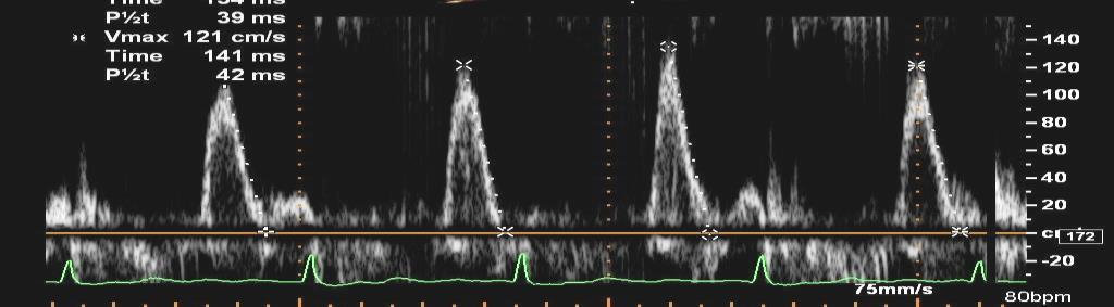
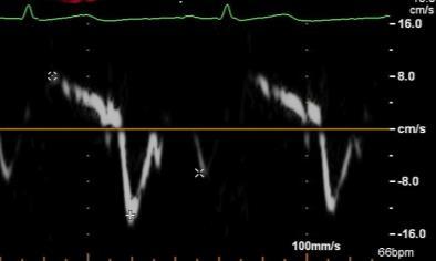
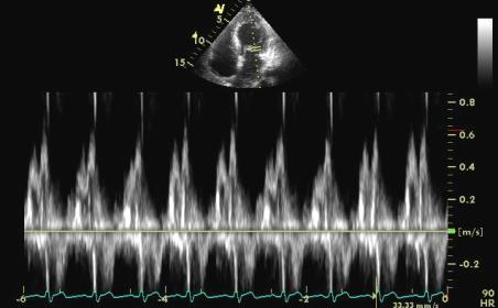
1 Question #1 Choose the grading of diastolic function in 82 yo woman E= 80 cm/s A= 70 cm/s LAVI < 34 ml/m 2 1= Grade 1 2= Grade 2 3= Grade 3 4= Normal 5= Indeterminate 2018 MFMER Choose the grading of diastolic function in 82 yo woman E= 80 cm/s A= 70 cm/s LAVI < 34 ml/m 2 E/e = 10 Medial e = 8 cm/s TR =2.7 m/sec 1= Grade 1 2= Grade 2 3= Grade 3 4= Normal 5= Indeterminate 2018 MFMER


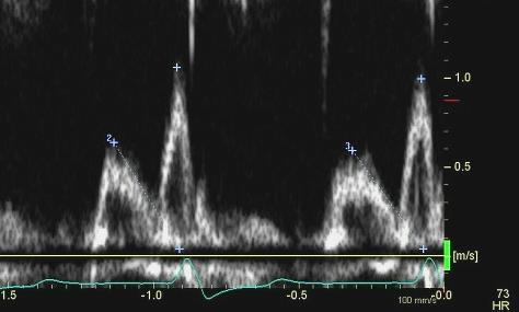
2 82 year old woman with normal diastolic function TR = 3.2 m/sec TR =2.7 m/sec 2018 MFMER Question #2 Grade diastolic function in 80 YO man with dyspnea TR = 3.5 m/sec E= 60 cm/s A= 80 cm/s E/A = 0.75 Medial e = 2 cm/s E/e =30 Lat e = 6 cm/s E/e = Grade 1 2. Grade 2 3. Grade 3 4. Normal 5. Indeterminate 2018 MFMER


3 80 YO man with dyspnea Septal e is reduced due to PHT 1. e is reduced 2. E/e +/- 3. LAVI normal 4. TR is high 2018 MFMER year old man with dyspnea and PHT Cardiac Cath Data RA pressure 2 mmhg RVSP 50 mmhg Mean PASP 28 mmhg PCWP 4 mmhg LVEDP 13 mmhg Cardiac Index 1.5 L/min/m MFMER


4 Question #3 Diastolic Function in 65 yo man with hypertension? LAVI = 20 ml/m 2 & TR = 3 m/s E=127 cm/s A= 135 cm/s Medial e = 5 cm/sec E/e = Grade 1 2. Grade 2 3. Grade 3 4. Normal Filling Pressure 5. Indeterminate Lat e = 8 cm/sec E/e = MFMER LV volume measurement 2018 MFMER


5 65 yo man with hypertension E/A = 1.0 E/A = MFMER Good Quality in Measurements is the Key! No data is better than wrong data! 2018 MFMER


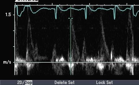
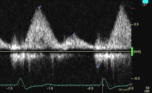
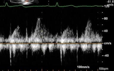
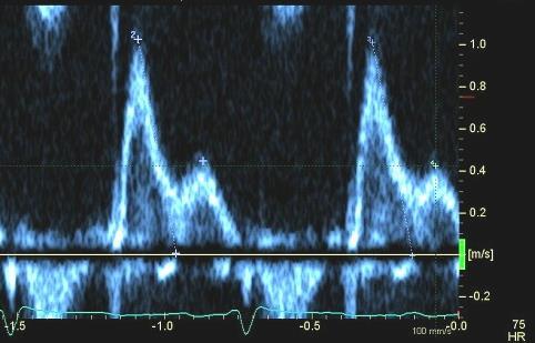
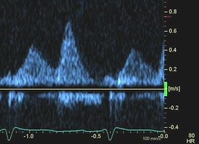
6 78 yo woman with 21 mm CE AVR 2010 for severe AS Referred for Valve in Valve with high gradient and dyspnea Aorta 3 cm 2018 MFMER yo female with 21 mm CE AV prosthesis and SOB How do you grade her diastolic function? E =45 A= 140 E/A = 3 4 m/sec i EOA < 0.65 AT 75 msec 1. Grade 1 2. Grade 2 3. Grade 3 4. Normal Filling Pressure 5. Indeterminate 2018 MFMER
7 78 yo female with 21 mm CE AV prosthesis and SOB What is you next diagnostic step? E =45 A= 140 E/A = 3 4 m/sec i EOA < 0.65 AT 75 msec 1. Aortic valve replacement 2. Refer to pulmonary 3. Exercise test 4. TEE 5. Old age and Observe 2018 MFMER Pericardial Aortic Prosthesis AT >100 ms AT 100 ms ieoa <0.65 cm 2 /m 2 ieoa <0.65 cm 2 /m 2 ieoa 0.85 cm 2 /m 2 MG >20 mmhg MG 20 mmhg Aortic root diameter 3 cm Aortic root diameter <3 cm MG >20 mmhg MG 20 mmhg Pathologic Obstruction (normal flow) Pathologic Obstruction (low flow) Severe PPM Severe PPM ± Pressure Recovery High flow Pathologic Regurgitation Norma l 2018 MFMER


8 Exercise Cardiac Catheterization PPM + Pressure Recovery Rest LVEDP 12, PAWP 8 CO 2.8, CI 1.5, MG 17 mmhg AVA 1.1 cm 2 Exercise PAWP 41, LVEDP 44, mean PA 46 Aortic pressure 220 CO 5.7, CI 3.1, gradient 10, AVA 2.0 cm MFMER Mitral inflow E/A E <50 cm/s E/A E >50 cm/s or E/A >0.8-<2 E/A 2 3 criteria to be evaluated* 2 of 3 negative 1 Average E/e >14 2 TR velocity >2.8 m/s 3 LA volume index >34 ml/m 2 2 of 3 or 3 of 3 positive When only 2 criteria are available 2 negative 1 positive and 1 negative 2 positive Normal LAP Grade I diastolic dysfunction Cannot determine LAP and diastolic dysfunction grade* LAP Grade II diastolic dysfunction LAP Grade III diastolic dysfunction If symptomatic consider CAD or proceed to diastolic stress test 2018 MFMER


 CP1082496-595/8/2018 2018")
9 73 yo woman with Hypertension and Exertional Dyspnea No ischemia E/e = 7 E = 50 cm/s DT = 250 ms This patient has delayed myocardial relaxation, but filling pressure is not increased at rest e = 7 cm/s CP /8/ MFMER Change in CI and PCWP with Exercise Normal and Abnormal Diastolic Function 8 6 Normal Cardiac index (L/min/m 2 ) 4 2 Diastolic dysfunction PCWP (mm Hg) CP /8/ MFMER
10 Dynamic Diastology Filling Pressure (E/e ) with Exercise E e E/e Normal Abnormal LV filling pressure (E/e ) does not increase much with exercise in normal heart, but increases in symptomatic patients with diastolic dysfunction. CP /8/ MFMER Diastolic Stress Test Baseline and Peak (or Post) Exercise Supine bike or Treadmill 25 watts (3 min) increments Assess LVEF, size, and wall motion Mitral inflow (E, A, and DT) Mitral annulus velocity E/e ratio TR velocity 5/8/ MFMER

11 E (cm/s) E/e septal e septal (cm/s) 5/8/2018 Diastology Exercise Test Normal Values Baseline Exercise E (cm/s) 73±19 90±25 A (cm/s) 69±17 87±22 DT (ms) 192±40 176±42 e (cm/s) 12±4 15±5 E/e 6.7± ±2.5 Mean age 59±14 yrs Ha J et al: AJC, 2003 CP /8/ MFMER LV Diastolic Function at Rest and With Stress Diastolic Stress Echocardiography in the Young: Endurance-Trained (ET) Healthy Subjects Rest Exercise 12 ET NA * Nonathletic (NA) and Rest Exercise Mean age 29 yrs 2 0 Rest Bruengger et al: JASE, 2014 Exercise 2018 MFMER


12 Exertional Dyspnea E = 50 DT = 250 E = 85 DT = 140 e = 7 E/e = 7 TR = 2.4 e = 7 E/e = 12 TR = 3.8 Baseline Supine Bike CP /8/ MFMER Echo-Cath Correlation E/e = 16 E/e = 25 Ha et al. JASE MFMER

 Peak A 50 P<0.")
13 E/e medial E/e medial 5/8/ Mean LVDP vs E/e Rest and Exercise Rest: y=0.44x r=0.67, P< Exercise: y=0.46x r=0.59, P<0.01 E/e Mean LVDP (mm Hg) Burgess et al: JACC 47:1891, 2006 CP /8/ MFMER Echocardiographic Hemodynamic and Ventricular Function Indices During Exercise Submaximal (20W) Peak A 50 P< % Not measurable B 50 P< % Non measurable NCD HFpEF 0 NCD HFpEF Obokata M et al: Circ 135: , MFMER


14 Exercise Echo for Dyspnea E= 50 cm/s E= 100 cm/s e = 5 cm/s e = 6 cm/s 5/8/ MFMER Exercise Echo for Dyspnea E= 100 cm/s E= 150 cm/s e = 5 cm/sec 5/8/ MFMER


15 Question #4 Which of following clinical situation is best suited for using E/A ratio as the initial diastolic assessment parameter? 1. Age > 80 years 2. Uncontrolled hypertension 3. LVEF 45% 4. AL Cardiac Amyloidosis 5. Hypertrophic cardiomyopathy 6. Marked mitral annulus calcification 2018 MFMER Question #5 79 year old with bigemini E= 60 A=70 E/A= 0.8 Med e = 7 E/e = 8 1. Grade 1 2. Grade 2 3. Grade 3 4. Normal Filling Pressure 5. Indeterminate 2018 MFMER


16 79 yo with bigemini TR 2.4 m/sec Valsalva E=40 A= 60 E/A= MFMER year old man with dyspnea PFO velocity LAP- RAP = 36 mmhg 2018 MFMER


17 PFO Velocity for estimating LA pressure Baseline After Diuresis Peak V = 3.2 m/s Peak V = 1.6 m/s From Chandra, MD & Will Miranda, MD 2018 MFMER Atrial Fib. And Mitral Regurgitation Medial e = 8 cm/s E= 90 cm/s E/e =11 TR = 2.5 m/sec 2018 MFMER


18 Atrial Fib. And Mitral Regurgitation with exercise E= 90 cm/s E= 130 cm/s and DT 120 msec Medial e = 8 cm/s Medial e = 9 cm/s TR = 2.5 m/sec TR = 3.2 m/sec 2018 MFMER Mitral E, e and Pulmonary Vein Normal Grade 1 Grade MFMER


19 Questions & Discussion 2018 MFMER Pericardial Diseases Questions Jae K. Oh, MD 2018 MFMER


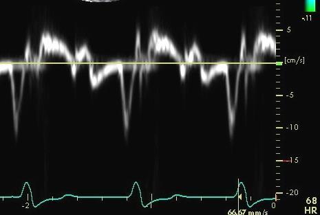
20 Question #6 Who from following patients has the highest medial e velocity expected? year old woman with CP after radiation treatment year old woman with CP after viral illness year old man with CP after CABG year old man with cardiac Fabry s year old woman with HFPEF 2018 MFMER VBaseline Primary CP Secondary CP Primary CP Secondary CP Baseline Post Pericardiectomy Medial e 14.6 ± ± ± ± 2.0 Lateral e 12.8 ± ± ± ± 2.0 e 15 cm/s e 10cm/s Veress et al. Circulation CV Imaging July MFMER


21 Tissue Doppler in Constriction vs Restriction E normal to high in constriction, low in myocardial disease 2018 MFMER Normal vs RCM vs CP Medial Mitral e velocity (LV Relaxation) Normal RCM CP Medial e 13 cm/s Medial e 3 cm/s Medial e 14 cm/s Usually > Lateral e (Annulus Reversus) 2018 MFMER


22 Question #7 A 26 year old woman with a previous pericardiectomy, for tbc CP presenting with edema Doppler demonstrates? 1. Recurrent CP 2. Normal 3. Restriction 4. Respiratory distress Lateral e = 10 cm/s Medial e = 15 cm/s 2018 MFMER A 26 year old woman with a previous pericardiectomy, presenting with edema 2018 MFMER


23 41 pts MFMER Cho and Schaff et al. Annal Thorac Surgery MFMER Question #8 Which of following statements is correct regarding Kussmaul sign? 1. Expiratory increase in JVP 2. Cannot be correct since opposite to Hepatic Vein 3. Due to increased IVC flow 2018 MFMER


24 T Meyer AJC MFMER Question #9 42 year old man with dyspnea Gradual onset of chest tightness and dyspnea Physical Examination HR 90 BPM JVP mildly elevated Increased S2 intensity GR 1/6 systolic murmur Mild pitting edema 2018 MFMER


25 42 year old man with dyspnea What is his diastolic function? E= 120 A= 30 cm/sec E/A =4 Lat e = 12 cm/sec E/e = 10 Med e = 9 cm/sec E/e = 13 1= Grade 1 2= Grade 2 3= Grade 3 4= Normal 5= Constriction 2018 MFMER year old man with dyspnea and high output LVOT D = 2.3 m/sec LVOT TVI = 26cm SV= (2.3) 2 x x 26 = 108 ml CO = SV x HR = 108 x 80 = 8.6 L CI = CO/BSA = 8.6 /1.93 = 4.48 L/m MFMER


26 What is the most common cause of HF seen in this 42 year old man? SV= (2.3) 2 x x 26 = 108 ml CO = SV x HR = 108 x 80 = 8.6 L CI = CO/BSA = 8.6 /1.93 = 4.48 L/m 2 1. Shunt 2. Obesity 3. Liver Disease 4. Pheochromocytoma 2018 MFMER year old man with high output HF Before and after treatment (Thyrotoxicosis) 2018 MFMER

 Shunt (22%) Diastolic function")


27 Etiologies of High Output Failure Cardiac Index > 4.0 L/m 2 Obesity (31%) Liver Disease (23%%) Shunt (22%) Diastolic function can be normal Reddy et al JACC MFMER year old man with chest pain What is your next step? Question #10 1. Cardiac Cath 2. TEE 3. CT 4. Stress Echo 2018 MFMER
28 Congenital Absence of the Pericardium Snoopy Sign 2018 MFMER Pericardial Cyst 2018 MFMER

29 A large pericardial cyst 2011 MFMER slide MFMER Cardiac Tamponade RV collapse Mitral inflow variation HV expiratory reversal 2018 MFMER
30 Hemo-pericardium 2018 MFMER Intramural Hematoma 2018 MFMER


31 66 year old woman with dyspnea Pneumo-pericardium PneGastro-pericardial fistulau 2018 MFMER year old woman from Middle East with ascites several month after acute pericarditis and pericardial effusion Medial e = 14 cm/s Lateral e = 11 cm/s 2018 MFMER


32 23 year old woman from Middle East with ascites several month after acute pericarditis and pericardial effusion (Thrombotic CP) Medial e = 14 cm/s Courtesy of H. Schaff, MD 2018 MFMER yo man presents with dyspnea and fever BP 80/40 mmhg Hepatic vein 2018 MFMER


33 35 yo man with tamponade and fever TEE after pericardiocentesis 2018 MFMER Final Pericardial Disease Case 45 yo male with chest pain x 4 days Pain preceded by heavy lifting while camping. No relationship to exercise. MVA with pelvic and rib fractures. Chest tube and IVC filter placement 6 yrs ago. Many CV risk factors 2012 MFMER MFMER

34 Echocardiographic images in 45 yo with chest pain 2018 MFMER MFMER Chest and Cardiac CT 2012 MFMER MFMER


35 CT scans now and 8 years ago 2012 MFMER MFMER Embolic strut from IVC Filter 2012 MFMER MFMER


36 2012 MFMER MFMER Constriction or Myocardial Disease? Diagnostic Algorithm Medial e 12 cm/s Medial e 5 cm/s Syed, Schaff, Oh Nature Review Sep MFMER


37 Thank You! MFMER Echo-Guided Pericardiocentesis Welles mov Chest wall 79% 3% 18% Subcostal Location 2018 MFMER
38 Questions & Discussion 2018 MFMER
Diastolic Function Assessment New Guideline Update Practical Approach
 Mayo Clinic Department of Cardiovascular Diseases Mayo Clinic Echocardiography Review Course for Boards and Recertification Diastolic Function Assessment New Guideline Update Practical Approach Jae K.
Constriction vs Restriction Case-based Discussion
 Mayo Clinic Department of Cardiovascular Diseases Mayo Clinic Echocardiography Review Course for Boards and Recertification Constriction vs Restriction Case-based Discussion Jae K. Oh, MD Samsung Professor
Pericardial Diseases/Tamponade Illustrative Cases
 Pericardial Diseases/Tamponade Illustrative Cases Jae K. Oh, MD Echo Hawaii 2017 2012 MFMER 3200268v3(2010)-1 Case #1 47 year old man Chest pain Not exertional Normal Examination 2016 MFMER slide-2 1 47
Constrictive Pericarditis
 Constrictive Pericarditis Never Confused with Anything Else Jae K. Oh, MD 2018 MFMER 3712003-1 ARS #1 CP Which of following patients has constrictive pericarditis? 1 2 3 Medial e 13 cm/s Medial e 3 cm/s
Diastolic Function Assessment Practical Ways to Incorporate into Every Echo
 Diastolic Function Assessment Practical Ways to Incorporate into Every Echo Jae K. Oh, MD Echo Hawaii 2018 2018 MFMER 3712003-1 Learning Objectives My presentation will help you to Appreciate the importance
10/1/2016. Constrictive Pericarditis Unique Hemodynamics. What s New in Pericardial Disease? Case-based Discussion
 Mayo Clinic Department of Cardiovascular Diseases Mayo Clinic Echocardiography Review Course for Boards and Recertification What s New in Pericardial Disease? Case-based Discussion Jae K. Oh, MD Samsung
Pericardial Disease: Case Examples. Echo Fiesta 2017
 Pericardial Disease: Case Examples Echo Fiesta 2017 2014 2014 MFMER MFMER 3346252-1 slide-1 Objectives Have a systematic approach to evaluation of constriction 2014 MFMER 3346252-2 CASE 1 2013 MFMER 3248567-3
DOPPLER HEMODYNAMICS (1) QUANTIFICATION OF PRESSURE GRADIENTS and INTRACARDIAC PRESSURES
 QUANTIFICATION OF PRESSURE GRADIENTS and INTRACARDIAC PRESSURES") THORAXCENTRE DOPPLER HEMODYNAMICS (1) QUANTIFICATION OF PRESSURE GRADIENTS and INTRACARDIAC PRESSURES J. Roelandt DOPPLER HEMODYNAMICS Intracardiac pressures and pressure gradients Volumetric measurement
THORAXCENTRE DOPPLER HEMODYNAMICS (1) QUANTIFICATION OF PRESSURE GRADIENTS and INTRACARDIAC PRESSURES J. Roelandt DOPPLER HEMODYNAMICS Intracardiac pressures and pressure gradients Volumetric measurement
Constrictive Pericarditis Curable Paradoxical Diastolic Heart Failure ASE EBRC 2018
 Constrictive Pericarditis Curable Paradoxical Diastolic Heart Failure ASE EBRC 2018 May 8 th, 2018 Jae K. Oh, MD Samsung Professor of CV Diseases Director, Pericardial Disease Clinic Co-Director, Integrated
Echo Doppler Assessment of Right and Left Ventricular Hemodynamics.
 Echo Doppler Assessment of Right and Left Ventricular Hemodynamics. Itzhak Kronzon, MD, FASE, FACC, FESC, FAHA, FACP, FCCP Northwell, Lenox Hill Hospital, New York Professor of Cardiology Hofstra University
P = 4V 2. IVC Dimensions 10/20/2014. Comprehensive Hemodynamic Evaluation by Doppler Echocardiography. The Simplified Bernoulli Equation
 Comprehensive Hemodynamic Evaluation by Doppler Echocardiography Itzhak Kronzon, MD North Shore LIJ/ Lenox Hill Hospital New York, NY Disclosure: Philips Healthcare St. Jude Medical The Simplified Bernoulli
Comprehensive Hemodynamics By Doppler Echocardiography. The Echocardiographic Swan-Ganz Catheter.
 Comprehensive Hemodynamics By Doppler Echocardiography. The Echocardiographic Swan-Ganz Catheter. Itzhak Kronzon, MD, FASE, FACC, FESC, FAHA, FACP, FCCP North Shore HS, LIJ/Lenox Hill Hospital, New York
Diastolic Heart Function: Applying the New Guidelines Case Studies
 Diastolic Heart Function: Applying the New Guidelines Case Studies Mitral Regurgitation The New ASE William Guidelines: A. Zoghbi Role MD, of FASE, 2D/3D MACCand CMR Professor and Chairman, Department
Pericardial Diseases. Smonporn Boonyaratavej, MD. Division of Cardiology, Department of Medicine Chulalongkorn University
 Pericardial Diseases Smonporn Boonyaratavej, MD Division of Cardiology, Department of Medicine Chulalongkorn University Cardiac Center, King Chulalongkorn Memorial Hospital 21 AUGUST 2016 Pericardial
Diastology State of The Art Assessment
 Diastology State of The Art Assessment Dr. Mohammad AlGhamdi Assistant professor, KSAU-HS Consultant Cardiologist King AbdulAziz Cardiac Center Ministry of National Guard Health Affairs Diagnostic Clinical
Μαρία Μπόνου Διευθύντρια ΕΣΥ, ΓΝΑ Λαϊκό
 Μαρία Μπόνου Διευθύντρια ΕΣΥ, ΓΝΑ Λαϊκό Diastolic HF DD: Diastolic Dysfunction DHF: Diastolic HF HFpEF: HF with preserved EF DD Pathophysiologic condition: impaired relaxation, LV compliance, LV filling
Evalua&on)of)Le-)Ventricular)Diastolic) Dysfunc&on)by)Echocardiography:) Role)of)Ejec&on)Frac&on)
of)Le-)Ventricular)Diastolic) Dysfunc&on)by)Echocardiography:) Role)of)Ejec&on)Frac&on)") Evalua&on)of)Le-)Ventricular)Diastolic) Dysfunc&on)by)Echocardiography:) Role)of)Ejec&on)Frac&on) N.Koutsogiannis) Department)of)Cardiology) University)Hospital)of)Patras)! I have no conflicts of interest
Evalua&on)of)Le-)Ventricular)Diastolic) Dysfunc&on)by)Echocardiography:) Role)of)Ejec&on)Frac&on) N.Koutsogiannis) Department)of)Cardiology) University)Hospital)of)Patras)! I have no conflicts of interest
Adel Hasanin Ahmed 1
 Adel Hasanin Ahmed 1 PERICARDIAL DISEASE The pericardial effusion ends anteriorly to the descending aorta and is best visualised in the PLAX. PSAX is actually very useful sometimes for looking at posterior
The Doppler Examination. Katie Twomley, MD Wake Forest Baptist Health - Lexington
 The Doppler Examination Katie Twomley, MD Wake Forest Baptist Health - Lexington OUTLINE Principles/Physics Use in valvular assessment Aortic stenosis (continuity equation) Aortic regurgitation (pressure
Hemodynamic Assessment. Assessment of Systolic Function Doppler Hemodynamics
 Hemodynamic Assessment Matt M. Umland, RDCS, FASE Aurora Medical Group Milwaukee, WI Assessment of Systolic Function Doppler Hemodynamics Stroke Volume Cardiac Output Cardiac Index Tei Index/Index of myocardial
ΚΑΡΔΙΟΛΟΓΟΣ EUROPEAN ACCREDITATION IN TRANSTHORACIC AND TRANSESOPHAGEAL ECHOCARDIOGRAPHY
 1 ΚΑΡΔΙΟΛΟΓΟΣ EUROPEAN ACCREDITATION IN TRANSTHORACIC AND TRANSESOPHAGEAL ECHOCARDIOGRAPHY 2 Constrictive pericarditis (CP) is characterized by impaired ventricular filling due to a stiffened or noncompliant
HEMODYNAMIC ASSESSMENT
 HEMODYNAMIC ASSESSMENT INTRODUCTION Conventionally hemodynamics were obtained by cardiac catheterization. It is possible to determine the same by echocardiography. Methods M-mode & 2D echo alone can provide
An Integrated Approach to Study LV Diastolic Function
 An Integrated Approach to Study LV Diastolic Function Assoc. Prof. Adriana Ilieşiu, FESC University of Medicine Carol Davila Bucharest, Romania LV Diastolic Dysfunction impaired relaxation (early diastole)
ASCeXAM / ReASCE. Practice Board Exam Questions. Tuesday Morning
 ASCeXAM / ReASCE Practice Board Exam Questions Tuesday Morning Congenital Heart Disease in Adults Congenital Heart Disease Cases Diastolic Function Pericardial Disease Congenital Heart Disease Cases Michael
THE PERICARDIUM: LOOKING OUTSIDE THE HEART
 THE PERICARDIUM: LOOKING OUTSIDE THE HEART DISCLOSURE Relevant relationships with commercial entities none Potential for conflicts of interest within this presentation none Steps taken to review and mitigate
GENERAL PRINCIPLES FOR ECHO ASSESSMENT OF DIASTOLIC FUNCTION (For full recommendation refer to the Left Ventricular Diastolic Function Guideline)
") 1 THE AMERICAN SOCIETY OF ECHOCARDIOGRAPHY RECOMMENDATIONS FOR THE EVALUATION OF LEFT VENTRICULAR DIASTOLIC FUNCTION BY ECHOCARDIOGRAPHY: A QUICK REFERENCE GUIDE FROM THE ASE WORKFLOW AND LAB MANAGEMENT
1 THE AMERICAN SOCIETY OF ECHOCARDIOGRAPHY RECOMMENDATIONS FOR THE EVALUATION OF LEFT VENTRICULAR DIASTOLIC FUNCTION BY ECHOCARDIOGRAPHY: A QUICK REFERENCE GUIDE FROM THE ASE WORKFLOW AND LAB MANAGEMENT
Imaging in Heart Failure: A Multimodality Approach. Thomas Ryan, MD
 Imaging in Heart Failure: A Multimodality Approach Thomas Ryan, MD Heart Failure HFrEF HFpEF EF50% Lifetime risk 20% Prevalence 6M Americans Societal costs - $30B 50% 5-year survival 1 Systolic
Dobutamine Stress testing In Low Flow, Low EF, Low Gradient Aortic Stenosis Case Studies
 Dobutamine Stress testing In Low Flow, Low EF, Low Gradient Aortic Stenosis Case Studies Mitral Regurgitation The New ASE Guidelines: Role of 2D/3D and CMR William A. Zoghbi MD, FASE, MACC Professor and
Case Reviews: Hemodynamic Calculations in Valvular Regurgitation
 Case Reviews: Hemodynamic Calculations in Valvular Regurgitation Case 5 History: 69-year-old man with orthotopic heart transplant 15 years ago. Inferior MI several years ago. Recurrent CHF. Currently dyspneic
OPTIMIZING ECHO ACQUISTION FOR STRAIN AND DIASTOLOGY
 OPTIMIZING ECHO ACQUISTION FOR STRAIN AND DIASTOLOGY October 8, 2017 Deborah Agler, ACS, RDCS, FASE Coordinator of Education and Training Cleveland Clinic General Principles Diastology Clinical Data Heart
Constrictive/Restrictive Cardiomyopathies: Diagnosis and Management Update; Radiation Induced Heart Disease. Alexander (Sandy) Dick, MD
 Dick, MD") Constrictive/Restrictive Cardiomyopathies: Diagnosis and Management Update; Radiation Induced Heart Disease Alexander (Sandy) Dick, MD Outline Pericardial Constriction Diagnosis: Imaging, Hemodynamics
Constrictive/Restrictive Cardiomyopathies: Diagnosis and Management Update; Radiation Induced Heart Disease Alexander (Sandy) Dick, MD Outline Pericardial Constriction Diagnosis: Imaging, Hemodynamics
2/4/2011. Nathan Kerner, M.D.
 Nathan Kerner, M.D. Definition Elevated pressures - cut off usually >40 mmhg pulmonary artery systolic pressure (PASP) Usually associated with elevated pulmonary vascular resistance (PVR) measured in dynessec/cm
HFpEF. April 26, 2018
 HFpEF April 26, 2018 (J Am Coll Cardiol 2017;70:2476 86) HFpEF 50% or more (40-71%) of patients with CHF have preserved LV systolic function. HFpEF is an increasingly frequent hospital discharge. Outcomes
Doppler Basic & Hemodynamic Calculations
 Doppler Basic & Hemodynamic Calculations August 19, 2017 Smonporn Boonyaratavej MD Division of Cardiology, Department of Medicine Chulalongkorn University Cardiac Center, King Chulalongkorn Memorial Hospital
Role of Stress Echo in Valvular Heart Disease. Satoshi Nakatani Osaka University Graduate School of Medicine Osaka, Japan
 Role of Stress Echo in Valvular Heart Disease Satoshi Nakatani Osaka University Graduate School of Medicine Osaka, Japan Exercise echocardiography Dobutamine echocardiography Usefulness of exercise echo
Prosthetic valve dysfunction: stenosis or regurgitation
 Prosthetic valve dysfunction: stenosis or regurgitation Jean G. Dumesnil MD, FRCP(C), FACC, FASE(Hon) Quebec Heart and Lung Institute, Québec, Québec No disclosures Possible Causes of High Gradients in
UPDATE ON CONSTRICTIVE PERICARDITIS ECHOCARDIOGRAPHY AND CARDIAC CATHETERISATION
 Arsen D. Ristić, MD, PhD, FESC (no conflicts of interest to disclose regarding this presentation) UPDATE ON CONSTRICTIVE PERICARDITIS ECHOCARDIOGRAPHY AND CARDIAC CATHETERISATION Department of Cardiology,
E/Ea is NOT an essential estimator of LV filling pressures
 Euroecho Kopenhagen Echo in Resynchronization in 2010 E/Ea is NOT an essential estimator of LV filling pressures Wilfried Mullens, MD, PhD December 10, 2010 Ziekenhuis Oost Limburg Genk University Hasselt
Diastolic Function: What the Sonographer Needs to Know. Echocardiographic Assessment of Diastolic Function: Basic Concepts 2/8/2012
 Diastolic Function: What the Sonographer Needs to Know Pat Bailey, RDCS, FASE Technical Director Beaumont Health System Echocardiographic Assessment of Diastolic Function: Basic Concepts Practical Hints
The Patient with Atrial Fibrilation
 Assessment of Diastolic Function The Patient with Atrial Fibrilation Assoc. Prof. Adriana Ilieşiu, FESC University of Medicine Carol Davila Bucharest, Romania Associated Conditions with Atrial Fibrillation
Stage of Valvular AS. Outline 10/14/16. Low-flow and Other Challenges to the Assessment of Aortic Stenosis. Severe AS
 Low-flow and Other Challenges to the Assessment of Aortic Stenosis Nithima Ratanasit, MD, FACC, FASE Siriraj Hospital, Mahidol University Bangkok, Thailand Outline Types of low-flow aortic stenosis Assessment
Managing the Low Output Low Gradient Aortic Stenosis Patient
 Managing the Low Output Low Gradient Aortic Stenosis Patient R A Nishimura MD Judd and Mary Leighton Professor of CV Mayo Clinic No disclosures Valvular Stenosis Severity of Aortic Stenosis Mean gradient
Echocardiography as a diagnostic and management tool in medical emergencies
 Echocardiography as a diagnostic and management tool in medical emergencies Frank van der Heusen MD Department of Anesthesia and perioperative Care UCSF Medical Center Objective of this presentation Indications
Review of Cardiac Imaging Modalities in the Renal Patient. George Youssef
 Review of Cardiac Imaging Modalities in the Renal Patient George Youssef ECHO Left ventricular hypertrophy (LVH) assessment Diastolic dysfunction Stress ECHO Cardiac CT angiography Echocardiography - positives
Clinical implication of exercise pulmonary hypertension: when should we measure it?
 Clinical implication of exercise pulmonary hypertension: when should we measure it? Jang-Young, Kim Wonju College of Medicine, Yonsei Univ. Exercise pulmonary hypertension (EPH) Introduction of pulmonary
ECHO HAWAII. Role of Stress Echo in Valvular Heart Disease. Not only ischemia! Cardiomyopathy. Prosthetic Valve. Diastolic Dysfunction
 Role of Stress Echo in Valvular Heart Disease ECHO HAWAII January 15 19, 2018 Kenya Kusunose, MD, PhD, FASE Tokushima University Hospital Japan Not only ischemia! Cardiomyopathy Prosthetic Valve Diastolic
Right Heart Catheterization. Franz R. Eberli MD Chief of Cardiology Stadtspital Triemli, Zurich
 Right Heart Catheterization Franz R. Eberli MD Chief of Cardiology Stadtspital Triemli, Zurich Right Heart Catheterization Pressure measurements Oxygen saturation measurements Cardiac output, Vascular
Left ventricular diastolic function and filling pressure in patients with dilated cardiomyopathy
 Left ventricular diastolic function and filling pressure in patients with dilated cardiomyopathy Bogdan A. Popescu University of Medicine and Pharmacy Bucharest, Romania My conflicts of interest: I have
Quantification of Mitral Stenosis: Planimetry, pressure Half time, Continuity Common Errors
 Quantification of Mitral Stenosis: Planimetry, pressure Half time, Continuity Common Errors Christopher J Kramer RDCS Advanced Cardiovascular Services Aurora Health Care Milwaukee, WI No Disclosures Baumgartner,
Integrative Clinical Hemodyamics
 Integrative Clinical Hemodyamics James A. Goldstein, MD Director, Research & Education Division of Cardiology William Beaumont Hospital Disclosure Information Integrative Clinical Hemodyamics James A.
Value of echocardiography in chronic dyspnea
 Value of echocardiography in chronic dyspnea Jahrestagung Schweizerische Gesellschaft für /Schweizerische Gesellschaft für Pneumologie B. Kaufmann 16.06.2016 Chronic dyspnea Shortness of breath lasting
MAKING SENSE OF MODERATE GRADIENTS IN PATIENTS WITH SYMPTOMATIC AORTIC STENOSIS
 MAKING SENSE OF MODERATE GRADIENTS IN PATIENTS WITH SYMPTOMATIC AORTIC STENOSIS David A. Orsinelli, MD, FACC, FASE Professor, Internal Medicine Director, Structural Heart Imaging The Ohio State University
ASCeXAM / ReASCE. Practice Board Exam Questions. Monday Afternoon
 ASCeXAM / ReASCE Practice Board Exam Questions Monday Afternoon Aortic Valve Prostheses Aortic Regurgitation Stress Echocardiography Ischemic Heart Disease and Myocardial Infarction 3D Echocardiography
Swan Song: Echocardiography as a Pulmonary Artery Catheter? Interdepartmental Division of Critical Care Medicine
 Swan Song: Echocardiography as a Pulmonary Artery Catheter? The swan is without spot, and it sings sweetly as it dies, that song ending its life Leonardo Da Vinci Curr Opin Anesthesiol 2016, 29:36 45 Circulation.
M-Mode Echocardiography Is it still Alive? Itzhak Kronzon, MD,FASE. Sampling Rate M-Mode: 1800 / sec 2D: 30 / sec
 M-Mode Echocardiography Is it still Alive? Itzhak Kronzon, MD,FASE Honoraria: Philips Classical M-mode Echocardiography M-Mode offers better time and image resolution. Sampling Rate M-Mode: 1800 / sec
Echo Emergencies. Outline. Michael H. Picard, MD Massachusetts General Hospital Harvard Medical School No disclosures
 Echo Emergencies Michael H. Picard, MD Massachusetts General Hospital Harvard Medical School No disclosures Outline Common emergency / on call scenarios Tamponade Pulmonary embolism/rv strain Cardiogenic
Exercise Testing/Echocardiography in Asymptomatic AS
 Exercise Testing/Echocardiography in Asymptomatic AS Raluca Dulgheru, MD Heart Valve Clinic, University of Liège, CHU Sart Tilman, BELGIUM Disclosure related to this presentation: None VALVULAR HEART DISEASE
Rest and Exercise Echocardiography in Hypertrophic Cardiomyopathy: Determinants of Exercise Peak Gradient and Predictors of Outcome
 Rest and Exercise Echocardiography in Hypertrophic Cardiomyopathy: Determinants of Exercise Peak Gradient and Predictors of Outcome G. Deswarte, AS. Polge, N. Lamblin, A. Millaire, M. Richardson, C. Bauters,
Ref 1. Ref 2. Ref 3. Ref 4. See graph
 Ref 1 Ref 2 Ref 3 1. Ages 6-23 y/o 2. Significant LVM differences by gender 3. For males 95 th percentiles: a. LVM/BSA = 103 b. LVM/height = 100 4. For females 95 th percentiles: a. LVM/BSA = 84 b. LVM/height
Congenital Heart Disease Cases
 Congenital Heart Disease Cases Sabrina Phillips, MD FACC FASE Mayo Clinic Congenital Heart Disease Center 2013 MFMER slide-1 No Disclosures 2013 MFMER slide-2 1 CASE 1 2013 MFMER slide-3 63 year old Woman
Δυναμική υπερηχοκαρδιογραφία στις μυοκαρδιοπάθειες : έχει θέση και ποια ;
 Ελληνική Καρδιολογική Εταιρεία Σεμινάρια ομάδων εργασίας Θεσσαλονίκη, 8-10 Φεβρουαρίου 2018 Ομάδα εργασίας Ηχωκαρδιολογίας Δυναμική υπερηχοκαρδιογραφία στις μυοκαρδιοπάθειες : έχει θέση και ποια ; ΑΓΑΘΗ-ΡΟΖΑ
Stress Testing in Valvular Disease
 2017 ASE Florida Orlando, FL October 10, 2017 2:40 2:50 PM 10 min Grand Harbor Ballroom South Stress Testing in Valvular Disease Muhamed Sarić MD, PhD, MPA Director of Noninvasive Cardiology Echo Lab Associate
Aortic Stenosis: Spectrum of Disease, Low Flow/Low Gradient and Variants
 Aortic Stenosis: Spectrum of Disease, Low Flow/Low Gradient and Variants Martin G. Keane, MD, FASE Professor of Medicine Lewis Katz School of Medicine at Temple University Basic root structure Parasternal
Echocardiographic Cardiovascular Risk Stratification: Beyond Ejection Fraction
 Echocardiographic Cardiovascular Risk Stratification: Beyond Ejection Fraction October 4, 2014 James S. Lee, M.D., F.A.C.C. Associates in Cardiology, P.A. Silver Spring, M.D. Disclosures Financial none
LV FUNCTION ASSESSMENT: WHAT IS BEYOND EJECTION FRACTION
 LV FUNCTION ASSESSMENT: WHAT IS BEYOND EJECTION FRACTION Jamilah S AlRahimi Assistant Professor, KSU-HS Consultant Noninvasive Cardiology KFCC, MNGHA-WR Introduction LV function assessment in Heart Failure:
LV geometric and functional changes in VHD: How to assess? Mi-Seung Shin M.D., Ph.D. Gachon University Gil Hospital
 LV geometric and functional changes in VHD: How to assess? Mi-Seung Shin M.D., Ph.D. Gachon University Gil Hospital LV inflow across MV LV LV outflow across AV LV LV geometric changes Pressure overload
March yr. old male, newspaper writer, with worsening dyspnea /orthopnea past few months
 Case 1 March 2016 59 yr. old male, newspaper writer, with worsening dyspnea /orthopnea past few months PMH diabetes, celiac disease Reports chest discomfort, positional coughing and pedal edema last 10
Doppler and Hemodynamics Muhamed Saric and Itzhak Kronzon
 LWBK739-C07_p77-119.qxd 9/1/10 11:50 PM Page 77 Aptara Doppler and Hemodynamics Muhamed Saric and Itzhak Kronzon CHAPTER 7 1. On echocardiography, the diameter of the inferior vena cava is measured at
ASE Guidelines on Aortic Regurgitation What Do I Measure? Case Studies
 ASE Guidelines on Aortic Regurgitation What Do I Measure? Case Studies Mitral Regurgitation The New ASE Guidelines: Role of 2D/3D and CMR William A. Zoghbi MD, FASE, MACC Professor and Chairman, Department
Interventions in Adult Congenital Heart Disease: Role of CV Imaging. Associate Professor. ACHD mortality. Pillutla. Am Heart J 2009;158:874-9
 Interventions in Adult Congenital Heart Disease: Role of CV Imaging Sangeeta Shah MD, FACC, FASE Associate Professor ACHD mortality Pillutla. Am Heart J 2009;158:874-9 Adult Congenital Heart Disease Heterogenity
Diastole is Not a Single Entity Four Components of Diastolic Dysfunction
 Physiology of Diastolic Function Made Easy James D. Thomas, MD, FACC, FASE Director, Center for Heart Valve Disease Bluhm Cardiovascular Institute Professor of Medicine, Feinberg School of Medicine, Northwestern
Evaluation of Left Ventricular Diastolic Dysfunction by Doppler and 2D Speckle-tracking Imaging in Patients with Primary Pulmonary Hypertension
 ESC Congress 2011.No 85975 Evaluation of Left Ventricular Diastolic Dysfunction by Doppler and 2D Speckle-tracking Imaging in Patients with Primary Pulmonary Hypertension Second Department of Internal
The new Guidelines: Focus on Chronic Heart Failure
 The new Guidelines: Focus on Chronic Heart Failure Petros Nihoyannopoulos MD, FRCP, FESC Professor of Cardiology Imperial College London and National & Kapodistrian University of Athens 2 3 4 The principal
ECHO HAWAII. My home. Pulmonary Hypertension and Pulmonary Embolism: Role of Echo U.S.A. Japan. Hawaii Island 1/9/2018
 Pulmonary Hypertension and Pulmonary Embolism: Role of Echo ECHO HAWAII January 15 19, 2018 Kenya Kusunose, MD, PhD, FASE Tokushima University Hospital Japan My home Japan U.S.A Hawaii Island 1 Economy
Pulmonary Hypertension: Another Use for Viagra
 Pulmonary Hypertension: Another Use for Viagra Kathleen Tong, MD Director, Heart Failure Program Assistant Clinical Professor University of California, Davis Disclosures I have no financial conflicts A
Diastolic Heart Failure
 Chronic Heart Failure Prevalence overall = 2-3 % Diastolic Heart Failure Patrick Wouters University Hospital Ghent Belgium (Heart Failure + Asymptomatic Ventricular Dysfunction) Prevalence > 70 y = 10-20
Outline. Echocardiographic Assessment of Pericardial Effusion/Tamponade: The Essentials
 Echocardiographic Assessment of Pericardial Effusion/Tamponade: The Essentials John R Schairer DO FACC Henry Ford Heart and Vascular Institute No Disclosures Outline Normal Anatomy and Physiology Pathophysiology
Echocardiography Conference
 Echocardiography Conference David Stultz, MD Cardiology Fellow, PGY-6 September 20, 2005 Atrial Septal Aneurysm Bulging of Fossa Ovalis Associated commonly with Atrial septal defect or small perforations
Conflicts of interest: GE, Abbott, Edwards (honoraria)
") Understanding Diastole and Its Contribution to Heart Failure: State of the Art in 2016 James D. Thomas, MD, FACC, FASE Director, Center for Heart Valve Disease Bluhm Cardiovascular Institute Professor
Understanding Diastole and Its Contribution to Heart Failure: State of the Art in 2016 James D. Thomas, MD, FACC, FASE Director, Center for Heart Valve Disease Bluhm Cardiovascular Institute Professor
Ejection across stenotic aortic valve requires a systolic pressure gradient between the LV and aorta. This places a pressure load on the LV.
 Valvular Heart Disease Etiology General Principles Cellular and molecular mechanism of valve damage Structural pathology Functional pathology - stenosis/regurgitation Loading conditions - pressure/volume
Highlights from EuroEcho 2009 Echo in cardiomyopathies
 Highlights from EuroEcho 2009 Echo in cardiomyopathies Bogdan A. Popescu University of Medicine and Pharmacy, Bucharest, Romania ESC Congress 2010 Hypertrophic cardiomyopathy To determine the differences
Prosthesis-Patient Mismatch or Prosthetic Valve Stenosis?
 EuroValves 2015, Nice Prosthesis-Patient Mismatch or Prosthetic Valve Stenosis? Philippe Pibarot, DVM, PhD, FACC, FAHA, FASE FESC Canada Research Chair in Valvular Heart Diseases Université LAVAL Disclosure
Adult Echocardiography Examination Content Outline
 Adult Echocardiography Examination Content Outline (Outline Summary) # Domain Subdomain Percentage 1 2 3 4 5 Anatomy and Physiology Pathology Clinical Care and Safety Measurement Techniques, Maneuvers,
Miscellaneous Cardiology Topics pregnancy - congenital - myocarditis - pericardial disease. Pregnancy and Cardiovascular Disease MCQ
 Miscellaneous Cardiology Topics pregnancy - congenital - myocarditis - pericardial disease Maan Jokhadar, MD, FACC Emory Center for Advanced Heart Failure Therapy Emory Adult Congenital Heart Center Pregnancy
Constrictive Pericarditis in the Modern Era
 Journal of the American College of Cardiology Vol. 51, No. 3, 2008 2008 by the American College of Cardiology Foundation ISSN 0735-1097/08/$34.00 Published by Elsevier Inc. doi:10.1016/j.jacc.2007.09.039
Appendix II: ECHOCARDIOGRAPHY ANALYSIS
 Appendix II: ECHOCARDIOGRAPHY ANALYSIS Two-Dimensional (2D) imaging was performed using the Vivid 7 Advantage cardiovascular ultrasound system (GE Medical Systems, Milwaukee) with a frame rate of 400 frames
How to Assess Diastolic Dysfunction?
 How to Assess Diastolic Dysfunction? Fausto J Pinto, MD, PhD, FESC, FACC, FASE Lisbon University Dyastolic Dysfunction Impaired relaxation Elevated filling pressures Ischemic heart disease Cardiomyopathies
Ejection across stenotic aortic valve requires a systolic pressure gradient between the LV and aorta. This places a pressure load on the LV.
 Valvular Heart Disease General Principles Etiology Cellular and molecular mechanism of valve damage Structural pathology Functional pathology - stenosis/regurgitation Loading conditions - pressure/volume
Echocardiographic evaluation of mitral stenosis
 Echocardiographic evaluation of mitral stenosis Euroecho 2011 Philippe Unger, MD, FESC Erasme Hospital, ULB, Brussels, Belgium I have nothing to declare EuroHeart Survey Etiology of single native left-sided
Usually we DON T need to go beyond the gradient
 Aortic Stenosis Going Beyond the Gradient James D. Thomas, MD, FACC, FASE Director, Center for Heart Valve Disease Bluhm Cardiovascular Institute Professor of Medicine, Feinberg School of Medicine, Northwestern
Comprehensive Echo Assessment of Aortic Stenosis
 Comprehensive Echo Assessment of Aortic Stenosis Smonporn Boonyaratavej, MD, MSc King Chulalongkorn Memorial Hospital Bangkok, Thailand Management of Valvular AS Medical and interventional approaches to
TAVR: Echo Measurements Pre, Post And Intra Procedure
 2017 ASE Florida, Orlando, FL October 10, 2017 8:00 8:25 AM 25 min TAVR: Echo Measurements Pre, Post And Intra Procedure Muhamed Sarić MD, PhD, MPA Director of Noninvasive Cardiology Echo Lab Associate
Role of echocardiography in the assessment of ischemic heart disease 분당서울대학교병원윤연이
 Role of echocardiography in the assessment of ischemic heart disease 분당서울대학교병원윤연이 Outline Evaluation of Chest pain Evaluation of MI complications Prediction of Outcomes Evaluation of Chest pain Evaluation
Susan P. D Anna MSN, APRN BC February 14, 2019
 Is there Equal Opportunity in Heart Failure?? Susan P. D Anna MSN, APRN BC February 14, 2019 Disclosures: I have no financial disclosures. I am not an expert on this topic, but see a lot of women with
Valve Disease Board Review Questions
 Valve Disease Board Review Questions Dennis A. Tighe, MD, FASE University of Massachusetts Medical School Worcester, MA Case 1 History A 61 year-old man Presents to hospital with worsening shortness of
Index. K Knobology, TTE artifact, image resolution, ultrasound, 14
 A Acute aortic regurgitation (AR), 124 128 Acute aortic syndrome (AAS) classic aortic dissection diagnosis, 251 263 evolutive patterns, 253 255 pathology, 250 251 classifications, 247 248 incomplete aortic
Transient Constrictive Pericarditis: Causes and Natural History
 Journal of the American College of Cardiology Vol. 43, No. 2, 2004 2004 by the American College of Cardiology Foundation ISSN 0735-1097/04/$30.00 Published by Elsevier Inc. doi:10.1016/j.jacc.2003.08.032
7. Echocardiography Appropriate Use Criteria (by Indication)
") Criteria for Echocardiography 1133 7. Echocardiography Criteria (by ) Table 1. TTE for General Evaluation of Cardiac Structure and Function Suspected Cardiac Etiology General With TTE 1. Symptoms or conditions
Criteria for Echocardiography 1133 7. Echocardiography Criteria (by ) Table 1. TTE for General Evaluation of Cardiac Structure and Function Suspected Cardiac Etiology General With TTE 1. Symptoms or conditions
Low Gradient Severe? AS
 Low Gradient Severe? AS Philippe Pibarot, DVM, PhD, FACC, FAHA, FESC, FASE Canada Research Chair in Valvular Heart Diseases Institut Universitaire de Cardiologie et de Pneumologie de Québec / Québec Heart
Case based learning: CMR in Heart Failure
 Case based learning: CMR in Heart Failure Milind Y Desai, MD FACC FAHA FESC Associate Professor of Medicine Heart and Vascular Institute, Cleveland Clinic Cleveland, OH Disclosures: none Use of Gadolinium