PROS AND CONS OF PERICARDIAL CLOSURE AFTER HEART SURGERY. Alfredo Rego MD, PhD. South Florida Heart and Lung Institute
|
|
|
- Vernon Cox
- 6 years ago
- Views:
Transcription
1 PROS AND CONS OF PERICARDIAL CLOSURE AFTER HEART SURGERY Alfredo Rego MD, PhD South Florida Heart and Lung Institute
2 Pericardial Repair Clinical Importance
3 Pericardial Functions: Well documented, seldom appreciated The majority of cardiac surgeons leave the Pericardium open after cardiac surgery
4 Pericardial Repair How important Is Pericardial Anatomy and Physiology? Should Pericardial Repair be Standard after Cardiac Operations?

5 Pericardial Repair Question: Is this really the most physiological and best operative strategy for patients???
6 Management of The Pericardial Sac After Open Heart Procedures Overwhelmingly, no management at all! Most common technique: Open sac Forget about it! Most common closure practice: Re-approximate sac with several interrupted sutures Limitations of direct reconstruction: Large gaps Very incomplete edge to edge reapproximation Sometimes not possible due to technical constraints
7 Pericardial Repair Attitudes about closing pericardium vary widely among heart surgeons Should it be done? How to do it / not to do it Does it even matter? Does it have any impact on clinical outcomes?
8 Pericardial Repair The benefits, drawbacks and technical aspects of this procedure have been complicated by: Conflicts between traditional believe and modern practice Lack of standard technique Lack of viable pericardial substitutes A virtual absence of convincing, high-quality prospective studies
9 Assumption 1: The pericardium in its normal anatomical state does not play a significant or meaningful role in healthy or pathological states

10 Assumption 2: The postoperative restoration of the pericardium to its normal anatomical state has a NO impact on early or late surgical outcomes
11 My Notion about what s important Restore/maintain normal barrier of intact pericardial sac Separate myocardium from sternum Safer re-entry at re-operation Discourage direct spread of infection from post-op sternal wound infection Protect myocardium and bypass grafts from acute injury in event of sternal dehiscence Prevent contamination of heart and pericardial sac from rundown of debris (blood, fat, necrotic tissue = soup ) from th sternal wound Compartmentalize and organize the surgical site Maintains proper lie of bypass grafts? Decrease bleeding post-op? Improve recognition of source of bleeding post-op Reduced incidence of post-op complications? (AF, etc)
12 Pericardial Repair - objectives 1. Understanding Pericardial Physiology 2. Why closing pericardium after surgery is a good idea 3. Arguments against pericardial closure 4. Why pericardial closure may impact postoperative atrial fibrillation 5. Other possible benefits of pericardial closure
13 Pericardial Repair In order to understand the implications and make informed decisions about repair, pericardial physiology must first be understood. What does the pericardium do?
14 Pericardial Functions: Well documented, seldom appreciated
15 Pericardium Blood Supply 1. Primarily from internal thoracic arteries. Via the Musculo-phrenic branches 2. Directly from the aorta.
16 Pericardium Venous Drainage 1. Via tributaries of the brachiocephalic veins
17 Pericardium Nerve Supply 1. Vagal fibers from esophageal plexus 2. Sympathetic trunk (vasomotor) 3. Phrenic nerves primary source of sensory fibers (C3 C5)
18 Pericardial Physiology Three Main Functions 1. Mechanical 2. Membranous 3. Ligamentous
19 Mechanical Physical protection of the heart Maintenance of normal position and orientation of heart within the mediastinum Secretion of pericardial fluid to reduce friction/work of contraction Hemodynamic Intact sac discourages acute (+ chronic) disproportionate chamber distension Promotes atrial filling during diastole Neurohumoral Normal Pericardial Function Fluid and neurohumoral exchange between pericardium and epicardium is constantly ongoing (many pharmacologic agents are directly absorbed across the epicardial surface) Neuroreceptors in sac contribute to autoregulation of cardiac cycle via the autonomic nervous system
20 Normal Pericardial Physiology Mechanical Functions I. Relatively inelastic cardiac envelope. A. Maintenance of normal ventricular compliance (volume-elasticity relation). B. Defense of the integrity of the Starling mechanism which operates uniformly at all intra-ventricular pressures because presence of pericardium. 1. Maintains ventricular function curves. 2. Limits effect of increased LV end-diastolic pressure. 3. Supports output responses to: a. Venous inflow loads and atrioventricular valve regurgitation (particularly when acute). b. Rate fluctuations.
21 Normal Pericardial Physiology Mechanical Functions B. Defense of the integrity of any Starling mechanism: 4. Hydrostatic system (pericardium plus pericardial fluid) distribute hydrostatic forces over epicardial surfaces. a. Favors equality of transmural end-diastolic pressure throughout ventricle, therefore uniform stretch of muscle fibers (preload). b. Constantly compensates for changes in gravitational and inertial forces, distributing them evenly around the heart.
22 Normal Pericardial Physiology Mechanical Functions C. Limitation of excessive acute dilation. D. Protection against excessive ventriculoatrial regurgitation (atrial support). E. Ventricular interaction: relative pericardial stiffness. 1. Provides a mutually restrictive chamber favoring balanced output from right and left ventricles integrated over several cardiac cycles. 2. Permits either ventricle to generate greater isovolumic pressure from any volume. 3. Reduces ventricular compliance with increased pressure in the opposite ventricle (e.g., limits right ventricular stroke work during increased impedance to left ventricular outflow.) F. Maintenance of functionally optimal cardiac shape.
23 Normal Pericardial Physiology Mechanical Functions II. Provision of closed chamber with slightly subatmospheric pressure in which: A. The level of transmural cardiac pressures will be low, relative to even large increases in filling pressures referred to atmospheric pressure. B. Pressure changes aid atrial filling via more negative pericardial pressure during ventricular ejection. III. Feedback cardiocirculatory regulation via pericardial servomechanisms. A. Neuroreceptors detect lung inflation and (via vagus): alter heart rate and blood pressure. B. Mechanoreceptors: Lower blood pressure and contract spleen.

24 Intact pericardium modulates left and right ventricular stroke volumes
25 Effect of Pericardium on Regional Myocardial Systolic Function in Acute Ischemia KUNIO SHIRATO1, KEN ISHIKAWA1), MASAHARU KANAZAWA1, TOSHIYUKI NAKAJIMA1, KEI MUNAKATA1, MASAHITO SAKUMA1, TAKASHI HANEDA and TAMOTSU TAKISHIMA1) These results indicate that, in acute ischemia, the pericardium inhibits paradoxical systolic expansion of the ischemic region and increase in end-systolic length of non-ischemic segment. Thus, it is concluded that the pericardium modifies the regional myocardial systolic function in acute ischemia The Tohoku Journal of Experimental Medicine Vol.165, No.4(1991)pp
26 Normal Pericardial Physiology Membranous Function Shields the heart by reducing external friction Production of pericardial fluid Generation of phospholipid surfactants
27 Normal Pericardial Physiology II. Membranous functions I. Reduction of external friction due to heart movements. A. Production of pericardial fluid. B. Generation of phospholipid surfactants. II. Buttressing of thinner portions of the myocardium (reciprocal variations in parietal pericardial thickness). A. Atria. B. Right ventricle. III. Defensive immunologic constituents in pericardial fluid. IV. Fibrinolytic activity in mesothelial lining. V. Prostacyclin (PGE2, PGI2 and eicosanoids) released into pericardial sac in response to stretch, hypoxia and increased myocardial loading/work. VI.Synthesis and release of endothelin, increased by angiotensin III stimulation. VII. Barrier to inflammation from contiguous structures.
28 Normal Pericardial Physiology Neuro-hormonal Feedback Prostaglandins and prostanoids constantly released by pericardial lining (mesothelium) in response to stretch Modulates caliber and tone of underlying coronary vessels (direct vasodilation by prostaglandin and indirectly by opposing coronary spasm by altering pericardial sympathetic neurotransmission) Since prostanoids inhibit efferent sympathetic effects may alter cardiac electrophysiology including reperfusion arrythmias and myocardial contraction
29 Normal Pericardial Physiology Neuro-hormonal Feedback Prostacyclins in the pericardium are a potent inhibitor of platelet aggegration: fibrinolysins in the intact mesothelial serosa oppose both intrapericardial clotting and formation of adhesions
30 Prostaglandins in the pericardial fluid modulate neural regulation of cardiac electrophysiological properties. the pericardium produces prostaglandins that play a role in neural regulation of cardiac electrophysiological properties by modulating epicardial nerve effects Miyazaki, T : Pride, H P : Zipes, D P Circ-Res Jan; 66(1):
31 Normal Pericardial Function Summary Significant modulator of ventricular filling, heart deformation during diastole, the structural behavior of the septum and the LV and RV free walls Enhances RV function and is a strong modulator of left-right ventricular interdependence Protects against abrupt changes in volume, and subsequent, stretch-related electrical phenomena such as postoperative atrial fibrillation
32 Normal Pericardial Function Summary Normal intra-cardiac pressures often result in markedly larger atrial and right ventricular volumes after the chest and the pericardium have been opened It is important to recognize, from more recent work on pericardial physiology, that the volume of any cardiac chamber depends upon its transmural pressure (i.e., intra-cardiac minus pericardial pressure), not upon intracardiac pressure per se.
33 Normal Pericardial Function Summary Increased LV volume subsequent to pericardiotomy changes coronary blood flow distribution causing reduced coronary artery perfusion and decreased myocardial blood flow. The constraining effects of the native pericardium prevents the increased oxygen and coronary flow requirements produced by ventricular dilatation
34 Why Close the Pericardium
35 Why Close Pericardium? Restore normal anatomy Restore normal physiology Re-compartmentalization of the intrapericardial microenvironment Re-compartmentalize mediastinum / thorax Maintenance of Retrosternal Distance Prevent complications (AF, adhesions, tamponade, CHF, etc)
36 Why close Pericardium? Minimize Postoperative Adhesions Reoperations comprise 10% of adult cardiac surgeries performed annually 50,000 resternotomies are performed in the US per year The documented rate of catastrophic injury during resternotomy is approximately 1% it can be estimated that a minimum of 5,000 catastrophic injuries are still occurring every year. Given the high risk of mortality associated with massive hemorrhage, many surgeons have advocated for closure of the pericardium or other protection of the epicardial surface whenever possible
37 Why close Pericardium? Maintenance of Retrosternal Distance Closure of the pericardium helps maintain a safe postoperative distance between the heart and the sternum Maintenance of native cardiac geometry within the mediastinal space is important for preserving LV function after cardiac operation Inadequate retrosternal distance has been cited as a potential risk factor for injury during re-sternotomy Primary pericardial closure results in a significant and clinically meaningful increase in retrosternal distance immediately and long-term after operation
38 Why Close Pericardium? Re-compartmentalization of the intrapericardial microenvironment Complete closure of the pericardium sequesters the heart from shed blood and its component cytokines and other pro-inflammatory mediators, as well as from infection. Inflammation has been implicated in the genesis of postoperative atrial fibrillation Exposure of the heart to pericardial and ex-trapericardial blood has been implicated in the development of cardiac adhesions and post-pericardiotomy syndrome
39 Why close Pericardium? Re-compartmentalization of the intra-pericardial microenvironment Studies have demonstrated that repairing the pericardium may improve postoperative outcomes and facilitate patient management by enabling accurate differentiation between postsurgical bleeding of cardiac or mediastinal origin.
40 Closure of pericardium after open heart surgery. A way to prevent postoperative cardiac tamponade. P Nandi, J S Leung and K L Cheung Absence of tamponade in the closed pericardium group can be explained by the fact that blood from extrapericardial sources of bleeding cannot collect round the heart because the pericardium is closed. British Heart Journal 1976;38:

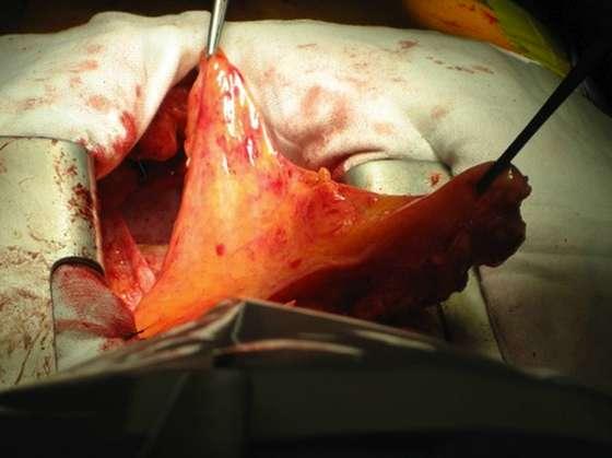
41 How do we do it? Examples of Pericardial Closure

42

43
44

45

46 Clinical Implantation
47 Is this information convincing enough?
48 Pericardial Closure: Patient outcomes


49 CorMatrix ECM is Derived from Porcine Small Intestinal Submucosa The SIS-ECM is obtained from the submucosa of the small intestine of pigs. {


50 CorMatrix ECM Derived from porcine small intestinal submucosa 100 mm H&E; 200X
51 Special Properties of the ECM Immuno-modulatory Anti inflammatory Antimicrobial Site specific remodeling Chemo-attractant to progenitor cells
52 Retrospective review of isolated CABG patients undergoing ECM pericardial repair Primary Isolated CABG Patients Repair Group treated with CorMatrix ECM Complete circumferential suturing of CorMatrix ECM to native pericardium Control Group not treated, pericardium left open 222 patients, 3 institutions
53 Control Group Repair Group P-value Characteristic N=111 N=111 Age range, years Mean age, years (SD) 64.5 (11.2) 62.3 (10.7) 0.13 Mean height, inches (SD) 67.7 (3.9) 67.9 (3.9) 0.69 Mean weight, pounds (SD) (40.9) (38.4) 0.46 Sex, male 64.90% 67.60% 0.78 Ethnicity2, white 85.2% (92/108) 81.7% (89/109) 0.59 Medical History Left ventricular hypertrophy3 5.0% (5/101) 6.9% (7/103) 0.77 Left bundle branch block3 2.0% (2/101) 3.0% (3/103) 1 Previous revascularization 11.70% 3.60% 0.04 Congestive heart failure 9.00% 8.10% 1 Chronic obstructive pulmonary disease 15.30% 22.50% 0.23 Hypertension 80.20% 83.80% 0.6 Peripheral vascular disease 9.90% 18.00% 0.12 Bleeding 0.00% 0.00% -- Medications Beta-adrenergic blocker 47.3% (52/110) 49.60% 0.79 Statin 61.0% (67/110) 54.10% 0.34 Anti-arrhythmic 3.6% (4/110) 3.60% 1 Calcium channel blocker 32.7% (36/110) 19.80% 0.03 Angiotensin converting enzyme inhibitor 28.2% (31/110) 28.80% 1 Angiotensin II receptor blocker 3.6% (4/110) 5.40% 0.75 Nonsteroidal anti-inflammatory Drug 38.2% (42/110) 33.30% 0.49 Steroids 2.7% (3/110) 1.80% 0.68 Magnesium 0.9% (1/110) 0.90% 1
54 Retrospective review of isolated CABG patients undergoing CorMatrix ECM pericardial repair Adverse Event Control Group Repair Group Postoperative Complications 29/111 37/111 Intra-operative complications 2/111 6/111 Acute inflammation 1/111 0/111 ARDS 6/111 3/111 Allergic reaction 0/111 1/111 Angina 0/111 0/111 Anti-coagulation complications 1/111 1/111 Bleeding 0/111 0/111 Chronic inflammation 0/111 0/111 Cognitive sequelae 1/111 0/111 Infection 0/111 0/111 ECM migration 0/111 0/111 Myocardial infarction 0/111 0/111 Pericarditis 0/111 0/111 Renal failure 2/111 1/111 Stroke 1/111 1/111 Tamponade 1/111 1/111 Death 1/111 0/111 Other 22/111 27/111
55 3 year clinical experience with CorMatrix ECM Provides an environment for tissue specific cardio-vascular remodeling Safe, reliable and cost-effective No adverse complications Provides structural integrity during remodeling Pericardial closure restores the normal anatomy, facilitates perioperative patient care Facilitates surgical approach for redo operations Protects underlying grafts and cardiac structures

56 Pericardial Repair

57 5 Pericardial Structure Repaired


58 9 month post implant re-op

59 3 YEARS POST-OP 59
60 Other important advantages Decreased Postoperative Atrial Fibrillation???
61 Atrial Fibrillation After Cardiac Surgery Occurs in about 30% of patients Associated with increased mortality (both early and late), increased morbidity, increased cost Incidence is reduced but not eliminated by the use of pre-operative antiarrhythmic agents B-Blockers and Amiodarone most effective

62 Pathogenesis of POAF Echahidi, N. et al. J Am Coll Cardiol 2008;51: Copyright 2008 American College of Cardiology Foundation. Restrictions may apply.
63 Circumferential Pericardial Reconstruction Using an Extracellular Matrix Implant Correlates with Reduced Risk of Postoperative Atrial Fibrillation in Coronary Artery Bypass Surgery Patients W. Douglas Boyd, MD, William E. Johnson, III, MD, Parvez K. Sultan, MD, Thomas F. Deering, MD, FACC, FACP, FHRS, Robert G. Matheny, MD, FACS University of California Davis Medical Center, Davis, California, USA; Mobile Infirmary Medical Center, Mobile, Alabama, USA; Trinity Medical Center, Birmingham, Alabama, USA; Piedmont Heart Institute, Atlanta, Georgia, USA; CorMatrix Cardiovascular, Atlanta, Georgia, USA Conclusions: In this retrospective study, circumferential pericardial reconstruction with the ECM implant contributed directly to a statistically significant and clinically meaningful reduction in the rate of postoperative AF in primary isolated CABG patients. A prospective, multicenter, randomized trial has been planned to further test this approach.
64 Methods: A retrospective comparison of the incidence of postoperative AF in 111 patients who underwent a circumferential pericardial reconstruction procedure with the CorMatrix ECM for Pericardial Closure (CorMatrix Cardiovascular Inc., Atlanta, GA) following primary isolated CABG, versus a control group of 111 patients who did not undergo pericardial reconstruction
65 Results: Postoperative AF occurred in 43 of 111 control patients (38.74%, LCL 29.64, UCL 48.45) but only 20 of 111 treated patients (18.02%, LCL 11.37, UCL 26.45) representing a 53.8% relative risk reduction in the treatment group (P=0.0003). There was a small but not statistically significant decrease in hospital LOS in treated patients. The two treatment groups exhibited a similar postoperative complication profile.
66 Conclusion Circumferential pericardial reconstruction with CorMatrix ECM after cardiac operations is: Safe Effective in re-establishing normal pericardial function in the immediate and long term postoperative periods Increases the safety of sternal re-entry Appears to reduce the incidence of post-op atrial fibrillation
67 How Might ECM Pericardial Reconstruction Discourage The Occurrence of Post-op Atrial Fibrillation? Prevent acute chamber distension Prevent rundown of soup from wound onto epicardial surface Inhibits inflammatory response to surgical wound Induces remodeling response in the surgical wound

68 CorMatrix for Pericardial Closure Potential Impact on Reducing New Onset Post-Op Atrial Fibrillation Published Article reporting a 54% reduction in post-op Atrial Fibrillation following pericardial reconstruction using CorMatrix Prospective Multicenter FDA approved study underway to confirm impact on reducing post-op Atrial Fibrillation
69 Agruments against closing the pericardium
70 Original Articles Should the pericardium be closed routinely after heart operations? Vivek Rao, MD, PhD a, Masashi Komeda, MD, PhD a, Richard D. Weisel, MD a, Gideon Cohen, MD a, Michael A. Borger, MD a, Tirone E. David, MD a The cardiac index and stroke work index were lower in the closure group compared to the open group (P<0.001), however, these difference were only present for one hour post operatively and at 4 h and 8 h post operatively no difference could be determined. Ann Thorac Surg 1999;67:
71 Pericardial Closure: Arguments against Adverse changes in hemodynamics A transient reduction in cardiac index and stroke work index following primary pericardial closure has been documented in humans in one small, prospective randomized clinical trial Several clinical case series have also reported mild-tomoderate cardiac constriction, reduction in cardiac output and a fall in arterial pressure resulting from primary pericardial closure
72 Pericardial Closure: Arguments against Adverse changes in hemodynamics Are consistent with the combined effects of contraction of the pericardium, dilation of the cardiac chambers in the absence of the pericardium, and the adverse effects of CPB. Studies have shown these effects to be transient in nature No study has ever reported an adverse clinical outcome
73 Pericardial Closure: Arguments against Perceived risks of graft compression Several authors have asserted that pericardial closure could lead to kinking of bypass grafts or IMA conduits No published studies regarding this phenomenon Not observed in >40,000 closures
74 Pericardial Closure: Arguments against Perceived risks of graft compression Pericardial substitutes can be tailored to allow circumferential closure that approximates normal pericardial constraint around the ventricles, while avoiding adverse effect on grafts.
75 Pericardial Closure : What I have learned There are no real contraindications to pericardial closure The details are important!!!! How you open pericardium can facilitate closure Preservation of Thymus, pre-closure of notches, early CT suction No gaps, separate chest tubes Blake tubes have to be frequently milked In my experience, hemodynamic compromise after closure has virtually NEVER been related to pericardial closure Until comfortable, do early post-op / pre-discharge echoes and CT scans to evaluate the pericardial sac.
76 Top Reasons to Close Pericardium 1) Restores normal anatomy 2) Allows normal function 3) Protects underlying bypass grafts early / late reentry 4) Helps guide postoperative patient management 5) Helps prevent tamponade 6) Potential for improving RV function 7) Normalizes postoperative hemodynamics 8) Takes minimal time, is reimbursable and cost effective 9) May prevent post-op AF 10) Improvement of patient outcomes 11) No evidence of adverse effects
Normal Pericardial Physiology
 Normal Pericardial Physiology Normal pericardium contains 20-30 ml of lymphoid fluid lubricating function that facilitates normal myocardial rotation and translation during each cardiac cycle in that the
CorMatrix ECM Bioscaffold
 CorMatrix ECM Bioscaffold REMODEL. REGROW. RESTORE. CorMatrix ECM Bioscaffold provides a natural bioscaffold matrix that enables the body s own cells to repair and remodel damaged cardio-vascular tissue.
The Cardiovascular System
 The Cardiovascular System The Cardiovascular System A closed system of the heart and blood vessels The heart pumps blood Blood vessels allow blood to circulate to all parts of the body The function of
Cardiovascular Nursing Practice: A Comprehensive Resource Manual and Study Guide for Clinical Nurses 2 nd Edition
 Cardiovascular Nursing Practice: A Comprehensive Resource Manual and Study Guide for Clinical Nurses 2 nd Edition Table of Contents Volume 1 Chapter 1: Cardiovascular Anatomy and Physiology Basic Cardiac
efferent fibers from t.. Heart Surface anatomy and heart sounds -Dry lecture -Gray s 169,
 A patient is diagnosed with ischemia (i.e., lack of blood flow) in a left lobar pulmonary vein. The attending physician determines that the ischemia is due to a vasospastic episode. Constriction of this
Cardiovascular Physiology. Heart Physiology. Introduction. The heart. Electrophysiology of the heart
 Cardiovascular Physiology Heart Physiology Introduction The cardiovascular system consists of the heart and two vascular systems, the systemic and pulmonary circulations. The heart pumps blood through
Major Function of the Cardiovascular System. Transportation. Structures of the Cardiovascular System. Heart - muscular pump
 Structures of the Cardiovascular System Heart - muscular pump Blood vessels - network of tubes Blood - liquid transport vehicle brachiocephalic trunk superior vena cava right pulmonary arteries right pulmonary
11/10/2014. Muscular pump Two atria Two ventricles. In mediastinum of thoracic cavity 2/3 of heart's mass lies left of midline of sternum
 It beats over 100,000 times a day to pump over 1,800 gallons of blood per day through over 60,000 miles of blood vessels. During the average lifetime, the heart pumps nearly 3 billion times, delivering
University of Florida Department of Surgery. CardioThoracic Surgery VA Learning Objectives
 University of Florida Department of Surgery CardioThoracic Surgery VA Learning Objectives This service performs coronary revascularization, valve replacement and lung cancer resections. There are 2 faculty
Function: Transportation of. Oxygen Nutrients Waste Hormones gases
 Function: Transportation of Oxygen Nutrients Waste Hormones gases Pericardium: double sac of serous membrane filled with fluid (pericardial fluid to be exact) that surrounds the heart. Parietal pericardium:
The Cardiovascular System
 The Cardiovascular System The Manila Times College of Subic Prepared by: Stevens B. Badar, RN, MANc THE HEART Anatomy of the Heart Location and Size approx. the size of a person s fist, hollow and cone-shaped,
The Cardiovascular System Part I: Heart Outline of class lecture After studying part I of this chapter you should be able to:
 The Cardiovascular System Part I: Heart Outline of class lecture After studying part I of this chapter you should be able to: 1. Describe the functions of the heart 2. Describe the location of the heart,
Ch 19: Cardiovascular System - The Heart -
 Ch 19: Cardiovascular System - The Heart - Give a detailed description of the superficial and internal anatomy of the heart, including the pericardium, the myocardium, and the cardiac muscle. Trace the
CARDIOVASCULAR SYSTEM
 CARDIOVASCULAR SYSTEM Overview Heart and Vessels 2 Major Divisions Pulmonary Circuit Systemic Circuit Closed and Continuous Loop Location Aorta Superior vena cava Right lung Pulmonary trunk Base of heart
Chapter 20: Cardiovascular System: The Heart
 Chapter 20: Cardiovascular System: The Heart I. Functions of the Heart A. List and describe the four functions of the heart: 1. 2. 3. 4. II. Size, Shape, and Location of the Heart A. Size and Shape 1.
The Heart & Pericardium Dr. Rakesh Kumar Verma Assistant Professor Department of Anatomy KGMU UP Lucknow
 The Heart & Pericardium Dr. Rakesh Kumar Verma Assistant Professor Department of Anatomy KGMU UP Lucknow Fibrous skeleton Dense fibrous connective tissue forms a structural foundation around AV & arterial
Cardiac Drugs: Chapter 9 Worksheet Cardiac Agents. 1. drugs affect the rate of the heart and can either increase its rate or decrease its rate.
 Complete the following. 1. drugs affect the rate of the heart and can either increase its rate or decrease its rate. 2. drugs affect the force of contraction and can be either positive or negative. 3.
10/23/2017. Muscular pump Two atria Two ventricles. In mediastinum of thoracic cavity 2/3 of heart's mass lies left of midline of sternum
 It beats over 100,000 times a day to pump over 1,800 gallons of blood per day through over 60,000 miles of blood vessels. During the average lifetime, the heart pumps nearly 3 billion times, delivering
Cardiovascular System. Heart
 Cardiovascular System Heart Electrocardiogram A device that records the electrical activity of the heart. Measuring the relative electrical activity of one heart cycle. A complete contraction and relaxation.
THE HEART. A. The Pericardium - a double sac of serous membrane surrounding the heart
 THE HEART I. Size and Location: A. Fist-size weighing less than a pound (250 to 350 grams). B. Located in the mediastinum between the 2 nd rib and the 5 th intercostal space. 1. Tipped to the left, resting
The Cardiovascular System
 Essentials of Human Anatomy & Physiology Elaine N. Marieb Seventh Edition Chapter 11 The Cardiovascular System Slides 11.1 11.19 Lecture Slides in PowerPoint by Jerry L. Cook The Cardiovascular System
Cardiovascular Physiology
 Cardiovascular Physiology Introduction The cardiovascular system consists of the heart and two vascular systems, the systemic and pulmonary circulations. The heart pumps blood through two vascular systems
The Cardiovascular System
 Essentials of Human Anatomy & Physiology Elaine N. Marieb Slides 11.1 11.19 Seventh Edition Chapter 11 The Cardiovascular System Functions of the Cardiovascular system Function of the heart: to pump blood
10. Thick deposits of lipids on the walls of blood vessels, called, can lead to serious circulatory issues. A. aneurysm B. atherosclerosis C.
 Heart Student: 1. carry blood away from the heart. A. Arteries B. Veins C. Capillaries 2. What is the leading cause of heart attack and stroke in North America? A. alcohol B. smoking C. arteriosclerosis
Cardiology. Objectives. Chapter
 1:44 M age 1121 Chapter Cardiology Objectives art 1: Cardiovascular natomy and hysiology, ECG Monitoring, and Dysrhythmia nalysis (begins on p. 1127) fter reading art 1 of this chapter, you should be able
Cardiovascular System
 Cardiovascular System The Heart Cardiovascular System The Heart Overview What does the heart do? By timed muscular contractions creates pressure gradients blood moves then from high pressure to low pressure
10/8/2018. Lecture 9. Cardiovascular Health. Lecture Heart 2. Cardiovascular Health 3. Stroke 4. Contributing Factor
 Lecture 9 Cardiovascular Health 1 Lecture 9 1. Heart 2. Cardiovascular Health 3. Stroke 4. Contributing Factor 1 The Heart Muscular Pump The Heart Receives blood low pressure then increases the pressure
Adult Echocardiography Examination Content Outline
 Adult Echocardiography Examination Content Outline (Outline Summary) # Domain Subdomain Percentage 1 2 3 4 5 Anatomy and Physiology Pathology Clinical Care and Safety Measurement Techniques, Maneuvers,
Cardiovascular Anatomy Dr. Gary Mumaugh
 Cardiovascular Anatomy Dr. Gary Mumaugh Location of Heart Approximately the size of your fist Location o Superior surface of diaphragm o Left of the midline in mediastinum o Anterior to the vertebral column,
Section 5.1 The heart and heart disease
 Section 5.1 The heart and heart disease Mammals are too large to rely on diffusion. They need a circulatory system to move substances around the body. Blood moves down pressure gradients, from high to
The Heart. Size, Form, and Location of the Heart. 1. Blunt, rounded point; most inferior part of the heart.
 12 The Heart FOCUS: The heart is composed of cardiac muscle cells, which are elongated, branching cells that appear striated. Cardiac muscle cells behave as a single electrical unit, and the highly coordinated
Cardiovascular Disorders. Heart Disorders. Diagnostic Tests for CV Function. Bio 375. Pathophysiology
 Cardiovascular Disorders Bio 375 Pathophysiology Heart Disorders Heart disease is ranked as a major cause of death in the U.S. Common heart diseases include: Congenital heart defects Hypertensive heart
Structure and organization of blood vessels
 The cardiovascular system Structure of the heart The cardiac cycle Structure and organization of blood vessels What is the cardiovascular system? The heart is a double pump heart arteries arterioles veins
2. right heart = pulmonary pump takes blood to lungs to pick up oxygen and get rid of carbon dioxide
 A. location in thorax, in inferior mediastinum posterior to sternum medial to lungs superior to diaphragm anterior to vertebrae orientation - oblique apex points down and to the left 2/3 of mass on left
Approximately the size of your fist Location. Pericardial physiology
 Heart Anatomy Approximately the size of your fist Location Superior surface of diaphragm Left of the midline Anterior to the vertebral column, posterior to the sternum Wednesday, March 28, 2012 Muscle
CMS Limitations Guide - Radiology Services
 CMS Limitations Guide - Radiology Services Starting October 1, 2015, CMS will update their existing medical necessity limitations on tests and procedures to correspond to ICD-10 codes. This limitations
P215 SPRING 2019: CIRCULATORY SYSTEM Chaps 13, 14 & 15: pp , , , I. Major Functions of the Circulatory System
 P215 SPRING 2019: CIRCULATORY SYSTEM Chaps 13, 14 & 15: pp 360-390, 395-404, 410-428 433-438, 441-445 I. Major Functions of the Circulatory System 1. 2. 3. 4. II. Structure of the Heart 1. atria 2. ventricles
BME 5742 Bio-Systems Modeling and Control. Lecture 41 Heart & Blood Circulation Heart Function Basics
 BME 5742 Bio-Systems Modeling and Control Lecture 41 Heart & Blood Circulation Heart Function Basics Dr. Zvi Roth (FAU) 1 Pumps A pump is a device that accepts fluid at a low pressure P 1 and outputs the
Cardiac MRI in ACHD What We. ACHD Patients
 Cardiac MRI in ACHD What We Have Learned to Apply to ACHD Patients Faris Al Mousily, MBChB, FAAC, FACC Consultant, Pediatric Cardiology, KFSH&RC/Jeddah Adjunct Faculty, Division of Pediatric Cardiology
Circulation. Sinoatrial (SA) Node. Atrioventricular (AV) Node. Cardiac Conduction System. Cardiac Conduction System. Linked to the nervous system
 Node. Atrioventricular (AV) Node. Cardiac Conduction System. Cardiac Conduction System. Linked to the nervous system") Circulation Cardiac Conduction System AHS A H S Your body resembles a large roadmap. There are routes or arteries that take you downtown to the heart of the city and veins that take you to the outskirts
Circulation Cardiac Conduction System AHS A H S Your body resembles a large roadmap. There are routes or arteries that take you downtown to the heart of the city and veins that take you to the outskirts
Circulation. Blood Pressure and Antihypertensive Medications. Venous Return. Arterial flow. Regulation of Cardiac Output.
 Circulation Blood Pressure and Antihypertensive Medications Two systems Pulmonary (low pressure) Systemic (high pressure) Aorta 120 mmhg Large arteries 110 mmhg Arterioles 40 mmhg Arteriolar capillaries
Figure ) The specific chamber of the heart that is indicated by letter A is called the. Diff: 1 Page Ref: 364
 The specific chamber of the heart that is indicated by letter A is called the. Diff: 1 Page Ref: 364") Essentials of Anatomy and Physiology, 9e (Marieb) Chapter 11 The Cardiovascular System Short Answer Figure 11.1 Using Figure 11.1, identify the following: 1) The Purkinje fibers are indicated by label.
Essentials of Anatomy and Physiology, 9e (Marieb) Chapter 11 The Cardiovascular System Short Answer Figure 11.1 Using Figure 11.1, identify the following: 1) The Purkinje fibers are indicated by label.
Heart Anatomy. 7/5/02 Stephen G Davenport 1
 Heart Anatomy Copyright 1999, Stephen G. Davenport, No part of this publication may be reproduced, stored in a retrieval system, or transmitted, in any form without prior written permission. 7/5/02 Stephen
Blood pressure. Formation of the blood pressure: Blood pressure. Formation of the blood pressure 5/1/12
 Blood pressure Blood pressure Dr Badri Paudel www.badripaudel.com Ø Blood pressure means the force exerted by the blood against the vessel wall Ø ( or the force exerted by the blood against any unit area
Using Figure 14.1, match the following: 1) Myelin sheath. 1) 2) Cell body of ANS preganglionic neuron. 2)
 Myelin sheath. 1) 2) Cell body of ANS preganglionic neuron. 2)") Practice Exam 1 AP 2 chapters 14 and 18 Name MATCHING: Match labeled areas with the appropriate terminology from the list below. Figure 14.1 Using Figure 14.1, match the following: 1) Myelin sheath. 1)
Practice Exam 1 AP 2 chapters 14 and 18 Name MATCHING: Match labeled areas with the appropriate terminology from the list below. Figure 14.1 Using Figure 14.1, match the following: 1) Myelin sheath. 1)
Chapter 13 The Cardiovascular System: Cardiac Function
 Chapter 13 The Cardiovascular System: Cardiac Function Overview of the Cardiovascular System The Path of Blood Flow through the Heart and Vasculature Anatomy of the Heart Electrical Activity of the Heart
Heart. Structure Physiology of blood pressure and heartbeat
 Heart Structure Physiology of blood pressure and heartbeat Location and Anatomy Location and Anatomy Pericardial cavity: surrounds, isolates, and anchors heart Parietal pericardium lined with serous membrane
Echocardiography as a diagnostic and management tool in medical emergencies
 Echocardiography as a diagnostic and management tool in medical emergencies Frank van der Heusen MD Department of Anesthesia and perioperative Care UCSF Medical Center Objective of this presentation Indications
The Heart. The Heart A muscular double pump. The Pulmonary and Systemic Circuits
 C H A P T E R 19 The Heart The Heart A muscular double pump circuit takes blood to and from the lungs Systemic circuit vessels transport blood to and from body tissues Atria receive blood from the pulmonary
The Cardiovascular System (Heart)
") The Cardiovascular System The Cardiovascular System (Heart) A closed system of the heart and blood vessels The heart pumps blood Blood vessels allow blood to circulate to all parts of the body The function
The Cardiovascular System The Cardiovascular System (Heart) A closed system of the heart and blood vessels The heart pumps blood Blood vessels allow blood to circulate to all parts of the body The function
Ischemic heart disease
 Ischemic heart disease Introduction In > 90% of cases: the cause is: reduced coronary blood flow secondary to: obstructive atherosclerotic vascular disease so most of the time it is called: coronary artery
Randomized comparison of single versus double mammary coronary artery bypass grafting: 5 year outcomes of the Arterial Revascularization Trial
 Randomized comparison of single versus double mammary coronary artery bypass grafting: 5 year outcomes of the Arterial Revascularization Trial Embargoed until 10:45 a.m. CT, Monday, Nov. 14, 2016 David
The Heart and Heart Disease
 The Heart and Heart Disease Illustration of the heart by Leonardo DaVinci heart-surgeon.com/ history.html 2/14/2010 1 I. Location, Size and Position of the Heart A. Triangular organ located 1. of mass
d) Cardiovascular System Higher Human Biology
 Cardiovascular System Higher Human Biology") d) Cardiovascular System Higher Human Biology What can your remember about the heart and blood vessels? What is the Cardiovascular System? The cardiovascular system, also known as the circulatory system,
d) Cardiovascular System Higher Human Biology What can your remember about the heart and blood vessels? What is the Cardiovascular System? The cardiovascular system, also known as the circulatory system,
Index of subjects. effect on ventricular tachycardia 30 treatment with 101, 116 boosterpump 80 Brockenbrough phenomenon 55, 125
 145 Index of subjects A accessory pathways 3 amiodarone 4, 5, 6, 23, 30, 97, 102 angina pectoris 4, 24, 1l0, 137, 139, 140 angulation, of cavity 73, 74 aorta aortic flow velocity 2 aortic insufficiency
Chapter 14. The Cardiovascular System
 Chapter 14 The Cardiovascular System Introduction Cardiovascular system - heart, blood and blood vessels Cardiac muscle makes up bulk of heart provides force to pump blood Function - transports blood 2
Ischemic Heart Disease Interventional Treatment
 Ischemic Heart Disease Interventional Treatment Cardiac Catheterization Laboratory Procedures (N = 89) is a regional and national referral center for percutaneous coronary intervention (PCI). A total of
Heart Pump and Cardiac Cycle. Faisal I. Mohammed, MD, PhD
 Heart Pump and Cardiac Cycle Faisal I. Mohammed, MD, PhD 1 Objectives To understand the volume, mechanical, pressure and electrical changes during the cardiac cycle To understand the inter-relationship
RV dysfunction and failure PATHOPHYSIOLOGY. Adam Torbicki MD, Dept Chest Medicine Institute of Tuberculosis and Lung Diseases Warszawa, Poland
 RV dysfunction and failure PATHOPHYSIOLOGY Adam Torbicki MD, Dept Chest Medicine Institute of Tuberculosis and Lung Diseases Warszawa, Poland Normal Right Ventricle (RV) Thinner wall Weaker myocytes Differences
Chapter 9, Part 2. Cardiocirculatory Adjustments to Exercise
 Chapter 9, Part 2 Cardiocirculatory Adjustments to Exercise Electrical Activity of the Heart Contraction of the heart depends on electrical stimulation of the myocardium Impulse is initiated in the right
CV Anatomy Quiz. Dr Ella Kim Dr Pip Green
 CV Anatomy Quiz Dr Ella Kim Dr Pip Green Q1 The location of the heart is correctly described as A) lateral to the lungs. B) medial to the sternum. C) superior to the diaphragm. D) posterior to the spinal
Outline. Echocardiographic Assessment of Pericardial Effusion/Tamponade: The Essentials
 Echocardiographic Assessment of Pericardial Effusion/Tamponade: The Essentials John R Schairer DO FACC Henry Ford Heart and Vascular Institute No Disclosures Outline Normal Anatomy and Physiology Pathophysiology
Cardiovascular System
 Cardiovascular System Purpose Transport oxygen and nutrients Take waste products away from tissues & organs Things we learned Blood pressure: the force of blood pushing against the walls of blood vessels
Collin County Community College. ! BIOL Anatomy & Physiology! WEEK 5. The Heart
 Collin County Community College! BIOL. 2402 Anatomy & Physiology! WEEK 5 The Heart 1 (1578-1657) A groundbreaking work in the history of medicine, English physician William Harvey s Anatomical Essay on
BUSINESS. Articles? Grades Midterm Review session
 BUSINESS Articles? Grades Midterm Review session REVIEW Cardiac cells Myogenic cells Properties of contractile cells CONDUCTION SYSTEM OF THE HEART Conduction pathway SA node (pacemaker) atrial depolarization
Chapter 18 - Heart. I. Heart Anatomy: size of your fist; located in mediastinum (medial cavity)
") Chapter 18 - Heart I. Heart Anatomy: size of your fist; located in mediastinum (medial cavity) A. Coverings: heart enclosed in double walled sac called the pericardium 1. Fibrous pericardium: dense connective
Chapter 18 - Heart I. Heart Anatomy: size of your fist; located in mediastinum (medial cavity) A. Coverings: heart enclosed in double walled sac called the pericardium 1. Fibrous pericardium: dense connective
Vasculature and innervation of the heart. A. Bendelic Human Anatomy Department
 Vasculature and innervation of the heart A. Bendelic Human Anatomy Department Plan: 1. Arterial blood supply of the heart. Coronary arteries 2. Venous drainage of the heart. Cardiac veins 3. Innervation
Anatomy of the Heart. Figure 20 2c
 Anatomy of the Heart Figure 20 2c Pericardium & Myocardium Remember, the heart sits in it s own cavity, known as the mediastinum. The heart is surrounded by the Pericardium, a double lining of the pericardial
Cardiovascular System
 Component 3-Terminology in Healthcare and Public Health Settings Unit 5-Cardiovascular System This material was developed by The University of Alabama at Birmingham, funded by the Department of Health
The Cardiovascular System
 The Cardiovascular System https://www.youtube.com/watch?v=ohmmtqkgs50 Human Anatomy & Physiology P. Wilson 1 Introduction The functions of the cardiovascular system are: to bring oxygen & nutrients to
McHenry Western Lake County EMS System Paramedic, EMT-B and PHRN Optional Continuing Education 2018 #12 Understanding Preload and Afterload
 McHenry Western Lake County EMS System Paramedic, EMT-B and PHRN Optional Continuing Education 2018 #12 Understanding Preload and Afterload Cardiac output (CO) represents the volume of blood that is delivered
The Cardiovascular System. Chapter 15. Cardiovascular System FYI. Cardiology Closed systemof the heart & blood vessels. Functions
 Chapter 15 Cardiovascular System FYI The heart pumps 7,000 liters (4000 gallons) of blood through the body each day The heart contracts 2.5 billion times in an avg. lifetime The heart & all blood vessels
CARDIOVASCULAR SYSTEM
 CARDIOVASCULAR SYSTEM 1. Resting membrane potential of the ventricular myocardium is: A. -55 to-65mv B. --65 to-75mv C. -75 to-85mv D. -85 to-95 mv E. -95 to-105mv 2. Regarding myocardial contraction:
Blood Functions. Blood and the Cardiovascular System. Blood. Plasma. Erythrocytes (RBCs) Erythrocytes (RBCs) 4/7/2017
 Erythrocytes (RBCs) 4/7/2017") Blood Functions Blood and the Cardiovascular System Distribution Delivery of oxygen and nutrients to all body cells; Transport of wastes to lungs and excretory organs; Transport of hormones Regulation
Blood Functions Blood and the Cardiovascular System Distribution Delivery of oxygen and nutrients to all body cells; Transport of wastes to lungs and excretory organs; Transport of hormones Regulation
Retrospective Study Of Redo Cardiac Surgery In A Single Centre. R Karthekeyan, K Selvaraju, L Ramanathan, M Rakesh, S Rao, M Vakamudi, K Balakrishnan
 ISPUB.COM The Internet Journal of Anesthesiology Volume 12 Number 2 Retrospective Study Of Redo Cardiac Surgery In A Single Centre R Karthekeyan, K Selvaraju, L Ramanathan, M Rakesh, S Rao, M Vakamudi,
STRUCTURES OF THE CARDIOVASCULAR SYSTEM
 STRUCTURES OF THE CARDIOVASCULAR SYSTEM CARDIOVASCULAR SYSTEM Also called the circulatory system Consists of the heart, arteries, veins, and capillaries Main function is to pump/circulate oxygenated blood
Cardiovascular System Notes: Heart Disease & Disorders
 Cardiovascular System Notes: Heart Disease & Disorders Interesting Heart Facts The Electrocardiograph (ECG) was invented in 1902 by Willem Einthoven Dutch Physiologist. This test is still used to evaluate
OPCAB IS NOT BETTER THAN CONVENTIONAL CABG
 OPCAB IS NOT BETTER THAN CONVENTIONAL CABG Harold L. Lazar, M.D. Harold L. Lazar, M.D. Professor of Cardiothoracic Surgery Boston Medical Center and the Boston University School of Medicine Boston, MA
BIPN100 F15 Human Physiol I (Kristan) Lecture 14 Cardiovascular control mechanisms p. 1
 Lecture 14 Cardiovascular control mechanisms p. 1") BIPN100 F15 Human Physiol I (Kristan) Lecture 14 Cardiovascular control mechanisms p. 1 Terms you should understand: hemorrhage, intrinsic and extrinsic mechanisms, anoxia, myocardial contractility, residual
BIPN100 F15 Human Physiol I (Kristan) Lecture 14 Cardiovascular control mechanisms p. 1 Terms you should understand: hemorrhage, intrinsic and extrinsic mechanisms, anoxia, myocardial contractility, residual
1. What kind of blood is found in the rt. atrium? (oxygenated or deoxygenated)
") Carl Christennsen, PhD Chap. 19, 20, & 21 - Circulatory System Bio. 2304 Human Anatomy HEART 1. What kind of blood is found in the rt. atrium? (oxygenated or deoxygenated) Where does this blood come from?
Carl Christennsen, PhD Chap. 19, 20, & 21 - Circulatory System Bio. 2304 Human Anatomy HEART 1. What kind of blood is found in the rt. atrium? (oxygenated or deoxygenated) Where does this blood come from?
The Circulatory System. The Heart, Blood Vessels, Blood Types
 The Circulatory System The Heart, Blood Vessels, Blood Types The Closed Circulatory System Humans have a closed circulatory system, typical of all vertebrates, in which blood is confined to vessels and
The Cardiovascular System
 PowerPoint Lecture Slide Presentation by Patty Bostwick-Taylor, Florence-Darlington Technical College The Cardiovascular System 11 PART A The Cardiovascular System A closed system of the heart and blood
Cardiac Output MCQ. Professor of Cardiovascular Physiology. Cairo University 2007
 Cardiac Output MCQ Abdel Moniem Ibrahim Ahmed, MD Professor of Cardiovascular Physiology Cairo University 2007 90- Guided by Ohm's law when : a- Cardiac output = 5.6 L/min. b- Systolic and diastolic BP
Cardiovascular System. Supplementary Information
 Cardiovascular System Supplementary Information THE CARDIOVASCULAR SYSTEM - 1 - THE CARDIOVASCULAR SYSTEM FUNCTION Transport system carrying nutrient, gases, hormones and waste products to and from the
1. Distinguish among the types of blood vessels on the basis of their structure and function.
 Blood Vessels and Circulation Objectives This chapter describes the structure and functions of the blood vessels Additional subjects contained in Chapter 13 include cardiovascular physiology, regulation,
The Heart. Happy Friday! #takeoutyournotes #testnotgradedyet
 The Heart Happy Friday! #takeoutyournotes #testnotgradedyet Introduction Cardiovascular system distributes blood Pump (heart) Distribution areas (capillaries) Heart has 4 compartments 2 receive blood (atria)
CORONARY ARTERY BYPASS GRAFT (CABG) MEASURES GROUP OVERVIEW
 MEASURES GROUP OVERVIEW") CONARY ARTERY BYPASS GRAFT (CABG) MEASURES GROUP OVERVIEW 2015 PQRS OPTIONS F MEASURES GROUPS: 2015 PQRS MEASURES IN CONARY ARTERY BYPASS GRAFT (CABG) MEASURES GROUP: #43 Coronary Artery Bypass Graft (CABG):
CONARY ARTERY BYPASS GRAFT (CABG) MEASURES GROUP OVERVIEW 2015 PQRS OPTIONS F MEASURES GROUPS: 2015 PQRS MEASURES IN CONARY ARTERY BYPASS GRAFT (CABG) MEASURES GROUP: #43 Coronary Artery Bypass Graft (CABG):
Cardiac Arrhythmias. Cathy Percival, RN, FALU, FLMI VP, Medical Director AIG Life and Retirement Company
 Cardiac Arrhythmias Cathy Percival, RN, FALU, FLMI VP, Medical Director AIG Life and Retirement Company The Cardiovascular System Three primary functions Transport of oxygen, nutrients, and hormones to
The pericardial sac is composed of the outer fibrous pericardium
 Pericardiectomy for Constrictive or Recurrent Inflammatory Pericarditis Mauricio A. Villavicencio, MD, Joseph A. Dearani, MD, and Thoralf M. Sundt, III, MD Anatomy and Preoperative Considerations The pericardial
Electrical Conduction
 Sinoatrial (SA) node Electrical Conduction Sets the pace of the heartbeat at 70 bpm AV node (50 bpm) and Purkinje fibers (25 40 bpm) can act as pacemakers under some conditions Internodal pathway from
Circulation. Circulation = is a process used for the transport of oxygen, carbon! dioxide, nutrients and wastes through-out the body
 Circulation Circulation = is a process used for the transport of oxygen, carbon! dioxide, nutrients and wastes through-out the body Heart = muscular organ about the size of your fist which pumps blood.
*Generating blood pressure *Routing blood: separates. *Ensuring one-way blood. *Regulating blood supply *Changes in contraction
 *Generating blood pressure *Routing blood: separates pulmonary and systemic circulations *Ensuring one-way blood flow: valves *Regulating blood supply *Changes in contraction rate and force match blood
Ventricular Tachycardia in Structurally Normal Hearts (Idiopathic VT) Patient Information
 Patient Information") Melbourne Heart Rhythm Ventricular Tachycardia in Structurally Normal Hearts (Idiopathic VT) Patient Information What is Ventricular Tachycardia? Ventricular tachycardia (VT) is an abnormal rapid heart
Melbourne Heart Rhythm Ventricular Tachycardia in Structurally Normal Hearts (Idiopathic VT) Patient Information What is Ventricular Tachycardia? Ventricular tachycardia (VT) is an abnormal rapid heart
The Cardiovascular and Lymphatic Systems Cardiovascular System Blood Vessels Blood Vessels Arteries Arteries Arteries
 CH 12 The Cardiovascular and s The Cardiovascular and s OUTLINE: Cardiovascular System Blood Vessels Blood Pressure Cardiovascular System The cardiovascular system is composed of Blood vessels This system
Ischemic Heart Disease Interventional Treatment
 Ischemic Heart Disease Interventional Treatment Cardiac Catheterization Laboratory Procedures (N = 11,61) is a regional and national referral center for percutaneous coronary intervention (PCI). A total
THE HEART OBJECTIVES: LOCATION OF THE HEART IN THE THORACIC CAVITY CARDIOVASCULAR SYSTEM
 BIOLOGY II CARDIOVASCULAR SYSTEM ACTIVITY #3 NAME DATE HOUR THE HEART OBJECTIVES: Describe the anatomy of the heart and identify and give the functions of all parts. (pp. 356 363) Trace the flow of blood
Strategies for the High Risk Redo in CHD
 Strategies for the High Risk Redo in CHD Joseph A. Dearani, MD AATS, Minneapolis 2013 Strategies for the High Risk Redo in CHD Joseph A. Dearani, MD AATS, Minneapolis 2013 No Disclosures 2011 MFMER slide-3
Anatomy Review: The Heart Graphics are used with permission of A.D.A.M. Software, Inc. and Benjamin/Cummings Publishing Co.
 Anatomy Review: The Heart Graphics are used with permission of A.D.A.M. Software, Inc. and Benjamin/Cummings Publishing Co. Anatomy Views Label the diagrams of the heart below: Interactive Physiology Study
Cardiogenic Shock. Carlos Cafri,, MD
 Cardiogenic Shock Carlos Cafri,, MD SHOCK= Inadequate Tissue Mechanisms: Perfusion Inadequate oxygen delivery Release of inflammatory mediators Further microvascular changes, compromised blood flow and