The Cardiovascular System
|
|
|
- Jonah Cody Shaw
- 5 years ago
- Views:
Transcription

1 PowerPoint Lecture Slide Presentation by Patty Bostwick-Taylor, Florence-Darlington Technical College The Cardiovascular System 11 PART A
2 The Cardiovascular System A closed system of the heart and blood vessels The heart pumps blood Blood vessels allow blood to circulate to all parts of the body The function of the cardiovascular system is transportation - it delivers oxygen and nutrients and removes carbon dioxide and other waste products
3 The Heart Location 2/3 to the left of the midline Thorax between the lungs in the inferior mediastinum medial cavity of the thorax Orientation Pointed apex directed toward left hip where you would place a stethoscope Base points toward right shoulder About the size of your fist and weighs less than a pound

4 The Heart Figure 11.1a b

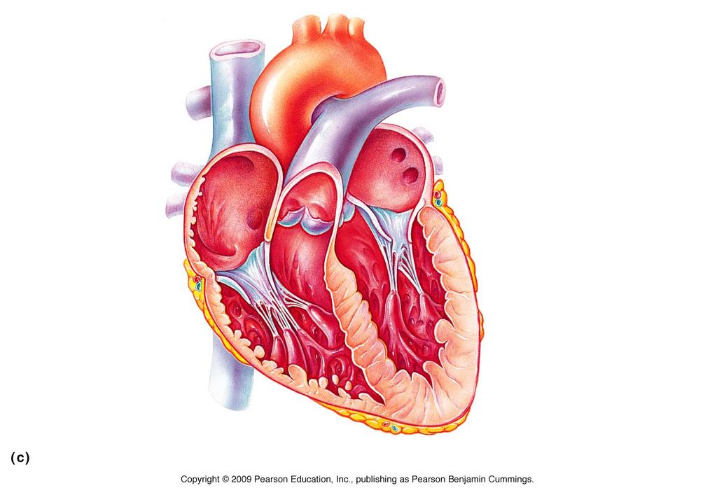
5 The Heart Figure 11.1c

6 The Heart Figure 11.2a
7 The Heart: Coverings Pericardium a double-walled sac Fibrous pericardium is loose and superficial Serous membrane is deep to the fibrous pericardium and composed of two layers Visceral pericardium Next to heart; also known as the epicardium Parietal pericardium Outside layer that lines the inner surface of the fibrous pericardium Serous fluid fills the space between the layers of pericardium
8 The Heart: Heart Wall Figure 11.2b
9 The Heart: Heart Wall Three layers Epicardium - Outside layer This layer is the visceral pericardium Connective tissue layer Myocardium - Middle layer Mostly cardiac muscle twisted and whorled into ringlike arrangement The layer that actually contracts Called the skeleton of the heart Endocardium - Inner layer Endothelium that lines heart chambers Continuous with blood vessels leaving & entering the heart
10 Homeostatic Imbalance: Pericarditis Inflammation of the pericardium Results in decrease of serous fluid Cause pericardial layers to stick together forming painful adhesions Interferes with heart movement

11 The Heart: Heart Wall Figure 11.2b
12 The Heart: Heart Wall Figure 11.2c
13 Review Questions 1 Where is the location of the heart in the thorax? What is the major function of the heart? What is the heart enclosed by? Name the two layers of the of the pericardial sac? Name the 3 layers of the heart wall What is and what causes pericarditis?
14 The Heart: Chambers Right and left side act as separate pumps Four hollow chambers 2 Atria Receiving chambers not important in pumping activity of heart 2 Ventricles Blood flows in from veins under low pressure Right atrium Left atrium Discharging chambers actual pumps of the heart Right ventricle Left ventricle
15 The Heart: Chambers Figure 11.2c

16 Differences in Right and Left Ventricles Figure 11.4
17 The Heart: Septa Septum divides the heart longitudinally Interventricular septum - Separates the two ventricles Interatrial septum - Separates the two atria
18 The Heart: Chambers Figure 11.2c
19 The Heart: Valves Allow blood to flow in only one direction to prevent backflow Four valves Atrioventricular (AV) valves between atria and ventricles Bicuspid (mitral) valve (left side of heart) Tricuspid valve (right side of heart) Anchored in place by chordae tendinae ( heart strings ) Open during heart relaxtion and closed during ventricular contrations
20 Valves Semilunar valves between ventricle and artery Pulmonary semilunar valve Aortic semilunar valve Closed during heart relaxation but open during ventricular contraction Notice these valves operate opposite of one another to force a one-way path of blood through the heart
21 **Homeostatic Imbalance Faulty Valves heart can function with leaky valves as long as damage is not too great Incompetent valve forces heart to pump and repump same blood because valves don t close properly & blood backflows Valvular stenosis valve flaps become stiff often because of repeated endocarditis (bacterial infection of the endocardium) Forces heart to contract more vigorously increasing workload Faulty valve replaced with synthetic valve, cryopreserved human valve, or chemically treated porcine valve (pig heart valve)
22 The Heart: Valves Figure 11.2c
 Figure 11.")
23 Operation of the AV valves Blood returning to the atria, puts pressure against AV valves; the AV valves are forced open Ventricles AV valves open (a) Figure 11.5a, step 1
 Figure 11.")
24 Operation of the AV valves Blood returning to the atria, puts pressure against AV valves; the AV valves are forced open As the ventricles fill, AV valve flaps hang limply into ventricles Ventricles AV valves open (a) Figure 11.5a, step 2
25 Operation of the AV valves Blood returning to the atria, puts pressure against AV valves; the AV valves are forced open As the ventricles fill, AV valve flaps hang limply into ventricles Atria contract, forcing additional blood into ventricles Ventricles AV valves open (a) Figure 11.5a, step 3
26 Ventricles contract, forcing blood against AV valve flaps (a) Figure 11.5a, step 4
27 Ventricles contract, forcing blood against AV valve flaps AV valves close (a) AV valves closed Figure 11.5a, step 5
28 Ventricles contract, forcing blood against AV valve flaps AV valves close Chordae tendineae tighten, preventing valve flaps from everting into atria (a) AV valves closed Figure 11.5a, step 6

 Semilunar valve open Figure 11.")
29 Operation of the semilunar valves Aorta Pulmonary trunk As ventricles contract and intraventricular pressure rises, blood is pushed up against semilunar valves, forcing them open (b) Semilunar valve open Figure 11.5b, step 1
 Semilunar valve open Semilunar valve closed Figure 11.")
30 Aorta Pulmonary trunk Operation of the semilunar valves As ventricles contract and intraventricular pressure rises, blood is pushed up against semilunar valves, forcing them open As ventricles relax, and intraventricular pressure falls, blood flows back from arteries, filling the leaflets of semilunar valves and forcing them to close (b) Semilunar valve open Semilunar valve closed Figure 11.5b, step 2
31 Systemic and Pulmonary Circulations Systemic circulation Blood flows from the left side of the heart through the body tissues and back to the right side of the heart Pulmonary circulation Blood flows from the right side of the heart to the lungs and back to the left side of the heart
32 Systemic and Pulmonary Circulations Figure 11.3
33 The Heart: Associated Great Vessels Arteries Aorta - Leaves left ventricle Pulmonary arteries - Leave right ventricle Veins Superior and inferior venae cavae - Enter right atrium Pulmonary veins (four) - Enter left atrium
34 The Heart: Associated Great Vessels Figure 11.2c
35 Blood Flow Through the Heart Superior and inferior venae cavae dump blood into the right atrium From right atrium, through the tricuspid valve, blood travels to the right ventricle From the right ventricle, blood leaves the heart as it passes through the pulmonary semilunar valve into the pulmonary trunk Pulmonary trunk splits into right and left pulmonary arteries that carry blood to the lungs
36 Blood Flow Through the Heart Oxygen is picked up and carbon dioxide is dropped off by blood in the lungs Oxygen-rich blood returns to the heart through the four pulmonary veins Blood enters the left atrium and travels through the bicuspid valve into the left ventricle From the left ventricle, blood leaves the heart via the aortic semilunar valve and aorta

37 Systemic and Pulmonary Circulations Figure 11.3
38 Coronary Circulation Blood in the heart chambers does not nourish the myocardium The heart has its own nourishing circulatory system consisting of Coronary arteries branch from the aorta to supply the heart muscle with oxygenated blood Cardiac veins drain the myocardium of blood Coronary sinus a large vein on the posterior of the heart, receives blood from cardiac veins Blood empties into the right atrium via the coronary sinus
39 ** Homeostatic Imbalance Angina Pectoris crushing chest pain caused by oxygen deprivation of the myocardium Infarct caused by prolonged oxygen deprivation where heart cells die Myocardial infarction heart attack or coronary

40 The Heart: Associated Great Vessels Figure 11.2c
41
42 Review Questions 3 1. Compare and contrast systemic versus pulmonary circulation. 2. Which side of the heart carries oxygenated blood? Deoxygenated blood? 3. What is the major artery leaving the heart? The major veins returning blood to the heart? 4. Where do the pulmonary vessels travel to & from? 5. In linear fashion list flow of blood with arrows? 6. How does the cardiac muscle tissue receive nourishment? 7. Compare and contrast angina pectoris with an infarct.
43 II. Physiology of the Heart Intrinsic conduction system (nodal system) Heart muscle cells contract, without nerve impulses, in a regular, continuous way
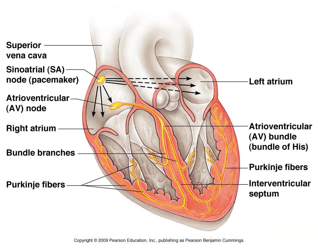
44 The Heart: Conduction System Special tissue sets the pace similar to cross between muscle & nervous tissue Sinoatrial node = SA node ( pacemaker ), is in the right atrium Atrioventricular node = AV node, is at the junction of the atria and ventricles Atrioventricular bundle = AV bundle (bundle of His), is in the interventricular septum Bundle branches are in the interventricular septum Purkinje fibers spread within the ventricle wall muscles
45 Heart Contractions Figure 11.6
46 Heart Contractions Contraction is initiated by the sinoatrial node (SA node) Sequential stimulation occurs at other autorhythmic cells Force cardiac muscle depolarization in one direction from atria to ventricles
47 Heart Contractions Once SA node starts the heartbeat Impulse spreads to the AV node Then the atria contract At the AV node, the impulse passes through the AV bundle, bundle branches, and Purkinje fibers Blood is ejected from the ventricles to the aorta and pulmonary trunk as the ventricles contract

48 Heart Contractions Figure 11.6
49 Heart Contractions Tachycardia rapid heart rate over 100 beats per minute Bradycardia slow heart rate less than 60 beats per minutes
50 **Homeostatic Imbalance Heart block when the AV node is damaged allowing the ventricles to beat at their own slower rate Ischemia lack of adequate blood supply which leads to fibrillation- rapid uncoordinated shudddering of heart muscles Pacemaker will need to be inserted to correct this condition.
51 The Heart: Cardiac Cycle In a healthy heart, atria contract simultaneously Atria relax, then ventricles contract Systole = contraction Diastole = relaxation
52 Filling Heart Chambers: Cardiac Cycle Left atrium Right atrium Left ventricle Right ventricle Ventricular filling Atrial contraction Isovolumetric Ventricular contraction phase ejection phase Isovolumetric relaxation Mid-to-late diastole (ventricular filling) Ventricular systole (atria in diastole) Early diastole Figure 11.7
 Figure 11.")
53 Filling Heart Chambers: Cardiac Cycle Left atrium Right atrium Left ventricle Right ventricle Ventricular filling Mid-to-late diastole (ventricular filling) Figure 11.7, step 1a
54 Filling Heart Chambers: Cardiac Cycle Left atrium Right atrium Left ventricle Right ventricle Ventricular filling Atrial contraction Mid-to-late diastole (ventricular filling) Figure 11.7, step 1b
 Ventricular systole (atria in diastole) Figure 11.")
55 Filling Heart Chambers: Cardiac Cycle Left atrium Right atrium Left ventricle Right ventricle Ventricular filling Atrial contraction Isovolumetric contraction phase Mid-to-late diastole (ventricular filling) Ventricular systole (atria in diastole) Figure 11.7, step 2a
 Ventricular systole (atria in diastole) Figure 11.")
56 Filling Heart Chambers: Cardiac Cycle Left atrium Right atrium Left ventricle Right ventricle Ventricular filling Atrial contraction Isovolumetric Ventricular contraction phase ejection phase Mid-to-late diastole (ventricular filling) Ventricular systole (atria in diastole) Figure 11.7, step 2b
57 Filling Heart Chambers: Cardiac Cycle Left atrium Right atrium Left ventricle Right ventricle Ventricular filling Atrial contraction Isovolumetric Ventricular contraction phase ejection phase Isovolumetric relaxation Mid-to-late diastole (ventricular filling) Ventricular systole (atria in diastole) Early diastole Figure 11.7, step 3
58 The Heart: Cardiac Cycle Cardiac cycle events of one complete heart beat Mid-to-late diastole blood flows from atria into ventricles Ventricular systole blood pressure builds before ventricle contracts, pushing out blood Early diastole atria finish refilling, ventricular pressure is low
59 Heart Sounds When using a stethoscope you hear two distinct sounds in a cardiac cycle. These heart sounds are described by two syllables lub and dup The lub is caused by the closing of the AV valves (bicuspid and tricuspid) The dup occurs when the semilunar valves close.
60 ** Homeostatic Imbalance - Murmurs Murmur abnormal or unusual heart sounds Fairly common in young children (and some elderly people) with perfectly healthy hearts probably due to thin heart walls. Outside of these groups a murmur may indicate a valve problem.
61 The Heart: Regulation of Heart Rate Increased heart rate Sympathetic nervous system Crisis stressors (physical or emotional trauma, high body temp.) Low blood pressure Hormones can also increase heart rate Epinephrine Thyroxine Exercise working muscles generate heat which increases heart rate by boosting metabolism
62 The Heart: Regulation of Heart Rate Decreased heart rate Parasympathetic nervous system slow and steady the heart giving it time to rest.

63 Cardiac Output Regulation Figure 11.8
64 ** Homeostatic Imbalance Congestive heart failure when the pumping efficiency of the heart is depressed so that circulation is inadequate to meet tissue needs. A progressive condition that may be caused by coronary atherosclerosis (blockage of coronary vessels with fatty buildup), persistent high blood pressure or multiple myocardial infarctions
65 Congestive Heart Failure (CHF) Each side can fail independently If the left side fails it is pulmonary congestion because the right side continues to send blood to the lungs but the left side can not discharge blood into systemic circuclation Lungs become swollen with blood, pressure increases and fluid leaks into lung tissue causing pulmonary edema If untreated the patient suffocates
66 Congestive Heart Failure (CHF) If the right side fails peripheral congestion occurs as blood backs up in the systemic circulation Edema is most noticeable in distal parts of the body; feet, ankles, and fingers become swollen and puffy Failure of one side puts a greater strain on the other side and eventually the whole heart fails
67 Heart Pop Quiz Word Bank Superior vena cava Inferior vena cava Right atrium Bicuspid valve Right ventricle Aortic Semilunar valve Right Pulmonary artery Left Pulmonary artery Right pulmonary vein Left pulmonary vein Left atrium Tricuspid valve Left ventricle Aorta Septum Pulmonary semilunar valve Pulmonary trunk
The Cardiovascular System
 Essentials of Human Anatomy & Physiology Elaine N. Marieb Slides 11.1 11.19 Seventh Edition Chapter 11 The Cardiovascular System Functions of the Cardiovascular system Function of the heart: to pump blood
The Cardiovascular System
 11 PART A The Cardiovascular System PowerPoint Lecture Slide Presentation by Jerry L. Cook, Sam Houston University ESSENTIALS OF HUMAN ANATOMY & PHYSIOLOGY EIGHTH EDITION ELAINE N. MARIEB The Cardiovascular
The Cardiovascular System (Heart)
") The Cardiovascular System The Cardiovascular System (Heart) A closed system of the heart and blood vessels The heart pumps blood Blood vessels allow blood to circulate to all parts of the body The function
The Cardiovascular System The Cardiovascular System (Heart) A closed system of the heart and blood vessels The heart pumps blood Blood vessels allow blood to circulate to all parts of the body The function
The Cardiovascular System
 The Cardiovascular System The Cardiovascular System A closed system of the heart and blood vessels The heart pumps blood Blood vessels allow blood to circulate to all parts of the body The function of
The Cardiovascular System
 Essentials of Human Anatomy & Physiology Elaine N. Marieb Seventh Edition Chapter 11 The Cardiovascular System Slides 11.1 11.19 Lecture Slides in PowerPoint by Jerry L. Cook The Cardiovascular System
The Heart. Happy Friday! #takeoutyournotes #testnotgradedyet
 The Heart Happy Friday! #takeoutyournotes #testnotgradedyet Introduction Cardiovascular system distributes blood Pump (heart) Distribution areas (capillaries) Heart has 4 compartments 2 receive blood (atria)
The HEART. What is it???? Pericardium. Heart Facts. This muscle never stops working It works when you are asleep
 This muscle never stops working It works when you are asleep The HEART It works when you eat It really works when you exercise. What is it???? Located between the lungs in the mid thoracic region Apex
The Cardiovascular System
 The Cardiovascular System The Manila Times College of Subic Prepared by: Stevens B. Badar, RN, MANc THE HEART Anatomy of the Heart Location and Size approx. the size of a person s fist, hollow and cone-shaped,
The Heart. The Heart A muscular double pump. The Pulmonary and Systemic Circuits
 C H A P T E R 19 The Heart The Heart A muscular double pump circuit takes blood to and from the lungs Systemic circuit vessels transport blood to and from body tissues Atria receive blood from the pulmonary
THE HEART OBJECTIVES: LOCATION OF THE HEART IN THE THORACIC CAVITY CARDIOVASCULAR SYSTEM
 BIOLOGY II CARDIOVASCULAR SYSTEM ACTIVITY #3 NAME DATE HOUR THE HEART OBJECTIVES: Describe the anatomy of the heart and identify and give the functions of all parts. (pp. 356 363) Trace the flow of blood
10. Thick deposits of lipids on the walls of blood vessels, called, can lead to serious circulatory issues. A. aneurysm B. atherosclerosis C.
 Heart Student: 1. carry blood away from the heart. A. Arteries B. Veins C. Capillaries 2. What is the leading cause of heart attack and stroke in North America? A. alcohol B. smoking C. arteriosclerosis
THE HEART. A. The Pericardium - a double sac of serous membrane surrounding the heart
 THE HEART I. Size and Location: A. Fist-size weighing less than a pound (250 to 350 grams). B. Located in the mediastinum between the 2 nd rib and the 5 th intercostal space. 1. Tipped to the left, resting
THE CARDIOVASCULAR SYSTEM. Part 1
 THE CARDIOVASCULAR SYSTEM Part 1 CARDIOVASCULAR SYSTEM Blood Heart Blood vessels What is the function of this system? What other systems does it affect? CARDIOVASCULAR SYSTEM Functions Transport gases,
Pearson's Comprehensive Medical Assisting Administrative and Clinical Competencies
 Pearson's Comprehensive Medical Assisting Administrative and Clinical Competencies THIRD EDITION CHAPTER 27 The Cardiovascular System Lesson 1: Overview of the Cardiovascular System Lesson Objectives Upon
2. right heart = pulmonary pump takes blood to lungs to pick up oxygen and get rid of carbon dioxide
 A. location in thorax, in inferior mediastinum posterior to sternum medial to lungs superior to diaphragm anterior to vertebrae orientation - oblique apex points down and to the left 2/3 of mass on left
The Cardiovascular System Part I: Heart Outline of class lecture After studying part I of this chapter you should be able to:
 The Cardiovascular System Part I: Heart Outline of class lecture After studying part I of this chapter you should be able to: 1. Describe the functions of the heart 2. Describe the location of the heart,
Chapter 14. The Cardiovascular System
 Chapter 14 The Cardiovascular System Introduction Cardiovascular system - heart, blood and blood vessels Cardiac muscle makes up bulk of heart provides force to pump blood Function - transports blood 2
Human Anatomy, First Edition
 Human Anatomy, First Edition McKinley & O'Loughlin Chapter 22 : Heart 1 Functions of the Heart Center of the cardiovascular system, the heart. Connects to blood vessels that transport blood between the
Ch 19: Cardiovascular System - The Heart -
 Ch 19: Cardiovascular System - The Heart - Give a detailed description of the superficial and internal anatomy of the heart, including the pericardium, the myocardium, and the cardiac muscle. Trace the
4. The two inferior chambers of the heart are known as the atria. the superior and inferior vena cava, which empty into the left atrium.
 Answer each statement true or false. If the statement is false, change the underlined word to make it true. 1. The heart is located approximately between the second and fifth ribs and posterior to the
Chapter 18 - Heart. I. Heart Anatomy: size of your fist; located in mediastinum (medial cavity)
") Chapter 18 - Heart I. Heart Anatomy: size of your fist; located in mediastinum (medial cavity) A. Coverings: heart enclosed in double walled sac called the pericardium 1. Fibrous pericardium: dense connective
Chapter 18 - Heart I. Heart Anatomy: size of your fist; located in mediastinum (medial cavity) A. Coverings: heart enclosed in double walled sac called the pericardium 1. Fibrous pericardium: dense connective
CV Anatomy Quiz. Dr Ella Kim Dr Pip Green
 CV Anatomy Quiz Dr Ella Kim Dr Pip Green Q1 The location of the heart is correctly described as A) lateral to the lungs. B) medial to the sternum. C) superior to the diaphragm. D) posterior to the spinal
The Cardiovascular System
 PowerPoint Lecture Slide Presentation by Patty Bostwick-Taylor, Florence-Darlington Technical College The Cardiovascular System 11PART A The Cardiovascular System A closed system of the heart and blood
the Cardiovascular System I
 the Cardiovascular System I By: Dr. Nabil A Khouri MD, MsC, Ph.D MEDIASTINUM 1. Superior Mediastinum 2. inferior Mediastinum Anterior mediastinum. Middle mediastinum. Posterior mediastinum Anatomy of
The Heart. Size, Form, and Location of the Heart. 1. Blunt, rounded point; most inferior part of the heart.
 12 The Heart FOCUS: The heart is composed of cardiac muscle cells, which are elongated, branching cells that appear striated. Cardiac muscle cells behave as a single electrical unit, and the highly coordinated
CIRCULATORY SYSTEM BLOOD VESSELS
 Name: Block: CIRCULATORY SYSTEM Multicellular organisms (above the level of roundworms) rely on a circulatory system to bring nutrients to, and take wastes away from, cells. In higher organisms such as
Figure ) The specific chamber of the heart that is indicated by letter A is called the. Diff: 1 Page Ref: 364
 The specific chamber of the heart that is indicated by letter A is called the. Diff: 1 Page Ref: 364") Essentials of Anatomy and Physiology, 9e (Marieb) Chapter 11 The Cardiovascular System Short Answer Figure 11.1 Using Figure 11.1, identify the following: 1) The Purkinje fibers are indicated by label.
Essentials of Anatomy and Physiology, 9e (Marieb) Chapter 11 The Cardiovascular System Short Answer Figure 11.1 Using Figure 11.1, identify the following: 1) The Purkinje fibers are indicated by label.
Approximately the size of your fist Location. Pericardial physiology
 Heart Anatomy Approximately the size of your fist Location Superior surface of diaphragm Left of the midline Anterior to the vertebral column, posterior to the sternum Wednesday, March 28, 2012 Muscle
Circulation. Circulation = is a process used for the transport of oxygen, carbon! dioxide, nutrients and wastes through-out the body
 Circulation Circulation = is a process used for the transport of oxygen, carbon! dioxide, nutrients and wastes through-out the body Heart = muscular organ about the size of your fist which pumps blood.
Unit 6: Circulatory System. 6.2 Heart
 Unit 6: Circulatory System 6.2 Heart Functions of Circulatory System 1. The heart is the pump necessary to circulate blood to all parts of the body 2. Arteries, veins and capillaries are the structures
The Heart and Cardiovascular System
 The Heart and Cardiovascular System What you will learn The location of the heart 3 layers and covering of the heart Explain the function of the heart as 2 separate pumps Identify the 4 chambers of the
Cardiovascular System. Heart Anatomy
 Cardiovascular System Heart Anatomy 1 The Heart Location & general description: Atria vs. ventricles Pulmonary vs. systemic circulation Coverings Walls The heart is found in the mediastinum, the medial
Anatomy of the Heart. Figure 20 2c
 Anatomy of the Heart Figure 20 2c Pericardium & Myocardium Remember, the heart sits in it s own cavity, known as the mediastinum. The heart is surrounded by the Pericardium, a double lining of the pericardial
The Cardiovascular System. Chapter 15. Cardiovascular System FYI. Cardiology Closed systemof the heart & blood vessels. Functions
 Chapter 15 Cardiovascular System FYI The heart pumps 7,000 liters (4000 gallons) of blood through the body each day The heart contracts 2.5 billion times in an avg. lifetime The heart & all blood vessels
Circulatory System Notes
 Circulatory System Notes Functions of Circulatory System A. Transports B. Transports C. Transports D. Transports E. of fluids F. G. Regulate temperature H. Blood clotting Characteristics of various blood
Chapter 20 (1) The Heart
 The Heart") Chapter 20 (1) The Heart Learning Objectives Describe the location and structure of the heart Describe the path of a drop of blood from the superior vena cava or inferior vena cava through the heart out
Chapter 20 (1) The Heart Learning Objectives Describe the location and structure of the heart Describe the path of a drop of blood from the superior vena cava or inferior vena cava through the heart out
Major Function of the Cardiovascular System. Transportation. Structures of the Cardiovascular System. Heart - muscular pump
 Structures of the Cardiovascular System Heart - muscular pump Blood vessels - network of tubes Blood - liquid transport vehicle brachiocephalic trunk superior vena cava right pulmonary arteries right pulmonary
The Heart and Heart Disease
 The Heart and Heart Disease Illustration of the heart by Leonardo DaVinci heart-surgeon.com/ history.html 2/14/2010 1 I. Location, Size and Position of the Heart A. Triangular organ located 1. of mass
Heart Anatomy. 7/5/02 Stephen G Davenport 1
 Heart Anatomy Copyright 1999, Stephen G. Davenport, No part of this publication may be reproduced, stored in a retrieval system, or transmitted, in any form without prior written permission. 7/5/02 Stephen
Cardiovascular System Notes: Physiology of the Heart
 Cardiovascular System Notes: Physiology of the Heart Interesting Heart Fact Capillaries are so small it takes ten of them to equal the thickness of a human hair. Review What are the 3 parts of the cardiovascular
Cardiovascular System Notes: Heart Disease & Disorders
 Cardiovascular System Notes: Heart Disease & Disorders Interesting Heart Facts The Electrocardiograph (ECG) was invented in 1902 by Willem Einthoven Dutch Physiologist. This test is still used to evaluate
Chapter 20: Cardiovascular System: The Heart
 Chapter 20: Cardiovascular System: The Heart I. Functions of the Heart A. List and describe the four functions of the heart: 1. 2. 3. 4. II. Size, Shape, and Location of the Heart A. Size and Shape 1.
Label Diagram #1 (Pg. 664)
") Chapter 18 The Cardiovascular System 18.1 Heart Anatomy The Pulmonary and Systemic Circuits Oxygen rich vs. Oxygen Poor Heart is a transport system consisting of two side-by-side pumps Right side receives
Chapter 18 The Cardiovascular System 18.1 Heart Anatomy The Pulmonary and Systemic Circuits Oxygen rich vs. Oxygen Poor Heart is a transport system consisting of two side-by-side pumps Right side receives
Blood Functions. Blood and the Cardiovascular System. Blood. Plasma. Erythrocytes (RBCs) Erythrocytes (RBCs) 4/7/2017
 Erythrocytes (RBCs) 4/7/2017") Blood Functions Blood and the Cardiovascular System Distribution Delivery of oxygen and nutrients to all body cells; Transport of wastes to lungs and excretory organs; Transport of hormones Regulation
Blood Functions Blood and the Cardiovascular System Distribution Delivery of oxygen and nutrients to all body cells; Transport of wastes to lungs and excretory organs; Transport of hormones Regulation
BIOLOGY 2060 LECTURE NOTES ANATOMY & PHYSIOLOGY II (A. IMHOLTZ) HEART P1 OF 5
 HEART P1 OF 5") BIOLOGY 2060 LECTURE NOTES ANATOMY & PHYSIOLOGY II (A. IMHOLTZ) HEART P1 OF 5 1. Heart Functions a. Generates pressure that propels blood thru blood vessels. (Tissue perfusion.) b. Separates oxygenated
BIOLOGY 2060 LECTURE NOTES ANATOMY & PHYSIOLOGY II (A. IMHOLTZ) HEART P1 OF 5 1. Heart Functions a. Generates pressure that propels blood thru blood vessels. (Tissue perfusion.) b. Separates oxygenated
Cardiovascular System
 Cardiovascular System The Heart Cardiovascular System The Heart Overview What does the heart do? By timed muscular contractions creates pressure gradients blood moves then from high pressure to low pressure
Cardiovascular system
 Cardiovascular system 1 Essential Question: How does the structure of the heart allow it to function in pumping blood? 2 Function? 3 Location of Heart 4 Heart coverings double sac of serous membrane pericardium
LECTURE 5. Anatomy of the heart
 LECTURE 5. Anatomy of the heart Main components of the CVS: Heart Blood circulatory system arterial compartment haemomicrocirculatory (=microvascular) compartment venous compartment Lymphatic circulatory
37 1 The Circulatory System
 H T H E E A R T 37 1 The Circulatory System The circulatory system and respiratory system work together to supply cells with the nutrients and oxygen they need to stay alive. a) The respiratory system:
Principles of Anatomy and Physiology
 Principles of Anatomy and Physiology 14 th Edition CHAPTER 20 The Cardiovascular System: The Heart Introduction The purpose of the chapter is to: 1. Learn about the components of the cardiovascular system
INTRODUCTORY REMARKS:
 INTRODUCTORY REMARKS: The circulatory system provides a way for the blood to be transported throughout the body. This provides nutrients to the cells and allows wastes to be removed. Open vs. Closed Circulatory
Cardiovascular System
 Cardiovascular System Purpose Transport oxygen and nutrients Take waste products away from tissues & organs Things we learned Blood pressure: the force of blood pushing against the walls of blood vessels
Unit 11 - The Cardiovascular System 1
 Unit 11 - The Cardiovascular System 1 I. Unit 11: The Cardiovascular System A. The Cardiovascular System 1. A closed system of the heart and blood vessels a) The heart pumps blood b) Blood vessels allow
CARDIOVASCULAR SYSTEM
 CARDIOVASCULAR SYSTEM Overview Heart and Vessels 2 Major Divisions Pulmonary Circuit Systemic Circuit Closed and Continuous Loop Location Aorta Superior vena cava Right lung Pulmonary trunk Base of heart
The Cardiovascular System: The Heart: Part A
 PowerPoint Lecture Slides prepared by Janice Meeking, Mount Royal College CHAPTER 18 The Cardiovascular System: The Heart: Part A Heart Anatomy Approximately the size of a fist Location In the mediastinum
- what other structures, besides the heart, does the mediastinum contain?
 Basic A & P II Dr. L. Bacha Chapter Outline (Martini & Nath 2010) An Introduction to the Cardiovascular System - read the paragraphs under this heading on page 580 The Heart is a Four Chambered Organ describe
Ch.15 Cardiovascular System Pgs {15-12} {15-13}
 Ch.15 Cardiovascular System Pgs {15-12} {15-13} E. Skeleton of the Heart 1. The skeleton of the heart is composed of rings of dense connective tissue and other masses of connective tissue in the interventricular
Function: Transportation of. Oxygen Nutrients Waste Hormones gases
 Function: Transportation of Oxygen Nutrients Waste Hormones gases Pericardium: double sac of serous membrane filled with fluid (pericardial fluid to be exact) that surrounds the heart. Parietal pericardium:
AP2 Lab 3 Coronary Vessels, Valves, Sounds, and Dissection
 AP2 Lab 3 Coronary Vessels, Valves, Sounds, and Dissection Project 1 - BLOOD Supply to the Myocardium (Figs. 18.5 &18.10) The myocardium is not nourished by the blood while it is being pumped through the
Health Science 20 Circulatory System Notes
 Health Science 20 Circulatory System Notes Functions of the Circulatory System The circulatory system functions mainly as the body s transport system. It transports: o Oxygen o Nutrients o Cell waste o
All About the Heart. Structures of the heart. Layers. Chambers
 All About the Heart Your heart is a muscle. It is slightly larger than your fist and weighs less than a pound. It is located to the left of the middle of your chest. Your heart pumps blood to the lungs
Chapter 11. The Cardiovascular System
 Chapter 11 The Cardiovascular System The Cardiovascular System The Cardiovascular System A closed system of the heart and blood vessels Heart pumps blood Blood vessels circulate blood to all parts of the
Heart Dissection. 5. Locate the tip of the heart or the apex. Only the left ventricle extends all the way to the apex.
 Heart Dissection Page 1 of 6 Background: The heart is a four-chambered, hollow organ composed primarily of cardiac muscle tissue. It is located in the center of the chest in between the lungs. It is the
THE HEART. Unit 3: Transportation and Respiration
 THE HEART Unit 3: Transportation and Respiration The Circulatory System Also called the Cardiovascular System Circulates blood in the body Transports nutrients, oxygen, carbon dioxide, hormones, and blood
11/10/2014. Muscular pump Two atria Two ventricles. In mediastinum of thoracic cavity 2/3 of heart's mass lies left of midline of sternum
 It beats over 100,000 times a day to pump over 1,800 gallons of blood per day through over 60,000 miles of blood vessels. During the average lifetime, the heart pumps nearly 3 billion times, delivering
Anatomy of the Heart
 Biology 212: Anatomy and Physiology II Anatomy of the Heart References: Saladin, KS: Anatomy and Physiology, The Unity of Form and Function 8 th (2018). Required reading before beginning this lab: Chapter
The cardiovascular system is composed of the heart and blood vessels that carry blood to and from the body s organs. There are 2 major circuits:
 1 The cardiovascular system is composed of the heart and blood vessels that carry blood to and from the body s organs. There are 2 major circuits: pulmonary and systemic. The pulmonary goes out to the
1. What kind of blood is found in the rt. atrium? (oxygenated or deoxygenated)
") Carl Christennsen, PhD Chap. 19, 20, & 21 - Circulatory System Bio. 2304 Human Anatomy HEART 1. What kind of blood is found in the rt. atrium? (oxygenated or deoxygenated) Where does this blood come from?
Carl Christennsen, PhD Chap. 19, 20, & 21 - Circulatory System Bio. 2304 Human Anatomy HEART 1. What kind of blood is found in the rt. atrium? (oxygenated or deoxygenated) Where does this blood come from?
Cardiovascular System
 Cardiovascular System I. Structure of the Heart A. Average adult heart is 14 cm long and 9 cm wide. B. Lies in the mediastinum. C. Enclosed in the pericardium. 1. Fibrous pericardium- Outer, tough connective
Test Review Circulatory System Chapters
 Test Review Circulatory System Chapters 13-2010 1. The tissue that forms the tight fitting sac around the heart is the a. parietal pericardium c. myocardium b. visceral pericardium d. endocardium 2. Which
The Cardiovascular System
 C H A P T E R 1 4 The Cardiovascular System OBJECTIVES After studying this chapter, you should be able to: 1. Describe how the heart is positioned in the thoracic cavity. 2. List and describe the layers
Biology Unit 3 The Human Heart P
 Biology 2201 Unit 3 The Human Heart P 314-321 Structure and Function of the Human Heart Structure of the Human Heart Has four Chambers (2 Atria and 2 Ventricles) Made of Cardiac Muscle Found in Chest Cavity
Cardiovascular System. I. Structures of the heart A. : Pericardium sack that surrounds the heart
 Cardiovascular System I. Structures of the heart A. : Pericardium sack that surrounds the heart 1. : Pericardial Cavity serous fluid filled space between the heart and the pericardium B. Heart Wall 1.
Collin County Community College. ! BIOL Anatomy & Physiology! WEEK 5. The Heart
 Collin County Community College! BIOL. 2402 Anatomy & Physiology! WEEK 5 The Heart 1 (1578-1657) A groundbreaking work in the history of medicine, English physician William Harvey s Anatomical Essay on
Lab 16. The Cardiovascular System Heart and Blood Vessels. Laboratory Objectives
 Lab 16 The Cardiovascular System Heart and Blood Vessels Laboratory Objectives Describe the anatomical structures of the heart to include the pericardium, chambers, valves, and major vessels. Describe
BIOLOGY 2060 LECTURE NOTES ANATOMY & PHYSIOLOGY II (A. IMHOLTZ) HEART P1 OF 7
 HEART P1 OF 7") BIOLOGY 2060 LECTURE NOTES ANATOMY & PHYSIOLOGY II (A. IMHOLTZ) HEART P1 OF 7 1. Heart a. Generates the pressure that propels blood thru blood vessels. b. Separates oxygenated and deoxygenated blood separate.
BIOLOGY 2060 LECTURE NOTES ANATOMY & PHYSIOLOGY II (A. IMHOLTZ) HEART P1 OF 7 1. Heart a. Generates the pressure that propels blood thru blood vessels. b. Separates oxygenated and deoxygenated blood separate.
The Heart. Made up of 3 different tissue: cardiac muscle tissue, nerve tissue, and connective tissue.
 The Heart The Heart Made up of 3 different tissue: cardiac muscle tissue, nerve tissue, and connective tissue. Your heart pumps with a regular beat (Heart Rate) Your heart rate can change depending on
Heart. Structure Physiology of blood pressure and heartbeat
 Heart Structure Physiology of blood pressure and heartbeat Location and Anatomy Location and Anatomy Pericardial cavity: surrounds, isolates, and anchors heart Parietal pericardium lined with serous membrane
Human Anatomy and Physiology Chapter 19 Worksheet 1- The Heart
 Human Anatomy and Physiology Chapter 19 Worksheet 1- The Heart Name Date Period 1. The "double pump" function of the heart includes the right side, which serves as the circuit pump, while the left side
Unit 11: The Cardiovascular System
 Unit 11: The Cardiovascular System I. The Cardiovascular System A. A closed system of the heart and blood vessels 1. The heart pumps blood 2. Blood vessels allow blood to circulate to all parts of the
A closed system of the heart/blood. Function: The heart pumps blood. Blood vessels allow blood to circulate throughout the body
 A closed system of the heart/blood The heart pumps blood It is no more than a transportation pump Blood vessels allow blood to circulate throughout the body MILES of blood vessels intricate network At
STRUCTURES OF THE CARDIOVASCULAR SYSTEM
 STRUCTURES OF THE CARDIOVASCULAR SYSTEM CARDIOVASCULAR SYSTEM Also called the circulatory system Consists of the heart, arteries, veins, and capillaries Main function is to pump/circulate oxygenated blood
CIRCULATORY SYSTEM TASK CARDS Worksheet
 CIRCULATORY SYSTEM TASK CARDS Worksheet Name: Date: Instructions: Put the answers to each task card in the numbered boxes on the chart. 1 a) left semilunar valve / aortic valve b) blood would backflow
Chapter 11. The Cardiovascular System. Clicker Questions Pearson Education, Inc.
 Chapter 11 The Cardiovascular System Clicker Questions Oxygen-poor blood is pumped through the venae cavae to the right side of the heart, and then through the pulmonary arteries to the lungs and back
Cardiovascular Anatomy Dr. Gary Mumaugh
 Cardiovascular Anatomy Dr. Gary Mumaugh Location of Heart Approximately the size of your fist Location o Superior surface of diaphragm o Left of the midline in mediastinum o Anterior to the vertebral column,
The Mammalian Circulatory System
 The Mammalian Heart The Mammalian Circulatory System Recall: What are the 3 cycles of the mammalian circulatory system? What are their functions? What are the three main vessel types in the mammalian circulatory
10/23/2017. Muscular pump Two atria Two ventricles. In mediastinum of thoracic cavity 2/3 of heart's mass lies left of midline of sternum
 It beats over 100,000 times a day to pump over 1,800 gallons of blood per day through over 60,000 miles of blood vessels. During the average lifetime, the heart pumps nearly 3 billion times, delivering
The Circulatory System. Lesson Overview. Lesson Overview The Circulatory System
 33.1 THINK ABOUT IT More than one-third of the 1.2 million Americans who suffer a heart attack each year die. This grim evidence shows that the heart and the circulatory system it powers are vital to life.
PART ONE General Characteristics of the Cardiovascular System
 HUMAN ANATOMY & PHYSIOLOGY CARDIOVASCULAR SYSTEM Chapter 15 Notes OBJECTIVES HOLE S HA&P CHAPTER FIFTEEN 1. Discuss the functions of the organs of the cardiovascular system. 2. Distinguish between the
Chapter 20 THE CARDIOVASCULAR SYSTEM: THE HEART
 Chapter 20 THE CARDIOVASCULAR SYSTEM: THE HEART INTRODUCTION A. The cardiovascular system consists of the blood, heart, and blood vessels. B. The heart is the pump that circulates the blood through an
If I Only Had a Brain
 If I Only Had a Brain A Heart. (The Nerve!) Regions of the Brain Cerebral hemisphere Diencephalon Cerebellum (b) Adult brain Brain stem Regions of the Brain: Cerebrum Precentral gyrus Frontal lobe Central
Cardiac Cycle. Each heartbeat is called a cardiac cycle. First the two atria contract at the same time.
 The Heartbeat Cardiac Cycle Each heartbeat is called a cardiac cycle. First the two atria contract at the same time. Next the two ventricles contract at the same time. Then all the chambers relax. http://www.youtube.com/watch?v=frd3k6lkhws
Part 1. Copyright 2011 Pearson Education, Inc. Figure Copyright 2011 Pearson Education, Inc.
 PowerPoint Lecture Slides prepared by Leslie Hendon University of Alabama, Birmingham C H A P T E R The Heart 19 Part 1 The Heart A muscular double pump circuit vessels transport blood to and from the
Lab Activity 23. Cardiac Anatomy. Portland Community College BI 232
 Lab Activity 23 Cardiac Anatomy Portland Community College BI 232 Cardiac Muscle Histology Branching cells Intercalated disc: contains many gap junctions connecting the adjacent cell cytoplasm, creates
Heart. Heart 2-Tunica media: middle layer (media ='middle') muscle fibers (smooth or cardiac).
 muscle fibers (smooth or cardiac).") t. innermost lumenal General Circulatory system heart and blood vessels walls have 3 layers (inside to outside) 1-Tunica interna: aka tunica intima layer--lumenal layer epithelium--endothelium simple squamous
t. innermost lumenal General Circulatory system heart and blood vessels walls have 3 layers (inside to outside) 1-Tunica interna: aka tunica intima layer--lumenal layer epithelium--endothelium simple squamous
Cardiovascular System- Heart. Miss Wheeler Unit 8
 Cardiovascular System- Heart Miss Wheeler Unit 8 Overview CARDIOVASCULAR SYSTEM heart vessels Made up of heart, blood vessels, and blood Functions Heart- pump blood Vessels- (veins, arteries, capillaries)
The Circulatory System
 The Circulatory System Key Questions What are the functions of the circulatory system? How does the heart pump blood through the body? What are three types of blood vessels? Vocabulary myocardium atrium
Cardiovascular system
 BIO 301 Human Physiology Cardiovascular system The Cardiovascular System: consists of the heart plus all the blood vessels transports blood to all parts of the body in two 'circulations': pulmonary (lungs)
1. Which of the following blood vessels has a thin elastic layer? A. Aorta. B. Pulmonary artery. C. Posterior vena cava. D. Mesenteric capillary.
 CIRCULATORY SYSTEM 1. Which of the following blood vessels has a thin elastic layer? A. Aorta. B. Pulmonary artery. C. Posterior vena cava. D. Mesenteric capillary. 2. Capillary beds are equipped with
Section 5.1 The heart and heart disease
 Section 5.1 The heart and heart disease Mammals are too large to rely on diffusion. They need a circulatory system to move substances around the body. Blood moves down pressure gradients, from high to