Endoleaks after endovascular aneurysm repair lead to nonuniform intra-aneurysm sac pressure.
|
|
|
- Amos Hodges
- 5 years ago
- Views:
Transcription
1 Endoleaks after endovascular aneurysm repair lead to nonuniform intra-aneurysm sac pressure. Dias, Nuno; Ivancev, Krassi; Resch, Tim; Malina, Martin; Sonesson, Björn Published in: Journal of Vascular Surgery DOI: /j.jvs Published: Link to publication Citation for published version (APA): Dias, N., Ivancev, K., Resch, T., Malina, M., & Sonesson, B. (2007). Endoleaks after endovascular aneurysm repair lead to nonuniform intra-aneurysm sac pressure. Journal of Vascular Surgery, 46(2), DOI: /j.jvs General rights Copyright and moral rights for the publications made accessible in the public portal are retained by the authors and/or other copyright owners and it is a condition of accessing publications that users recognise and abide by the legal requirements associated with these rights. Users may download and print one copy of any publication from the public portal for the purpose of private study or research. You may not further distribute the material or use it for any profit-making activity or commercial gain You may freely distribute the URL identifying the publication in the public portal L UNDUNI VERS I TY PO Box L und
2 Take down policy If you believe that this document breaches copyright please contact us providing details, and we will remove access to the work immediately and investigate your claim. Download date: 10. Jul. 2018


3 LU:research Institutional Repository of Lund University This is an author produced version of a paper published in Journal of vascular surgery. This paper has been peerreviewed but does not include the final publisher proofcorrections or journal pagination. Citation for the published paper: Dias, Nuno V and Ivancev, Krassi and Resch, Timothy A and Malina, Martin and Sonesson, Bjorn. "Endoleaks after endovascular aneurysm repair lead to nonuniform intra-aneurysm sac pressure." J Vasc Surg, 2007, Vol: 46, Issue: 2, pp Access to the published version may require journal subscription. Published with permission from: Elsevier
4 1 Endoleaks after endovascular aneurysm repair lead to non-uniform intra-aneurysm sac pressure Nuno V. Dias, Krassi Ivancev, Timothy A. Resch, Martin Malina, Björn Sonesson Department of Vascular Diseases Malmö-Lund and Endovascular Centre, Malmö University Hospital, Lunds University, Malmö, Sweden Corresponding Author: Nuno V. Dias Dept Vascular Diseases Malmö-Lund Malmö University Hospital Entrance 59 2nd floor Malmö Sweden Telephone: Fax:
5 2 Abstract Objective: to study intra-aneurysm sac pressures in patients who developed endoleaks after endovascular repair (EVAR) of abdominal aortic aneurysms (AAA). Methods: 25 patients with endoleaks (18 men, 7 women; 76 (68-80) years old) underwent 31 direct intra-aneurysm sac pressure measurements (DISP) 16 (14-26) months after EVAR (AAA diameter 59 (52-67) mm). Six patients underwent DISP twice. Tip-pressure sensors were used through direct translumbar puncture of the AAA except in three patients (transabdominal puncture in 2 and endoluminal in 1). Mean pressure index (MPI) was calculated between simultaneously registered intra-aneurysm sac and systemic pressures. Values presented are medians with interquartile range between parentheses. Results: Type I EL (n=1) showed MPI of 93% in the nidus and 62% in the thrombus. Type II ELs were associated with lower MPIs in the thrombus (35(24-38)%) when AAAs shrank (n=4), compared to when the aneurysms remained unchanged (n=11, MPI 78 (47-85)%) or expanded (n=6, MPI 74 (58-87)%, P=.019). The nidus of type II EL (MPI 79 (70-90)%) had higher pressure than the thrombus (45 (34-85)%, P=.047, n=7). Successful embolization of type II ELs led to AAA shrinkage (n=3, MPI reduction to 13-31%) or diameter stability (n=3, unchanged MPIs of 37-44%). Type III ELs (n=3) had MPIs in the thrombus of 33%-70%. Conclusions: Endoleaks after EVAR pressurize the AAA-sac non-uniformly with higher, near-systemic, pressure in the EL nidus comparing with the thrombus. Nevertheless, type
6 3 II EL in shrinking AAAs have lower intra-sac pressure than expanding or stable aneurysms and successful EL embolization reduces pressure. Introduction Endovascular aneurysm repair (EVAR) of abdominal aortic aneurysms (AAA) is successful when the aneurysm-sac is completely excluded. 1 Type I and III endoleaks are considered treatment failures given their association with adverse clinical outcomes. 1 In contrast, the importance of type II endoleaks is controversial. On the one hand they tend to seal spontaneously and are frequently associated with aneurysm stability or shrinkage, 2, 3 but on the other hand, aneurysm expansion, and even sporadic rupture, has been reported with type II endoleaks. 4, 5 Follow-up after EVAR has focused on the evaluation of endoleaks and AAA size. Imaging techniques allow assessment of flow within the aneurysm sac detecting contrast enhancement and/or Doppler signals. However, intra-sac pressure after EVAR is only indirectly evaluated by these imaging methods by assessing AAA size changes. The intra-aneurysm sac pressure is one of the main determinants of the tension applied on the AAA wall. A high or low tension will lead to aneurysm expansion or shrinkage, respectively. We have previously verified this relationship in the absence of endoleaks by performing direct intra-aneurysm sac pressure measurements (DISP). 6, 7 Pressure has also been measured in patients with endoleaks, but mostly in type II endoleaks and shortly after EVAR. 8, 9 The aim of this study was to evaluate intra-aneurysm sac pressures in patients who developed endoleaks after EVAR of AAA).
7 4 Methods Patients 553 patients underwent EVAR of AAA at our tertiary university center since Twenty-five of these patients (18 men, 7 women; 76 (68-80) years old) underwent 31 DISP 16 (14-26) months after EVAR (figure 1). The median duration of DISP was 174 ( ) minutes. DISP was performed in 21 patients while a type II endoleak was present or had been embolized. Three patients had type III endoleaks at the time of DISP and one patient had a type I endoleak. Inclusion criteria for the study were based on anatomic suitability for direct sac puncture and the presence of an endoleak after EVAR or the status after endoleak embolization. Patients were grouped according to the type of endoleak or status after its embolization (table I). Our initial experience with DISP in 8 of these patients has been included in a previous report. 7 The study was approved by the local ethics committee and all patients gave informed consent before the procedure. Imaging The imaging protocols included contrast-enhanced spiral-computer tomography (CT) scans preoperatively and yearly after EVAR. More frequent CT-scans were performed when indicated. Preoperative CT scans included two spiral scans, i.e., before and after intravenous non-ionic iodinated contrast injection. Postoperative CTs had an additional delayed spiral scan. CT scans were reconstructed with mm axial slices. An additional CT-scan was obtained the month before DISP (pre-disp CT-scan).
8 5 The shortest transverse diameter of the AAA was measured at its widest portion on axial CT-scans by the same observer. Diameter changes were calculated to express the diameter evolution before DISP (Diameter at the time of DISP Initial diameter). 7 A 5 mm cutoff was used for grouping AAAs into shrinking ( -5 mm), unchanged and expanding ( 5 mm). 1 At the time of DISP a digital subtraction aortogram was performed. This included selective angiography of the superior mesenteric and hypogastric arteries whenever the origin of the type II endoleak was not clearly established by CT-scan and non-selective aortography. Endoleaks were classified according to the recommended reporting standards. 1 Endoleak nidus was defined as the contrast filled area within the aneurysm sac. A pressure measurement was considered within the endoleak nidus when this location was verified by contrast injection and free blood flow was obtained from the needle. Pressure measurement system and DISP technique Anatomical suitability was assessed in the pre-disp CT-scan and defined as the possibility of inserting a pressure sensor in the AAA-sac without damaging any viscera or jeopardizing stent-graft integrity. The pressure measurement system and DISP technique have been previously described in detail. 6, 7, 10 Wired tip-pressure sensors mounted on.014-inch guide-wires were used (PressureWire4, RADI Medical AB, Uppsala, Sweden). The pressure guide-wire used for intra-aneurysm sac pressure measurements had a shorter tip (1 mm instead of 3 cm) in order to allow precise placement of the sensor. Systemic pressure was measured in the
9 6 lumen of the stent-graft. Access to the AAA-sac was obtained by translumbar puncture using fluoroscopic guidance in 28 occasions (including the 6 late embolization controls). Two DISP were done through direct transabdominal ultrasound-guided AAA puncture. During the direct puncture of the AAA-sac efforts were made to pin-point the endoleak nidus (figure 2-A). Measurements in the thrombus were made when the sensor was located approximately mid-way between the nidus and the stent-graft. In one occasion, the pressure guide-wire was passed through a coaxial catheter placed in the ostium of the inferior mesenteric artery via the superior mesenteric artery. The system recorded systemic and AAA-sac pressures simultaneously. Intra-aneurysm sac pressures were analyzed as systolic, diastolic, mean and pulse pressures. Mean pressure index (MPI) was calculated as the percentage of the mean intra-aneurysmal pressure relative to the simultaneous mean systemic pressure. MPI = mean AAA pressure / mean systemic pressure * 100 Measurements were only considered of good quality if the drift in recalibration of the pressure sensor did not exceed 5 mm Hg in the end of the measurements. DISP has been previously validated in patients with a median intraobserver variability of MPI of 0 (-7 17)% , 11 The sensor was also tested in vitro with an accuracy better than 2 mm Hg. Endoleak embolization Embolization of the endoleak was performed once access to the endoleak nidus or its feeding vessels was achieved (figure 2-B). The embolizing materials used were coils,
10 7 radioopaque glue (Histoacryl, Braun, Tuttlingen, Germany and Lipiodol, Laboratoire Guerbet, Aulnay-Sous-Bois, France) and/or gel-foam sponge (Spongostan Standard, Johnsson & Johnsson Medical Limited, UK). Late embolization control was defined as DISP being performed on a different occasion than the embolization. Statistical analysis Normal distribution was not assumed. Values are presented as medians and interquartile range (IQR) between parentheses, if not stated otherwise. Non-parametric exact tests were used for paired and unpaired comparisons. Results were considered significant when P <.05. Statistical analysis was done using SPSS (SPSS Inc, Chicago, USA).
11 8 Results Type I endoleak One patient (81 years old male, AAA diameter of 55 mm) with a proximal type I endoleak and unchanged AAA diameter (0 mm) underwent DISP 15 months after EVAR. MPI and pulse pressure were, respectively, 62% and 21 mm Hg in the thrombus and 93% and 66 mm Hg in the nidus. The AAA expanded 9 mm in the 28 months following DISP until the patient consented a reintervention Type II endoleaks Nineteen patients (12 men; table I) underwent 21 DISP while a type II endoleak was present. Two patients underwent DISP twice, since no embolization was performed on the first occasion and the aneurysms subsequently expanded (6 mm after 8 months and 11 mm after 11 months, respectively). At the time of the pressure measurement 4 AAAs had decreased in size, 11 remained unchanged and 6 had expanded. MPI, intra-aneurysm mean and pulse pressures were significantly lower in the thrombus of shrinking AAAs when compared to AAAs that expanded or remained unchanged in size (P of.019,.030 and.019, respectively; table II). Pressure in the thrombus and nidus of type II endoleaks Pressure was measured within the thrombus and inside the endoleak nidus during the same puncture in 7 patients (figure 3). Pressure was significantly higher inside the endoleak nidus (MPI of 79 (70-90)% and pulse pressure of 22 (18-57) mm Hg) than
12 9 within the thrombus (MPI of 45 (34-85)% and pulse pressure of 11 (7-21) mm Hg; P =.043 and P=.028, respectively). The median MPI difference between the nidus and the thrombus was 32 (4-44)%. Type II endoleak embolization Thirteen patients underwent embolization of type II endoleaks at the time of DISP. Glue was used in 12 cases and coils and gel-foam particles in one. Twelve of the 13 procedures were considered successful and the AAA diameter changed 0 (-7 2) mm during the following 13 (6-37) months. In 5 of these patients pressure was measured immediately after the embolization at the same procedure. MPI changed from 70 (57-84)% before the embolization to 53 (30-79)% immediately after (not significant). Intra-aneurysm sac mean and pulse pressure changed from 86 (56-98) and 13 (12-19) mm Hg before the embolization to, respectively, 56 (31-111) and 12 (8-40) mm Hg after the embolization (not significant in both). There was no association between the final MPI and the AAA diameter change afterwards. One embolization was unsuccessful since the endoleak persisted at the end of the procedure. This patient was considered unfit for any further reintervention and the aneurysm continued to expand (19 mm during the following 26 months). Late control of type II endoleak embolization Six patients (5 men, 1 women; 74 (70-79) years old; table I) underwent DISP 16 (6-31) months after successful embolization of type II endoleaks.
13 10 Three patients with MPIs of 31% or less at the late embolization control showed AAA shrinkage. The MPIs were lower after the embolization (table III). Three patients with unchanged AAA diameter after the embolization of the type II endoleak had MPIs between 37% and 44% at the late embolization control. The MPIs had not changed significantly at the late control comparing to before the embolization (table III). Type III endoleaks Three patients (3 men, table I) underwent DISP in the presence of a type III endoleak. One patient with a small type III endoleak persisting after EVAR underwent DISP 2 months after the EVAR. MPIs ranged from 70% close to the endoleak to 42% in the periphery of the AAA-sac. The endoleak sealed spontaneously afterwards and the AAA diameter shrank by 12 mm. The other two patients underwent DISP when late type III endoleaks were identified (one partial separation of the components of an aortic uni-iliac system and one small fabric disruption) and the AAAs remained unchanged in diameter (+2 and +1 mm, respectively). Intra-sac pressure within the thrombus decreased with increasing distance to the endoleak (MPIs ranging from 57 to 35% and 44 to 33%, respectively). No measurements were performed in the endoleak nidus. One patient refused any further reinterventions and died within one month of unknown cause. The other one showed a 6 mm expansion of the AAA in the 4 months following DISP and was converted to open repair.
14 11 Discussion The aim of EVAR is the exclusion of the aneurysm sac from blood flow and pressure. AAA size and endoleak status have been the key points in the evaluation of the treatment success. However, intra-aneurysm sac pressure has, until recently, been mostly indirectly assessed. The use of tip pressure sensors for direct intra-aneurysm sac pressure measurement after EVAR has been previously validated. 6, 7, 10 Shrinking AAAs without endoleaks were shown to have lower intra-aneurysm sac pressure within the thrombus compared to expanding aneurysms. 7 Furthermore, DISP was able to predict future AAA diameter changes in the absence of endoleaks. However, the effect of endoleaks after EVAR in intra-sac pressure is incompletely understood, especially considering the varied clinical outcome associated with the different types of endoleaks. The results of this study show that the endoleak nidus (channel) has consistently higher pressure than the intra-sac thrombus. Type II endoleaks seem to be associated with varying intra-sac pressures in the thrombus relating to the AAA diameter changes. Moreover, type II endoleaks appear to be dynamic since the variation can occur within the same patient at different time points. Successful embolization of a type II endoleak leads to depressurization of the AAA-sac with shrinkage or stabilization of the aneurysm diameter. Endoleaks of type I and III usually lead to a pressurization of the AAA-sac, as it was anticipated. DISP in one patient with a type I endoleak showed a high pulsatile intra-aneurysm sac pressure in the thrombus and even higher and near-systemic in the nidus. This was followed by AAA expansion and reinforces the perception of these endoleaks as treatment failures.
15 12 Type II endoleaks were associated with varying degrees of intra-sac pressurization. The previously reported difference in intra-sac pressure between shrinking and expanding AAAs without endoleaks 7 was also verified in type II endoleaks. However, AAA shrinkage with a type II endoleak was associated with higher intra-sac pressures in the thrombus than what was previously seen in AAA shrinkage without endoleaks (MPI of 35% and 19%, respectively). 7 Reports in the literature have consistently shown near-systemic pressure in patients with type II endoleaks in the early follow-up after EVAR. 8, 9 However, these measurements were performed mainly within the endoleak nidus and, as our results show, there is a pressure difference between the EL nidus and the AAA thrombus. The difference in pressure between the nidus and the thrombus seems to be dependent on several factors including the size of the endoleak nidus, 12, 13 its flow and perfusion pressure and thrombus composition. In our experience some cases displayed a discrepancy in the endoleak size as seen on the CT-scan and on the aneurysmogram (with contrast injection into the nidus). The composition and structural properties of the thrombus, and their dependency on the proximity of blood circulation, may also influence the transmission of pressure. 17 Furthermore, the amplitude of the pressure gradient measured between the nidus and the thrombus may also be influenced by the distance between the measurement locations. This varying distribution of sac pressure in patients with endoleaks, although consistently higher in expanding aneurysms, may question the reliability of systems based on single-spot pressure measurements such as implantable pressure sensors. 18 Recent literature report the use of implantable devices in the intra-operative identification
16 13 of endoleaks 19 and in canine models of type II endoleaks. 20 However, the location of the pressure transducer in relation to the endoleak nidus may influence the measurements obtained with those devices. For instance, if the pressure transducer is placed within the endoleak nidus, measurements can be high without necessarily implying AAA expansion. Type II endoleaks seem to be a dynamic entity since many seal spontaneously early after EVAR. This dynamic character of type II endoleaks, the difference in intra-sac pressure between patients with and without endoleaks and the above mentioned pressure gradient between the nidus and thrombus, suggest the need for a cautious interpretation of intrasac pressure in patients with endoleaks. Direct percutaneous puncture of the AAA allows not only intra-sac pressure measurement but also embolization of type II endoleaks. This is a significant advantage, as translumbar embolization has been shown to be more efficient 9, than endoluminal methods Successful type II endoleak embolization was associated with shrinkage or stabilization of the aneurysm diameter. The diameter stability may be partially explained by the incompressibility of the embolic material injected into the sac. Intra-aneurysm sac pressure appears, nevertheless, to decrease upon successful embolization. In contrast to others 8 we did not find this pressure reduction directly after the embolization but only later during follow-up. It is possible that by performing our initial pressure measurement later in the follow-up after EVAR we have measured a more selected group of patients where the endoleaks that sealed spontaneously in the early follow-up hade been excluded. In conclusion, the endoleak nidus had high, near-systemic pressure while the degree of thrombus pressurization varied. Thus, pressure is not uniformly distributed through the
17 14 AAA sac. Shrinking AAAs with type II endoleaks had lower pressure than those with expanding or unchanged AAA diameter. Successful endoleak embolization leads to a delayed depressurization of the AAA sac. Type I and III endoleaks are associated with high intra-sac pressures.
18 15 Acknowledgments We are very grateful to Kristina Lindholm, PhD and Christel Ekberg-Jönsson for their assistance.
19 16 References 1. Chaikof EL, Blankensteijn JD, Harris PL, White GH, Zarins CK, Bernhard VM, et al. Reporting standards for endovascular aortic aneurysm repair. J Vasc Surg 2002;35: Resch T, Ivancev K, Lindh M, Nyman U, Brunkwall J, Malina M, et al. Persistent collateral perfusion of abdominal aortic aneurysm after endovascular repair does not lead to progressive change in aneurysm diameter. J Vasc Surg 1998;28: Rhee RY, Eskandari MK, Zajko AB, Makaroun MS. Long-term fate of the aneurysmal sac after endoluminal exclusion of abdominal aortic aneurysms. J Vasc Surg 2000;32: White RA, Donayre C, Walot I, Stewart M. Abdominal aortic aneurysm rupture following endoluminal graft deployment: report of a predictable event. J Endovasc Ther 2000;7: van Marrewijk CJ, Fransen G, Laheij RJ, Harris PL, Buth J. Is a type II endoleak after EVAR a harbinger of risk? Causes and outcome of open conversion and aneurysm rupture during follow-up. Eur J Vasc Endovasc Surg 2004;27: Sonesson B, Dias N, Malina M, Olofsson P, Griffin D, Lindblad B, Ivancev K. Intra-aneurysm pressure measurements in successfully excluded abdominal aortic aneurysm after endovascular repair. J Vasc Surg 2003;37: Dias NV, Ivancev K, Malina M, Resch T, Lindblad B, Sonesson B. Intraaneurysm sac pressure measurements after endovascular aneurysm repair: differences
20 17 between shrinking, unchanged, and expanding aneurysms with and without endoleaks. J Vasc Surg 2004;39: Baum RA, Carpenter JP, Cope C, Golden MA, Velazquez OC, Neschis DG, et al. Aneurysm sac pressure measurements after endovascular repair of abdominal aortic aneurysms. J Vasc Surg 2001;33: Rial R, Serrano Fj F, Vega M, Rodriguez R, Martin A, Mendez J, et al. Treatment of type II endoleaks after endovascular repair of abdominal aortic aneurysms: translumbar puncture and injection of thrombin into the aneurysm sac. Eur J Vasc Endovasc Surg 2004;27: Dias NV, Ivancev K, Malina M, Hinnen JW, Visser M, Lindblad B, Sonesson B. Direct intra-aneurysm sac pressure measurement using tip-pressure sensors: In vivo and in vitro evaluation. J Vasc Surg 2004;40: Kälvesten E, Smith L, Tenerz L, Stemme G. The first surface micromachined pressure sensor for cardiovascular pressure measurements. Proceedings of the 11th Annual International Workshop on Micro-Electro-Mechanical Systems, Heidelberg, Germany 1998: Timaran CH, Ohki T, Rhee SJ, Veith FJ, Gargiulo NJ, 3rd, Toriumi H, et al. Predicting aneurysm enlargement in patients with persistent type II endoleaks. J Vasc Surg 2004;39: Timaran CH, Ohki T, Veith FJ, Lipsitz EC, Gargiulo NJ, 3rd, Rhee SJ, et al. Influence of type II endoleak volume on aneurysm wall pressure and distribution in an experimental model. J Vasc Surg 2005;41:
21 Arko FR, Filis KA, Siedel SA, Johnson BL, Drake AR, Fogarty TJ, et al. Intrasac flow velocities predict sealing of type II endoleaks after endovascular abdominal aortic aneurysm repair. J Vasc Surg 2003;37: Parent FN, Meier GH, Godziachvili V, LeSar CJ, Parker FM, Carter KA, et al. The incidence and natural history of type I and II endoleak: a 5-year follow-up assessment with color duplex ultrasound scan. J Vasc Surg 2002;35: Li Z, Kleinstreuer C. Computational analysis of type II endoleaks in a stented abdominal aortic aneurysm model. J Biomech 2006;39: Vallabhaneni SR, Gilling-Smith GL, Brennan JA, Heyes RR, Hunt JA, How TV, et al. Can Intrasac Pressure Monitoring Reliably Predict Failure of Endovascular Aneurysm Repair? Journal of Endovascular Therapy 2003;10: Ellozy SH, Carroccio A, Lookstein RA, Jacobs TS, Addis MD, Teodorescu VJ, et al. Abdominal aortic aneurysm sac shrinkage after endovascular aneurysm repair: correlation with chronic sac pressure measurement. J Vasc Surg 2006;43: Ohki T, Ouriel K, Silveira PG, Katzen B, White R, Criado F, et al. Initial results of wireless pressure sensing for endovascular aneurysm repair: The APEX Trial-- Acute Pressure Measurement to Confirm Aneurysm Sac EXclusion. J Vasc Surg 2007;45: Chaer RA, Trocciola S, Derubertis B, Hynecek R, Xu Q, Lam R, et al. Evaluation of the accuracy of a wireless pressure sensor in a canine model of retrogradecollateral (type II) endoleak and correlation with histologic analysis. J Vasc Surg 2006;44:
22 Baum RA, Carpenter JP, Golden MA, Velazquez OC, Clark TW, Stavropoulous SW, et al. Treatment of type 2 endoleaks after endovascular repair of abdominal aortic aneurysms: comparison of transarterial and translumbar techniques. J Vasc Surg 2002;35: Martin ML, Dolmatch BL, Fry PD, Machan LS. Treatment of type II endoleaks with Onyx. J Vasc Interv Radiol 2001;12: Schmid R, Gurke L, Aschwanden M, Stierli P, Jacob AL. CT-guided percutaneous embolization of a lumbar artery maintaining a type II endoleak. J Endovasc Ther 2002;9: Kasirajan K, Matteson B, Marek JM, Langsfeld M. Technique and results of transfemoral superselective coil embolization of type II lumbar endoleak. J Vasc Surg 2003;38: Haulon S, Tyazi A, Willoteaux S, Koussa M, Lions C, Beregi JP. Embolization of type II endoleaks after aortic stent-graft implantation: Technique and immediate results. J Vasc Surg 2001;34: Karch LA, Henretta JP, Hodgson KJ, Mattos MA, Ramsey DE, McLafferty RB, et al. Algorithm for the diagnosis and treatment of endoleaks. Am J Surg 1999;178: Chuter TA, Faruqi RM, Sawhney R, Reilly LM, Kerlan RB, Canto CJ, et al. Endoleak after endovascular repair of abdominal aortic aneurysm. J Vasc Surg 2001;34:
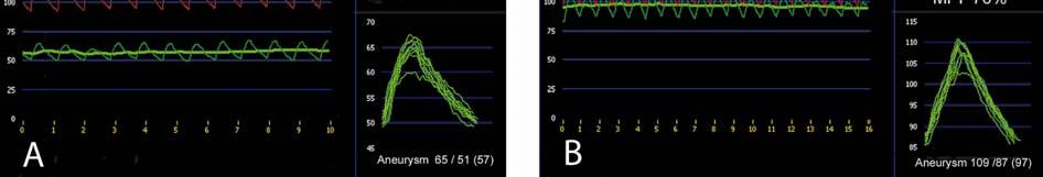
23 20 Figure Legends: Figure 1 Patients undergoing direct intra-aneurysm sac pressure measurements (DISP) according to the type of endoleak and AAA diameter change. Six patients underwent DISP after successful embolization of type II endoleaks. *Two patients with type II endoleaks underwent DISP twice. Initially after AAA diameter shrinkage and thereafter once the AAA had expanded. Figure 2 A Digital subtraction angiography with contrast injection inside a type II endoleak nidus (arrow heads) through a translumbar puncture of the AAA at the time of direct intra-aneurysm sac pressure measurement. Two lumbar arteries are filled (arrows). B Status after embolization of the endoleak with Hystoacril-Lipiodol. The glue fills the endoleak nidus and the proximal part of two lumbar arteries (arrow). Figure 3 Recording from direct intra-aneurysm pressure measurement. A Measurement within the thrombus. B Measurement in the endoleak nidus. Red curves represent the aortic pressure, and green curves represent the intra-aneurysm-sac pressure. Each figure has three tracings. The large one to the left is the tracing obtained during each recording where the aortic and intra-aneurysm sac pressures are registered simultaneously. The pressure recorded during 10 heart cycles is gathered separately in two different tracings, one for the aortic (upper smaller tracing to the right) and the other intra-aneurysm sac pressures (lower smaller tracing to the right). The system calculates also the mean of the systolic, diastolic and mean pressures for both the aortic and intra-
24 21 sac pressures (values bellow the small tracings). In the smaller tracings the scale is adjusted by the software to the amplitude of the curves.
25 Table I Characteristics of patients Group No. of Age AAA Ø Time between EVAR and patients (mm) DISP (mo) Type I EL Type II EL 19* 77 (68-81) 62 (53-73) 16 (14-22) Embolization control after 6 74 (70-79) 58 (53-62) 30 (20-48) type II EL Type III EL 3 73 (64-75) 55 (52-62) 41 (2-97) *Two patients of the 19 patients with type II endoleaks were measured twice. The first time after diameter shrinkage and thereafter once the diameter had expanded. Four of these patients were also measured before the embolization of the endoleak and are, therefore, also included in type II endoleak group (ie, 2 patients were measured only after a previous embolization of the endoleak). Values are shown as median with interquertile range between parenthesis when not stated otherwise. AAA Ø AAA diameter; EL endoleak, DISP direct intra aneurysm-sac pressure measurement.
26 Table II Intra-aneurysm sac pressure in the presence of type II endoleaks. Diameter change before DISP N Follow-up before DISP (mo) Ø AAA (mm) AAA mean pressure (mm Hg) AAA pulse pressure (mm Hg) MPI (%) Shrinking 4 19 (7-43) -6 (-9, -5) 40 (24-47) * 7 (6-10) 35 (25-38) Unchanged (13-16) 2 (0, 3) 86 (53-97) * 21 (12-22) 78 (47-85) Expanding 6 22 (20-31) 7 (6,10) 76 (59-109) * 17 (11-31) 74 (58-87) Grouping was done according to the AAA diameter change. *P =.019, P =.030, P =.019. N number of DISP performed. Ø diameter change; MPI Mean Pressure Index.
27 Table III Pressure in six patients undergoing late endoleak embolization control Patient MPI before MPI after Intra-aneurysm sac pressure Follow-up Ø AAA after embolization embolization mm Hg after embolization (%) (%) Systolic Diastolic Mean embolization (mm) (mo) NA NA The follow-up after the embolization includes even the imaging follow-up after the second DISP. MPI Mean Pressure Index; Ø diameter change.


28 Figure 1 Figure 2

29 Figure 3
Description. Section: Surgery Effective Date: April 15, Subsection: Surgery Original Policy Date: December 6, 2012 Subject:
 Last Review Status/Date: March 2015 Page: 1 of 6 Description Wireless sensors implanted in an aortic aneurysm sac after endovascular repair are being investigated to measure post procedural pressure. It
Intra-aneurysm pressure measurements in successfully excluded abdominal aortic aneurysm after endovascular repair
 Intra-aneurysm pressure measurements in successfully excluded abdominal aortic aneurysm after endovascular repair Björn Sonesson, MD, a Nuno Dias, MD, b Martin Malina, MD, a PerÅke Olofsson, c Dennis Griffin,
Treatment options for endoleaks: stents, embolizations and conversions
 Treatment options for endoleaks: stents, embolizations and conversions Poster No.: C-0861 Congress: ECR 2012 Type: Authors: Keywords: DOI: Scientific Exhibit G. Lombardi; napoli/it Arteries / Aorta, Abdomen,
Aneurysm sac pressure monitoring: Does the direction of pressure measurement matter in fibrinous thrombus?
 Aneurysm sac pressure monitoring: Does the direction of pressure measurement matter in fibrinous thrombus? Jan-Willem Hinnen, MD, a Daniel J. Rixen, PhD, b Olivier H. Koning, MD, a Hajo J. Van Bockel,
Intrasac flow velocities predict sealing of type II endoleaks after endovascular abdominal aortic aneurysm repair
 Intrasac flow velocities predict sealing of type II endoleaks after endovascular abdominal aortic aneurysm repair Frank R. Arko, MD, Konstantinos A. Filis, MD, PhD, Scott A. Siedel, MD, Bonnie L. Johnson,
Effect of type II endoleaks and antiplatelet therapy on abdominal aortic aneurysm shrinkage after endovascular repair
 Effect of type II endoleaks and antiplatelet therapy on abdominal aortic aneurysm shrinkage after endovascular repair Atsushi Aoki, MD, a Takanori Suezawa, MD, a Kenji Sangawa, MD, b and Mamoru Tago, MD,
Approaches to type II Endoleaks: Transcaval, transarterial, translumbar. Saher Sabri,MD University of Virginia
 Approaches to type II Endoleaks: Transcaval, transarterial, translumbar Saher Sabri,MD University of Virginia Saher Sabri, M.D. Speakers Bureau: W.L.Gore & Associates, Abbott Type 2 Endoleaks after EVAR
An Overview of Post-EVAR Endoleaks: Imaging Findings and Management. Ravi Shergill BSc Sean A. Kennedy MD Mark O. Baerlocher MD FRCPC
 An Overview of Post-EVAR Endoleaks: Imaging Findings and Management Ravi Shergill BSc Sean A. Kennedy MD Mark O. Baerlocher MD FRCPC Disclosure Slide Mark O. Baerlocher: Current: Consultant for Boston
Effectiveness of coiling in the treatment of endoleaks after endovascular repair
 From the Society for Clinical Vascular Surgery Effectiveness of coiling in the treatment of endoleaks after endovascular repair Maureen K. Sheehan, MD, a Joel Barbato, MD, a Christopher N. Compton, MD,
Mechanism of failure in the treatment of type II endoleak with percutaneous coil embolization
 Mechanism of failure in the treatment of type II endoleak with percutaneous coil embolization Maurice M. Solis, MD, a Juan Ayerdi, MD, a Gregory A. Babcock, MD, b Jose R. Parra, MD, a Robert B. McLafferty,
Type-II Endoleaks Following Endovascular AAA Repair: Preoperative Predictors and Long-term Effects
 503 VASCULAR FELLOWS FORUM 2001, FIRST PLACE Type-II Endoleaks Following Endovascular AAA Repair: Preoperative Predictors and Long-term Effects Frank R. Arko, MD; Geoffrey D. Rubin, MD; Bonnie L. Johnson,
Length Measurements of the Aorta After Endovascular Abdominal Aortic Aneurysm Repair
 Eur J Vasc Endovasc Surg 18, 481 486 (1999) Article No. ejvs.1999.0882 Length Measurements of the Aorta After Endovascular Abdominal Aortic Aneurysm Repair J. J. Wever, J. D. Blankensteijn, I. A. M. J.
Embolization for type 2 endoleak with sac expansion after endovascular repair of abdominal aortic aneurysm: safety and effectiveness
 DOI 10.1186/s40064-016-1934-x RESEARCH Open Access Embolization for type 2 endoleak with sac expansion after endovascular repair of abdominal aortic aneurysm: safety and effectiveness Kenji Kajiwara 1*,
Faculty Disclosure. Glue, Particulates, Thrombin, Coils and the Kitchen Sink for Type II Endoleak Management. Background.
 Glue, Particulates, Thrombin, Coils and the Kitchen Sink for Type II Endoleak Management Faculty Disclosure I disclose the following financial relationships: UCSF Vascular Symposium 2013 Receive grant/research
Management of Endoleaks
 Management of Endoleaks Sarah Ikponmwosa, MD Brooklyn VA 6/20/08 Questions Advantages of endovascular repair Definition of an endoleak Types of endoleaks Management of type lll endoleak Diagnosis of type
Percutaneous Transabdominal Approach for the Treatment of Endoleaks after Endovascular Repair of Infrarenal Abdominal Aortic Aneurysm
 Percutaneous Transabdominal Approach for the Treatment of Endoleaks after Endovascular Repair of Infrarenal Abdominal Aortic Aneurysm Sun Young Choi, MD 1 Jong Yun Won, MD 2 Do Yun Lee, MD 1 Donghoon Choi,
An endoleak is radiographic or ultrasonic evidence
 Complex Coil Embolization of Multiple Type II Endoleaks Liquid embolics, detachable coils, and plugs to repair an enlarging abdominal aortic aneurysm sac 5 years after EVAR. BY FRANK R. ARKO, MD; ABRAHAM
Duplex ultrasound factors predicting persistent type II endoleak and increasing AAA sac diameter after EVAR
 From the Society for Clinical Vascular Surgery Duplex ultrasound factors predicting persistent type II endoleak and increasing AAA sac diameter after EVAR Brian R. Beeman, MD, Kathy Murtha, RVT, Kevin
Type II Endoleak Embolization Choice of Materials: EVOH, Glue, Thrombin & Coils. Michael S. Rosenberg, MD Assistant Professor of Radiology
 Type II Endoleak Embolization Choice of Materials: EVOH, Glue, Thrombin & Coils Michael S. Rosenberg, MD Assistant Professor of Radiology Michael Rosenberg, M. D. No relevant financial relationship reported
Technique and results of transfemoral superselective coil embolization of type II lumbar endoleak
 Technique and results of transfemoral superselective coil embolization of type II lumbar endoleak Karthikeshwar Kasirajan, MD, Brian Matteson, MD, John M. Marek, MD, and Mark Langsfeld, MD, Albuquerque,
Use of cine magnetic resonance angiography in quantifying aneurysm pulsatility associated with endoleak
 From the Society for Clinical Vascular Surgery Use of cine magnetic resonance angiography in quantifying aneurysm pulsatility associated with endoleak Peter L. Faries, MD, a Gautam Agarwal, MD, b Robert
Disclosures. EVAR follow-up: actual recommendation. EVAR follow-up: critical issues
 Disclosures is it time to discuss individualized follow-up schemes based on preoperative anatomy and high quality completion angiography? Consultant / Speaker / Proctor Cook Cordis Medtronic Invatec W.L.
Increased Flexibility of AneuRx Stent-Graft Reduces Need for Secondary Intervention Following Endovascular Aneurysm Repair
 583 Increased Flexibility of AneuRx Stent-Graft Reduces Need for Secondary Intervention Following Endovascular Aneurysm Repair Frank R. Arko, MD; W. Anthony Lee, MD; Bradley B. Hill, MD; Paul Cipriano,
Type II endoleak after endovascular abdominal aortic aneurysm repair: A conservative approach with selective intervention is safe and cost-effective
 From the American Association for Vascular Surgery Type II endoleak after endovascular abdominal aortic aneurysm repair: A conservative approach with selective intervention is safe and cost-effective Eric
CLINICAL RESEARCH STUDIES
 CLINICAL RESEARCH STUDIES From the Southern Association for Vascular Surgery First experience in human beings with a permanently implantable intrasac pressure transducer for monitoring endovascular repair
Eccentric stent graft compression: An indicator of insecure proximal fixation of aortic stent graft
 Eccentric stent graft compression: An indicator of insecure proximal fixation of aortic stent graft Yehuda G. Wolf, MD, Bradley B. Hill, MD, W. Anthony Lee, MD, Christine M. Corcoran, RN, MS, Thomas J.
Selective Inferior Mesenteric Artery Embolization during Endovascular Abdominal Aortic Aneurysm Repair to Prevent Type II Endoleak
 Kobe J. Med. Sci., Vol. 63, No. 5, pp. E130-E135, 2017 Selective Inferior Mesenteric Artery Embolization during Endovascular Abdominal Aortic Aneurysm Repair to Prevent Type II Endoleak TETSUYA FUKUDA
Computed Tomography versus Magnetic Resonance Imaging of Endoleaks after EVAR
 Eur J Vasc Endovasc Surg 32, 361e365 (2006) doi:10.1016/j.ejvs.2006.02.011, available online at http://www.sciencedirect.com on Computed Tomography versus Magnetic Resonance Imaging of Endoleaks after
Persistent type 2 endoleak after endovascular repair of abdominal aortic aneurysm is associated with adverse late outcomes
 CLINICAL RESEARCH STUDIES Persistent type 2 endoleak after endovascular repair of abdominal aortic aneurysm is associated with adverse late outcomes John E. Jones, MD, Marvin D. Atkins, MD, David C. Brewster,
Abdominal Aortic Aneurysm (AAA)
") Abdominal Aortic Aneurysm (AAA) Vascular Workshop: Objectives Anatomy Keith VanHaltren Indications Technique Cases Abdominal Aorta: Normal Size Abdominal aortic aneurysm: Definition Normal diameter of
Abdominal Aortic Aneurysm (AAA) Vascular Workshop: Objectives Anatomy Keith VanHaltren Indications Technique Cases Abdominal Aorta: Normal Size Abdominal aortic aneurysm: Definition Normal diameter of
Nellix Endovascular System: Clinical Outcomes and Device Overview
 Nellix Endovascular System: Clinical Outcomes and Device Overview Jeffrey P. Carpenter, MD Professor and Chief, Department of Surgery CAUTION: Investigational device. This product is under clinical investigation
Changes in aneurysm volume after endovascular repair of abdominal aortic aneurysm
 Changes in aneurysm volume after endovascular repair of abdominal aortic aneurysm Yehuda G. Wolf, MD, a Manfred Tillich, MD, b W. Anthony Lee, MD, a Thomas J. Fogarty, MD, a Christopher K. Zarins, MD,
PAPER. Morphologic Changes and Outcome Following Endovascular Abdominal Aortic Aneurysm Repair as a Function of Aneurysm Size
 PAPER Morphologic Changes and Outcome Following Endovascular Abdominal Aortic Aneurysm Repair as a Function of Aneurysm Size Frank R. Arko, MD; Konstantinos A. Filis, MD; Bradley B. Hill, MD; Thomas J.
Chapter 8. Aortic compliance following EVAR and the. influence of different endografts: Determination using dynamic MRA
 Aortic compliance following EVAR and the influence of different endografts: Determination using dynamic MRA J.A. van Herwaarden B.E. Muhs K.L. Vincken J. van Prehn A. Teutelink L.W. Bartels F.L. Moll H.J.M.
Treatment options of late failures of EVAS. Michel Reijnen Rijnstate Arnhem The Netherlands
 Treatment options of late failures of EVAS Michel Reijnen Rijnstate Arnhem The Netherlands Disclosure Speaker name: Michel Reijnen I have the following potential conflicts of interest to report: Consulting
- to discuss the limits of traditional treatment options of type II endoleak after endovascular aneurysms repair (EVAR);
;") Transgluteal echo-guided arterial access: an unusual approach to treat type II endoleak following endovascular repair of an aortic and internal iliac artery aneurysm. Poster No.: C-0824 Congress: ECR 2014
Transgluteal echo-guided arterial access: an unusual approach to treat type II endoleak following endovascular repair of an aortic and internal iliac artery aneurysm. Poster No.: C-0824 Congress: ECR 2014
Endovascular aneurysm repair (EVAR) is universally accepted as an
 is universally accepted as an") Diagn Interv Radiol 2012; 18:307 313 Turkish Society of Radiology 2012 INTERVENTIONAL RADIOLOGY ORIGINAL ARTICLE Risk factors for the development of persistent type II endoleaks after endovascular repair
Diagn Interv Radiol 2012; 18:307 313 Turkish Society of Radiology 2012 INTERVENTIONAL RADIOLOGY ORIGINAL ARTICLE Risk factors for the development of persistent type II endoleaks after endovascular repair
ndovascular abdominal aortic aneurysm repair (EVAR) has been established as a safe and effective
 has been established as a safe and effective") Vascular and Interventional Radiology Original Research Mursalin et al. Imaging-Based Predictors of Persistent Type II Endoleak After EVAR Vascular and Interventional Radiology Original Research Rafael
Vascular and Interventional Radiology Original Research Mursalin et al. Imaging-Based Predictors of Persistent Type II Endoleak After EVAR Vascular and Interventional Radiology Original Research Rafael
Morphological Study of Abdominal Aortic Aneurysm: Optimal Stent-graft Size for Japanese Patients
 Original Article Morphological Study of Abdominal Aortic Aneurysm: Optimal Stent-graft Size for Japanese Patients Hirofumi Midorikawa, MD, Tomohiro Ogawa, MD, Kouichi Satou, MD, and Shunichi Hoshino, MD
Endoleak Sealing after AAA Endovascular Repair. When and How?
 Endoleak Sealing after AAA Endovascular Repair. When and How? Poster No.: C-1086 Congress: ECR 2013 Type: Educational Exhibit Authors: D. Quintana Blanco, B. González Humara, E. Torres Diez, C. Jimenez
Educational Exhibit Authors:
 Endoleaks in Abdominal Aortic Aneurysm Endoprosthesis: What radiologists need to know about Diagnostic, Characterization and Basic Management Strategies Poster No.: C-0150 Congress: ECR 2013 Type: Educational
From 1996 to 1999, a total of 1,193 patients with
 THE ANEURX CLINICAL TRIAL AT 8 YEARS Lessons learned following the US AneuRx clinical trial from 1996 to 2004. BY CHRISTOPHER K. ZARINS, MD From 1996 to 1999, a total of 1,193 patients with infrarenal
Long-term follow-up of type II endoleak embolization reveals the need for close surveillance
 From the Society for Vascular Surgery Long-term follow-up of type II endoleak embolization reveals the need for close surveillance Timur P. Sarac, MD, Connor Gibbons, Lina Vargas, MD, Jane Liu, MD, Sunita
Management of Endoleaks. Michael Meuse, M.D Vascular and Interventional Radiology 12/14/09
 Management of Endoleaks Michael Meuse, M.D Vascular and Interventional Radiology 12/14/09 Endoleak Failure to totally exclude the abdominal aortic aneurysm (AAA) from continued perfusion and pressurization
Prognosis in patients with type 2 endoleak after endovascular repair of abdominal aortic aneurysm
 Prognosis in patients with type 2 endoleak after endovascular repair of abdominal aortic aneurysm D.A. Lier a, T.D.G. Nyheim b, J.J. Jørgensen b,c, A.H. Krog b,c a Faculty of Medicine, University of Oslo,
Efficacy of ultrasound scan contrast agents in the noninvasive follow-up of aortic stent grafts
 Efficacy of ultrasound scan contrast agents in the noninvasive follow-up of aortic stent grafts Phillip J. Bendick, PhD, Paul G. Bove, MD, Graham W. Long, MD, Gerald B. Zelenock, MD, O. William Brown,
MODERN METHODS FOR TREATING ABDOMINAL ANEURYSMS AND THORACIC AORTIC DISEASE
 MODERN METHODS FOR TREATING ABDOMINAL ANEURYSMS AND THORACIC AORTIC DISEASE AAA FACTS 200,000 New Cases Each Year Ruptured AAA = 15,000 Deaths per Year in U.S. 13th Leading Cause of Death 80% Chance of
Iliac fixation inhibits migration of both suprarenal and infrarenal aortic endografts
 From the Society for Vascular Surgery Iliac fixation inhibits migration of both suprarenal and infrarenal aortic endografts Peyman Benharash, MD, Jason T. Lee, MD, Oscar J. Abilez, MD, Tami Crabtree, MS,
Case Report Early and Late Endograft Limb Proximal Migration with Resulting Type 1b Endoleak following an EVAR for Ruptured AAA
 Hindawi Case Reports in Vascular Medicine Volume 2017, Article ID 4931282, 5 pages https://doi.org/10.1155/2017/4931282 Case Report Early and Late Endograft Limb Proximal Migration with Resulting Type
The Natural History of Type 2 Endoleaks after EVAR Justifies Conservative Management
 Research Article imedpub Journals www.imedpub.com Journal of Vascular and Endovascular Surgery DOI: 10.21767/2573-4482.100067 The Natural History of Type 2 Endoleaks after EVAR Justifies Conservative Management
Surgery for Acquired Cardiovascular Disease
 Resch et al Changes in aneurysm morphology and stent-graft configuration after endovascular repair of aneurysms of the descending thoracic aorta Timothy Resch, MD, PhD a Bansi Koul, MD, PhD d Nuno V. Dias,
SANWICH TECHNIQUE TO REDUCE COMPLICATIONS WHEN TREATING BILATERAL INTERNAL ILIAC ARTERY
 SANWICH TECHNIQUE TO REDUCE COMPLICATIONS WHEN TREATING BILATERAL INTERNAL ILIAC ARTERY TRAN TRA GIANG.MD Interventional cardiovascular department Hanoi Heart Hospital, Hanoi, Viet Nam Nothing to Disclose
Right Choice for Right Angles
 Right Choice for Right Angles The Anatomy of Technology Aorfix gives you technology that conforms to patient anatomy, optimising both procedure and post-operative performance. Fishmouth for optimum neck
Transcaval embolization as an alternative technique for the treatment of type II endoleak after endovascular aortic aneurysm repair
 VASCULAR AND ENDOVASCULAR TECHNIQUES Peter F. Lawrence, MD, Section Editor Transcaval embolization as an alternative technique for the treatment of type II endoleak after endovascular aortic aneurysm repair
Initial experience characterizing a type I endoleak from velocity profiles using time-resolved three-dimensional phase-contrast MRI
 Initial experience characterizing a type I endoleak from velocity profiles using time-resolved three-dimensional phase-contrast MRI Thomas A. Hope, MD, a Christopher K. Zarins, MD, b and Robert J. Herfkens,
Treatment of type 2 endoleaks after endovascular repair of abdominal aortic aneurysms: Comparison of transarterial and translumbar techniques
 Treatment of type 2 endoleaks after endovascular repair of abdominal aortic aneurysms: Comparison of transarterial and translumbar techniques Richard A. Baum, MD, a Jeffrey P. Carpenter, MD, b Michael
Improved results using Onyx glue for the treatment of persistent type 2 endoleak after endovascular aneurysm repair
 From the Eastern Vascular Society Improved results using Onyx glue for the treatment of persistent type 2 endoleak after endovascular aneurysm repair Christopher J. Abularrage, MD, a,b Virendra I. Patel,
Endovascular Treatment of Type II Endoleak Following TEVAR for Thoracic Aortic Aneurysm: Squeeze Technique to Reach the Aneurysmal Sac
 Endovascular Treatment of Type II Endoleak Following TEVAR for Thoracic Aortic Aneurysm: Squeeze Technique to Reach the Aneurysmal Sac Chang Won Kim Department of Radiology Pusan National University Hospital
3. Endoluminal Treatment of Infrarenal Abdominal Aortic Aneurysm
 3. Endoluminal Treatment of Infrarenal Abdominal Aortic Aneurysm Hence J. M. Verhagen, Geoffrey H. White, Tom Daly and Theodossios Perdikides A 78-year-old male was referred for investigation and management
FEVAR FIFTEEN YEARS OF EFFICIENCY E.DUCASSE MD PHD FEBVS CHU DE BORDEAUX
 FEVAR FIFTEEN YEARS OF EFFICIENCY E.DUCASSE MD PHD FEBVS CHU DE BORDEAUX 2018 A BIT OF HISTORY First use of F-EVAR : 1990s Park et al. J Vasc Interv Radiol. 1996;7:819-823. Faruqi et al. J Endovasc Surg.
Outcomes of original and low-permeability Gore Excluder endoprosthesis for endovascular abdominal aortic aneurysm repair
 From the Society for Vascular Surgery Outcomes of original and low-permeability Gore Excluder endoprosthesis for endovascular abdominal aortic aneurysm repair William Tanski, III, MD, and Mark Fillinger,
150 Aortic Aneurysm - Recent Advances is the occurrence and significance of endoleaks. White et al were the first to systematically describe and class
 Chapter 8 Endovascular Treatment of Endoleaks Following EVAR Zaiping Jing, Qingsheng Lu, Jiaxuan Feng and Jian Zhou Additional information is available at the end of the chapter http://dx.doi.org/10.5772/54836
Aortic compliance following EVAR and the influence of different endografts: determination using dynamic MRA.
 3/1/2007 o/06-3928 muss diss pag 61 5 Aortic compliance following EVAR and the influence of different endografts: determination using dynamic MRA. Joost A. van Herwaarden, MD 1,3 ;BartE.Muhs,MD 1 ; Koen
Anatomical challenges in EVAR
 Anatomical challenges in EVAR M.H. EL DESSOKI, MD,FRCS PROFESSOR OF VASCULAR SURGERY CAIRO UNIVERSITY Disclosure Speaker name:... I have the following potential conflicts of interest to report: Consulting
Thrombus within an aortic aneurysm does not reduce pressure on the aneurysmal wall
 Thrombus within an aortic aneurysm does not reduce pressure on the aneurysmal wall G. W. H. Schurink, MD, J. M. van Baalen, MD, M. J. T. Visser, MD, and J. H. van Bockel, MD, Leiden, The Netherlands Purpose:
Optimizing Accuracy of Aortic Stent Grafts in Short Necks
 Optimizing Accuracy of Aortic Stent Grafts in Short Necks Venkatesh Ramaiah, MD, FACS Medical Director Arizona Heart Hospital Director Peripheral Vascular and Endovascular Research Arizona Heart Institute
WHAT IS THE BEST OPTION FOR ARCH ANEURYSMS?
 WHAT IS THE BEST OPTION FOR ARCH ANEURYSMS? Prof. Furuzan Numan M.D Chief of Interventional Radiology Department Cerrahpasa Medical Faculty & Memorial Hospital, ISTANBUL, TURKIYE 3ad INTERNATIONAL MEETING
Device migration after endoluminal abdominal aortic aneurysm repair: Analysis of 113 cases with a minimum follow-up period of 2 years
 Device migration after endoluminal abdominal aortic aneurysm repair: Analysis of 113 cases with a minimum follow-up period of 2 years Piergiorgio Cao, MD, a Fabio Verzini, MD, a Simona Zannetti, MD, a
symptomatic aneurysms or aneurysms that grow >1cm/yr
 1. Elective repair for aneurysm >5.5 cm, symptomatic aneurysms or aneurysms that grow >1cm/yr 2. Ruptured AAA Aneurysm Detection and Management Study (ADAM) and UK Small Aneurysm Trial early open surgery
ENCORE, a Study to Investigate the Durability of Polymer EVAR with Ovation A Contemporary Review of 1296 Patients
 ENCORE, a Study to Investigate the Durability of Polymer EVAR with Ovation A Contemporary Review of 1296 Patients The Ovation System is approved to treat infrarenal abdominal aortic aneurysms and is not
Periprosthetic leak and rupture after endovascular repair of abdominal aortic aneurysm: The significance of device design for long-term results
 CASE REPORTS Periprosthetic leak and rupture after endovascular repair of abdominal aortic aneurysm: The significance of device design for long-term results Kirsten Krohg-Sørensen, MD, PhD, Magne Brekke,
Durability of The Endurant Stent-Graft through 5 Years
 Durability of The Endurant Stent-Graft through 5 Years Michel S. Makaroun MD Co-Director, UPMC Heart and Vascular Institute Professor and Chair, Division of Vascular Surgery University of Pittsburgh School
Mid-term results of 300+ patients treated by endovascular aortic sealing (EVAS)
") Mid-term results of 300+ patients treated by endovascular aortic sealing (EVAS) Jean-Paul P.M. de Vries Dept Vascular Surgery St. Antonius Hospital, Nieuwegein,The Netherlands On behalf of the DEVASS study
Mid-term results of 300+ patients treated by endovascular aortic sealing (EVAS) Jean-Paul P.M. de Vries Dept Vascular Surgery St. Antonius Hospital, Nieuwegein,The Netherlands On behalf of the DEVASS study
Important Update to Field Safety Notice Nellix EndoVascular Aneurysm Sealing System Updated Instructions for Use (IFU)
") October 6, 2017 Important Update to Field Safety Notice Nellix EndoVascular Aneurysm Sealing System Updated Instructions for Use (IFU) Dear Physician, This notification is to provide you with further information
October 6, 2017 Important Update to Field Safety Notice Nellix EndoVascular Aneurysm Sealing System Updated Instructions for Use (IFU) Dear Physician, This notification is to provide you with further information
Long-term sac behavior after endovascular abdominal aortic aneurysm repair with the Excluder low-permeability endoprosthesis
 Long-term sac behavior after endovascular abdominal aortic aneurysm repair with the Excluder low-permeability endoprosthesis Melissa E. Hogg, MD, a Mark D. Morasch, MD, a Taeyoung Park, PhD, b Walker D.
ChEVAR Vs. fevar for juxtarenal Aneurysm. E.Ducasse MD PhD FEVBS Unit of vascular surgery CHU bordeaux
 ChEVAR Vs. fevar for juxtarenal Aneurysm E.Ducasse MD PhD FEVBS Unit of vascular surgery CHU bordeaux CH-EVAR VS. F-EVAR IN JAAAs Meta-analysis, 2001-2012 5 CH-EVAR studies (94 patients, 151 target vessels)
Use of glue and microcoils for transarterial catheter embolization of a type 1 endoleak
 Diagn Interv Radiol 2008; 14:111-115 Turkish Society of Radiology 2008 INTERVENTIONAL RADIOLOGY CASE REPORT Use of glue and microcoils for transarterial catheter embolization of a type 1 endoleak Bora
The Short Proximal AAA Neck
 The Short Proximal AAA Neck A comparison of EVAR outcomes among groups of patients with different proximal neck lengths. BY ALEXANDRA A. MACLEAN, MD, AND BARRY T. KATZEN, MD, FOR THE BCVI ENDOVASCULAR
Thrombin injection vs Conventional Surgical Repair in Treatment of Iatrogenic Post-cath Femoral Artery Pseudoaneurysm (IFAP)
") Kasr El Aini Journal of Surgery VOL., 11, NO 3 September 2010 31 Thrombin injection vs Conventional Surgical Repair in Treatment of Iatrogenic Post-cath Femoral Artery Pseudoaneurysm (IFAP) Farghaly A,
Kasr El Aini Journal of Surgery VOL., 11, NO 3 September 2010 31 Thrombin injection vs Conventional Surgical Repair in Treatment of Iatrogenic Post-cath Femoral Artery Pseudoaneurysm (IFAP) Farghaly A,
Safety of coil embolization of the internal iliac artery in endovascular grafting of abdominal aortic aneurysms
 Safety of coil embolization of the internal iliac artery in endovascular grafting of abdominal aortic aneurysms Frank J. Criado, MD, a Eric P. Wilson, MD, a Omaida C. Velazquez, MD, b Jeffrey P. Carpenter,
The natural history of abdominal aortic aneurysm (AAA) is enlargement
 is enlargement") Diagn Interv Radiol 2006; 12:99-104 Turkish Society of Radiology 2006 INTERVENTIONAL RADIOLOGY ORIGINAL ARTICLE Secondary interventions following endovascular repair of abdominal aortic aneurysm Krishnan
Diagn Interv Radiol 2006; 12:99-104 Turkish Society of Radiology 2006 INTERVENTIONAL RADIOLOGY ORIGINAL ARTICLE Secondary interventions following endovascular repair of abdominal aortic aneurysm Krishnan
Endovascular Abdominal Repair: Technical Tips to Achieve Best Results and Avoid Disaster
 Endovascular Abdominal Repair: Technical Tips to Achieve Best Results and Avoid Disaster RICHARD R. HEUSER, MD, FACC, FACP, FESC, FASCI Director Of Cardiology, St. Luke s Medical Center, Phoenix, Arizona
RadRx Your Prescription for Accurate Coding & Reimbursement Copyright All Rights Reserved.
 Interventional Radiology Coding Case Studies Prepared by Stacie L. Buck, RHIA, CCS-P, RCC, CIRCC, AAPC Fellow President & Senior Consultant INDICATION: Abdominal aortic aneurysm. INTERVENTIONAL RADIOLOGIST:
Time-resolved magnetic resonance angiography as a noninvasive method to characterize endoleaks: Initial results compared with conventional angiography
 From the Society for Vascular Surgery Time-resolved magnetic resonance angiography as a noninvasive method to characterize endoleaks: Initial results compared with conventional angiography Robert A. Lookstein,
Ultrasound Evaluation after EVAR: (Trying to) Let the CAT Scan Out of the Bag
 Let the CAT Scan Out of the Bag") Ultrasound Evaluation after EVAR: (Trying to) Let the CAT Scan Out of the Bag Joseph-Vincent V. Blas, MD Division of Vascular Surgery Department of Surgery Greenville Health System University of South
Ultrasound Evaluation after EVAR: (Trying to) Let the CAT Scan Out of the Bag Joseph-Vincent V. Blas, MD Division of Vascular Surgery Department of Surgery Greenville Health System University of South
Outcomes of endovascular repair of isolated iliac artery aneurysms. A. Stella
 Alma Mater Studiorum Bologna University S.Orsola-Malpighi, Bologna, Italy Vascular Surgery Outcomes of endovascular repair of isolated iliac artery aneurysms A. Stella Isolated iliac artery aneurysms treated
Renal Arteries Covered by Aortic Stents: Clinical Experience from Endovascular Grafting of Aortic Aneurysms
 Eur J Vasc Endovasc Surg 14, 109-113 (1997) Renal Arteries Covered by Aortic Stents: Clinical Experience from Endovascular Grafting of Aortic Aneurysms M. Malinat 1, J. BrunkwaIG K. Ivancev 2, M. Lindh
Endologix PowerWeb System EPW?
 13 579 583 2004 Endologix PowerWeb System EPW? Endologix PowerWeb System EPW (AAA) 1993 7 2003 11 AAA 176 155 21 52 897240 120mm 53.5mm EPWEPW 1 2 proximal neck PN 15mm 3 PN 23mm 4 distal neck DN 15mm
Abdominal Aortic Aneurysms. A Surgeons Perspective Dr. Derek D. Muehrcke
 Abdominal Aortic Aneurysms A Surgeons Perspective Dr. Derek D. Muehrcke Aneurysm Definition The abnormal enlargement or bulging of an artery caused by an injury or weakness in the blood vessel wall A localized
The Auckland Experience with the Nellix EVAS System. Andrew Holden, MBChB, FRANZCR
 The Auckland Experience with the Nellix EVAS System Andrew Holden, MBChB, FRANZCR Disclosure Speaker name: Associate Professor Andrew Holden I have the following potential conflicts of interest to report:
Endovascular aneurysm repair alters renal artery movement: a preliminary evaluation using dynamic CTA.
 3/1/2007 o/06-3928 muss diss pag 77 Endovascular aneurysm repair alters renal artery movement: a preliminary evaluation using dynamic CTA. 6 Bart E. Muhs MD 1, Arno Teutelink MD 1, Matthias Prokop MD,
The diagnostic and treatment challenge of type IIIb endoleaks
 The diagnostic and treatment challenge of type IIIb endoleaks Rodolfo Pini, MD, Gianluca Faggioli, MD, Chiara Mascoli, MD, Antonio Freyrie, MD, Mauro Gargiulo, MD, and Andrea Stella, MD, Bologna, Italy
Ancillary Components with Z-Trak Introduction System
 Ancillary Components with Z-Trak Introduction System Zenith Flex AAA Endovascular Graft Ancillary Components Converter Converters can be used to convert a bifurcated graft into an aortouniiliac graft if
Chimney endovascular aneurysm sealing (ch-evas) for ruptured abdominal aortic aneurysms (AAA) due to type Ia endoleak following failed EVAS
 for ruptured abdominal aortic aneurysms (AAA) due to type Ia endoleak following failed EVAS") Chimney endovascular aneurysm sealing (ch-evas) for ruptured abdominal aortic aneurysms (AAA) due to type Ia endoleak following failed EVAS Saritphat Orrapin MD FRCS (Thailand), Thoetphum Benyakorn, Tunyarat
Chimney endovascular aneurysm sealing (ch-evas) for ruptured abdominal aortic aneurysms (AAA) due to type Ia endoleak following failed EVAS Saritphat Orrapin MD FRCS (Thailand), Thoetphum Benyakorn, Tunyarat
Alma Mater Studiorum Bologna University. S.Orsola-Malpighi, Bologna, Italy Vascular Surgery. CO 2 angiography for. EVAR procedure. E.
 Alma Mater Studiorum Bologna University S.Orsola-Malpighi, Bologna, Italy Vascular Surgery CO 2 angiography for EVAR procedure E. Gallitto CO 2 in aortic abdominal procedures Why How Limits Tips and Tricks
Color-flow duplex ultrasound scan versus computed tomographic scan in the surveillance of endovascular aneurysm repair
 From the Society for Clinical Vascular Surgery Color-flow duplex ultrasound scan versus computed tomographic scan in the surveillance of endovascular aneurysm repair Kathleen G. Raman, MD, MPH, Nita Missig-Carroll,
Aneurysm Sac Thrombization and Stabilization in EVAR: A Technique to Reduce the Risk of Type II Endoleak
 J ENDOVASC THER 517 CLINICAL INVESTIGATION Aneurysm Sac Thrombization and Stabilization in EVAR: A Technique to Reduce the Risk of Type II Endoleak Salvatore Ronsivalle, MD 1 ; Francesca Faresin, MD 1
LOWERING THE PROFILE RAISING THE BAR
 LOWERING THE PROFILE RAISING THE BAR INNOVATIVE LOW PROFILE. ADVANCED CLINICAL PERFORMANCE. The AFX TM Endovascular AAA System integrates anatomical fixation with an advanced delivery system and graft
Recommendations for Follow-up After Vascular Surgery Arterial Procedures SVS Practice Guidelines
 Recommendations for Follow-up After Vascular Surgery Arterial Procedures 2018 SVS Practice Guidelines vsweb.org/svsguidelines About the guidelines Published in the July 2018 issue of Journal of Vascular
Tips and techniques for optimal stent graft placement in angulated aneurysm necks
 VASCULAR AND ENDOVASCULAR TECHNIQUES Thomas L. Forbes, MD, Section Editor Tips and techniques for optimal stent graft placement in angulated aneurysm necks Jasper W. van Keulen, MD, Frans L. Moll, MD,