CARDIAC REMODELLING IN HYPERTROPHIC CARDIOMYOPATHY AND DIABESITY
|
|
|
- Annice Preston
- 5 years ago
- Views:
Transcription
1 CARDIAC REMODELLING IN HYPERTROPHIC CARDIOMYOPATHY AND DIABESITY Wei Wen Lim BSc. (Hons) Centre for Heart Rhythm Disorders The School of Medicine The University of Adelaide A thesis submitted to the University of Adelaide in fulfilment of the requirements for the degree of DOCTOR OF PHILOSOPHY DECEMBER 2015
2 To my parents, Yong Meng and Florence, my sister, Shu Ping, and my partner, Melissa
3 Table of Contents ABSTRACT... IX THESIS DECLARATION... XII ACKNOWLEDGEMENTS... XIII PUBLICATIONS AND COMMUNICATION TO LEARNED SOCIETIES... XIV PRIZES AND AWARDS DURING CANDIDATURE... XVI CHAPTER ONE... 1 LITERATURE REVIEW INTRODUCTION Incidence and Prevalence of Atrial Fibrillation RISK FACTORS FOR ATRIAL FIBRILLATION Major Risk Factors for Atrial Fibrillation Novel Risk Factors for AF MECHANISMS OF AF Rapid Ectopic Activity Single and Multiple Circuit Re-entry Endocardial-epicardial Wave Breakthrough Summary ATRIAL REMODELLING IN AF Atrial Tachycardia-induced Remodelling: AF begets AF Atrial Structural Remodelling: Atrial Remodelling of a Different Sort Summary CARDIAC AUTONOMIC NERVOUS SYSTEM AND ATRIAL FIBRILLATION Physiology of Heart Rate Variability Vagal Contributions to Heart Rate Variability Sympathetic Contributions to Heart Rate Variability Heart Rate Variability and Atrial Fibrillation Summary METABOLIC SYNDROME, OBESITY, DIABETES AND ITS ROLE IN AF DEVELOPMENT Metabolic Syndrome Association of Obesity to AF development Atrial Remodelling in Obesity Association of Diabetes with AF development Atrial Remodelling in Diabetes HYPERTROPHIC CARDIOMYOPATHY AS A CAUSE FOR AF Hypertrophic Cardiomyopathy and its association with AF development Atrial Remodelling in Hypertrophic Cardiomyopathy iii
4 1.8. SUMMARY AND DIRECTIONS CHAPTER TWO ELECTROPHYSIOLOGICAL AND STRUCTURAL REMODELLING OF THE ATRIA IN HYPERTROPHIC CARDIOMYOPATHY: IMPLICATIONS FOR ATRIAL FIBRILLATION INTRODUCTION METHODS Animal Model Heart Rate and Blood Pressure Electrophysiology Study (Multi-Electrode Array) Atrial Refractoriness Conduction Analysis Intracellular Action Potential Recording Structural Analysis Multiplex Enzyme-linked Immunoassay Statistical Analysis RESULTS Animal Characteristics Atrial Action Potential Duration and Effective Refractory Period Atrial Conduction Arrhythmia Induction Atrial Structural Remodelling Extracellular Matrix Remodelling Changes in Cellular Adhesion and Inflammation DISCUSSION Substrate predisposing to AF Atrial Remodelling in HCM Clinical Implications for AF Study Limitations CONCLUSIONS TABLE 1. ANIMAL CHARACTERISTICS AND PATHOLOGY ASSESSMENT FIGURE LEGENDS FIGURE FIGURE FIGURE FIGURE FIGURE FIGURE iv
5 FIGURE FIGURE CHAPTER THREE SLOWED ATRIAL AND ATRIOVENTRICULAR CONDUCTION AND DEPRESSED HRV IN A MURINE MODEL OF HYPERTROPHIC CARDIOMYOPATHY INTRODUCTION METHODS Animals Data Processing and Analyses Statistical Analysis RESULTS Study Design ECG Parameters HRV Parameters DISCUSSION Study Limitations Conclusions TABLE 1. COMPARISON OF ECG PARAMETERS BETWEEN CONTROL AND TG MICE AT 30 AND 50 WEEKS OF AGE TABLE 2. COMPARISON OF HRV PARAMETERS BETWEEN CONTROL AND TG MICE AT 30 AND 50 WEEKS OF AGE FIGURE LEGENDS FIGURE FIGURE CHAPTER FOUR ATRIAL ELECTROPHYSIOLOGICAL AND STRUCTURAL REMODELLING IN DIABESITY: IMPLICATIONS FOR ARRHYTHMOGENESIS IN DIET-INDUCED OBESITY IN A MURINE MODEL OF TYPE II DIABETES INTRODUCTION METHODS Animal Models Blood and Urine Glucose levels Electrophysiology Study v
6 Intracellular Action Potential Recording Atrial Refractoriness Conduction Analysis Structural Analysis Slide-based Multiplex ELISA Statistical Analysis RESULTS Animal Characteristics Atrial Effective Refractory Period and Action Potential Duration in Diabesity Slowed and Heterogeneous Atrial Conduction in Diabesity Arrhythmia Inducibility in Diabesity Mice Atrial Histopathology in Diabesity Biomarkers of Extracellular Matrix Remodelling Biomarkers of Inflammation/Leukocyte and Monocyte Activation DISCUSSION Electrophysiology of the Diabesity Atria Histopathology of the Diabesity Atria Biomarkers of Extracellular Matrix Remodelling and Inflammation in Diabesity Clinical Implications for AF Study Limitations CONCLUSIONS TABLE 1. ANIMAL CHARACTERISTICS FIGURE LEGENDS FIGURE FIGURE FIGURE FIGURE FIGURE FIGURE FIGURE FIGURE FIGURE FIGURE CHAPTER FIVE vi
7 DEPRESSED HRV AND SLOWED ATRIAL AND ATRIOVENTRICULAR CONDUCTION IN DIABESITY: A POLYGENIC MOUSE MODEL OF DIABETES AND OBESITY INTRODUCTION METHODS Mouse Models Mouse Electrocardiography Data Processing and Analyses Statistical Analysis RESULTS Animal Characteristics ECG parameters HRV parameters Association of P-wave and RMSSD to Obesity and Hyperglycaemia DISCUSSION Clinical Implications Study Limitations CONCLUSIONS FIGURE LEGENDS FIGURE FIGURE FIGURE FIGURE FIGURE FIGURE FIGURE FIGURE CHAPTER SIX FINAL DISCUSSIONS ATRIAL REMODELLING IN HYPERTROPHIC CARDIOMYOPATHY: IMPLICATIONS FOR ATRIAL FIBRILLATION Atrial electrophysiological and structural remodelling in the TnI-203 HCM mouse Biomarkers of extracellular matrix remodelling and inflammation in HCM vii
8 6.2. SLOWED ATRIAL AND ATRIOVENTRICULAR CONDUCTION IN HYPERTROPHIC CARDIOMYOPATHY: BASED ON ELECTROCARDIOGRAM PARAMETERS IN THE TNI-203 MOUSE AUTONOMIC NEUROPATHY IN HYPERTROPHIC CARDIOMYOPATHY: EVIDENCE FROM HEART RATE VARIABILITY STUDIES IN TNI-203 MICE ATRIAL REMODELLING IN DIABESITY: IMPLICATIONS FOR ATRIAL FIBRILLATION Diet-induced Obesity, Hyperglycaemia, Leptin, and Adiponectin Alterations in RCS10 Mice Atrial Electrophysiological Remodelling and Structural Remodelling in diabesity Alterations in Biomarkers of Extracellular Remodelling and Inflammation SLOWED ATRIAL AND ATRIOVENTRICULAR CONDUCTION IN DIABESITY: BASED ON ELECTROCARDIOGRAM PARAMETERS IN THE NONCNZO10/LTJ MOUSE HEART RATE VARIABILITY AS A MEASURE OF CARDIAC AUTONOMIC NEUROPATHY IN DIABESITY TABLE 1. CURRENT LITERATURE ON HEART RATE VARIABILITY IN RODENT MODELS OF TYPE 2 DIABETES AND DIET-INDUCED OBESITY CHAPTER SEVEN FUTURE DIRECTIONS INTRINSIC VS. SECONDARY ATRIAL MYOPATHY IN HYPERTROPHIC CARDIOMYOPATHY? REVERSE ATRIAL REMODELLING: TARGETING INFLAMMATION, OXIDATIVE STRESS, AND ATTENUATING FIBROSIS LIFESTYLE MODIFICATIONS AND PHARMACOTHERAPIES TO REVERSE ATRIAL REMODELLING IN HYPERTROPHIC AND DIABESITY-INDUCED CARDIOMYOPATHY: ROLE OF INFLAMMATION, OXIDATIVE STRESS, AND FIBROSIS Lifestyle Changes Through Physical Activity and Diet Modifications Pharmacotherapies Targeting Inflammation, Oxidative Stress and Fibrosis REFERENCES viii
9 ABSTRACT Atrial fibrillation (AF) is the most common arrhythmia encountered in clinical practice and contributes to significant morbidity and mortality risks in patients. Hypertrophic cardiomyopathy (HCM) is the most common heritable cardiac disorder and whilst ventricular cardiomyopathy has been well characterised, the atrial substrate contributing to AF has not. Chapter 2 assessed the underlying atrial electrophysiological, structural and biomarkers alterations in transgenic mice with a monogenic mutation in Troponin- I as a model of HCM. Also described are the changes in the atrial substrate with chronicity of HCM (30 vs. 50 weeks of age). Mice with established HCM (30 weeks) demonstrate increased bi-atrial mass associated with atrial myocyte hypertrophy, increased fibrosis and inflammatory cell infiltration. Electrophysiological parameters demonstrate decreased action potential durations, normal refractoriness, normal but heterogeneous conduction in the HCM atria. Conversely, biomarkers of extracellular matrix remodelling and inflammation were not altered in 30-week old HCM mice. With older age, HCM mice demonstrate progressively increased refractoriness and slowed conduction, as well as changes in biomarkers for extracellular matrix remodelling and inflammation. Chapter 3 characterizes electrocardiography (ECG) changes that are indicative of conduction time within compartments of the heart and HRV as a measure of cardiac autonomic function in HCM mice. HCM mice demonstrated slowed atrial and atrioventricular conduction (as measured by P wave duration ix
10 and PR intervals respectively), as well as depressed HRV (as shown by decreased standard deviation of RR intervals (SDRR), coefficient of variation of RR intervals (CVRR), and standard deviation of heart rate (SDHR)). No significant age-related difference was observed in all ECG and HRV parameters. Although diabetes and obesity (collectively termed as diabesity) are increasingly prevalent in humans and independently associated with AF development; the atrial substrate accounting for AF has not been fully understood. Animal models used in diabetes research are often of monogenic aetiology that demonstrates severe clinical phenotypes uncharacteristic of humans. Studies in polygenic models that better represent human diabesity are warranted. Chapter 4 describes electrophysiological and structural changes in the atria of young (10-week old) and matured (30-week old) polygenic NONcNZO10/LtJ (RCS10) mice prone to diabesity development with high fat diet and/or increasing age. Young RCS10 mice with diet-induced progressive obesity and hyperglycaemia demonstrated increased refractoriness and action potential durations, slowed and heterogeneous conduction, increased myocyte hypertrophy, fibrosis and inflammatory cell infiltration in the atria. Matured RCS10 mice demonstrated similar electrophysiological and structural substrates with the exception of unchanged atrial refractoriness and action potential durations. Furthermore, biomarkers of extracellular matrix remodelling and inflammation in this model were altered. x
11 Chapter 5 examines ECG and HRV modulations in the RCS10 mice with age and diet-induced diabesity. RCS10 mice demonstrated atrial and atrioventricular conduction delay (as evident by increased PR intervals and progressively prolonged P wave durations respectively). RCS10 mice also demonstrated reduced SDRR and RMSSD indicative of reduced HRV. Age was not a significant contributor of ECG and HRV changes. Increased severity of obesity and diabetes had a minor but significant correlation to worse P wave duration and depressed RMSSD. xi
12 THESIS DECLARATION I certify that this work contains no material which has been accepted for the award of any other degree or diploma in my name, in any university or other tertiary institution and, to the best of my knowledge and belief, contains no material previously published or written by another person, except where due reference has been made in the text. In addition, I certify that no part of this work will, in the future, be used in a submission in my name, for any other degree or diploma in any university or other tertiary institution without the prior approval of the University of Adelaide and where applicable, any partner institution responsible for the jointaward of this degree. I give consent to this copy of my thesis when deposited in the University Library, being made available for loan and photocopying, subject to the provisions of the Copyright Act The author acknowledges that copyright of published works contained within this thesis resides with the copyright holder(s) of those works. I also give permission for the digital version of my thesis to be made available on the web, via the University s digital research repository, the Library Search and also through web search engines, unless permission has been granted by the University to restrict access for a period of time. Wei Wen Lim DECEMBER 2015 xii
13 ACKNOWLEDGEMENTS I would like to thank Professor Prashanthan Sanders, my primary supervisor, for his mentorship and support during the course of my studies. I am particularly encouraged for his clinical insights, originality of ideas, and driven attitude to accomplish and lead multiple projects to completion. I would like to thank Professor David Saint, my secondary supervisor, for his continued support and wealth of knowledge that has helped me through my projects and writing. I am also thankful to my co-supervisors, Dr Pawel Kuklik, Dr Anand Ganesan, Dr Mathias Baumert, Professor Christopher Semsarian, for their guidance throughout the course of my study. I am grateful to the University of Adelaide for the scholarship support (Adelaide Graduate Research Scholarship). I am thankful to Professor Christopher Semsarian for his provision of the Gly203Ser cardiac Troponin-I mutant mice and Dr Elizabeth Beckett for providing me with the NONcNZO10/LtJ and SWR/J mice colonies to perform my studies. I am indebted to the staff of the Laboratory Animal Services (especially Pacita Wissell, Alice Parmiter, & Xavier Curlija) for their support and assistance in maintaining the mice breeding colonies without which these studies would not be possible. I am blessed by the unwavering support of my beloved partner, Melissa Neo who has supported me throughout the course of my study sharing my tribulations and joys. I am also blessed to have the emotional support and financial assistance from my parents whom have understood and given their full support in my pursuit for higher education. Lastly, I thank our God for His unreserved favour, love and blessings for me. xiii
14 PUBLICATIONS AND COMMUNICATION TO LEARNED SOCIETIES Chapter 2 1. Manuscript: Lim WW, Neo M, Thanigaimani S, Kuklik P, Ganesan AN, Baumert M, Lau DH, Tsoutsman T, Kalman JM, Semsarian C, Saint DA, Sanders P. Atrial Electrophysiological and Structural Remodelling in Hypertrophic Cardiomyopathy in a Murine Model of Troponin-I Mutations: Implications for Atrial Fibrillation (prepared in publication format) 2. Presentation: Presented at the Australian Physiological Society November 2014, Brisbane, Australia 3. Presentation: Presented at the Florey Postgraduate Research Conference September 2014, Adelaide, Australia Chapter 3 1. Manuscript: Lim WW, Baumert M, Neo M, Kuklik P, Ganesan AN, Lau DH, Tsoutsman T, Semsarian C, Sanders P, Saint DA. Slowed Atrial and Atrioventricular Conduction and Depressed HRV in a Murine Model of Hypertrophic Cardiomyopathy. Clinical and Experimental Pharmacology and Physiology (accepted manuscript) 2. Presentation: Presented at the Australian Physiological Society November 2014, Brisbane, Australia 3. Presentation: Presented at the Cardiac Society of Australia and New Zealand August 2015, Melbourne, Australia xiv
15 Chapter 4 1. Manuscript: Lim WW, Neo M, Kuklik P, Ganesan AN, Baumert M, Lau DH, Kalman JM, Semsarian C, Saint DA, Sanders P. Atrial Electrophysiological and Structural Remodelling in Diabesity: Implications for Arrhythmogenesis in Diet-induced Obesity in a Murine Model of Type II Diabetes. (prepared in publication format) 2. Presentation: Presented at the Cardiac Society of Australia and New Zealand August 2015, Melbourne, Australia and published in abstract form (Heart, Lung and Circulation 22 (2013): S94) 3. Presentation: Presented at the Cardiac Society of Australia and New Zealand August 2015, Melbourne, Australia and published in abstract form (Heart, Lung and Circulation 22 (2013): S96) 4. Presentation: Presented at the Cardiac Society of Australia and New Zealand August 2015, Melbourne, Australia and published in abstract form (Heart, Lung and Circulation 21 (2012): S100) Chapter 5 1. Manuscript: Lim WW, Neo M, Kuklik P, Ganesan AN, Baumert M, Lau DH, Kalman JM, Semsarian C, Saint DA, Sanders P. Depressed HRV and Slowed Atrial and Atrioventricular Conduction in Diabesity: A Polygenic Mouse Model of Diabetes and Obesity (prepared in publication format) xv
16 PRIZES AND AWARDS DURING CANDIDATURE 1. Adelaide Graduate Research Scholarship ( ) 2. Australian Physiological Society Student Travel Claim for AUPS International Society for Heart Research Travel Bursary for CSANZ 2015 xvi
17 Chapter One Literature Review 1.1. INTRODUCTION Atrial fibrillation (AF) is the most common cardiac rhythm disturbance treated in clinical practice, accounting for one third of hospitalizations for cardiac arrhythmias (1). In AF, the electrical discharges within the atrium are rapid and irregular, resulting in asynchronous atrial depolarisation without efficient hemodynamic contraction. Increased age is a major determinant of AF with a doubling in prevalence with each decade of age in individuals aged 50 years (2). The prevalence of AF is recognized as a major public health epidemic worldwide, and contributes to substantial morbidity and mortality in the population (1). This is primarily mediated through the dislodgement of thrombus from the fibrillating atrial appendage leading to stroke. One in six ischemic strokes occurs in AF patients and this increases to one in three elderly patients over the age of 75 (3). AF-related strokes are also twice as fatal, more likely to recur, and cause severe functional deficits (4). Due to the high risk of AF patients developing cardio-embolic strokes, anticoagulation therapies such oral vitamin K antagonists or aspirin are often prescribed (5). Current therapies for AF include either rhythm- or rate-control strategies in patients. Under rate-control strategies, the ventricular rate is controlled with the use of beta-blockers or calcium channel blockers in the absence 1
18 of any attempt to restore or maintain sinus rhythm. Rhythm control strategies aim to convert to and maintain individuals in sinus rhythm; these include pharmacological approaches with anti-arrhythmic agents, ablative therapies, surgical approaches, and cardioversion (6). While anti-arrhythmic drugs such as amiodarone can be effective in the maintenance of sinus rhythm, they are associated with adverse effects of pro-arrhythmias and increased mortality risk (7). Additionally, atrial contractility may be reduced following surgical approaches causing stasis and increase risk of thromboembolism (8). At present, whilst catheter ablation may improve quality of life in drug-refractory patients, there is insufficient evidence showing that catheter ablation improves survival or the risk of developing stroke and heart failure (9). Furthermore, ablative therapies do not always guard against AF recurrences (10). The current lack of pharmacological specificity and elevated risks for adverse events in the present therapies have made the understanding of AF mechanisms and subsequent translation of basic science concepts into clinical practice an attractive avenue for research in the hope of improving success rates with reduced risks Incidence and Prevalence of Atrial Fibrillation AF is recognized as a burgeoning epidemic of the millennium (11). It has been projected that million individuals in the United States will be affected by AF by 2050 (12), and the direct costs to the country has been estimated at $6 billion annually (13). In the United Kingdom, AF accounted for % of total national health expenditure in 2000, of which 50% was attributed to hospitalization alone (14). In Australia between , hospitalization due to AF was significantly 2
19 increased by 155% as compared with myocardial infarction (50%) and heart failure (-2%) demonstrating the enormous public health care burden of AF (15). The increase in hospitalizations may in part be explained by the aging populations worldwide. A systematic review of the current worldwide epidemiological data on AF confirms higher rates of AF in older age groups where males aged years have a 2- and 5-fold increased prevalence of AF compared to males aged and respectively. Improvements in medical care have also resulted in prolonging exposure to traditional as well as novel risk factors for AF (16,17). 3
20 1.2. RISK FACTORS FOR ATRIAL FIBRILLATION Major Risk Factors for Atrial Fibrillation Age Age is an independent risk factor for developing AF, with a 100-fold increase in prevalence in octogenarians compared to individuals younger than 55 years (18). With advances in medicine and therapies, the global population is expected to age further putting more people at risk of developing AF. The aging process clearly affects both the electrophysiology and structural substrates in the myocardium (19). The majority of elderly patients develop AF in the setting of structural heart disease, whereas only small percentages have a primarily electrical disorder as the cause of AF (18). Importantly, the number of prescribed drug use increases with age due to co-morbidities, which makes drug interactions more likely and sideeffects more risky in the elderly population (20). In older age, it has been demonstrated that action potential duration and the effective refractory period lengthens, and the atrioventricular conduction slows (21,22). Age-related fibrosis is known to occur in the ventricles resulting in a reduction in ventricular compliance and a greater ventricular diastolic pressure which restricts atrial flow and increases atrial pressure and diameter (20). Additionally, experimental data from several large (23,24) and small (25,26) animal models of AF demonstrates that age-related fibrosis also accumulates in the atria, whereas others do not (27,28). The presence of interstitial fibrosis can have implications on the electrophysiology of the atrial myocardium by producing areas 4
21 of localized conduction slowing and heterogeneity, providing a substrate of unidirectional block and macro-reentry circuit formation (29) Congestive Heart Failure Heart failure (HF) and AF frequently coexist (30) and HF can predispose to the development of AF and vice versa (31). The increased morbidity and mortality with the onset of AF in HF patients may be potentially mediated by a high ventricular rate resulting in a loss of atrial contraction and irregular ventricular filling time, or thromboembolism (32-35). Of note, increasing evidence suggest that genetic factors may be associated with an increased risk of AF in HF patients and AF is more pronounced in HF patients than the general population (36). In HF patients, the atria often undergo hypertrophy and eventually become dilated as a result of chronic pressure and volume overload. This increase in surface area allows for multiple wavelet formation (37) and thus increases AF probability (38). Chronic atrial dilatation can activate stretch-activated channels (11,39,40), which alters the atrial electrophysiology and can cause heterogeneous reduction in effective refractory periods, conduction slowing and spontaneous triggered activity (41,42) Hypertension Hypertension is the most common risk factor associated with AF, primarily due to its high prevalence in the general population (43). In a 38-year follow-up of the Framingham cohort, it was found that hypertension conferred a 1.5- and 1.4-fold increased risk of AF development in males and females respectively (2). Left 5
22 ventricular hypertrophy usually occurs as a maladaptive response to chronic pressure overload due to systolic hypertension and is an important risk factor for AF development. Hypertension is also related to structural changes in the left atrium that are associated with AF development. These include left atrial dilatation, changes in mechanical function, altered electrophysiology, and increased ectopic activity (41,42), all of which contribute to AF Diabetes Mellitus Diabetes mellitus (DM) is one of the most insidious epidemics affecting humans and is projected to increase from 135 million in 1995 to 300 million worldwide by 2025 (44). A meta-analysis by Huxley et al. (2011) estimates that patients with DM have 40% greater risk of developing AF (45). The possible mechanisms of how DM relates to AF remain to be elucidated. However, hypotheses include: 1) insulin resistance, 2) inflammation, 3) left ventricular hypertrophy, and 4) atrial mechanical stress Myocardial Infarction Myocardial infarction (MI) is one of the most common causes of death in Australia. However, as medical practice improves patient outcomes, it has been suggested that 20% of MI survivors go on to develop AF (46,47). MI confers a 1.4- and 1.2- fold increased risk of developing AF in males and females respectively (2). Post-MI AF is postulated to be due to a lack of ventricular function and decreased left ventricular ejection fraction (48,49), resulting in an increase in atrial pressure and volume and consequently AF. Alternatively, atrial ischemia due to coronary artery 6
23 disease affecting the atrial branches independent of increased filling pressures has been associated with new-onset AF post-mi (50) Novel Risk Factors for AF Obesity Obesity is associated with ventricular diastolic dysfunction (51), pro-inflammatory state (52), cardiac autonomic dysfunction (53), and increased atrial size (54,55) all of which are known to promote AF (56). Therefore, it comes as no surprise that recent studies have demonstrated the association of obesity to atrial fibrillation (57-60). A meta-analysis by Wanahita et al. (2008) demonstrates that obesity increases the risk of developing AF by 49% in the general population and the AF risk increases in parallel with increased body mass index (BMI) (61). Obesity also implicates progression of paroxysmal to permanent AF (55). Furthermore, weight reduction and risk factor management reduces atrial fibrillation symptom burden and improves beneficial cardiac morphology (62). Recent evidence suggests that cardiac or pericardial adipose tissue has direct implications of AF development. Atrial pericardial fat in AF patients has been associated with the presence and severity of AF, as well as long-term AF recurrence following first-time AF ablation (63). Furthermore, these associations persisted after adjustments for body weight and were stronger than other systemic measures of adiposity including body mass index and body surface area. Potential mechanisms underlying the association of pericardial fat to AF development may be via 1) paracrine activity due to secretions of cytokines that 7
24 influence inflammation and extracellular matrix remodelling and 2) infiltration of the atrial myocardium resulting in disorganization in conduction propagation and local conduction block contributing to an arrhythmogenic substrate (64) Metabolic Syndrome Metabolic syndrome (MetS) involves a cluster of factors including obesity, hypertension, insulin resistance, dyslipidaemia. MetS is a growing epidemic in the United States and currently affects 20% of the population (65). In the past few decades, several small cohort studies have suggested that weight at birth may implicate adult onset of metabolic disorders including hypertension (66), diabetes (67,68), and obesity (69). Subsequently, Curhan et al. (1996) confirmed in large cohorts studies that low birth weights correlated to adult hypertension, diabetes and obesity in men (70) and adult hypertension and obesity in women (71). Recent evidence suggest that MetS is associated with an increased risk of AF (72,73) and also an increased risk of developing recurrent AF following catheter ablation (74). Additionally, Chamberlain et al. (2010) demonstrated that the risk of AF was increased with each of the components of MetS, and cumulative risk of AF increased with the number of risk factors present (75) Smoking Cigarette smoking is a leading cause of cardiovascular diseases such as hypertension, myocardial infarction and cardiac arrhythmias in humans, and smoking cessation alleviates these cardiovascular risks (76-78). However, the role 8
25 of cigarette smoking as a new risk factor for AF is unclear. Large population studies have been conflicting. The Rotterdam study suggests that smoking results in an increased risk of AF (79). In contrast, after age-adjustments, smoking was a weak risk factor for AF occurrence in the Framingham Study (16). Nonetheless, this may be due to a less important role of cigarette smoking to AF development in the presence of other confounding comorbidities. Furthermore, published case reports suggest that nicotine, the main ingredient of cigarette smoke, can lead to serious and sometimes fatal cardiac arrhythmias (80-83), highlighting the pro-arrhythmic effects of nicotine alone. Chronic smoking in humans has been demonstrated to result in atrial fibrosis due to the effects of nicotine (84) and atrial fibrosis has been postulated as a substrate for AF by impairing local atrial conduction and promotes induction of re-entry circuits, causing abnormal electrical activity and consequently fibrillation (85,86) Inflammation Inflammation has been found to contribute to some types of AF such as postcardiac surgery and pericarditis (87,88); and implicated in genetic causes of elevated inflammatory cytokines (89). Inflammation has also been implicated in lone AF by the presence of inflammatory infiltrates, myocytes necrosis, and fibrosis producing a substrate to sustain and progress AF (90) Familial AF Atrial fibrillation is often regarded as a condition resulting from structural and electrical remodelling in the atria due to cardiac and systemic disorders. However, 9
26 a proportion of patients that do not present with clinical comorbidities develop unexplained AF, known as Lone AF. It is only in recent years that studies have documented evidence of inherited AF (91-93), of which accounting for 30% of lone AF patients having a family history of AF. However, recent studies have demonstrated atrial structural abnormalities in lone AF patients that may contribute to the development and progression of AF (90,94). These structural abnormalities suggest evidence for a consistent atrial substrate in AF patients, even in the absence of comorbidities Hypertrophic Cardiomyopathy Hypertrophic cardiomyopathy (HCM) is the most common heritable cardiovascular disease affecting 1 in every 500 of the general population (95). AF is the most prevalent cardiac arrhythmia clinically presented in patients with HCM and a significant contributor to heart failure progression and embolic stroke deaths (96-99). A recent meta-analysis by Guttmann et al. (2013) observed that AF was prevalent in 22% of HCM patients, with an annual AF incidence rate of 3 per hundred patients (100). This suggests a 4- to 6-fold increased likelihood of AF in HCM patients compared to the general population (43,101). 10
27 1.3. MECHANISMS OF AF Despite significant research being done in understanding AF development, the underlying mechanism(s) responsible for the pathogenesis of this disease remains uncertain. There are currently four major theories attributed to AF development: a focal source of ectopy originating from pulmonary veins (102), coexistence of multiple re-entry circuits (103) or a stable, localized spiral wave emitting multiple daughter wavelets (104), and more recently transmural conduction leading to electric dissociation between the endocardial muscle bundle network and the epicardial surface (EED) (105) Rapid Ectopic Activity In a healthy heart, the sinus node which is intrinsically the fastest pacemaker determines the heart rate. However, spontaneous initiation of AF may be triggered by rapid sources other than the sinus node (102, ), with the majority of atrial premature beats originating in the pulmonary veins (102). Automaticity in areas outside the sinus node may be due to a change in the balance of inward and outward currents, such that the predominance of inward currents leads to progressive diastolic depolarization allowing for the premature reaching of current threshold and the cell fires rapidly as a result (110). Afterdepolarizations may also contribute to abnormal focal activity. This may be due to Ca 2+ dysregulation when there is a reduction in sarcoplasmic reticulum reuptake and/or extrusion through the electrogenic transmembrane Na + /Ca 2+ exchanger (110). The inward current generated by the latter process can lead to 11
28 cellular depolarization resulting in afterdepolarizations that may generate ectopic beats if and when the threshold potential is reached. Furthermore, studies utilising experimental atrial tachypacing as a method for rapid sources have clearly demonstrated progressive, and sometimes persistent, changes in atrial electrophysiology that translates to progression of atrial arrhythmias (Chapter 1.4.1). Clinical therapies for suppressing abnormal automaticity or triggered activity includes rate control approaches such as β-adrenergic receptor blockers and calcium and sodium channel blockers (106) or rhythm control strategies such as anti-arrhythmic drugs (111) and radiofrequency ablation (102,107). Interestingly, catheter ablation therapy has been recently proposed as a superior first-line treatment of AF over the use of rate control and anti-arrhythmic medications by preventing sources of atrial premature beats and thereby the primary cause of fibrillatory conduction (112). However, it should also be noted that structural substrates such as atrial fibrosis are associated with conduction abnormalities and perpetuation of re-entrant arrhythmias (113), and as such may not be rectified solely through ablation. Recently, results from the DECAAF study suggest that post-ablation recurrent arrhythmias are associated with increased atrial fibrosis (114). This further complicates AF ablation strategies in structural heart disease which often presents with extensive fibrosis, and may have longterm implications on AF recurrence in this subset of patients. 12
29 Single and Multiple Circuit Re-entry A dissociation of refractoriness between different zones of tissue can result in reentry formation thereby causing abnormal impulse propagation (110). Re-entry can exist either as a single rotor with regular yet rapid firing, multiple unstable and self-sustaining rotors or even a transition between the two (104,110,115). The multiple-circuit re-entry theory has been the long standing predominant notion for initiation of AF, supporting the maintenance of continuous electrical activity by coexistence of simultaneous wavelets varying in spatial and temporal characteristics propagating through excitable tissue (110). The excitability of the atria is dependent on the cellular, structural and functional alterations in the atrial tissue. The number and size of wavelets (110), slowed myocardial conduction velocity (116), alterations in action potential duration (APD), effective refractory period (ERP) and consequent changes in action potential wavelength (117), and spatial heterogeneity in impulse propagation (118) can all contribute to the initiation and sustenance of AF. The concept that AF induces atrial remodelling which in turn further increases the likelihood of arrhythmia maintenance is well established (117). This is primarily manifested by a shortening of the ERP, which reduces the functional wavelength of wave propagation and facilitates functional reentry circuits. Furthermore, increasing evidence suggests that atrial fibrosis and its resultant role in conduction heterogeneity can be an alternative substrate for AF sustenance, presenting with prolonged rather than shortened refractoriness in animal models 13
30 of congestive heart failure (29) and hypertension (119). Additionally, contributors of slowed conduction velocity such as the reduction in fast sodium current (INa), increased axial resistance due to gap junctional decoupling, and fibrosis change resulting in myocyte-myocyte and myocyte-fibroblasts decoupling have been demonstrated in pathophysiological conditions predisposing to arrhythmogenesis through slow conducting re-entry circuits (116) Endocardial-epicardial Wave Breakthrough Apart from automaticity and re-entrant arrhythmias, recent evidence proposes electric dissociation between the endocardial and epicardial muscle bundle network (EED) as a new 3-dimensional substrate for the induction of AF in goats, explanted human hearts and persistent AF patients ( ). Present hypotheses for the cause of breakthrough events include transmural conduction, transmural micro-reentry, or ectopy foci (102,121,124). Eckstein et al. (2013) demonstrated with simultaneous endo-epicardial mapping of the left atrial free walls in goats that the leading mechanism of epicardial breakthrough was transmural conduction (86%) with ectopic focal discharges being a limited source in breakthrough (13%) and 1% being unexplained (105). Their data also suggested that transmural microreentry was an unlikely contributor of breakthrough events due to the lack of increased incidence non-peripheral waves preceding a breakthrough event at half the AF cycle length which would be expected for transmural micro-reentry (105). On the other hand, heterogeneous conduction properties like fibrosis can cause the disruption of transmural connections leading to more breakthrough 14
31 events (121). Breakthrough events are rarely due to single rotors but often widespread, not clustered and not repetitive in both computer simulations (125) and persistent AF in the goat model(105), lending support to the existence of multiple wavelets resulting in fibrillatory conduction rather than local sources of AF. Furthermore, recent studies by Hansen et al. (2015) suggest that AF is driven by intramural re-entry anchored to interstitial fibrosis and angle differences in transmural fiber orientation in explanted human hearts (123). This study contrasted with earlier studies by Eckstein and colleagues (2013) and may be due to several experimental differences. These include differences in experimental model, optical mapping vs. high density surface electrode mapping, and right as opposed to the left atria. Additionally, the added use of gadolinium-enhanced magnetic resonance imaging of the atrial wall structure in the latter study allows for the 3D mapping of intramural conduction; contrasting from the interpretation of intramural conduction in the prior study based on overlapping of two planes of conduction on the endo-epicardial surface in both time and direction. The latter study demonstrates with histological evidence that intramural re-entry pathways are anchored to areas of fibrosis, opposing fiber orientation, and thickness of the myocardium that would not be seen by endocardial and epicardial surface mapping alone Summary Over the last century, we have seen an evolvement in our understanding and knowledge in regards to mechanisms that initiate and maintain AF. Despite evidence of the various theories, there is no clear mechanism that lies superior 15
32 over the others. There is a current gap in the knowledge bridging the underlying atrial substrate to the initiation and/or sustenance of AF. 16
33 1.4. ATRIAL REMODELLING IN AF AF and congestive heart failure (CHF) are two conditions commonly encountered in clinical practice with both leading to an array of processes resulting in changes in atrial function and structure, cumulatively known as atrial remodelling (126). As will be further explained below, the progression of atrial remodelling in these two diseases take on a very different profile despite both often coinciding and have reciprocal relationships. Atrial remodelling comprises of several changes within the atria including electrophysiological, structural and contractile changes that may be responsible for the genesis and maintenance of AF. Electrophysiological remodelling refers to alterations in atrial action potential profile and duration due to changes in ionic currents, atrial refractoriness, conduction velocity, and heterogeneity of atrial conduction and atrial refractoriness, all of which provides an electrical substrate for re-entry mechanisms by creating a dispersion of non-uniformed repolarization in the myocardium resulting in conduction block and re-entry. Alternatively, changes in structural substrates leading to increased tissue anisotropy such as accumulation of tissue fibrosis and gap junctional remodelling within the myocardium may be a slower occurring second factor responsible for longer time course of sustained AF and recurrence of AF Atrial Tachycardia-induced Remodelling: AF begets AF It has been known for some time that AF is a progressive arrhythmia with chronicity rates depending on the underlying aetiology (127). However, close to 20% 17
34 of patients with paroxysmal AF go on to develop sustained AF even in the absence of progressive underlying heart disease (128), suggesting that AF itself results in electrophysiological and/or structural changes within the atria promoting the perpetuation of AF. This was first demonstrated in dogs and goats whereby chronic rapid pacing or fibrillation resulted in reductions in ERP and stabilization of increasingly sustained AF (129,130). AF-induced electrical remodelling has also been confirmed in humans (131). Furthermore, Lu et al. (2008) demonstrated with ganglionated plexi ablation that the interruption of the intrinsic cardiac autonomic nervous system prevented the progression of paroxysmal AF to persistent AF, suggesting autonomic modulation to be important in atrial electrical remodelling (132). In early studies, Attuel et al. (1982) demonstrated that increased vulnerability to sustained atrial tachyarrhythmia correlated with a loss of physiologic adaptation of the ERP to increasing heart rates (133). Similarly, virtually all studies of tachycardia-induced atrial electrical remodelling over time courses ranging from days to weeks demonstrated decreased atrial ERP and reduced rate adaptations of ERP (129, ). Interestingly, even AF as short as several minutes has been demonstrated to decrease ERP (137). Decreases in ERP result in a reduction in minimal wavelength required for the electrical wave to travel to support re-entry and thereby allow more functional re-entry circuits to co-exist in the atria and decreases the chances of spontaneous termination (138,139). Atrial tachycardia also results in atrial conduction slowing (129,134,136), this in turn reduces wavelength by increasing the chances of the reentry circuit to be re-initiated following recovery from refractoriness. The spatial 18
35 heterogeneity of atrial ERP is also an important factor to determine the selfpropagation of AF. Spatial heterogeneity of refractoriness increases over time in dogs subjected to rapid atrial pacing, and has been shown to be a predictor of atrial tachycardia-induced changes in AF duration (136). Additionally, changes in atrial ERP caused by sustained atrial tachycardia are spatially variable among and within atrial regions resulting in increased ERP heterogeneity and contributing to atrial remodelling that promotes AF (140). Decreases in APD and APD restitution mirror the ERP and ERP rate adaptation caused by rapid pacing in dogs and lends support to the notion of APD changes as the mechanism for ERP alterations caused by tachycardia (141). Shortening of the APD can be either due to an increase in outward repolarising current or a decrease in inward depolarising current. The major contributors of the outward current and plateau phase of the inward current lie primarily in potassium (K + ) ions and calcium (Ca 2+ ) ions respectively. Therefore, it was hypothesized that AF-induced shortening of the APD was due to an upregulation of K + channels or a downregulation of Ca 2+ channels. However, Van Wagoner et al demonstrated that the channels carrying the transient outward current (Ito) as well as the sustained potassium current (IKsus) were reduced instead of elevated in patients with chronic AF(142). Additionally, the expression of Kv1.5 (the alpha subunit of the delayed rectifier K + channel) was reduced by more than 50%. Taken together, these findings suggested that K + channels were not a sole contributor of the observed shortening of APD in chronic AF patients. Yue et al. (1997) studied the alternative hypothesis in a canine model of rapid-pacing induced atrial fibrillation, where they demonstrated the density of the transient outward current 19
36 (Ito) and the L-type Ca 2+ current to be markedly reduced (141). On the other hand, no changes in voltage clamp studies of IK1, IKr, IKs, IKur.d, ICaT, or IClCa were observed. The shortened action potential in the remodelled cells also resembled those in normal cells subjected to nifedipine (an L-type Ca 2+ blocker) which was restored upon administration of an L-type Ca 2+ channel agonist, thereby suggesting that APD changes were largely due to reduced ICa,L. Yue and colleagues further went on to demonstrate reduced mrna concentrations of Kv4.3 (putative gene encoding transient outward current channels), the α1c subunit of L-type Ca 2+ channels and the α subunit of the cardiac Na + channel in the same model, which was paralleled by a decrease in protein expressions of Kv4.3 and Na + channel α subunits; therefore providing an possible explanation for the self-perpetuating nature of AF (143). These findings were mirrored in human subjects whereby the action potential was shortened in atrial myocytes from chronic AF patients due to a reduction in ICa,L and Ito coupled with increases in IK1 and IKACh (144). This leads one to question as to why the atrial myocytes adapt in such a way to tachycardia pacing. One theory is that atrial electrical remodelling occurs as a result of Ca 2+ overload due to the rapid activation resulting in a continuously open state of the L-type Ca 2+ channel. Therefore, alterations of ion channels, particularly the decreased ICa,L, occurs to subsequently shorten the action potential duration and refractoriness (143,144). Rapid atrial activation through AF itself or by rapid atrial pacing causes electrical remodelling via decreased atrial ERP, slowed conduction and increased electrophysiological heterogeneity (129,130). However, it is unlikely that rate- 20
37 related remodelling forms the substrate predisposing to AF development. This is observed in the longer time course of development of sustained AF not running in parallel with the refractoriness alterations nor the cumulative effects of repetitive 1-month episodes of AF (130,145). This supports the notion of a second factor needed for the persistence of AF, of which tissue anisotropy due to changes in local gap junctional proteins or tissue fibrosis may be the cause (146). Electrical coupling in the atria is mediated by gap junctional proteins, of which connexin40 (Cx40) and Cx43 are the two principle connexins co-expressed in combinations in intercalated discs between cardiac myocytes (147,148). Cx43 is the predominant connexin expressed by cardiomyocytes and Cx40 is expressed in lower quantities (148). Gap junctional remodelling can play a part in AF-induced atrial remodelling by contributing to abnormal impulse propagation and arrhythmia; however, findings to date have been contradictory. Elvan et al. (1997) demonstrated an increase in Cx43 protein in the atria dogs with pacing-induced AF which was reversed with ablation (135), left atrial tissue in AF patients demonstrated increases in Cx40 and Cx43 compared to patients in sinus rhythm (149), whereas there was no change in connexin43 and a heterogeneous distribution of Cx40 observed in the goat model of AF (150). Furthermore, the speed of atrial impulse propagation is only affected when connexins are reduced by more than 40% (151). Spatial heterogeneities of connexins may therefore be the more important factor for AF by altering the route of impulse propagation and resulting in a fragmented wavefront. Redistribution of Cx40 to predominate at the lateral borders have been observed in human atrial samples from patients suffering from AF and are suggested to contribute to dispersion and heterogeneity 21
38 in tissue anisotropy contributing to perpetuation of re-entrant pathways (152,153). An additional complexity in gap junctional remodelling lies in the co-expression of multiple connexins having differing properties in propagation velocities. Using in vitro expression systems, Cx40 when expressed alone results in high conductance gap junctions (154). However in cultured neonatal atrial myocytes with coexpression of Cx40 and Cx43, progressive reduction in Cx40 expression against a background of normal Cx43 expression using partial or total knockout mice lead to further increases in conduction velocity (155). Notably, whilst specific expression of the junctions and their channels at the molecular level are important in intermyocyte electrophysiological properties using in vitro systems, determining gap junction expression at the molecular level in pathological states can be technically challenging. On the cellular level, Morillo et al. (1995) demonstrated for the first time changes in the structure of atrial myocytes due to rapid atrial pacing (129). Since then, several following studies in dog and goat models of AF (135, ) have shown that AF-induced structural changes in atrial myocytes include myocyte hypertrophy, glycogen accumulation, myolysis, alterations in connexin expression, changes in the mitochondrial shape, fragmentation of sarcoplasmic reticulum, homogeneous nuclear chromatin distribution, and changes in the amount and localization of structural cellular proteins. 22
39 Atrial Structural Remodelling: Atrial Remodelling of a Different Sort As the mechanisms of AF begets AF explains the progression of AF but not the underlying predisposing substrate for AF initiation, investigations have since been concentrated on evaluating atrial abnormalities in conditions that predispose to AF development. A study by Li et al. (1999) in a canine model of congestive heart failure (CHF) demonstrated a different atrial remodelling to that commonly seen in tachycardia-induced remodelling (29). Rather than a decrease in refractoriness favouring AF maintenance as is seen in AF begets AF, their study demonstrated a significant increase in ERP at shorter cycle lengths which was associated with increased conduction heterogeneity and extensive interstitial fibrosis was also observed. These findings are in concert with those seen in dogs with mitral valve fibrosis (160) and cats with cardiomyopathy (161), both of which develop spontaneous AF, as well as in a canine model of atrial enlargement due to mitral regurgitation (162). Atrial enlargement appears to be a common structural trait in most experimental models of CHF (29), mitral valve disease (160,162), and ventricular cardiomyopathy (161). Left atrial size (163) and volume (164) are also important risk factors for AF development. Boyden and colleagues found increased connective tissue accumulation between hypertrophied myocytes in the dilated atria of dogs and cats with mitral valve disease and cardiomyopathy that develop spontaneous AF, with degeneration and loss of myofilaments (160,161). They also found transmembrane action potential was not altered in the dilated atria lending support to the fact that the enlarged atria may accommodate multiple re-entry 23
40 circuit formation simply by increasing surface area. Li et al. (1999) also demonstrated that treatment with class III agent dofetilide strongly suppressed CHF-related AF, consistent with a re-entry mechanism, whereas flunarizine that suppresses abnormal automaticity, did not significantly affect either duration or inducibility of AF in their dog model (29), suggesting that re-entry mechanisms may play a more important role than triggered activity in development of AF at least in the setting of heart failure. Fibrosis is a fundamental hallmark of atrial structural remodelling, contributing to a sustained substrate for AF progression. Local conduction abnormalities have been observed in relation to fibrotic tissue infiltrates in atrial tissue preparations (165), in left atrial appendage tissue of patients with rheumatic mitral stenosis (166), and animal models of hypertension (28,119), obesity (167), and heart failure (29,168). Therefore, it appears that fibrosis may be a common endpoint in a range of different cardiac diseases. A major electrophysiological change in the mentioned animal models consists of an increase in spatial heterogeneity in atrial conduction velocity which may be explained by the development of regional fibrosis resulting in the electrical uncoupling of adjacent myocytes. Myocardial electrical impulse propagation is governed by low-resistance connexin channels located in the gap junction of adjacent myocytes, which can be disrupted by fibrosis interrupting inter-myocyte coupling. As a result, electrical propagation in the transverse direction is reduced, leading to focal areas of slowed conduction and block, thereby increasing heterogeneity and promoting focal and macro-reentry mechanisms driving AF (113). 24
41 Tissue fibrosis commonly occurs as a reparative process to replace degenerative myocardial cells with reactive fibrosis through accumulation of fibrillar collagen deposits, resulting in interstitial expansion (169,170). Interestingly, the atria myocardium is more susceptible to fibrosis as compared to the ventricles, at least in the setting of ventricular tachypacing-induced remodelling (171). Atrial fibrosis involves a complex interplay among neurohormonal and cellular mediators including angiotensin II (AngII), transforming growth factor (TGF)-beta 1, platelet-derived growth factor (PDGF) and connective tissue growth factor (CTGF). The renin-angiotensin-aldosterone system has been implicated in the development of myocardial fibrosis in hypertensive heart disease, myocardial infarction, cardiomyopathy and chronic heart failure (172), as well as in vitro experiments where AngII and aldosterone stimulate collagen synthesis (173,174). Additionally, AngII also suppresses the activities of matrix metalloproteinase I, a key enzyme that degrades interstitial collagen, thereby synergistically resulting in progressive interstitial collagen accumulation in the myocardium (173). Several animal studies have since shown that administration of ACE inhibitors and angiotensin 1 receptor blockers attenuated atrial fibrosis with consequent reduction in AF duration and/or inducibility ( ). TGF-β1 is a pivotal player in the signalling cascade, as a downstream mediator of AngII effects, and the two work hand in hand in the process of cardiac fibrosis (180). The stimulation of AngII has been demonstrated to upregulate both 25
42 mrna and protein expression of TGF-β1 in cardiomyocytes and cardiac fibroblasts (181,182). Furthermore, ACE inhibitors or angiotensin 1 receptor blockers significantly reduced TGF-β1 levels in hypertrophied (183) and infarcted hearts (184), lending support to the notion that TGF-β1 is mediated, at least partially, through the effects of AngII. Additionally, cardiac overexpression of TGF-β1 causes atrial fibrosis, heterogeneous conduction and increased propensity for AF (185,186). Interestingly, the selective fibrosis occurring in the atria and not the ventricles in this model have significant implications by suggesting that TGF-β1 is a crucial activator of atrial fibrosis and that regional differences exist within cardiac remodelling in the sense that the atria is more particularly prone to fibrosis. Inflammation is another crucial initiator of atrial fibrosis because proinflammatory stimuli such as oxidative stress and cytokines, growth factors, AngII and other hormones, stimulate fibroblast proliferation, migration and differentiation into myofibroblast (187,188). Of note, myofibroblasts are rarely seen in healthy atrial myocardium, but are the predominant cell type in diseased states (187). Myofibroblasts in turn play a pivotal role in the fibrotic process by secreting more growth factors, cytokines, matrix metalloproteinases (MMPs) and extracellular matrix proteins (189,190). Importantly, enhanced MMP expression and activity degrades extracellular matrix proteins, fuels the turnover of connective tissue and in turn results in increased extracellular matrix deposition (191,192) and predisposes to AF occurrences (193,194). Pro-inflammatory cytokines including tumour necrosis factor-α (TNFα) and interleukin-1β (IL-1β) are present in post-infarct myocardium and have been shown to upregulate the 26
43 angiotensin 1 receptor density through the process of NF-ĸβ activation in cardiac fibroblasts (195) Summary Initial studies in animal models demonstrated the progression of AF whereby artificial maintenance of AF resulted in electrophysiological changes in the atria, contributing to increased AF inducibility and stability, termed AF begets AF. However, the mechanisms did not seem to explain for all forms of AF, especially those with underlying atrial substrates, leading to a second theory atrial remodelling of a different sort. Indeed, increasing evidence suggests that the complex atrial substrates promote the sustenance of AF in their own ways that are very different from those of atrial tachycardia-induced AF. To be able to treat clinical AF effectively, we need greater understanding in the substrates, electrophysiological or structural, that contributes to AF in the setting of different disease states. 27
44 1.5. CARDIAC AUTONOMIC NERVOUS SYSTEM AND ATRIAL FIBRILLATION Physiology of Heart Rate Variability The heart is an organ that is richly innervated by the parasympathetic and sympathetic limbs of the ANS, which can modify the rate as well as strength of contraction. Although the heart is self-sufficient in regulating heart rate via intrinsic cardiac pacemaker cells (in most cases the sino-atrial (SA) node is the pacemaker of the heart), these functions are largely regulated by autonomic influences (196). Normal variation in heart rate as seen in healthy humans is due to autonomic neural regulation (either vagal or sympathetic) of the heart and the circulatory system (197), in response to match cardiac output with body needs during various stimuli such as physical and mental stress, postural changes, and exercise. The atrium, in particular the SA node and atrioventricular (AV) node, receives both vagal and sympathetic stimulation, whereas in the ventricles autonomic innervation is predominantly sympathetic in origin (198). Additionally, the heart also has afferent receptors that can subserve various reflexes originating from the heart (199). Instantaneous fluctuations of heart rate mirror the interplay between ongoing perturbations to the cardiovascular system in response to its regulatory mechanisms. Heart rate variability (HRV), which is the temporal variation between consecutive heart beats, is thought to reflect the ability of the heart to adapt to 28
45 changing circumstances, and can be used as a tool to investigate the overall state of the autonomic nervous system (ANS) and cardiac health (200). The vagal and sympathetic nervous systems both bring about changes to the heart rate via a complex, dynamic, and often antagonistic interaction (201,202). These interactions are altered via the activity of the second messenger system (cyclic AMP and cyclic GMP) through to the innervated cardiac cells Vagal Contributions to Heart Rate Variability Parasympathetic nervous stimulation is supplied by the vagal nerves, innervating the SA and AV node, and the atrial muscle. Parasympathetic innervations on the ventricular muscle remains controversial depending on the animal model, and is thought to be of trivial effect if any in humans (199). Vagal efferents from the medulla extend to postganglionic nerves that innervate the atria via ganglia located in cardiac epicardial fat pads that are anatomically located around great vessels such as the pulmonary veins (203). Neurotransmission through the vagal pathway is mediated via nicotinic receptors in the ganglia and muscarinic receptors on target tissue (204). The most obvious effect of vagal stimulation is heart rate reduction either through inhibiting sympathetic contribution presynaptically and/or direct hyperpolarization of sinus node cells (204). Acetylcholine released as a result of enhanced parasympathetic activity increases the permeability of the SA node to potassium ions (K + ) by slowing the closure of K + channels, resulting in a reduction in the rate of spontaneous action potentials via a reduction in resting potential, thereby decreasing the rate of spontaneous 29
46 depolarization (196). Therefore, a low sympathetic activity or high parasympathetic activity, primarily from trauma, allergic reactions and inhalation of irritants, decreases the firing rate of pacemaker cells and the HR (205) Sympathetic Contributions to Heart Rate Variability The cardiac sympathetic nerves innervate the entire heart, including the SA and AV node, and the atrial and ventricular myocardium (199). The sympathetic nervous system controls the heart action in emergency and exercise situations, speeds up the heart rate through its effects on the pacemaker tissue. Noradrenaline released from the sympathetic nerve endings decreases K + permeability by accelerating inactivation of the K + channels, thereby causing a drift to threshold under sympathetic influence and permits more frequent action potentials and a correspondingly faster heart rate (196). Sympathetic stimulation or parasympathetic inhibition, in response to stress, exercise and heart disease, results in acceleration of the heart rate by increasing the firing rate of the pacemaker cells in the sino-atrial node Heart Rate Variability and Atrial Fibrillation The most important clinical application of HRV lies in its overwhelming presence in pathophysiological states and its usefulness in risk stratification of patients at risk of mortality. A depressed HRV has been implicated in patients with epilepsy (206), myocardial infarction (207), hypertension (208), obesity (53), diabetes (209), and congestive heart failure (210,211). Decreased HRV has also been 30
47 implicated in identifying patients at risk for sudden cardiac death (212,213) and prospective risk for developing cardiac events such as angina pectoris, coronary insufficiency, myocardial infarction, and congestive heart failure (214). Recent evidence suggests that the autonomic nervous system plays an important role in AF initiation and sustenance. In 1997, Armour et al. (1997) highlighted the anatomical findings that concentrations of intrinsic cardiac autonomic nerves converge in epicardial fat pads, termed ganglionated plexi, innervating great vessels such as the pulmonary veins (203). Additionally, the pulmonary veins contribute to the majority of atrial ectopy in AF, and ablation of the pulmonary veins appears to be effective in terminating AF (102). Taken together, this suggests that autonomic regulation may be important in AF development. Several studies have since demonstrated favourable results with ablation of ganglionated fat pads (135,215,216), whereas others have shown increased vulnerability (217) and lack of long term suppression of AF induction in canine models (218). Similarly, clinical studies have demonstrated conflicting results ( ). Nonetheless while the contributions of the autonomic regulation to AF initiation and maintenance is debatable, changes in HRV, which acts as a noninvasive measure of cardiac autonomic function, have been implicated in AF. Changes in HRV have been reported in the last 5 mins prior to an episode of AF reflecting two types of AF initiation: 1) vagal tone whereby a decrease in low frequency power (LF) and increase in high frequency power (HF) is observed or 2) sympathetic tone with an increase in LF and decrease in HF (222,223). Vagal- 31
48 mediated AF tend to occur in patients with structurally normal hearts, whereas sympathetic tone plays a greater role in patients with structural heart disease (224). Furthermore, a decrease in RR interval complexity and altered fractal properties in RR interval dynamics, both independent measures of HRV, has been demonstrated in lone AF patients preceding spontaneous AF initiation (225). Interestingly, Tomita et al. (2003) demonstrated differences in HRV alterations before and after episodes of paroxysmal AF in patients suspected of vagal AF and sympathetic AF did not correlated with duration or termination time of AF (226). Lastly, it has been noted that in AF patients that have undergone cardioversion, presenting with a high LF/HF ratio is associated with a 2-fold risk of having AF recurrence (227). Notably, most of these studies either looked solely into lone AF, without structural heart disease, or did not state any structural heart disease in their subject population Summary The complex interplay between vagal and sympathetic arms of the autonomic nervous system likely contributes AF initiation and maintenance. However, operative neural modulation as a therapy to terminate AF has remained inconclusive thus far. HRV as a surrogate indicator of autonomic regulation is a useful tool in risk stratification in a variety of cardiac diseases, as well as being implicated in AF development. Current evidence suggests a heightened sympathetic tone in patients with vagal AF, and vice versa, contributing to the initiation of AF (222,223) and its termination (226). However, the majority of studies have been concentrated on patients without underlying structural heart 32
49 disease. Further studies are warranted in patients who have underlying risk factors for AF in order to elucidate their contributions towards AF initiation and maintenance. 33
50 1.6. METABOLIC SYNDROME, OBESITY, DIABETES AND ITS ROLE IN AF DEVELOPMENT Metabolic Syndrome MetS consists of a combination of atherosclerotic risk factors that includes hypertension, obesity, insulin resistance, and dyslipidaemia, many of which are also independent risk factors for AF (72,228). Hypertension is one of the most prominent risk factors for AF (43), and confers an increased risk of developing AF by 1.5 and 1.4 times in males and females respectively (2). Although data from individual studies regarding the association of diabetes and AF have been conflicting, a meta-analysis by Huxley et al. (2011) estimates that patients with type 2 diabetes have a 40% increased risk of developing AF (45). Furthermore, it has been suggested that insulin resistance may be the critical factor in type 2 diabetes and hypertension responsible for AF development (229). Obesity is associated with a 49% increased risk of AF in the general population (61). Taking these factors into consideration, it is very likely that metabolic syndrome is possibly related to AF development. Indeed, obesity and hypertension have been recognized as factors correlating to atrial stretch and dilatation (230,231). Patients with MetS demonstrate moderately increased atrial dimensions associated with increased risk of developing new-onset AF as compared to patients without MetS (232). Additionally, inflammation and oxidative stress has been associated with MetS and its individual components ( ). Considering that inflammation has been implicated in atrial 34
51 remodelling through inflammatory infiltrates and fibrosis (90), it is reasonable to assume that these remodelling processes provide the necessary substrate that facilitates AF in the context of MetS. Furthermore, MetS is also independently associated with increased AF recurrence risk following coronary artery bypass graft surgery (236) and catheter ablation (237,238). Together, these suggest that despite successful arrhythmia cure with intervention, the underlying substrate promoting AF may continue to progress in the MetS population Association of Obesity to AF development Adult obesity has risen to global epidemic proportions in the past few decades. Body mass index (BMI), a measure of human body shape, has been associated with increased risk of death, type 2 diabetes, cardiovascular morbidity and mortality (239,240). Similarly, AF incidence similarly has encountered a parallel rise, and is also a significant contributor to cardiovascular morbidity and mortality (2). A systematic review of the literature has since revealed an excess AF risk of 49% due to obesity in the general population that increases in parallel with the BMI (61). Obesity also exacerbates the progression of AF (55). Furthermore, obesity has been demonstrated as a modifiable risk factor where weight reduction and risk factor management reduces AF burden and improves cardiac structure (62). Obesity is associated with ventricular diastolic dysfunction (51), proinflammatory state (52), cardiac autonomic dysfunction (53), and atrial size (54,55) all of which are known to promote AF (56). Increased left atrial size appears to be a particularly strong predictor of AF. In the first prospective study associating 35
52 obesity and AF, Wang et al. (2004) demonstrated a 4-5% excess AF risk for each unit of BMI increase (231). Furthermore, the association remained significant following adjustments for cardiac co-morbidities including myocardial infarction, hypertension and diabetes, but not with adjustment for left atrial size. This suggests that AF risk in obese subjects is, at least in part, mediated through an enlarged atrial size, a similar finding observed in subjects with MetS (232,241). In the MONICA/KORA study, age, obesity, and hypertension independently predicted for left atrial enlargement; of which obesity being the most powerful predictor and the combination of hypertension and obesity demonstrating the largest atrial size (54). Although the mechanisms associating obesity, left atria enlargement, and AF are not fully understood, it is possibly multifactorial involving a combination of hemodynamic dysfunction (51), autonomic dysfunction (53), inflammation (52) and activation of the renin-angiotensin-aldosterone system (188), all of which results in pathological atria structural and electrical remodelling and contributing to arrhythmogenesis Atrial Remodelling in Obesity Pericardial adipose tissue depots have recently been implicated in AF development (63,242,243). Furthermore, pericardial fat is associated with AF presence, AF severity, left atrial volumes, and AF recurrence following ablation (63). Furthermore, total and interatrial epicardial fat are independently associated with left atrial structural remodelling in AF patients (244), and epicardial fat thickness 36
53 correlates to enlarged atrial dimensions and diastolic dysfunction in morbidly obese patients (245). Additionally, adipocytes secrete pro-inflammatory cytokines enhancing inflammation and oxidative stress in patients with obesity and MetS. Recent evidence suggests that adipocytes are capable of modulating electrophysiological characteristics and contribute to atrial arrhythmogenesis (246). In a study by Lin et al. (2012), this effect has been postulated to be in part due to secretary cytokines that prolongs action potential duration and changes the resting membrane potential which in turn decreases the depolarizing threshold and facilitates arrhythmia induction (246). Notably, incubation of atrial myocytes in this study with supernatant produced a lesser extent of changes, suggesting at least some direct effects of adipocyte on modulation of myocyte electrophysiological properties. In rodent studies, mice and rat fed diets consisting of 45%-46% fat of total calories develop several cardiac effects including oxidative stress, inflammation, hypertrophy, fibrosis, apoptosis, contractile dysfunction, and cardiomyocyte intracellular Ca 2+ defects ( ). On the other hand, other studies report absence of overt cardiac remodelling and contractile dysfunction despite higher fat content diets (60%), indicating that specific lipid components of the diet (i.e. the composition of saturated and unsaturated fatty acids) are important in fatty acid metabolism and cardiac dysfunction was likely due to changes in fatty acid utilization pathways ( ). Of note, these studies were conducted on ventricular tissue and the effects of obesity directly on the atria are less established. 37
54 In an ovine model of obesity, Abed et al. (2013) demonstrated that by using an ad libitum calorie-dense diet, progressive obesity was associated with atrial electro-structural remodelling (167). These changes included an increased atrial size, slowed and heterogeneous conduction as measured by epicardial electrophysiology studies, increased fibrosis, inflammatory cell infiltrate, and lipidosis that were associated with spontaneous and more persistent AF. This model in a following study has also demonstrated atrial structural and endocardial electro-anatomical remodelling (255), which can be partially reversed with weight loss (256) Association of Diabetes with AF development Diabetes mellitus (DM) and pre-diabetes continues to be a rising epidemic affecting humans worldwide with diabetes prevalence projected to reach 439 million worldwide by 2030, a 54% increase up from 2010 (257). DM is characterized by hyperglycaemia arising from defects in insulin secretion, insulin action, or both (258). There are 2 major classifications of DM, namely type I DM (T1DM; insulin-dependent) characterised by a lack of insulin production due to cellular-mediated autoimmune destruction of insulin-producing β-cells of the pancreas and type II DM (T2DM; insulin-resistant) characterised by insulin resistance and relative insulin deficiency. T2DM accounts for 90% or more of the total diabetic population (44). Nonetheless, T1DM is also on the rise and its incidence is projected to exceed 30 per people worldwide annually. T2DM confers a 40% excess risk of developing AF (45). The possible mechanisms of how DM relates to AF remains to be fully elucidated, however several hypotheses 38
55 include: 1) insulin resistance, 2) inflammation, 3) left ventricular hypertrophy, and 4) atrial mechanical stress as possible underlying mechanisms. A large community-based study in Sweden by Ostgren et al. (2004) found that patients with both DM and hypertension had 3-fold increased risk of developing AF than control patients (229). However, combined DM and hypertension was no longer significant for AF following adjustment for insulin resistance, suggesting insulin resistance to be a common underlying mechanism behind hypertension and DM. Inflammation is another possible mechanism that may influence AF development, as pro-inflammatory cytokines such as C-reactive protein and IL-6 have been shown to be similarly elevated in cohorts of patients with DM (259) as well as AF patients (260). In the Framingham study, Rutter et al. (2003) found that the LV mass and wall thickness was increased in patients with worsening glucose intolerance and insulin resistance (261). Left ventricular hypertrophy in itself is a major risk factor for AF (163) and AF risk was increased by 28% for every 4-mm increase in left ventricular thickness (2) Atrial Remodelling in Diabetes Abnormal glucose metabolism has been implicated in changes in the atrial substrate, and contributing to AF development and/or recurrence. Chao et al. (2010) demonstrated through bi-atrial electro-anatomical mapping that patients with DM or impaired fasting glucose demonstrated inter-atrial conduction delay, decreased voltage in both atria, and increased AF recurrence following catheter ablation (262). Inter- and intra-atrial electromechanical delay as measured with 39
56 tissue Doppler imaging was also identified in patients with impaired fasting glucose (263) and T2DM (264). One serious, yet often overlooked, complication of DM is cardiovascular autonomic neuropathy (CAN) (265) which consists of damage to the autonomic nerve fibers innervating the heart and blood vessels, which can result in dysfunctional heart-rate control and impaired vascular dynamics (266). CAN has been associated with abnormalities in left ventricular systolic and diastolic dysfunction in patients with chronic T2DM (267) as well as T1DM (268). Recent studies suggest that CAN, in particular the sympathetic arm, has direct implications on heterogeneous electrophysiological properties in the atria. Olgin et al. (1998) demonstrated in dogs that heterogeneous sympathetic denervation of the atrial epicardium by phenol application in 3 distinct areas of the right atrium resulted in inducible sustained AF and increased dispersion of refractoriness and AF cycle length (269). On the other hand, Jayachandran et al. (2000) demonstrated that rapid pacing in dogs led to increased heterogeneity of sympathetic innervation that is spatially related to heterogeneity of atrial refractoriness as measured with AF cycle length (270). Furthermore, increased heterogeneity of atrial refractoriness was seen with sympathetic nerve stimulation in streptozotocininduced diabetic rats, and parasympathetic nerve stimulation increased AF incidence (271). These studies suggest that the cardiovascular autonomic system plays a crucial role in regulation of atrial electrophysiology and neural remodelling can negatively affect atrial conduction, resulting in AF development. However, the chronic effects of CAN and atrial structural remodelling were not investigated and should not be disregarded. 40
57 Although the balance of evidence indicates that DM is likely an independent risk factor for AF, its arrhythmogenic substrates remain unclear. Recently, Watanabe et al. (2012) demonstrated greater atrial tachycardia vulnerability, increased conduction slowing and heterogeneity, prolonged and spatially dispersed action potential duration, and incidence of APD alternans in the right atrium of streptozotocin-induced diabetic rats (272). They also reported increased interstitial fibrosis and decreased expression of connexin40 in the right atrium of diabetic rats, suggesting atrial structural remodelling may be implicated in AF development in this rat model. In a genetic model of type II diabetes, Kato et al. (2008) reported that the left atrial appendage demonstrated increased atrial tachycardia vulnerability, longer intra-atrial conduction, increased fibrotic deposition but no change in refractoriness in the isolated heart. Furthermore, the atria of streptozotocin-induced diabetic rats demonstrates diffuse interstitial fibrosis with abundant expression of receptor for advance glycation end products (RAGE) and CTGF, which was prevented with the use of an inhibitor of advance glycation end products (AGEs) formation (273). This suggests that the AGEs-RAGE system mediates atrial structural remodelling in diabetic rats, at least in this particular model. 41
58 1.7. HYPERTROPHIC CARDIOMYOPATHY AS A CAUSE FOR AF Hypertrophic Cardiomyopathy and its association with AF development Hypertrophic cardiomyopathy (HCM) is the most common heritable cardiac disease affecting 1 in 500 of the general population (95). It is clinically characterized by a small left ventricular cavity and marked hypertrophy of the myocardium with myofibril disarray in the absence of any cardiac or systemic secondary causes of hypertrophy (274,275). Although initial studies in tertiary care centres associate HCM patients with a poor prognosis (276,277), subsequent studies in unselected populations have since suggested a generally benign course in most patients with an overall mortality rate of 1-2% per annum (96, ). Nonetheless, a significant proportion of HCM patients are subjected to several adverse outcomes including progression to heart failure, increased risk of strokes predominantly due to atrial fibrillation, and the most unpredictable risks for sudden cardiac death (95). AF contributes to a variety of adverse clinical profiles with a 3-fold increased risk of HCM-related deaths in HCM patients with AF compared to patients in sinus rhythm (284). AF is also a common disease variable (two-thirds of the HCM patients in a large population study) associated with progressive heart failure (285), proving to be the most important factor in heart failure evolution overall. The increase in HCM-related deaths in AF patients is believed to occur due to: 1) loss of atrial systole leading to deterioration of cardiac function, 2) rapid ventricular rate resulting in less filling and a resultant greater degree of outflow obstruction, and 3) stroke-related death (284). 42
59 The fundamental cause of HCM has been elucidated to lie predominantly with mutations in the sarcomeric proteins (275,286). Importantly, mutations within the beta-myosin heavy chain (β-mhc), cardiac myosin binding protein C (MyBP-C), cardiac troponin T (TnT) and the cardiac troponin I (TnI) sarcomeric proteins account for 60-70% of all cases of HCM ( ). It should also be noted that clinical disease expression is variable between different mutations in sarcomeric proteins. For example, patients with β-mhc mutations tend to develop mild or severe HCM, whereas MyBP-C mutations develop late onset HCM, troponin-t mutations results in mild hypertrophy with risk of sudden death, and troponin-i mutations in patients tend not to develop sudden death in the absence of severe disease (289). The mechanisms by which mutations in the sarcomere cause left ventricular hypertrophy remain unresolved, although it has been suggested that myocardial hypertrophy acts in compensation to the sarcomeric dysfunction in HCM (286,290). This is supported by findings that functional abnormalities often precede development of cardiac hypertrophy and myocyte disarray in both human (291,292) and animal HCM (293). This hypothesis is further supported by the up-regulation of compensatory cardiac hypertrophy markers including insulin-like growth factor-i(294), atrial natriuretic peptide (295), endothelin-i (296), and brain natriuretic peptide (297). Furthermore, the possible reversal of interstitial fibrosis and/or hypertrophy by angiotensin II blockade (298), and statins (299,300) substantiates a secondary nature of the histological and morphological phenotypes in HCM. Noticeably, there is a current lack of literature investigating the prospective and retrospective cardiac remodelling process and their correlation to arrhythmogenesis in HCM. 43
60 Despite the advances in the treatment and management of HCM, current therapies of affected individuals remain suboptimal. This is due to the heterogeneous clinical course of HCM varying between benign asymptomatic forms to most dramatically, sudden cardiac death (SCD). Despite the substantial literature regarding the pathophysiology and natural history of HCM (95), the mechanistic linkage between the genetic basis of developing HCM and its heterogeneous manifestation remains poorly understood Atrial Remodelling in Hypertrophic Cardiomyopathy Recent meta-analyses by Guttmann and colleagues revealed atrial fibrillation to be present in 22% of HCM patients, with an annual AF incidence of 3 per 100 patients (100). This suggests a 4- to 6-fold increased likelihood of AF in HCM patients compared to the general population (43,101). Guttmann et al. (2013) also identified left atria size and age to be significant predictors for AF development and stroke in the majority of studies (100). Age is a well-established risk factor for AF development (16), Olivotto et al. (2001) however demonstrated in a community-based HCM population, that unlike the general population, more than a third of this population developed AF prior to 50 years of age (284). One proposed mechanism behind the increased AF prevalence in HCM lies in diastolic dysfunction and/or mitral regurgitation that increases left atria pressure leading to chronic dilatation and increased size, which in turn influences left atria electrical properties (301,302). However, Olivotto et al. (2001) showed that the left atria size, but not left ventricular thickness nor outflow obstruction, 44
61 independently predicted AF development in their cohort (284). Losi et al. (2004) suggested that a decreased LA function, a measure of LA dysfunction and dilatation as determined by M-mode echocardiography to be a predictor of AF (303). Additionally, Maron et al. (2014) also recently demonstrated left atrial structural and functional changes in decreased left atria ejection fraction and increased left atria end diastolic volume, without significant changes in left ventricular outflow tract gradient and wall thickness, by cardiac magnetic resonance associating AF occurrence with HCM (304). These factors also reliably identified a small subset of 41 HCM patients at risk for future AF development upon prospective follow-up. These results suggest that atrial dysfunction, irrespective but not excluding ventricular dysfunction, is directly related to AF susceptibility. An alternative hypothesis yet to be determined is the coexistence of atrial myopathy, which may determine LA dysfunction and the development of AF due to fragmentation of atrial conduction; this hypothesis is indirectly supported by observations that individuals with a specific Arg633His β-mhc mutation, having less cardiac hypertrophy as compared to other β-mhc mutations, have a higher risk of developing AF (305). Furthermore, atrial myopathy may provide the explanation for AF development in the absence of LA dilatation in a minority of patients (284). To date, there is no clear evidence of atrial myopathy in HCM. However, left atrial myopathy has been previously suggested in patients with idiopathic dilated cardiomyopathy who developed left atrial systolic dysfunction that could not be explained by LA dilatation or tension at the end of atrial systole (306). 45
62 Increasing emphasis has been placed in recent years on establishing animal models replicating phenotypes of human HCM due to two main benefits, 1) full access to tissues, cells and invasive techniques that would otherwise be restricted in human studies, and 2) carefully controlled animal model variables to separate concomitant factors in the clinical setting. Most transgenic animal models to date have replicated a combination or all of human HCM phenotype consisting of myocardial fibrosis, hypertrophy, and myofibrillar disarray, and left ventricular dysfunction in mice ( ), rats (310,311), rabbits (300,312), and cats (313). Notably, most if not all of these histological findings were determined in ventricular tissue, and not in the atria. Furthermore, involvement of the atria in cardiac remodelling is less investigated, with only a few studies noting atria enlargement in transgenic models ( ). Only one study to date has demonstrated cardiomyocyte disarray and sarcomere dysgenesis in the atria of a mouse model of MyBP-C mutation (317), although in this study they did not observe significant heart enlargement nor myocardium wall thickening presumably due to the relatively young age at time of experimentation mirroring the clinical picture in HCM patients with this mutation. 46
63 1.8. SUMMARY AND DIRECTIONS It is now well-established that AF involves a highly complex interplay of triggers, substrate and modulating factors influencing arrhythmia perpetuation. Given the rising prevalence of AF with age, the number of people affected by AF is expected to rise significantly. The lack of highly effective therapies further complicates the AF epidemic. The major reason for this lies in our incomplete understanding of the complex pathophysiology of the arrhythmia, especially in the context of differing cardiovascular morbidities. In diabetes, obesity, and metabolic syndrome, the majority of animals studies conducted thus far have concentrated on monogenic causes of the pathophysiology phenotype such as the db/db mice, ob/ob mice, Zucker Lean and Fatty rats. Whilst these studies have clearly demonstrated direct abnormalities in cardiac structure and electrophysiology, they often exhibit extreme disease phenotypes not commonly observed in humans. Therefore, these models do not adequately represent human conditions where a polygenetic nature arising from dietary, environmental factors and life-style habits likely contribute to the disease. Recent studies have established new recombinant congenic models of diabetes and obesity that more accurately reflect the human phenotypes ( ). It will be interesting to examine the effects of diabetes and obesity in such models and its corresponding effects on atrial electrical, structural and autonomic remodelling and their corresponding impact on AF. 47
64 In HCM, it is well established that monogenetic mutations in sarcomeric proteins are the main cause of left ventricle hypertrophy. Additionally, compounding mutations in multiple genes can also be the cause of HCM and often results in a more severe phenotype (316,321). However, there is a lack of understanding in atrial response in this disorder and if intrinsic atrial myopathy co-exist that may explain the increase AF incidence in HCM. Finally, the possibility of reverse remodelling of the atria as a therapeutic option to treat AF remains to be investigated in these models, and would be interesting to determine if pharmacological agents capable of reversing atrial remodelling are translatable to human studies to treat AF. 48
65 49
66 50
67 51
68 52
69 Chapter Two Electrophysiological and Structural Remodelling of the Atria in Hypertrophic Cardiomyopathy: Implications for Atrial Fibrillation 2.1. INTRODUCTION Hypertrophic cardiomyopathy (HCM) is the most common heritable cardiac disease affecting 1 in 500 of the general population (95). Although frequently undiagnosed, HCM is also a global disorder across all geographical regions, regardless of race groups and gender differences (322). HCM is associated with significant adverse outcomes such as risks of developing sudden cardiac death (1-2% per annum) often in young patients (96,278,279,281,283,323,324), whereas progressive heart failure and increased risk of strokes develops in the older cohort (95). Atrial fibrillation (AF) is the most common rhythm disorder affecting patients with HCM and confers an added risk of developing heart failure, death and stroke (284). A recent meta-analysis revealed the overall prevalence of AF to be 22.5% in HCM patients (325), suggesting a 4 to 6-fold increased likelihood of AF in HCM patients compared to the general population (43,326). Increased left atrial (LA) size and age are major predictors for AF and stroke development (325). In a large community-based HCM population assembled from Italy and the US, Olivotto et al. (2001) demonstrated that AF prevalence increased with age, but was not rare in younger patients (36% developed AF at 50 years of age) (284). Similarly, a recent 53
70 large single-centre study by Siontis et al. (2014) demonstrated that AF was more prevalent in older HCM patients and was associated with worse survival compared to AF-free HCM patients (327). Interestingly in this study, AF was associated with larger left atria, larger wall thickness and worse cardiopulmonary exercise tolerance; however, AF was less common in HCM patients with the obstructive phenotype. With the economic burden posed by AF being on the rise (14,328), there is a need for better targeted therapies of AF particularly in the HCM cohort where AF incidence occur at a younger age compared to the general population with greater mortality risks. Thromboembolism prophylaxis with oral anticoagulation is recommended for all HCM patients with paroxysmal, persistent or permanent AF regardless of the CHA2DS2-VASc score due to the high stroke risk despite the younger age of AF presentation in HCM patients (329,330). Treatment of AF in HCM patients in accordance to European Society of Cardiology and American Heart Association guidelines involves rate control therapies with the use of beta blockers and calcium channel antagonists, or the use of rhythm control strategies such as antiarrhythmic agents and radiofrequency ablation (329,330). However, it should be noted that randomized controlled trials with rhythm control therapies in HCM patients with AF are currently lacking. The limited published data on the use of amiodarone in HCM patients suggests safety and efficacy in maintaining sinus rhythm (331), however no significant benefit was observed for sinus rhythm maintenance over treatment with beta blockers and calcium channel blockers (96,284). Long-term benefits of radiofrequency ablation in HCM patients with drug-resistant AF are also unclear, however early success rates (50-60%) in HCM 54
71 patients have been comparable to that of other heart diseases ( ), with left atria size and older age being the major reasons for failure of AF suppression (332,333). Underlying atrial myopathy creates the substrate for AF development (336). However, the evaluation of the atrial substrate in HCM that predispose to the development of AF remains uncertain. Better delineation of the progression of the atrial substrate in HCM may allow for the early prevention of atrial myopathy and AF development. The aim of the current study was to characterise the atrial substrates that predispose to AF in HCM. In particular, we utilised an established Gly203Ser cardiac troponin-i transgenic male mouse that has been previously shown to develop characteristic features of human HCM by 21 weeks of age (337), to characterise the atrial structural and electrophysiological abnormalities and blood biomarkers alterations due to HCM. In addition, given the change in the occurrence of AF with age, we also evaluated the impact of age on the HCM atria. 55
72 2.2. METHODS Animal Model HCM was characterised in the Gly203Ser cardiac troponin-i transgenic mouse model as previously described (337). The mouse model was bred on the C57BL/6 genetic background and has been shown to have a normal life span. Transgenic (HCM) mice with the mutation develop phenotypic hallmarks of HCM by 21 weeks of age. A total of 12 HCM animals were studied in 2 groups at age 30 and 50 weeks of age. As a control group, non-transgenic C57BL/6 (control) mice were used. Again these have been previously demonstrated to be indistinguishable from transgenic wild-type overexpression mice (337). A total of 12 control animals were studied in 2 groups at age 30 and 50 weeks of age. HCM and control mice were bred by crossing a HCM female with a wild-type male mouse, and were tail genotyped at 2-3 weeks of age for identification. Mice were then allocated to the two groups. Mice were housed at controlled temperature (24ᵒ C) and lighting (12-hour light-dark cycles) with free access to standard chow and water ad libitum. All experiments were approved by the University of Adelaide Animal Ethics Committee and conducted in accordance with the Australian Code of Practice for the Care and Use of Animals for Scientific Purposes Heart Rate and Blood Pressure Non-invasive tail-cuff systolic blood pressures and heart rates of mice were measured using the NIBP controller (AD Instruments, Australia) under anaesthesia 56
73 with intraperitoneal injection (75mg/kg Ketamine (100mg/ml) and 0.5mg/kg Medetomidine (1mg/ml)) immediately prior to sacrifice. Depth of sedation was checked periodically for paw- and tail-pinch reflexes. Reported values are derived from a mean of 3 consecutive readings Electrophysiology Study (Multi-Electrode Array) At the respective endpoints for sacrifice (30 or 50 weeks of age) and following anaesthesia, a single intraperitoneal injection of heparin (2-IU/g body weight) was administered prior to blood sampling. A midline thoracotomy incision was performed and the heart promptly removed and rinsed with ice-chilled bicarbonate buffer solution. The left atrium was carefully dissected from the heart and the epicardial surface placed onto the multi-electrode array (MEA) housed within a flow chamber irrigated with bicarbonate buffered solution (in mm: 130 NaCl, 4 KCl, 0.6 MgCl, 24 NaHCO3, 1.2 NaH2PO4, 12 D-glucose, 1.5 CaCl2) maintained at 37 ᵒC and ph 7.4 when aerated with 95% oxygen and 5% carbon dioxide, as previously described (338). The MEA was custom made, consisting of 6 x 6 electrodes of 0.1 mm diameter and 0.5 mm inter-electrode distance yielding a total of 25 bipolar electrograms. Electrograms from the MEA digitally sampled at a rate of 2 KHz and filtered from Hz. These were recorded to commercially available computerized recording system (LabSystem Pro, Bard Electrophysiology, USA). This system allows off-line analysis using digital callipers at a sweep speed of 200 mm/s. Stimulation was performed a commercially available cardiac stimulator (Micropace EPS cardiac stimulator, Micropace Pty Ltd, Australia). 57
74 For consistency, the placement of the atrial tissue was oriented in the same cranial-caudal and medial-lateral orientation with the epicardial surface in contact with the electrodes. A nylon weighting harp was placed over the tissue to improve contact with underlying electrodes. Stimulation of the atrial tissue was performed from two corners of the plaque (Stimulation site 1: inferior LA appendage (LAA), site 2: LA free wall (LAFW)) at twice the pacing capture threshold with a pulse width of 0.5 ms (see Figure 1) Atrial Refractoriness Refractoriness of the tissue was evaluated from the two corners of the plaque as outlined previously using an eight beat (S1) stimuli drive train followed by a premature (S2) stimulus delivered in 10 ms decrements from an initial coupling interval of 100 ms. Effective refractory period (ERP) was defined as the longest S1- S2 interval failing to propagate an electrical impulse. This was repeated at 4 cyclelengths (100, 200, 300, and 400 ms). ERP measurements were conducted thrice during each CL and averaged. Induced arrhythmia was assessed during ERP testing and characterised as self-propagating atrial arrhythmias lasting more than 2 seconds Conduction Analysis Conduction was assessed during constant capture at each CL using the local activation time maps. Activation maps were generated offline using semiautomated custom designed software to determine mean conduction velocity (CV) and conduction heterogeneity index (CHI). The impact of premature extra-stimuli 58
75 was also evaluated to determine the effects of functional conduction abnormalities and performed by evaluating the shortest coupled extra-stimulus that resulted in a propagated response. Each annotation was manually verified by annotating the local activation time to the maximum deviation of the largest amplitude from the baseline on bipolar electrograms. CV was calculated from local vectors within each triangle of electrodes (85), while CHI was assessed using established phasemapping techniques (118). In brief, the phase distribution was obtained by calculating the largest difference in activation between every 4 adjacent electrodes. Absolute conduction phase delay was established through subtracting the 5 th from 95 th percentile of the phase distribution (P5-95), which was then divided by the median (P50) to derive the CHI. Total activation time (TAT) was defined as the longest conduction time in a given annotated map Intracellular Action Potential Recording To perform simultaneous intracellular action potential recordings during electrophysiology studies, an aluminosilicate glass electrode filled with 3M KCl (100MΩ) was inserted in the atrium endocardium and intracellular membrane potential recordings made with a high input impedance amplifier (WPI model 725) (339). Action potentials were digitised at 10 khz, 16-bit resolution and recorded and analysed with Chart 5 (AD Instruments, NSW, Australia). Signal ground was via a silver/silver chloride wire in the bath (MEA electrodes are allowed to float relative to this ground). Action potentials were recorded from opposing two regions (Recording region 1, region 2) of the MEA from the two stimulation sites during atrial pacing (see Figure 1). 59
76 Structural Analysis The left atrial tissue was fixed in 10% formalin before being wax embedded following standard routine procedures. Transverse sections (6μm) were cut (Leica RM2235 Rotary Microtome), mounted on to albumin coated slides and stained with Masson s Trichrome to determine the extent of fibrosis (blue staining), and presence of inflammatory infiltrates with H&E straining. Histological slides were scanned at 40x magnification using the NanoZoomer Digital Pathology System (Hamamatsu Photonics, Hamamatsu, Japan). Five fields at 400x magnification were randomly selected per atrium, and images were exported and myocyte crosssectional diameter (100 cells in total per atrium) were independently analysed by a blinded observer using the ruler function on NDP view 2 (Hamamatsu Photonics, Hamamatsu, Japan). Images were processed with Adobe Photoshop CC ver (Adobe Systems, CA, USA) in accordance to published protocols (340), before the pixel content of staining for each atrium was measured relative to the total tissue area using ImageJ (ver ; NIH, USA) with a batch macro following background subtraction Multiplex Enzyme-linked Immunoassay Blood samples were collected immediately before heart excision in 4 ml EDTA tubes with 1 ml heparinised syringes with a 21G needle via cardiac puncture in the left ventricle. Tubes were then centrifuged for 10 minutes at 1,500 g at 4 ᵒC refrigerated centrifuge and the plasma was transferred using a Pasteur pipette into 1.5 ml microcentrifuge tubes and stored at -20 ᵒC. To explore the potential 60
77 mechanisms by which HCM leads to the substrate for AF, plasma samples were analysed for peptide levels of matrix metalloproteinase (MMP)-2 and -3, pro-mmp- 9, tissue inhibitor of metalloproteinase-1 (TIMP-1), transforming growth factor beta-1 (TGFβ1), tumour necrosis factor alpha (TNFα), intercellular adhesion molecule-1 (ICAM-1), and vascular cell adhesion molecule-1 (VCAM-1) using multiplex sandwich ELISA arrays (Custom Quantibody Array, Raybiotech, GA, USA) according to manufacturer s instructions Statistical Analysis All data are presented as means ± standard error of means (SEM) unless indicated otherwise. Two-way ANOVA was conducted to determine differences of age and HCM mutation contributions on animal characteristics using GraphPad Prism 6 (GraphPad Software Inc., USA). Two-tailed student t test was used as appropriate to identify for strain differences when ANOVA was significant. General linear model analysis of variance (GLM ANOVA) was used to determine significant differences of age and HCM mutation contributions on electrophysiological and histological parameters as appropriate using PASW Statistics 18 (IBM Corp., USA). Statistical significance was established at P <
78 2.3. RESULTS Animal Characteristics Table 1 presents the detailed animal characteristics of both groups. There were no significant differences in body weight, heart rates and blood pressures between HCM and control mice at both age groups, although significantly lower blood pressures were noted in old mice as compared to young mice (85.8 ± 0.6 vs ± 0.4 mmhg; P < 0.001). Young HCM animals demonstrated a greater left ventricular and septal wall thickness compared to controls (both P <0.0001) along with significant bi-atrial hypertrophy evident by the increased of left and right atrial mass (P < 0.01 and P < 0.05 respectively) (Table 1). Similarly, old HCM animals demonstrated ventricular and bi-atrial hypertrophy compared to controls. Increased age was not a significant factor for any parameter for cardiac hypertrophy in HCM mice Atrial Action Potential Duration and Effective Refractory Period Atrial APD20, 50 and 90 were significantly reduced in young HCM mice compared to controls (APD20: 4.74 ± 0.07 vs ± 0.25 ms, APD50: ± 0.32 vs ± 0.90, APD90: ± 1.5 vs ± 2.75 respectively; all P < 0.001; Figure 2A). Looking at rate adaptations changes in APD20, 50 and 90, the decrease in APD20, 50 and 90 were uniformly reduced in the young HCM mice compared to controls across all pacing cycle lengths tested (Figure 2B-D). Similarly, older HCM animals demonstrated reduced APD20, 50, and 90 as compared to controls (Figure 2E-H). 62
79 In contrast, we observed that changes in atrial ERPs were not similar to APD results (Figure 3). Atrial ERP in the young HCM mice was not different from controls across all pacing intervals (51 ± 6 vs. 52 ± 3 ms respectively at pacing interval of 200 ms; P = NS). At 50 weeks of age, HCM mice demonstrated significant prolongation of ERP at across all pacing intervals (78 ± 4 vs. 57 ± 2 ms respectively at pacing interval of 200 ms; P < 0.001). Additionally, a significant decrease in ERP was observed at faster pacing rates (400 vs. 100 ms pacing) in both HCM and control mice (HCM: 84 ± 5 vs. 71 ± 4 ms respectively; Control: 64 ± 3 vs. 54 ± 2 ms respectively; both P < 0.01) Atrial Conduction Figure 4 shows the activation time maps and corresponding phase histograms from representative LA of HCM and control mice during S1 pacing at 200 ms. Young HCM mice demonstrate unchanged conduction velocity (0.323 ± vs ± m/s respectively at 200 ms pacing; P = NS) but increased conduction heterogeneity (2.29 ± 0.27 vs ± 0.17 respectively at 200 ms pacing; P = 0.001) across all pacing cycle lengths as compared to age-matched controls. In the older cohort, HCM animals demonstrated a reduction in conduction velocity (0.251 ± vs ± m/s respectively at 200 ms pacing; P = 0.001) and also greater conduction heterogeneity index (2.21 ± 0.10 vs ± 0.10 respectively at 200 ms pacing; P = 0.001) across all pacing cycle lengths compared to controls (Figure 5). 63
80 Arrhythmia Induction There were no incidence of self-propagating atrial arrhythmias lasting more than 2 seconds observed during the standard extra-stimulus pacing protocol in the current study Atrial Structural Remodelling Representative images of transverse sections of the left atrium from (A) young and (B) old mice stained with H&E and Masson s Trichrome are illustrated in Figure 6. HCM mice atria demonstrates increased cardiomyocyte hypertrophy (Figure 6C, left), increased interstitial fibrosis (Figure 6C, middle), and increased inflammatory cell infiltration (Figure 6C, right) as compared to controls. Additionally, no statistical difference was observed between older mice compared to young mice for any of the atrial structural parameters assessed Extracellular Matrix Remodelling To investigate biomarkers of extracellular matrix remodelling that may explain the elevated atrial fibrosis in the HCM atria, we assessed the plasma concentrations of MMP-2, MMP-3, pro MMP-9, TIMP-1, and TGFβ1 in HCM and control mice at both 30 and 50 weeks of age (Figure 7). No significant changes in plasma levels of MMP- 2, -3, pro MMP-9, TIMP-1 and TGFβ1 were observed in the young HCM mice as compared to controls. In contrast, significant elevations in circulating MMP-2 and MMP-3 were observed in old HCM mice compared to age-matched controls (MMP- 2: ± 41.5 vs ± 7.4 ng/ml, MMP-3: 5.5 ± 0.7 vs. 2.0 ± 0.4 ng/ml; P < 64
81 0.05 and P < 0.01 respectively). Pro MMP-9 and TIMP-1 and TGFβ1 levels were not statistically altered in the old HCM mice as compared to controls Changes in Cellular Adhesion and Inflammation To investigate the increased inflammatory cell infiltration in the HCM atria, we assessed plasma concentrations of TNFα, ICAM-1, and VCAM-1 in both HCM and control mice at both ages (Figure 8). Young HCM mice demonstrated unchanged concentrations of TNFα, ICAM-1, and VCAM-1 compared to controls. In contrast, plasma levels of VCAM-1 were elevated in the old HCM mice as compared to controls (13.9 ± 0.8 vs ± 0.3 ng/ml; P < 0.05), whereas concentrations of TNFα and ICAM-1 were not different to controls. 65
82 2.4. DISCUSSION Using an established model of hypertrophic cardiomyopathy, this study characterises the structural and electrophysiological remodelling of the atria that potentially predisposes to the development of AF. This study presents the following new information regarding the atrial remodelling in HCM compared to controls: 1. Both atria were enlarged and demonstrate an increase in atrial myocardial mass 2. Marked structural abnormalities with myocyte hypertrophy, cellular infiltrates and interstitial fibrosis 3. Electrophysiological abnormalities within the atria were observed from a young age with abbreviation of the action potential duration, and an increase in conduction heterogeneity. However, abnormalities in refractoriness and conduction velocity were only observed with increasing age. 4. Older HCM mice demonstrated increased circulating levels of MMP-2, MMP- 3, and VCAM-1, which were not changed in the younger cohort. These observations demonstrate that HCM is associated with an abnormal atrial substrate that is further potentiated with increasing age. This milieu of abnormalities may in part result in the increased burden of AF in patients with HCM. 66
83 Substrate predisposing to AF Rapid atrial activation, either through artificial AF maintenance (130) or rapid atrial pacing (129), has been demonstrated to induce electrical alterations such as decreased atrial refractoriness, slowed and heterogeneous conduction thereby promoting AF, namely termed AF begets AF. Whilst this may explain the progression of paroxysmal to permanent AF, it does not explain for the substrate leading to AF development. Li et al. (1999) previously demonstrated that experimental congestive heart failure in dogs modifies atrial substrates by promoting atrial interstitial fibrosis, resulting in slowed and heterogeneous conduction and thereby increasing inducibility and duration of AF (29). These findings have also been demonstrated in humans with congestive heart failure (341). Localized conduction abnormalities have also been observed implicating AF in the setting of atrial ischemia (342,343), obesity (167,344), obstructive sleep apnoea (345), hypertension (119,346), mitral valve fibrosis (347), cardiomyopathy (161,348), and mitral regurgitation (162). Likewise, slower and heterogeneous atrial conduction associated with increased age was observed in the current model of HCM. However, in contrast to the notion of AF begets AF where atrial refractoriness is known to be progressively reduced to drive the stability of AF (130), atrial refractoriness has been variably reported to be either decreased (342), unchanged (167,344,345) or increased (119,346,348) depending on the underlying heart disorder. These contrasting results may perhaps suggest that atrial refractoriness is less likely to represent a defined component of the substrate for AF in all types of heart disorders. 67
84 Atrial Remodelling in HCM Most transgenic mouse models to date have replicated a combination or all of the human HCM phenotype, mainly consisting of ventricular myocardial fibrosis, hypertrophy, and myofibrillar disarray, and left ventricular dysfunction (314,337, ). Atrial enlargement has occasionally been observed in some HCM mice models (349,350) and not others (351); however the characterisation of electrophysiological and structural substrates have not been investigated. We observed significant atrial and myocyte hypertrophy in troponin I mutant HCM mice in the current study. Atrial enlargement is a common structural trait in experimental models of CHF (29), mitral valve disease (162,347), and ventricular cardiomyopathy (161). Additionally, LA size and volume are important risk factors for AF development (163,352). However, we did not observe any inducible AF despite achieving significant atrial enlargement with slowed and heterogeneous atrial conduction in this mouse model of HCM. This may be due to a lack of an appropriate trigger to perpetuate AF in the presence of vulnerable atrial substrate. Alternatively, increased refractoriness in the older HCM mice may counteract the slower conduction by normalizing the electrical impulse wavelength that accounts for re-entrant activity (353). Nonetheless, our observations of increased refractoriness in the older HCM mice are in agreement with human studies demonstrating age-associated increased refractoriness (19). Our observations of reduced atrial APD recorded from the atrial endocardium in HCM mice may also reflect spatial and transmural heterogeneity in both APD and ERP in the atrium. Transmural repolarization differences in APD have been previously demonstrated in mouse ventricular tissue (354) and may be replicated in atrial myocardium. 68
85 Fibrosis is a fundamental hallmark of atrial structural remodelling contributing to a sustained substrate for AF progression. Local conduction abnormalities have been observed in relation to fibrotic tissue infiltrates in atrial tissue preparations (165), in LA appendage tissue of patients with rheumatic mitral stenosis (166), animal models of hypertension (338,355), obesity (167) and heart failure (29,356). Similarly, interstitial fibrosis and inflammatory infiltration are key histological findings in the current study. A major electrophysiological change in the aforementioned animal models consists of an increase in spatial heterogeneity in atrial conduction velocity which may be explained by the development of regional fibrosis resulting in the electrical uncoupling of adjacent myocytes. Myocardial electrical impulse propagation is governed by low-resistance connexin channels located in the gap junction of adjacent myocytes, which can be disrupted by fibrosis interrupting inter-myocyte coupling. As a result, electrical propagation is reduced, leading to focal areas of slowed conduction and block, contributing to development of AF substrate (113). Alterations in plasma matrix metalloproteinases (MMPs) and tissue inhibitors of metalloproteinases (TIMPs), key proteins involved in the homeostasis of collagen turnover and resultant fibrosis, have been demonstrated in adverse structural and functional manifestations of ventricular remodelling in the setting of hypertension (357), myocardial infarction (358), and HCM ( ). Increased circulating TIMP-1 and MMP-2 have been observed in HCM patients with systolic dysfunction (359), elevated plasma MMP-2, MMP-9 and NT-proBNP have been 69
86 observed in HCM patients (360), whereas MMP-2 correlates with reduced LV systolic function and MMP-9 associated with small LV size and degree of LV hypertrophy in HCM patients (361). Additionally, the effect of aging changes the balance of plasma MMPs and TIMPs with one study demonstrating elevated MMP- 2, -7, TIMP-1, -2 and -4, with decreased MMP-9 with increasing subject age (362). In atrial biopsies, downregulation of TIMP-2 and upregulation of MMP-2 correlates with development of sustained AF in patients with cardiomyopathy and heart failure (363). Furthermore, plasma levels of MMP-9, MMP-3, and TIMP-4 independently predicted AF recurrence in post-cardioversion patients (364). In the current study, we observed elevations in plasma MMP-2 and -3 in old HCM mice although pro-mmp-9 and TIMP-1 levels were unchanged. In contrast, plasma levels of TGFβ1 in HCM mice were not statistically altered. This may have been due to high variability in the severity of disease progression in these mice. In our study model of moderate HCM, increased age differentially modulates biomarkers of extracellular matrix remodelling in the pathogenesis of HCM possibly reflecting the severity and/or chronicity of the disorder. Inflammation has been found to contribute to certain types of AF such as post-cardiac surgery (87) and pericarditis (88). Inflammatory infiltrates have been observed in atrial endothelium in AF patients and animal models (162,365), and inflammatory biomarkers are elevated in AF patients and associated with progression of AF (366,367). Our results demonstrate a progressive increase in inflammatory infiltration in the atrial myocardium in older mice with HCM. Cardiac mast cells have been suggested to contribute to fibrosis leading to AF (368) and is 70
87 associated with the pathogenesis of dilated cardiomyopathy, cardiac hypertrophy and heart failure ( ). Furthermore, TNFα is released from cardiac mast cells, initiating the cytokine cascade leading to myocyte ICAM-1 induction (373). Schultz et al. (2014) have previously demonstrated that circulating levels of ICAM- 1 were significantly greater in AF patients compared to subjects with supraventricular tachycardia and non-af controls (374). ICAM-1 levels in our study may therefore be unchanged as our model did not demonstrate inducible AF. We also found increased inflammatory infiltration without significant changes in circulating TNFα and ICAM-1 levels; however, we did not study their expression in the atrial myocardium. In contrast, the increase in circulating VCAM-1 levels, an endothelial promoter of monocyte and lymphocyte recruitment and a marker of inflammation (375,376), was observed in older mice with HCM Clinical Implications for AF Evidence suggest that atrial myopathy plays a crucial role in providing a substrate that predisposes AF development (336). However, the delineation of the pathophysiological traits in the diseased atria in the setting of HCM has not been adequately characterized although atrial size, fibrosis and inflammation are likely perpetuators for AF development. Catheter ablation therapies have been proposed as possible first-line treatment of AF over the use of rate control strategies or antiarrhythmic medication for rhythm control by treating the primary causes of fibrillatory conduction (112). In light of recent evidence from the DECAAF study suggesting that left atrial fibrosis is associated with arrhythmia recurrence (114), catheter ablation strategies may not be effective given the fibrotic, inflamed and 71
88 hypertrophied atria in HCM. This may also explain the reason behind early success rates of catheter ablation in HCM patients to be around 50-60% (329,330), that is considerably lower than the 80-90% success of catheter ablation in AF patients of unspecified comorbidities (5). Nonetheless, large randomised studies of catheter ablation in HCM patients with AF are currently lacking and are warranted. Alternatively, the use of pharmacological agents of renin-angiotensin-aldosterone blockade (377,378), antioxidants (379), statins (300) and calcium channel antagonists (380) have shown promise in reversal or prevention of ventricular cardiomyopathy in transgenic models of human HCM although their effects on the atria remains to be studied Study Limitations This study aimed to characterise the atrial substrate associated with HCM predisposing to AF development. A detailed examination of the temporal course of atrial remodelling in parallel to the severity and rate of ventricular pathology in HCM that may help delineate the cause of atrial myopathy was not undertaken. More robust analysis of molecular remodelling using mrna profiling may be possible with these models in future studies. The pulmonary veins that are a known source of ectopic beats that initiate AF (381) were excluded from the left atrial preparation in the current study. Notably, none of the animals evaluated had incidence of AF. In addition, the initiation and maintenance of AF is recognised to be related to the complex interplay between triggers, initiators and the substrate (382). This study has only provided information on the potential substrate for AF. We have studied only one of the transgenic models of HCM, and the atrial 72
89 substrates presented here may not necessary replicate all other sarcomeric protein mutations. 73
90 2.5. CONCLUSIONS To conclude, mice with established HCM demonstrate key pathological findings of atrial myopathy with overt structural and electrophysiological abnormalities in the atrial tissue. Additionally, increased age resulted in significant ERP prolongation, slower conduction velocity, and elevations in some biomarkers of extracellular matrix remodelling and inflammation. These changes may potentially explain the age-related predisposition of HCM patients to the development of AF. Modulation of the atrial electrophysiological and structural changes may therefore represent new therapeutic approaches in the early treatment and prevention of AF in HCM. 74
91 TABLE 1. ANIMAL CHARACTERISTICS AND PATHOLOGY ASSESSMENT 30 weeks of age 50 weeks of age P-value (Strain)* P-value (Age)ǂ Controls HCM Controls HCM Body Weight 31.3 ± ± ± ± (g) Heart Rate ± ± ± ± (bpm) 12.4 Blood Pressure 94.5 ± ± ± 1.6ǂ 85.2 ± 1.7ǂ (mmhg) LA weight (mg) 3.9 ± ± 0. 9* 3.7 ± ± 2.0* RA weight (mg) 4.1 ± ± 0.6* 3.6 ± ± 0.5* LV wall thickness (mm) Septal wall thickness (mm) 1.2 ± ± 0.1* 1.2 ± ± 0.1* < ± ± 0.1* 0.9 ± ± 0.1* < * P < 0.05 for strain on two-tailed student t test 75
92 FIGURE LEGENDS Figure 1 (Left) Schematic drawing of the multi-electrode array (MEA) system with simultaneous intracellular action potential recording. (Right) Orientation of the excised left atrium with epicardium surface down on the MEA, the atrial free wall on the bottom and the atrial appendage on the top (Image reproduced and redrawn from Neo et al., Reference 350). Figure 2 Action potential durations at 20, 50 and 90% repolarisations in the HCM atria. (A) Young HCM mice demonstrated reduced APD20, 50, and 90 compared to control mice (P < vs. controls). (B to D) Atrial APD20, 50, and 90 were reduced in young HCM compared to controls across all pacing cycle lengths tested (all P < vs. controls). (E) Similarly, older HCM mice had decreased APD20, 50 and 90 compared to controls (P < vs. controls). (F to H) Atrial APD20, 50, and 90 were reduced in old HCM compared to controls across all pacing cycle lengths tested (all P < vs. controls). * P < Figure 3 Atrial effective refractory periods in the HCM mice. (A) Young HCM mice demonstrate unchanged ERP compared to controls across all pacing cycle lengths. (B) Old HCM mice exhibit increased ERP across all pacing cycle lengths in the 50- week old HCM mice were observed (P < vs. control). Pacing intervals at 400 ms demonstrated significantly higher ERP compared to pacing at 100 ms intervals in both HCM and control mice (P < 0.01 vs. 100 ms). ** P < 0.01 and *** P <
93 Figure 4 Representative LA time maps and corresponding phase histograms of mice at 30-weeks (A) and 50-weeks (B) at 200 ms pacing. HCM mice demonstrate unchanged total activation time but increased conduction heterogeneity when young progressing to slowed heterogeneous conduction in older HCM mice. Isochrones (black lines) are constructed at 1ms intervals. Red indicates the start of the electrical impulse and violet represents the longest activation time. Figure 5 HCM mice demonstrated age-related slowing and heterogeneous conduction in the atria. (A) Mean conduction velocity was unchanged in 30-week old HCM mice as compared to controls (left), whereas slowed atrial conduction was observed in 50-week old HCM mice across all pacing cycle lengths (right) (P < vs. controls). (B) Increased conduction heterogeneity was observed in both 30-week old (left) and 50-week old HCM mice (right) as compared to controls across all cycle lengths tested (both P < 0.001). *** P < Figure 6 HCM mice exhibited myocyte hypertrophy, elevated interstitial fibrosis and inflammatory cell infiltrate in the atria. (A) Photomicrographs of transverse sections of the LA stained with H&E (top) and Masson s Trichrome stain (bottom) from young HCM mice demonstrated increased inflammatory cell infiltration (arrows) and interstitial fibrosis (stained blue) in the atrial myocardium. (B) Similarly, older HCM mice demonstrate elevated inflammatory cell infiltration (arrows) and interstitial fibrosis (stained blue) in the atrial myocardium as compared to controls. Scale = 50 µm. (C) Pooled data on LA cardiomyocyte diameter (left), atrial fibrosis (middle) and inflammatory cell infiltrate (right) 77
94 shows that atrial myocyte hypertrophy, interstitial fibrosis and inflammatory cell infiltrates were greater in HCM mice compared to controls. No significant age differences were observed. * P < 0.05, ** P < 0.01 and *** P < Figure 7 Circulating Markers of Extracellular Matrix Remodelling in HCM mice. Plasma levels of MMP-2, MMP-3, pro MMP-9, TIMP-1 and TGFβ1 were unchanged in young HCM mice. In contrast, older HCM mice demonstrated elevated plasma levels of MMP-2 and MMP-3 compared to controls (P < 0.05 and P < 0.01 vs. controls respectively). Although plasma levels of TGFβ1 were elevated in old HCM mice compared to controls, this was not statistically significant (P = 0.189). * P < 0.05 and ** P <0.01. Figure 8 Circulating Markers of Inflammation and Cell Adhesion Molecules in HCM mice. Plasma levels of TNFα, ICAM-1 in both young and old HCM mice compared to controls. In contrast, old HCM mice showed elevated VCAM-1 levels in comparison to age-matched controls (P < 0.05). * P <

95 FIGURE 1 79
96 FIGURE 2 A Young Mice (30 weeks) E Old Mice (50 weeks) B F C G D H 80
97 Effective Refractory Period (ms) Effective Refractory Period (ms) FIGURE 3 A Young (30 weeks) Control HCM Pacing Interval (ms) B Old (50 weeks) ** Control HCM *** Pacing Interval (ms) 81

98 FIGURE 4 82
99 Conduction Heterogeneity Index Conduction Heterogeneity Index Conduction Velocity (m/s) Conduction Velocity (m/s) FIGURE 5 A Young (30 weeks) Old (50 weeks) 0.5 Control 0.5 HCM Control HCM *** Pacing Interval (ms) Pacing Interval (ms) B Young (30 weeks) Old (50 weeks) 4 Control 4 HCM Control HCM *** 2 *** Pacing Interval (ms) Pacing Interval (ms) 83


100 FIGURE 6 A Control HCM Young (30 weeks) B Control HCM Old (50 weeks) C ** Control 8 HCM
101 FIGURE 7 85
102 FIGURE 8 86
103 87
104 88
105 89
106 90
107 Chapter Three Slowed Atrial and Atrioventricular Conduction and Depressed HRV in a Murine Model of Hypertrophic Cardiomyopathy 3.1. INTRODUCTION Hypertrophic cardiomyopathy (HCM) is the most common heritable cardiac disease affecting 1 in 500 of the general population (95). Although initial studies in tertiary care centres associate HCM patients with a poor prognosis (276,277), subsequent studies in unselected populations have suggested a generally benign course in most patients with an overall mortality rate of 1-2% per annum (96,278,279, ,323). Nonetheless, a significant proportion of HCM patients are subjected to several adverse outcomes including progression to heart failure, increased risk of stroke predominantly due to atrial fibrillation, and risks for developing sudden cardiac death (95). The clinical course of HCM in human patients is very variable ranging from benign carriers to sudden cardiac death particularly in young patients (95). This is fundamentally due to the variety of inter- and intragenic mutations of the individual sarcomeric proteins. Furthermore, the clinical phenotype is different between mutations in different proteins. For example, patients with beta myosin heavy chain (β-mhc) mutations tend to develop mild or severe HCM, whereas 91
108 myosin binding protein-c (MyBP-C) mutations develop late onset HCM, troponin-t mutations results in mild hypertrophy with risk of sudden death, and patients with troponin-i (TnI) mutations tend not to develop sudden death in the absence of severe disease (289). Additionally, recent evidence suggests multiple mutations can occur in the same HCM patients, often resulting with greater severity in clinical disease (321). This highlights the importance of identification of mutations within HCM families, and greater risk stratification in this population. Animal models of HCM, such as the Gly203Ser cardiac TnI mice, serve as a tool in isolating disease pathogenesis due to the targeted mutations without any co-morbidity as is often found in human patients. Heart rate variability (HRV), the naturally occurring temporal variation between consecutive heart beats, is thought to reflect the overall state of the autonomic nervous system and cardiac health (200). Depressed HRV has been previously implicated in patients with congestive heart failure (210,211), identifying patients at risk for sudden cardiac death (212,213). Changes in HRV have also been identified prior to atrial fibrillation (AF) initiation (223), as well as predictive of AF recurrence following ablation (227). As these clinical sequelae are not uncommon in the course of HCM, we propose that similar changes in HRV may be present in HCM. Previous studies characterizing the heterozygous MyBP-C mutant mice past 125 weeks of age demonstrated later onset of less severe hypertrophy, unchanged electrophysiological interval conduction times and less inducible arrhythmias compared to the alpha myosin heavy chain (αmhc) mutant mouse (314). In 92
109 contrast, homozygous MyBP-C mutant mice at 55 weeks of age whilst demonstrating normal HRV as well as electrophysiological interval conduction times compared to heterozygous mutation and wild type controls, developed more inducible ventricular arrhythmias (383). Although ventricular effects of HCM have been well characterised in the Gly203Ser cardiac TnI transgenic male mouse (337). The electrophysiology and cardiac autonomic changes that occur in this model have not been studied. The present study sought to characterise ECG and HRV changes in this model of HCM. As several HCM mutations have been associated with late onset HCM in the elderly (384,385). and the TnI mutant mice being previously demonstrated having normal lifespans at 52 weeks of age (337), we investigated age-related ECG and HRV changes in 30- and 50-week old mice in attempt to identify any potential long-term changes underlying the course of HCM. 93
110 3.2. METHODS Animals We used the Gly203Ser cardiac TnI transgenic mouse model that has been previously described 1 (337). The mouse model was bred on the C57BL/6 genetic background and has been shown to have a normal life span. TG mice with the mutation develop phenotypic hallmarks of HCM by 21 weeks of age. We used nontransgenic C57BL/6 mice as controls, as they have been previously demonstrated to be indistinguishable from transgenic wild-type overexpression mice (337). TG and Con mice were bred by crossing a TG female with a Con male mouse, and were tail genotyped at 2-3 weeks of age for identification. Mice were then allocated to two age groups (n=7-11 per group), 30 weeks or 50 weeks of age before experiments were performed. Mice were housed at controlled temperature (24ᵒC) and lighting (12h light-dark cycles) with free access to standard chow and water ad libitum. All experiments were approved by the University of Adelaide Animal Ethics Committee and conducted in accordance with the Australian Code of Practice for the Care and Use of Animals for Scientific Purposes Data Processing and Analyses All ECG recordings were analysed with the investigator blinded to the animal groupings. ECG and HRV parameters were analysed offline with the respective modules from LabChart Pro (ADInstruments, NSW, Australia). ECG parameters including P-wave duration, PR, QRS, QT, and QTc intervals were determined in consecutive beats, with averages generated for every four successive beats. We used the rate-corrected Bazett s formula to calculate for QTc values. HRV 1 The cohort of animals used in this study was separate from that of Chapter Two. 94
111 quantification was conducted in 2 mins continuous ECG tracings by the use of time-domain analysis, and frequency-domain analysis. Time-domain parameters included total beats, mean RR intervals, SDRR, CVRR, average HR, SD HR, and (RMSSD. CVRR was calculated as a ratio by the formulae SDRR/meanRR as per LabChart Pro software. Frequency-domain parameters included TP, LF ( Hz) expressed in normalized units, HF (1.5-5 Hz) expressed in normalized units, and the LF/HF ratio. These frequency spectra selected were in accordance with the majority of mice HRV studies (386) Statistical Analysis All values are reported in mean ± standard deviation unless indicated otherwise. Analysis of variance (ANOVA) was used to determine significant differences, and Tukey s post-hoc test was conducted to identify between group differences. A value of p<0.05 was used to determine significance. 95
112 3.3. RESULTS Study Design We studied four groups of mice: (1) Non-transgenic controls (Con) at 30 weeks of age; (2) Gly203Ser transgenic mice (TG) at 30 weeks of age; (3) Con at 50 weeks of age; and (4) TG at 50 weeks of age. Following anaesthesia, mice underwent 30- minute surface 3-lead electrocardiography recording and ECG and HRV parameters were analysed offline ECG Parameters Overall ECG results are summarised in Table 1 with example ECG tracings from 50- weeks old mice presented in Figure 1B. General linear model ANOVA demonstrated no significant differences in age and strain age interactions across all ECG parameters tested. QRS, QT, and corrected QT (QTc) intervals do not significantly differ between TG mice compared to controls at both age groups. On the other hand, we observed significantly prolonged P-wave duration in TG mice compared to controls at both ages (p<0.001). A significant prolongation of PR interval was also observed in TG mice compared to controls (p<0.001) HRV Parameters Figure 1 depicts ECG, heart rate (HR) fluctuations, and power spectra in representative 50-week old mice and Figure 2 illustrates Poincaré plots depicting the correlation of consecutive RR intervals in representative 50-week old mice. Summarized HRV results are presented in Table 2. General linear model ANOVA 96
113 demonstrated no significant differences in age and strain age interactions across all HRV parameters tested. There were no significant changes in total beats, beatto-beat (RR) intervals, average HR, root mean square of successive heart-beat interval differences (RMSSD) between the two mouse strains. In contrast, standard deviation of RR intervals (SDRR) was significantly decreased in TG mice compared to controls (p<0.01). Additionally, coefficient of variation of RR intervals (CVRR) and standard deviation of heart rate (SDHR) were both significantly decreased in TG mice compared to controls (both p<0.01). Lastly, total power (TP) was significantly decreased in TG compared to controls (p<0.05), although low frequency (LF), high frequency (HF) and LF/HF ratio was not significantly different between mouse strains. 97
114 3.4. DISCUSSION This study has shown that Gly203Ser cardiac TnI transgenic mice demonstrate several electrophysiological and heart rate variability alterations: 1. Prolonged atrial and atrioventricular conduction as evident by a longer P- wave and PR interval duration 2. Depressed overall HRV evident by a decrease in time-domain parameters of SDRR, CVRR, and SDHR 3. Age upon established HCM phenotype did not significantly impact electrophysiological and heart rate variability in TG mice Previous studies by Tsoutsman et al. (2006) have identified that animals from this mouse model develop all characteristic features of human HCM by 21 weeks of age (337). In that study, with the primary focus on the ventricular myocardium, they demonstrated left ventricular hypertrophy, increased PR intervals, myocyte hypertrophy, myofibrillar disarray, interstitial fibrosis, and abnormal myocyte Ca 2+ cycling. In accordance with their ECG data, we observed that in addition to PR prolongation, HCM mice show prolonged P-wave duration, demonstrating atrial and atrioventricular conduction slowing. Interestingly, no significant change was observed in ventricular depolarisation and repolarisation times (QRS, QT and QTc; Table 1) in this animal model despite evident ventricular hypertrophy (337). In a different model of HCM, Berul et al. (1997), in the setting of an in vivo electrophysiological study with concurrent 12-lead ECG recordings, demonstrated evidence of prolonged repolarization times (QT and QTc) in 30-week old αmhc403 mice (387), as well as gender differences with males demonstrating greater 98
115 histopathology and hemodynamic derangements (388). On the other hand, mice with homozygous MyBP-C mutations demonstrate normal ECG intervals with normal sinus node, atrial, and ventricular conduction and repolarisation despite prominent histopathology and ventricular dysfunction (383). Variability in the electrophysiological phenotype in different models may implicate genotypespecific risk factors, in addition to other important environmental or somatic contributions that contribute to disease and clinical outcome (389). Notably, TnI together with troponin C and T forms the troponin complex and acts as a molecular switch mediating muscle contraction through actin-myosin interactions depending on Ca 2+ concentrations (390). The mechanisms of slow atrial and atrioventricular conduction may therefore be due to Ca 2+ handling abnormalities with expression of mutant troponin I. This was suggested by observations of reduced protein interactions between the troponin I with troponin T and C in the transgenic mouse, as well as increased decay constant in cardiomyocytes (337). Alternatively, atrial enlargement and intra- as well as interatrial conduction slowing due to local disruptions of fibrosis or gap junctional remodelling can also contribute to prolonged P wave durations and PR intervals ( ). Further, we have demonstrated depressed HRV in our model of TnI mutations that is not seen in the mouse model of homozygous MyBP-C mutations where HRV was found to be unchanged (383). Our findings of decreased SDRR, CVRR, and SDHR are indicative of depressed vagal activity in the TnI mutant mouse. Similarly, global HRV parameters in HCM patients such as standard deviation of normal beat- 99
116 to-beat interval (SDNN), RMSSD and specific vagal influences on HRV such as the proportion of beat-to-beat time difference by more than 50 ms (pnn50) and HF were reduced ( ), and correlated well to severity of heart failure (397,398). Furthermore, predictors for parasympathetic withdrawal in HCM patients were attributed to degree of left ventricular hypertrophy, enlarged left atrium and small left ventricular end-systolic dimension (395,398). However, genetic mutation contributions have not been identified in these clinical studies. The shift towards compromised parasympathetic regulation may be related to increased left ventricular wall thickness and decreased end-diastole diameter previously observed in this mouse model (337). It is generally accepted that HF power is a marker of vagal tone (399), whereas LF power reflects both vagal and sympathetic activity (400). Hence, the LF/HF ratio is believed to be an indicator of sympatho-vagal balance (399). However, recent evidence has suggested that that LF and LF/HF ratio do not accurately reflect sympatho-vagal balance (401,402). Furthermore, the results from frequency domain measures of vagal or sympathetic activity in HCM patients are inconclusive. Some studies suggest a reduction in vagal control with a reduction of HF power ( ,398), whereas Fei et al. (1995) demonstrated normal HF power with reduced LF power and LF/HF ratio in HCM patients, suggesting of reduced sympathetic tone instead (403). In the current study, we did not observe any significant changes in LF, HF, and LF/HF ratio in HCM mice. 100
117 Study Limitations The influence of anaesthetics is known to cause depress sympathetic tone (404). However, unlike other murine strains, C57BL/6 mice have robust vagal tone making them more comparable to that of humans featuring vagal dominance (405,406). Likewise, other experimenters have assessed cardiac autonomic function by measuring HRV in mice under other anaesthetics such as urethane, isoflurane, thiopental, and a combination ketamine, acepromazine and xylazine ( ). Nevertheless, we observed significant differences in various HRV parameters in the HCM mice despite similar treatment compared to controls. Telemetric ECG is the ideal methodology of reflecting true resting heart rates of the mice (404). However, the majority of studies with implantable telemetric devices employ a designated 2-4 post-surgery recovery days ( ), when postsurgical resting heart rates can remain elevated up to 10 days (386,415). This suggests that surgery-related stress or other implantation-related factors could have a significant impact on HRV as well. The design of 50-week old TG mice may not be long enough to observe late progression of HCM and hence demonstrating a lack of significant difference compared to 30-week old mice. However, there is no existing data to date in the TnI mutant mice characterizing further deterioration of the cardiac phenotype past 52 weeks of age Conclusions In summary, the results from the present study demonstrate electrophysiological and heart rate alterations in the Gly203Ser cardiac TnI mutant mouse model of HCM. The prolongation in atrial conduction may implicate electro-anatomical 101
118 changes within the atria that remain to be investigated. Furthermore, a depressed HRV in these mice may reflect the degree of disease severity; although we could not correlate HRV parameters to mortality rates as the strain of mice demonstrate normal longevity (337). The current model of HCM does not demonstrate spontaneous arrhythmias (337), although sudden deaths and inducible arrhythmias have been reported infrequently in other models (349,383,387). Utilizing various animal models with differing genetic mutations may increase our understanding of cardiac autonomic regulation and improve outcomes in HCM, a disease with variable clinical outcomes. More research is warranted to minimise adverse autonomic traits and improve cardiovascular outcomes in HCM. 102
119 TABLE 1. COMPARISON OF ECG PARAMETERS BETWEEN CONTROL AND TG MICE AT 30 AND 50 WEEKS OF AGE 30 weeks 50 weeks Con TG Con TG (n=11) (n=7) (n=7) (n=8) P wave duration (ms) ± ± 1.97*** ± ± 1.80*** PR interval (ms) ± ± 2.01*** ± ± 4.16*** QRS interval (ms) ± ± ± ± 1.10 QT interval (ms) ± ± ± ± 0.90 QTc interval (ms) ± ± ± ± 4.51 *** p<0.001 vs. age-matched controls 103
120 Frequency-domain Time-domain TABLE 2. COMPARISON OF HRV PARAMETERS BETWEEN CONTROL AND TG MICE AT 30 AND 50 WEEKS OF AGE 30 weeks 50 weeks Con TG Con TG (n=11) (n=7) (n=7) (n=8) Total Beats (n) ± ± ± ± RR interval (ms) ± ± ± ± HR (bpm) ± ± ± ± SDRR (ms) 3.33 ± ± 0.56** 3.28 ± ± 0.80** CVRR (%) 1.6 ± ± 0. 2*** 1.6 ± ± 0.3*** SD HR (BPM) 4.98 ± ± 0.45*** 4.49 ± ± 0.70*** RMSSD 3.78 ± ± ± ± 1.66 Total power (µs 2 ) ± ± 5.73* ± ± 9.03* LF (nu) ± ± ± ± 6.44 HF (nu) ± ± ± ± LF/HF ratio 0.23 ± ± ± ± 0.10 * denotes p<0.05, ** p<0.01, and *** p<0.001 vs. age-matched controls 104
121 FIGURE LEGENDS Figure 1 Example of ECG recording, single ECG tracing, HR fluctuations, and power spectra of 50-week old mice. (A) ECG recordings from representative TG and control mice. (B) TG mice demonstrate longer P wave duration and PR interval compared to control. Black bar represents 10 ms. (C) Comparison of beat-to-beat changes in RR intervals plotted against successive beats. (D) Power spectra of the RR signal from transgenic and control mice. VLF = Hz, LF = Hz, and HF = Hz. Figure 2 Poincaré plots depicting the correlation between consecutive RR intervals in representative 50-week old mice. TG mice demonstrated diminished scattering of the plot compared to controls, highlighting the hallmark of a decreased HRV. 105

122 FIGURE 1 106
123 FIGURE 2 107
124 108
125 109
126 110
127 111
128 Chapter Four Atrial Electrophysiological and Structural Remodelling in Diabesity: Implications for Arrhythmogenesis in Dietinduced Obesity in a Murine Model of Type II Diabetes 4.1. INTRODUCTION Both type II diabetes mellitus and obesity have risen to global epidemic proportions over the past few decades (416,417), and are associated with elevated risk of developing atrial fibrillation (AF). Given the growing aging population worldwide, AF has similarly encountered a progressive increase in prevalence with significant implications on patient morbidity and mortality, and health care burden (418). Meta-analyses of cohort and case-control series determine that diabetes and obesity confer approximately 40% and 50% elevated risk of AF respectively (45) (61). In the Atherosclerosis Risk in Communities (ARIC) study, diabetes but not pre-diabetes attributed an increased 35% risk of developing AF (419). Similarly, insulin-resistance, a common manifestation of pre-diabetes and obesity, did not independently correlate with new-onset AF in the Framingham Heart Study (420). In the Women s Health Study, investigators found that whilst baseline diabetes was a modest but significant risk factor for AF development, increased AF risk was associated with other diabetes-related comorbidities such as obesity over time 112
129 (421). Furthermore, poor glycaemic control resulting in glucose fluctuations and/or hypoglycaemic episodes has been linked to AF in diabetic patients (422,423). Obesity contributes to the progression of paroxysmal to permanent AF (424), and weight reduction and risk factor management have demonstrated significant alleviation of the AF burden and improvements to the cardiac structure (62,425,426). Pathophysiological links between diabetes, obesity and AF are multifactorial and can include insulin resistance, cardiac autonomic, electrophysiological, and structural remodelling. Such factors are known to contribute to oxidative stress and can alter myocardial electrical properties (273,427). Several mouse models have been developed to reflect human obesity and diabetes (428). The two most commonly used mouse models for diabetes and obesity research are the leptin-deficient (ob/ob) and leptin-resistant (db/db) mice of monogenic causes of disease. However, leptin signalling which has been suggested to be crucial for development of atrial fibrosis and AF is impaired in these models (429,430). Therefore, polygenic mice models presenting a more accurate representation of the complex metabolic abnormalities resembling the progressive development of human disease (431), provides for an attractive tool to investigate atrial remodelling. One method to mimic the polygenic nature of human metabolic syndrome is by enriching fat content of diets in established rodent models. Prolonged feeding of high fat diets in susceptible rodent models is known to increase 10-20% body weight, whereas effects on blood glucose, insulin levels and sensitivity have been 113
130 largely variable depending on the diet type (432). The polygenic NONcNZO10/LtJ (RCS10) mouse is one such model derived from the combination of independent risk-conferring quantitative traits from the New Zealand Obese and the Non-obese Non-diabetic mice (433). These mice develop fasted hyperglycaemia, liver and skeletal muscle insulin resistance at 8 weeks and transitions to moderate obesity, and chronic hyperglycaemia by 13 weeks (433,434). The condition of maturityonset obesity-induced diabetes has been termed as diabesity (433). RCS10 mice do not develop severe hypercorticism, hyperphagia, severe insulin resistance and morbid obesity unlike monogenic diabesity (434). The current study sought to characterise the effects of age maturity and differential diets on atrial electrophysiological and structural remodelling in the RCS10 polygenic mouse model of maturity-onset obesity and diabetes. We also characterised the plasma biomarkers involved in extracellular matrix remodelling and inflammation and the impact of sustained diabesity in the current model. 114
131 4.2. METHODS Animal Models Male and Female NONcNZO10/LtJ (RCS10) and SWR/J (SWR) mice were purchased from Jackson Laboratory (Bar Harbor, USA) and a breeding colony was set up for each strain and maintained in the animal facility in the University of Adelaide. RCS10 mice weaned onto 10% fat diets develop moderate obesity without hyperplasia and transitions from impaired glucose tolerance to stable non-fasting hyperglycaemia by 10 weeks of age (435). RCS10 mice develop obesity and hyperglycaemic state by 13 weeks with increased insulin resistance in skeletal muscle, liver, and heart associated with increased organ-specific lipid content (433). Moderate elevation in plasma insulin is observed, and pancreatic islets transitions from hypertrophy and hyperplasia to atrophy by 24 weeks of age (435). The SWR mouse is a Swiss-derived inbred strain that is resistant to diet-induced obesity and does not exhibit impaired glucose tolerance (Strain #000689, Jackson Laboratory, Bar Harbour, USA). Mice were housed at controlled temperature (24ᵒC) and lighting (12h lightdark cycles) with free access to normal chow (5% fat), high fat diet (HFD; 10% fat), and HFD with chocolate bar supplement (Choco) and water ad libitum. We attempted to drive obesity in young RCS10 mice by supplementing HFD with chocolate bars (268 calorie breakdown: 50% fat, 42% carbohydrate, 8% protein) that were replenished fortnightly to ensure ad libitum supply. All experiments were approved by the University of Adelaide Animal Ethics Committee and 115
132 conducted in accordance with the Australian Code of Practice for the Care and Use of Animals for Scientific Purposes Blood and Urine Glucose levels At the respective endpoints for sacrifice (30 or 50 weeks of age), mice were weighed and anaesthetised with an intraperitoneal injection of ketamine (75mg/kg) and medetomidine (0.5mg/kg). Depth of sedation was checked periodically for paw- and tail-pinch reflexes. Following anaesthesia, heparin (2- IU/g body weight) was administered. Blood glucose levels were measured with the Accu-Chek Performa glucometer (Roche Diagnostics, Mannheim, Germany) and urine glucose measured with Keto-Diastix (Bayer, Indiana, USA) Electrophysiology Study A midline thoracotomy incision was performed under deep anaesthesia and the heart promptly removed and placed in ice-cold bicarbonate-buffered solution. The left atrium was then carefully dissected from the heart and the epicardial surface placed onto the MEA housed within a flow chamber irrigated with bicarbonate buffered solution (in mm: 130 NaCl, 4 KCl, 0.6 MgCl, 24 NaHCO3, 1.2 NaH2PO4, 12 D-glucose, 1.5 CaCl2) maintained at 37ᵒC and ph 7.4 when aerated with 95% oxygen and 5% carbon dioxide (338). Electrophysiology studies were conducted with a custom-designed multielectrode array (MEA), consisting of 6 x 6 electrodes of 0.1mm diameter and 0.5mm inter-electrode distance yielding a total of 25 bipolar electrograms (338). 116
133 The MEA was attached to a cardiac stimulator (Micropace EPS cardiac stimulator, Micropace Pty Ltd, Australia) and a computerized recording system (LabSystem Pro, Bard Electrophysiology, USA). For consistency, the placement of the atrial tissue was oriented in the same cranial-caudal and medial-lateral orientation with the epicardial surface in contact with the electrodes. A nylon weighting harp was placed over the tissue to improve contact with underlying electrodes. Stimulation of the atrial tissue was provided individually from the two corners (corner 1: inferior left atria appendage (LAA), corner 2: left atrial free wall (LAFW) with 1ms bipolar constant current pulses at 2-times threshold. Electrograms were sampled at 2 khz and filtered from Hz. Inducible arrhythmias were identified and characterised as self-propagating atrial arrhythmias lasting more than 2 seconds during electrophysiology study Intracellular Action Potential Recording Action potential recordings were conducted simultaneously with the electrophysiology study (339). Briefly, an aluminosilicate glass electrode filled with 3M KCl (100MΩ) was inserted in the atrial endocardium and intracellular membrane potential recordings made with a high input impedance amplifier (WPI model 725), digitised at 10 KHz, 16-bit resolution and recorded with Chart 5 (AD Instruments). Signal ground was via a silver/silver chloride wire in the bath (MEA electrodes are allowed to float relative to this ground). Action potentials were recorded from opposing two regions (region 1, region 2) of the MEA from the two stimulation sites (corner 1, corner 2) during atrial pacing. Action potential 117
134 duration at 90% of repolarization (APD90) was measured offline with the use of Chart 5 (peak parameters module, AD Instruments) Atrial Refractoriness Electrograms were recorded by employing eight beat (S1) stimuli drive train followed by a premature (S2) stimulus delivered in 10ms decrements from an initial coupling interval of 100 ms. Atrial effective refractory period (ERP) was defined as the longest S1-S2 interval failing to propagate an atrial depolarization. This was repeated at 4 cycle-lengths (100, 200, 300, 400ms CL). ERP measurements were conducted thrice during each CL and values averaged Conduction Analysis Conduction was assessed during S1 pacing and local activation time maps were generated offline using semi-automated custom designed software (Nucleus Medical, Australia) to determine mean conduction velocity (CV) and conduction heterogeneity index (CHI). Functional conduction was also evaluated for the last S2 extra-stimulus pacing that resulted in a propagated response. Each annotation was manually verified by annotating the local activation time to the maximum deviation of the largest amplitude from the baseline on bipolar electrograms. CV was calculated from local vectors within each triangle of electrodes (85), while CHI was assessed using an established phase-mapping technique (118). In brief, the phase distribution was obtained by calculating the largest difference in activation between every 4 adjacent electrodes. Absolute conduction phase delay was established through subtracting the 5 th from 95 th percentile of the phase 118
135 distribution (P5-95), which was then divided by the median (P50) to derive the CHI. Total activation time (TAT) was defined as the longest conduction time in a given annotated map Structural Analysis The LA was fixed in 10% formalin before being wax embedded following standard routine procedures. Transverse sections (6μm) were cut (Leica RM2235 Rotary Microtome), mounted onto albumin coated slides and stained with Masson s Trichrome to determine the extent of fibrosis (blue staining), and presence of mast cell infiltrates with H&E straining. Histological slides were scanned at 40x magnification using the NanoZoomer Digital Pathology System (NDP) (Hamamatsu Photonics, Hamamatsu, Japan). Five fields at 400x magnification were randomly selected per atrium, and images were exported and myocyte cross-sectional diameter (100 cells in total per atrium) were independently analysed by a blinded observer using the ruler function on NDP view 2 (Hamamatsu Photonics, Hamamatsu, Japan). Images were processed with Adobe Photoshop CC ver (Adobe Systems, CA, USA) in accordance to published protocols (340). The pixel content of staining for each atrium was measured relative to the total tissue area using ImageJ (ver ; NIH, USA) with a batch macro following background subtraction with colour correction Slide-based Multiplex ELISA Blood samples from 10 weeks old mice (n=6 per age per strain) were collected in 4mL EDTA tubes with 1mL heparinised syringes with a 21G needle via cardiac 119
136 puncture in the left ventricle prior to heart excision. Tubes were then centrifuged for 10 minutes at 1,500g at 4ᵒC refrigerated centrifuge and the plasma was transferred using a Pasteur pipette into 1.5mL Eppendorf tubes and stored at - 20ᵒC. Plasma samples were analysed for peptide levels of leptin, adiponectin, matrix metalloproteinase (MMP)-2 and -3, pro-mmp-9, tissue inhibitor of metalloproteinase-1 (TIMP-1), transforming growth factor beta-1 (TGFβ1), tumour necrosis factor alpha (TNFα), intercellular adhesion molecule-1 (ICAM-1), and vascular cell adhesion molecule-1 (VCAM-1) using multiplex sandwich ELISA arrays (Custom Quantibody Array, Raybiotech, GA, USA) according to manufacturer s instructions Statistical Analysis All data are presented as means ± standard error of means (SEM) unless indicated otherwise. General linear model analysis of variance (GLM ANOVA) was used to determine significant differences of age and HCM mutation contributions on electrophysiological (ERP, CV, TAT and CHI) and histological parameters as appropriate using PASW Statistics 18 (IBM Corp., USA). Multiple comparisons with Tukey s post hoc test were conducted to identify strain and diet interactions. Statistical significance was established at P <
137 4.3. RESULTS Animal Characteristics Table 1 highlights the animal characteristics in RCS10 mice and SWR controls. In young 10-week old mice, RCS10 mice fed CHOC diet had significantly heavier body weights than controls (33.3 ± 0.4 g vs ± 0.6 g, P < 0.001), whereas NOM and HFD diet did not significantly affect body weight. Additionally, post-hoc analysis demonstrated that CHOC diet significantly accelerated weight gain in young RCS10 mice compared to NOM and HFD diets (both P < 0.001). Unfasted blood glucose levels were significantly greater in young RCS10 mice fed CHOC diet (P < 0.001), but were unchanged with NOM and HFD diet. Lastly, urine glucose was not detectable in all SWR animals, but were elevated in all young RCS10 mice fed NOM, HFD, and CHOC diets with a trend towards greater glycosuria in RCS10 mice fed increasing fat concentrated diets (diet interaction: P = 0.073). Older 30-week old RCS10 mice demonstrated significantly increased body weights compared to controls across all three diets (all P < 0.001). With sustained diabesity, 30-week old RCS10 mice fed NOM, HFD and CHOC diets developed hyperglycaemia compared to controls (all P < 0.001). Additionally, there was no significant diet interaction on blood glucose levels in RCS10 mice. Urine glucose levels were significantly elevated in 30-week old RCS10 mice fed NOM, HFD, and CHOC diets compared to controls (all P < 0.001). However, diet interaction on urine glucose levels was not significantly greater with increasing fat concentrated diets. 121
138 Additionally, we looked at circulating levels of adiponectin and leptin in the young RCS10 mice that have demonstrated diet-induced accelerated obesity. Adiponectin levels did not differ between RCS10 and SWR animals irrespective of diet feeding. Plasma leptin levels were significantly elevated in RCS10 mice fed CHOC diets compared to controls ( ± vs ± 61.4 pg/ml, P < 0.001), whereas leptin levels in RCS10 mice fed NOM or HFD diets did not differ from controls Atrial Effective Refractory Period and Action Potential Duration in Diabesity Figure 1 and 2 demonstrates atrial ERP and APD90 in RCS10 mice and SWR controls respectively. Atrial ERP was increased in 10-week old RCS10 mice fed HFD and CHOC diets compared to controls (49.3 ± 2.9 vs ± 3.4, P < 0.05; 75.7 ± 4.7 vs ± 2.2 ms, P < respectively), but did not significantly differ in the NOM diet group. Figure 1B depicts changes in atrial ERP with increasing pacing rates. GLM ANOVA revealed an overall increased ERPs in RCS10 mice fed CHOC diet compared to controls (P < 0.001), with greatest difference observed at higher pacing rates (100 ms: 83.9 ± 8.8 vs ± 15.7 ms, P < 0.01). In contrast, sustained diabesity in RCS10 mice resulted in unchanged ERPs compared to controls. Similarly, APD90 was greater in 10-week old RCS10 mice fed NOM, HFD and CHOC diet compared to controls (31.7 ± 0.3 vs ± 0.4 ms, P < 0.001; 37.4 ± 0.3 vs ± 0.2 ms, P < 0.001; 44.5 ± 0.8 vs ± 0.3 ms, P < respectively). Additionally, GLM ANOVA revealed that APD90 was similarly reduced in both 122
139 RCS10 and SWR animals fed NOM, HFD and CHOC diets at increased pacing rates (cycle-length interaction: all P < 0.01) without significant strain*cycle-length interaction. Increased age at 30 weeks demonstrated unchanged APD90 in RCS10 animals as compared to controls Slowed and Heterogeneous Atrial Conduction in Diabesity Figure 3 illustrates representative activation time maps of 10- and 30-week old RCS10 and SWR animals and their respective phase histograms. Development of diabesity resulted in progressive and significant conduction slowing and heterogeneity with increasingly fat diets in the young RCS10 mice. In the older age group, RCS10 mice with established diabesity demonstrated progressively heterogeneous conduction with increasing fat concentrated diets, whereas conduction velocity was significantly slowed compared to controls in all diets. In young mice, all RCS10 animals fed NOM, HFD, and CHOC diets demonstrate reduced conduction velocity compared to controls (0.303 ± vs ± m/s, P < 0.05; ± vs ± m/s, P < 0.01; ± vs ± m/s, P < respectively; Figure 4A). Additionally, GLM ANOVA revealed significant diet interactions (P < 0.05), with RCS10 animals fed HFD and CHOC diets demonstrating significantly lower CV compared to NOM diet animals on post hoc analyses (P < 0.01 and P < respectively), but were not statistically different between HFD and CHOC diet animals. Additionally, conduction velocity was reduced in RCS10 mice fed NOM, HFD and CHOC diets 123
140 across all pacing cycle-lengths, without significant cycle-length interactions (Figure 4B). 10-week old RCS10 mice also demonstrated progressive increase in conduction heterogeneity with increasing fat content diets with significant increase CHI in HFD and CHOC diets compared to controls (1.90 ± 0.06 vs ± 0.03, P < 0.01; 2.30 ± 0.07 vs ± 0.05, P < respectively), but not in NOM diet animals (1.70 ± 0.05 vs ± 0.05; Figure 5A). In addition, the elevated CHI in RCS10 mice fed CHOC diets was significantly greater than RCS10 mice fed NOM and HFD diets (both P < 0.001). The increase in CHI in RCS10 mice fed HFD was predominantly evident at 300 and 400 ms pacing (both P < 0.01), whereas RCS10 mice fed CHOC demonstrated elevated CHI across all pacing cycle-lengths tested (P < 0.001). Conduction velocity was significantly reduced in all 30-week old RCS10 mice fed NOM, HFD, and CHOC diets compared to controls (0.260 ± vs ± 0.005, P < 0.001; ± vs ± 0.008, P < 0.001; ± vs ± 0.007, P < 0.001; Figure 4A), without significant diet interaction observed. Additionally, the reduction in CV was significant across all cycle-lengths in all 3 diets (Figure 4B). Older RCS10 mice at 30 weeks of age demonstrated overall increased CHI across NOM, HFD, and CHOC diets compared to controls (2.02 ± 0.05 vs ± 0.08, P < 0.05; 2.61 ± 0.07 vs ± 0.10, P < 0.001; 2.59 ± 0.06 vs ± 0.04, P < respectively; Figure 5A). GLM ANOVA revealed significant diet interactions with older RCS10 mice fed HFD and CHOC diets similarly demonstrated increased CHI compared to the NOM diet on post hoc analyses (P < and P < 0.05), without significant difference between HFD and CHOC diets. 124
141 The increased conduction heterogeneity in RCS10 mice fed NOM diets was evident increasing pacing rates, whereas CHI was increased across all pacing cycle-lengths in RCS10 mice fed HFD and CHOC diets (both P < 0.001) Arrhythmia Inducibility in Diabesity Mice We observed self-propagating atrial tachyarrhythmias during electrophysiological study in 1 SWR (30 weeks; HFD), 1 RCS10 (10 weeks; NOM), 1 RCS10 (30 weeks; HFD), and 3 RCS10 (30 weeks; CHOC). Averaged arrhythmia duration in the single SWR mouse was 3.0 ± 0.4 s (total 2 episodes). Averaged arrhythmia duration in RCS10 mice was 3.6 ± 0.5 s (total 17 episodes). Unfortunately, this study was not sufficiently statistically powered to assess the effect of diet and strain on inducibility of arrhythmias Atrial Histopathology in Diabesity Figure 6 and 7 are representative photomicrographs of atrial myocardium from 10- and 30-week old RCS10 and SWR animals stained with H&E or Masson s Trichrome respectively, and Figure 8 illustrates the overall cardiomyocyte hypertrophy, fibrosis accumulation and inflammatory infiltration in the atrial myocardium. 10-week old RCS10 animals fed NOM, HFD, and CHOC diet demonstrated increased cardiomyocyte diameter compared to controls (8.33 ± 0.23 vs ± 125
142 0.17 µm, P < 0.001; 8.35 ± 0.14 vs ± 0.16 µm, P < 0.001; 8.76 ± 0.22 vs ± 0.20 µm, P < respectively; Figure 8A). Additionally, no significant diet interaction was observed with cardiomyocyte hypertrophy. We observed a progressive increase in diet-induced fibrosis accumulation in 10-week old RCS10 mice compared to controls with increasing fat content diets (strain interaction: P < 0.001, diet interaction: P < 0.05, and strain*diet interaction: P < 0.05). Fibrosis was increased in 10-week old RCS10 animals fed HFD and CHOC diets compared to controls (2.80 ± 0.53 vs ± 0.08 %, P < 0.05; 3.31 ± 0.81 vs ± 0.05 %, P < 0.05 respectively) but not were unchanged with NOM diet (1.11 ± 0.08 vs ± 0.16 %, P = 0.171; Figure 8B). Additionally, post hoc analyses demonstrated significantly greater fibrosis in RCS10 animals fed CHOC diet compared to NOM diet (P < 0.05). Lastly, inflammatory cell infiltration was elevated in 10-week old RCS10 mice compared to controls without significant influence of diet (strain interaction: P < 0.001, diet interaction: P = 0.4). RCS10 mice fed NOM, HFD and CHOC demonstrated greater inflammatory cell infiltrates compared to controls (4.89 ± 0.32 vs ± 0.25, P = 0.06; 5.12 ± 0.30 vs ± 0.14, P < 0.01; 5.57 ± 0.43 vs ± 0.28, P < 0.05 respectively; Figure 8C). 30-week old RCS10 animals fed NOM, HFD, and CHOC diet demonstrated increased cardiomyocyte diameter compared to controls (8.74 ± 0.16 vs ± 0.13 µm, P < 0.01; 8.74 ± 0.12 vs ± 0.21 µm, P < 0.01; 8.81 ± 0.38 vs ± 0.23 µm, P < 0.01 respectively; Figure 8A). Additionally, no significant diet interaction was observed with cardiomyocyte hypertrophy. We observed a progressive increase in diet-induced fibrosis accumulation in 30-week old RCS10 126
143 mice compared to controls with increasing fat content diets (strain interaction: P < 0.01, diet interaction: P < 0.05, and strain*diet interaction: P < 0.05). Fibrosis was increased in 30-week old RCS10 animals fed NOM, HFD and CHOC diets compared to controls (2.07 ± 0.42 vs ± 0.05 %, P < 0.05; 2.29 ± 0.55 vs ± 0.07 %, P < 0.05 respectively; 4.50 ± 0.91 vs ± 0.12 %, P < 0.01; Figure 8B). Additionally, post hoc analyses demonstrated significantly greater fibrosis in RCS10 animals fed CHOC diet compared to NOM diet (P < 0.05). Lastly, inflammatory cell infiltration was elevated in 30-week old RCS10 mice compared to controls without significant influence of diet (strain interaction: P < 0.001, diet interaction: P = 0.2). RCS10 mice fed NOM, HFD and CHOC demonstrated greater inflammatory cell infiltrates compared to controls (4.87 ± 0.32 vs ± 0.08, P < 0.05; 5.24 ± 0.23 vs ± 0.14, P < 0.01; 5.76 ± 0.29 vs ± 0.28, P < respectively; Figure 8C) Biomarkers of Extracellular Matrix Remodelling Figure 9 illustrates the plasma concentrations of MMP-2, MMP-3, pro-mmp-9, TIMP-1 and TGFβ1 in RCS10 and SWR animals. Plasma MMP-2 levels were significantly elevated in RCS10 mice compared to controls (70.1 ± 4.5 vs ± 4.1 ng/ml, P < 0.05) with no significant effect with increasing fat content diets (P = 0.14). Increase in pro-mmp-9 levels trended towards significance in RCS10 mice compared to controls (300.3 ± 77.2 vs ± 30.7 pg/ml, P = 0.055) without significant difference between diets (P = 0.23). TIMP-1 was significantly decreased in RCS10 animals compared to controls (138.4 ± 32.2 vs ± 40.3 pg/ml, P < 0.001). Additionally, TIMP-1 levels in RCS10 mice fed CHOC diets trended towards further decrease compared to normal diets (P = 0.062). TGFβ1 was significantly 127
144 increased in RCS10 mice compared to controls (704.2 ± vs ± 22.4 pg/ml, P < 0.001). Additionally, the increased plasma concentration of TGFβ1 was greatest in RCS10 mice fed CHOC compared to NOM and HFD (P < and P < 0.01 respectively). On the other hand, plasma MMP-3 was unchanged between RCS10 and SWR mice Biomarkers of Inflammation/Leukocyte and Monocyte Activation Figure 10 represents plasma concentrations of TNFα, ICAM-1 and VCAM-1 in RCS10 and SWR controls. Plasma levels of TNFα was significantly elevated in RCS10 mice compared to controls (246.6 ± 17.0 vs ± 23.0 pg/ml, P < 0.001), regardless of diets fed (P = 0.321). Overall concentration of ICAM-1 was significantly elevated in RCS10 mice compared to controls (223.2 ± 24.6 vs ± 15.4 pg/ml, P < 0.05), regardless of diet (P = 0.145). VCAM-1 was significantly increased in RCS10 mice compared to controls (12.4 ± 0.3 vs ± 0.2 ng/ml, P < 0.001). RCS10 mice fed CHOC diet demonstrated significantly greater VCAM-1 levels compared to NOM diet. 128
145 4.4. DISCUSSION In this study, we have demonstrated in NONcNZO10/LtJ (RCS10) mice, a polygenic model of diabesity that the following occurs: 1. RCS10 mice demonstrate maturity-onset diabetes and obesity (young vs. old RCS10 fed normal diets). 2. This maturity-onset of diabesity can be progressively accelerated with diets of higher fat content, demonstrating a parallel increase of leptin levels but normal adiponectin levels (young mice fed normal to CHOC diet). 3. RCS10 mice demonstrated progressively increased atrial refractoriness and APD in the young but not the older animals with established diabesity. 4. Diet and age-related slowed and heterogeneous conduction in the atria. 5. RCS10 mice demonstrate atrial myocyte hypertrophy and diet-induced increase in fibrosis and inflammatory infiltrates. 6. Diabesity mice demonstrate altered plasma concentrations of proteins involved in extracellular matrix maintenance and inflammatory cell recruitment. We believe that this is the first study that has characterised electrophysiological and structural remodelling in the atria of the polygenic RCS10 mouse. These findings have significant implications by highlighting diet-induced changes in the atrial myocardium that can predispose to atrial fibrillation in a polygenic cause of diabetes and obesity, a closer mimic to the human diabesity syndrome. 129
146 The polygenic RCS10 mouse model was developed to harvest the quantitative trait loci contributing to both diabetes and obesity, exhibiting diabesity at frequencies of % in males when fed an elevated fat diet (434). Our results demonstrated diet-induced increase in body weights and glucose levels in both the bloodstream and urine with progressively elevated fat content in post-weaning diets in the young animals. Surprisingly, the RCS10 mice in the current study were obese and hyperglycaemic despite the 5% diet after 30 weeks of age. We do not know of any long-term studies that investigated the use of 5% fat diets in these animals. Although it has been noted that >11% fat diets, instead of the original 6% fat, was required to drive diabesity in these animals following re-derivation into a higher barrier colony (434). Our results may suggest that the RCS10 mice develop maturity-onset diabetes and obesity even at the low fat diets and the rate of maturation may be accelerated with greater fat content diets. Alternatively, inbreeding of the RCS10 mice at our animal facility may have contributed to increasing susceptibility of the RCS10 offspring to diabetes and obesity despite the low fat diet. Increased plasma leptin levels in 10-week old RCS10 mice were observed when fed chocolate supplemented high fat diet, but not in the normal and high fat diet groups. Leptin, an adipocyte-derived hormone, inhibits β cell insulin secretion through both direct and indirect mechanisms (436). Additionally, circulating leptin levels have been suggested to reflect body lipid content in mice (437). As expected, we observed a progressive increase in leptin levels of RCS10 mice weaned onto normal to high fat and chocolate diets that parallels the increase in body mass. Despite the fact that obese patients often demonstrate reduced adiponectin levels 130
147 (438,439), we did not observe any differences in adiponectin levels of RCS10 mice fed all 3 diets. Adiponectin is an adipocyte secreted hormone that increases insulin sensitivity by reducing triglyceride levels in the muscles and liver (440). Circulating adiponectin has been previously shown to correlate positively insulin sensitivity and improvement in body mass index (438,441). Notably, adiponectin has been previously demonstrated to be transiently elevated in the RCS10 mouse at 6 weeks before declining over a period of 16 weeks (431). This transient increase in adiponectin levels may reflect the transient insulin resistance also observed in human patients (442), and may explain for the unchanged adiponectin levels in animals with chronic hyperglycaemia in the current study Electrophysiology of the Diabesity Atria Atrial ERP and APD90 were significantly greater in the 10-week old RCS10 mice fed chocolate-supplemented high fat diet in parallel with the significant weight gain and hyperglycaemia. In contrast, sustained diabesity resulted in normalization of the ERP and APD. The reasons for this remain unclear. However, we noted that atrial myocytes were larger in the 30-week old that may be a manifestation of atrial stretch or dilatation which is known to result in shortening of the atrial ERP (443). Furthermore, oxidative stress can contribute to ERP shortening (444), although we did not investigate changes in reactive oxygen species expression in the current study. ERP and APD prolongation has been previously observed in some diabetic studies (272,445), as well as no change in ERP despite AF vulnerability (271, ). 131
148 With progression of hyperglycaemia and obesity with increasing fat diets, the isolated atria in RCS10 animals demonstrate progressive reductions in conduction velocity and heterogeneous conduction propagation. These findings are similar to that of other established diabetic models that have reported slowed inter- or intra-atrial activation time (445,446), reduced conduction velocity and increased heterogeneity (272). Likewise, other models of obesity such as the high caloric diet fed sheep results in progressive decrease in conduction velocity and heterogeneity, whereas atrial refractoriness was unchanged (167,449). These were primarily associated with structural remodelling including atria enlargement, fibrosis and inflammatory infiltrates. In total, we observed 1 SWR control and 5 RCS10 diabesity mice demonstrated self-propagating atrial arrhythmias during the electrophysiological study. However, there was insufficient statistical power to identify any diet or age effect on the vulnerability of arrhythmias in the current study. The reasons for the low incidence of inducible arrhythmias remains unclear but may have been due to the small size of the mouse atria to sustain arrhythmias in the setting of the isolated atria protocol Histopathology of the Diabesity Atria In the current study, we observed myocyte hypertrophy in RCS10 mice regardless of diet that may be due to the polygenic cause for diabesity. Myocyte hypertrophy in the ventricles of diabetic and/or obese animals have been commonly reported by others ( ), although evidence of atrial myocyte hypertrophy has been 132
149 scarce. Additionally, increased interstitial fibrosis and inflammatory cell infiltration was associated with progressively increased fat-enriched diet in the current model of diabesity. Likewise, others have observed increased atrial fibrosis in other diabetic models that can contribute to the vulnerable substrate for arrhythmias (272,273,446). Furthermore, inflammatory infiltrates such as macrophages and leukocytes are known to secrete growth factors, such as TGF-β1, that activate fibroblasts to secrete pro-fibrotic and pro-apoptotic factors resulting in extracellular matrix deposition and apoptosis in the diabetic and/or obese myocardium (453,454). Similarly, we have observed increased plasma TGF-β1 levels in the RCS10 mice. It has been postulated that fibrosis can disrupt intermyocyte coupling, resulting in areas of slowed conduction and block and contributing to a sustained substrate for AF development (113). Accordingly, enhanced atrial fibrosis by overexpressing TGF-β1 in mice has been demonstrated to alter electrophysiological properties and increase AF vulnerability (455) Biomarkers of Extracellular Matrix Remodelling and Inflammation in Diabesity We observed elevated plasma concentrations of MMP-2, pro-mmp-9, TGF-β1, unchanged MMP-3, and decreased TIMP-1 in the RCS10 mouse. In contrast, Adi et al. (2012) demonstrated altered gene expressions in a variety of extracellular matrix remodelling proteins including increased MMP-2, increased MMP-3, unchanged MMP-9, increased TIMP-1 in the visceral adipose tissue of 25 week old RCS10 mice compared to age-matched SWR controls when fed a 27% fat diet (456). The disparity between our findings and Adi et al. (2012) may reflect the 133
150 differences in sampling (plasma vs. visceral adipose tissue), high fat diets (11% fat vs. 27% fat), type of analyses (protein vs. gene expression) or age (10 weeks vs. 25 weeks). Our findings of elevated plasma concentrations of MMP-2 and/or pro- MMP-9 are in accordance with diabetic patients ( ) or patients with dyslipidaemia (460), but not others (461). In streptozotocin-induced type I and II diabetes rats, increased activity and expression of MMP-9, but not MMP-2, were observed in the vasculature and plasma (462). On the other hand, alterations in plasma TIMP-1 levels have been controversial in diabetes with some reporting an increased in TIMP-1 levels (457,458), whereas other reporting a decrease (461). Decreased plasma TIMP-1 levels have also been previously reported in obese children with hypoadiponectinaemia (463). Recent evidence have suggested that adiponectin increases TIMP-1 secretion through macrophages (464), whereas adiponectin levels were unchanged in the current study. Alternatively, TIMP-1 levels are known to be decreased in patients with diabetic nephropathy (465) and RCS10 mice are known to develop diabetic nephropathy (466). Noting that TIMP-1 is a potent inhibitor of activated MMPs, a decrease in TIMP-1 levels and a concurrently increase in MMPs may suggest enhanced extracellular matrix turnover. Coupled with elevations in the concentration of pro-fibrotic TGF-β1, this may explain for our observations of increased fibrosis in the atria. Recent evidence suggests that type II diabetes mellitus is a condition mediated by inflammatory cytokines (467). In obesity-linked insulin resistant animals (ob/ob mouse, db/db mouse, and Zucker fatty rats), Hotamisligil et al. (1993) demonstrated that TNFα expression is increased in adipocytes and insulin 134
151 resistance in these animals can be modulated by neutralisation of TNFα (468). In the RCS10 mice, we noted diet-induced increases in TNFα, as well as cell adhesion molecules ICAM-1 and VCAM-1 concentrations. Increased expression of ICAM-1 and VCAM-1 have previously been associated with increased glomerular macrophage recruitment in streptozotocin-induced diabetes (469). The dietinduced elevation of these inflammatory cytokines may explain for our observation of increased inflammatory infiltrates in the atrial myocardium in the RCS10 mice Clinical Implications for AF Evidence suggest that atrial myopathy plays a crucial role in providing a substrate that predisposes AF development (336). In particular, recent interests have been focused on atrial fibrosis based on modern MRI techniques used to visualize atrial fibrosis in humans that have demonstrated prognostic benefits in AF treatment (114). The current model demonstrates primary atrial histological and electrophysiological substrates that are associated with diabesity and fatty diets. Treating the underlying atrial histopathology with anti-inflammatory, anti-oxidant, and anti-fibrotic agents shows promise in improving the atrial substrate in diabesity. In addition, lifestyle modifications such as targeted weight loss goals and risk factor management have been shown to reduce AF burden in humans (425), and may likely elicit its benefits by treating the primary atrial myopathy. Identification of altered biomarkers may also be useful in assessment of AF risk and identify molecular pathways that can be targeted for treatment. 135
152 Study Limitations Plasma biomarker assays were only carried out in the young group in order to identify early changes in biomarkers of the RCS10 mice in response to the different diets, which to the best of our knowledge has not been demonstrated before. We did not investigate if changes in these biomarkers are altered with increased age. Further studies looking at gene and protein expression are warranted to identify how these markers change in response to increasing age, dietary or lifestyle modifications. 136
153 4.5. CONCLUSIONS This study has identified key pathological changes in both structural and electrophysiological remodelling of the atria in response to high-fat feeding of mice with polygenic diabesity. Additionally, biomarkers of extracellular matrix remodelling and inflammation are altered in the diabesity mouse. Further work is warranted to investigate if physical activity, dietary modifications, and/or pharmacological interventions can modulate these atrial electrophysiological and structural remodelling. 137
154 TABLE 1. ANIMAL CHARACTERISTICS Young Mice (10 weeks) NOM HFD CHOC SWR RCS10 SWR RCS10 SWR RCS10 BW (g) 23.3 ± ± ± ± ± ± 0.4***/ǂǂǂ Blood glucose (mmol/l) 11.6 ± ± ± ± ± ± 2.4***/ǂǂǂ Urine glucose (mmol/l) N.D ± 4.0*** N.D ± 4.1*** N.D ± 8.5*** Adiponectin (ng/ml) 14.4 ± ± ± ± ± ±1.3 Leptin (pg/ml) Below LOD Below LOD Below LOD ± ± ± 515.9***/ǂǂ Old Mice (30 weeks) NOM HFD CHOC SWR RCS10 SWR RCS10 SWR RCS10 BW (g) 30.3 ± ± 2.4*** 29.0 ± ± 1.4*** 29.0 ± ± 2.1*** Blood glucose (mmol/l) 13.0 ± ± 3.4*** 10.7 ± ± 2.1*** 11.9 ± ± 2.9*** Urine glucose (mmol/l) N.D ± 8.2*** N.D ± 6.2*** N.D ± 8.8*** Adiponectin (pg/ml) Leptin (pg/ml) *** P < vs. age-matched SWR. ǂǂ P < 0.01 and ǂǂǂ P < vs. NOM diet. Multiplex ELISAs were conducted for circulating adiponectin and leptin levels in the young animals but not in the old animals. 138
155 FIGURE LEGENDS Figure 1. Diet-induced early prolongation of atrial effective refractory period (ERP) in RCS10 mice that normalised with sustained diabesity. (A) Mean atrial ERP in 10-week old (left) and 30-week old mice (right) fed NOM, HFD, or CHOC diets. The graph was pooled for pacing cycle lengths. (B) Mean atrial ERP in 10- week old (top) and 30-week old mice (bottom) at increasing pacing rates. * P <0.05, ** P < 0.01, *** P < vs. diet-matched SWR, ǂǂǂ P < vs. strain-matched NOM and HFD. Figure 2. Diet-induced early prolonged atrial action potential duration at 90% repolarisation (APD90) in RCS10 mice that normalised with sustained diabesity. (A) Mean APD90 in 10-week old (left) and 30-week old mice (right) fed NOM, HFD, or CHOC diets. The graph was pooled for pacing cycle lengths. (B) Mean APD90 in 10-week old (top) and 30-week old mice (bottom) with pacing rate adaptations at increasing pace rates. * P <0.05, ** P < 0.01, *** P < vs. dietmatched SWR, ǂǂǂ P < vs. strain-matched NOM and HFD. Figure 3. Representative activation time maps and corresponding phase histograms. (A) Activation time maps were constructed with corresponding phase histograms in representative 10-week old RCS10 and SWR mice. Young RCS10 mice demonstrated progressive conduction slowing with associated increase in conduction heterogeneity when fed HFD and CHOC diets. (B) Activation time maps were constructed with corresponding phase histograms in 30-week old RCS10 and 139
156 SWR mice. Slowed and heterogeneous conduction were observed in all RCS10 mice fed all three diets, with progressive increase in conduction heterogeneity when fed increasing fat content diets. Isochronal bars were constructed at 1 ms intervals and a time bar presented with red indicating the pacing site and violet representing the longest activation time recorded. Figure 4. Diabesity-induced slowed atrial conduction velocity (CV) with diet and age. (A) Mean atrial CV in 10-week old (left) and 30-week old mice (right) fed NOM, HFD, or CHOC diets. The graph was pooled for pacing cycle lengths. (B) Mean atrial CV in 10-week old (top) and 30-week old mice (bottom) at increasing pacing rates. * P < 0.05, ** P < 0.01, *** P < vs. diet-matched SWR, ǂ P < vs. strain-matched NOM. Figure 5. Diabesity-induced atrial conduction heterogeneity index (CHI) with diet and age. (A) Mean atrial CHI in 10-week old (left) and 30-week old mice (right) fed NOM, HFD, or CHOC diets. The graph was pooled for pacing cycle lengths. (B) Mean atrial CHI in young mice (top) and old mice (bottom) at increasing pacing rates. * P < 0.05, ** P < 0.01, *** P < vs. diet-matched SWR, ǂ P < vs. strain-matched NOM. Figure 6. Diabesity-associated cardiomyocyte hypertrophy and inflammatory cell infiltration in atrial myocardium. (A) Representative haematoxylin and eosin stained atrial sections from 10-week old mice fed NOM, HFD, and CHOC diet. (B) Representative haematoxylin and eosin stained sections of atrial myocardium 140
157 from 30-week old mice fed NOM, HFD, and CHOC diet. Atrial myocardium of RCS10 mice demonstrated larger cardiomyocytes and increased density of inflammatory cell infiltration (white arrows) with increasing fat content diets. Black bar represents 50 µm in length. Figure 7. Diabesity-associated interstitial fibrosis in atrial myocardium (A) Representative Masson s Trichrome stained atrial sections from 10-week old mice fed NOM, HFD, and CHOC diet. (B) Representative Masson s Trichrome stained sections of atrial myocardium from 30-week old mice fed NOM, HFD, and CHOC diet. Atrial myocardium of RCS10 mice demonstrated evidence of increased infiltrating collagen deposition (stained blue). Black bar represents 50 µm in length. Figure 8. Diet-induced atrial cardiomyocyte hypertrophy, fibrosis and inflammatory cell infiltration in RCS10 mice. (A) Cumulative data on atrial myocyte diameter in 10- and 30-week old RCS10 and control mice. (B) Cumulative data on atrial fibrosis was in 10- and 30-week old RCS10 and control mice. (C) Cumulative data on inflammatory cell infiltration in 10- and 30-week old RCS10 and control mice. * P < 0.05, ** P < 0.01, and *** P < Figure 9. Biomarkers of extracellular matrix remodelling. * P < 0.05, ** P < 0.01, and *** P < vs. diet-matched controls; ## P < 0.01, ### P < vs. strain-matched NOM and HFD. 141
158 Figure 10. Biomarkers of inflammatory mediators. * P < 0.05, ** P < 0.01, and *** P < 0.001; # P < 0.05 vs. strain-matched NOM. 142
 Old")
159 FIGURE 1. A Young Mice (10 weeks) Old Mice (30 weeks) B 143
 Old")
160 FIGURE 2. A Young Mice (10 weeks) Old Mice (30 weeks) B 144


161 FIGURE 3. FIGURE


162 FIGURE 4. A Young Mice (10 weeks) Old Mice (30 weeks) B 146

")


163 FIGURE 5. A Young Mice (10 weeks) Old Mice (30 weeks) B 147

164 FIGURE 6. A B FIGURE 6 148

165 FIGURE 7. A B FIGURE 7 149


166 FIGURE 8. A B C 150


167 FIGURE

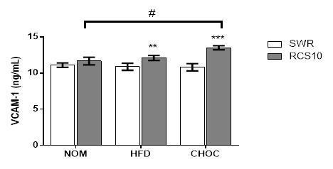
168 FIGURE
169 153
170 154
171 155
172 156
173 Chapter Five Depressed HRV and Slowed Atrial and Atrioventricular Conduction in Diabesity: A Polygenic Mouse Model of Diabetes and Obesity 5.1. INTRODUCTION Both type II diabetes mellitus (DM) and obesity are rising epidemics affecting millions of humans worldwide (44,470). Additionally, obesity itself promotes development of insulin resistance and can lead to type II DM (471,472). In the Nurses Health Study whereby 85,000 females underwent a 120 itemised, semiquantitative food frequency questionnaire and were followed up from , apart from excess body fat being the most important determinant of diabetes, increased dietary intake of trans-fat and increased glycaemic load also predicted risk of DM development (473). In contrast, increased intake of dietary fibre and polyunsaturated fat was found to reduce DM risk. This study underscores dietary contributions in the promotion and prevention of DM. Heart rate variability (HRV), a measure of the temporal variation of consecutive heart beats, is a non-invasive indicator of cardiac autonomic function (200). Depressed HRV has been previously implicated in various metabolic disorders such as hypertension (208), obesity (53), diabetes (209), and metabolic syndrome (474). In the Atherosclerosis Risk in Communities study, diabetic subjects 157
174 demonstrated depressed HRV at baseline and a more rapid temporal decline in HRV over a 9 year follow up compared to non-diabetics (475). Similarly, the Framingham Heart study also reported lower HRV in diabetic subjects compared to those with normal fasting glucose (476). However, these studies did not adequately address the contributions of obesity which is often concomitantly present in diabetic patients. In small case-control studies, obesity has been associated with reduced HRV and weight loss through diet, gastroplasty or bariatric surgery improves these autonomic disturbances (53,477,478). Previous studies looking into HRV modulation, as a measure of cardiac autonomic neuropathy, in rodent models include the Streptozotocin-induced (STZ) rat and mouse ( ), leptin receptor-deficient db/db mouse (482,483), Zucker diabetic fatty rat (484), Akita mouse (485) and Goto-Kakizaki rat (486). Most of these studies demonstrated depressed HRV parameters (time- and/or frequency domain analyses of HRV) in the presence of diabetes but not others (483). Additionally, although decreased nerve sympathetic nerve density has been observed in most models (485,487,488), the changes in norepinephrine content have been variable and inconsistent (487, ). One possible explanation for this disparity of results is the severity of disease in different animal strains and duration of chronic diabetes (ranging from 1 to 9 months of chronic diabetes). However, several limitations remain in the existing models of diabetes, especially animal models of type II DM. The current models for type II DM are mostly derived from monogenic causes which may not fully represent the clinical presentation in humans. Additionally, the presence of obesity which is common in type II diabetes is itself a modulator of HRV (492), and has not been sufficiently factored into the 158
175 HRV modulations from previous studies. It is well established that long-term highfat diet consumption not only results in increased body weight but also can cause insulin resistance and that itself is a risk factor for developing cardiac autonomic dysfunction (493,494). Additionally, free fatty acids and visceral fat have been demonstrated to correlate well with indices of HRV in high-fat induced obese rats (494). However, most high-fat diet consumption studies investigating neuropathy in rodent models employ diets of variable but extremely high fat contribution (ranging from 36-59% energy from fat) ( ). The NONcNZO10/LtJ (RCS10) male mouse is a polygenic model of diet-induced type II DM and obesity, generated from the a cross of the New Zealand Obese and Non-obese Non-diabetic mice (433). RCS10 mice are thought to provide a more accurate representation of human diabetes and obesity by producing moderate obesity through polygenic interactions rather than massive obesity elicited by monogenic mutations in the leptin/leptin receptor axis. RCS10 mice weaned onto a moderately high fat diet (10-11% fat) have been characterised to develop moderate obesity, maturity-onset chronic hyperglycaemia (12-20 weeks), glucose intolerance, visceral obesity, dyslipidaemia, moderate liver steatosis and pancreatic islet atrophy (434,497). Additionally, diet differences in fat content have obvious implications on the genetic contributions of RCS10 mice. Adi et al. (2012) demonstrated that RCS10 mice fed control chow (27% calories in fat, 19% protein, 54% carbohydrates) demonstrated alterations in visceral adipose tissue expression of matrix metalloproteinases (MMPs) and tissue inhibitor of metalloproteinases (TIMPs), proteins involved in extracellular matrix regulation and remodelling, when compared to either a diet of high protein and fish oil 159
176 and/or SWR/J mice, a strain of mice that is diet-resistant to obesity and diabetes (456). This suggests that both diet regulation and genetic contributions play important roles in driving obesity and progression of type II diabetes in this model. Furthermore, high fat diets in RCS10 mice demonstrate elevations of peroxisome proliferator-activated receptor-γ gene expression (456), known to contribute to adipocyte hypertrophy and insulin resistance (498). It is currently unclear if the polygenic model of diet-induced diabetes and obesity is associated with cardiac autonomic dysfunction. In this study, we aim to determine the impact of diabetes, diet contribution to obesity, and age on ECG characteristics and HRV parameters in a polygenic mouse model of diabetes and maturity-onset obesity. 160
177 5.2. METHODS Mouse Models NONcNZO10/LtJ (RCS10) and SWR/J (control) mice were obtained from The Jackson Laboratory (Bar Harbour, ME) and housed in an in-house animal facility (The University of Adelaide, SA) to set up breeding colonies. SWR/J mice are suggested as diet-resistant non-obese controls for physiologic comparisons (Jackson Laboratory). Male mice were randomly allocated in two steps: 1) according to age groups, one at 8-10 weeks of age and the other at 30 weeks of age and 2) according to diets, standard chow (NOM: 5% fat by calories, Meat-free Rat and Mouse Cubes, Specialty Feeds, Glen Forrest, Australia), high fat chow (HFD: 10% fat; SF06-105, Specialty Feeds, Glen Forrest, Australia), and high fat chow with chocolate bar supplement (CHOC: 268 calories breakdown: 50% fat, 42% carbohydrate, 8% protein) that was replenished every fortnight 1. Mice were housed at controlled temperature (24ᵒC) and lighting (12h light-dark cycles) with free access to respective chow and water ad libitum. All experiments were approved by the University of Adelaide Animal Ethics Committee and conducted in accordance with the Australian Code of Practice for the Care and Use of Animals for Scientific Purposes Mouse Electrocardiography 3-lead ECG studies were performed in deeply anesthetized mice. Powerlab with LabChart v8.0.5 software (ADInstruments, NSW, Australia) was used for electrogram acquisition. In brief, mice were anesthetized with an intraperitoneal 1 The cohort of animals used in this study was separate from that of Chapter Four. 161
178 injection of 75mg/kg ketamine and 0.5mg/kg medetomidine. Sedation state was checked by toe- and tail-pinch testing. Mice were placed on a heating pad maintained at 37ᵒC, and gel-coated electrode pads were attached to both front feet and left back feet. Each mouse then underwent 30 minutes of continuous ECG recording. The ECG channel was filtered between 0.5 to 150 Hz and sampled at 1 khz Data Processing and Analyses ECG recordings were grouped and blinded to the analysing experimenter. ECG and HRV parameters were analysed offline with the respective modules from LabChart Pro (ADInstruments, NSW, Australia). ECG parameters including P-wave duration, PR, QRS, QT, and corrected QT (QTc) intervals were determined in consecutive beats, with averages generated for every four successive beats. We used the rate-corrected Bazett s formula to calculate for QTc values. HRV quantification was conducted in 2 mins continuous ECG tracings by the use of time-domain and frequency-domain analyses. Time-domain parameters included the mean of normal R-R intervals (RR), standard deviation of normal RR intervals (SDRR), average heart rate (HR) and root mean square of successive heartbeat interval differences (RMSSD). Frequency-domain parameters included total power (TP), low frequency (LF; Hz) expressed in normalized units, high frequency (HF; Hz) expressed in normalized units, and the LF/HF ratio. These frequency spectrums selected were pre-set in LabChart Pro in accordance to mouse characteristics. 162
179 Statistical Analysis All values are reported in mean ± standard error unless indicated otherwise. General Linear Model (GLM) ANOVA was used to determine significant differences between strain, diet and age factors, and Tukey s post-hoc test was conducted to determine diet differences. Student t-test was used to compare inter-group differences when ANOVA was significant. Linear regression was conducted to determine correlation of P-wave duration and RMSSD values to increased body weight or blood glucose levels. A value of p<0.05 was used to determine significance. 163
180 5.3. RESULTS Animal Characteristics Figure 1 illustrates the body weight, unfasted plasma and urine glucose levels of RCS10 and SWR/J mice. Both young and old RCS10 mice fed NOM diets were not significantly overweight (young: 26.2 ± 1.2 g vs ± 0.7 g, p=ns; old: 33.7 ± 1.7 g vs ± 0.4 g, p=ns) nor diabetic compared to age-matched controls (young: 14.5 ± 1.5 mmol/l vs ± 0.9 mmol/l, p=ns; old: 13.7 ± 1.2 mmol/l vs ± 0.6 mmol/l, p=ns). Young RCS10 mice fed HFD trended towards increased body weights (17% difference) (27.4 ± 1.7 g vs ± 0.7 g, p=0.06) and significant hyperglycaemia (plasma glucose: 20.7 ± 2.6 mmol/l vs ± 1.2 mmol/l, p<0.05) compared to controls. Additionally, increased age resulted in progressed obesity (30% difference body weight). Young RCS10 mice fed CHOC developed severe (41% difference) obesity (32.5 ± 0.5 g vs ± 0.6 g, p<0.0001) and hyperglycaemia (plasma glucose: 23.4 ± 2.2 mmol/l vs ± 1.1 mmol/l, p<0.001) which persisted with increased age. Older age was associated with increased body weight regardless of strain or diet (p<0.001) possibly due to maturation growth, but no significant age effect was seen in unfasted plasma and urine glucose levels. Presence of glycosuria was uniformly greater in RCS10 mice in all diets whereas urine glucose was not detectable in all SWR/J controls (Figure 1C) ECG parameters Figure 2 illustrates representative ECG recordings depicting example of prolonged p- wave duration in young and old RCS10 compared to age-matched SWR/J mice fed HFD diets. P-wave duration was prolonged in all RCS10 mice compared to controls in 164
181 NOM diets (Young: 15.3 ± 0.7 vs ± 0.6 ms, p<0.05; Old: 18.3 ± 0.3 vs ± 0.3 ms, p<0.001), HFD (Young: 19.7 ± 0.9 vs ±0.5 ms, p<0.0001; Old: 18.2 ± 0.7 vs ± 0.5ms, p<0.0001) and CHOC diets (Young: 19.2 ± 1.2 vs ± 0.4 ms, p<0.0001; Old: 18.5 ± 0.5 vs ± 0.7 ms, p<0.0001) (Figure 3B). PR intervals were elevated in RCS10 mice compared to SWR/J controls (p<0.001) without significant diet effect. Increased age had a small but significant increased PR interval regardless of strain (Age: p<0.05; Strain*Age interaction: p=ns). No significant difference in ECG parameters QRS, QT and QTc intervals were observed in RCS10 mice of all diet and age groups HRV parameters Figure 4 depicts representative ECG, HR fluctuations and power spectrum of 10 weeks old SWR/J and RCS10 mice fed CHOC diet. As observed in this example, RCS10 mice demonstrated reduced HR fluctuations when compared to SWR/J mice (Figure 4B). In time-domain analyses of HRV, mean RR intervals and average heart rate were not significantly different between RCS10 and SWR/J mice in all diets and age groups (all P=NS, Figure 5A and 5B). Both young and old RCS10 mice fed NOM diets had unchanged SDRR and RMSSD as compared to age-matched SWR/J controls (Figure 5C and 5D). In RCS10 mice fed HFD diet, SDRR was significantly reduced in young mice (3.41 ± 0.35 ms vs ± 0.54 ms, p<0.05) and trended to be reduced in older mice (3.12 ± 0.25 ms vs ± 0.58 ms, p=0.051). When fed CHOC diet, RCS10 mice demonstrated reduced SDRR compared to age-matched SWR/J mice (young: 2.46 ± 0.22 ms vs ± 0.73 ms, p<0.001; old: 2.66 ± 0.13 ms vs ± 0.38 ms, p<0.01). No significant age and diet effect in SDRR was observed (both p=ns). In RCS10 mice 165
182 fed HFD, RMSSD trended towards reduction in young mice and was significantly reduced in old mice (young: 2.62 ± 0.38 vs ± 0.55, p=0.08; old: 1.82 ± 0.30 vs ± 0.48, p<0.01). When fed CHOC diet, RCS10 showed reduced RMSSD compared to SWR/J controls (young: 2.38 ± 0.35 vs ± 0.47, p<0.05; old: 2.23 ± 0.23 vs ± 0.24, p<0.01). No significant age effect in RMSSD was observed (p=ns), however RCS10 mice fed HFD and CHOC diets trended towards lower RMSSD in both diets compared to NOM diets (Strain*Age interaction: p=0.079). In frequency-domain analyses of HRV, RCS10 mice demonstrated increased LF power compared to SWR/J controls, however this did not reach statistical significance (23.9 ± 0.7 vs ± 0.7 n.u., p=0.078; Figure 6A). Additionally, increased age and fatcontent in diets trended towards significance for LF power (Age: p=0.089; Diet: p=0.055). HF power was significantly reduced in RCS10 mice compared to controls (76.2 ± 0.8 vs ± 0.7 n.u., p<0.05; Figure 6B), with no significant age and diet effect observed (both p=ns). LF/HF ratio was not significantly altered between RCS10 and SWR/J mice in all diet and age groups Association of P-wave and RMSSD to Obesity and Hyperglycaemia Figure 7 depicts the distribution of increasing body weight and blood glucose levels according to mouse strain and diets. SWR/J mice in all three diets remained relatively lean and normoglycaemic whereas RCS10 mice fed HFD or CHOC diets tend to demonstrate greater body weights and blood glucose levels. Figure 8 depicts linear regression correlates of P-wave durations and RMSSD to body weight and blood glucose levels. P-wave duration was positively correlated with 166
183 increasing body weight and blood glucose levels in the RCS10 mice (R 2 value: and respectively, both p<0.05). Conversely, SWR/J controls demonstrated poor correlation of the P-wave duration to body weights or blood glucose (R 2 value: and respectively) and was statistically different to RCS10 animals (p<0.001). RCS10 mice demonstrated reduced RMSSD compared to SWR/J controls (p<0.01), and a negative correlation of RMSSD to increased body weight (R 2 value: 0.156, p<0.05). Additionally, RCS10 mice trended towards significantly reduced RMSSD compared to controls when assessed for blood glucose levels (p=0.074), and increased blood glucose levels correlated with further reduction in RMSSD (p<0.01). Conversely, SWR/J controls demonstrated poor correlation of the RMSSD to body weights or blood glucose (R 2 value: and respectively). 167
184 5.4. DISCUSSION To determine the effects of diet-induced diabetes and obesity on cardiac autonomic regulation and electrophysiology, we studied a relatively new and understudied polygenic mouse model of Type II diabetes. The RCS10 mouse is a strain of mice that varies in progression of diabetes and/or obesity based on the diet and the maturation age. In this study, we characterised the following ECG and HRV alterations using non-invasive ECG recordings in a polygenic model of dietinduced diabetes and obesity: 1) RCS10 mice fed normal low fat content diets do not develop overt diabetes and obesity and have normal HRV but demonstrate slight to moderate prolongation of the atrial conduction as evidenced by increased p-wave durations. 2) RCS10 mice fed HFD or CHOC diets develop diabetes and obesity demonstrating severe slowing of atrial conduction and reduced heart rate variability as evident by time-domain HRV parameters of reduced SDRR and RMSSD and trended towards significant increase and decrease of LF and HF power respectively. 3) Prolonged p-wave duration and depressed RMSSD had a weak but significant correlation to increased obesity and hyperglycaemia. 4) Older age in RCS10 mice was not a main contributor to more marked ECG abnormalities and reduced HRV. Cardiac function is controlled by cardiac autonomic innervations through a balance of sympatho-vagal modulation and reflected through HRV. Although autonomic 168
185 neuropathy has been originally thought to be a late complication of diabetes, HRV has been demonstrated to detect small changes in the autonomic dysfunction occurring much earlier in the clinical course of diabetes (499). Cardiac autonomic dysfunction has been described in a variety of diabetic animal models. In Type I diabetes, rodent models such as the STZ-induced, NOD and Akita mouse commonly demonstrate a decrease in both heart rate and HRV ( ,491). In Type II diabetic rodent models including the db/db mice, Goto-Kakizaki rat, Zucker Diabetic Fatty rat, and high-fat diet induced obese rats have commonly demonstrated decreased HRV, however the average HR change in these studies have been inconclusive. Some studies have demonstrated a decrease in HR due to diabetes (483,486), whereas others have shown an increase (482,484,493). It should be noted that assessment of HRV has not been consistent throughout, such as 1) methods of HRV analysis including time-domain, frequency-domain or both, 2) the use of β-adrenergic and/or muscarinic blockade has been variable, and 3) method of ECG recording (telemetric, restraint, or under anaesthesia). One study demonstrated that feeding state of the animal can have a significant impact on HR and HRV, suggesting that glycaemic state itself can alter HR and HRV at the time of recording (500). Soltynsinska et al. demonstrated in the anesthetized db/db mouse model of Type II diabetes that hypoglycaemia due to fasting resulted in a decrease in HR, and increased time-domain HRV parameters of standard deviation of the difference between successive RR intervals (SDARR) and percentage of normal consecutive RR intervals differing by >6 ms (prr6) whereas frequency-domain analyses of HRV were unchanged. In contrast, in fed animals HR and HRV were unaffected (500). In the current study, we observed unchanged HR, diet-induced decrease in SDRR and RMSSD, and unchanged frequency-domain analyses of HRV 169
186 in anesthetized unfasted RCS10 mice. Our results contrasts with the prior study in HR and HRV despite animals being unfasted, and may be explained by the difference in animal strain (RCS10 vs. db/db) and age (10 and 30 weeks vs. 8 weeks). Our results in this polygenic model suggest similar depression of HRV compared to previous monogenic models, this despite being a closer mimic to human obesity/diabetes syndrome (497). To the best of our knowledge, we believe that this is the first study that has looked at non-invasive ECG recordings in a polygenic model of type II diabetes. In our study, we observed diet-specific prolongation of P wave durations in the diabetic and obese mice compared to non-obese non-diabetic controls is in the absence of any ventricular depolarisation and repolarisation abnormalities. P wave duration is known to be highly correlated with inter-atrial conduction times both in normal and patients with LA enlargement (501). Increased maximum P-wave duration and P-wave dispersion have been previously demonstrated in diabetic as well as obese patients ( ). Autonomic nervous system activity is known to affect the p- wave duration. β-adrenergic blockage is known to prolong p-wave duration whereas parasympathetic blockade shortens p-wave duration (506). Additionally, patients with prolonged p-wave durations have an increased risk of developing atrial fibrillation ( ) and it is postulated that prolonged p-wave duration may be due to areas of delayed atrial conduction predisposing to AF. In isolated hearts or atria of diabetic animals, inter-atrial conduction time and intra-atrial conduction has been shown to be prolonged in concert with increased atrial interstitial fibrosis and higher AF vulnerability (272,445,446,510). 170
187 Clinical Implications Evidence of depressed HRV in the polygenic mouse model for diabesity are consistent with studies in monogenic disorders such as leptin deficiency. Additionally, the polygenic cause is a closer representation of human diabesity with less severe organ defects (434). The results from the current study likely suggest electrophysiological abnormalities in ECG and HRV in humans with diabetes and obesity. Evidently depressed HRV has been observed in cohort and case-controlled studies in relation to diabetes and obesity independently (53, ). However, their combined and individual contributions to HRV in a population where multiple metabolic disorders are concomitantly expressed remain to be investigated. Linear correlation studies also suggest that depressed RMSSD and prolonged P-wave duration are weak but significantly correlated with the progression of obesity and hyperglycaemia in the polygenic mouse Study Limitations Firstly, we did not monitor food consumption as food was given ad libitum and we acknowledge that the effects of glycaemic status in these animals may impact heart rate and HRV. Future studies could identify the differences in HRV of these animals under fasting and non-fasting conditions. We chose to give chocolate supplement in addition of HFD to boost obesity in the RCS10 mice because extremely high fat diets often lose their solid pellet form and are not as palatable to the RCS10 mice. Chocolate bars were replenished every two weeks for both strains of mice, with visual observation that both RCS10 and SWR/J mice consumed the chocolate. Our ECG recordings were conducted under anaesthesia and it is known that 171
188 anaesthesia reduces the power of HRV and conclusions from the frequency domain analysis should be drawn with caution (511). However, all our animal groups underwent the same protocol and were randomly allotted and blinded before ECG and HRV analyses. Therefore, any significant difference is unlikely to be due to anaesthesia alone. 172
189 5.5. CONCLUSIONS In conclusion, diabetes of polygenic causes results in slowed atrial conduction and depressed HRV. Prolonged P-wave duration was observed in ECG recordings from diabetic and obese mice. High fat diets and chocolate supplemented diets significantly prolonged p-wave durations and demonstrated depressed RMSSD in the diabetic and obese mice. Moreover, there was a slight but significant correlation of these markers to both increased hyperglycaemia and body weight. The results highlighted in the current study suggest of dietary-induced electrophysiological abnormalities and cardiac autonomic dysfunction in polygenic diabetes. Further studies are warranted to investigate effects of managing body weight and glycaemic state on these changes. 173
190 FIGURE LEGENDS Figure 1. RCS10 mice develop obesity and diabetes at 10- and 30-weeks of age when fed high fat (HFD) and chocolate supplemented (CHOC) diets. (A) Body weights of 10- and 30-week old RCS10 mice were significantly greater compared to SWR/J controls when fed HFD and CHOC diets post-weaning. 30 week old mice regardless of strain demonstrated elevated body weights compared to 10 week old mice (Age: p<0.001; Age*Strain interaction: p=0.151). (B) Elevated levels of unfasted blood glucose were demonstrated in 10- and 30-week old RCS10 mice when fed HFD and CHOC diets post-weaning. The increase in unfasted blood glucose of RCS10 mice fed HFD and CHOC diets was significantly greater compared to RCS10 mice fed NOM diets (Strain*Diet interaction: p<0.01). No significant age effect was observed (p=ns). (C) Elevated urine glucose levels were observed in RCS10 mice fed all three diets whereas urine glucose was not detectable in SWR/J mice. The increase in urine glucose was significantly greater in all RCS10 mice compared to SWR/J controls regardless of age and diet (Strain: p<0.001; Age: p=0.112; Diet: p=0.292). Figure 2. Prolonged P-wave durations in diabetic mice. (A) Representative surface ECG recordings demonstrating prolonged P-wave durations in young and old RCS10 mice fed HFD diet (dashed lines). Figure 3. ECG recordings demonstrate slowed atrial and AV node conduction delay but normal ventricular conduction in RCS10 mice as compared to SWR/J controls. (A) PR interval, a measure of AV node conduction, was elevated 174
191 in RCS10 mice compared to SWR/J controls (p<0.001) without significant diet effect (p=ns). 30 weeks old mice demonstrated increased PR intervals compared to younger mice regardless of strain (Age: p<0.05; Strain*Age interaction: p=0.586). (B) Both 10 and 30 weeks old RCS10 mice demonstrated similarly prolonged P-wave durations compared to SWR/J mice (Strain: p<0.001; Age: p=0.317). Additionally, RCS10 mice fed HFD and CHOC diets demonstrated significantly prolonged P-wave durations compared to RCS10 mice fed NOM diets (Strain*diet interaction: p<0.001). (C-E) QRS intervals, QT intervals and corrected QT intervals were not significantly different between RCS10 mice and controls in all diets and both age groups (all p=ns). Figure 4. ECG, HR fluctuations and power spectra of 10 weeks old SWR/J and RCS10 mice fed CHOC diet. (A) One-lead ECG recorded from representative RCS10 and SWR/J (control) mice. (B) Comparison of beat-to-beat changes in RR intervals plotted against successive beats. (C) Power spectrum of the RR signal from RCS10 and control mice. VLF = Hz, LF = Hz, and HF = Hz. (D) Poincaré plot demonstrating distribution of RR intervals for successive R-R beats from RCS10 and control mice. Figure 5. Time domain indices of HRV parameters. (A) RCS10 mice demonstrated similar mean RR intervals as compared to SWR/J controls in all diets and age groups. (B) RCS10 mice demonstrated similar mean HR as compared to SWR/J controls in all diets and age groups. (C) RCS10 mice had significantly reduced SDRR as compared to SWR/J controls when fed HFD and CHOC diets, but was not significantly reduced in the NOM diets. No significant age and diet effect 175
192 was observed (both p=ns). (D) RMSSD was significantly reduced in RCS10 mice as compared to SWR/J controls when fed the HFD and CHOC diets, but was unchanged in the NOM diets. Reduced RMSSD in RCS10 mice fed HFD and CHOC diets trended towards significance when compared to RCS10 mice fed NOM diets (strain*diet interaction: p=0.079). No significant age effect on RMSSD was observed (p=ns). Figure 6. Frequency domain indices of HRV parameters. (A) RCS10 mice demonstrated increased LF power compared to SWR/J controls but this did not reach statistical significance (Strain: p=0.078). Additionally, increased LF power in 30 weeks old RCS10 mice fed HFD trended towards significance (Diet: p=0.055, Age: p=0.089). (B) RCS10 mice demonstrated reduced HF power compared to SWR/J controls (Strain: p<0.05), although no significant diet and age effect was observed (both p=ns). (C) LF/HF ratio was not significantly altered in RCS10 mice compared to SWR/J controls (p=ns). Figure 7. Distribution of increasing body weight and blood glucose levels in accordance to mouse strain and diets. SWR/J mice given all 3 diets remained relatively lean and normoglycaemic whereas RCS10 mice fed HFD and/or CHOC diets were more obese and hyperglycaemic. Figure 8. Correlation of P-wave duration (top) and RMSSD (bottom) to body weight (left) and blood glucose levels (right). (A & B) RCS10 mice that developed significantly prolonged p-wave durations had a small but significant positive correlation to both increasing body weight and blood glucose level. (C) 176
193 RCS10 mice with increasing body weight had a small but significant negative correlation with RMSSD. (D) RCS10 mice trended towards significantly reduced RMSSD and increase blood glucose levels correlated with further reduction in RMSSD. 177


194 FIGURE 1. A Young Old B Young Old C Young Old RCS10 178

195 FIGURE 2. SWR/J RCS10 179


196 FIGURE 3. A Young Old B Young Old C Young Old D Young Old E Young Old RCS10 180


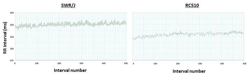
197 FIGURE 4. A SWR/J 500 ms RCS10 B C D 181


198 FIGURE 5. A Young Old B Young Old C Young Old D Young Old RCS10 182

199 FIGURE 6. A Young Old B Young Old C Young Old RCS10 183
Cardiac arrhythmias. Janusz Witowski. Department of Pathophysiology Poznan University of Medical Sciences. J. Witowski
 Cardiac arrhythmias Janusz Witowski Department of Pathophysiology Poznan University of Medical Sciences A 68-year old man presents to the emergency department late one evening complaining of increasing
Where are the normal pacemaker and the backup pacemakers of the heart located?
 CASE 9 A 68-year-old woman presents to the emergency center with shortness of breath, light-headedness, and chest pain described as being like an elephant sitting on her chest. She is diagnosed with a
Deposited on: 29 October 2009
 Workman, A.J. (2009) Mechanisms of postcardiac surgery atrial fibrillation: more pieces in a difficult puzzle. Heart Rhythm, 6 (10). pp. 1423-1424. ISSN 1547-5271 http://eprints.gla.ac.uk/7847/ Deposited
AF Today: W. For the majority of patients with atrial. are the Options? Chris Case
 AF Today: W hat are the Options? Management strategies for patients with atrial fibrillation should depend on the individual patient. Treatment with medications seems adequate for most patients with atrial
Chapter 16: Arrhythmias and Conduction Disturbances
 Complete the following. Chapter 16: Arrhythmias and Conduction Disturbances 1. Cardiac arrhythmias result from abnormal impulse, abnormal impulse, or both mechanisms together. 2. is the ability of certain
High%Density%Mapping%Of%Ventricular%Scar7%Insights% Into%Mechanisms%Of%Ventricular%Tachycardia%%
 High%Density%Mapping%Of%Ventricular%Scar7%Insights% Into%Mechanisms%Of%Ventricular%Tachycardia%% By% Sachin%Nayyar% MBBS,%MD,%DM% % Centre%for%Heart%Rhythm%Disorders% University%of%Adelaide%and%Royal%Adelaide%Hospital%
Arrhythmias. Simple-dysfunction cause abnormalities in impulse formation and conduction in the myocardium.
 Arrhythmias Simple-dysfunction cause abnormalities in impulse formation and conduction in the myocardium. However, in clinic it present as a complex family of disorders that show variety of symptoms, for
Arrhythmias. 1. beat too slowly (sinus bradycardia). Like in heart block
. Like in heart block") Arrhythmias It is a simple-dysfunction caused by abnormalities in impulse formation and conduction in the myocardium. The heart is designed in such a way that allows it to generate from the SA node electrical
Arrhythmias It is a simple-dysfunction caused by abnormalities in impulse formation and conduction in the myocardium. The heart is designed in such a way that allows it to generate from the SA node electrical
Introduction. Circulation
 Introduction Circulation 1- Systemic (general) circulation 2- Pulmonary circulation carries oxygenated blood to all parts of the body carries deoxygenated blood to the lungs From Lt. ventricle aorta From
Cardiac Arrhythmias. Cathy Percival, RN, FALU, FLMI VP, Medical Director AIG Life and Retirement Company
 Cardiac Arrhythmias Cathy Percival, RN, FALU, FLMI VP, Medical Director AIG Life and Retirement Company The Cardiovascular System Three primary functions Transport of oxygen, nutrients, and hormones to
Cardiac Properties MCQ
 Cardiac Properties MCQ Abdel Moniem Ibrahim Ahmed, MD Professor of Cardiovascular Physiology Cairo University 2007 1- Cardiac Valves: a- Prevent backflow of blood from the ventricles to the atria during
Chapter 12: Cardiovascular Physiology System Overview
 Chapter 12: Cardiovascular Physiology System Overview Components of the cardiovascular system: Heart Vascular system Blood Figure 12-1 Plasma includes water, ions, proteins, nutrients, hormones, wastes,
PART I. Disorders of the Heart Rhythm: Basic Principles
 PART I Disorders of the Heart Rhythm: Basic Principles FET01.indd 1 1/11/06 9:53:05 AM FET01.indd 2 1/11/06 9:53:06 AM CHAPTER 1 The Cardiac Electrical System The heart spontaneously generates electrical
Chapter 13 The Cardiovascular System: Cardiac Function
 Chapter 13 The Cardiovascular System: Cardiac Function Overview of the Cardiovascular System The Path of Blood Flow through the Heart and Vasculature Anatomy of the Heart Electrical Activity of the Heart
Atrial Fibrillation: Classification and Electrophysiology. Saverio Iacopino, MD, FACC, FESC
 Atrial Fibrillation: Classification and Electrophysiology Saverio Iacopino, MD, FACC, FESC Sinus Rythm Afib (first episode) AFib Paroxistic AFib Spontaneous conversion Permanent AFib Recurrence Sinus Rythm
Aberrant sodium influx causes cardiomyopathy and atrial fibrillation in mice
 Aberrant sodium influx causes cardiomyopathy and atrial fibrillation in mice Elaine Wan, Jeffrey Adams, Richard L. Weinberg, Alexander N. Katchman, Joseph Bayne, Sergey I. Zakharov, Lin Yang, John P. Morrow,
EHRA/EUROPACE 2011 Madrid, Spain June
 EHRA/EUROPACE 2011 Madrid, Spain June 26.-29.2011 Implementing modern management in atrial fibrillation patients Proceedings from the 3rd AFNet/EHRA consensus conference EHRA Special Session Different
CASE 10. What would the ST segment of this ECG look like? On which leads would you see this ST segment change? What does the T wave represent?
 CASE 10 A 57-year-old man presents to the emergency center with complaints of chest pain with radiation to the left arm and jaw. He reports feeling anxious, diaphoretic, and short of breath. His past history
Atrial Cardiomyopathy is it relevant?
 Atrial Cardiomyopathy is it relevant? Dr Carl Shakespeare MD FRCP FACC FESC Consultant Cardiologist Relevance Progressive nature of AF Implications for RF Targeting Risk Factors CHADS vasc scoring? Could
Tachycardia-induced heart failure - Does it exist?
 Tachycardia-induced heart failure - Does it exist? PD Dr Etienne Delacrétaz Clinique Cecil et Hôpital de Fribourg SSC Cardiology meeting 2015 Zürich Rapid atrial fibrillation is a common cause of heart
Ventricular arrhythmias in acute coronary syndromes. Dimitrios Manolatos, MD, PhD, FESC Electrophysiology Lab Evaggelismos General Hospital
 Ventricular arrhythmias in acute coronary syndromes Dimitrios Manolatos, MD, PhD, FESC Electrophysiology Lab Evaggelismos General Hospital introduction myocardial ischaemia and infarction leads to severe
THE CHARACTERISATION OF RISK FACTORS, SUBSTRATE AND MANAGEMENT STRATEGIES FOR ATRIAL FIBRILLATION
 THE CHARACTERISATION OF RISK FACTORS, SUBSTRATE AND MANAGEMENT STRATEGIES FOR ATRIAL FIBRILLATION Geetanjali Rangnekar BSc (Honours) Centre for Heart Rhythm Disorders School of Medicine The University
Electrocardiography Abnormalities (Arrhythmias) 7. Faisal I. Mohammed, MD, PhD
 7. Faisal I. Mohammed, MD, PhD") Electrocardiography Abnormalities (Arrhythmias) 7 Faisal I. Mohammed, MD, PhD 1 Causes of Cardiac Arrythmias Abnormal rhythmicity of the pacemaker Shift of pacemaker from sinus node Blocks at different
Electrocardiography Abnormalities (Arrhythmias) 7 Faisal I. Mohammed, MD, PhD 1 Causes of Cardiac Arrythmias Abnormal rhythmicity of the pacemaker Shift of pacemaker from sinus node Blocks at different
THE ISSUE OF RESTITUTION AND MAINTENANCE OF SINUS RHYTHM IN ATRIAL FIBRILLATION
 UNIVERSITY LUCIAN BLAGA SIBIU UNIVERSITY OF MEDICINE VICTOR PAPILIAN SIBIU DOCTORAL THESIS THE ISSUE OF RESTITUTION AND MAINTENANCE OF SINUS RHYTHM IN ATRIAL FIBRILLATION - ABSTRACT - PhD student Mentor
How do arrhythmias occur?
 How do arrhythmias occur? An arrhythmia is an abnormal heart rhythm (= dysrhythmia). Can be fast (tachy) or slow (brady). Brady arrhythmias are usually due to conduc;on block, while tachyarrhythmias are
UNDERSTANDING YOUR ECG: A REVIEW
 UNDERSTANDING YOUR ECG: A REVIEW Health professionals use the electrocardiograph (ECG) rhythm strip to systematically analyse the cardiac rhythm. Before the systematic process of ECG analysis is described
Catheter Ablation: Atrial fibrillation (AF) is the most common. Another Option for AF FAQ. Who performs ablation for treatment of AF?
 is the most common. Another Option for AF FAQ. Who performs ablation for treatment of AF?") : Another Option for AF Atrial fibrillation (AF) is a highly common cardiac arrhythmia and a major risk factor for stroke. In this article, Dr. Khan and Dr. Skanes detail how catheter ablation significantly
: Another Option for AF Atrial fibrillation (AF) is a highly common cardiac arrhythmia and a major risk factor for stroke. In this article, Dr. Khan and Dr. Skanes detail how catheter ablation significantly
ATRIAL FIBRILLATION AND OBESITY: CHARACTERIZATION OF ELECTRO-STRUCTURAL ATRIAL UPON WEIGHT REDUCTION
 ATRIAL FIBRILLATION AND OBESITY: CHARACTERIZATION OF ELECTRO-STRUCTURAL ATRIAL SUBSTRATE WITH SUSTAINED OBESITY AND REVERSAL UPON WEIGHT REDUCTION Rajiv Mahajan MBBS, MD, FRACP Department of Cardiology
Cardiac Drugs: Chapter 9 Worksheet Cardiac Agents. 1. drugs affect the rate of the heart and can either increase its rate or decrease its rate.
 Complete the following. 1. drugs affect the rate of the heart and can either increase its rate or decrease its rate. 2. drugs affect the force of contraction and can be either positive or negative. 3.
Cardiovascular system
 BIO 301 Human Physiology Cardiovascular system The Cardiovascular System: consists of the heart plus all the blood vessels transports blood to all parts of the body in two 'circulations': pulmonary (lungs)
Evolving pharmacologic antiarrhythmic treatment targets Ready for clinical practice?
 Evolving pharmacologic antiarrhythmic treatment targets Ready for clinical practice? David O. Arnar MD PhD EMPH Landspitali The National University Hospital of Iceland, Reykjavik Disclosures Consultant
The Electrocardiogram
 The Electrocardiogram Chapters 11 and 13 AUTUMN WEDAN AND NATASHA MCDOUGAL The Normal Electrocardiogram P-wave Generated when the atria depolarizes QRS-Complex Ventricles depolarizing before a contraction
Cardiovascular Nursing Practice: A Comprehensive Resource Manual and Study Guide for Clinical Nurses 2 nd Edition
 Cardiovascular Nursing Practice: A Comprehensive Resource Manual and Study Guide for Clinical Nurses 2 nd Edition Table of Contents Volume 1 Chapter 1: Cardiovascular Anatomy and Physiology Basic Cardiac
Chapter 20: Cardiovascular System: The Heart
 Chapter 20: Cardiovascular System: The Heart I. Functions of the Heart A. List and describe the four functions of the heart: 1. 2. 3. 4. II. Size, Shape, and Location of the Heart A. Size and Shape 1.
Index. cardiology.theclinics.com. Note: Page numbers of article titles are in boldface type.
 Index Note: Page numbers of article titles are in boldface type. A AADs. See Antiarrhythmic drugs (AADs) ACE inhibitors. See Angiotensin-converting enzyme (ACE) inhibitors ACP in transseptal approach to
QUIZ/TEST REVIEW NOTES SECTION 1 CARDIAC MYOCYTE PHYSIOLOGY [CARDIOLOGY]
![QUIZ/TEST REVIEW NOTES SECTION 1 CARDIAC MYOCYTE PHYSIOLOGY [CARDIOLOGY]](/thumbs/96/126998162.jpg "QUIZ/TEST REVIEW NOTES SECTION 1 CARDIAC MYOCYTE PHYSIOLOGY [CARDIOLOGY]") QUIZ/TEST REVIEW NOTES SECTION 1 CARDIAC MYOCYTE PHYSIOLOGY [CARDIOLOGY] Learning Objectives: Describe the ionic basis of action potentials in cardiac contractile and autorhythmic cells Explain the relationship
QUIZ/TEST REVIEW NOTES SECTION 1 CARDIAC MYOCYTE PHYSIOLOGY [CARDIOLOGY] Learning Objectives: Describe the ionic basis of action potentials in cardiac contractile and autorhythmic cells Explain the relationship
2) Heart Arrhythmias 2 - Dr. Abdullah Sharif
 Heart Arrhythmias 2 - Dr. Abdullah Sharif") 2) Heart Arrhythmias 2 - Dr. Abdullah Sharif Rhythms from the Sinus Node Sinus Tachycardia: HR > 100 b/m Causes: o Withdrawal of vagal tone & Sympathetic stimulation (exercise, fight or flight) o Fever
2) Heart Arrhythmias 2 - Dr. Abdullah Sharif Rhythms from the Sinus Node Sinus Tachycardia: HR > 100 b/m Causes: o Withdrawal of vagal tone & Sympathetic stimulation (exercise, fight or flight) o Fever
Lecture outline. Electrical properties of the heart. Automaticity. Excitability. Refractoriness. The ABCs of ECGs Back to Basics Part I
 Lecture outline The ABCs of ECGs Back to Basics Part I Meg Sleeper VMD, DACVIM (cardiology) University of Florida Veterinary School Electrical properties of the heart Action potentials Normal intracardiac
Management strategies for atrial fibrillation Thursday, 20 October :27
 ALTHOUGH anyone who has had to run up a flight of steps or has had a frightening experience is quite familiar with a racing heartbeat, for the more than 2 million Americans who suffer from atrial fibrillation
BIPN100 F15 Human Physiology I (Kristan) Problem set #5 p. 1
 Problem set #5 p. 1") BIPN100 F15 Human Physiology I (Kristan) Problem set #5 p. 1 1. Dantrolene has the same effect on smooth muscles as it has on skeletal muscle: it relaxes them by blocking the release of Ca ++ from the
BIPN100 F15 Human Physiology I (Kristan) Problem set #5 p. 1 1. Dantrolene has the same effect on smooth muscles as it has on skeletal muscle: it relaxes them by blocking the release of Ca ++ from the
A Dynamic model of Pulmonary Vein Electrophysiology. Harry Green 2 nd year Ph.D. student University of Exeter
 A Dynamic model of Pulmonary Vein Electrophysiology Harry Green 2 nd year Ph.D. student University of Exeter Background to the Project Cardiac disease is the leading cause of death in most developed countries
Chapter 26. Media Directory. Dysrhythmias. Diagnosis/Treatment of Dysrhythmias. Frequency in Population Difficult to Predict
 Chapter 26 Drugs for Dysrythmias Slide 33 Slide 35 Media Directory Propranolol Animation Amiodarone Animation Upper Saddle River, New Jersey 07458 All rights reserved. Dysrhythmias Abnormalities of electrical
Cardiac Arrhythmias. For Pharmacists
 Cardiac Arrhythmias For Pharmacists Agenda Overview of the normal Classification Management Therapy Conclusion Cardiac arrhythmias Overview of the normal Arrhythmia: definition From the Greek a-, loss
Step by step approach to EKG rhythm interpretation:
 Sinus Rhythms Normal sinus arrhythmia Small, slow variation of the R-R interval i.e. variation of the normal sinus heart rate with respiration, etc. Sinus Tachycardia Defined as sinus rhythm with a rate
Invasive and Medical Treatments for Atrial Fibrillation. Thomas J Dresing, MD Section of Electrophysiology and Pacing Cleveland Clinic
 Invasive and Medical Treatments for Thomas J Dresing, MD Section of Electrophysiology and Pacing Cleveland Clinic Disclosures Fellow s advisory panel for St Jude Medical Speaking honoraria from: Boston
Emergency Medical Training Services Emergency Medical Technician Paramedic Program Outlines Outline Topic: WPW Revised: 11/2013
 Emergency Medical Training Services Emergency Medical Technician Paramedic Program Outlines Outline Topic: WPW Revised: 11/2013 Wolff-Parkinson-White syndrome (WPW) is a syndrome of pre-excitation of the
Heart Failure (HF) Treatment
 Treatment") Heart Failure (HF) Treatment Heart Failure (HF) Complex, progressive disorder. The heart is unable to pump sufficient blood to meet the needs of the body. Its cardinal symptoms are dyspnea, fatigue, and
Heart Failure (HF) Treatment Heart Failure (HF) Complex, progressive disorder. The heart is unable to pump sufficient blood to meet the needs of the body. Its cardinal symptoms are dyspnea, fatigue, and
Mr. Eknath Kole M.S. Pharm (NIPER Mohali)
") M.S. Pharm (NIPER Mohali) Drug Class Actions Therapeutic Uses Pharmacokinetics Adverse Effects Other Quinidine IA -Binds to open and inactivated Na+ -Decreases the slope of Phase 4 spontaneous depolarization
M.S. Pharm (NIPER Mohali) Drug Class Actions Therapeutic Uses Pharmacokinetics Adverse Effects Other Quinidine IA -Binds to open and inactivated Na+ -Decreases the slope of Phase 4 spontaneous depolarization
V. TACHYCARDIAS Rapid rhythm abnormalities
 V. TACHYCARDIAS Rapid rhythm abnormalities Tachyarrhythmias currently account for up to 350,000 deaths annually in the US. In addition to these clearly dangerous rhythm disturbances, other forms of more
AF :RHYTHM CONTROL BY DR-MOHAMMED SALAH ASSISSTANT LECTURER CARDIOLOGY DEPARTMENT
 AF :RHYTHM CONTROL BY DR-MOHAMMED SALAH ASSISSTANT LECTURER CARDIOLOGY DEPARTMENT 5-2014 Atrial Fibrillation therapeutic Approach Rhythm Control Thromboembolism Prevention: Recommendations Direct-Current
THE CARDIOVASCULAR SYSTEM. Heart 2
 THE CARDIOVASCULAR SYSTEM Heart 2 PROPERTIES OF CARDIAC MUSCLE Cardiac muscle Striated Short Wide Branched Interconnected Skeletal muscle Striated Long Narrow Cylindrical PROPERTIES OF CARDIAC MUSCLE Intercalated
Full file at
 MULTIPLE CHOICE. Choose the one alternative that best completes the statement or answers the question. 1) What electrical event must occur for atrial kick to occur? 1) A) Atrial repolarization B) Ventricular
Declaration of conflict of interest. None to declare
 Declaration of conflict of interest None to declare Risk management of coronary artery disease Arrhythmias and diabetes Hercules Mavrakis Cardiology Department Heraklion University Hospital Crete, Greece
Management of ATRIAL FIBRILLATION. in general practice. 22 BPJ Issue 39
 Management of ATRIAL FIBRILLATION in general practice 22 BPJ Issue 39 What is atrial fibrillation? Atrial fibrillation (AF) is the most common cardiac arrhythmia encountered in primary care. It is often
University of Groningen. Atrial electrical remodeling from barn to bedside Tieleman, Robert George
 University of Groningen Atrial electrical remodeling from barn to bedside Tieleman, Robert George IMPORTANT NOTE: You are advised to consult the publisher's version (publisher's PDF) if you wish to cite
Is cardioversion old hat? What is new in interventional treatment of AF symptoms?
 Is cardioversion old hat? What is new in interventional treatment of AF symptoms? Joseph de Bono Consultant Electrophysiologist University Hospitals Birmingham Atrial Fibrillation (AF) Affects 2% of the
Pediatrics. Arrhythmias in Children: Bradycardia and Tachycardia Diagnosis and Treatment. Overview
 Pediatrics Arrhythmias in Children: Bradycardia and Tachycardia Diagnosis and Treatment See online here The most common form of cardiac arrhythmia in children is sinus tachycardia which can be caused by
The Cardiovascular System
 Chapter 18 Part A The Cardiovascular System 1/19/16 1 Annie Leibovitz/Contact Press Images Similarities of Cardiac and Skeletal Muscle RMP Ion concentration Deploarization Action Potential Repolarization
Arrhythmia 341. Ahmad Hersi Professor of Cardiology KSU
 Arrhythmia 341 Ahmad Hersi Professor of Cardiology KSU Objectives Epidemiology and Mechanisms of AF Evaluation of AF patients Classification of AF Treatment and Risk stratification of AF Identify other
ARRHYTHMIAS IN THE ICU: DIAGNOSIS AND PRINCIPLES OF MANAGEMENT
 ARRHYTHMIAS IN THE ICU: DIAGNOSIS AND PRINCIPLES OF MANAGEMENT Nora Goldschlager, M.D. MACP, FACC, FAHA, FHRS SFGH Division of Cardiogy UCSF CLINICAL VARIABLES IN ARRHYTHMOGENESIS Ischemia/infarction (scar)
Electrical Conduction
 Sinoatrial (SA) node Electrical Conduction Sets the pace of the heartbeat at 70 bpm AV node (50 bpm) and Purkinje fibers (25 40 bpm) can act as pacemakers under some conditions Internodal pathway from
Antiarrhythmic Drugs
 Antiarrhythmic Drugs DR ATIF ALQUBBANY A S S I S T A N T P R O F E S S O R O F M E D I C I N E / C A R D I O L O G Y C O N S U L T A N T C A R D I O L O G Y & I N T E R V E N T I O N A L E P A C H D /
Gene annotation for heart rhythm. 1. Control of heart rate 2. Action Potential 3. Ion channels and transporters 4. Arrhythmia 5.
 Gene annotation for heart rhythm 1. Control of heart rate 2. Action Potential 3. Ion channels and transporters 4. Arrhythmia 5. EC coupling Control of heart rate Autonomic regulation of heart function
Cardiac physiology. b. myocardium -- cardiac muscle and fibrous skeleton of heart
 I. Heart anatomy -- general gross. A. Size/orientation - base/apex B. Coverings D. Chambers 1. parietal pericardium 2. visceral pericardium 3. Layers of heart wall a. epicardium Cardiac physiology b. myocardium
Cardiovascular System: The Heart
 Cardiovascular System: The Heart I. Anatomy of the Heart (See lab handout for terms list) A. Describe the size, shape and location of the heart B. Describe the structure and function of the pericardium
Systems Biology Across Scales: A Personal View XXVII. Waves in Biology: Cardiac Arrhythmia. Sitabhra Sinha IMSc Chennai
 Systems Biology Across Scales: A Personal View XXVII. Waves in Biology: Cardiac Arrhythmia Sitabhra Sinha IMSc Chennai The functional importance of biological waves Spiral Waves Cardiac Arrhythmias Arrhythmias:
ARRHYTHMIAS IN THE ICU
 ARRHYTHMIAS IN THE ICU Nora Goldschlager, MD MACP, FACC, FAHA, FHRS SFGH Division of Cardiology UCSF IDENTIFIED VARIABLES IN ARRHYTHMOGENESIS Ischemia/infarction (scar) Electrolyte imbalance Proarrhythmia
Dysrhythmias 11/7/2017. Disclosures. 3 reasons to evaluate and treat dysrhythmias. None. Eliminate symptoms and improve hemodynamics
 Dysrhythmias CYDNEY STEWART MD, FACC NOVEMBER 3, 2017 Disclosures None 3 reasons to evaluate and treat dysrhythmias Eliminate symptoms and improve hemodynamics Prevent imminent death/hemodynamic compromise
Paramedic Rounds. Tachyarrhythmia's. Sean Sutton Dallas Wood
 Paramedic Rounds Tachyarrhythmia's Sean Sutton Dallas Wood Objectives At the end of this session, the paramedic will be able to: State the key components of the cardiac conduction pathway, along with the
11/10/2014. Muscular pump Two atria Two ventricles. In mediastinum of thoracic cavity 2/3 of heart's mass lies left of midline of sternum
 It beats over 100,000 times a day to pump over 1,800 gallons of blood per day through over 60,000 miles of blood vessels. During the average lifetime, the heart pumps nearly 3 billion times, delivering
ECG Interpretation Cat Williams, DVM DACVIM (Cardiology)
") ECG Interpretation Cat Williams, DVM DACVIM (Cardiology) Providing the best quality care and service for the patient, the client, and the referring veterinarian. GOAL: Reduce Anxiety about ECGs Back to
ECG Interpretation Cat Williams, DVM DACVIM (Cardiology) Providing the best quality care and service for the patient, the client, and the referring veterinarian. GOAL: Reduce Anxiety about ECGs Back to
WHAT S THAT RHYTHM I AM HEARING? GUIDE TO AUSCULTATION OF ARRHYTHMIAS IN HORSES
 WHAT S THAT RHYTHM I AM HEARING? GUIDE TO AUSCULTATION OF ARRHYTHMIAS IN HORSES Michelle Henry Barton DVM, PhD, DACVIM University of Georgia, Athens, GA INTRODUCTION The purpose of this talk is to review
Chapter 14. Agents used in Cardiac Arrhythmias
 Chapter 14 Agents used in Cardiac Arrhythmias Cardiac arrhythmia Approximately 50% of post-myocardial infarction fatalities result from ventricular tachycarida (VT) or ventricular fibrillation (VF). These
10/23/2017. Muscular pump Two atria Two ventricles. In mediastinum of thoracic cavity 2/3 of heart's mass lies left of midline of sternum
 It beats over 100,000 times a day to pump over 1,800 gallons of blood per day through over 60,000 miles of blood vessels. During the average lifetime, the heart pumps nearly 3 billion times, delivering
Pediatrics ECG Monitoring. Pediatric Intensive Care Unit Emergency Division
 Pediatrics ECG Monitoring Pediatric Intensive Care Unit Emergency Division 1 Conditions Leading to Pediatric Cardiology Consultation 12.7% of annual consultation Is arrhythmias problems Geggel. Pediatrics.
Index of subjects. effect on ventricular tachycardia 30 treatment with 101, 116 boosterpump 80 Brockenbrough phenomenon 55, 125
 145 Index of subjects A accessory pathways 3 amiodarone 4, 5, 6, 23, 30, 97, 102 angina pectoris 4, 24, 1l0, 137, 139, 140 angulation, of cavity 73, 74 aorta aortic flow velocity 2 aortic insufficiency
Who Gets Atrial Fibrilla9on..?
 Birmingham October 20 th 2013 AFA Pa9ents Day Symptoma9c Atrial Fibrilla9on What therapies are available? GENERAL BACKGROUND Andrew Grace Papworth Hospital and University of Cambridge Consultant: Medtronic
PERMANENT PACEMAKERS AND IMPLANTABLE DEFIBRILLATORS Considerations for intensivists
 PERMANENT PACEMAKERS AND IMPLANTABLE DEFIBRILLATORS Considerations for intensivists Craig A. McPherson, MD, FACC Associate Professor of Medicine Constantine Manthous, MD, FACP, FCCP Associate Clinical
AF ABLATION Concepts and Techniques
 AF ABLATION Concepts and Techniques Antony F Chu, M.D. Director of Complex Ablation Arrhythmia Services Section Division of Cardiology at the Rhode Island and Miriam Hospital HIGHLIGHTS The main indications
Chapter 20 (2) The Heart
 The Heart") Chapter 20 (2) The Heart ----------------------------------------------------------------------------------------------------------------------------------------- Describe the component and function of
Chapter 20 (2) The Heart ----------------------------------------------------------------------------------------------------------------------------------------- Describe the component and function of
TEST BANK FOR ECGS MADE EASY 5TH EDITION BY AEHLERT
 Link download full: http://testbankair.com/download/test-bank-for-ecgs-made-easy-5thedition-by-aehlert/ TEST BANK FOR ECGS MADE EASY 5TH EDITION BY AEHLERT Chapter 5 TRUE/FALSE 1. The AV junction consists
EKG Abnormalities. Adapted from:
 EKG Abnormalities Adapted from: http://www.bem.fi/book/19/19.htm Some key terms: Arrhythmia-an abnormal rhythm or sequence of events in the EKG Flutter-rapid depolarizations (and therefore contractions)
Paroxysmal Supraventricular Tachycardia PSVT.
 Atrial Tachycardia; is the name for an arrhythmia caused by a disorder of the impulse generation in the atrium or the AV node. An area in the atrium sends out rapid signals, which are faster than those
Figure 2. Normal ECG tracing. Table 1.
 Figure 2. Normal ECG tracing that navigates through the left ventricle. Following these bundle branches the impulse finally passes to the terminal points called Purkinje fibers. These Purkinje fibers are
Rhythmical Excitation of the Heart
 Rhythmical Excitation of the Heart KALEB HOOD AND JIMMY JOHNSON Special Excitory and Conductive System of the Heart Sinus Node (or sinoatrial node or S-A): A small node with almost no contractile muscle,
Human Anatomy and Physiology II Laboratory Cardiovascular Physiology
 Human Anatomy and Physiology II Laboratory Cardiovascular Physiology 1 This lab involves two exercises: 1) Conduction System of the Heart and Electrocardiography and 2) Human Cardiovascular Physiology:
Chapter 9. Learning Objectives. Learning Objectives 9/11/2012. Cardiac Arrhythmias. Define electrical therapy
 Chapter 9 Cardiac Arrhythmias Learning Objectives Define electrical therapy Explain why electrical therapy is preferred initial therapy over drug administration for cardiac arrest and some arrhythmias
How does the heart work? The heart is muscle whose main function is a pump; to push blood the rest of your body.
 1 You have a condition called atrial fibrillation. I would like you to learn more about this condition. You should read about it below, and can also watch an Internet program about it. After reading about
Atrial Fibrillation and Heart Failure: A Cause or a Consequence
 Atrial Fibrillation and Heart Failure: A Cause or a Consequence Rajat Deo, MD, MTR Assistant Professor of Medicine Division of Cardiology, Electrophysiology Section University of Pennsylvania November
Atrial Fibrillation and Common Supraventricular Tachycardias. Sunil Kapur MD
 Atrial Fibrillation and Common Supraventricular Tachycardias Sunil Kapur MD Cardiac Electrophysiology Brigham and Women s Hospital Instructor, Harvard Medical School No disclosures Cardiac Conduction:
Reentry in a Pulmonary Vein as a Possible Mechanism of Focal Atrial Fibrillation
 824 Reentry in a Pulmonary Vein as a Possible Mechanism of Focal Atrial Fibrillation BERNARD BELHASSEN, M.D., AHARON GLICK, M.D., and SAMI VISKIN, M.D. From the Department of Cardiology, Tel-Aviv Sourasky
(D) (E) (F) 6. The extrasystolic beat would produce (A) increased pulse pressure because contractility. is increased. increased
 (E) (F) 6. The extrasystolic beat would produce (A) increased pulse pressure because contractility. is increased. increased") Review Test 1. A 53-year-old woman is found, by arteriography, to have 5% narrowing of her left renal artery. What is the expected change in blood flow through the stenotic artery? Decrease to 1 2 Decrease
Review Test 1. A 53-year-old woman is found, by arteriography, to have 5% narrowing of her left renal artery. What is the expected change in blood flow through the stenotic artery? Decrease to 1 2 Decrease
Skin supplied by T1-4 (medial upper arm and neck) T5-9- epigastrium Visceral afferents from skin and heart are the same dorsal root ganglio
 T5-9- epigastrium Visceral afferents from skin and heart are the same dorsal root ganglio") Cardio 2 ECG... 3 Cardiac Remodelling... 11 Valvular Diseases... 13 Hypertension... 18 Aortic Coarctation... 24 Erythropoiesis... 27 Haemostasis... 30 Anaemia... 36 Atherosclerosis... 44 Angina... 48 Myocardial
Cardio 2 ECG... 3 Cardiac Remodelling... 11 Valvular Diseases... 13 Hypertension... 18 Aortic Coarctation... 24 Erythropoiesis... 27 Haemostasis... 30 Anaemia... 36 Atherosclerosis... 44 Angina... 48 Myocardial
Atrial fibrillation in the ICU
 Atrial fibrillation in the ICU Atrial fibrillation Preexisting or incident (new onset) among nearly one in three critically ill patients Formation of arrhythogenic substrate usually fibrosis (CHF, hypertension,
Cardiovascular Physiology. Heart Physiology. Introduction. The heart. Electrophysiology of the heart
 Cardiovascular Physiology Heart Physiology Introduction The cardiovascular system consists of the heart and two vascular systems, the systemic and pulmonary circulations. The heart pumps blood through
Principles of Anatomy and Physiology
 Principles of Anatomy and Physiology 14 th Edition CHAPTER 20 The Cardiovascular System: The Heart Introduction The purpose of the chapter is to: 1. Learn about the components of the cardiovascular system
Non-Invasive Ablation of Ventricular Tachycardia
 Non-Invasive Ablation of Ventricular Tachycardia Dr Shaemala Anpalakhan Newcastle upon Tyne Hospitals NHS Foundation Trust Freeman Road, Newcastle Upon Tyne, NE7 7DN Contact: shaemala@doctors.org.uk Introduction
Approximately the size of your fist Location. Pericardial physiology
 Heart Anatomy Approximately the size of your fist Location Superior surface of diaphragm Left of the midline Anterior to the vertebral column, posterior to the sternum Wednesday, March 28, 2012 Muscle
Ventricular Tachycardia in Structurally Normal Hearts (Idiopathic VT) Patient Information
 Patient Information") Melbourne Heart Rhythm Ventricular Tachycardia in Structurally Normal Hearts (Idiopathic VT) Patient Information What is Ventricular Tachycardia? Ventricular tachycardia (VT) is an abnormal rapid heart
Melbourne Heart Rhythm Ventricular Tachycardia in Structurally Normal Hearts (Idiopathic VT) Patient Information What is Ventricular Tachycardia? Ventricular tachycardia (VT) is an abnormal rapid heart
Creating Order out of Chaos: New Research into Treatment of Atrial Fibrillation
 Creating Order out of Chaos: New Research into Treatment of Atrial Fibrillation Peter Spector, MD Professor of Medicine, Director, Cardiac Electrophysiology Fletcher Allen Health Care University of Vermont