Liver. Function of the liver
|
|
|
- Gregory Barton
- 6 years ago
- Views:
Transcription
1 Liver & Gallbladder
2 Liver The liver is the largest gland in the body and has a wide variety of functions Weight: 1/50 of body weight in adult & 1/20 of body weight in infant It is exocrine(bile) & endocrine organ(albumen, prothrombin & fibrinogen) Function of the liver Secretion of bile & bile salt Metabolism of carbohydrate, fat and protein Formation of heparin & anticoagulant substances Detoxication Storage of glycogen and vitamins Activation of vita.d

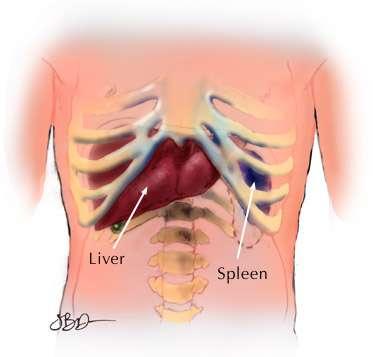
3 Occupies right hypochondrium + epigastrium &extends to left hypochondrium Location


4 Surface anatomy of the liver -The greater part of the liver is situated under cover of the right costal margin - Diaphragm separates it from the pleura, lungs, pericardium, and heart.

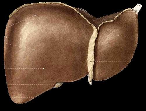
5 Ant. View of the liver Right lobe Cut edge of the Falciform ligament left lobe Diverging cut edges of the superior part of the coronary ligament Fundus of the gall bladder
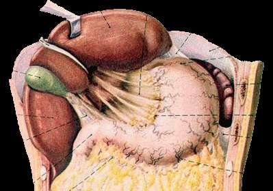
6 Surfaces of the liver, their relations & impressions Postero - inferior surface= visceral surface Superior surface = Diaphragmatic surface Anterior surface Posterior surface Right surface
7 Postero- infero surface= visceral surface Relations I.V.C the esophagus the stomach the duodenum the right colic flexure the right kidney Rt. Suprarenal gland the gallbladder. Porta hepatic( bile duct,h.a.h.v) Fissure for lig. Venoosum & lesser omentum Tubular omentum Lig.teres

8 Postero-inferior surface of the liver

9 Right & left lobes Cut edge of the Falciform ligament The cut edges of the superior and inferior parts of the coronary ligament The left triangular ligament The right triangular ligament Bare area of the liver (where there is no peritoneum covering the liver Groove for the inferior vena cava and the hepatic veins Caudate lobe of the liver more or less wrapping around the groove of the inferior vena cava Fundus of gall bladder Lig.teres Sup. Surface of the liver

10 Relations of Sup. surface of liver Diaphragm Pleura & lung Pericardium & heart
11 Relations of the liver Anteriorly Diaphragm Rt & Lt pleura and lung Costal cartilage Xiphoid process Ant. abdominal wall
12

13 Posterior relation of the liver Diaphragm Rt. Kidney Supra renal gland T.colon(hepatic flexure Duodenum Gall bladder I.V.C Esophagus Fundus of stomach
14 Lobes of the liver Rt. Lobe Lt.lobe Quadrate lobe Caudate lobe
 - bile ducts, portal vein, hepatic")
15 Separation of the four lobes of the liver: Right sagittal fossa - groove for inferior vena cava and gall bladder left sagittal fissure - contains the Ligamentum Venoosum and round ligament of liver Transverse fissure (also porta hepatis) - bile ducts, portal vein, hepatic arteries
16 Rt. Lobe -Largest lobe - Occupies the right hypochondrium - Divided into anterior and posterior sections by the right hepatic vein - Reidel s Lobe extend as far caudally as the iliac crest
17 Left Lobe Varied in size Lies in the epigastric and left hypochondriac regions Divided into lateral and medial segments by the left hepatic vein
18 Lobes of the liver..cont Rt. & Lt lobe separated by Falciform ligament Ligamentum Venoosum Ligamentum teres
19 Caudate Lobe -present in the posterior surface from the Rt. Lobe Two processes 1- c- process 2- papillary process Relations of caudate lobe - Inf. the porta hepatis - The right the fossa for the inferior vena cava - The left the fossa for the lig.venosum.
20 Quadrate lobe Present on the inferior surface from the Rt. Lobe Relation - Ant. anterior margin of the liver - Sup. porta hepatis - Rt. fossa for the gallbladder - Lt by the fossa for lig.teres

21 Porta hepatis -It is the hilum of the liver -It is found on the posteroinferior surface - lies between the caudate and quadrate lobes -Lesser omentum attach to its margin Contents - Gallbladder ant. - Hepatic. Art + nerve+ lymphatic node middle. - Portal vein post.

22 Peritoneum of the liver The liver is covered by peritoneum (intraperitoneal organ)except at bare area(it is origin from septum transversum) Inferior surface covered with peritoneum of greater sac except porta hepatis, G.B & Lig.teres fissure Rt. Lateral surface covered by peritoneum, related to diaphragm which separate it from Rt. Pleura, lung and the Rt Ribs (6-11)
23 1. The ligaments of the liver 1- The Falciform ligament of liver 2- The Ligamentum teres hepatis 3- The coronary ligament 4- The right triangular ligament 5- The left triangular ligament 6- The Hepatogastric ligament 7- The hepatoduonedenal ligament 8- The Ligamentum Venoosum
24 Falciform ligament of liver Consists of double peritoneal layer Sickle shape Extends from anterior abdominal wall (umbilicus) to liver Free border of the ligament contains Ligamentum teres (obliterated umbilical vein)


25 Coronary ligament the area between upper and lower layer of the coronary ligament is the bare area of liver which contract with the diaphragm; Left and right triangular ligaments formed by left and right extremity of coronary ligament

26 Hepatogastric ligament Hepatoduodenal ligament

27 The Ligamentum Venoosum -Fibrous band that is the remains of the ductus venosus - Is attached to the left branch of the portal vein and ascends in a fissure on the visceral surface of the liver to be attached above to the inferior vena cava

28 lobules >> roughly hexagonal structures consisting of hepatocytes. Radiate outward from a central vein. At each of the six corners of a lobule is a portal triad ( p.arteriole,p.venule & bile duct) Between the hepatocytes are the liver sinusoids. LIVER Histology


29 Where do the two blood supplies mix? Liver surrounded by a thin capsule at portahepatic(it is thick) Glisson s capsule invests the liver and send septa into liver subset subdivide the parenchyma into lobules
 True morphological and physiological division by a line extend from fossa of GD to fossa of I.V.")
30 Segmental anatomy of the liver Rt.& Lt. lobes anatomically no morphological significance. Separation by ligaments (Falciform, lig. Venoosum & Lig.teres) True morphological and physiological division by a line extend from fossa of GD to fossa of I.V.C each has its own arterial blood supply, venous drainage and biliary drainage No anastomosis between divisions 3 major hepatic veins Rt, Lt & central 8 segments based on hepatic and portal venous segments

31 Segmental anatomy of the liver Liver segments are based on the portal and hepatic venous segments

32 Blood supply of the liver

33 Blood supply of the liver Proper hepatic artery The right and left hepatic arteries enter the porta hepatis. The right hepatic artery usually gives off the cystic artery, which runs to the neck of the gallbladder.

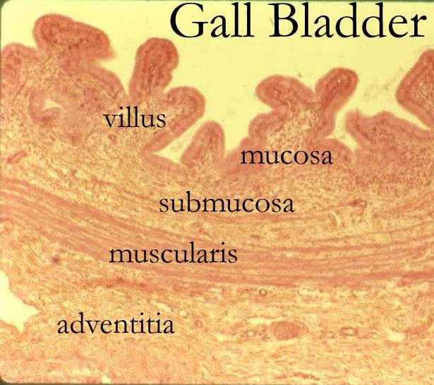
34 Blood Circulation through the Liver The blood vessels conveying blood to the liver are the hepatic artery (30%) and portal vein (70%). The hepatic artery brings oxygenated blood to the liver, and the portal vein brings venous blood rich in the products of digestion, which have been absorbed from the gastrointestinal tract. The arterial and venous blood is conducted to the central vein of each liver lobule by the liver sinusoids. The central veins drain into the right and left hepatic veins, and these leave the posterior surface of the liver and open directly into the inferior vena cava.
 emerge from the posterior surface of the liver and drain into the inferior vena cava.")
35 Vein drainage of the liver The portal vein divides into right and left terminal branches that enter the porta hepatis behind the arteries. The hepatic veins (three or more) emerge from the posterior surface of the liver and drain into the inferior vena cava.

36
37 Lymphatic drainage of the liver Liver produce large amount of lymph~ one third one half of total body lymph Lymph leave the liver and enters several lymph nod in porta hepatis efferent vessels pass to celiac nods A few vessels pass from the bare area of the liver through the diaphragm to the posterior Mediastinal lymph nodes. Nerve supply Sympathetic hepatic plexus>>> celiac plexuses thoracic ganglion chain T1- T12 Parasympathetic vagous nerve( anterior part) Sympathetic and parasympathetic nerves form the celiac plexus. The anterior vagal trunk gives rise to a large hepatic branch, which passes directly to the liver
38 Endoscopic retrograde cholangiopancreatography (ERCP) It is a technique that combines the use of endoscopy and fluoroscopy to diagnose and treat certain problems of the biliary or pancreatic ductal systems. Through the endoscope, the physician can see the inside of the stomach and duodenum, and inject dyes into the ducts in the biliary tree and pancreas so they can be seen on X-rays. ERCP is used primarily to diagnose and treat conditions of the bile ducts, including gallstones, inflammatory strictures (scars), leaks (from trauma and surgery), and cancer.

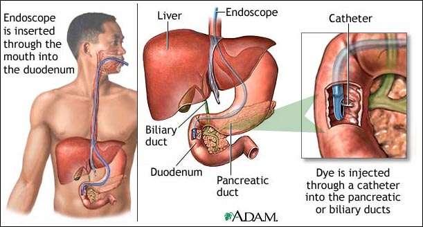
39 ERCP

40 Liver cirrhosis
41 GALLBLADDER
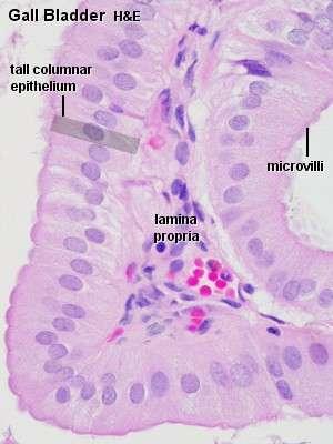
42 Anatomical position of GB - Epigastric - Right hypochondrium region - At the tip of the 9 th RT. C.C - Green muscular organ - Pear-shaped, hollow structure - On inferior surface of liver - Between quadrate and right lobes - Has a short mesentery - Capacity cc - Body and neck Directed toward porta hepatis
43 Structure of GB Fundus -Ant:ant.abdominal wall - Post.inf: transverscolon Body sup: liver Neck post.inf: Tr.colon. End of 1 st part of doudenum, begins of 2 nd part of doudenum - Form the cystic duct, 4cm Hartmann s Pouch 1. Lies between body and neck of gallbladder 2. A normal variation 3. May obscure cystic duct 4. If very large, may see cystic duct arising from pouch
44
45

46 Cystic duct - It joins common hepatic duct
47 Arterial Supply to the Gallbladder Cystic artery Right hepatic artery Proper hepatic artery Common hepatic artery
48 Blood supply of GB: - Cystic artery branch of Rt. Hepatic artery - Cystic vein end in portal vein - Small branches ( arteries and veins run between liver and gall bladder Proper Hepatic Artery Common Hepatic Artery Gastroduodenal Artery
49 Lymphatic drainage of GB 1. celiac nodes 2. Cystic node at neck of GB a. Actually a hepatic node b. Lies at junction of cystic & common hepatic ducts 3. Other lymph vessels also drain into hepatic nodes
50 Nerve supply Sympathetic and parasympathetic from celiac plexus Parasympathetic ---- vagous nerve Hormone cholecystokini duodenum
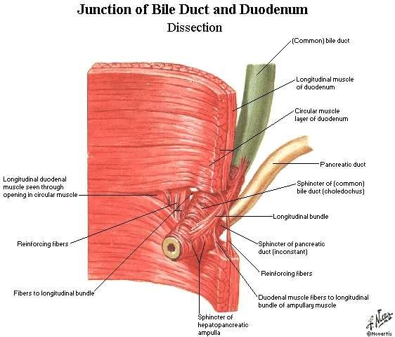
51 Common bile duct Extra hepatic biliary system Rt. hepatic duct + Lt hepatic duct Common hepatic duct + Cystic duct Common bile duct - 4cm - Descend in free edge of lesser omentum - Supra duodenal part Retro duodenal part Retro pancreatic part
52 Bile duct. parts and relations -3 inc long -1 st part -Located in right free margin of lesser omentum - in front of the opening into the lesser sac (Epiploic opening) -Rt to hepatic artery and portal vein - 2 nd part -Behind the 1 st part of the duodenum -Rt to the gastroduodenal artery -3 rd part -Posterior surface of the head of the pancreas -Contact with main pancreatic duct -Related with IVC, gastroduodenal artery, portal vein -End in the half second part of duodenum at ampulla of Vater
53
54 Ampulla of Vater with CBD and Pancreatic Duct Ampulla of Vater

55 Hepaticopancreatic ampulla (Ampulla of Vater)
56 Blood supply of CBD Small arteries supplying CBD a. Arise from cystic artery b. Posterior branch of superior pancreaticoduodenal artery
 Gallstones are")
 Contains waste products from RBC breakdown and other metabolic processing (color of feces from bilirubin")
57 What is bile? Bile composed of water, ions, bile acids, organic molecules (including cholesterol, phospholipids, bilirubin) Gallstones are mostly cholesterol Acids and salts emulsify fats for absorption across wall of small intestines into lacteal lymph capillaries (review) Contains waste products from RBC breakdown and other metabolic processing (color of feces from bilirubin in bile)(review) Ions buffer chyme from stomach (review) Liver Physiology, Larry Frolich, Yavapai College, March 10, 2006

58 Cholelithiasis GB shows likely sites of stone formation/deposition Gangrene of gallbladder is rare Stone in C.B.D obstruct jaundice & pancreatitis
59 Gallbladder Diseases 1- Cholelithiasis & Cholecystitis Cholecystitis = inflammation of GB Cholelithisi = Stone(s) in GB 2- Obstructive jaundice: liver patterns 3- Gangrene of gall bladder rare 4- Congenital defects
60 PANCREAS

61 Anatomical position - Epigastric - left upper hypochondrium region Right lobe of liver Falciform ligament Gallbladder Pancreas Duodenum L-3

62 Common relation Anterior -Transverse colon -Transvers mesocolon -Lesser sac -Stomach -Posterior --Bile duct -Portalvein -Splenic vein -IVC -Aorta - origin of Sup.mesentric.a -Lt.Psoas muscle -Lt.Suuprarenal gland -Left kidney -Hilum of the spleen PANCREAS

63 Posterior view of duodenum/pancreas

64 Histology of pancreas Exocrine part Pancreatic juice Endocrine part Insulin, glucagon and somatostatin
65 Parts of the pancreas
66 Parts Head Neck body Tail

67 The head -It is disc shaped - lies within the concavity of the duodenum - A part of the head extends to the left behind the superior mesenteric vessels and is called the Uncinate process.
68 The neck - It is the constricted portion of the pancreas - connects the head to the body. - It lies in front of the beginning of the portal vein the origin of the
69 The body -Runs upward and to the left across the midline - It is somewhat triangular in cross section.
70 Body of pancreas cont - Three surfaces: anterior, posterior, and inferior. - Three borders: ant,post & inf The anterior surface 1- Covered by peritoneum of post. Wall of lesser sac 2- Tuber omental : where the ant. surface of pancreas join the neck

71 Body of pancreas cont The posterior surface - devoid of peritoneum - in contact with 1- the aorta 2- the splenic vein 3- the left kidney and its vessels 4- the left suprarenal gland 5- the origin of the superior mesenteric artery 6- and the crura of the diaphragm.
72 Body of pancreas cont The inferior surface - Narrow on the right but broader on the left - Covered by peritoneum of greater omentum - lies upon the duodenojejunal flexure - Some coils of the jejunum - its left extremity rests on the left colic flexure The superior border - Blunt and flat to the right; - Narrow and sharp to the left near the tail It commences on the right in the omental tuberosity In relation with 1- The celiac artery 2- Hepatic artery 3- The splenic artery runs toward the left in a groove along this border.
73 Body of pancreas cont The anterior border separates the anterior surface from the inferior surface along this border the two layers of the transverse mesocolon diverge from one another; one passing upward over the anterior surface, the other backward over the inferior surface.
74 Body of pancreas The inferior border separates the posterior from the inferior surface the superior mesenteric vessels emerge under its right extremity.

75 The Tail - Passes forward in the splenicorenal ligament and comes in contact with the hilum of the spleen
76 Pancreatic ducts The main duct - Begins in the tail and runs the length of the gland - Receiving numerous tributaries on the way. - It opens into the second part of the duodenum at about its middle with the bile duct on the major duodenal papilla Accessory duct - When present, drains the upper part of the head - Then opens into the duodenum a short distance above the main duct on the minor duodenal papilla. - The accessory duct frequently communicates with the main duct

77 Blood Supply of pancreas Arteries The splenic.a The superior pancreaticoduodenal.a Inferior pancreaticoduodenal arteries.a Veins The corresponding veins drain into the portal system.

78 pancreaticoduod enal artery and its branches
79 Lymphatic drainage of pancreas Lymph nodes are situated along the arteries that supply the gland. The efferent vessels ultimately drain into the celiac and superior mesenteric lymph nodes.
80 Nerve supply Sympathetic and parasympathetic chain Parasympathetic = vagus nerve
81 Congenital defects of pancreas Annular Pancreas (pancreas encircles duodenum) (rare) Ectopic Pancreas (very common)= Outside the gastrointestinal tract
82 Clinical notes Cancer head of pancreas Obstruction jaundices Cancer body of pancreas pressure I.V.C & portal vein Acute pancreatitis= inflammation of pancreas
83 Spleen
84 Location and Description it is reddish & oval shaped the largest single mass of lymphoid tissue in the body. and Spleen has a notched anterior border. location: Lt hypochondrium - It lies just beneath the left half of the diaphragm - under the 9th, 10th, and 11th ribs. - Its long axis parallel to the 10th rib - Medial end is 4 cm away from mid line post - Lat.end is in left mid axillary line
 between the spleen & the greater curvature")
.")
85 Peritoneum Spleen The spleen is completely covered with peritoneum intraperitoneal organ Two ligaments 1- the gastrosplenic omentum (ligament) between the spleen & the greater curvature of the stomach (carrying the short gastric and left gastroepiploic vessels) 2- splenicorenal ligament between spleen & kidney (carrying the splenic vessels and the tail of the pancreas).

86 Spleen...cont Size 1 inch thick 3 inch broad 5 inch long Weight 7 ounce Shape variable 2 ends 2 borders 2 surfaces Notched - Due to lobulation in embryo

87 Surfaces of spleen 2 surfaces - Diaphragmatic surface - Visceral surface 1- Diaphragmatic surface - Has Post- lat.relation - Convex - Smooth -Diaphragm separates it from - Pleura & lung - Ribs 9,10,11
88 Spleen..cont 2- Visceral surface Has Ant- med. Relations It is divided by a ridge into 1- An anterior or gastric 2- A posterior or renal portion. Lower extremity has Colic surface Pancreatic surface
89 Spleen cont Gastric surface - Extends forward, upward, and medialward - Broad and concave - Related to stomach Renal surface - Directed medialward and downward. -It is somewhat flattened - Related to Lt.kidney
90 Spleen..cont The lower extremity or colic surface - It is flat - Triangular in shape - Rests upon the left flexure of the colon and the phrenicocolic ligament, and is generally in contact with the tail of the pancreas(pancreatic surface)
91 Spleen.cont Hilum of spleen - Splenic. A ant - Splenic. v post - Tail of pancreas
92 Spleen..cont Borders of spleen 1- sup. Border - It is free - Sharp - Thin - Often notched(sup.notch), especially below - It separates the diaphragmatic surface from the gastric surface
93 Spleen cont 2- inferior border - More rounded and blunter - Separates the renal from the diaphragmatic surface; - It corresponds to the lower border of the eleventh rib - lies between the diaphragm and left kidney. The
94 Spleen.cont Intermediate margin is the ridge which separates the renal and gastric surfaces. Internal border separates the diaphragmatic from the colic surface.


95 Spleen cont 2 Ends Med.end sup & back 4cm away from mid line post Lat.end in left mid axillary line
96 Blood supply The large splenic artery is the largest branch of the celiac artery. It has a tortuous course It runs along the upper border of the pancreas The splenic artery then divides into about six branches, which enter the spleen at the hilum
97 Blood supply of spleen Veins - The splenic vein leaves the hilum and runs behind the tail and the body of the pancreas. - Behind the neck of the pancreas, the splenic vein joins the superior mesenteric vein to form the portal vein.

98 Lymphatic Drainage of spleen The lymph vessels emerge from the hilum and pass through a few lymph nodes along the course of the splenic artery and then drain into the celiac nodes.
99 Nerve Supply of spleen The nerves accompany the splenic artery and are derived from the celiac plexus.
100 Thank you
Accessory Glands of Digestive System
 Accessory Glands of Digestive System The liver The liver is soft and pliable and occupies the upper part of the abdominal cavity just beneath the diaphragm. The greater part of the liver is situated under
To describe the liver. To list main structures in porta hepatis.
 GI anatomy Lecture: 6 د. عصام طارق Objectives: To describe the liver. To list main structures in porta hepatis. To define portal system & portosystemic anastomosis. To list parts of biliary system. To
Pancreas & Biliary System. Dr. Vohra & Dr. Jamila
 Pancreas & Biliary System Dr. Vohra & Dr. Jamila 1 Objectives At the end of the lecture, the student should be able to describe the: Location, surface anatomy, parts, relations & peritoneal reflection
Lecture 02 Anatomy of the LIVER
 Lecture 02 Anatomy of the LIVER BY Dr Farooq Khan Aurakzai Dated: 02.01.2018 Introduction to Liver Largest gland in the body. 2 nd largest organ of the body. Weight approximately 1500 gm, and is roughly
Surface Anatomy. Location Shape Weight Role of Five Surfaces Borders Fissures Lobes Peritoneal Lig
 The Liver Functions Bile production and secretion Detoxification Storage of glycogen Protein synthesis Production of heparin and bile pigments Erythropoiesis (in fetus) Surface Anatomy Location Shape Weight
-12. -Renad Habahbeh. -Dr Mohammad mohtasib
 -12 -Renad Habahbeh - -Dr Mohammad mohtasib The Gallbladder -The gallbladder has a body, a fundus (a rounded end), a neck, Hartmann s pouch before the neck and a cystic duct that meets the common hepatic
د. عصام طارق. Objectives:
 GI anatomy Lecture: 5 د. عصام طارق Objectives: To describe anatomy of stomach, duodenum & pancreas. To list their main relations. To define their blood & nerve supply. To list their lymph drainage. To
Liver o The liver is the largest gland in the body and has a wide variety of functions. - It s an accessory organ of GIT
 بسم رلاهللا You don t need to refer to the slides, we included everything here In this lecture we will talk about Liver & Gallbladder Liver o The liver is the largest gland in the body and has a wide variety
Duodenum retroperitoneal
 Duodenum retroperitoneal C shaped Initial region out of stomach into small intestine RETROperitoneal viscus Superior 1 st part duodenal cap ; moves upwards and backwards to lie on the R crura medial to
Pancreas and Biliary System
 Pancreas and Biliary System Please view our Editing File before studying this lecture to check for any changes. Color Code Important Doctors Notes Notes/Extra explanation Objectives At the end of the lecture,
Common Bile Duct (CBD)
") Liver Last time we talked about the liver and the doctor started by revising some information about it: It has five surfaces. It reaches the 5 th intercostal space ; some books write that it reaches the
Liver Last time we talked about the liver and the doctor started by revising some information about it: It has five surfaces. It reaches the 5 th intercostal space ; some books write that it reaches the
The Spleen. Dr Fahad Ullah
 The Spleen BY Dr Fahad Ullah Spleen The spleen is an largest lymphoid organ shaped like a shoe that lies relative to the 9th and 11th ribs and is located in the left hypochondrium. Thus, the spleen is
The abdominal Esophagus, Stomach and the Duodenum. Prof. Oluwadiya KS
 The abdominal Esophagus, Stomach and the Duodenum Prof. Oluwadiya KS www.oluwadiya.com Viscera of the abdomen Abdominal esophagus: Terminal part of the esophagus The stomach Intestines: Small and Large
Done by: nisreen obeidat
 Sheet: liver and pancreas Done by: nisreen obeidat Embryology of the liver The liver develops in the ventral mesentery of the foregut and divides the ventral mesentery :into 1)lesser omentum (between the
-Ensherah Mokheemer. -Shatha Al-Jaberi محمد المحتسب- 1 P a g e
 9-9 -Ensherah Mokheemer -Shatha Al-Jaberi محمد المحتسب- 1 P a g e Small intestine has three regions: ( االثني عشر( The duodenum The jejunum The ileum Small intestine Duodenum: -c-shaped -The concavity
Dr. Zahiri. In the name of God
 Dr. Zahiri In the name of God small intestine = small bowel is the part of the gastrointestinal tract Boundaries: Pylorus Ileosecal junction Function: digestion and absorption of food It receives bile
BLOCK IV: OFFICIAL BODY PARTS LIST FOR ANTERIOR ABDOMINAL WALL AND ABDOMINAL CONTENTS
 BLOCK IV: OFFICIAL BODY PARTS LIST FOR ANTERIOR ABDOMINAL WALL AND ABDOMINAL CONTENTS External oblique muscle Muscular portion Aponeurotic portion Superficial inguinal ring Lateral (inferior) crus Medial
Block 3: DISSECTION 2 CELIAC TRUNK, JEJUNUM/ILEUM, LARGE INTESTINE, DUODENUM, PANCREAS, PORTAL VEIN; MOBILIZATION OF THE LIVER
 1 Block 3: DISSECTION 2 CELIAC TRUNK, JEJUNUM/ILEUM, LARGE INTESTINE, DUODENUM, PANCREAS, PORTAL VEIN; MOBILIZATION OF THE LIVER Attempt to complete as much as you can of the dissection explained in the
Peritoneum: Def. : It is a thin serous membrane that lines the walls of the abdominal and pelvic cavities and clothes the viscera.
 Peritoneum: Def. : It is a thin serous membrane that lines the walls of the abdominal and pelvic cavities and clothes the viscera. Layers of the peritoneum: 1. Outer Layer ( Parietal Peritoneum) : lines
ANATOMY OF THE DIGESTIVE SYSTEM PART II
 ANATOMY OF THE DIGESTIVE SYSTEM PART II 9.12.2014 Kaan Yücel M.D., Ph.D. http://fhs121.org Dr.Kaan Yücel http://fhs121.org Digestive system Part II 1. LIVER The liver is the largest gland in the body and,
-the stones will obstruct the common bile duct and it might also be precancerous. -so the best treatment is chlolycyctoctomy.
 At the beginning this sheet includes the rest of last lecture s slides liver +gallbladder and the new lecture posterior abdominal wall and its vessels. We will start talking about Cholelithiasis -it means
BY DR NOMAN ULLAH WAZIR
 BY DR NOMAN ULLAH WAZIR The stomach (from ancient Greek word stomachos, stoma means mouth) is a muscular, hollow and the most dilated part of the GIT. It starts from the point where esophagus ends. It
Mousa Salah. Dr. Mohammad Al. Mohtasib. 1 P a g e
 8 Mousa Salah Dr. Mohammad Al. Mohtasib 1 P a g e In the previous lecture we talked about the peritoneum, and we said that the peritonium is a serous sac, and it consists of two layers, visceral and parietal.
Anatomy of the SMALL INTESTINE. Dr. Noman Ullah Wazir PMC
 Anatomy of the SMALL INTESTINE Dr. Noman Ullah Wazir PMC SMALL INTESTINE The small intestine, consists of the duodenum, jejunum, and illium. It extends from the pylorus to the ileocecal junction were the
The peritoneum. Prof. Oluwadiya KS, MBBS, FMCS(Orthop) Website:
 Website:") The peritoneum Prof. Oluwadiya KS, MBBS, FMCS(Orthop) Website: http://oluwadiya.com The peritoneum Serous membrane that lines the abdominopelvic cavity and invests the viscera The largest serous membrane
The peritoneum Prof. Oluwadiya KS, MBBS, FMCS(Orthop) Website: http://oluwadiya.com The peritoneum Serous membrane that lines the abdominopelvic cavity and invests the viscera The largest serous membrane
It passes through the diaphragm at the level of the 10th thoracic vertebra to join the stomach
 The esophagus is a tubular structure (muscular, collapsible tube ) about 10 in. (25 cm) long that is continuous above with the laryngeal part of the pharynx opposite the sixth cervical vertebra The esophagus
Anatomy: Know Your Abdomen
 Anatomy: Know Your Abdomen Glossary Abdomen - part of the body below the thorax (chest cavity); separated by the diaphragm. Anterior - towards the front of the body. For example, the umbilicus is anterior
1 Right & left Hepatic ducts Gastric Impression of spleen
 Pancreatic Model 1 Right & left Hepatic ducts 14 Gastric Impression of spleen 2 Common hepatic duct 15 Renal Impression of spleen 3 Cystic Duct 16 Colic Impression of spleen 4 Common Bile Duct 17 Splenic
Small Plicae Circularis. Short Closely packed together. Sparse, completely absent at distal part Lymphoid Nodule
 Intestines Differences Between Jejunum and Ileum Types Jejunum Ileum Color Deeper red Paler pink Calibre Bigger Smaller Thickness of wall Thick and Heavy Thin and Lighter Vascularity Highly vascularised
Preview from Notesale.co.uk Page 1 of 34
 Abdominal viscera and digestive tract Digestive tract Abdominal viscera comprise majority of the alimentary system o Terminal oesophagus, stomach, pancreas, spleen, liver, gallbladder, kidneys, suprarenal
Anatomy of the Large Intestine
 Large intestine Anatomy of the Large Intestine 2 Large Intestine Extends from ileocecal valve to anus Length = 1.5-2.5m = 5 feet Regions Cecum = 2.5-3 inch Appendix= 3-5 inch Colon Ascending= 5 inch Transverse=
Anatomy of Liver and Spleen
 Anatomy of Liver and Spleen Please view our Editing File before studying this lecture to check for any changes. Color Code Important Doctors Notes Notes/Extra explanation Objectives At the end of the lecture,
Jhia Anjela D. Rivera 1 1. BS Biology, Department of Biology, College of Science, Polytechnic University of the Philippines
 DIGESTIVE SYSTEM Jhia Anjela D. Rivera 1 1 BS Biology, Department of Biology, College of Science, Polytechnic University of the Philippines DIGESTIVE SYSTEM Consists of the digestive tract (gastrointestinal
Netter's Anatomy Flash Cards Section 4 List 4 th Edition
 Netter's Anatomy Flash Cards Section 4 List 4 th Edition https://www.memrise.com/course/1577335/ Section 4 Abdomen (31 cards) Plate 4-1 Bony Framework of Abdomen 1.1 Costal cartilages 1.2 Iliac crest 1.3
LECTURE 11 & 12: ABDOMINAL VISCERA ABDOMINAL CONTENTS DIVISION. The location of abdominal viscera is divided into 4 quadrants:
 LECTURE 11 & 12: ABDOMINAL VISCERA ABDOMINAL CONTENTS DIVISION The location of abdominal viscera is divided into 4 quadrants: - horizontal line across the umbilicus divides the upper quadrants from the
Anatomy of the liver and pancreas
 Anatomy of the liver and pancreas Prof. Abdulameer Al-Nuaimi E-mail: a.al-nuaimi@sheffield.ac.uk abdulameerh@yahoo.com Liver Aorta Pulm. Trunk Rt. At, Duct. Art. Lt. Ven. Rt. Ven. Internal Posterior
Fareed Khdair, MD Assistant Professor Chief, Section of Pediatric Gastroenterology, Hepatology, and Nutrition University of Jordan School of Medicine
 Fareed Khdair, MD Assistant Professor Chief, Section of Pediatric Gastroenterology, Hepatology, and Nutrition University of Jordan School of Medicine Outline Lecture one : Gut formation Foregut: esophagus,
Digestive System Module 6: Accessory Organs in Digestion: The Liver, Pancreas, and Gallbladder
 Connexions module: m49293 1 Digestive System Module 6: Accessory Organs in Digestion: The Liver, Pancreas, and Gallbladder Donna Browne Based on Accessory Organs in Digestion: The Liver, Pancreas, and
GI module Lecture: 9 د. عصام طارق. Objectives:
 GI module Lecture: 9 د. عصام طارق Objectives: To list structures forming posterior abdominal wall. To follow aorta & its main branches. To describe IVC & its main tributaries. To list nerves of posterior
ABDOMEN - GI. Duodenum
 TALA SALEH ABDOMEN - GI Duodenum - Notice the shape of the duodenum, it looks like capital G shape tube which extends from the pyloroduodenal junction to the duodenojejunal junction. - It is 10 inches
Development of the Liver and Pancreas
 Development of the Liver and Pancreas Professor Alfred Cuschieri Department of Anatomy University of Malta Three glandular buds arise from the distal end of the foregut during the fourth week Day 22 -The
In the name ofgod. Abdomen 3. Dr. Zahiri
 In the name ofgod Abdomen 3 Dr. Zahiri Peritoneum Peritoneum It is the serous membrane(a type of loose connective tissue and is covered by mesothelium) that lines the abdominal cavity. Extensions of the
Exploring Anatomy: the Human Abdomen
 Exploring Anatomy: the Human Abdomen PERITONEUM AND PERITONEAL CAVITY PERITONEUM The peritoneum is a thin serous membrane that lines the abdominal cavity and covers, in variable amounts, the viscera within
Done by: Dina Sawadha & Mohammad Abukabeer
 Done by: Dina Sawadha & Mohammad Abukabeer The stomach *the stomach is a dilated part of the gastro intestinal tract, it's "J" shape. *the lower surface of the stomach ( the greater curvature ) reaches
Anatomy of the spleen. Oluwadiya KS
 Anatomy of the spleen Oluwadiya KS www.oluwadiya.com Introduction The spleen is an ovoid, usually purplish, pulpy mass about the size and shape of one's fist. It is the largest lymphoid tissue in the body
ANATOMY OF THE SMALL & LARGE INTESTINES. Semester 1, 2011 A. Mwakikunga
 ANATOMY OF THE SMALL & LARGE INTESTINES Semester 1, 2011 A. Mwakikunga LEARNING OBJECTIVES 1. List the parts and anatomical regions of the small and large intestines 2. State anatomical relations of the
GASTROINTESTINAL SYSTEM
 GASTROINTESTINAL SYSTEM Topographic Anatomy of the Abdomen Surface Landmarks Xiphoid process T9/T10 Inferior costal margin L2/L3 Iliac Crest L4 level ASIS L5/S1 level Pubic symphysis level of greater trochanter
بسم هللا الرحمن الرحيم
 بسم هللا الرحمن الرحيم **As we remember from the last lecture: The arterial supply which comes from the single branches of the aorta drains in the portal vein (venous drainage of the gut = portal vein).
DEVELOPMENT OF THE LIVER
 THE LIVER DEVELOPMENT OF THE LIVER THE LIVER The hepa-c diver-culum develops as an outgrowth of the endoderm of the foregut in the region of the second por-on of duodenum. This diver-culum enters the ventral
Lab Monitor Images Dissection of the Abdominal Vasculature + Lower Digestive System
 Lab Monitor Images Dissection of the Abdominal Vasculature + Lower Digestive System Stomach & Duodenum Frontal (AP) View Nasogastric tube 2 1 3 4 Stomach Pylorus Duodenum 1 Duodenum 2 Duodenum 3 Duodenum
Clinical Anatomy of the Biliary Apparatus: Relations & Variations
 Clinical Anatomy of the Biliary Apparatus: Relations & Variations Handout download: http://www.oucom.ohiou.edu/dbms-witmer/gs-rpac.htm 27 March 2007 Lawrence M. Witmer, PhD Professor of Anatomy Department
Nasogastric tube. Stomach. Pylorus. Duodenum 1. Duodenum 2. Duodenum 3. Duodenum 4
 Esophagus Barium Swallow Stomach and Duodenum 4 year old Upper GI Nasogastric tube Stomach and Duodenum 4 year old Upper GI Nasogastric tube Stomach Pylorus Duodenum 1 Duodenum 2 Duodenum 3 Duodenum 4
- Tamara Wahbeh. - Fareed Khdair. 0 P a g e
 -1 - Tamara Wahbeh - - Fareed Khdair 0 P a g e GI Embryology Note: I included everything in the records and slides; anything in the slide not included in this sheet was not mentioned by the doctor during
Biology Human Anatomy Abdominal and Pelvic Cavities
 Biology 351 - Human Anatomy Abdominal and Pelvic Cavities Please place your name and I.D. number on the back of the last page of this exam. You must answer all questions on this exam. Because statistics
Right lung. -fissures:
 -Right lung is shorter and wider because it is compressed by the right copula of the diaphragm by the live.. 2 fissure, 3 lobes.. hilum : 2 bronchi ( ep-arterial, hyp-arterial ), one artery mediastinal
Large veins of the thorax Brachiocephalic veins
 Large veins of the thorax Brachiocephalic veins Right brachiocephalic vein: formed at the root of the neck by the union of the right subclavian & the right internal jugular veins. Left brachiocephalic
ORAL CAVITY, ESOPHAGUS AND STOMACH
 ORAL CAVITY, ESOPHAGUS AND STOMACH 1 OBJECTIVES By the end of the lecture you should be able to: Describe the anatomy the oral cavity, (boundaries, parts, nerve supply). Describe the anatomy of the palate,
Lab 9 Abdomen MUSCLES
 Lab 9 Abdomen MUSCLES External abdominal oblique continuous with the external intercostal muscle; its fibers point in a caudal direction as it moves anteriorly until it inserts on the linea alba via its
Accessory Organs in Digestion: The Liver, Pancreas, and Gallbladder *
 OpenStax-CNX module: m46496 1 Accessory Organs in Digestion: The Liver, Pancreas, and Gallbladder * OpenStax This work is produced by OpenStax-CNX and licensed under the Creative Commons Attribution License
Dissection Lab Manuals: Required Content
 Dissection Lab Manuals: Required Content 1. Introduction a. Basic terminology (directions) b. External features of the cat c. Adaptations to predatory niche d. How to skin a cat e. How to make the incisions
The Foregut. At first the esophagus is short. but with descent of the heart and lungs it lengthens rapidly
 GI embryology 2 The Foregut At first the esophagus is short but with descent of the heart and lungs it lengthens rapidly The muscular coat, which is formed by surrounding splanchnic mesenchyme, is striated
Day 5 Respiratory & Cardiovascular: Respiratory System
 Day 5 Respiratory & Cardiovascular: Respiratory System Be very careful not to damage the heart and lungs while separating the ribs! Analysis Questions-Respiratory & Cardiovascular Log into QUIA using your
- Digestion occurs during periods of low activity - Produces more energy than it uses. - Mucosa
 Introduction Digestive System Chapter 29 Provides processes to break down molecules into a state easily used by cells - A disassembly line: Starts at the mouth and ends at the anus Digestive functions
The jejunum and the Ileum. Prof. Oluwadiya KS
 The jejunum and the Ileum Prof. Oluwadiya KS www.oluwadiya.siteled.com Introduction Introduction The small intestine (SI) comprises of the duodenum, jejunum and the ileum The jejunum is the second part
The posterior abdominal wall. Prof. Oluwadiya KS
 The posterior abdominal wall Prof. Oluwadiya KS www.oluwadiya.sitesled.com Posterior Abdominal Wall Lumbar vertebrae and discs. Muscles opsoas, quadratus lumborum, iliacus, transverse, abdominal wall
The Whipple Operation Illustrations
 The Whipple Operation Illustrations Fig. 1. Illustration of the sixstep pancreaticoduodenectomy (Whipple operation) as described in a number of recent text books by Dr. Evans. The operation is divided
Portal System & Lymphatic System. When the vein of any organ of the body does not open in the caval vein or heart.
 1. Introduction of portal system 2. Renal portal system 3. Hepatic portal system 4. Hypophysial portal system 5. Introduction of lymphatic system 6. The lymph 7. Lymph vessels 8. Lymph nodes 9. Lymphoid
Anatomy of the renal system. Professor Nawfal K. Al-Hadithi
 Anatomy of the renal system Professor Nawfal K. Al-Hadithi Objectives To describe the posterior abdominal wall To identify the main anatomical landmarks of the kidneys & ureters To describe the suprarenal
Digestive System 7/15/2015. Outline Digestive System. Digestive System
 Digestive System Biology 105 Lecture 18 Chapter 15 Outline Digestive System I. Functions II. Layers of the GI tract III. Major parts: mouth, pharynx, esophagus, stomach, small intestine, large intestine,
Midterm 2 is Tuesday 5/28/13
 Business Reminder: No class Monday (Memorial Day) Midterm 2 is Tuesday 5/28/13 Optional review session tomorrow @ 5pm Homework due in Lab 1. PreLab 8 (1pt) 2. Replace a Missing Assignment (4 pts) Homework
1. A stab wound into the abdomen transected the hepatoduodenal ligament. Each of the following structures would have been cut EXCEPT the:
 YR 1 GROSS ANATOMY UNIT EXAM 3 -- November 07, 1997. CHOOSE THE SINGLE BEST ANSWER FOR QUESTION 1-42. 1. A stab wound into the abdomen transected the hepatoduodenal ligament. Each of the following structures
4/9/2018 OBJECTIVES PANCREAOTO BILIARY ULTRASOUND: BEYOND CHOLECYSTITIS
 PANCREAOTO BILIARY ULTRASOUND: BEYOND CHOLECYSTITIS Jean Yves Sewah Kaiser Permanente West Los Angeles 1 OBJECTIVES Discuss the role of ultrasound in the evaluation of the gallbladder, biliary tree and
ACTIVITY 11: RESPIRATORY AND DIGESTIVE SYSTEMS RESPIRATORY SYSTEM
 ACTIVITY 11: RESPIRATORY AND DIGESTIVE SYSTEMS OBJECTIVES: 1) How to get ready: Read Chapters 25 and 26, McKinley et al., Human Anatomy, 4e. All text references are for this textbook. 2) Identify structures
Lecturer: Ms DS Pillay ROOM 2P24 25 February 2013
 Lecturer: Ms DS Pillay ROOM 2P24 25 February 2013 Thoracic Wall Consists of thoracic cage Muscle Fascia Thoracic Cavity 3 Compartments of the Thorax (Great Vessels) (Heart) Superior thoracic aperture
RESPIRATORY SYSTEM. described: pp. 744,746 fig. 25.1, described: p. 746 fig described: p. 776 fig. 26.3
 ACTIVITY 11: RESPIRATORY AND DIGESTIVE SYSTEMS OBJECTIVES: 1) How to get ready: Read Chapters 25 and 26, McKinley et al., Human Anatomy, 5e. All text references are for this textbook. 2) Identify structures
STERNUM. Lies in the midline of the anterior chest wall It is a flat bone Divides into three parts:
 STERNUM Lies in the midline of the anterior chest wall It is a flat bone Divides into three parts: 1-Manubrium sterni 2-Body of the sternum 3- Xiphoid process The body of the sternum articulates above
Dr. Weyrich G07: Superior and Posterior Mediastina. Reading: 1. Gray s Anatomy for Students, chapter 3
 Dr. Weyrich G07: Superior and Posterior Mediastina Reading: 1. Gray s Anatomy for Students, chapter 3 Objectives: 1. Subdivisions of mediastinum 2. Structures in Superior mediastinum 3. Structures in Posterior
Omar Sami --- Muhammad Al-Muhatasib
 8 Omar Sami --- Muhammad Al-Muhatasib This sheet is a remake from 2015 s sheet for the same lecture; I have checked the record, added, omitted, edited & illustrated all what the Professor mentioned in
ANATOMY OF PELVICAYCEAL SYSTEM -DR. RAHUL BEVARA
 1 ANATOMY OF PELVICAYCEAL SYSTEM -DR. RAHUL BEVARA 2 KIDNEY:ANATOMY OVERVIEW Kidneys are retroperitoneal, in posterior abdominal region, extending from T12 L3 Bean-shaped Right kidney is lower than left
- Digestion occurs during periods of low activity - Produces more energy than it uses. 3 Copyright 2016 by Elsevier Inc. All rights reserved.
 Introduction Digestive System Chapter 29 Provides processes to break down molecules into a state easily used by cells - A disassembly line: Starts at the mouth and ends at the anus Digestive functions
SUBJECTS 2nd year, 1st semester I. 1. Primitive gut - limits, derivatives 2. Foregut -limits, evolution, derivatives 3. Midgut -limits, evolution,
 SUBJECTS 2nd year, 1st semester I. 1. Primitive gut - limits, derivatives 2. Foregut -limits, evolution, derivatives 3. Midgut -limits, evolution, derivatives 4. Hindgut- limits, evolution, derivatives
Omran Saeed. Mohammad Al-muhtaseb. 1 P a g e
 13 Omran Saeed Mohammad Al-muhtaseb 1 P a g e Posterior abdominal wall - The diaphragm separates between thoracic cavity and abdominal cavity. Structures of posterior abdominal wall: (below diaphragm)
The Digestive System. Chapter 25
 The Digestive System Chapter 25 Introduction Structure of the digestive system A tube that extends from mouth to anus Accessory organs are attached Functions include Ingestion Movement Digestion Absorption
EFSUMB Course Book, 2 nd Edition
 Ultrasound of the liver. 11.04.2018 10:01 1 EFSUMB Course Book, 2 nd Edition Editor: Christoph F. Dietrich Ultrasound of the liver Christoph F. Dietrich, Carla Serra 2, Maciej Jedrzejczyk 3 1 Caritas-Krankenhaus
slide 23 The lobes in the right and left lungs are divided into segments,which called bronchopulmonary segments
 Done By : Rahmeh Alsukkar Date : 26 /10/2017 slide 23 The lobes in the right and left lungs are divided into segments,which called bronchopulmonary segments Each segmental bronchus passes to a structurally
CT abdomen and pelvis
 CT abdomen and pelvis General indications: Assessment of vague abdominal symptoms (pain, colics,distenstion,...) Varifecation of a lesion discovered by other diagnostic modalities as US, barium,ivp, Staging
The Thoracic wall including the diaphragm. Prof Oluwadiya KS
 The Thoracic wall including the diaphragm Prof Oluwadiya KS www.oluwadiya.com Components of the thoracic wall Skin Superficial fascia Chest wall muscles (see upper limb slides) Skeletal framework Intercostal
Benha University. Faculty of Medicine. Anatomy Department Course code (MED 0701) Model answer of Anatomy examination. (Abdomen,Pelvis and Thorax)
 Model answer of Anatomy examination. (Abdomen,Pelvis and Thorax)") 1 Benha University Faculty of Medicine Anatomy Department Course code (MED 0701) Model answer of Anatomy examination (Abdomen,Pelvis and Thorax) 1 st year 2 nd term Date :18 /5 /2013 2 I-Short account
1 Benha University Faculty of Medicine Anatomy Department Course code (MED 0701) Model answer of Anatomy examination (Abdomen,Pelvis and Thorax) 1 st year 2 nd term Date :18 /5 /2013 2 I-Short account
MCAT Biology Problem Drill 20: The Digestive System
 MCAT Biology Problem Drill 20: The Digestive System Question No. 1 of 10 Question 1. During the oral phase of swallowing,. Question #01 A. Initially, the food bolus is moved to the back of the tongue and
DESCRIPTION: This is the part of the trunk, which is located between the root of the neck and the superior border of the abdominal region.
 1 THE THORACIC REGION DESCRIPTION: This is the part of the trunk, which is located between the root of the neck and the superior border of the abdominal region. SHAPE : T It has the shape of a truncated
Identify the lines used in anatomical surface descriptions of the thorax. median line mid-axillary line mid-clavicular line
 L 14 A B O R A T O R Y Thorax THORACIC WALL Identify the lines used in anatomical surface descriptions of the thorax. median line mid-axillary line mid-clavicular line Identify the surface landmarks of
Bronchioles. Alveoli. Type I alveolar cells are very thin simple squamous epithelial cells and form most of the lining of an alveolus.
 276 Bronchioles Bronchioles continue on to form bronchi. The primary identifying feature is the loss of hyaline cartilage. The epithelium has become simple ciliated columnar, and there is a complete ring
Development of pancreas and Small Intestine. ANATOMY DEPARTMENT DR.SANAA AL-AlSHAARAWY DR.ESSAM Eldin Salama
 Development of pancreas and Small Intestine ANATOMY DEPARTMENT DR.SANAA AL-AlSHAARAWY DR.ESSAM Eldin Salama OBJECTIVES At the end of the lecture, the students should be able to : Describe the development
Anatomy of the Thorax
 Anatomy of the Thorax A) THE THORACIC WALL Boundaries Posteriorly by the thoracic part of the vertebral column Anteriorly by the sternum and costal cartilages Laterally by the ribs and intercostal spaces
Anatomical Considerations for Lab Practical II
 Anatomical Considerations for Lab Practical II For each of the following please be prepared to provide: Identification System Organ(s) or ducts to Function(s) location which it is attached Use your lecture
Surgical anatomy of the biliary tract
 HPB, 2008; 10: 7276 REVIEW ARTICLE Surgical anatomy of the biliary tract DENIS CASTAING Centre hépato-biliaire, Hôpital Paul Brousse, Assistance Publique- Hôpitaux de Paris, Université Paris XI, Paris,
THE ABDOMEN SUPRARENAL GLANDS KIDNEY URETERS URINARY BLADDER
 THE ABDOMEN SUPRARENAL GLANDS KIDNEY URETERS URINARY BLADDER THE SUPRARENAL GLANDS The suprarenal (adrenal) glands lie immediately superior and slightly anterior to the upper pole of either kidney. Golden
Blood Vessels. Types of Blood Vessels Arteries carry blood away from the heart Capillaries smallest blood vessels. Veins carry blood toward the heart
 C H A P T E R Blood Vessels 20 Types of Blood Vessels Arteries carry blood away from the heart Capillaries smallest blood vessels The site of exchange of molecules between blood and tissue fluid Veins