Development of the Liver and Pancreas
|
|
|
- Damian Hodge
- 6 years ago
- Views:
Transcription

1 Development of the Liver and Pancreas Professor Alfred Cuschieri Department of Anatomy University of Malta

2 Three glandular buds arise from the distal end of the foregut during the fourth week Day 22 -The hepatic bud projects into the ventral mesogastrium and grows in the septum transversum. Day 26 The dorsal pancreatic bud grows into the dorsal mesogastrium Day 28 The ventral pancreatic bud grows into the ventral mesogastrium

3 Two pancreatic buds develop from the distal end of the foregut endoderm during the fourth week Day 26 The dorsal pancreatic bud grows into the dorsal mesentery Day 28 The ventral pancreatic bud grows into the ventral mesentery just distal to the bile duct

4 The Position of the Pancreatic Buds Is Altered by Two Rotations 1. The ventral pancreas migrates to the dorsal aspect of the foregut. The superior mesentric artey is trapped between the two pancreatic rudiments

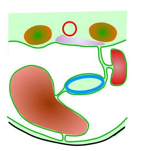
5 2. The foregut and dorsal mesentery fold to the right As a result: The dorsal mesentery is obliterated The pancreas becomes retroperitoneal

6 The dual origin of the pancreas explains the anatomical relations of the pancreas The two pancreatic buds fuse in the 6 th week to form the definitive pancreas The dorsal pancreas forms the head, body and tail The ventral pancreas forms the uncinate process The superior mesenteric artery emerges dorsal to the body and ventral to the uncinate process

7 The two pancreatic ducts are re-arranged to form a common opening into the duodenum. The main (dorsal) duct anastomosis with the ventral duct. The main duct loses its connection with the duodenum. The opening of the ventral duct becomes the main opening. The pancreatic duct and bile duct open into a common ampulla of Vater. Sphincter of Oddi develops from mesenchyme at 12 weeks
8 An Accessory Pancreatic Duct (of Santorini) Is Often Present in Normal Individuals. It is due to anomalous anostomoses between the ventral pancreatic duct and the opening of the dorsal duct. When present, the accessory pancreatic duct: receives the ducts from the uncinate process, opens into the duodenum proximal to the major papilla often communicates with the main duct.

.")

9 The acini and islets of Langerhans both differentiate from the foregut endoderm. They arise from distinct cell clusters. Islet cells Acinar cells The pancreatic acini begin to form zymogen granules from the 8 th week. They secrete digestive enzymes at 30 weeks (7 months). The islets of Langerhans differentiate into distinct cell types during the 8 th -9 th weeks in the following order: α-cells: glucagon β-cells: insulin δ-cells: somatostatin PP-cells: pancreatic polypeptide Specific granules are evident by the 9 th week. Islets secrete insulin at 21 weeks (5 months).
10 Development and Differentiation of the Pancreas The dorsal pancreas is induced by the notochord. The ventral pancreas is induced by hepatic mesoderm Differentiation of the acini depends on the presence of mesoderm, but this need not be pancreatic mesoderm. Differentiation of the islets of Langerhans does not require the presence of mesoderm or of pancreatic acini. It was originally thought that the Islets were derived from the neural crest. Evidence from quail-chick chimera cell tracers and transgenic animals indicates that islet cells are derived from forgut endoderm.
11 Anomalies of the Pancreas 1. Annular pancreas: thought to arise from the ventral pancreas migrating on both sides of the dodenum It may cause duodenal obstruction It is often associated with duodenal atresia 2. Ectopic pancreatic tissue: may occur in the duodenum, stomach or Meckel s diverticulum or rarely in the appendix, biliary tract, ileum or lungs causes vague abdominal pain due to the ectopic secretion of proteolytic enzymes may simulate peptic ulcer or appendicitis difficult to diagnose clinically

12 The liver and biliary system develop from the hepatic diverticulum The distal end proliferates rapidly in the septum transversum to form the hepatocytes and intra-hepatic biliary system The proximal part in the ventral mesogastrium proliferate slowly, becomes relatively narrow and forms the extrahepatic biliary system. A secondary outgrowth from it forms the gall bladder and cystic duct during the 4 th week (day 24)
13 Rudiments of the Liver and Biliary system Hepatic diverticulum Liver cords & Biliary duct epithelium Vitelline and umbilical veins Septum transversum mesoderm Sinusoids & portal vein Space of Disse Kupffer cells Haemopoietic tissue Hepatic artery Connective tissue
14 Rudiments of the Liver and Biliary system Hepatic diverticulum Liver cords & Biliary duct epithelium Vitelline and umbilical veins Septum transversum mesoderm Sinusoids & portal vein Space of Disse Kupffer cells Haemopoietic tissue Hepatic artery Connective tissue
15 Septum transversum mesoderm C-met receptor The septum transversum mesoderm has an inductive influence on hepatic bud endoderm. Hepatic growth factor It releases hepatic growth factor that interacts with receptors on the liver cells. Liver cords Hepatic diverticulum
16 Development of Liver Function Synthesis of serum albumin 5 th week Secretion of bile 12 th week -From the 4 th month till birth the intestinal contents are dark green and termed meconium. Glycogen storage late foetal life Haemopoiesis is a major function of the liver from the 4 th week to birth -Haemopoieticcells: are derived from septum transversum mesoderm are situated in clusters in spaces between hepatic cells.

17 Changes in the veins coursing through the septum transversum account for some anatomical features of the liver Initially veins are symmetrical

18 Paired umbilical and vitelline veins course through the septum transversum and drain into the sinus venosus Right Left Umbilical veins Vitelline veins Sinus venosus Diaphragm Septum transversum A series of anastomoses connect the right and left vitelline veins. Two anastomoses lie ventral and one dorsal to the terminal part of the foregut. The most cranial anastomosis lies within the septum transversum.
19 Proliferation of cells of the hepatic diverticulum disrupts the symmetrical pattern of the veins coursing through the septum transversum The vitelline veins break up into a plexus that surrounds the rapidly growing cords of hepatic cells and forms the liver sinusoids The hepatic veins drain the liver sinusoids into the right vitelline veins.
20 As the left horn of the sinus venosus regresses, all the veins are shunted to the right vitelline vein. 1. The left vitelline and umbilical veins lose their connections with the left horn of the sinus venosus 2. The right umbilical vein disappears completely 3. The ductus venosus shunts blood from the left umbilical to the cranial end of the right vitelline vein. 4. The liver sinusoids drain into the right vitelline veins through the hepatic veins 5. The cranial part of the right vitelline vein enlarges and forms part of the of the IVC
21 The Umbilical Veins 1. The right umbilical vein disappears completely 2. The left umbilical veins lose its connection with the left horn of the sinus venosus 3. The ductus venosus shunts blood from the left umbilical to the cranial end of the right vitelline vein. Remember! The umbilical cord has only one vein (the left umbilical vein)

22 Re-arrangement of the paired vitelline veins gives rise to the portal vein In the septum transversum - Parts of the right and left vitelline veins and the ventral anastomosis give rise to the right and left branches of the portal vein Outside the septum transversum - The portal vein develops from parts of the right and left vitelline veins and the dorsal anastomosis. The splenic and superior mesenteric veins develop join the left vitelline vein; later, the inferior mesenteric vein joins the splenic vein.

23 The portal vein develops partly from the dorsal anastomosis. This explains why it passes dorsal to the first part of the duodenum. Portal vein Superior mesenteric vein Splenic vein Inferior mesenteric vein
24 The development of the veins explain some of the anatomical features of the liver. Inferior vena cava (right vitelline v.) Hepatic veins drain into IVC Ductus venosus Portal vein Umbilical vein (left)
25 Embryological veins and their derivatives in the mature liver. A mirror image of the previous diagram illustrates the liver as seen from the posterior aspect, in order to facilitate comparison with the derivatives in the mature liver. L R Right vitelline V Inferior Vena Cava L Ductus venosus Fissure for ligamentum venosum Umbilical vein Fissure for ligamentum teres R
26 Anomalies of the Liver and Biliary System 1. Duplication of the gall bladder, sharing the same common bile duct 2. Extrahepatic biliary atresia variable absence of part or all of the biliary tree causes severe jaundice shortly after birth requires surgery to relieve the obstruction (e.g. cholecysto-gastrostomy) prognosis depends on site and extent of the atresia; more severe if proximal part is involved 3. Biliary hypoplasia due to intrahepatic biliary atresia
27
28
29
30


31 The stomach develops in the septum transversum and has dorsal and ventral mesenteries Lateral structures Paraxial mesoderm Intermediate cell mass Pleuro-peritoneal canal 28 day embryo: TS at the level of septum transversum Midline structures Neural tube Notochord Dorsal aorta Dorsal mesentery Developing Stomach Ventral mesentery The spleen develops in the dorsal mesentery. The liver develops in the ventral mesentery.
32 The liver and biliary system develop from the hepatic diverticulum The distal end of the hepatic divertivculum proliferates rapidly in the septum transversum to form the hepatocytes and intra-hepatic biliary system The proximal part in the ventral mesogastrium does not proliferate rapidly, becomes relatively narrow and forms the extrahepatic biliary system. A secondary outgrowth from it forms the gall bladder and cystic duct during the 4 th week (day 24) The vitelline and umbilical veins, disrupted by growth of hepatic cells in the septum transversum, form the liver sinusoids The septum transversum mesoderm surrounding the hepatic cords forms the Kuppfer cells and haemopoietic cells
The Foregut. At first the esophagus is short. but with descent of the heart and lungs it lengthens rapidly
 GI embryology 2 The Foregut At first the esophagus is short but with descent of the heart and lungs it lengthens rapidly The muscular coat, which is formed by surrounding splanchnic mesenchyme, is striated
Fareed Khdair, MD Assistant Professor Chief, Section of Pediatric Gastroenterology, Hepatology, and Nutrition University of Jordan School of Medicine
 Fareed Khdair, MD Assistant Professor Chief, Section of Pediatric Gastroenterology, Hepatology, and Nutrition University of Jordan School of Medicine Outline Lecture one : Gut formation Foregut: esophagus,
Development of pancreas and Small Intestine. ANATOMY DEPARTMENT DR.SANAA AL-AlSHAARAWY DR.ESSAM Eldin Salama
 Development of pancreas and Small Intestine ANATOMY DEPARTMENT DR.SANAA AL-AlSHAARAWY DR.ESSAM Eldin Salama OBJECTIVES At the end of the lecture, the students should be able to : Describe the development
- Tamara Wahbeh. - Fareed Khdair. 0 P a g e
 -1 - Tamara Wahbeh - - Fareed Khdair 0 P a g e GI Embryology Note: I included everything in the records and slides; anything in the slide not included in this sheet was not mentioned by the doctor during
Development of the Digestive System. W.S. O The University of Hong Kong
 Development of the Digestive System W.S. O The University of Hong Kong Plan for the GI system Then GI system in the abdomen first develops as a tube suspended by dorsal and ventral mesenteries. Blood
Development of the Digestive System. W.S. O School of Biomedical Sciences, University of Hong Kong.
 Development of the Digestive System W.S. O School of Biomedical Sciences, University of Hong Kong. Organization of the GI tract: Foregut (abdominal part) supplied by coeliac trunk; derivatives include
The sinus venosus represent the venous end of the heart It receives 3 veins: 1- Common cardinal vein body wall 2- Umbilical vein from placenta 3-
 1 2 The sinus venosus represent the venous end of the heart It receives 3 veins: 1- Common cardinal vein body wall 2- Umbilical vein from placenta 3- Vitelline vein from yolk sac 3 However!!!!! The left
The embryonic endoderm initially is widely connected with the yolk sac. As a consequence of cephalocaudal and lateral folding, a portion of the
 DIGESTIVE SYSTEM The embryonic endoderm initially is widely connected with the yolk sac. As a consequence of cephalocaudal and lateral folding, a portion of the endoderm-lined yolk sac cavity is incorporated
Accessory Glands of Digestive System
 Accessory Glands of Digestive System The liver The liver is soft and pliable and occupies the upper part of the abdominal cavity just beneath the diaphragm. The greater part of the liver is situated under
Embryology - GIT - Lecture 2
 Embryology - GIT - Lecture 2 Last time we talked about embryology of the GIT. We said that the development of the stomach is accompanied with the development of the duodenum and the pancreas. Also we talked
Embryology of the Midgut and Hind gut
 Embryology of the Midgut and Hind gut Prof. Abdulameer Al-Nuaimi E-mail: a.al-nuaimi@sheffield.ac.uk E-mail: abdulameerh@yahoo.com Abdominal organs www.google.co.uk/search? Development of Duodenum The
Anatomy of the liver and pancreas
 Anatomy of the liver and pancreas Prof. Abdulameer Al-Nuaimi E-mail: a.al-nuaimi@sheffield.ac.uk abdulameerh@yahoo.com Liver Aorta Pulm. Trunk Rt. At, Duct. Art. Lt. Ven. Rt. Ven. Internal Posterior
Done by: nisreen obeidat
 Sheet: liver and pancreas Done by: nisreen obeidat Embryology of the liver The liver develops in the ventral mesentery of the foregut and divides the ventral mesentery :into 1)lesser omentum (between the
MICROSCOPIC STRUCTURE OF LIVER, GALLBLADDER, GALL DUCTS, AND PANCREAS OVERVIEW OF DEVELOPMENT OF THE ALIMENTARY CANAL
 Lecture 2 ESS_3rd semester MICROSCOPIC STRUCTURE OF LIVER, GALLBLADDER, GALL DUCTS, AND PANCREAS OVERVIEW OF DEVELOPMENT OF THE ALIMENTARY CANAL MICROSCOPIC STRUCTURE OF LIVER - is the largest gland of
Pancreas & Biliary System. Dr. Vohra & Dr. Jamila
 Pancreas & Biliary System Dr. Vohra & Dr. Jamila 1 Objectives At the end of the lecture, the student should be able to describe the: Location, surface anatomy, parts, relations & peritoneal reflection
1 Right & left Hepatic ducts Gastric Impression of spleen
 Pancreatic Model 1 Right & left Hepatic ducts 14 Gastric Impression of spleen 2 Common hepatic duct 15 Renal Impression of spleen 3 Cystic Duct 16 Colic Impression of spleen 4 Common Bile Duct 17 Splenic
Embryology of the Heart
 *Page 1A: Embryology of the Heart Human embryonic disc is divided into three layers: ectoderm, intraembryonic mesoderm, and endoderm. The embryonic disc lies between the amniotic cavity and the primary
Anatomy of the SMALL INTESTINE. Dr. Noman Ullah Wazir PMC
 Anatomy of the SMALL INTESTINE Dr. Noman Ullah Wazir PMC SMALL INTESTINE The small intestine, consists of the duodenum, jejunum, and illium. It extends from the pylorus to the ileocecal junction were the
-Ensherah Mokheemer. -Shatha Al-Jaberi محمد المحتسب- 1 P a g e
 9-9 -Ensherah Mokheemer -Shatha Al-Jaberi محمد المحتسب- 1 P a g e Small intestine has three regions: ( االثني عشر( The duodenum The jejunum The ileum Small intestine Duodenum: -c-shaped -The concavity
Duodenum retroperitoneal
 Duodenum retroperitoneal C shaped Initial region out of stomach into small intestine RETROperitoneal viscus Superior 1 st part duodenal cap ; moves upwards and backwards to lie on the R crura medial to
-12. -Renad Habahbeh. -Dr Mohammad mohtasib
 -12 -Renad Habahbeh - -Dr Mohammad mohtasib The Gallbladder -The gallbladder has a body, a fundus (a rounded end), a neck, Hartmann s pouch before the neck and a cystic duct that meets the common hepatic
Surface Anatomy. Location Shape Weight Role of Five Surfaces Borders Fissures Lobes Peritoneal Lig
 The Liver Functions Bile production and secretion Detoxification Storage of glycogen Protein synthesis Production of heparin and bile pigments Erythropoiesis (in fetus) Surface Anatomy Location Shape Weight
Midgut. Over its entire length the midgut is supplied by the superior mesenteric artery
 Gi Embryology 3 Midgut the midgut is suspended from the dorsal abdominal wall by a short mesentery and communicates with the yolk sac by way of the vitelline duct or yolk stalk Over its entire length the
2/2/2011. Primitive Gut Tube Proctodeum and Stomodeum Stomach Duodenum Pancreas Liver and Biliary Apparatus Spleen Midgut
 DEVELOPMENT OF THE DIGESTIVE SYSTEM Development of Endodermal Organs Primitive Gut Tube Proctodeum and Stomodeum Stomach Duodenum Pancreas Liver and Biliary Apparatus Spleen Midgut Wednesday, February
Block 3: DISSECTION 2 CELIAC TRUNK, JEJUNUM/ILEUM, LARGE INTESTINE, DUODENUM, PANCREAS, PORTAL VEIN; MOBILIZATION OF THE LIVER
 1 Block 3: DISSECTION 2 CELIAC TRUNK, JEJUNUM/ILEUM, LARGE INTESTINE, DUODENUM, PANCREAS, PORTAL VEIN; MOBILIZATION OF THE LIVER Attempt to complete as much as you can of the dissection explained in the
Anatomy: Know Your Abdomen
 Anatomy: Know Your Abdomen Glossary Abdomen - part of the body below the thorax (chest cavity); separated by the diaphragm. Anterior - towards the front of the body. For example, the umbilicus is anterior
To describe the liver. To list main structures in porta hepatis.
 GI anatomy Lecture: 6 د. عصام طارق Objectives: To describe the liver. To list main structures in porta hepatis. To define portal system & portosystemic anastomosis. To list parts of biliary system. To
DIGESTIVE. CHAPTER 17 Lecture: Part 1 Part 2 BIO 212: ANATOMY & PHYSIOLOGY II
 BIO 212: ANATOMY & PHYSIOLOGY II CHAPTER 17 Lecture: DIGESTIVE Part 1 Part 2 Dr. Lawrence G. Altman www.lawrencegaltman.com Some illustrations are courtesy of McGraw-Hill. SMALL INTESTINE DUODENUM > JEJUNUM
Slide 154: Pancreas, H&E
 Slide 154: Pancreas, H&E the pancreas, located adjacent to the duodenum, is a mixed exocrine and endocrine gland; it is usually readily identifiable by the presence of the interspersed endocrine pancreatic
Pancreas and Biliary System
 Pancreas and Biliary System Please view our Editing File before studying this lecture to check for any changes. Color Code Important Doctors Notes Notes/Extra explanation Objectives At the end of the lecture,
Small Plicae Circularis. Short Closely packed together. Sparse, completely absent at distal part Lymphoid Nodule
 Intestines Differences Between Jejunum and Ileum Types Jejunum Ileum Color Deeper red Paler pink Calibre Bigger Smaller Thickness of wall Thick and Heavy Thin and Lighter Vascularity Highly vascularised
Digestive system L 4. Lecturer Dr. Firdous M. Jaafar Department of Anatomy/Histology section
 Digestive system L 4 Lecturer Dr. Firdous M. Jaafar Department of Anatomy/Histology section objectives 1-Describe the structure of liver. 2-Define liver lobule, and identify its zones. 3-Define portal
Common Bile Duct (CBD)
") Liver Last time we talked about the liver and the doctor started by revising some information about it: It has five surfaces. It reaches the 5 th intercostal space ; some books write that it reaches the
Liver Last time we talked about the liver and the doctor started by revising some information about it: It has five surfaces. It reaches the 5 th intercostal space ; some books write that it reaches the
When you see this diagram, remember that you are looking at the embryo from above, through the amniotic cavity, where the epiblast appears as an oval
 When you see this diagram, remember that you are looking at the embryo from above, through the amniotic cavity, where the epiblast appears as an oval disc 2 Why the embryo needs the vascular system? When
Digestive System Module 6: Accessory Organs in Digestion: The Liver, Pancreas, and Gallbladder
 Connexions module: m49293 1 Digestive System Module 6: Accessory Organs in Digestion: The Liver, Pancreas, and Gallbladder Donna Browne Based on Accessory Organs in Digestion: The Liver, Pancreas, and
Gastric Contrac,le Ac,vity. Regula,on of Gastric Emptying
 Gastric Contrac,le Ac,vity Figure 23.18 Regula,on of Gastric Emptying Gastric emptying is regulated by: Neural enterogastric reflex Hormonal (enterogastrone) mechanisms In the presence of gastric gastrin
8. Development of digestive system II. Rotation if intestine. Liver, pancreas, spleen. Development of respiratory passages and lung.
 8. Development of digestive system II. Rotation if intestine. Liver, pancreas, spleen. Development of respiratory passages and lung. Duodenum originates partially from the terminal part of the foregut,
ABDOMEN - GI. Duodenum
 TALA SALEH ABDOMEN - GI Duodenum - Notice the shape of the duodenum, it looks like capital G shape tube which extends from the pyloroduodenal junction to the duodenojejunal junction. - It is 10 inches
Dr. Zahiri. In the name of God
 Dr. Zahiri In the name of God small intestine = small bowel is the part of the gastrointestinal tract Boundaries: Pylorus Ileosecal junction Function: digestion and absorption of food It receives bile
The abdominal Esophagus, Stomach and the Duodenum. Prof. Oluwadiya KS
 The abdominal Esophagus, Stomach and the Duodenum Prof. Oluwadiya KS www.oluwadiya.com Viscera of the abdomen Abdominal esophagus: Terminal part of the esophagus The stomach Intestines: Small and Large
BLOCK IV: OFFICIAL BODY PARTS LIST FOR ANTERIOR ABDOMINAL WALL AND ABDOMINAL CONTENTS
 BLOCK IV: OFFICIAL BODY PARTS LIST FOR ANTERIOR ABDOMINAL WALL AND ABDOMINAL CONTENTS External oblique muscle Muscular portion Aponeurotic portion Superficial inguinal ring Lateral (inferior) crus Medial
Lab Monitor Images Dissection of the Abdominal Vasculature + Lower Digestive System
 Lab Monitor Images Dissection of the Abdominal Vasculature + Lower Digestive System Stomach & Duodenum Frontal (AP) View Nasogastric tube 2 1 3 4 Stomach Pylorus Duodenum 1 Duodenum 2 Duodenum 3 Duodenum
The Cardiovascular System (Part II)
") The Cardiovascular System (Part II) 黃敏銓 mchuang@ntu.edu.tw 解剖學暨細胞生物學研究所 1 Development of veins Three paired veins drain into the tubular heart of a 4-week embryo Vitelline veins: poorly oxygenated blood
The Cardiovascular System (Part II) 黃敏銓 mchuang@ntu.edu.tw 解剖學暨細胞生物學研究所 1 Development of veins Three paired veins drain into the tubular heart of a 4-week embryo Vitelline veins: poorly oxygenated blood
Embryology of the Gut and Mesenteries, slide 7 This is very similar to a cross-section we looked at when were talking about formation of the diaphragm
 Embryology of the Gut and Mesenteries, slide 2 This is a median sagittal section of a four week embryo, most of you can probably draw it in your sleep. I made a few modifications from some of the earlier
Nasogastric tube. Stomach. Pylorus. Duodenum 1. Duodenum 2. Duodenum 3. Duodenum 4
 Esophagus Barium Swallow Stomach and Duodenum 4 year old Upper GI Nasogastric tube Stomach and Duodenum 4 year old Upper GI Nasogastric tube Stomach Pylorus Duodenum 1 Duodenum 2 Duodenum 3 Duodenum 4
Biology 340 Comparative Embryology Lecture 10 Dr. Stuart Sumida. Further Development of the Mesoderm (and Endoderm)
") Biology 340 Comparative Embryology Lecture 10 Dr. Stuart Sumida Further Development of the Mesoderm (and Endoderm) Further Development: Digestive System Foregut, Midgut, Hindgut Heart and Aortic Arches
Biology 340 Comparative Embryology Lecture 10 Dr. Stuart Sumida Further Development of the Mesoderm (and Endoderm) Further Development: Digestive System Foregut, Midgut, Hindgut Heart and Aortic Arches
Development of the Great Vessels and Conduc6on Tissue
 Development of the Great Vessels and Conduc6on Tissue Development of the heart fields h:p://php.med.unsw.edu.au/embryology/ index.php?6tle=advanced_- _Heart_Fields! 2 Septa6on of the Bulbus Cordis Bulbus
In the name ofgod. Abdomen 3. Dr. Zahiri
 In the name ofgod Abdomen 3 Dr. Zahiri Peritoneum Peritoneum It is the serous membrane(a type of loose connective tissue and is covered by mesothelium) that lines the abdominal cavity. Extensions of the
The jejunum and the Ileum. Prof. Oluwadiya KS
 The jejunum and the Ileum Prof. Oluwadiya KS www.oluwadiya.siteled.com Introduction Introduction The small intestine (SI) comprises of the duodenum, jejunum and the ileum The jejunum is the second part
16 April 2010 Resident Teaching Conference. Pancreatitis. W. H. Nealon, M.D., F.A.C.S. J.J. Smith, M.D., D.W.D.
 16 April 2010 Resident Teaching Conference Pancreatitis W. H. Nealon, M.D., F.A.C.S. J.J. Smith, M.D., D.W.D. Santorini Wirsung anatomy.med.umich.edu/.../ duodenum_ans.html Bud and ductology Ventral pancreatic
Heart & vascular system I. Dawei Dong
 Heart & vascular system I Dawei Dong Lecture goal Learn the basics of heart and vascular development. Development of Heart, Blood, and Blood Vessels LEARNING GOALS: 1. explain the early development of
Exploring Anatomy: the Human Abdomen
 Exploring Anatomy: the Human Abdomen PERITONEUM AND PERITONEAL CAVITY PERITONEUM The peritoneum is a thin serous membrane that lines the abdominal cavity and covers, in variable amounts, the viscera within
LECTURE 11 & 12: ABDOMINAL VISCERA ABDOMINAL CONTENTS DIVISION. The location of abdominal viscera is divided into 4 quadrants:
 LECTURE 11 & 12: ABDOMINAL VISCERA ABDOMINAL CONTENTS DIVISION The location of abdominal viscera is divided into 4 quadrants: - horizontal line across the umbilicus divides the upper quadrants from the
DEVELOPMENT OF THE CIRCULATORY SYSTEM L E C T U R E 5
 DEVELOPMENT OF THE CIRCULATORY SYSTEM L E C T U R E 5 REVIEW OF CARDIAC ANATOMY Heart 4 chambers Base and apex Valves Pericardial sac 3 layers: epi, myo, endo cardium Major blood vessels Aorta and its
Development of Gastrointestinal Tract
 11 Development of Gastrointestinal Tract Learning Objectives At the end of this chapter, students would be able to define and understand the following: Development of the esophagus and stomach Rotation
Cardiovascular System Anatomy & Embryology
 بسم رلاهللا Cardiovascular System Anatomy & Embryology Portal circulation: Portal vessel: a vessel or a vein running between two sets of capillaries, the portal vein of the liver starts from a set of capillaries
SLIDES 6 AND 10 MM PIG SLIDES; TRANSVERSE SECTIONS AND SAGITAL; FETAL PIGS-1-8INCH; HUMAN SAGITAL DIAGRAMS:DRAWINGS OF THE PIG SECTIONS TO BE
 SLIDES 6 AND 10 MM PIG SLIDES; TRANSVERSE SECTIONS AND SAGITAL; FETAL PIGS-1-8INCH; HUMAN SAGITAL SECTION DIAGRAMS:DRAWINGS OF THE PIG SECTIONS TO BE LABELLED, AND FOUR DRAWINGS TO BE MADE. REFERENCES:PATTEN:
DIGESTIVE SYSTEM II ACCESSORY DIGESTIVE ORGANS
 DIGESTIVE SYSTEM II ACCESSORY DIGESTIVE ORGANS Dr. Larry Johnson Texas A& M University Objectives Distinguish between the parotid and submandibular salivary glands. Understand and identify the structural
ANATOMY OF THE DIGESTIVE SYSTEM PART II
 ANATOMY OF THE DIGESTIVE SYSTEM PART II 9.12.2014 Kaan Yücel M.D., Ph.D. http://fhs121.org Dr.Kaan Yücel http://fhs121.org Digestive system Part II 1. LIVER The liver is the largest gland in the body and,
Pathology of Intestinal Obstruction. Dr. M. Madhavan, MBBS., MD., MIAC, Professor of Pathology Saveetha Medical College
 Pathology of Intestinal Obstruction Dr. M. Madhavan, MBBS., MD., MIAC, Professor of Pathology Saveetha Medical College Pathology of Intestinal Obstruction Objectives list the causes of intestinal obstruction
Development of the Urinary System
 Development of the Urinary System Lecture Objectives Understand the development of the kidney and related organs of the urinary system. Define the pronephrons, mesonephrons and metanephrons. Understand
STRUCTURAL BASIS OF MEDICAL PRACTICE EXAMINATION 3. October 17, 2014
 STRUCTURAL BASIS OF MEDICAL PRACTICE EXAMINATION 3 October 17, 2014 PART l. Answer in the space provided. (12 pts) 1. Identify the structures. (2 pts) A. B. A B C. D. C D 2. Identify the structures. (2
Lecture 02 Anatomy of the LIVER
 Lecture 02 Anatomy of the LIVER BY Dr Farooq Khan Aurakzai Dated: 02.01.2018 Introduction to Liver Largest gland in the body. 2 nd largest organ of the body. Weight approximately 1500 gm, and is roughly
3 Circulatory Pathways
 40 Chapter 3 Circulatory Pathways Systemic Arteries -Arteries carry blood away from the heart to the various organs of the body. -The aorta is the longest artery in the body; it branches to give rise to
Pancreas Quizzes c. Both A and B a. Directly into the blood stream (not using ducts)
") Pancreas Quizzes Quiz 1 1. The pancreas produces hormones. Which type of hormone producing organ is the pancreas? a. Endocrine b. Exocrine c. Both A and B d. Neither A or B 2. Endocrine indicates hormones
Pancreas Quizzes Quiz 1 1. The pancreas produces hormones. Which type of hormone producing organ is the pancreas? a. Endocrine b. Exocrine c. Both A and B d. Neither A or B 2. Endocrine indicates hormones
Preview from Notesale.co.uk Page 1 of 34
 Abdominal viscera and digestive tract Digestive tract Abdominal viscera comprise majority of the alimentary system o Terminal oesophagus, stomach, pancreas, spleen, liver, gallbladder, kidneys, suprarenal
GASTROINTESTINAL SYSTEM
 GASTROINTESTINAL SYSTEM Topographic Anatomy of the Abdomen Surface Landmarks Xiphoid process T9/T10 Inferior costal margin L2/L3 Iliac Crest L4 level ASIS L5/S1 level Pubic symphysis level of greater trochanter
Development of the Pancreas and Response to Disease
 Chapter 1 Development of the Pancreas and Response to Disease The pancreas develops from the primitive gut, which is derived from endoderm. An interdependent series of signals is necessary to form the
Clinical Anatomy of the Biliary Apparatus: Relations & Variations
 Clinical Anatomy of the Biliary Apparatus: Relations & Variations Handout download: http://www.oucom.ohiou.edu/dbms-witmer/gs-rpac.htm 27 March 2007 Lawrence M. Witmer, PhD Professor of Anatomy Department
DEVELOPMENT OF THE LIVER
 THE LIVER DEVELOPMENT OF THE LIVER THE LIVER The hepa-c diver-culum develops as an outgrowth of the endoderm of the foregut in the region of the second por-on of duodenum. This diver-culum enters the ventral
(Wirsung) whose opening into the duodenum, therefore, will inevitably
 whose opening into the duodenum, therefore, will inevitably") THE APPLIED ANATOMY OF THE GALL-BLADDER AND EXTRA-IEPATIC BILIARY PASSAGES, WITH SPECIAL REFERENCE TO THEIR DEVELOPMENT Lecture delivered at The Royal College of Surgeons of England on 1st July, 1947 by
THE APPLIED ANATOMY OF THE GALL-BLADDER AND EXTRA-IEPATIC BILIARY PASSAGES, WITH SPECIAL REFERENCE TO THEIR DEVELOPMENT Lecture delivered at The Royal College of Surgeons of England on 1st July, 1947 by
Paneth Cells. Road Map to the Finish. No Review this Friday. Today 11/29 Finish digestion/accessory organs. Wednesday 12/1 Immune System I
 Road Map to the Finish No Review this Friday Today 11/29 Finish digestion/accessory organs Wednesday 12/1 Immune System I Paneth Cells - base of intestinal glands -! large -! intense acidophilic granules
Bio 322 Human Anatomy Objectives for the laboratory exercise Digestive System
 Bio 322 Human Anatomy Objectives for the laboratory exercise Digestive System Required reading before beginning this lab: Saladin, KS: Human Anatomy 5 th ed (2017) Chapter 24 For this lab you will use
BIOLOGY 30S: Fetal Pig Dissection Worksheet
 BIOLOGY 30S: Fetal Pig Dissection Worksheet Name: Part A: External Anatomy & Oral Cavity 1. How long (metric) is your fetal pig? 2. What is the age of your fetal pig? 3. What sense organs are located on
ACTIVITY 11: RESPIRATORY AND DIGESTIVE SYSTEMS RESPIRATORY SYSTEM
 ACTIVITY 11: RESPIRATORY AND DIGESTIVE SYSTEMS OBJECTIVES: 1) How to get ready: Read Chapters 25 and 26, McKinley et al., Human Anatomy, 4e. All text references are for this textbook. 2) Identify structures
Netter's Anatomy Flash Cards Section 4 List 4 th Edition
 Netter's Anatomy Flash Cards Section 4 List 4 th Edition https://www.memrise.com/course/1577335/ Section 4 Abdomen (31 cards) Plate 4-1 Bony Framework of Abdomen 1.1 Costal cartilages 1.2 Iliac crest 1.3
Chapter 7 The digestive system
 41 Chapter 7 The digestive system Primitive gut tube 41 Foregut 42 Other foregut derivatives 44 Midgut 45 Hindgut 47 4 Fig. 7.1 The gut tube in a 4-week embryo. Pharynx Foregut Respiratory diverticulum
Laboratory exercises for abdominal organs
 Laboratory exercises for abdominal organs Slide #77 (C007- H- 107A). Pancreas, dog. pancreatic islets CENTROACINAR CELLS ARE THE BEGINNING CELLS OF THE INTERCALATED DUCTS THAT DRAIN THE SECRETORY ACINI
RESPIRATORY SYSTEM. described: pp. 744,746 fig. 25.1, described: p. 746 fig described: p. 776 fig. 26.3
 ACTIVITY 11: RESPIRATORY AND DIGESTIVE SYSTEMS OBJECTIVES: 1) How to get ready: Read Chapters 25 and 26, McKinley et al., Human Anatomy, 5e. All text references are for this textbook. 2) Identify structures
Development of the Heart
 Development of the Heart Thomas A. Marino, Ph.D. Temple University School of Medicine Stages of Development of the Heart 1. The horseshoe-shaped pericardial cavity. 2. The formation of the single heart
Liver o The liver is the largest gland in the body and has a wide variety of functions. - It s an accessory organ of GIT
 بسم رلاهللا You don t need to refer to the slides, we included everything here In this lecture we will talk about Liver & Gallbladder Liver o The liver is the largest gland in the body and has a wide variety
د. عصام طارق. Objectives:
 GI anatomy Lecture: 5 د. عصام طارق Objectives: To describe anatomy of stomach, duodenum & pancreas. To list their main relations. To define their blood & nerve supply. To list their lymph drainage. To
Midterm 2 is Tuesday 5/28/13
 Business Reminder: No class Monday (Memorial Day) Midterm 2 is Tuesday 5/28/13 Optional review session tomorrow @ 5pm Homework due in Lab 1. PreLab 8 (1pt) 2. Replace a Missing Assignment (4 pts) Homework
Jhia Anjela D. Rivera 1 1. BS Biology, Department of Biology, College of Science, Polytechnic University of the Philippines
 DIGESTIVE SYSTEM Jhia Anjela D. Rivera 1 1 BS Biology, Department of Biology, College of Science, Polytechnic University of the Philippines DIGESTIVE SYSTEM Consists of the digestive tract (gastrointestinal
Biology Human Anatomy Abdominal and Pelvic Cavities
 Biology 351 - Human Anatomy Abdominal and Pelvic Cavities Please place your name and I.D. number on the back of the last page of this exam. You must answer all questions on this exam. Because statistics
SUBJECTS 2nd year, 1st semester I. 1. Primitive gut - limits, derivatives 2. Foregut -limits, evolution, derivatives 3. Midgut -limits, evolution,
 SUBJECTS 2nd year, 1st semester I. 1. Primitive gut - limits, derivatives 2. Foregut -limits, evolution, derivatives 3. Midgut -limits, evolution, derivatives 4. Hindgut- limits, evolution, derivatives
Digestive Anatomy Lab
 Digestive Anatomy Lab In-Lab Exercises I have included the word list in this document. Any descrepencies between this document and the wordlist, you should default to this document. There is a lot of repetition
Circumcaval Ureter: Embryology
 european urology supplements 5 (2006) 444 448 available at www.sciencedirect.com journal homepage: www.europeanurology.com Circumcaval Ureter: Embryology Arianna Lesma *, Aldo Bocciardi, Patrizio Rigatti
A. Incorrect! Think of a therapy that reduces prostaglandin synthesis. B. Incorrect! Think of a therapy that reduces prostaglandin synthesis.
 USMLE Step 1 - Problem Drill 02: Embryology Question No. 1 of 10 1. A premature infant is born with a patent ductus arteriosis. Which of the following treatments may be used as part of the treatment regimen?
OVARIES URETER FALLOPIAN TUBES BLADDER UROGENITAL OPENINGS (BOTH SEXES) PENIS VAGINA UTERUS
 PENIS VAGINA UTERUS") URETER OVARIES FALLOPIAN TUBES BLADDER UROGENITAL OPENINGS (BOTH SEXES) PENIS VAGINA UTERUS REPRODUCTIVE PRODUCE FEMALE HORMONES EXCRETORY FROM KIDNEY TO BLADDER EXCRETORY STORES URINE REPRODUCTIVE TRANSPORTS
URETER OVARIES FALLOPIAN TUBES BLADDER UROGENITAL OPENINGS (BOTH SEXES) PENIS VAGINA UTERUS REPRODUCTIVE PRODUCE FEMALE HORMONES EXCRETORY FROM KIDNEY TO BLADDER EXCRETORY STORES URINE REPRODUCTIVE TRANSPORTS
Blood Vessels. Types of Blood Vessels Arteries carry blood away from the heart Capillaries smallest blood vessels. Veins carry blood toward the heart
 C H A P T E R Blood Vessels 20 Types of Blood Vessels Arteries carry blood away from the heart Capillaries smallest blood vessels The site of exchange of molecules between blood and tissue fluid Veins
Respiratory & Digestive Organs of the Head and Neck, Human;
 Name Date Lab Exercise 5: Lab Exercise 6: Lab Exercise 7: Lab Exercise 8: Respiratory & Digestive Organs of the Head and Neck, Human; Histology of the Respiratory System Digestive System Models, Human
Fetal Pigs and You BIO 171 WEEK 10
 Fetal Pigs and You BIO 171 WEEK 10 The Domestic Pig: Sus scrofa Kingdom: Animalia Phylum: Chordata Class: Mammalia - Skin covered in hair or fur; Milk-producing glands (mammary glands) in the female to
The peritoneum. Prof. Oluwadiya KS, MBBS, FMCS(Orthop) Website:
 Website:") The peritoneum Prof. Oluwadiya KS, MBBS, FMCS(Orthop) Website: http://oluwadiya.com The peritoneum Serous membrane that lines the abdominopelvic cavity and invests the viscera The largest serous membrane
The peritoneum Prof. Oluwadiya KS, MBBS, FMCS(Orthop) Website: http://oluwadiya.com The peritoneum Serous membrane that lines the abdominopelvic cavity and invests the viscera The largest serous membrane
The Digestive System
 The Digestive System Identify the Structure and Function. Mesentery of the Large Intestine The mesentery functions to connect the visceral organs to the abdominal wall. Identify the Structure. Nasal Cavity
Accessory Organs in Digestion: The Liver, Pancreas, and Gallbladder *
 OpenStax-CNX module: m46496 1 Accessory Organs in Digestion: The Liver, Pancreas, and Gallbladder * OpenStax This work is produced by OpenStax-CNX and licensed under the Creative Commons Attribution License
Lectures of Human Embryology
 Lectures of Human Embryology "Body Cavities & GIT" By DR. ABDEL-MONEM AWAD HEGAZY M.B. with honor 1983, Dipl."Gynecology and Obstetrics "1989, Master "Anatomy and Embryology" 1994, M.D. "Anatomy and Embryology"
Pancreas Fox Chapter 18 part 2 (also Chapter 19.3 & 19.4)
") Vert Phys PCB3743 Pancreas Fox Chapter 18 part 2 (also Chapter 19.3 & 19.4) T. Houpt, Ph.D. Anatomy of Digestive System Peristalsis Stomach and Acid Secretion Liver and Bile Secretion Pancreas and pancreatic
Vert Phys PCB3743 Pancreas Fox Chapter 18 part 2 (also Chapter 19.3 & 19.4) T. Houpt, Ph.D. Anatomy of Digestive System Peristalsis Stomach and Acid Secretion Liver and Bile Secretion Pancreas and pancreatic