Sterile Distal Radius Kit
|
|
|
- Camron Sparks
- 5 years ago
- Views:
Transcription


1 For Fragment-Specific Fracture Fixation Using the Variable Angle LCP Two-Column Volar Distal Radius Plate 2.4 With Variable Angle Locking Technology Sterile Distal Radius Kit Surgical Technique
2 Image intensifier control Warning This description alone does not provide sufficient background for direct use of DePuy Synthes products. Instruction by a surgeon experienced in handling these products is highly recommended. Processing, Reprocessing, Care and Maintenance For general guidelines, function control and dismantling of multi-part instruments, as well as processing guidelines for implants, please contact your local sales representative or refer to: For general information about reprocessing, care and maintenance of Synthes reusable devices, instrument trays and cases, as well as processing of Synthes non-sterile implants, please consult the Important Information leaflet (SE_023827) or refer to:
3 Table of Contents Introduction Indications 3 Sterile Kit Variations and Added Instruments 4 Distal Radius Sterile Kit Key Steps 6 Sterile Tube Packaging 7 AO Principles 8 Clinical Cases 9 Surgical Technique Approach 10 Implantation Steps 11 Postoperative Treatment and Implant Removal 21 Product Information 22 MRI Information 28 Bibliography 29 Sterile Distal Radius Kit Surgical Technique DePuy Synthes 1


4 2 DePuy Synthes Sterile Distal Radius Kit Surgical Technique
5 Indications Variable Angle LCP Two-Column Volar Distal Radius Plates 2.4 are indicated for the fixation of intra- and extra-articular fractures and osteotomies of the distal radius. Sterile Distal Radius Kit Surgical Technique DePuy Synthes 2

6 Sterile Kit Variations and Added Instruments Variable Angle LCP Volar and Dorsal Distal Radius Plates 2.4. Sterile Instruments and Implants for multi-fragment fracture fixation with variable angle locking technology. Core sterile kits Core sterile kits contain both the implants and instruments to perform one distal radius surgery with a 3-shaft hole VA-LCP Two-Column Distal Radius Plate 2.4. All implants are available in both Stainless Steel and Titanium. Extra screws are provided single-packed in sterile packaging. 4 DePuy Synthes Sterile Distal Radius Kit Surgical Technique


7 Sterile Kit Variations and Added Instruments Sterile instrument kits with individually packed sterile plates and screws Instrument kits are part of a modular system with individually packed sterile plates and screws. They contain only the instruments needed to perform one distal radius surgery with the following DePuy Synthes Variable Angle radius plates: VA-LCP Two-Column Distal Radius Plates 2.4 VA-LCP Two-Column Distal Radius Plates, 2.4/2.7, Extra-Long VA-LCP Extra-articular Distal Radius Plates 2.4 VA-LCP Dorsal Distal Radius Plates 2.4 Plates and screws are available single-packed in sterile packaging. Trial implants In addition, sterile trial implants are available separately to determine the correct plate length and width of VA-LCP Two-Column Distal Radius Plates. Implant removal screwdriver The single-use T8 screwdriver is designed to facilitate implant removal without the need for a sterile instrument kit. Note that the screwdriver tip is non-retaining to facilitate maximum torque transmission. Note: All implants and instruments of the Sterile Distal Radius Kit are single use. Do not resterilize. Dispose of all unused items after surgery. Sterile Distal Radius Kit Surgical Technique DePuy Synthes 2


8 Distal Radius Sterile Kit Key Steps Note: Please refer to the corresponding IFU ( and this surgical technique for full usage information. 1 DePuy Synthes Sterile Distal Radius Kit Surgical Technique


: Remove the inner tube by pulling and if necessary slightly twisting the inner tube back and forth to release it from the outer cap.")


9 Sterile Tube Packaging The amount and lengths of screws contained in the Distal Radius Single Use Kit has been identified both scientifically to fit various anatomies and through historical sales data. For most cases, the kit will contain screws of sufficient length. It is recommended to measure each hole s depth to determine the correct screw length based on the current recommendation of the literature. 3,10 Extra screws that are not contained in the kit are available individually packed in sterile tubes. Step 3: Circulating Nurse (non-sterile field): Hold the outer cap and point the inner tube towards the sterile field. Scrub Nurse (sterile field): Remove the inner tube by pulling and if necessary slightly twisting the inner tube back and forth to release it from the outer cap. When twisting the tube, hold on to the tube and not the cap. Leading the Way to Sterile Implants The Sterile Tube concept consists of a solution that uses 2 tubes where one fits into the other to allow a streamlined and secure transfer of sterile implants into the sterile field. sterile field non-sterile field Step 4: Open the inner tube by unscrewing the inner cap from the inner tube and remove the holder with the screw. Follow the following steps to access a sterile screw: Step 1: Tear the pull-strip open to remove the sleeve. sterile field Step 5: Engage the screwdriver in the screw recess and if necessary secure with finger. Remove the screw from the holder by tilting it out of the holder/tilting the holder away at the opening. non-sterile field Step 2: Open the outer tube by unscrewing the outer cap from the outer tube. Do not touch the inner tube to maintain sterility. sterile field non-sterile field Sterile Distal Radius Kit Surgical Technique DePuy Synthes 1

10 4 DePuy Synthes Expert Lateral Femoral Nail Surgical Technique AO Principles AO PRINCIPLES In 1958, the AO formulated four basic principles, which have become the guidelines for internal fixation. 1,2 In 1958, the AO formulated four basic principles, which have become the guidelines for internal fixation 1, 2. 4_Priciples_03.pdf :08 Anatomic Anatomic reduction reduction Fracture Fracture reduction reduction and and fixation fixation to to restore restore anatomical anatomical relationships. relationships. 1 2 Stable Stable fixation fixation Fracture Fracture fixation fixation providing providing absolute absolute or relative or relative stability, stability, as required as by the required patient, by the the injury, patient, and the the injury, personality and the personality of the fracture. of the fracture. Early, Early, active active mobilization mobilization Early Early and and safe safe mobilization mobilization and and rehabilitation rehabilitation of of the the injured injured part part and and the the patient patient as as a whole. a whole. 4 3 Preservation Preservation of of blood blood supply supply Preservation Preservation of of the the blood blood supply supply to to soft soft tissues tissues and and bone bone by by gentle reduction gentle reduction techniques techniques and and careful careful handling. handling. 1 Müller ME, M Allgöwer, R Schneider, H Willenegger. Manual of Internal Fixation. 3rd ed. Berlin Heidelberg New York: Springer Rüedi 1. Müller TP, RE ME, Buckley, Allgöwer CG M, Moran. Schneider AO Principles R, Willenegger of Fracture H. Manual Management. of Internal Fixation. 2nd ed. 3rd Stuttgart, ed. Berlin, New Heidelberg, York: Thieme. New York: Springer-Verlag; Rüedi TP, RE Buckley, CG Moran. AO Principles of Fracture Management. 2nd ed. Stuttgart New York: Thieme; DePuy Synthes Sterile Distal Radius Kit Surgical Technique


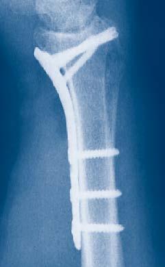
11 Clinical Cases Case 1 24-year-old male with AO 23C2.1 fracture, fall from scaffold Preoperative, AP view Preoperative, lateral view Postoperative, AP view Postoperative, lateral view Case 2 77-year-old female with AO 23C1 fracture, fall Preoperative, AP view Preoperative, lateral view Postoperative, AP view Postoperative, lateral view Sterile Distal Radius Kit Surgical Technique DePuy Synthes 9

12 Approach Make a longitudinal incision slightly radial to the flexor carpi radialis tendon (FCR). Dissect between the FCR and the radial artery, exposing the pronator quadratus. Detach the pronator quadratus from the lateral border of the radius and elevate it toward the ulna. Note: Leave the volar wrist capsule intact to avoid devascularization of the fracture fragments and destabilization of the volar wrist ligaments. 10 DePuy Synthes Sterile Distal Radius Kit Surgical Technique

. Trial implants are marked with Left (L) and Right (R) as well as the corresponding plate type (narrow, standard, wide).")
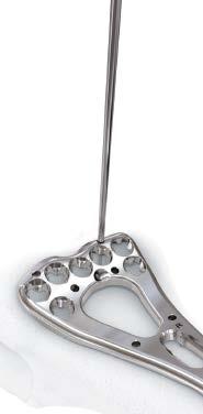
13 Implantation Steps 1. Select implant Instrument S Trial Implants for VA-LCP Two-Column Distal Radius Plate 2.4, sterile Trial implants Double-sided sterile trial implants may be used to identify the desired plate size prior to opening a distal radius sterile kit. Trial implants with 3 shaft holes are available for VA-LCP Two-Column Volar Distal Radius Plates 2.4. Trial implant choices, for VA-LCP Two-Column Volar Distal Radius Plates 2.4 (narrow, standard, wide head sizes). Trial implants are marked with Left (L) and Right (R) as well as the corresponding plate type (narrow, standard, wide). Select the correct distal radius sterile kit according to the trial implant matching the bone and fracture pattern with respect to side (left and right, facing up) and size (narrow, standard, wide). Precaution: Ensure proper plate selection by verifying the L (left) and R (right) etching on the trial implant. The trial implant s distal lip is slightly lower on the radial side. Note: Excessive and repetitive bending of the trial implants should be avoided. Trial implants are double-sided (L and R version). Sterile Distal Radius Kit Surgical Technique DePuy Synthes 11


14 Implantation Steps 2. Reduce fracture and position plate Instrument Kirschner Wires Ø 1.25 mm, with trocar tip Reduce the fracture The reduction method will be fracture-specific. Position plate Apply the plate to fit the volar surface. The order of screw insertion and the use of K-wires may vary depending on the fracture pattern and reduction technique. If necessary, use 1.25 mm K-wires inserted through the desired Kirschner-wire hole to temporarily fix the plate distally. The distal K-wire holes of the plate are designed to indicate the nominal screw trajectory angle of the screw hole medial to the respective K-wire hole. Perform several radiographic views of the distal radius to ensure alignment, plate placement, and fracture reduction. Additional options for preliminary Kirschner wire fixation 12 DePuy Synthes Sterile Distal Radius Kit Surgical Technique

15 Implantation Steps 3. Insertion of proximal screws Instruments mm Drill Bit, quick coupling mm VA/Cortex Drill Guide mm Coaxial Drill Guide /2.7 mm Depth Gauge Nm Torque-limiting Screwdriver, T8 Determine where 2.4 mm locking screws and 2.4 mm cortex screws will be used in the plate. The order of screw insertion in the shaft and metaphysis may vary depending on the fracture pattern and reduction technique. Verify plate and distal screw location with a drill bit or K-wires before inserting multiple screws Cortex Screws Drill using VA/Cortex Drill Guide Beginning with the center of the elongated hole in the shaft of the plate, drill with the 1.8 mm drill bit using the 1.8 mm VA/Cortex drill guide. Note: The 1.8 mm VA/Cortex drill guide should be fully inserted in cortex or VA plate holes. Moderate pressure should be applied to keep guide inserted in plate hole when drilling. Precaution: Excessive force should be avoided while drilling to avoid bending or damaging the drill. Sterile Distal Radius Kit Surgical Technique DePuy Synthes 12
.")
 screw Insert an appropriate length 2.")


16 Implantation Steps Insertion of proximal screws (continued) Determine screw length of cortex screws Engage the hook of the 2.4/2.7 mm depth gauge at the opposite cortex and shift the sleeve down into the hole (1). Take the reading from the edge of the sleeve (2) and select a screw length corresponding to the reading Insert cortex (non-locking) screw Insert an appropriate length 2.4 mm cortex screw in the elongated hole in the plate shaft. Adjust the plate position if necessary and tighten the screw with the screwdriver. Depending on fixation requirements, additional cortex screws may be inserted into remaining unthreaded shaft holes following the steps above. Note: If dense bone is preventing screw insertion by engaging the torque-limiting function of the screwdriver before the screw head is seated, the torque-limiting stop should be attached to the torque-limiting screwdriver. Then carefully tighten the screw paying attention not to overtighten it. For assembly of the torque-limiting stop refer to chapter Torque-limiting Stop for Dense Bone. 14 DePuy Synthes Sterile Distal Radius Kit Surgical Technique


17 Implantation Steps Insertion of proximal screws (continued) Locking Shaft Screws Drill using the Coaxial Drill Guide For locking screws, carefully insert the 1.8 mm coaxial drill guide into the threaded portion of the shaft hole until it is seated in the desired locking hole. Drill with the 1.8 mm drill bit. Note: The 1.8 mm coaxial drill guide should be inserted in-axis to the plate hole and tightened until held in plate. Excessive torque should be avoided. Precaution: Excessive force should be avoided while drilling to avoid bending or damaging the drill. Determine screw length Use the 2.4/2.7 mm depth gauge to determine the screw length.take the reading from the edge of the sleeve (see arrow). Insert a locking screw Using the T8 torque-limiting screwdriver, insert the screw perpendicular to the plate surface until the screw head is seated. Note: If dense bone is preventing screw insertion by engaging the torque-limiting function of the screwdriver before the screw head is seated, the torque-limiting stop should be attached to the torque-limiting screwdriver. Then carefully tighten the screw paying attention not to overtighten it. For assembly of the torque-limiting stop refer to chapter Torque-limiting Stop for Dense Bone. Sterile Distal Radius Kit Surgical Technique DePuy Synthes 12
 or at a variable angle (option 2).")

18 Implantation Steps 4. Insertion of distal screws For used instruments, please refer to page 13. 4a. Determine whether screws will be inserted at the pre-defined nominal angle (option 1) or at a variable angle (option 2). Option 1: Pre-defined angle drilling To follow the nominal trajectory of a locking hole, insert the 1.8 mm coaxial drill guide in-axis to the nominal angle of the plate hole. Drill with the 1.8 mm drill bit. Precaution: Excessive force should be avoided while drilling to avoid bending or damaging the drill. Option 2: Variable angle drilling Use the 1.8 mm VA/cortex drill guide. Fully seat it into the VA locking hole. Drill holes at the desired angle with the 1.8 mm drill bit. Moderate pressure should be applied to keep the guide inserted in the plate hole when drilling. Precaution: To ensure that the screw is locked correctly, do not angle it in excess of +/ 15 from the nominal trajectory of the hole. Excessive force should be avoided while drilling to avoid bending or damaging the drill. To achieve the desired angle, verify the drill bit angle under image intensifier control. If necessary, drill at a different angle and verify again under image intensifier control. 11 DePuy Synthes Sterile Distal Radius Kit Surgical Technique


19 Implantation Steps 4b. Determine screw length Use the 2.4/2.7 mm depth gauge to determine the screw length. If the fracture type allows, choose a screw length shorter than measured to ensure the screw does not protrude from the dorsal cortex. 3,10 4c. Insert VA locking screws Using the T8 torque-limiting screwdriver, insert the screw in the direction of the previously drilled hole until the screw head is seated. Sterile Distal Radius Kit Surgical Technique DePuy Synthes 11

20 Implantation Steps 5. Ensure proper joint reconstruction Ensure proper joint reconstruction, screw placement and screw length using multiple radiographic views. Verify that the distal screws are not in the joint by using additional views such as a 10 dorsally tilted, 20 inclined lateral, 45 pronated oblique, and the dorsal tangential view. 5,6 11 DePuy Synthes Sterile Distal Radius Kit Surgical Technique
 03.")


21 Implantation Steps 6. Final fixation of VA locking screws Instruments Torque-limiting Screwdriver, T Torque-limiting Stop Optional: intended to be used only in case of dense bone (optional) Use the T8 torque-limiting screwdriver to perform the final locking step for VA locking screws. Insert the screw until you hear a click. The torque-limiting screwdriver prevents over-tightening and ensures that the VA locking screws are securely locked into the plate. Note: For dense bone, visually inspect if the screw is appropriately threaded and seated after tightening with the torque-limiting screwdriver. If dense bone is preventing screw insertion, the torque-limiting stop should be attached to the torque-limiting screwdriver. Then carefully tighten the screw until the head is flush with the plate surface. For a detailed description of screw insertion and assembly of the torque-limiting stop refer to chapter Torque-limiting Stop for Dense Bone. Precaution: Remove the torque-limiting stop from the screwdriver when not necessary to overcome dense bone. Sterile Distal Radius Kit Surgical Technique DePuy Synthes 19

 03.111.")


22 Implantation Steps Torque-limiting Stop for Dense Bone (optional) Instruments Torque-limiting Screwdriver, T Torque-limiting Stop Optional: intended to be used only in case of dense bone (optional) Visually inspect if the screw is appropriately threaded and seated after tightening with the torque-limiting handle. If dense bone is preventing screw insertion, the torque-limiting stop should be attached to the torque-limiting handle. To attach the torque-limiting stop ensure that the triangles on both the torque-limiting stop and the handle are aligned. Rotation may be required for alignment. Then carefully tighten so that the screw head is flush with the plate surface. Confirm final locking with the torque-limiting stop removed. Precaution: Remove the torque-limiting stop from the screwdriver when not necessary to overcome dense bone. 20 DePuy Synthes Sterile Distal Radius Kit Surgical Technique


23 Postoperative Treatment and Implant Removal Postoperative treatment Postoperative treatment with VA locking compression plates does not differ from conventional internal fixation procedures and is at the discretion of the clinician. Implant removal Instrument S Screwdriver, T8, non-retaining, sterile For implant removal The screwdriver for implant removal is not designed for screw retention. Before attempting screw removal clean all screw head recesses of tissue or ingrown/overgrown bone using a dental pick or other appropriate instrument. To remove locking screws, first unlock all screws from the plate; then remove the screws completely from the bone. The last screw removed should be a non-locking screw on the shaft. This prevents the plate from spinning when locking screws are removed. Sterile Distal Radius Kit Surgical Technique DePuy Synthes 22
 holes holes (mm) Right 02.111.530KS narrow 19.5 6 3 51 R 02.111.531KS narrow 19.")
 holes holes (mm) Right 04.111.530KS narrow 19.")


, and screws are made from titanium alloy (Ti-6Al-7Nb).")
24 Product Information Sterile Implant and Instrument Kits with VA-LCP Two-Column Volar Distal Radius Plates Stainless Steel* Head size Head Shaft Length Left/ Part number (mm) holes holes (mm) Right KS narrow R KS narrow L KS standard R KS standard L KS wide R KS wide L Titanium** Head size Head Shaft Length Left/ Part number (mm) holes holes (mm) Right KS narrow R KS narrow L KS standard R KS standard L KS wide R KS wide L Trial Implants for Two-Column Distal Radius Plates S Trial Implants for VA-LCP Two-Column Distal Radius Plates 2.4, sterile S Trial Implants for VA-LCP Two-Column Distal Radius Plates 2.4/2.7, Extra-Long, sterile Implant Removal Screwdriver S Screwdriver, T8, non-retaining, sterile For implant removal S S * Plates and screws are made from stainless steel (316L). ** Plates are made from commercially pure titanium (TiCP), and screws are made from titanium alloy (Ti-6Al-7Nb). 22 DePuy Synthes Sterile Distal Radius Kit Surgical Technique

 Right 02.")
 Right 02.111.720S 04.111.720S 7 2 47 R 02.")
 and commercially pure titanium (TiCP) materials.")
25 Product Information Instrument-only Kit S VA-LCP Instrument Kit 2.4, sterile Individually Packed Sterile Plates Available for use with Instrument-only Kit VA-LCP Two-Column Volar Distal Radius Plates 2.4, sterile Narrow, width 19.5 mm Head Shaft Length Left/ Stainless Steel Titanium holes holes (mm) Right S S R S S L S S R S S L S S R S S L S S R S S L Standard, width 22 mm Head Shaft Length Left/ Stainless Steel Titanium holes holes (mm) Right S S R S S L S S R S S L S S R S S L S S R S S L Wide, width 25.5 mm Head Shaft Length Left/ Stainless Steel Titanium holes Holes (mm) Right S S R S S L S S R S S L S S R S S L S S R S S L All plates are sterile packed. Images show selected right plates in both stainless steel (316L) and commercially pure titanium (TiCP) materials. Sterile Distal Radius Kit Surgical Technique DePuy Synthes 22

26 Product Information VA-LCP Two-Column Volar Distal Radius Plates 2.4/2.7, Extra-Long, sterile Head size Head Shaft Length Left/ Stainless Steel Titanium (mm) holes holes (mm) Right S S narrow R S S narrow L S S standard R S S standard L S S wide R S S wide L S S standard R S S standard L S S standard R S S standard L VA-LCP Extra-Articular Volar Distal Radius Plates 2.4, sterile Head Shaft Length Left/ Stainless Steel Titanium holes holes (mm) Right S S R S S L S S R S S L S S R S S L S S R S S L All plates are sterile packed. Images show selected right plates in both stainless steel (316L) and commercially pure titanium (TiCP) materials. 24 DePuy Synthes Sterile Distal Radius Kit Surgical Technique
 are angled left and the plates for the left radius (0x.")
 Angle 02.115.130S 04.115.130S 3 37 +90 02.115.131S 04.115.131S 3 37-90 02.115.150S 04.115.150S 5 51 +90 02.")
27 Product Information VA-LCP Dorsal Distal Radius Plates 2.4, sterile Radial Column Shaft Length Stainless Steel Titanium holes (mm) S S S S 6 57 Intermediate Column, 2 head holes Note: The plates for the right radius (0x and 0x ) are angled left and the plates for the left radius (0x and 0x ) are angled right. Shaft Length Stainless Steel Titanium holes (mm) Angle S S S S S S S S L-Plates, 2 head holes Shaft Length Stainless Steel Titanium holes (mm) Angle S S S S S S S S L-Plates, 3 head holes Shaft Length Stainless Steel Titanium holes (mm) Angle S S S S S S S S All plates are sterile packed. Images show selected right plates in both stainless steel (316L) and commercially pure titanium (TiCP) materials. Sterile Distal Radius Kit Surgical Technique DePuy Synthes 22


 and commercially pure titanium (TiCP)")
28 Product Information L-Plates, oblique, 3 head holes Shaft Length Stainless Steel Titanium holes (mm) Angle S S S S S S S S T-Plates, 3 head holes Shaft Length Stainless Steel Titanium holes (mm) S S Note: This Surgical Technique does not include information necessary for selection and use of Variable Angle LCP Extra-Articular Volar Distal Radius Plates 2.4 and Variable Angle LCP Dorsal Distal Radius Plates 2.4. Please refer to the corresponding Surgical Technique for all necessary implant-specific information. All plates are sterile packed. Images show selected right plates in both stainless steel (316L) and commercially pure titanium (TiCP) materials. 21 DePuy Synthes Sterile Distal Radius Kit Surgical Technique
 02.210.108TS 04.210.108TS 8 02.210.110TS 04.210.110TS 10 02.210.112TS 04.210.112TS 12 02.")


29 Product Information Individually Packed Sterile Screws Ordering Information Variable Angle Locking Screws Ø 2.4 mm, with T8 StarDrive recess, tube sterile Stainless Steel* Titanium* Length (mm) TS TS TS TS TS TS TS TS TS TS TS TS TS TS TS TS TS TS TS TS TS TS TS TS 30 Cortex Screws Ø 2.4 mm, with T8 StarDrive recess, tube sterile Stainless Steel* Titanium* Length (mm) TS TS TS TS TS TS TS TS TS TS TS TS TS TS TS TS TS TS TS TS TS TS TS TS 30 * Stainless Steel (316L) Titanium Alloy (Ti-6AI-7Nb) Sterile Distal Radius Kit Surgical Technique DePuy Synthes 22
30 MRI Information Torque, Displacement and Image Artifacts according to ASTM F , ASTM F e1 and ASTM F Non-clinical testing of worst case scenario in a 3 T MRI system did not reveal any relevant torque or displacement of the construct for an experimentally measured local spatial gradient of the magnetic field of 3.69 T/m. The largest image artifact extended approximately 169 mm from the construct when scanned using the Gradient Echo (GE). Testing was conducted on a 3 T MRI system. Radio-Frequency-(RF-)induced heating according to ASTM F a Non-clinical electromagnetic and thermal testing of worst case scenario lead to peak temperature rise of 9.5 C with an average temperature rise of 6.6 C (1.5 T) and a peak temperature rise of 5.9 C (3 T) under MRI Conditions using RF Coils [whole body averaged specific absorption rate (SAR) of 2 W/kg for 6 minutes (1.5 T) and for 15 minutes (3 T)]. Precautions: The above mentioned test relies on non-clinical testing. The actual temperature rise in the patient will depend on a variety of factors beyond the specific absorption rate (SAR) and time of RF application. Thus, it is recommended to pay particular attention to the following points: It is recommended to thoroughly monitor patients undergoing MR scanning for perceived temperature and/or pain sensations. Patients with impaired thermo regulation or temperature sensation should be excluded from MR scanning procedures. Generally it is recommended to use a MR system with low field strength in the presence of conductive implants. The employed SAR should be reduced as far as possible. Using the ventilation system may further contribute to reduce temperature increase in the body. 22 DePuy Synthes Sterile Distal Radius Kit Surgical Technique
31 Bibliography 1. Brink PR, Rikli DA. Four-Corner Concept: CT-Based Assessment of Fracture Patterns in Distal Radius. J Wrist Surg. 2016;5: Hart A, Collins M, Chhatwal D, Steffen T, Harvey EJ, Martineau PA. Can the use of variable-angle volar locking plates compensate for suboptimal plate positioning in unstable distal radius fractures? A biomechanical study. J Orthop Trauma. 2015;29: Löw S, Herold D, Eingartner C. Standardized Palmar Plating of Dorsally Displaced Distal Radius Fractures. Tech Hand Up Extrem Surg. 2013;17: Oppermann J, Wacker M, Stein G, Springorum HP, Neiss WF, Burkhart KJ, Eysel P, Dargel J. Anatomical fit of seven different palmar distal radius plates. Arch Orthop Trauma Surg. 2014;134: Ozer K, Wolf JM, Watkins B, Hak DJ. Comparison of 4 fluoroscopic views for dorsal cortex screw penetration after volar plating of the distal radius. J Hand Surg Am. 2012;37: Pace A, Cresswell T. Use of articular wrist views to assess intra-articular screw penetration in surgical fixation of distal radius fractures. J Hand Surg Am. 2010;35: Rausch S, Klos K, Stephan H, Hoffmeier K, Gras F, Windolf M, Gueorguiev B, Hofmann GO, Mückley T. Evaluation of a polyaxial angle-stable volar plate in a distal radius C-fracture model--a biomechanical study. Injury. 2011;42: Rausch S, Schlonski O, Klos K, Gras F, Gueorguiev B, Hofmann GO, Mückley T. Volar versus dorsal latest-generation variable-angle locking plates for the fixation of AO type 23C 2.1 distal radius fractures: a biomechanical study in cadavers. Injury. 2013;44: Stanbury SJ, Salo A, Elfar JC. Biomechanical analysis of a volar variable-angle locking plate: the effect of capturing a distal radial styloid fragment. J Hand Surg Am. 2012;37: Tarallo L, Mugnai R, Zambianchi F, Adani R, Catani F. Volar plate fixation for the treatment of distal radius fractures: analysis of adverse events. J Orthop Trauma. 2013;27: Sterile Distal Radius Kit Surgical Technique DePuy Synthes 22
32 Synthes GmbH Eimattstrasse 3 Not all products are currently available in all markets Oberdorf Switzerland This publication is not intended for distribution in the USA. Tel: Fax: All surgical techniques are available as PDF files at DePuy Synthes Trauma, a division of Synthes GmbH All rights reserved. DSEM/TRM/1216/0773 1/17
2.4 mm Variable Angle LCP Volar Extra-Articular Distal Radius System. For fragment-specific fracture fixation with variable angle locking technology.
 2.4 mm Variable Angle LCP Volar Extra-Articular Distal Radius System. For fragment-specific fracture fixation with variable angle locking technology. Surgical Technique This publication is not intended
LCP Proximal Radius Plates 2.4. Plates for radial head rim and for radial head neck address individual fracture patterns of the proximal radius.
 LCP Proximal Radius Plates 2.4. Plates for radial head rim and for radial head neck address individual fracture patterns of the proximal radius. Surgical Technique This publication is not intended for
LCP Metaphyseal Plates. For extra-articular fractures.
 LCP Metaphyseal Plates. For extra-articular fractures. Surgical Technique This publication is not intended for distribution in the USA. Instruments and implants approved by the AO Foundation. Image intensifier
LCP Proximal Radius Plates 2.4. Plates for radial head rim and for radial head neck address individual fracture patterns of the proximal radius.
 LCP Proximal Radius Plates 2.4. Plates for radial head rim and for radial head neck address individual fracture patterns of the proximal radius. Surgical Technique This publication is not intended for
Long Volar Plates for Diaphyseal-Metaphyseal Radius Fractures LCP. Dia-Meta Volar Distal Radius Plates. Surgical Technique
 Long Volar Plates for Diaphyseal-Metaphyseal Radius Fractures LCP Dia-Meta Volar Distal Radius Plates Surgical Technique Table of Contents Introduction LCP Dia-Meta Volar Distal Radius Plates 2 AO Principles
Technique Guide. 2.4 mm Variable Angle LCP Distal Radius System. For fragment-specific fracture fixation with variable angle locking technology.
 Technique Guide 2.4 mm Variable Angle LCP Distal Radius System. For fragment-specific fracture fixation with variable angle locking technology. Table of Contents Introduction 2.4 mm Variable Angle LCP
LCP DISTAL TIBIA PLATE
 LCP DISTAL TIBIA PLATE Instruments and implants approved by the AO Foundation. This publication is not intended for distribution in the USA. SURGICAL TECHNIQUE Image intensifier control This description
2.4 mm Variable Angle LCP Volar Extra-Articular Distal Radius System. For fragment-specific fracture fixation with variable angle locking technology.
 Technique Guide 2.4 mm Variable Angle LCP Volar Extra-Articular Distal Radius System. For fragment-specific fracture fixation with variable angle locking technology. Table of Contents Introduction 2.4
OBSOLETED. LCP Medial Distal Tibia Plate, without Tab. The Low Profile Anatomic Fixation System with Angular Stability and Optimal Screw Orientation.
 LCP Medial Distal Tibia Plate, without Tab. The Low Profile Anatomic Fixation System with Angular Stability and Optimal Screw Orientation. Surgical Technique LCP Small Fragment System This publication
Variable Angle LCP Volar Rim Distal Radius Plate 2.4. For fragment-specific fracture fixation with variable angle locking technology.
 Technique Guide Variable Angle LCP Volar Rim Distal Radius Plate 2.4. For fragment-specific fracture fixation with variable angle locking technology. Image intensifier control Warning This description
LCP Low Bend Medial Distal Tibia Plates 3.5 mm. Anatomic plates with low profile head for intra- and extraarticular fractures.
 LCP Low Bend Medial Distal Tibia Plates 3.5 mm. Anatomic plates with low profile head for intra- and extraarticular fractures. Surgical Technique This publication is not intended for distribution in the
The Calcaneal Plate. The Synthes non-locking solution for the Calcaneus.
 The Calcaneal Plate. The Synthes non-locking solution for the Calcaneus. Surgical Technique This publication is not intended for distribution in the USA. Instruments and implants approved by the AO Foundation.
Distal Radius Plate 2.4/2.7 dorsal and volar
 Distal Radius Plate 2.4/2.7 dorsal and volar Surgical Technique This publication is not intended for distribution in the USA. Instruments and implants approved by the AO Foundation. Distal Radius Plate
LCP Distal Fibula Plates. Part of the Synthes locking compression plate (LCP) system.
 system.") LCP Distal Fibula Plates. Part of the Synthes locking compression plate (LCP) system. Surgical Technique This publication is not intended for distribution in the USA. Instruments and implants approved
LCP Distal Fibula Plates. Part of the Synthes locking compression plate (LCP) system. Surgical Technique This publication is not intended for distribution in the USA. Instruments and implants approved
3.5 mm LCP Olecranon Plates
 Part of the DePuy Synthes Locking Compression Plate (LCP ) System 3.5 mm LCP Olecranon Plates Surgical Technique Table of Contents Introduction 3.5 mm LCP Olecranon Plates 2 AO Principles 3 Indications
2.4 mm Variable Angle LCP Volar Rim Distal Radius Plates
 For Fragment-Specific Fracture Fixation With Variable Angle Locking Technology 2.4 mm Variable Angle LCP Volar Rim Distal Radius Plates Surgical Technique Table of Contents Introduction 2.4 mm Variable
Surgical Technique. This publication is not intended for distribution in the USA. Instruments and implants approved by the AO Foundation.
 LCP Extra-articular Distal Humerus Plate. The anatomically shaped and angular stable fixation system for extraarticular fractures of the distal humerus. Surgical Technique This publication is not intended
Low Bend Distal Tibia Plates
 Part of the DePuy Synthes Locking Compression Plate (LCP ) System 3.5 mm LCP Low Bend Medial Distal Tibia Plates Surgical Technique Table of Contents Introduction 3.5 mm LCP Low Bend Medial Distal Tibia
3.5 mm LCP Extra-articular Distal Humerus Plate
 Part of the DePuy Synthes Locking Compression Plate (LCP ) System 3.5 mm LCP Extra-articular Distal Humerus Plate Surgical Technique Table of Contents Introduction 3.5 mm LCP Extra-articular Distal Humerus
PHILOS and PHILOS Long. The anatomic fixation system for the proximal humerus.
 PHILOS and PHILOS Long. The anatomic fixation system for the proximal humerus. Surgical Technique This publication is not intended for distribution in the USA. Instruments and implants approved by the
VA-LCP Olecranon Plates 2.7/3.5. The fracture-specific low-profile fixation system with variable angle locking technology.
 VA-LCP Olecranon Plates 2.7/3.5. The fracture-specific low-profile fixation system with variable angle locking technology. Surgical Technique This publication is not intended for distribution in the USA.
LCP Distal Fibula Plates. Part of the Synthes locking compression plate (LCP) system.
 system.") LCP Distal Fibula Plates. Part of the Synthes locking compression plate (LCP) system. Surgical Technique This publication is not intended for distribution in the USA. Instruments and implants approved
LCP Distal Fibula Plates. Part of the Synthes locking compression plate (LCP) system. Surgical Technique This publication is not intended for distribution in the USA. Instruments and implants approved
VA LOCKING CALCANEAL PLATES 2.7
 VA LOCKING CALCANEAL PLATES 2.7 Instruments and Implants approved by the AO Foundation. This publication is not intended for distribution in the USA. SURGICAL TECHNIQUE Image intensifier control Warning
LCP Proximal Radius Plates 2.4. Plates for radial head rim and for radial head neck address individual fracture patterns of the proximal radius.
 Technique Guide LCP Proximal Radius Plates 2.4. Plates for radial head rim and for radial head neck address individual fracture patterns of the proximal radius. Table of Contents Introduction LCP Proximal
LCP Ulna Osteotomy System 2.7. Low profile angular stable fixation for ulna shortening osteotomies.
 LCP Ulna Osteotomy System 2.7. Low profile angular stable fixation for ulna shortening osteotomies. Surgical Technique This publication is not intended for distribution in the USA. Instruments and implants
VA-Locking Intercarpal Fusion System. Variable angle locking technology for mediocarpal partial arthrodesis.
 VA-Locking Intercarpal Fusion System. Variable angle locking technology for mediocarpal partial arthrodesis. Surgical Technique This publication is not intended for distribution in the USA. Image intensifier
LCP Periarticular Proximal Humerus Plate 3.5. The anatomic fixation system with anterolateral shaft placement.
 LCP Periarticular Proximal Humerus Plate 3.5. The anatomic fixation system with anterolateral shaft placement. Surgical Technique This publication is not intended for distribution in the USA. Instruments
VA-LCP Anterior Clavicle Plate. The anatomically precontoured fixation system with angular stability for clavicle shaft and lateral clavicle.
 VA-LCP Anterior Clavicle Plate. The anatomically precontoured fixation system with angular stability for clavicle shaft and lateral clavicle. Surgical Technique This publication is not intended for distribution
Technique Guide. 3.5 mm LCP Low Bend Medial Distal Tibia Plates. Part of the Synthes locking compression plate (LCP) system.
 system.") Technique Guide 3.5 mm LCP Low Bend Medial Distal Tibia Plates. Part of the Synthes locking compression plate (LCP) system. Table of Contents Introduction 3.5 mm LCP Low Bend Medial Distal Tibia Plates
Technique Guide 3.5 mm LCP Low Bend Medial Distal Tibia Plates. Part of the Synthes locking compression plate (LCP) system. Table of Contents Introduction 3.5 mm LCP Low Bend Medial Distal Tibia Plates
Button Plate. Reinforcement for transosseous fixations.
 Button Plate. Reinforcement for transosseous fixations. Product Information This publication is not intended for distribution in the USA. Instruments and implants approved by the AO Foundation. Image intensifier
3.5 mm LCP Distal Humerus Plates
 Part of the DePuy Synthes Locking Compression Plate (LCP ) System 3.5 mm LCP Distal Humerus Plates Surgical Technique Table of Contents Introduction 3.5 mm LCP Distal Humerus Plates 2 AO Principles 4 Indications
Wrist Fusion Instrument and Implant Set.
 Wrist Fusion Instrument and Implant Set. Surgical Technique Discontinued December 2016 DSEM/TRM/0815/0479(2) This publication is not intended for distribution in the USA. Instruments and implants approved
LCP Superior Anterior Clavicle Plate. The anatomically precontoured fixation system with angular stability for clavicle shaft and lateral clavicle.
 LCP Superior Anterior Clavicle Plate. The anatomically precontoured fixation system with angular stability for clavicle shaft and lateral clavicle. Surgical Technique This publication is not intended for
2.7 mm/3.5 mm LCP Distal Fibula Plate
 Part of the DePuy Synthes Locking Compression Plate (LCP ) System 2.7 mm/3.5 mm LCP Distal Fibula Plate Surgical Technique Table of Contents Introduction 2.7 mm/3.5 mm LCP Distal Fibula Plates 2 AO Principles
Technique Guide. 3.5 mm LCP Olecranon Plates. Part of the Synthes locking compression plate (LCP) system.
 system.") Technique Guide 3.5 mm LCP Olecranon Plates. Part of the Synthes locking compression plate (LCP) system. Table of Contents Introduction 3.5 mm LCP Olecranon Plates 2 AO Principles 3 Indications 3 Clinical
Technique Guide 3.5 mm LCP Olecranon Plates. Part of the Synthes locking compression plate (LCP) system. Table of Contents Introduction 3.5 mm LCP Olecranon Plates 2 AO Principles 3 Indications 3 Clinical
2.4 mm Variable Angle LCP Dorsal Distal Radius Plate
 For Fragment-Specific Fracture Fixation With Variable Angle (VA) Locking Technology 2.4 mm Variable Angle LCP Dorsal Distal Radius Plate Surgical Technique Table of Contents Introduction 2.4 mm VA LCP
LCP Medial Proximal Tibial Plate 3.5. Part of the Synthes small fragment Locking Compression Plate (LCP) system.
 system.") LCP Medial Proximal Tibial Plate 3.5. Part of the Synthes small fragment Locking Compression Plate (LCP) system. Surgical Technique This publication is not intended for distribution in the USA. Instruments
LCP Medial Proximal Tibial Plate 3.5. Part of the Synthes small fragment Locking Compression Plate (LCP) system. Surgical Technique This publication is not intended for distribution in the USA. Instruments
2.4 mm LCP Radial Head Plates. Part of the Synthes LCP Distal Radius Plate System.
 2.4 mm LCP Radial Head Plates. Part of the Synthes LCP Distal Radius Plate System. Technique Guide Instruments and Implants approved by the AO Foundation Table of Contents Introduction 2.4 mm LCP Radial
LCP Condylar Plate 4.5/5.0. Part of the LCP Periarticular Plating System.
 LCP Condylar Plate 4.5/5.0. Part of the LCP Periarticular Plating System. Surgical Technique This publication is not intended for distribution in the USA. Instruments and implants approved by the AO Foundation.
3.5 mm LCP Clavicle Hook Plates
 Part of the Synthes Locking Compression Plate (LCP ) System 3.5 mm LCP Clavicle Hook Plates Surgical Technique Table of Contents Introduction 3.5 mm LCP Clavicle Hook Plates 2 AO Principles 4 Indications
3.5 mm LCP Hook Plate
 Part of the DePuy Synthes Locking Compression Plate (LCP ) System 3.5 mm LCP Hook Plate Surgical Technique Table of Contents Introduction 3.5 mm LCP Hook Plate 2 AO Principles 4 Indications 5 Clinical
ANGLED BLADE PLATES FOR ADULTS
 ANGLED BLADE PLATES FOR ADULTS Instruments and implants approved by the AO Foundation. This publication is not intended for distribution in the USA. SURGICAL TECHNIQUE Image intensifier control This description
3.5 mm LCP Low Bend Medial Distal Tibia Plate Aiming Instruments
 Part of the 3.5 mm LCP 3.5 mm LCP Low Bend Medial Distal Tibia Plate Aiming Instruments Surgical Technique TABLE OF CONTENTS INTRODUCTION 3.5 mm LCP Low Bend Medial Distal Tibia Plate 2 Aiming Instruments
Femoral Neck System. Surgical Technique
 Femoral Neck System Surgical Technique Image intensifier control This description alone does not provide sufficient background for direct use of DePuy Synthes products. Instruction by a surgeon experienced
Part of the DePuy Synthes Locking Compression Plate (LCP ) System. 3.5 mm LCP Medial Proximal Tibia Plates
 System. 3.5 mm LCP Medial Proximal Tibia Plates") Part of the DePuy Synthes Locking Compression Plate (LCP ) System 3.5 mm LCP Medial Proximal Tibia Plates Surgical Technique Table of Contents Introduction 3.5 mm LCP Medial Proximal Tibia Plates 2 AO
Part of the DePuy Synthes Locking Compression Plate (LCP ) System 3.5 mm LCP Medial Proximal Tibia Plates Surgical Technique Table of Contents Introduction 3.5 mm LCP Medial Proximal Tibia Plates 2 AO
3.0/3.5/4.0/4.5/6.5/7.0/7.3. Cannulated Screws. Surgical Technique
 3.0/3.5/4.0/4.5/6.5/7.0/7.3 Cannulated Screws Surgical Technique Image intensifier control This description alone does not provide sufficient background for direct use of DePuy Synthes products. Instruction
LCP Percutaneous Aiming System 3.5 for PHILOS. For less invasive surgery at the proximal humerus.
 LCP Percutaneous Aiming System 3.5 for PHILOS. For less invasive surgery at the proximal humerus. Surgical Technique This publication is not intended for distribution in the USA. Instruments and implants
VA-LCP Anterior Clavicle Plate. The anatomically precontoured fixation system with angular stability for clavicle shaft and lateral clavicle.
 Technique Guide VA-LCP Anterior Clavicle Plate. The anatomically precontoured fixation system with angular stability for clavicle shaft and lateral clavicle. Table of Contents Introduction VA-LCP Anterior
LCP Superior Clavicle Plate. The anatomically precontoured fixation system with angular stability for clavicle shaft and lateral clavicle.
 LCP Superior Clavicle Plate. The anatomically precontoured fixation system with angular stability for clavicle shaft and lateral clavicle. Surgical Technique This publication is not intended for distribution
LCP Wrist Fusion Set. Anatomic plates for total wrist fusion.
 LCP Wrist Fusion Set. Anatomic plates for total wrist fusion. Surgical Technique This publication is not intended for distribution in the USA. Instruments and implants approved by the AO Foundation. Image
LCP Medial Distal Tibia Plate, without Tab. The Low Profile Anatomic Fixation System with Angular Stability and Optimal Screw Orientation.
 LCP Medial Distal Tibia Plate, without Tab. The Low Profile Anatomic Fixation System with Angular Stability and Optimal Screw Orientation. Technique Guide LCP Small Fragment System Table of Contents Introduction
VA-LCP Distal Humerus Plates 2.7/3.5. The low-profile fixation system with variable angle locking technology.
 VA-LCP Distal Humerus Plates 2.7/3.5. The low-profile fixation system with variable angle locking technology. Surgical Technique This publication is not intended for distribution in the USA. Instruments
VA LCP MEDIAL COLUMN FUSION PLATES 3.5
 VA LCP MEDIAL COLUMN FUSION PLATES 3.5 Instruments and Implants approved by the AO Foundation. This publication is not intended for distribution in the USA. SURGICAL TECHNIQUE Image intensifier control
Technique Guide. LCP Distal Fibula Plates. Part of the Synthes locking compression plate (LCP) system.
 system.") Technique Guide LCP Distal Fibula Plates. Part of the Synthes locking compression plate (LCP) system. Table of Contents Introduction LCP Distal Fibula Plates 2 AO Principles 4 Indications 5 Surgical Technique
Technique Guide LCP Distal Fibula Plates. Part of the Synthes locking compression plate (LCP) system. Table of Contents Introduction LCP Distal Fibula Plates 2 AO Principles 4 Indications 5 Surgical Technique
Instrument and Implant for wrist fracture
 Instrument and Implant for wrist fracture Jansri Janpanya Product specialist The Bangkok Unitrade Co,.ltd. Objectives Type of LCP for distal radius Fx. The new LCP design for distal radius Fx. Have knowledge
LCP Condylar Plate 4.5/5.0. Part of the LCP Periarticular Plating System.
 LCP Condylar Plate 4.5/5.0. Part of the LCP Periarticular Plating System. Surgical Technique This publication is not intended for distribution in the USA. Instruments and implants approved by the AO Foundation.
2.4 mm Variable Angle Locking Intercarpal Fusion System
 For Partial Wrist Arthrodesis With Variable Angle Locking Technology 2.4 mm Variable Angle Locking Intercarpal Fusion System Surgical Technique Table of Contents Introduction 2.4 mm Variable Angle Locking
Technique Guide. PHILOS and PHILOS Long. The anatomic fixation system for the proximal humerus.
 Technique Guide PHILOS and PHILOS Long. The anatomic fixation system for the proximal humerus. Table of Contents Introduction PHILOS and PHILOS Long 2 AO Principles 4 Indications 5 Surgical Technique
2.4 mm Variable Angle LCP Intercarpal Fusion System. Variable angle locking technology for mediocarpal partial arthrodesis.
 2.4 mm Variable Angle LCP Intercarpal Fusion System. Variable angle locking technology for mediocarpal partial arthrodesis. Technique Guide Instruments and implants approved by the AO Foundation Table
LOW PROFILE NEURO. This publication is not intended for distribution in the USA. SURGICAL TECHNIQUE
 LOW PROFILE NEURO This publication is not intended for distribution in the USA. SURGICAL TECHNIQUE TABLE OF CONTENTS INTRODUCTION Low Profile Neuro Plating System 2 Intended Use, Indications, Contraindications
MEFiSTO. Monolateral External Fixation System for Trauma and Orthopaedics.
 MEFiSTO. Monolateral External Fixation System for Trauma and Orthopaedics. Surgical Technique This publication is not intended for distribution in the USA. Instruments and implants approved by the AO Foundation.
Variable Angle LCP Two-Column Volar Distal Radius Plate 2.4. For fragment-specific fracture fixation with variable angle locking technology.
 Technique Guide Variable Angle LCP Two-Column Volar Distal Radius Plate 2.4. For fragment-specific fracture fixation with variable angle locking technology. Image intensifier control Warning This description
LCP Proximal Tibial Plate 4.5/5.0 with Periarticular Aiming Arm Instruments
 LCP Proximal Tibial Plate 4.5/5.0 with Periarticular Aiming Arm Instruments Surgical Technique This publication is not intended for distribution in the USA. Instruments and implants approved by the AO
The Locking Calcaneal Plate Instrument and Implant Sets
 Part of the DePuy Synthes Locking Compression Plate (LCP ) System The Locking Calcaneal Plate Instrument and Implant Sets Surgical Technique Table of Contents Introduction Locking Calcaneal Plate 2 AO
Technique Guide. 3.5 mm LCP Low Bend Medial Distal Tibia Plate Aiming Instruments. Part of the 3.5 mm LCP Percutaneous Instrument System.
 Technique Guide 3.5 mm LCP Low Bend Medial Distal Tibia Plate Aiming Instruments. Part of the 3.5 mm LCP Percutaneous Instrument System. Table of Contents Introduction 3.5 mm LCP Low Bend Medial Distal
LCP Anterolateral Distal Tibia Plate 3.5. The low profile anatomic fixation system with optimal plate placement and angular stability.
 LCP Anterolateral Distal Tibia Plate 3.5. The low profile anatomic fixation system with optimal plate placement and angular stability. Surgical Technique LCP Small Fragment System This publication is not
3.5 mm Clavicle Hook Plates
 A Single Solution for Lateral Clavicle Fractures and Acromioclavicular Joint Dislocations 3.5 mm Clavicle Hook Plates Surgical Technique Discontinued December 2017 DSUS/TRM/1016/1126(1) Table of Contents
LCP Proximal Femoral Hook Plate 4.5/5.0. Part of the LCP Periarticular Plating System.
 LCP Proximal Femoral Hook Plate 4.5/5.0. Part of the LCP Periarticular Plating System. Surgical Technique This publication is not intended for distribution in the USA. Instruments and implants approved
LCP Medial Proximal Tibial Plate 3.5. Part of the Synthes small fragment Locking Compression Plate (LCP) system.
 system.") LCP Medial Proximal Tibial Plate 3.5. Part of the Synthes small fragment Locking Compression Plate (LCP) system. Technique Guide This publication is not intended for distribution in the USA. Instruments
LCP Medial Proximal Tibial Plate 3.5. Part of the Synthes small fragment Locking Compression Plate (LCP) system. Technique Guide This publication is not intended for distribution in the USA. Instruments
LCP Superior Clavicle Plate. The anatomically precontoured fixation system with angular stability for clavicle shaft and lateral clavicle.
 Technique Guide LCP Superior Clavicle Plate. The anatomically precontoured fixation system with angular stability for clavicle shaft and lateral clavicle. Table of Contents Introduction LCP Superior Clavicle
2.4 mm LCP Distal Radius System
 A Comprehensive Plating System to Address a Variety of Fracture Patterns 2.4 mm LCP Distal Radius System Surgical Technique Table of Contents Introduction 2.4 mm LCP Distal Radius System 2 AO Principles
Olecranon Osteotomy Nail. For simple fractures and osteotomies of the olecranon.
 Olecranon Osteotomy Nail. For simple fractures and osteotomies of the olecranon. Technique Guide Discontinued June 2016; AVAILABLE FOR IMPLANT REMOVAL PURPOSES ONLY DSEM/TRM/0517/0843 Table of Contents
Technique Guide. 2.7 mm/3.5 mm LCP Distal Fibula Plates. Part of the Synthes locking compression plate (LCP) system.
 system.") Technique Guide 2.7 mm/3.5 mm LCP Distal Fibula Plates. Part of the Synthes locking compression plate (LCP) system. Table of Contents Introduction 2.7 mm/3.5 mm LCP Distal Fibula Plates 2 AO Principles
Technique Guide 2.7 mm/3.5 mm LCP Distal Fibula Plates. Part of the Synthes locking compression plate (LCP) system. Table of Contents Introduction 2.7 mm/3.5 mm LCP Distal Fibula Plates 2 AO Principles
Distal Radius Plate Instrument and Implant Set. Discontinued December 2017 DSUS/TRM/0916/1063(1)
") Distal Radius Plate Instrument and Implant Set Surgical Technique Discontinued December 2017 DSUS/TRM/0916/1063(1) The Distal Radius Plates Indications For fixation of fractures and osteotomies, including
Distal Radius Plate Instrument and Implant Set Surgical Technique Discontinued December 2017 DSUS/TRM/0916/1063(1) The Distal Radius Plates Indications For fixation of fractures and osteotomies, including
Cannulated Pediatric Osteotomy System (CAPOS). A single system of osteotomy blade plates and cannulated instrumentation.
. A single system of osteotomy blade plates and cannulated instrumentation.") Cannulated Pediatric Osteotomy System (CAPOS). A single system of osteotomy blade plates and cannulated instrumentation. Surgical Technique This publication is not intended for distribution in the USA.
Cannulated Pediatric Osteotomy System (CAPOS). A single system of osteotomy blade plates and cannulated instrumentation. Surgical Technique This publication is not intended for distribution in the USA.
External Distal Radius Fixator. Supplement to the 8 mm rod fixator system
 External Distal Radius Fixator. Supplement to the 8 mm rod fixator system Surgical technique This publication is not intended for distribution in the USA. Instruments and implants approved by the AO Foundation
3.5 mm Locking Attachment Plate
 For Treatment of Periprosthetic Fractures 3.5 mm Locking Attachment Plate Surgical Technique Table of Contents Introduction 3.5 mm Locking Attachment Plate 2 Indications 4 Surgical Technique Preparation
Distal Radius Sterile Kit. Optimization And Efficiency You Can Rely On
 Distal Radius Sterile Kit Optimization And Efficiency You Can Rely On Introducing The Distal Radius Sterile Kit Improved OR Workflow The Distal Radius Sterile Kit provides high-quality single-use implants
VA-LCP Ankle Trauma System 2.7/3.5. Our most comprehensive ankle plating system.
 VA-LCP Ankle Trauma System 2.7/3.5. Our most comprehensive ankle plating system. Surgical Technique This publication is not intended for distribution in the USA. Instruments and implants approved by the
TSLP Thoracolumbar Spine Locking Plate
 Anterior thoracolumbar spine locking plate TSLP Thoracolumbar Spine Locking Plate Surgical Technique Image intensifier control This description alone does not provide sufficient background for direct use
2.7 mm/3.5 mm Variable Angle LCP. Ankle Trauma System
 Part of the DePuy Synthes Variable Angle Locking Compression Plate (VA LCP ) System 2.7 mm/3.5 mm Variable Angle LCP Ankle Trauma System Surgical Technique Table of Contents Introduction 2.7 mm/3.5 mm
3.5 mm LCP Anterolateral Distal Tibia Plates
 Part of the DePuy Synthes Locking Compression Plate (LCP ) System 3.5 mm LCP Anterolateral Distal Tibia Plates Surgical Technique Table of Contents Introduction 3.5 mm LCP Anterolateral Distal Tibia Plates
4.5 mm LCP Medial Proximal Tibia Plates
 Part of the DePuy Synthes LCP Periarticular Plating System 4.5 mm LCP Medial Proximal Tibia Plates Surgical Technique Table of Contents Introduction 4.5 mm LCP Medial Proximal Tibia Plates 2 AO Principles
LCP Medial Proximal Tibial Plate 4.5/5.0. Part of the Synthes LCP periarticular plating system.
 LCP Medial Proximal Tibial Plate 4.5/5.0. Part of the Synthes LCP periarticular plating system. Technique Guide This publication is not intended for distribution in the USA. Instruments and implants approved
Technique Guide. LCP Posterior Medial Proximal Tibial Plate 3.5. Part of the Synthes small fragment LCP system.
 Technique Guide LCP Posterior Medial Proximal Tibial Plate 3.5. Part of the Synthes small fragment LCP system. Table of Contents Introduction LCP Posterior Medial Proximal Tibial Plate 3.5 2 AO Principles
DOUBLE/TRIPLE PELVIC OSTEOTOMY PLATES For Treating Coxofemoral Joint Instability and Subluxation in Immature Dogs
 DOUBLE/TRIPLE PELVIC OSTEOTOMY PLATES For Treating Coxofemoral Joint Instability and Subluxation in Immature Dogs Instruments and implants approved by the AO Foundation. This publication is not intended
VA-LCP Ankle Trauma System 2.7/3.5. Our most comprehensive ankle plating system.
 VA-LCP Ankle Trauma System 2.7/3.5. Our most comprehensive ankle plating system. Surgical Technique This publication is not intended for distribution in the USA. Instruments and implants approved by the
Orthodontic Bone Anchor (OBA) System
 System") Skeletal implants for the orthodontic movement of the teeth Orthodontic Bone Anchor (OBA) System Surgical Technique Image intensifier control This description alone does not provide sufficient background
Skeletal implants for the orthodontic movement of the teeth Orthodontic Bone Anchor (OBA) System Surgical Technique Image intensifier control This description alone does not provide sufficient background
Technique Guide. 3.5 mm LCP Periarticular Proximal Humerus Plate. Part of the Synthes locking compression plate (LCP) system.
 system.") Technique Guide 3.5 mm LCP Periarticular Proximal Humerus Plate. Part of the Synthes locking compression plate (LCP) system. Table of Contents Introduction 3.5 mm LCP Proximal Humerus Plate 2 AO Principles
Technique Guide 3.5 mm LCP Periarticular Proximal Humerus Plate. Part of the Synthes locking compression plate (LCP) system. Table of Contents Introduction 3.5 mm LCP Proximal Humerus Plate 2 AO Principles
LCP Anterolateral Distal Tibia Plate 3.5. The low profile anatomic fixation system with optimal plate placement and angular stability.
 LCP Anterolateral Distal Tibia Plate 3.5. The low profile anatomic fixation system with optimal plate placement and angular stability. Technique Guide LCP Small Fragment System Table of Contents Introduction
Technique Guide. LCP Proximal Femoral Hook Plate 4.5/5.0. Part of the LCP Periarticular Plating System.
 Technique Guide LCP Proximal Femoral Hook Plate 4.5/5.0. Part of the LCP Periarticular Plating System. Table of Contents Introduction Features and Benefits 2 AO ASIF Principles 4 Indications 5 Surgical
Mandible External Fixator II. Provides treatment for fractures of the maxillofacial area.
 Mandible External Fixator II. Provides treatment for fractures of the maxillofacial area. Technique Guide This publication is not intended for distribution in the USA. Instruments and implants approved
CSLP-Cervical Spine Locking Plate
 For anterior, cervical fixation CSLP-Cervical Spine Locking Plate Surgical Technique Image intensifier control This description alone does not provide sufficient background for direct use of DePuy Synthes
LCP Anterolateral Distal Tibia Plate 3.5. The low profile anatomic fixation system with optimal plate placement and angular stability.
 LCP Anterolateral Distal Tibia Plate 3.5. The low profile anatomic fixation system with optimal plate placement and angular stability. Technique Guide LCP Small Fragment System Table of Contents Introduction
Cannulated Pediatric Osteotomy System (CAPOS). A single system of osteotomy blade plates and cannulated instrumentation.
. A single system of osteotomy blade plates and cannulated instrumentation.") Cannulated Pediatric Osteotomy System (CAPOS). A single system of osteotomy blade plates and cannulated instrumentation. Surgical Technique This publication is not intended for distribution in the USA.
Cannulated Pediatric Osteotomy System (CAPOS). A single system of osteotomy blade plates and cannulated instrumentation. Surgical Technique This publication is not intended for distribution in the USA.
Distal Ulnar Locking Plate
 INDEX Indications Patient Position Surgical Technique - Step 1 Approach - Step 2 Plate Contouring - Step 3 Fracture Reduction - Step 4 Distal Plate Fixation - Step 5 Confirm Proper Reconstruction - Step
LCP Extra-articular Distal Humerus Plate.
 Technique Guide LCP Extra-articular Distal Humerus Plate. The anatomically shaped and angular stable fixation system for extraarticular fractures of the distal humerus. Table of Contents Introduction
3.5 mm LCP Distal Tibia T-Plates
 Part of the DePuy Synthes Locking Compression Plate (LCP ) System 3.5 mm LCP Distal Tibia T-Plates Surgical Technique Table of Contents Introduction 3.5 mm LCP Distal Tibia T-Plates 2 AO Principles 4 Indications
Mini External Fixator.
 Mini External Fixator. Assembly and Surgical Technique This publication is not intended for distribution in the USA. Instruments and implants approved by the AO Foundation. Image intensifier control Warning
VECTRA-T SURGICAL TECHNIQUE. The Translational Anterior Cervical Palate System. This publication is not intended for distribution in the USA.
 VECTRA-T The Translational Anterior Cervical Palate System This publication is not intended for distribution in the USA. SURGICAL TECHNIQUE Image intensifier control This description alone does not provide