TRAUMATOLOGY. Vortex. Clavicle plate ±15
|
|
|
- Hector Russell
- 5 years ago
- Views:
Transcription

1 Vortex TRAUMATOLOGY Clavicle plate ±15 1
2 References Dr. Ferenc Tóth Chief Medical Director Introduction The implant The instruments Indications Péterfy Sándor úti Kórház Rendelőintézet és Baleseti Központ, Budapest, Hungary 2. Implant range 5-6 Dr. Béla Turchányi PhD. Associate Professor, Head of Department University of Debrecen Medical and Health Science Center Department of Traumatology and Hand Surgery Debrecen, Hungary Prof. Dr. Endre Varga Professor of trauma surgery University of Szeged Albert Szentgyörgyi Medical and Pharmaceutical Center Department of Trauma Surgery Szeged, Hungary Surgical description Patient positioning Incision Temporary fixation Plate selection Plate positioning Screw insertion Screw insertion - head Monoaxial screw insertion Poliaxial screw insertion Screw insertion - tail Monoaxial screw insertion Poliaxial screw insertion Compression locking Closing of the wound Dr. László Vámhidy PhD. 4. Implant list Head of Department, Clinical Director MSI Department of Traumatology and Hand Surgery 5. Instrument list Pécs, Hungary The following surgical description contains general outlines for clavicle plate fixation with the Vortex clavicle plate system. However, the operating surgeon shall adapt the content to the patient, fracture type and all other relevant factors that may have influence on the outcome of the surgery. Therefore, Sanatmetal Ltd. strongly recommends participation on workshops and trainings prior to the initial operation. 2 3


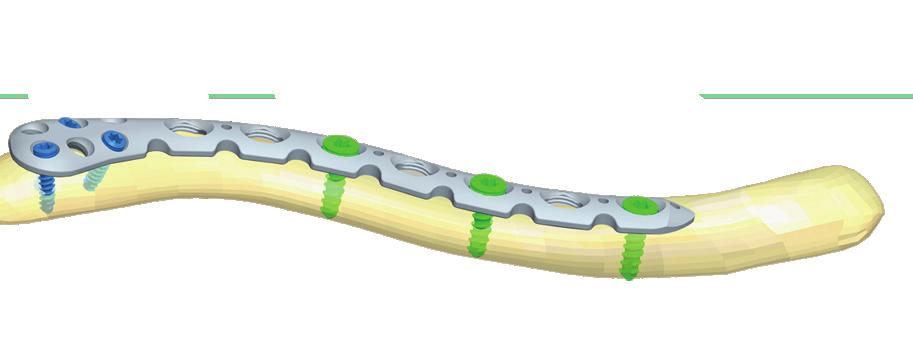
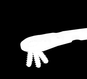
3 1 Introduction Implant range 2 Vortex polyaxial plating system has 4 members for the most complete care of clavicle fractures. Apart from the anatomically formed superior and anterior versions the headed and hooked plates are also available. All this diversity of shapes have polyaxial locking features to make this system the most modern clavicle plating family. 1.1 The implant Polyaxial angle stabilized system in step - free ±15 deg angulation of insertion ± The instruments Capable of drilling in preset and ±15 deg directions step free 2.1 Vortex Clavicle plate Holes on stem Side 6 right/left 7 right/left 8 right/left 10 right/left 2.2 Vortex Clavicle plate H Holes on stem Side 4 right/left 5 right/left 6 right/left 7 right/left Polyaxial and compression holes on the stem 4 different plate shapes: 2.3 Vortex Clavicle plate HK 2.4 Vortex Clavicle plate MA Minimally invasive technique supporting system Bending facilitating holes no screw hole deformation coded Torx screws Double and dynamic drill sleeves for multifunctional locking Easy-to-use bending tools Instruments and implants in one tray Optimized instruments coded torque limiting screwdriver Holes on stem Side 3 right/left 5 right/left Length of hook S: 15 mm M: 18 mm L: 21 mm Holes on stem Indications For medial and distal third fractures of clavicle 4 5
 8-14 blue Length (mm) 10-22")
 10-22 2.")
4 2 Implant range 2.5 Vortex screw Ø 2,7 mm 2.6 Vortex screw Ø 3,5 mm Length (mm) 8-14 blue Length (mm) green 2.7 Cortical screw - TX Ø 2,7 mm Length (mm) Cortical screw - TX Ø 3,5 mm Length (mm) Cancellous screw - Ø 4 mm Length (mm)


 3.")


5 3 Surgical description The VCL system conatins 4 types of plates. Apart from the shape and slightly different function of the plates the surgical steps are identical. These are to be introduced on the example of the Vortex Clavicula plate H (plate with head). 3.1 Patient positioning In beach chair or supine position on translucent surgical table. The head is rotated away from the affected shoulder. Reposition is helped by a tightly packed surgical tissue between the shoulder blades. 3.2 Incision Horizontal incision above or below the clavicula. Pay extra attention not to damage any of the local danger zones (i.e.: supraclavicular nerves) 3.3 Temporary fixation Reduce the fracture and use 1,6 mm Kirschner wires for temporary fixation. 3.4 Plate selection Choose the most appropriate plate size according to the nature of the fracture. If needed, modellate the plates with the bending tools supplied in the instrument tray. Avoid the extensive and the multiple to-and-back bending of the plate. There are bending facilitating holes on the plates which make the bending of the plate possible without the screw hole deformation. 3.5 Plate positioning Place the plate to its position and fix it temporarily with 1,6 mm Kirschner wires. 3.6 Screw insertion The VCL headed plate fixes itself with 2,7 mm polyaxial screws on the head while on the tail 3,5 mm polyaxial or 4,0 mm cancellous screws. 3.7 Screw insertion on the head First insert 2,7 mm polyaxial screws into the holes of the head of the plate. This can be performed monoor polyaxially Monoaxial screw insertion (2,7 mm screws) Place the straight part of the 2,0 mm double drill sleeve into the appropriate hole and perform drilling with 2,0 mm drillbit through that. Determine necessary screw length with a depth gauge. Alternative length gauging method: Use the blue drill stop placed on the drillbit before drilling, just above the spiral part. 8 9


 3.")


6 3 Surgical description Drill through the 2.0 mm sleeve. Below the stop the necessary value can be read. Perform length gauging accurately to avoid bicortical screws hurting soft tissues below the clavicula. For the same reason the protection of the area is also necessary during drilling. Apply image intensifier control! Drive the 2,7 mm polyaxial screws in with the T9 screwdriver. The final tightening of the screw is always performed with the red handle and blue cap torque screwdriver. If you use 4,0 mm cancellous screw you need to apply 2,5 hexagonal screwdriver. Drive the 2,7 mm polyaxial screws in with the T9 screwdriver. The final tightening of the screw is always performed with the red handle and blue cup torque screwdriver. 3.8 Screw insterion on the tail On the tail mono- or polyaxial and compression locking can be applied Monoaxial screw insertion (3,5 mm screws) Poliaxial screw insertion (2,7 mm screws) Fit the end of the conical part of the 2,0 mm double drill sleeve into the hole of the plate. There is a ±15 degree freedom with reference to this position. Perform drilling with the 2,8 mm drillbit in the optimal direction under image intensifier control. Make sure to protect tissues below the clavicula. Perform length gauging. Note that the drillbit and drillstop cannot be used for length gauging when using the conical side of the double sleeve. Place the straight part of the 2,8 mm double drill sleeve into the hole of the plate and perform drilling through that with the 2,8 mm drillbit. Measure screw length. Use the green drill stop placed on the drillbit before drilling, just above the spiral part. Drill through the 2,8 mm sleeve. Below the stop the necessary value can be read. Perform length gauging accurately to avoid bicortical screws hurting soft tissues below the clavicula. For the same reason the protection of the area is also necessary during drilling. Apply image intensifier control! Perform length gauging accurately to avoid bicortical screws hurting soft tissues below the clavicula


7 3 Surgical description Drive the screw in with T15 screwdriver. For the final tightening of the locking screws always use red handle and green cap torque screwdriver. Drive the 3,5 mm polyaxial screws in with the T15 screwdriver. The final tightening of the screw is always performed with the red handle and blue cap torque screwdriver. If you use 4,0 mm cancellous screw you need to apply 2,5 hexagonal screwdriver Compression screw insertion Poliaxial screw insertion (3,5 mm screws) Fit the end of the conical part of the 2,8 mm double drill sleeve into the hole of the plate. There is a ±15 degree freedom with reference to this position. Perform drilling with the 2,8 mm drillbit in the optimal direction under image intensifier control. Make sure to protect tissues below the clavicula. Perform length gauging. Note that the drillbit and drillstop cannot be used for length gauging when using the conical side of the double sleeve. Perform length gauging accurately to avoid bicortical screws hurting soft tissues below the clavicula. Compression technique can also be used on the tail of the plate. To achieve compression use the double compression sleeve. On side is marked 1,0, which is the compression side while the other functions as neutral. Put the required part of the double compression sleeve into the hole of the plate. Make sure that the small arrow shows towards the direction of the fracture. Perform drilling and length gauging. Use only the length gauge. The drillbit and drillstop technique cannot be used! Drive in the appropriate screw. By using this technique 1 mm compression can be achieved. 3.9 Closing of the wound After image intensifier control close the wound in the usual fashion Using cancellous screws For the VCL plates AO type cancellous screws can also be used. Please note that they require some separate instruments which cannot be found in VCL tray. See section Implant list for reference


8 4 Implant list 4.1 Vortex Clavicle plate 4.3 Vortex Clavicle plate - HK 4.2 Vortex Clavicle plate - H Left/6H Left/7H Left/8H Left/10H Right/6H Right/7H Right/8H Right/10H S Left 3H M Left 3H L Left 3H S Left 5H M Left 5H L Left 5H S Right 3H M Right 3H L Right 3H S Right 5H M Right 5H L Right 5H Left/4H Left/5H Left/6H Left/7H Right/4H Right/5H Right/6H Right/7H 4.4 Vortex Clavicle plate - MA H H H 14 15

9 4 Implant list 4.8 Cortical screw - TX Ø 3,5 mm 4.5 Vortex screw Ø 2,7 mm mm mm mm mm mm mm mm mm mm mm mm 4.6 Vortex screw Ø 3,5 mm mm mm mm mm mm mm mm 4.7 Cortical screw - TX Ø 2,7 mm mm mm mm mm 16 17
10 4 Implant list 4.9 Cancellous screw Ø 4,0 mm Instrument set for 4,0 mm screws Surgical instruments Description Quantity. Spiral drill with quick connecting end 2,5x180 mm Screwdriver 2,5 mm Double drill sleeve 3,5/2,5 mm

")


 5 pcs 937516150")
 210510044 Screw")
 Drill stop")

 (2,8 mm) 2 pcs")
")
11 5 Instrument list 5.1 Instruments Screwdriver (T9) Double drill sleeve - PAS (2,8 mm) Screwdriver (T15) Double drill sleeve - V (Small) Torque screwdriver ( T9 / 1 Nm) Kirschner wire (1,6x150 mm) 5 pcs Torque screwdriver ( T15 / 15 Nm) Screw forceps Spiral drill (2x125 mm) Drill stop (blue) (2 mm) 2 pcs Spiral drill (2,8x135 mm) Drill stop (green) (2,8 mm) 2 pcs Double drill sleeve - PAS (2 mm) Depth gauge (2,7-3,5 mm)
12 5 Instrument list Notes Instruments Plate bender (4 mm) 2 pcs Plate bender - V (6 mm) 2 pcs Filled up tray VCL Optional instruments Power torque screwdriver (T9/1 Nm) (for blue screw) Power torque screwdriver (T15/1,5 Nm) (for green screw)

13 Product family TRAUMATOLOGY 1.1. Intramedullary nails 1.2. Plates Vortex plates VDR VDH VDT VPT VPH VCL 1.3. Screws 1.4. Fixateur ORTHOPAEDICS DENTAL SPINE Contact Address: 5 Faiskola str Eger metal@sanatmetal.hu Phone: Fax: TRAUMA Vortex_Clavicle_en REV_B 2016/03/01
TRAUMATOLOGY. Vortex. Distal Tibia plate
 TRAUMATOLOGY Vortex Distal Tibia plate 1 References Dr. László Vámhidy PhD. Head of Department, Clinical Director MSI Department of Traumatology and Hand Surgery Pécs, Hungary Prof. Dr. Endre Varga Professor
TRAUMATOLOGY. Trochanter
 TRAUMATOLOGY Trochanter 1 References Prof. Dr. András Sárváry Head of Department Fővárosi Önkormányzat Péterfy Sándor úti Kórház Rendelőintézet és Baleseti Központ Budapest The following surgical description
TRAUMATOLOGY. Humerus
 TRAUMATOLOGY Humerus 1 References Dr. Zoltán DETRE Lead Surgeon Szent János Kórház, Budapest Prof. Dr. András SÁRVÁRY Head of Department, Director Főv. Önk. Péterfy Sándor úti Kórház Rendelőintézet és
Clavicle Hook Locking Plate
 990210003 Clavicle Hook Locking Plate Clavicle Hook Locking Plate Clavicle Hook Locking Plate INDEX Indications Patient Position Surgical Technique Step 1 Approach Step 2 Reduction Step 3 Temporary Fixation
WINSTA-C. Clavicle Plating System
 Clavicle Plating System Clinical Advisor Michael Kurer FRCS FRCS (Orth) Consultant Orthopaedic and Shoulder Surgeon North Middlesex University Hospital NHS Trust Table of Contents Introduction Indication
LCP Distal Tibia Plate
 Surgical Technique LCP Locking Compression Plate Original Instruments and Implants of the Association for the Study of Internal Fixation AO/ASIF Table of contents Indications 3 Implants/Instruments 5 Surgical
A locking plate system that expands a surgeon s options in trauma surgery. Zimmer NCB Plating System
 A locking plate system that expands a surgeon s options in trauma surgery Zimmer NCB Plating System The Power of Choice The power of having true intraoperative options is at your fingertips. Using standard
Technique Guide. Locking Attachment Plate. For treatment of periprosthetic fractures.
 Technique Guide Locking Attachment Plate. For treatment of periprosthetic fractures. Table of Contents Introduction Locking Attachment Plate 2 Indications 4 Surgical Technique Patient Positioning 5 Preparation
LCP Superior Clavicle Plate. The anatomically precontoured fixation system with angular stability for clavicle shaft and lateral clavicle.
 Technique Guide LCP Superior Clavicle Plate. The anatomically precontoured fixation system with angular stability for clavicle shaft and lateral clavicle. Table of Contents Introduction LCP Superior Clavicle
A locking plate system that expands a surgeon s options in trauma surgery. Zimmer NCB Plating System
 A locking plate system that expands a surgeon s options in trauma surgery Zimmer NCB Plating System The Power of Choice The power of having true intraoperative options is at your fingertips. Using standard
LCP Medial Distal Tibia Plate, without Tab. The Low Profile Anatomic Fixation System with Angular Stability and Optimal Screw Orientation.
 LCP Medial Distal Tibia Plate, without Tab. The Low Profile Anatomic Fixation System with Angular Stability and Optimal Screw Orientation. Technique Guide LCP Small Fragment System Table of Contents Introduction
3. Insert Tocar Sleeves Insert the NCB tissue protection sleeve assembly 1.6 to 10mm through a skin incision (Fig. 38).
.") NCB Proximal Humerus Plating System Surgical Technique 19 2. Temporary Plate Fixation The plate can be temporary fixed to the bone with 1.6mm K-wire through the proximal cannulated fixation screw of the
NCB Proximal Humerus Plating System Surgical Technique 19 2. Temporary Plate Fixation The plate can be temporary fixed to the bone with 1.6mm K-wire through the proximal cannulated fixation screw of the
VA-LCP Anterior Clavicle Plate. The anatomically precontoured fixation system with angular stability for clavicle shaft and lateral clavicle.
 Technique Guide VA-LCP Anterior Clavicle Plate. The anatomically precontoured fixation system with angular stability for clavicle shaft and lateral clavicle. Table of Contents Introduction VA-LCP Anterior
3.5 mm LCP Clavicle Hook Plates
 Part of the Synthes Locking Compression Plate (LCP ) System 3.5 mm LCP Clavicle Hook Plates Surgical Technique Table of Contents Introduction 3.5 mm LCP Clavicle Hook Plates 2 AO Principles 4 Indications
LCP Distal Humerus Plates
 The anatomic fixation system for the distal humerus with angular stability Surgical technique LCP Locking Compression Plate Contents Indications and contraindications 2 Implants 3 Instruments 5 Preparation
NCB Proximal Humerus Plating System
 NCB Proximal Humerus Plating System Surgical Technique The right locking option for tough fractures Disclaimer This document is intended exclusively for experts in the field, i.e. physicians in particular,
Distal Ulnar Locking Plate
 INDEX Indications Patient Position Surgical Technique - Step 1 Approach - Step 2 Plate Contouring - Step 3 Fracture Reduction - Step 4 Distal Plate Fixation - Step 5 Confirm Proper Reconstruction - Step
LCP Superior Clavicle Plate. The anatomically precontoured fixation system with angular stability for clavicle shaft and lateral clavicle.
 LCP Superior Clavicle Plate. The anatomically precontoured fixation system with angular stability for clavicle shaft and lateral clavicle. Surgical Technique This publication is not intended for distribution
Technique Guide. 3.5 mm LCP Low Bend Medial Distal Tibia Plates. Part of the Synthes locking compression plate (LCP) system.
 system.") Technique Guide 3.5 mm LCP Low Bend Medial Distal Tibia Plates. Part of the Synthes locking compression plate (LCP) system. Table of Contents Introduction 3.5 mm LCP Low Bend Medial Distal Tibia Plates
Technique Guide 3.5 mm LCP Low Bend Medial Distal Tibia Plates. Part of the Synthes locking compression plate (LCP) system. Table of Contents Introduction 3.5 mm LCP Low Bend Medial Distal Tibia Plates
Technique Guide. 3.5 mm LCP Low Bend Medial Distal Tibia Plate Aiming Instruments. Part of the 3.5 mm LCP Percutaneous Instrument System.
 Technique Guide 3.5 mm LCP Low Bend Medial Distal Tibia Plate Aiming Instruments. Part of the 3.5 mm LCP Percutaneous Instrument System. Table of Contents Introduction 3.5 mm LCP Low Bend Medial Distal
3.5 mm Locking Attachment Plate
 For Treatment of Periprosthetic Fractures 3.5 mm Locking Attachment Plate Surgical Technique Table of Contents Introduction 3.5 mm Locking Attachment Plate 2 Indications 4 Surgical Technique Preparation
Surgical Technique. Clavicle Locking Plate
 Surgical Technique Clavicle Locking Plate PERI-LOC Locked Plating System Clavicle Locking Plate Surgical Technique Table of Contents Introduction...2 Indications...3 Plate Features...3 Patient Positioning...4
LCP Low Bend Medial Distal Tibia Plates 3.5 mm. Anatomic plates with low profile head for intra- and extraarticular fractures.
 LCP Low Bend Medial Distal Tibia Plates 3.5 mm. Anatomic plates with low profile head for intra- and extraarticular fractures. Surgical Technique This publication is not intended for distribution in the
Olecranon Locking Plate II
 INDEX Indications Patient Position Fracture Reduction and Fixation Surgical Technique Step 1 Surgical Approach Step 2 Implantation Step 3 Proximal Locking Screw Insertion Step 4 Distal Screw Insertion
3.5 MM VA-LCP PROXIMAL TIBIA PLATE SYSTEM
 3.5 MM VA-LCP PROXIMAL TIBIA PLATE SYSTEM Part of the DePuy Synthes Variable Angle Periarticular Plating System SURGICAL TECHNIQUE TABLE OF CONTENTS INTRODUCTION 3.5 mm VA-LCP Proximal Tibial Plate 2 AO
OBSOLETED. LCP Medial Distal Tibia Plate, without Tab. The Low Profile Anatomic Fixation System with Angular Stability and Optimal Screw Orientation.
 LCP Medial Distal Tibia Plate, without Tab. The Low Profile Anatomic Fixation System with Angular Stability and Optimal Screw Orientation. Surgical Technique LCP Small Fragment System This publication
Technique Guide. 3.5 mm LCP Olecranon Plates. Part of the Synthes locking compression plate (LCP) system.
 system.") Technique Guide 3.5 mm LCP Olecranon Plates. Part of the Synthes locking compression plate (LCP) system. Table of Contents Introduction 3.5 mm LCP Olecranon Plates 2 AO Principles 3 Indications 3 Clinical
Technique Guide 3.5 mm LCP Olecranon Plates. Part of the Synthes locking compression plate (LCP) system. Table of Contents Introduction 3.5 mm LCP Olecranon Plates 2 AO Principles 3 Indications 3 Clinical
LCP Superior Anterior Clavicle Plate. The anatomically precontoured fixation system with angular stability for clavicle shaft and lateral clavicle.
 LCP Superior Anterior Clavicle Plate. The anatomically precontoured fixation system with angular stability for clavicle shaft and lateral clavicle. Surgical Technique This publication is not intended for
LCP Medial Proximal Tibial Plate 3.5. Part of the Synthes small fragment Locking Compression Plate (LCP) system.
 system.") LCP Medial Proximal Tibial Plate 3.5. Part of the Synthes small fragment Locking Compression Plate (LCP) system. Technique Guide This publication is not intended for distribution in the USA. Instruments
LCP Medial Proximal Tibial Plate 3.5. Part of the Synthes small fragment Locking Compression Plate (LCP) system. Technique Guide This publication is not intended for distribution in the USA. Instruments
P.R.C.T II FIXATION PLATE FOR ARTICULAR FRACTURE OF THE PROXIMAL HUMERUS SURGICAL TECHNIQUE
 P.R.C.T II FIXATION PLATE FOR ARTICULAR FRACTURE OF THE PROXIMAL HUMERUS SURGICAL TECHNIQUE INDICATIONS - Surgical technique...page 0 - Mini invasive approach...page 04 - Diaphyseal plates...page 05 -
USS II ILIO-SACRAL Modular System for Stable Fixation in the Sacrum and Illium
 USS II ILIO-SACRAL Modular System for Stable Fixation in the Sacrum and Illium Instruments and implants approved by the AO Foundation. This publication is not intended for distribution in the USA. TECHNIQUE
Technique Guide. LCP Distal Fibula Plates. Part of the Synthes locking compression plate (LCP) system.
 system.") Technique Guide LCP Distal Fibula Plates. Part of the Synthes locking compression plate (LCP) system. Table of Contents Introduction LCP Distal Fibula Plates 2 AO Principles 4 Indications 5 Surgical Technique
Technique Guide LCP Distal Fibula Plates. Part of the Synthes locking compression plate (LCP) system. Table of Contents Introduction LCP Distal Fibula Plates 2 AO Principles 4 Indications 5 Surgical Technique
Surgical Technique International Version. Clavicle Locking Plate
 Surgical Technique International Version Clavicle Locking Plate PERI-LOC Upper Extremity Locked Plating System Clavicle Surgical Techniquefor Table of Contents Introduction........................................................2
Technique Guide. 3.5 mm LCP Periarticular Proximal Humerus Plate. Part of the Synthes locking compression plate (LCP) system.
 system.") Technique Guide 3.5 mm LCP Periarticular Proximal Humerus Plate. Part of the Synthes locking compression plate (LCP) system. Table of Contents Introduction 3.5 mm LCP Proximal Humerus Plate 2 AO Principles
Technique Guide 3.5 mm LCP Periarticular Proximal Humerus Plate. Part of the Synthes locking compression plate (LCP) system. Table of Contents Introduction 3.5 mm LCP Proximal Humerus Plate 2 AO Principles
VA-LCP Anterior Clavicle Plate. The anatomically precontoured fixation system with angular stability for clavicle shaft and lateral clavicle.
 VA-LCP Anterior Clavicle Plate. The anatomically precontoured fixation system with angular stability for clavicle shaft and lateral clavicle. Surgical Technique This publication is not intended for distribution
NCB Distal Femur System. Surgical Technique
 NCB Distal Femur System Surgical Technique NCB Distal Femur System Surgical Technique 3 Surgical Technique NCB Distal Femur System Table of Contents Introduction 4 Indications 8 Preoperative Planning
Pediatric LCP Plate System. For osteotomies and fracture fixation of the proximal and distal femur.
 Pediatric LCP Plate System. For osteotomies and fracture fixation of the proximal and distal femur. Angular stability Intraoperative correction and flexibility Universal design Indications The Pediatric
Pediatric LCP Hip Plate. For osteotomy and trauma applications in the proximal femur.
 Pediatric LCP Hip Plate. For osteotomy and trauma applications in the proximal femur. Angular stability Intraoperative correction and flexibility Universal design Pediatric LCP Hip Plate The Pediatric
Technique Guide. 2.7 mm/3.5 mm LCP Distal Fibula Plates. Part of the Synthes locking compression plate (LCP) system.
 system.") Technique Guide 2.7 mm/3.5 mm LCP Distal Fibula Plates. Part of the Synthes locking compression plate (LCP) system. Table of Contents Introduction 2.7 mm/3.5 mm LCP Distal Fibula Plates 2 AO Principles
Technique Guide 2.7 mm/3.5 mm LCP Distal Fibula Plates. Part of the Synthes locking compression plate (LCP) system. Table of Contents Introduction 2.7 mm/3.5 mm LCP Distal Fibula Plates 2 AO Principles
3.5 mm LCP Superior and Superior Anterior Clavicle Plates. Part of the Synthes modular clavicle plate system.
 3.5 mm LCP Superior and Superior Anterior Clavicle Plates. Part of the Synthes modular clavicle plate system. Technique Guide Instruments and implants approved by the AO Foundation Table of Contents Introduction
PROXIMAL TIBIAL PLATE
 SURGICAL NÁSTROJE TECHNIQUE PRO ARTROSKOPII PROXIMAL INSTRUMENTS TIBIAL FOR PLATE ARTHROSCOPY Proximal Tibial Plate Description of medical device The Proximal Tibial Plate is used in epyphyseal and metaphyseal
3.5 mm LCP Superior Anterior Clavicle Plates
 Part of the DePuy Synthes Locking Compression Plate (LCP ) System 3.5 mm LCP Superior Anterior Clavicle Plates Surgical Technique Table of Contents Introduction 3.5 mm LCP Superior Anterior Clavicle Plates
Double Engine Orthopedic Bone Nail System Universal Humeral Nail
 Double Engine Orthopedic Bone Nail System ----------- Universal Humeral Nail Surgical Technique Manual Note: The surgical procedures should be performed under the guidance of qualified skilled orthopedic
2.7 mm/3.5 mm LCP Distal Fibula Plate System. Part of the Synthes locking compression plate (LCP) system.
 system.") 2.7 mm/3.5 mm LCP Distal Fibula Plate System. Part of the locking compression plate (LCP) system. Anatomically contoured Multiple screw options for fixedangle support Coaxial holes minimize screw head
2.7 mm/3.5 mm LCP Distal Fibula Plate System. Part of the locking compression plate (LCP) system. Anatomically contoured Multiple screw options for fixedangle support Coaxial holes minimize screw head
LCP Anterolateral Distal Tibia Plate 3.5. The low profile anatomic fixation system with optimal plate placement and angular stability.
 LCP Anterolateral Distal Tibia Plate 3.5. The low profile anatomic fixation system with optimal plate placement and angular stability. Technique Guide LCP Small Fragment System Table of Contents Introduction
ACTIV ANKLE DISTAL TIBIA PLATE MEDIAL MALLEOLUS INNOVATION MEANS MOTION. DUALTEC SYSTEM II and III
 INNOVATION MEANS MOTION ACTIV ANKLE DISTAL TIBIA PLATE MEDIAL MALLEOLUS POLYAXIAL LOCKING FIXATION DUALTEC SYSTEM II and III Precontoured implants Polyaxiality of 20 2 types of plates: hook and standard
Technique Guide. LCP Proximal Femoral Hook Plate 4.5/5.0. Part of the LCP Periarticular Plating System.
 Technique Guide LCP Proximal Femoral Hook Plate 4.5/5.0. Part of the LCP Periarticular Plating System. Table of Contents Introduction Features and Benefits 2 AO ASIF Principles 4 Indications 5 Surgical
LCP Anterolateral Distal Tibia Plate 3.5. The low profile anatomic fixation system with optimal plate placement and angular stability.
 LCP Anterolateral Distal Tibia Plate 3.5. The low profile anatomic fixation system with optimal plate placement and angular stability. Technique Guide LCP Small Fragment System Table of Contents Introduction
Surgical Technique. Anterolateral and Medial Distal Tibia Locking Plates
 Surgical Technique Anterolateral and Medial Distal Tibia Locking Plates PERI-LOC Periarticular Locked Plating System Anterolateral and Medial Distal Tibia Locking Plates Surgical Technique Contents Product
WINSTA-R. Distal Radius System
 Distal Radius System Table of Contents Introduction WINSTA-R System 2 Indication 2 Surgical Technique Palmar Access for Radius Plate 3 Dorsal Access for Radius Plate 3 Positioning of the Radius Plate
Surgical Technique. Calcaneal Locking Plate
 Surgical Technique Calcaneal Locking Plate PERI-LOC Locked Plating System Calcaneal Locking Plate Surgical TechniqueCatalog Infor Table of Contents Introduction...2 Indications...3 Plate Features...3 Patient
The Calcaneal Plate. The Synthes non-locking solution for the Calcaneus.
 The Calcaneal Plate. The Synthes non-locking solution for the Calcaneus. Surgical Technique This publication is not intended for distribution in the USA. Instruments and implants approved by the AO Foundation.
LCP Metaphyseal Plates. For extra-articular fractures.
 LCP Metaphyseal Plates. For extra-articular fractures. Surgical Technique This publication is not intended for distribution in the USA. Instruments and implants approved by the AO Foundation. Image intensifier
Technique Guide. TomoFix Osteotomy System. A comprehensive plating system for stable fixation of osteotomies around the knee.
 Technique Guide TomoFix Osteotomy System. A comprehensive plating system for stable fixation of osteotomies around the knee. Table of Contents Introduction TomoFix Osteotomy System 2 AO Principles 4 Indications
LCP Anterior Ankle Arthrodesis Plates. Part of the Synthes Locking Compression Plate (LCP) System.
 System.") LCP Anterior Ankle Arthrodesis Plates. Part of the Synthes Locking Compression Plate (LCP) System. Technique Guide Instruments and implants approved by the AO Foundation Table of Contents Introduction
LCP Anterior Ankle Arthrodesis Plates. Part of the Synthes Locking Compression Plate (LCP) System. Technique Guide Instruments and implants approved by the AO Foundation Table of Contents Introduction
modular ClaVICle PlaTe system
 modular ClaVICle PlaTe system 3.7 mm/3.3 mm VA LCP Anterior Clavicle Plates and 3.3 mm Superior Anterior Clavicle Plates surgical TeChnIque Table of Contents Introduction Modular Clavicle Plate System
LCP Medial Proximal Tibial Plate 4.5/5.0. Part of the Synthes LCP periarticular plating system.
 LCP Medial Proximal Tibial Plate 4.5/5.0. Part of the Synthes LCP periarticular plating system. Technique Guide This publication is not intended for distribution in the USA. Instruments and implants approved
Technique Guide. Compact 2.0 LOCK Mandible. The locking system for the mandible.
 Technique Guide Compact 2.0 LOCK Mandible. The locking system for the mandible. Table of Contents Introduction Compact 2.0 LOCK Mandible 2 AO Principles 4 Indications and Contraindications 5 Surgical
2.4 mm LCP Radial Head Plates. Part of the Synthes LCP Distal Radius Plate System.
 2.4 mm LCP Radial Head Plates. Part of the Synthes LCP Distal Radius Plate System. Technique Guide Instruments and Implants approved by the AO Foundation Table of Contents Introduction 2.4 mm LCP Radial
PediLoc 3.5mm and 4.5mm Contour Femur Plate Surgical Technique
 PediLoc 3.5mm and 4.5mm Contour Femur Plate Surgical Technique Surgical Technique Contour Femur Plate The technique description herein is made available to the healthcare professional to illustrate the
LCP Proximal Radius Plates 2.4. Plates for radial head rim and for radial head neck address individual fracture patterns of the proximal radius.
 Technique Guide LCP Proximal Radius Plates 2.4. Plates for radial head rim and for radial head neck address individual fracture patterns of the proximal radius. Table of Contents Introduction LCP Proximal
2.7 mm/3.5 mm VA-LCP Anterior Clavicle Plates. Part of the Synthes modular clavicle plate system.
 2.7 mm/3.5 mm VA-LCP Anterior Clavicle Plates. Part of the Synthes modular clavicle plate system. Technique Guide Instruments and implants approved by the AO Foundation Table of Contents Introduction 2.7
Surgical Technique. Proximal Humerus Locking Plate
 Surgical Technique Proximal Humerus Locking Plate PERI-LOC Upper Extremity Locked Plating System 3.5mm & 4.5mm Proximal Humerus Locking PlatesCatalog Infor Table of Contents Introduction.........................................................2
3.5 mm LCP Extra-articular Distal Humerus Plate
 Part of the DePuy Synthes Locking Compression Plate (LCP ) System 3.5 mm LCP Extra-articular Distal Humerus Plate Surgical Technique Table of Contents Introduction 3.5 mm LCP Extra-articular Distal Humerus
INNOVATION means MOTION ALIANS ELBOW OLECRANON. Narrow and standard plates Polyaxial locking technology Bendable precontoured implants
 surgical technique ALIANS ELBOW OLECRANON Narrow and standard plates Polyaxial locking technology Bendable precontoured implants ALIANS ELBOW - OLECRANON Indications: The ALIANS ELBOW range of plates is
Distal Lateral Femur Plate PP. Surgical Technique
 1 Disclaimer This surgical technique is exclusively intended for medical professionals, especially physicians, and therefore may not be regarded as a source of information for non-medical persons. The
Low Bend Distal Tibia Plates
 Part of the DePuy Synthes Locking Compression Plate (LCP ) System 3.5 mm LCP Low Bend Medial Distal Tibia Plates Surgical Technique Table of Contents Introduction 3.5 mm LCP Low Bend Medial Distal Tibia
Zimmer Small Fragment Universal Locking System. Surgical Technique
 Zimmer Small Fragment Universal Locking System Surgical Technique Zimmer Small Fragment Universal Locking System 1 Zimmer Small Fragment Universal Locking System Surgical Technique Table of Contents Introduction
Small Fragment Plating System. Securing optimal fixation through locked and compression plating technology
 Small Fragment Plating System Securing optimal fixation through locked and compression plating technology Contents Design Rationale Introduction Interfragmentary Fixation Insertion of a 3.5 mm Cortical
The Locking Calcaneal Plate Instrument and Implant Sets
 Part of the DePuy Synthes Locking Compression Plate (LCP ) System The Locking Calcaneal Plate Instrument and Implant Sets Surgical Technique Table of Contents Introduction Locking Calcaneal Plate 2 AO
AxSOS Locking Plate System
 AxSOS Locking Plate System Operative Technique Small Fragment Basic Fragment 1 2 Contents Page 1. Introduction 4 2. Features & Benefits 5 4 and 5mm Compression Plates 5 Reconstruction and 1/3 Tubular Locking
3.5 mm LCP Olecranon Plates
 Part of the DePuy Synthes Locking Compression Plate (LCP ) System 3.5 mm LCP Olecranon Plates Surgical Technique Table of Contents Introduction 3.5 mm LCP Olecranon Plates 2 AO Principles 3 Indications
Surgical Technique Guide
 Guide CAUTION: Federal Law (USA) restricts this device to sale by or on the order of a physician. INDICATIONS FOR USE The Align Anterior Ankle Fusion Plate is intended to facilitate arthrodesis of the
3.5 mm LCP Distal Humerus Plates
 Part of the DePuy Synthes Locking Compression Plate (LCP ) System 3.5 mm LCP Distal Humerus Plates Surgical Technique Table of Contents Introduction 3.5 mm LCP Distal Humerus Plates 2 AO Principles 4 Indications
NCB Proximal Humerus System. Surgical Technique
 NCB Proximal Humerus System Surgical Technique NCB Proximal Humerus System Surgical Technique 3 Surgical Technique NCB Proximal Humerus System Table of Contents Introduction 4 Cable Fixation Options 5
Technique Guide. PHILOS and PHILOS Long. The anatomic fixation system for the proximal humerus.
 Technique Guide PHILOS and PHILOS Long. The anatomic fixation system for the proximal humerus. Table of Contents Introduction PHILOS and PHILOS Long 2 AO Principles 4 Indications 5 Surgical Technique
USS Variable Axis Screw
 USS Variable Axis Screw Polyaxial side-opening pedicle screw Surgical technique Original Instruments and Implants of the Association for the Study of Internal Fixation AO/ASIF USS Variable Axis Screw
3.5 mm LCP Hook Plate
 Part of the DePuy Synthes Locking Compression Plate (LCP ) System 3.5 mm LCP Hook Plate Surgical Technique Table of Contents Introduction 3.5 mm LCP Hook Plate 2 AO Principles 4 Indications 5 Clinical
Small Fragment Plating System
 Small Fragment Plating System Securing optimal fixation through locked and compression plating technology SURGICAL TECHNIQUE RECOVERY FUNCTION SURVIVORSHIP DePuy believes in an approach to trauma surgery
Orthopedic Bone Nail System - Distal Femoral Nail Surgical Technique Manual
 Orthopedic Bone Nail System - Distal Femoral Nail Surgical Technique Manual Note: The surgical procedures should be performed under the guidance of qualified skilled orthopedic surgeons, and this surgical
operative technique Universal Application
 operative technique Universal Application Introduction Introduction Building upon the design rationale of the Xia Spinal System, the new Xia Spinal System represents the latest advancement in spinal implant
3.5 mm LCP Low Bend Medial Distal Tibia Plate Aiming Instruments
 Part of the 3.5 mm LCP 3.5 mm LCP Low Bend Medial Distal Tibia Plate Aiming Instruments Surgical Technique TABLE OF CONTENTS INTRODUCTION 3.5 mm LCP Low Bend Medial Distal Tibia Plate 2 Aiming Instruments
ACtiv ankle. INNOVATION means MOTION. Precontoured implants Polyaxiality of 20 2 surgical approaches : lateral and posterolateral
 ACtiv ankle distal AND diaphyseal FIBULA POLYAXIAL LOCKING FIXATION DUALTEC SYSTEM II Precontoured implants Polyaxiality of 20 2 surgical approaches : lateral and posterolateral ACTIV ANKLE The ACTIV ANKLE
Surgical Technique. Olecranon Locking Plate
 Surgical Technique Olecranon Locking Plate PERI-LOC Locked Plating System Olecranon Locking Plate Surgical Techniquealog Infor Table of Contents Introduction...2 Indications...3 Plate Features...3 Patient
Instrument and Implant for wrist fracture
 Instrument and Implant for wrist fracture Jansri Janpanya Product specialist The Bangkok Unitrade Co,.ltd. Objectives Type of LCP for distal radius Fx. The new LCP design for distal radius Fx. Have knowledge
LCP Extra-articular Distal Humerus Plate.
 Technique Guide LCP Extra-articular Distal Humerus Plate. The anatomically shaped and angular stable fixation system for extraarticular fractures of the distal humerus. Table of Contents Introduction
The Wrist Fusion Set. Stainless Steel and Titanium TECHNIQUE GUIDE. Instruments and implants approved by the AO Foundation
 The Wrist Fusion Set Stainless Steel and Titanium TECHNIQUE GUIDE Instruments and implants approved by the AO Foundation Three Plate Options Stainless Steel or Titanium* Standard Bend Stainless Steel [242.510]
2.7 mm/3.5 mm LCP Distal Fibula Plate
 Part of the DePuy Synthes Locking Compression Plate (LCP ) System 2.7 mm/3.5 mm LCP Distal Fibula Plate Surgical Technique Table of Contents Introduction 2.7 mm/3.5 mm LCP Distal Fibula Plates 2 AO Principles
Surgical Technique. Distal Humerus Locking Plate
 Surgical Technique Distal Humerus Locking Plate PERI-LOC Locked Plating System Distal Humerus Locking Plate Surgical Technique Table of Contents Introduction...2 Indications...3 Plate Features...3 Patient
Variable Angle LCP Volar Rim Distal Radius Plate 2.4. For fragment-specific fracture fixation with variable angle locking technology.
 Technique Guide Variable Angle LCP Volar Rim Distal Radius Plate 2.4. For fragment-specific fracture fixation with variable angle locking technology. Image intensifier control Warning This description
MetaCun DuoMetaCun. Midfoot Fusion Locking Plate System. Surgical Technique and Ordering Information
 MetaCun DuoMetaCun Midfoot Fusion Locking Plate System Surgical Technique and Ordering Information 2 Table of contents Table of contents Description... 4 Indications for use...5 Contraindications...5 Surgical
Distal Radius Plate 2.4/2.7 dorsal and volar
 Distal Radius Plate 2.4/2.7 dorsal and volar Surgical Technique This publication is not intended for distribution in the USA. Instruments and implants approved by the AO Foundation. Distal Radius Plate
LCP Condylar Plate 4.5/5.0. Part of the LCP Periarticular Plating System.
 LCP Condylar Plate 4.5/5.0. Part of the LCP Periarticular Plating System. Surgical Technique This publication is not intended for distribution in the USA. Instruments and implants approved by the AO Foundation.
Distal Radius Plate Instrument and Implant Set. Discontinued December 2017 DSUS/TRM/0916/1063(1)
") Distal Radius Plate Instrument and Implant Set Surgical Technique Discontinued December 2017 DSUS/TRM/0916/1063(1) The Distal Radius Plates Indications For fixation of fractures and osteotomies, including
Distal Radius Plate Instrument and Implant Set Surgical Technique Discontinued December 2017 DSUS/TRM/0916/1063(1) The Distal Radius Plates Indications For fixation of fractures and osteotomies, including
VA LOCKING CALCANEAL PLATES 2.7
 VA LOCKING CALCANEAL PLATES 2.7 Instruments and Implants approved by the AO Foundation. This publication is not intended for distribution in the USA. SURGICAL TECHNIQUE Image intensifier control Warning
Dates and images Intrauma S.p.A. reserves the right to modify the design and finishing of the products shown and described in the present catalog
 Product catalog Dates and images Intrauma S.p.A. reserves the right to modify the design and finishing of the products shown and described in the present catalog without any notice. The images are informative
Locking Compression Technology by aap
 Minimally Invasive Locking Compression Technology by aap Minimally Invasive 1 Disclaimer This surgical technique is exclusively intended for medical professionals, especially physicians, and therefore
humerus InSafeLOCK Nail
 humerus InSafeLOCK Nail Introduction Content Humerus InSafeLOCK Nail is an innovative intramedullary nailing system, developed for humerus problems. Humerus fractures have 5-6 % incidence of all bone fractures.