Utility of Dual-Energy CT to Evaluate Patients with Hip and Pelvis Pain in the ER Setting
|
|
|
- Garry Jenkins
- 6 years ago
- Views:
Transcription


1 Utility of Dual-Energy CT to Evaluate Patients with Hip and Pelvis Pain in the ER Setting Johnson, T., Moran, E., Glazebrook, K., Leng, S., Fletcher, J., and McCollough, C. An educational review ER011 Tucker F. Johnson MD Mayo Clinic, Rochester MN
2 Disclosure CHM: Research grant, Siemens Healthcare. The other authors have no financial disclosures.
3 Goals and objectives Understand the concept of Virtual Non-Calcium (VNCa) dual energy CT imaging. Review several cases involving VNCa dual energy CT. Discuss the benefits of VNCa dual energy CT images.
4 Emergency department scenario Numerous visits are paid each year to the emergency department for musculoskeletal pelvic pain. Radiographs obtained in this situation are often inconclusive in the setting of nondisplaced fractures. CT can be helpful in assessing indeterminate findings, but is often limited by osteopenia in elderly patients.
5 Emergency department scenario MRI has traditionally been the gold standard for detecting osseous pathology related to bone marrow edema. Unfortunately, MRI is time intensive and is not universally available during off hours. In contradistinction, CT is often readily available in the emergency setting. CT can also be useful when MRI is contraindicated.
6 Dual energy CT Dual Energy CT (DECT) is a new and powerful imaging technique. DECT works by acquiring attenuation data using two different x-ray spectra. Attenuation data acquired at a second x-ray spectrum allows decomposition of different materials: Ex. separation of bone marrow from trabecular bone. Same radiation dose as single energy CT.
7 Dual energy CT Dual energy data can be acquired through multiple techniques: Rapid switching of x-ray tube potential Dual x-ray sources Multilayer detector New technique that obtains low and high energy data sets at the same time. Photon-counting detectors Experimental technique that performs signal analysis to separate high and low energy data sets.
8 Virtual non-calcium imaging Through removal of calcium signal from cancellous bone, VNCa images can be obtained to visualize the marrow space. Conventional images are also provided from this same data set, enabling evaluation for both fractures and edema from a single exam.
9 Virtual non-calcium imaging VNCa DECT is extremely useful in demonstrating osseous bruising from: Occult fractures involving the hips and sacrum Avascular necrosis (AVN) of the hip Other painful pathology such as metastatic disease
10 Virtual non-calcium imaging Pache et al investigated 21 patients with acute knee trauma (2 observers) using MRI as gold standard: Sensitivity 86.4/86.4% and specificity 94.4/95.5% Good to excellent agreement Femur κ = 0.78, tibia κ = 0.87 Guggenberger et al investigated DECT VNCa detection of traumatic bone marrow edema in the ankle of 30 patients (2 observers) using MRI as gold standard: Visual grading Sensitivity 90/90% and specificity 80.5/81.6%, κ = 0.66
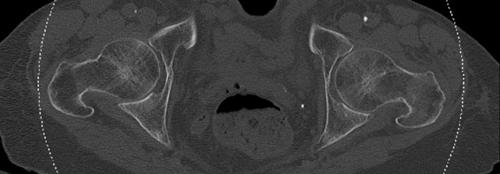
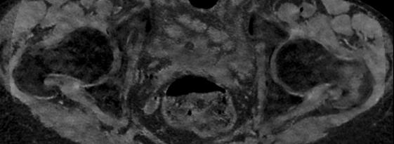
11 Cases I III Fractures


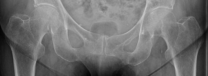
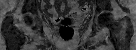
12 Case I Sacral fracture Acute S5 fracture November 2014 Note edema Healed S5 fracture April 2014 Note resolution of previous edema


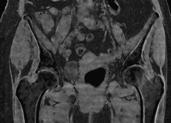
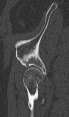
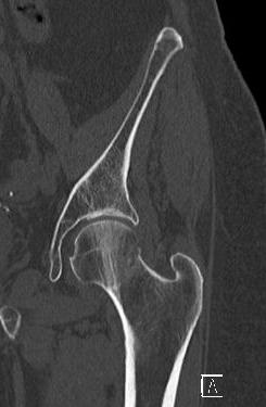
13 Case II Femoral head/neck fracture Possible fracture VNCa shows edema on left Cortical disruption Normal right Fx. left Fx. left VNCa shows edema on left
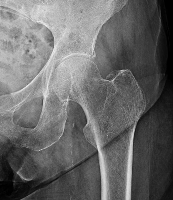
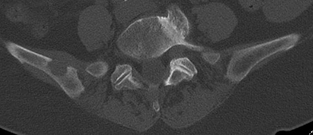
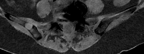
14 Case III Intertrochanteric fracture VNCa edema at fracture Radiographically occult fracture VNCa edema at fracture
15 Discussion Reddy et al. in 2014 examined VNCa DECT in patients with suspected hip fractures. DECT was compared to a gold standard of clinical or imaging follow-up with confirmation of hip fracture: Sensitivity 90% Positive predictive value 86% Mean age 77 years Demonstrate utility in osteoporotic patients Teaching point: DECT with VNCa can help identify fractures that are occult on radiographs or single energy CT, particularly in osteoporotic patients.
16 Case IV Avascular necrosis


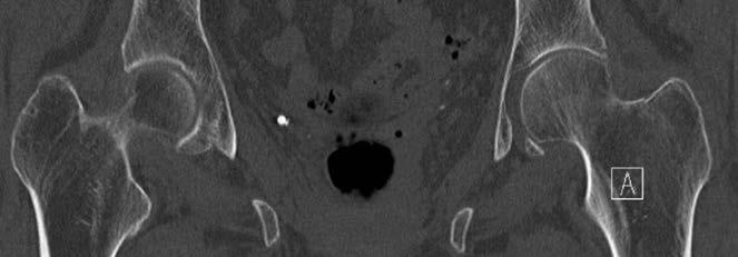
17 Case IV AVN 59 year old woman: Presents with right hip pain Radiographs and CT were negative for acute abnormality CT showed healed right sacral insufficiency fracture Healed sacral insufficiency fracture


18 Case IV AVN continued DECT shows bone marrow edema in both femoral heads suspicious for AVN


19 Case IV AVN continued Scout Coronal T1 Right Coronal T2 fat sat Sagittal T2 fat sat Bilateral femoral head abnormalities Serpiginous low T1 signal T2 edema T2 edema Follow-up MRI: Performed 12 days after radiographs & CT. Demonstrated changes related to bilateral AVN.
20 Discussion In 2014 Barile et al. examined the use of single energy CT in patients with MRI proven avascular necrosis of the femoral head. CTs were performed either before or after MRI: 89% of MRI proven cases were missed with single energy CT. Teaching point: DECT with VNCa can help distinguish acute and chronic osseous pathology. For example, old trauma from acute avascular necrosis.
21 Case V Metastatic disease


22 Case V Metastatic disease 63 year old woman with new left hip pain: Known lung cancer Negative CT abdomen/pelvis 2 months previously Metastatic lesions
23 Discussion 18% of all health care visits are related to musculoskeletal conditions. Commonly this includes: Arthritis, back and neck pain, injuries, and osteoporosis. Although less common, approximately 350,000 people in the U.S. die with bone metastases: Often silent, but can cause pain/fracture. Teaching point: DECT with VNCa can help identify non-traumatic osseous pathology such as metastatic disease.
24 Conclusion VNCa DECT imaging can be helpful in identifying occult radiographic and single energy CT fractures. Osseous edema serves as an important indicator of pathology. Edema can be helpful in differentiating acute and chronic processes.
25 Conclusion VNCa DECT can detect pathology such as avascular necrosis or metastatic disease that may be occult on radiography or single energy CT. These findings have been shown to parallel edema seen on T2 weighted MRI sequences.
26 Conclusion Advantages of VNCa DECT: Increased availability Decreased cost Useful in patients who are unable to undergo MR imaging Provides detailed information about osseous architecture
27 Conclusion Advantages of VNCa DECT: Ability to exclude pathology and thereby obviate the need for additional imaging with MR.
28 Conclusion Limitations: Large body habitus will attenuate low kvp potentially limiting this technique. Normal red marrow can confound findings by replacing fatty marrow.
29 Summary VNCa imaging is a new technology that is readily available with dual energy CT machines. We believe this is a tremendously powerful tool that can be helpful in assessing acute, subacute, and chronic musculoskeletal pelvic pathology in the emergency setting and thereby help prevent further patient morbidity.
30 References Ai, S.T., et al., Use of dual-energy CT and virtual non-calcium techniques to evaluate post-traumatic bone bruises in knees in the subacute setting. Skeletal radiology, (9): p Barille, M.F., J.S. Wu, and C.J. McMahon, Femoral head avascular necrosis: a frequently missed incidental finding on multidetector CT. Clinical radiology, (3): p Bierry, G., et al., Dual-energy CT in vertebral compression fractures: performance of visual and quantitative analysis for bone marrow edema demonstration with comparison to MRI. Skeletal radiology, (4): p Guggenberger, R., et al., Diagnostic performance of dual-energy CT for the detection of traumatic bone marrow lesions in the ankle: comparison with MR imaging. Radiology, (1): p Henes, F.O., et al., Quantitative assessment of bone marrow attenuation values at MDCT: An objective tool for the detection of bone bruise related to occult sacral insufficiency fractures. European Radiology, (10): p McCollough, C.H., et al., Dual- and Multi-Energy CT: Principles, Technical Approaches, and Clinical Applications. Radiology, (3): p Mundy, G.R., Metastasis to bone: causes, consequences and therapeutic opportunities. Nature reviews. Cancer, (8): p Pache, G., et al., Dual-energy CT virtual noncalcium technique: detecting posttraumatic bone marrow lesions--feasibility study. Radiology, (2): p Reddy, T., et al., Detection of occult, undisplaced hip fractures with a dual-energy CT algorithm targeted to detection of bone marrow edema. Emergency radiology, (1): p
31 Contact
32 CT Clinical Innovation Center
66 yr old female with groin and hip pain. Paul Jabour, MD
 66 yr old female with groin and hip pain Paul Jabour, MD 2 months later 12 months later 14 months after initial presentation Acetabular Insufficiency Fracture Pelvic stress fracture Fatigue
Musculoskeletal Imaging What to order? Brian Cole, MD
 Musculoskeletal Imaging What to order? Brian Cole, MD my background: 1994 University of Illinois 1998 MD University of Illinois College of Medicine 1999-2003 Diagnostic Radiology Mayo Clinic 2004 Fellowship
11/4/2018 SUBTLETIES OF LOWER EXTREMITY TRAUMA IMAGING SPEAKER DISCLOSURES
 SUBTLETIES OF LOWER EXTREMITY TRAUMA IMAGING Charles S. Resnik, M.D. Professor of Radiology University of Maryland School of Medicine Upon completion of this presentation, participants will be better able
Neural Network Diagnosis of Avascular Necrosis from Magnetic Resonance Images
 Neural Network Diagnosis of Avascular Necrosis from Magnetic Resonance Images Armando Manduca Dept. of Physiology and Biophysics Mayo Clinic Rochester, MN 55905 Paul Christy Dept. of Diagnostic Radiology
Detection of occult vertebral fractures by quantitative assessment of bone marrow attenuation values at MDCT
 Detection of occult vertebral fractures by quantitative assessment of bone marrow attenuation values at MDCT Poster No.: C-1582 Congress: ECR 2014 Type: Scientific Exhibit Authors: F. O. Henes, M. Groth,
FOR CMS (MEDICARE) MEMBERS ONLY NATIONAL COVERAGE DETERMINATION (NCD) FOR MAGNETIC RESONANCE IMAGING:
 MEMBERS ONLY NATIONAL COVERAGE DETERMINATION (NCD) FOR MAGNETIC RESONANCE IMAGING:") National Imaging Associates, Inc. Clinical guidelines BONE MARROW MRI Original Date: July 2008 Page 1 of 5 CPT Codes: 77084 Last Review Date: September 2014 NCD 220.2 MRI Last Effective Date: July 2011
National Imaging Associates, Inc. Clinical guidelines BONE MARROW MRI Original Date: July 2008 Page 1 of 5 CPT Codes: 77084 Last Review Date: September 2014 NCD 220.2 MRI Last Effective Date: July 2011
Complex Fractures and Hip Dislocations
 IMAGING OF HIP PAIN Patients may present with acute (< 2 weeks) or chronic hip pain. Acute pain may be related or not related to an acute traumatic event such as fall or trauma from a motor vehicle accident.
Imaging Choices in Occult Hip Fracture
 Introduction Imaging Choices in Occult Hip Fracture Jesse Cannon, MD; Salvatore Silvestri, MD; Mark Munro, MD J Emerg Med. 2009;32(3):144-152 Reporter PGY 宋兆家 Supervisor VS 侯勝文 990220 High dependence on
Bone Densitometry Radiation dose: what you need to know
 Bone Densitometry Radiation dose: what you need to know John Damilakis, PhD Associate Professor and Chairman University of Crete, Iraklion, Crete, GREECE Estimation of bone status using X-rays Assessment
Retrospective review of radiographically occult femoral and pelvic fractures detected by MRI following low-energy trauma.
 Retrospective review of radiographically occult femoral and pelvic fractures detected by MRI following low-energy trauma. Poster No.: P-0129 Congress: ESSR 2015 Type: Scientific Poster Authors: P. M. Yeap,
Dual-Energy CT Applications in Radiation Therapy
 THE UNIVERSITY OF WISCONSIN MADISON Dual-Energy CT Applications in Radiation Therapy - Jessica Miller 1 Disclosures Funding provided by Siemens Medical 2 Learning objectives General principles of dual
Basic Principles of Fractures & Easily Missed Fractures. Mr Irfan Merchant Trauma & Orthopaedic Registrar Bedford Hospital, East of England
 Basic Principles of Fractures & Easily Missed Fractures Mr Irfan Merchant Trauma & Orthopaedic Registrar Bedford Hospital, East of England Objectives Types Fracture Patterns Fracture Healing Assessing
Translating Protocols Across Patient Size: Babies to Bariatric
 Translating Protocols Across Patient Size: Babies to Bariatric Cynthia H. McCollough, PhD, FACR, FAAPM Professor of Radiologic Physics Director, CT Clinical Innovation Center Department of Radiology Mayo
Trabecular bone analysis with tomosynthesis in diabetic patients: comparison with CT-based finite-element method
 Trabecular bone analysis with tomosynthesis in diabetic patients: comparison with CT-based finite-element method Poster No.: C-1789 Congress: ECR 2015 Type: Scientific Exhibit Authors: M. Fujii, T. Aoki,
Acquired Hip Disorders in Children and Adolescents. Sarah D. Bixby Department of Radiology Boston Children s Hospital Boston, MA
 Acquired Hip Disorders in Children and Adolescents Sarah D. Bixby Department of Radiology Boston Children s Hospital Boston, MA Don t Miss Acquired Hip Disorders SCFE Posterior Hip Dislocation Osteoid
Screening for and Assessment of Osteonecrosis in Oncology Patients. Sue C. Kaste, DO SPR Postgraduate Course 2015
 Screening for and Assessment of Osteonecrosis in Oncology Patients Sue C. Kaste, DO SPR Postgraduate Course 2015 The author declares no potential conflicts of interest or financial disclosures Osteonecrosis
PACS: ERGONOMIC CONSIDERATIONS 1
 RADIOLOGY RESEARCH Radiographic Tomosynthesis: Acquisition Parameters Michael J. Flynn, PhD Henry Ford Health System Detroit, MI Learning Objectives Learn.. 1. Appreciate the importance of scan direction,
Dual-Energy CT: The Technological Approaches
 Dual-Energy CT: The Technological Approaches Dushyant Sahani, M.D Director of CT Associate Professor of Radiology Massachusetts General Hospital Harvard Medical School Email-dsahani@partners.org Disclosure
Radiologic Imaging Magnetic Resonance Imaging (MRI)
") Radiologic Imaging X-ray has always been the golden rule in diagnosing and treating podiatric patients. Unfortunately, for some patients the diagnosis is not as evident. That is when we need to utilize
Radiologic Imaging X-ray has always been the golden rule in diagnosing and treating podiatric patients. Unfortunately, for some patients the diagnosis is not as evident. That is when we need to utilize
Validation of in vivo cancellous bone assessment using high resolution imaging and histologic examination
 Validation of in vivo cancellous bone assessment using high resolution imaging and histologic examination Luke Arentsen, PhD Medical Physics Resident University of Minnesota Cancellous bone has a higher
MANAGEMENT OF FRACTURE. Sudi maiteh (seminar 2 )
") MANAGEMENT OF FRACTURE Sudi maiteh (seminar 2 ) Management of fracture Subjects : _ general management of fractures & Orthopedic patient evaluation _ Closed and open fractures management (Conservative,
MANAGEMENT OF FRACTURE Sudi maiteh (seminar 2 ) Management of fracture Subjects : _ general management of fractures & Orthopedic patient evaluation _ Closed and open fractures management (Conservative,
X-ray (Radiography) - Bone
 - Bone") Scan for mobile link. X-ray (Radiography) - Bone Bone x-ray uses a very small dose of ionizing radiation to produce pictures of any bone in the body. It is commonly used to diagnose fractured bones or
Scan for mobile link. X-ray (Radiography) - Bone Bone x-ray uses a very small dose of ionizing radiation to produce pictures of any bone in the body. It is commonly used to diagnose fractured bones or
MRI of the Hips and Pelvis
 MRI of the Hips and Pelvis Hips and Pelvis Protocols Vascular abnormalities Fractures Soft tissues Labrum and FAI Hips and Pelvis Protocols Vascular abnormalities Fractures Soft tissues Labrum and FAI
Radiologic Pitfalls. Objectives: High Risk! Occult Fracture? 2/16/2014
 Objectives: Radiologic Pitfalls Gregory W. Hendey, MD, FACEP Professor of Clinical Emergency Medicine UCSF Fresno, Medical Education Program To discuss plain film and physical findings that suggest an
Message of the Month for GPs June 2013
 Message of the Month for GPs June 2013 Dr Winn : Consultant Musculoskeletal Radiologist, Manchester Royal Infirmary Imaging of the musculoskeletal system Musculoskeletal pain is a common problem in the
Are radiographs needed when MR imaging is performed for non-acute knee symptoms in patients younger than 45 years of age?
 Skeletal Radiol (2007) 36:1129 1139 DOI 10.1007/s00256-007-0384-5 SCIENTIFIC ARTICLE Are radiographs needed when MR imaging is performed for non-acute knee symptoms in patients younger than 45 years of
Digital tomosynthesis in diagnosis of occult hip fractures
 Digital tomosynthesis in diagnosis of occult hip fractures Poster No.: B-0781 Congress: ECR 2013 Type: Authors: Keywords: DOI: Scientific Paper M. Geijer 1, D. Collin 2, J. H. Göthlin 2 ; 1 Lund/SE, 2
Learning from Discrepancies Meetings - What we've learned from Musculoskeletal Diagnostic Errors in 2014
 Learning from Discrepancies Meetings - What we've learned from Musculoskeletal Diagnostic Errors in 2014 Poster No.: P-0104 Congress: ESSR 2015 Type: Scientific Poster Authors: B. Batohi, R. Chhabra, S.
Objectives. Discuss bone health and the consequences of osteoporosis on patients medical and disability status.
 Objectives Discuss bone health and the consequences of osteoporosis on patients medical and disability status. Discuss the pathophysiology of osteoporosis and major risk factors. Assess the major diagnostic
National Imaging Associates, Inc. Clinical guidelines
 National Imaging Associates, Inc. Clinical guidelines Original Date: September 1997 THORACIC SPINE CT Page 1 of 5 CPT Codes: 72128, 72129, 72130 Last Review Date: May 2013 Guideline Number: NIA_CG_043
Missed hip fractures M. J. PARKER. undisplaced, but as a consequence of the delay in diagnosis displacement occurred SUMMARY
 Archives of Emergency Medicine, 1992, 9, 23-27 Missed hip fractures M. J. PARKER Peterborough District Hospital, SUMMARY Thorpe Road, Peterborough From a series of 825 consecutive admissions with a hip
Cpt code for bone density of hips only
 Cpt code for bone density of hips only Enter a location: Find a13 This policy may apply to the following codes. Inclusion of a code in this section does not guarantee that it will be reimbursed. For further
CIC Edizioni Internazionali. Severe osteoporosis: diagnosis of non-hip non-vertebral (NHNV) fractures. Mini-review
 fractures. Mini-review") Severe osteoporosis: diagnosis of non-hip non-vertebral (NHNV) fractures Giovanni D Elia 1 Giuliana Roselli 1 Loredana Cavalli 2 Paolo Innocenti 1 Maria Luisa Brandi 2 1 Department of Diagnostic Imaging
Severe osteoporosis: diagnosis of non-hip non-vertebral (NHNV) fractures Giovanni D Elia 1 Giuliana Roselli 1 Loredana Cavalli 2 Paolo Innocenti 1 Maria Luisa Brandi 2 1 Department of Diagnostic Imaging
SPECT CT in the Evaluation of Musculoskeletal Disease
 SPECT CT in the Evaluation of Musculoskeletal Disease Dr. Kevin Banks Department of Radiology San Antonio Military Medical Center and Health Education Consortium Assistant Professor of Radiology and Nuclear
2
 1 2 3 4 5 6 7 8 9 10 11 12 13 Cine loop of tomosynthesis slice images through the chest. 14 Standard PA chest radiograph (left) and single slice from the tomosynthesis image dataset (right) of a patient
Topics. Musculoskeletal Infection Extremities. Detection of Infection. Role of Imaging in Extremity Infection. Detection of Infection
 Topics Musculoskeletal Infection Extremities Nuttaya Pattamapaspong M.D. Department of Radiology, Faculty of Medicine, Chiang Mai University, Chiang Mai, Thailand Role of imaging in extremity infection
Sequential Sacral Insufficiency Fracture After Unilateral Pubic Fractures - A Case Report -
 CASE REPORT Vol. 19, No. 1, 2012 Sequential Sacral Insufficiency Fracture After Unilateral Pubic Fractures - A Case Report - Kyung-Soon Park, Dong-Hyun Lee, Indra Peni, Taek-Rim Yoon * Department of Orthopaedic
Spinal LCH Joseph Junewick, MD FACR
 Spinal LCH Joseph Junewick, MD FACR 05/16/2009 History 16 year old female with multiply recurrent Langerhans Cell Histiocytosis now with severe left sided neck pain. Diagnosis Langerhans Cell Histiocytosis
The Painful Hip. Jennifer R Marks, MD
 The Painful Hip Jennifer R Marks, MD The Painful Hip A 64 yo F presents to clinic complaining of a sore hip What further questions do you have for this patient? What is on your differential diagnosis?
Les Outils Cliniques de Demain en Scanner Cardiaque. Cardiaque Status en ECR 2018 From Diagnosis to Prognosis
 ECR 2018 From Diagnosis to Prognosis ECR 2018 From Diagnosis to Prognosis Thursday, March 1, 2018/08:30-10:00/Room N Les Outils Cliniques de Demain en Scanner Cardiaque Cardiaque Status en 2018 Rodrigo
Annotations Part III Vertebral Fracture Initiative. International Osteoporosis Foundation March 2011
 Annotations Part III Vertebral Fracture Initiative International Osteoporosis Foundation March 2011 Slide 1-3 Topics to be covered: What is vertebral fracture assessment? How does VFA compare to standard
Revised Dec Spine MR Protocols
 Spine MR Protocols Sp 1: Cervical spine MRI without contrast Sp 2: Pre- and post-contrast cervical spine MRI Sp 3: Pre- and post-contrast cervical spine MRI (multiple sclerosis protocol) Sp 4: Thoracic
Osteonecrosis of the knee Treatment with ESWT. Dr Shrenik Shah Shrey hospital Ahmedabad
 Osteonecrosis of the knee Treatment with ESWT Dr Shrenik Shah Shrey hospital Ahmedabad Osteonecrosis(ON) of the knee SPONK- Spontaneous ON of the knee Secondary ON of the knee Postarthrosopic ON of the
Cone-Beam CT for MSK Extremities
 8/6/0 Diagnostic Image Quality Evaluation of an Extremity Cone-Beam CT Scanner: Pre-Clinical and First Clinical Results Abdullah Muhit Wojciech Zbijewski, J Webster Stayman John Yorkston, Nathan Packard,
MRI XR, CT, NM. Principal Modality (2): Case Report # 2. Date accepted: 15 March 2013
: Case Report # 2. Date accepted: 15 March 2013") Radiological Category: Musculoskeletal Principal Modality (1): Principal Modality (2): MRI XR, CT, NM Case Report # 2 Submitted by: Hannah Safia Elamir, D.O. Faculty reviewer: Naga R. Chinapuvvula, M.D.
Radiological Category: Musculoskeletal Principal Modality (1): Principal Modality (2): MRI XR, CT, NM Case Report # 2 Submitted by: Hannah Safia Elamir, D.O. Faculty reviewer: Naga R. Chinapuvvula, M.D.
4/28/2010. Fractures. Normal Bone and Normal Ossification Bone Terms. Epiphysis Epiphyseal Plate (physis) Metaphysis
 Metaphysis") Fractures Normal Bone and Normal Ossification Bone Terms Epiphysis Epiphyseal Plate (physis) Metaphysis Diaphysis 1 Fracture Classifications A. Longitudinal B. Transverse C. Oblique D. Spiral E. Incomplete
Fractures Normal Bone and Normal Ossification Bone Terms Epiphysis Epiphyseal Plate (physis) Metaphysis Diaphysis 1 Fracture Classifications A. Longitudinal B. Transverse C. Oblique D. Spiral E. Incomplete
Radiology Update 2017
 Radiology Update 2017 John K. Phillips, MD Affiliated Assistant Professor of Radiology University of Tennessee Health Sciences Center Chief, Radiology and Nuclear Medicine VA Memphis Disclosures Financial:
Use of DXA / Bone Density in the Care of Your Patients. Brenda Lee Holbert, M.D. Associate Professor Senior Staff Radiologist
 Use of DXA / Bone Density in the Care of Your Patients Brenda Lee Holbert, M.D. Associate Professor Senior Staff Radiologist Important Websites Resources for Clinicians and Patients www.nof.org www.iofbonehealth.org
New Dual-energy X-ray Absorptiometry Machines (idxa) and Vertebral Fracture Assessment
 and Vertebral Fracture Assessment") Case 1 New Dual-energy X-ray Absorptiometry Machines (idxa) and Vertebral Fracture Assessment (VFA) History and Examination Your wealthy friend who is a banker brings his 62-year-old mother to your office
Case 1 New Dual-energy X-ray Absorptiometry Machines (idxa) and Vertebral Fracture Assessment (VFA) History and Examination Your wealthy friend who is a banker brings his 62-year-old mother to your office
Dual-Energy 101: Principles, Methods and Dose
 Dual-Energy 101: Principles, Methods and Dose Juan Carlos Ramirez-Giraldo, Ph.D Staff Scien2st, Collabora2ons Manager SE Region ISCT San Francisco, 2017 Siemens Medical Solu2ons USA, Inc., 2017 Page 1
Diagnostic Imaging Exams
 Guide for Chiropractors Diagnostic Imaging Exams CREATED FOR OUR CHIROPRACTIC PARTNERS This document has been prepared by the specialized, board-certified radiologists who interpret patient exams for Center
Austin Radiological Association Nuclear Medicine Procedure BONE MINERAL STUDY (Tc-99m-MDP, Tc-99m-HMDP)
") Austin Radiological Association Nuclear Medicine Procedure BONE MINERAL STUDY (Tc-99m-MDP, Tc-99m-HMDP) Overview The Bone Mineral Study, with either Tc-99m-MDP or Tc-99m-HMDP, depicts the distribution
Austin Radiological Association Nuclear Medicine Procedure BONE MINERAL STUDY (Tc-99m-MDP, Tc-99m-HMDP) Overview The Bone Mineral Study, with either Tc-99m-MDP or Tc-99m-HMDP, depicts the distribution
Dual-Energy Imaging of Bone Marrow Edema on a Dedicated Multi-Source Cone-Beam CT System for the Extremities
 Dual-Energy Imaging of Bone Edema on a Dedicated Multi-Source Cone-Beam CT System for the Extremities W Zbijewski, 1 A Sisniega, 1 JW Stayman, 1 N Packard, 2 J Yorkston, 2 G Thawait, 3 S Demehri, 3 J Fritz,
Research Article Investigation of Occult Hip Fractures: The Use of CT and MRI
 The Scientific World Journal Volume 2013, Article ID 830319, 4 pages http://dx.doi.org/10.1155/2013/830319 Research Article Investigation of Occult Hip Fractures: The Use of CT and MRI S. K. Gill, J. Smith,
PEM GUIDE CHILDHOOD FRACTURES
 PEM GUIDE CHILDHOOD FRACTURES INTRODUCTION Skeletal injuries account for 10-15% of all injuries in children; 20% of those are fractures, 3 out of 4 fractures affect the physis or growth plate. Always consider
Biomarkers and the Future of. John R. Votaw CBIS 5 th Year Anniversary Celebration/Look to the future February 8, 2013
 Biomarkers and the Future of Radiology John R. Votaw CBIS 5 th Year Anniversary Celebration/Look to the future February 8, 2013 Statistics/Radiology Collaboration The utility of Radiologic procedures
DENOSUMAB (PROLIA & XGEVA )
") DENOSUMAB (PROLIA & XGEVA ) UnitedHealthcare Oxford Clinical Policy Policy Number: PHARMACY 306.3 T2 Effective Date: July 2, 2018 Table of Contents Page INSTRUCTIONS FOR USE... 1 CONDITIONS OF COVERAGE...
DENOSUMAB (PROLIA & XGEVA ) UnitedHealthcare Oxford Clinical Policy Policy Number: PHARMACY 306.3 T2 Effective Date: July 2, 2018 Table of Contents Page INSTRUCTIONS FOR USE... 1 CONDITIONS OF COVERAGE...
Clinical Appropriateness Guidelines: Advanced Imaging
 Clinical Appropriateness Guidelines: Advanced Imaging Appropriate Use Criteria: Quantitative CT (QCT) Bone Mineral Densitometry Effective Date: September 5, 2017 Proprietary Date of Origin: 05/21/2007
RADIOGRAPHY OF THE KNEE, PATELLA, and FEMUR
 RADIOGRAPHY OF THE KNEE, PATELLA, and FEMUR KNEE AP Projection Patient Position: Part Position: Leg in Center Femoral condyles Central Ray: - Asthenic patient - if ASIS to tabletop is < 19 cm Sthenic patient
Seemingly isolated greater trochanter fractures do not exist
 Seemingly isolated greater trochanter fractures do not exist Poster No.: B-0950 Congress: ECR 2012 Type: Scientific Paper Authors: D. Dunker, J. H. Göthlin, M. Geijer ; Gothenburg/SE, Lund/SE Keywords:
Interpreting DEXA Scan and. the New Fracture Risk. Assessment. Algorithm
 Interpreting DEXA Scan and the New Fracture Risk Assessment Algorithm Prof. Samir Elbadawy *Osteoporosis affect 30%-40% of women in western countries and almost 15% of men after the age of 50 years. Osteoporosis
The long term fate of the fibula when used as an intraosseous graft
 Acta Orthop. Belg., 2004, 70, 322-326 The long term fate of the fibula when used as an intraosseous graft Onkar N. NAGI, Mandeep S. DHILLON, Sameer AGGARWAL From the Post Graduate Institute of Medical
Basic Radiographic Principles Part II
 Basic Radiographic Principles Part II Kristopher Avant, D.O. October 19 th, 2016 I have no disclosures relevant to the material presented in this discussion. Good Stuff!!! 1 Really? Really! Musculoskeletal
Radiologic Finding of Failed Percutaneous Vertebroplasty
 Radiologic Finding of Failed Percutaneous Vertebroplasty Liu, Wei Chiang 1, M.D., Sang-Ho Lee 2, M.D., Won Gyu Choi 2, M.D., Dong-Yeob Lee 2, M.D., Sung Suk Paeng 3, M.D., Amy Kwon 4, Ph.D. Department
Musculoskeletal MR Protocols
 Musculoskeletal MR Protocols Joint-based protocols MSK 1: Shoulder MRI MSK 1A: Shoulder MR arthrogram MSK 1AB: Shoulder MR arthrogram (instability protocol) MSK 2: Elbow MRI MSK 2A: Elbow MR arthrogram
EXAMINATION CONTENT SPECIFICATIONS ARRT BOARD APPROVED: JANUARY 2017 IMPLEMENTATION DATE: JULY 1, 2017
 EXAMINATION CONTENT SPECIFICATIONS Bone Densitometry The purpose of the bone densitometry examination is to assess the knowledge and cognitive skills underlying the intelligent performance of the tasks
Osteoporosis. Dr. C. C. Visser. MBChB MMed (Med Phys) Diploma Musculoskeletal Medicine (UK) Member: Society of Orthopaedic Medicine (UK)
 Diploma Musculoskeletal Medicine (UK) Member: Society of Orthopaedic Medicine (UK)") Osteoporosis Dr. C. C. Visser MBChB MMed (Med Phys) Diploma Musculoskeletal Medicine (UK) Member: Society of Orthopaedic Medicine (UK) Effect of age on trabecular bone. Fatfree dry bone cylinders obtained
Osteoporosis Dr. C. C. Visser MBChB MMed (Med Phys) Diploma Musculoskeletal Medicine (UK) Member: Society of Orthopaedic Medicine (UK) Effect of age on trabecular bone. Fatfree dry bone cylinders obtained
5/31/2018. Ipsilateral Femoral Neck And Shaft Fractures. Ipsilateral Neck-Shaft Fractures Introduction. Ipsilateral Neck-Shaft Fractures Introduction
 Ipsilateral Femoral Neck And Shaft Fractures Exchange Nailing For Non- Union Donald Wiss MD Cedars-Sinai Medical Center Los Angeles, California Introduction Uncommon Injury Invariably High Energy Trauma
Bilateral Insufficiency Fracture of Medial Subtrochanteric Area of the Femur: A Case Report
 CASE REPORT Hip Pelvis 25(3): 232-236, 2013 http://dx.doi.org/10.5371/hp.2013.25.3.232 Print ISSN 2287-3260 Online ISSN 2287-3279 Bilateral Insufficiency Fracture of Medial Subtrochanteric Area of the
Evangelia E. Vassalou MD,PhD Radiologist Department of Medical Imaging, Heraklion University Hospital Department of Medical Imaging, Sitia General
 Evangelia E. Vassalou MD,PhD Radiologist Department of Medical Imaging, Heraklion University Hospital Department of Medical Imaging, Sitia General Hospital Osteonecrosis pathophysiology epidemiology imaging
Nutritional Aspects of Osteoporosis Care and Treatment Cynthia Smith, FNP-BC, RN, MSN, CCD Pars Osteoporosis Clinic, Belpre, Ohio
 Osteoporosis 1 Nutritional Aspects of Osteoporosis Care and Treatment Cynthia Smith, FNP-BC, RN, MSN, CCD Pars Osteoporosis Clinic, Belpre, Ohio 1) Objectives: a) To understand bone growth and development
DEFENSE TRIGGERS 9/15/16. Learning Objective. Learning Objective
 DEFENSE TRIGGERS Learning Objective Understand the triggers which result in Defense Medical Examinations and Defense Record Reviews Learning Objective Recognize diagnostic and treatment difficulties /
Using Animal Models to Help Solve Clinical Problems
 Using Animal Models to Help Solve Clinical Problems -Overview -Epidemiology -Improving the Standards for Initial Wound Care -Use of dual purpose bone grafts -Developing a more relevant animal model Research
THE DIAGNOSIS OF OSTEOPOROSIS BY MEASURING LUMBAR VERTEBRAE DENSITY WITH MDCT: A COMPARATIVE STUDY WITH QUANTITATIVE COMPUTERIZED TOMOGRAPHY (QCT)
") Acta Medica Mediterranea, 2013, 29: 775 THE DIAGNOSIS OF OSTEOPOROSIS BY MEASURING LUMBAR VERTEBRAE DENSITY WITH MDCT: A COMPARATIVE STUDY WITH QUANTITATIVE COMPUTERIZED TOMOGRAPHY (QCT) KEMAL KARA 1,
Acta Medica Mediterranea, 2013, 29: 775 THE DIAGNOSIS OF OSTEOPOROSIS BY MEASURING LUMBAR VERTEBRAE DENSITY WITH MDCT: A COMPARATIVE STUDY WITH QUANTITATIVE COMPUTERIZED TOMOGRAPHY (QCT) KEMAL KARA 1,
Rad Lab 6 Unknowns: Musculoskeletal
 Rad Lab 6 Unknowns: Musculoskeletal Peter Clarke MD Associate Clerkship Director for Radiology Harvard Medical School Brigham and Women s Hospital Dana Farber Cancer Institute Here are two men, one 70,
MDCT of the Elbow in Pediatric Patients with Posttraumatic Elbow Effusions
 Chapman et al. MDCT of the Pediatric Elbow Pediatric Imaging Original Research C M E D E N T U R I C L I M G I N G JR 2006; 187:812 817 0361 803X/06/1873 812 merican Roentgen Ray Society Y O Vernon Chapman
Observer variation for radiography, computed tomography, and magnetic resonance imaging of occult hip fractures
 Observer variation for radiography, computed tomography, and magnetic resonance imaging of occult hip fractures Collin, David; Dunker, Dennis; Gothlin, Jan H.; Geijer, Mats Published in: Acta Radiologica
Tibia metaphysis fracture icd 9
 Tibia metaphysis fracture icd 9 The Borg System is 100 % Tibia metaphysis fracture icd 9 S82.244F is a billable/specific ICD-10-CM code that can be used to indicate a diagnosis for reimbursement purposes.
Publication for the Philips MRI Community
 FieldStrength Publication for the Philips MRI Community Issue 38 Summer 2009 Pediatric MSK imaging benefits from tailored scan protocols Vanderbilt University Children s Hospital builds dedicated scans
PREAMBLE GENERAL DIAGNOSTIC RADIOLOGY
 PREAMBLE The General Diagnostic Radiology category is intended to cover the body of knowledge a practicing board certified Diagnostic Radiologist should know. Since the range of content relevant to the
CASE PRESENTATION. Dr. Faseeh Shahab PGY3 Orthopaedic Resident, Khyber Teaching Hospital, Peshawar, PAKISTAN
 CASE PRESENTATION Dr. Faseeh Shahab PGY3 Orthopaedic Resident, Khyber Teaching Hospital, Peshawar, PAKISTAN CASE PRESENTATION - History Ms. SB, 30yo Afghan National Presented with 3 months history of Swelling
Chapter 39: Exercise prescription in those with osteoporosis
 Chapter 39: Exercise prescription in those with osteoporosis American College of Sports Medicine. (2010). ACSM's resource manual for guidelines for exercise testing and prescription (6th ed.). New York:
Case Report Marked Subchondral Bandlike Osteopenia on Radiography after Trauma and Inactivity: A Report of four Cases
 Case Reports in Orthopedics Volume 2013, Article ID 234278, 4 pages http://dx.doi.org/10.1155/2013/234278 Case Report Marked Subchondral Bandlike Osteopenia on Radiography after Trauma and Inactivity:
FOR CMS (MEDICARE) MEMBERS ONLY NATIONAL COVERAGE DETERMINATION (NCD) FOR MAGNETIC RESONANCE IMAGING:
 MEMBERS ONLY NATIONAL COVERAGE DETERMINATION (NCD) FOR MAGNETIC RESONANCE IMAGING:") National Imaging Associates, Inc. Clinical guidelines TEMPOROMANDIBULAR JOINT (TMJ) MRI Original Date: May 23, 2003 Page 1 of 5 CPT Code: 70336 Last Review Date: May 2016 NCD 220.2 MRI Last Effective Date:
National Imaging Associates, Inc. Clinical guidelines TEMPOROMANDIBULAR JOINT (TMJ) MRI Original Date: May 23, 2003 Page 1 of 5 CPT Code: 70336 Last Review Date: May 2016 NCD 220.2 MRI Last Effective Date:
Case Report Three-Dimensional Dual-Energy Computed Tomography for Enhancing Stone/Stent Contrasting and Stone Visualization in Urolithiasis
 Case Reports in Urology Volume 2013, Article ID 646087, 4 pages http://dx.doi.org/10.1155/2013/646087 Case Report Three-Dimensional Dual-Energy Computed Tomography for Enhancing Stone/Stent Contrasting
Osteonecrosis - Spectrum of imaging findings
 Osteonecrosis - Spectrum of imaging findings Poster No.: C-1861 Congress: ECR 2016 Type: Educational Exhibit Authors: P. Ninitas, A. L. Amado Costa, A. Duarte, I. Távora ; Lisbon/ 1 1 2 1 1 2 PT, Costa
Traumatic and Non Traumatic Adrenal Emergencies
 Traumatic and Non Traumatic Adrenal Emergencies Michael N. Patlas, MD, FRCPC (1), Christine O. Menias, MD (2), Douglas S. Katz, MD, FACR (3), Ania Z. Kielar, MD, FRCPC (4), Alla M. Rozenblit, MD (5), Jorge
ISPUB.COM. Spectrum Of MRI Findings In Musculoskeletal Tuberculosis: Pictoral Essay. P Chudgar INTRODUCTION SPINE
 ISPUB.COM The Internet Journal of Radiology Volume 8 Number 2 Spectrum Of MRI Findings In Musculoskeletal Tuberculosis: Pictoral Essay P Chudgar Citation P Chudgar.. The Internet Journal of Radiology.
Chapter 3 Diagnostic Imaging. 1 Diagnostic Imaging
 Chapter 3 Diagnostic Imaging 1 Diagnostic Imaging Radiographic Examination: Standards and Indications Position Statement Radiography should only be performed on the basis of clinical necessity as judged
Stress Injuries in the Young Athlete 3 rd Annual Young Athlete Conference Greg Canty, MD Medical Director, Center for Sports Medicine Asst Professor
 Stress Injuries in the Young Athlete 3 rd Annual Young Athlete Conference Greg Canty, MD Medical Director, Center for Sports Medicine Asst Professor of Orthopaedics & Pediatrics Disclosures Neither I,
MY PATIENT HAS KNEE PAIN. David Levi, MD Chief, Division of Musculoskeletal l limaging Atlantic Medical Imaging
 MY PATIENT HAS KNEE PAIN David Levi, MD Chief, Division of Musculoskeletal l limaging Atlantic Medical Imaging Causes of knee pain Non traumatic Trauma Osteoarthritis Patellofemoral pain Menisci or ligaments
Institutional review board approval was obtained prior to the start of this study.
 Lower Limb Alignment and Length Measurements - Comparison of Computed Tomography, Upright Full-Length Conventional Radiography and Upright Biplanar Linear-Low Dose X-ray Scanner Poster No.: C-1382 Congress:
Bilateral rib fractures 2 on right and 1 on left In different stages of healing, with left fracture older than right fractures
 More history: Seen by PCP yesterday because of vomiting and fussinesss. Called by ED today because Mom presents with same complaints. ED found nothing but got an abdominal x ray. ED now wants kid admitted
QCT and CT applications in Osteoporosis Imaging
 Q appli in Osteoporosis Imaging Thomas M. Link, MD, PhD Department of Radiology Biomedical Imaging University of California, San Francisco Goals 1. To identify advantages disadvantages of Q compared to
Femoral Neck Fractures
 Femoral Neck Fractures Michael Monge, Harvard Medical School Agenda Epidemiology Normal anatomy of the femur Garden classifications Patients Summary 1 Epidemiology 1 250,000 yearly hip fractures in the
Medical Coverage Policy Percutaneous Vertebroplasty and Scaroplasty
 Medical Coverage Policy Percutaneous Vertebroplasty and Scaroplasty EFFECTIVE DATE: 02 01 2011 POLICY LAST UPDATED: 07 02 2013 OVERVIEW Percutaneous vertebroplasty is an interventional technique involving
Osteoporosis. Overview
 v2 Osteoporosis Overview Osteoporosis is defined as compromised bone strength that increases risk of fracture (NIH Consensus Conference, 2000). Bone strength is characterized by bone mineral density (BMD)
Epidemiology, Diagnosis and Management of the Female Athlete Triad
 URMC Orthopaedics and Rehabilitation Epidemiology, Diagnosis and Management of the Female Athlete Triad Katie Rizzone MD MPH Assistant Professor of Orthopaedics and Rehabilitation and Pediatrics Team Physician,
B. CT protocols for the spine
 B. CT protocols for the spine Poster No.: A-003 Congress: ECR 2010 Type: Invited Speaker Topic: Neuro Authors: B. Tins; Oswestry/UK Keywords: CT, spine, diagnostic imaging protocol DOI: 10.1594/ecr2010/A-003