Rad Lab 6 Unknowns: Musculoskeletal
|
|
|
- Betty Campbell
- 5 years ago
- Views:
Transcription
1 Rad Lab 6 Unknowns: Musculoskeletal Peter Clarke MD Associate Clerkship Director for Radiology Harvard Medical School Brigham and Women s Hospital Dana Farber Cancer Institute
2 Here are two men, one 70, the other 28. Focus on the bones on the next two slides (the second is a magnification view). They share a common radiographic appearance with different causes. Is there a generalized abnormality you can pick up on? Describe it. Is there a more focal abnormality of bone in the older man? Locate and describe it. Come up with a differential diagnosis. (clue: consider the age and sex of these patients) Case 1
3 Elderly man with weight loss Case 1 Young black man with recurrent bone pain


4 Compare the abnormal bones on the left with the normal comparison below. normal 70 year old Case 1 28 year old
5 Football player, the victim of a rough tackle! What happened to this patient s shoulder? How can you tell in which direction, anterior vs. posterior? clue: examine the lateral view of the scapular Case 2


6 frontal lateral Y view Can you outline on both views the clavicle? acromion? coracoid? Case 2 glenoid? and scapula? Why is the lateral called the Y view?
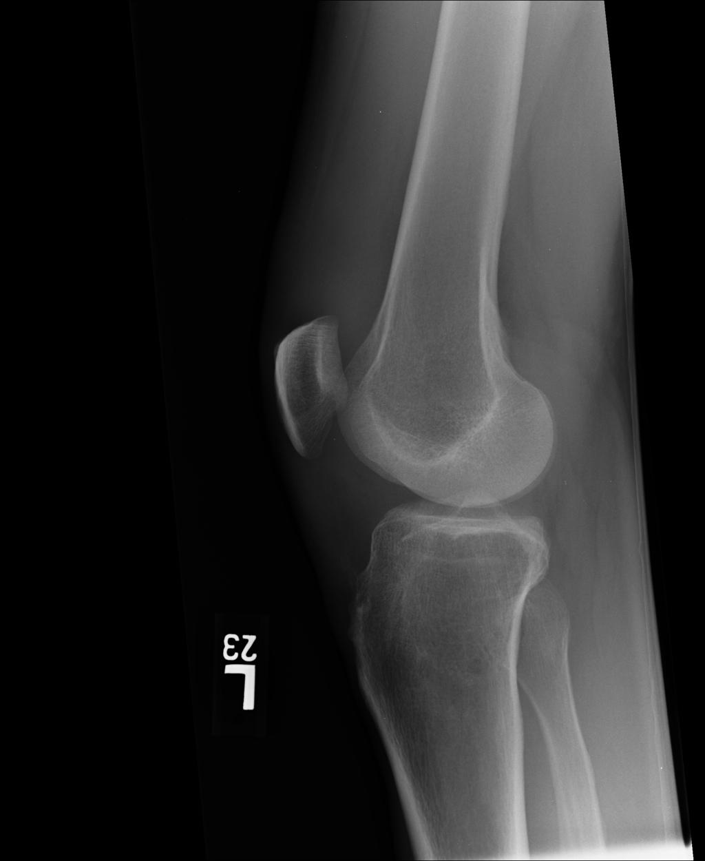
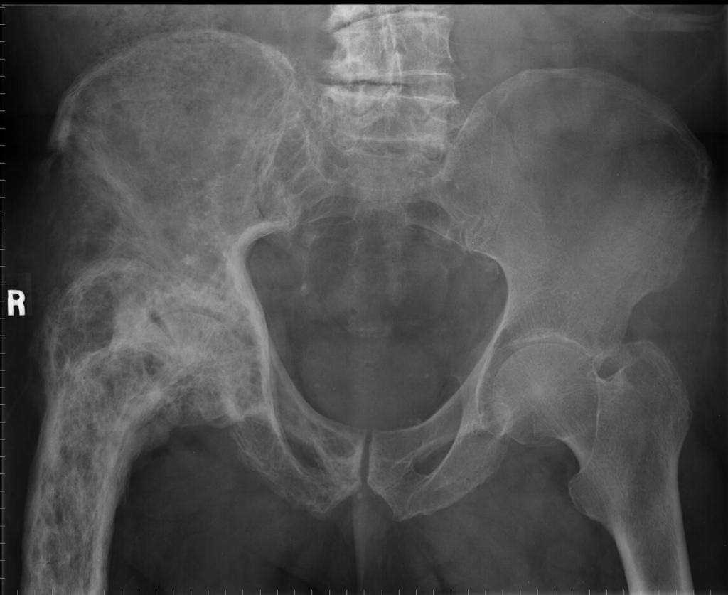
7 This patient, on steroids for organ transplant, complains of hip pain. Which hip hurts? What findings support this conclusion? In this setting, what s the most likely diagnosis? Case 3
 or lysis Case 3 (abnormal")
8 Do you see areas of sclerosis (increased bone density) or lysis Case 3 (abnormal lucency)?
9 What imaging modality is this? Why is it now easier to identify subtle abnormalities? Case 3
10 This patient went on for an additional study. What modality is this? Case 3
11 modality A modality B Compare the appearance of cortical bone and fat between these two studies. Can you match the following terms to the appropriate modality? attenuation, intensity, T1 weighting, bone setting, signal, Hounsfield Case 3 unit, gadolinium, iodine
12 Patient with renal disease and elbow pain Identify evidence for soft tissue swelling (clue: palpate the back of your own elbow for comparison) Distinguish fat from fluid/water opacity How does this help identify the elbow fat pad or sail sign? Case 4

13 Can you identify both the subcutaneous fat and the elbow joint fat pad? Case 4 Can you point out the transition between normal and edematous subcutaneous fat? What s your differential diagnosis?
14 Pedestrian struck by car with knee pain the first image is a normal lateral knee film for comparison again, can you distinguish between the opacities of air, fat, and fluid/soft tissue? Case 5

15 normal lateral knee Case 5 Identify the quadriceps muscle, the quadriceps tendon, and subcutaneous fat. How would the opacity of fluid compare?

16 Case 5 Our unfortunate patient: Can you identify the fracture?
17 Do you see the horizontal level cephalad to the patella?* How does this help indicate how the image was taken...with a Case 5 vertical or a horizontal beam (cross-table)? *see close-up on next slide
18 What is the nature of the fluids that comprise the horizontal interface you see? How can you tell? Case 5 clue: think balsamic vinegraitte!
19 55 year old diabetic male with painless foot swelling Can you identify 3 of the 4 basic radiographic opacities on this plain film? Which opacity is missing, and why? Which opacity is present in the wrong tissue, and what does that signify? Why do you think this patient experienced no pain? Case 6

20 Case 6 Can you appreciate the swelling? Feel the dorsum of your own foot.
21 This 42 year old woman complains of several months of worsening knee pain. identify cancellous vs. cortical bone do you see a lytic area, i.e. a focal area of missing bone? describe the location of the lesion Case 6
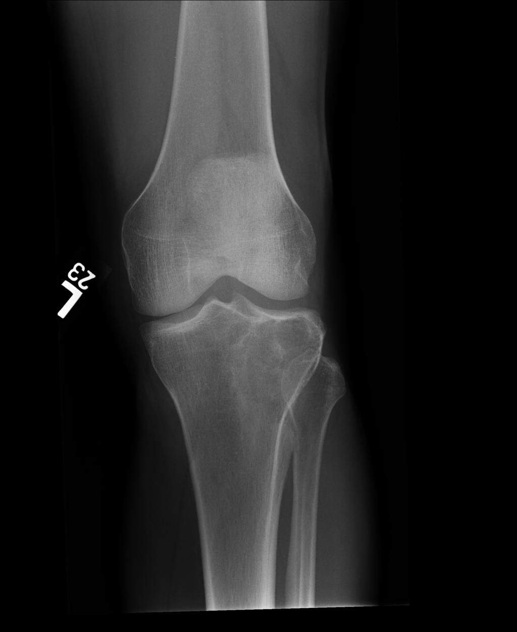
22 Case 6


23 axial CT coronal Describe the changes to both the cancellous and cortical bone Are the margins sharply defined or indistinct? Sclerotic or permeative? What does this say about the biology of the lesion? Case 6
24 Tripped on curb why do you always need two views at 90 degrees, especially in trauma? what has occurred at the ankle joint? identify the ankle mortise, medial and lateral malleoli, tibiotalar joint, and calcaneus Case 7


25 lateral frontal mortise view Case 7
26 85 year old male with deformity of the thigh compare the trabecula, cortex, and size of the femur between the two sides what s the second exam? is this an isolated bone abnormality Case 8
27 Case 8

28 What study did the patient then undergo? Why are the two images reversed? Does this disease involve one or multiple bones? What is causing certain areas to appear more white? Case 8

29 Shortly afterwards, this gentleman felt a sudden snap while getting to his car. Discuss why such a simple action could lead to this complication. Case 8
Imaging Modalities: Clinical Reasoning and Key Instructional Elements: Radiography
 Imaging Modalities: Clinical Reasoning and Key Instructional Elements: Radiography Michael D. Ross, PT, DHSc, OCS mross@daemen.edu Disclosure No relevant financial relationship exists Objectives Determine
Rad Lab 4 Unknowns: Genitourinary!
 Rad Lab 4 Unknowns: Genitourinary! Peter Clarke MD! Don Di Salvo, MD! Clerkship Directors for Radiology! Harvard Medical School! Brigham and Women s Hospital! Dana Farber Cancer Institute! Case 1: 69 year
RADIOGRAPHY OF THE ANKLE and LOWER LEG
 RADIOGRAPHY OF THE ANKLE and LOWER LEG Patient Position: ANKLE AP Projection Part Position: True Slight to place foot s long axis Center to Central Ray: to IR Midway Note: Ankle joint is to tips of malleoli
Imaging the musculoskeletal system. An Introduction
 Imaging the musculoskeletal system An Introduction Objectives Discuss: commonly used imaging modalities in the musculoskeletal system normal imaging anatomy in the extremities fracture description Imaging
SMALL GROUP SESSION 16 January 8 th or 10 th Shoulder pain case/ Touch workshop/ Upper and Lower Extremity Examination
 SMALL GROUP SESSION 16 January 8 th or 10 th Shoulder pain case/ Touch workshop/ Upper and Lower Extremity Examination Suggested Readings: Opatrny L. The Healing Touch. Ann Int Med 2002; 137:1003. http://www.annals.org/cgi/reprint/137/12/1003.pdf
Commonly Missed Injuries of the Extremities
 Commonly Missed Injuries of the Extremities Dr. Tudor H. Hughes M.D., FRCR Department of Radiology University of California School of Medicine San Diego, California 1. Base of skull 2. Odontoid process
Chealon Miller, HMS IV Gillian Lieberman, MD. November Stress Fractures. Chealon Miller, Harvard Medical School Year IV Gillian Lieberman, MD
 November 2005 Stress Fractures Chealon Miller, Harvard Medical School Year IV Our Patient G.F. 29 year old female runner c/o left shin pain and swelling Evaluated at OSH with MRI showing a mass Referred
emoryhealthcare.org/ortho
 COMMON SOCCER INJURIES Oluseun A. Olufade, MD Assistant Professor, Department of Orthopedics and PM&R 1/7/18 GOALS Discuss top soccer injuries and treatment strategies Simplify hip and groin injuries in
Radiology Positioning Practical Test #2 Table (By Jung Park):
:") Radiology Positioning Practical Test #2 Table (By Jung Park): (Lower Extremity): patient is fully gowned / no artifacts / properly shielded (exposure for femur and below : hold still, don t move ) (exposure
Radiology Positioning Practical Test #2 Table (By Jung Park): (Lower Extremity): patient is fully gowned / no artifacts / properly shielded (exposure for femur and below : hold still, don t move ) (exposure
MRI XR, CT, NM. Principal Modality (2): Case Report # 2. Date accepted: 15 March 2013
: Case Report # 2. Date accepted: 15 March 2013") Radiological Category: Musculoskeletal Principal Modality (1): Principal Modality (2): MRI XR, CT, NM Case Report # 2 Submitted by: Hannah Safia Elamir, D.O. Faculty reviewer: Naga R. Chinapuvvula, M.D.
Radiological Category: Musculoskeletal Principal Modality (1): Principal Modality (2): MRI XR, CT, NM Case Report # 2 Submitted by: Hannah Safia Elamir, D.O. Faculty reviewer: Naga R. Chinapuvvula, M.D.
ORTHOSCAN MOBILE DI POSITIONING GUIDE
 ORTHOSCAN MOBILE DI POSITIONING GUIDE Table of Contents SHOULDER A/P of Shoulder... 4 Tangential (Y-View) of Shoulder... 5 Lateral of Proximal Humerus... 6 ELBOW A/P of Elbow... 7 Extended Elbow... 8 Lateral
SMALL GROUP SESSION 16 January 8 th or 10 th. Shoulder pain case/ Touch workshop/ Upper and Lower Extremity Examination
 SMALL GROUP SESSION 16 January 8 th or 10 th Shoulder pain case/ Touch workshop/ Upper and Lower Extremity Examination Suggested Readings: Opatrny L. The Healing Touch. Ann Int Med 2002; 137:1003. http://www.annals.org/cgi/reprint/137/12/1003.pdf
11/4/2018 SUBTLETIES OF LOWER EXTREMITY TRAUMA IMAGING SPEAKER DISCLOSURES
 SUBTLETIES OF LOWER EXTREMITY TRAUMA IMAGING Charles S. Resnik, M.D. Professor of Radiology University of Maryland School of Medicine Upon completion of this presentation, participants will be better able
ADOLESCENT SPORTS INJURIES. Orthopaedics in Motion April 5, 2017 John Lammli, MD
 ADOLESCENT SPORTS INJURIES Orthopaedics in Motion April 5, 2017 John Lammli, MD OVERVIEW: Shoulder Injuries Knee Injuries Leg/Ankle Injuries SHOULDER INJURIES Injuries to the shoulder girdle are vast and
Exercise 11. The Appendicular Skeleton
 Exercise 11 The Appendicular Skeleton The Appendicular Skeleton The appendicular skeleton contains 126 bones. Consists of the upper and lower limbs, the pectoral girdles, and the pelvic girdles. The pectoral
AAP Boot Camp KNEE AND ANKLE EXAM
 AAP Boot Camp KNEE AND ANKLE EXAM Disclosures I have no relevant financial relationships with the manufacturers of any commercial products and or providers of commercial services discussed in this CME
Emerging Applications in Musculoskeletal CT Imaging
 Emerging pplications in Musculoskeletal CT Imaging y K Murali MD(RD), PDCC, Director of Interventional Radiology, G. Francis DMRD, DN (RD), Consultant Radiologist, and R. Madan, MS, MD, Consultant Radiologist,
Case. 5 year old with 2 weeks leg pain and now refusing to walk + Fevers, lower leg swelling, warmth Denies and history of trauma or wounds
 Case 5 year old with 2 weeks leg pain and now refusing to walk + Fevers, lower leg swelling, warmth Denies and history of trauma or wounds Exam I: Swelling over entire tibia extending to foot P: Tenderness
Dr.Israa H. Mohsen. Lecture 5. The vertebral column
 Anatomy Lecture 5 Dr.Israa H. Mohsen The vertebral column The vertebral column a flexible structure consisting of 33 vertebrae holds the head and torso upright, serves as an attachment point for the legs,
Orthopedics - Dr. Ahmad - Lecture 2 - Injuries of the Upper Limb
 The shoulder and the upper arm Fractures of the clavicle 1. Fall on the shoulder. 2. Fall on outstretched hand. In mid shaft fractures, the outer fragment is pulled down by the weight of the arm and the
Appendicular skeleton: ABCs Image Interpretation Search strategy
 NOVEMBER 2013 volume 51 number 2 THE SOUTH AFRICAN RADIOGRAPHER peer reviewed ARTICLE OF INTEREST Appendicular skeleton: ABCs Image Interpretation Search strategy IJ Williams MSc in Medical Imaging; B
Bone Composition. Bone is very strong for its relatively light weight The major components of bone are:
 Human Bones Bone Composition Bone is very strong for its relatively light weight The major components of bone are: Calcium carbonate Calcium phosphate Collagen Water Cortical Bone Spongy Bone Medullary
CLINICAL PRESENTATION AND RADIOLOGY QUIZ QUESTION
 Donald L. Renfrew, MD Radiology Associates of the Fox Valley, 333 N. Commercial Street, Suite 100, Neenah, WI 54956 11/24/2012 Radiology Quiz of the Week # 100 Page 1 CLINICAL PRESENTATION AND RADIOLOGY
CLINICAL PRESENTATION AND RADIOLOGY QUIZ QUESTION
 Donald L. Renfrew, MD Radiology Associates of the Fox Valley, 333 N. Commercial Street, Suite 100, Neenah, WI 54956 12/29/2012 Radiology Quiz of the Week # 105 Page 1 CLINICAL PRESENTATION AND RADIOLOGY
Musculoskeletal Examination Benchmarks
 Musculoskeletal Examination Benchmarks _ The approach to examining the musculoskeletal system is the same no matter what joint or limb is being examined. The affected and contralateral region should both
Human Anatomy, First Edition McKinley & O'Loughlin
 Human Anatomy, First Edition McKinley & O'Loughlin Chapter 8 : Appendicular Skeleton 8-1 Appendicular Skeleton Includes the bones of the upper and lower limbs. The girdles of bones that attach the upper
Anatomy of the Musculoskeletal System
 Anatomy of the Musculoskeletal System Kyle E. Rarey, Ph.D. Department of Anatomy & Cell Biology and Otolaryngology University of Florida College of Medicine Outline of Presentation Vertebral Column Upper
Anatomy and Physiology 2016
 Anatomy and Physiology 2016 O = Temporal line I = coronoid process (Mandible) A = elevates mandible (chewing) O = galea aponeurotica (layer of dense fibrous tissue which covers the upper part of the cranium)
Topics. Musculoskeletal Infection Extremities. Detection of Infection. Role of Imaging in Extremity Infection. Detection of Infection
 Topics Musculoskeletal Infection Extremities Nuttaya Pattamapaspong M.D. Department of Radiology, Faculty of Medicine, Chiang Mai University, Chiang Mai, Thailand Role of imaging in extremity infection
Lecture (10) Bone Fractures. Resources: - Lecture by dr.alboukai - Diagnostic imaging book
 Bone Fractures. Resources: - Lecture by dr.alboukai - Diagnostic imaging book") Lecture (10) Bone Fractures Hanan Alsalman Hanan Alrabiah Reem Aljurayyad Ayshah Almahboob Ghadeer Alwuhyad Khawlah AlOthman Dalal Alqadi Suliman Alshammari Maha AlKubaidan Rawabi Alghamdi Resources: -
Lecture (10) Bone Fractures Hanan Alsalman Hanan Alrabiah Reem Aljurayyad Ayshah Almahboob Ghadeer Alwuhyad Khawlah AlOthman Dalal Alqadi Suliman Alshammari Maha AlKubaidan Rawabi Alghamdi Resources: -
EPIPHYSEAL PLATE IN FEMUR
 Reviewing: Epiphyseal Plates (younger skeletons) eventually will disappear. Bones grow lengthwise up and down from each plate, and in a circular collar like fashion around the diaphysis. These plates will
P V S MEMORIAL HOSPITAL LTD.
 SHOULDER XRAYS Instability Series o True AP (Grashey s) o Axillary o Stryker Notch view o True AP in Internal rotation o Scapular Y view o West Point view for Bony Bankart ( looks like modif axillary view)
Shoulder Position: Supine arm in the neutral position. Collateral arm above head Indication: fracture humerus, fracture scapula
 Shoulder Position: Supine arm in the neutral position. Collateral arm above head Indication: fracture humerus, fracture scapula No instrumentation With metal or cast KV/ Effective mas/rotation time 140/300/1.0
Radiologic Pitfalls. Objectives: High Risk! Occult Fracture? 2/16/2014
 Objectives: Radiologic Pitfalls Gregory W. Hendey, MD, FACEP Professor of Clinical Emergency Medicine UCSF Fresno, Medical Education Program To discuss plain film and physical findings that suggest an
Review relevant anatomy of the foot and ankle. Learn the approach to examining the foot and ankle
 Objectives Review relevant anatomy of the foot and ankle Learn the approach to examining the foot and ankle Learn the basics of diagnosis and treatment of ankle sprains Overview of other common causes
3/3/2017. Acute spine disorder (< 4weeks duration) Subacute spine disorder (4-12 weeks duration) Chronic spine disorder (>12 weeks duration)
 Subacute spine disorder (4-12 weeks duration) Chronic spine disorder (>12 weeks duration)") William Hsu BSc DC DACBR March 4, 2017 Acute spine disorder (< 4weeks duration) Subacute spine disorder (4-12 weeks duration) Chronic spine disorder (>12 weeks duration) Neurologic symptoms and signs pain
William Hsu BSc DC DACBR March 4, 2017 Acute spine disorder (< 4weeks duration) Subacute spine disorder (4-12 weeks duration) Chronic spine disorder (>12 weeks duration) Neurologic symptoms and signs pain
CLINICAL PRESENTATION AND RADIOLOGY QUIZ QUESTION
 Donald L. Renfrew, MD Radiology Associates of the Fox Valley, 333 N. Commercial Street, Suite 100, Neenah, WI 54956 12/01/2012 Radiology Quiz of the Week # 101 Page 1 CLINICAL PRESENTATION AND RADIOLOGY
Radiographic Positioning Summary (Basic Projections RAD 222)
") Lower Extremity Radiographic Positioning Summary (Basic Projections RAD 222) AP Pelvis AP Hip (Unilateral) (L or R) AP Femur Mid and distal AP Knee Lateral Knee Pt lies supine on table Align MSP to Center
Lower Extremity Radiographic Positioning Summary (Basic Projections RAD 222) AP Pelvis AP Hip (Unilateral) (L or R) AP Femur Mid and distal AP Knee Lateral Knee Pt lies supine on table Align MSP to Center
Evaluation of the Knee and Shoulder
 Evaluation of the Knee and Shoulder Karen J. Boselli, MD Northeast Regional Nurse Practitioner Conference May 2018 Knee Overview History Examination Top 5 diagnoses When to image When to refer Pain most
Chapter 8B. The Skeletal System: Appendicular Skeleton. The Appendicular Skeleton. Clavicle. Pectoral (Shoulder) Girdle
 Girdle") The Appendicular Skeleton Chapter 8B The Skeletal System: Appendicular Skeleton 126 bones Pectoral (shoulder) girdle Pelvic (hip) girdle Upper limbs Lower limbs Functions primarily to facilitate movement
The Appendicular Skeleton Chapter 8B The Skeletal System: Appendicular Skeleton 126 bones Pectoral (shoulder) girdle Pelvic (hip) girdle Upper limbs Lower limbs Functions primarily to facilitate movement
Radiologic Imaging Magnetic Resonance Imaging (MRI)
") Radiologic Imaging X-ray has always been the golden rule in diagnosing and treating podiatric patients. Unfortunately, for some patients the diagnosis is not as evident. That is when we need to utilize
Radiologic Imaging X-ray has always been the golden rule in diagnosing and treating podiatric patients. Unfortunately, for some patients the diagnosis is not as evident. That is when we need to utilize
Osteoporosis. Dr. C. C. Visser. MBChB MMed (Med Phys) Diploma Musculoskeletal Medicine (UK) Member: Society of Orthopaedic Medicine (UK)
 Diploma Musculoskeletal Medicine (UK) Member: Society of Orthopaedic Medicine (UK)") Osteoporosis Dr. C. C. Visser MBChB MMed (Med Phys) Diploma Musculoskeletal Medicine (UK) Member: Society of Orthopaedic Medicine (UK) Effect of age on trabecular bone. Fatfree dry bone cylinders obtained
Osteoporosis Dr. C. C. Visser MBChB MMed (Med Phys) Diploma Musculoskeletal Medicine (UK) Member: Society of Orthopaedic Medicine (UK) Effect of age on trabecular bone. Fatfree dry bone cylinders obtained
2017 Resident Advanced Trauma Techniques Course COMPLICATIONS / CHALLENGES MALUNIONS/DEFORMITY
 2017 Resident Advanced Trauma Techniques Course COMPLICATIONS / CHALLENGES MALUNIONS/DEFORMITY What is a Malunion? Definition: a fracture that has healed in a nonanatomic (i.e. deformed) position Must
ESSEX COUNTY COLLEGE Nursing and Allied Health Division RTC 101 Radiographic Positioning Principles I Course Outline
 ESSEX COUNTY COLLEGE Nursing and Allied Health Division RTC 101 Radiographic Positioning Principles I Course Outline Course Number & Name: RTC 101 Radiographic Positioning Principles I Credit Hours: 4.0
Musculoskeletal Injuries
 Musculoskeletal Injuries KNOWLEDGE OBJECTIVES 1. Identify the four main structures of the musculoskeletal system. 2. List five common signs or symptoms of musculoskeletal injuries. 3. List seven signs
Injuries to Muscles, Bones and Joints. Emergency Medical Response
 Injuries to Muscles, Bones and Joints Lesson 33: Injuries to Muscles, Bones and Joints You Are the Emergency Medical Responder You are patrolling the state park where you are the emergency medical responder
Hands PA; Obl. Lat.; Norgaard s Thumb AP; Lat. PA. PA; Lat.: Obls.; Elongated PA with ulnar deviation
 Projections Region Basic projections Additional / Modified projections Upper Limbs Hands PA; Obl. Lat.; Norgaard s Thumb ; Lat. PA Fingers PA; Lat. Wrist PA; Lat. Obls. Scaphoid Lunate Trapezium Triquetral
MY PATIENT HAS KNEE PAIN. David Levi, MD Chief, Division of Musculoskeletal l limaging Atlantic Medical Imaging
 MY PATIENT HAS KNEE PAIN David Levi, MD Chief, Division of Musculoskeletal l limaging Atlantic Medical Imaging Causes of knee pain Non traumatic Trauma Osteoarthritis Patellofemoral pain Menisci or ligaments
The Appendicular Skeleton
 8 The Appendicular Skeleton PowerPoint Lecture Presentations prepared by Jason LaPres Lone Star College North Harris 8-1 The Pectoral Girdle The Pectoral Girdle Also called shoulder girdle Connects the
Functional Orthopedic Imaging Capturing Motion, Flow and Perfusion. Case Study Brochure Centre University Hospital Nancy.
 Capturing Motion, Flow and Perfusion dynamic volume CT Case Study Brochure Centre University Hospital Nancy http://www.toshibamedicalsystems.com Toshiba Medical Systems Corporation 2013. All rights reserved.
14b Swedish: Technique Review and Practice Feet, Anterior Lower Body, and Abs
 14b Swedish: Technique Review and Practice Feet, Anterior Lower Body, and Abs 14b Swedish:! Technique Review & Practice - Feet, Anterior Lower Body, and Abs! Class Outline 5 minutes Attendance, Breath
Radiography. 1. Introduction. 2. Documentation of Compliance. 3. Didactic Competency Requirements. 4. Clinical Competency Requirements
 PRIMARY CERTIFICATION AND REGISTRATION Radiography 1. Introduction Candidates for certification and registration are required to meet the Professional Education Requirements specified in the ARRT Rules
The examination of the painful knee. Maja K Artandi, MD, FACP Clinical Associate Professor of Medicine Stanford University
 The examination of the painful knee Maja K Artandi, MD, FACP Clinical Associate Professor of Medicine Stanford University Objectives of the talk By the end of this talk you will know The important anatomy
The Skeletal System THE APPENDICULAR SKELETON
 The Skeletal System THE APPENDICULAR SKELETON The appendicular skeleton consists of the girdles and the skeleton of the limbs. The upper (anterior) limbs are attached to the pectoral (shoulder) girdle
 17.2 A-P Lower Leg Measure: A-P at mid-lower leg Protection: Apron draped over pelvis SID: 40 Table top No Tube Angle Film: 7 x17 I.D. down or diagonal 14 x 17 www.fisiokinesiterapia.biz A-P Lower Leg
2015 AFL Injury Survey
 2015 AFL Injury Survey Collaborators: AFL Doctors Association AFL Physiotherapists Association AFL Football Operations Department 1 Key Indicators Table 1 Key indicators for all injuries over the 24 seasons
Radiology 10/28/2013 COLLIMATION CAN IMPROVE YOUR IMAGES COLLIMATION CAN IMPROVE YOUR IMAGES REVIEW OF BASIC X-RAY PHYSICS
 Radiology Hector RiveraMelo, DC, DACBR Director, Center for Diagnostic Imaging Southern California University of Health Sciences COLLIMATION CAN IMPROVE YOUR IMAGES This film demonstrates limited collimation.
NEW YORK CITY COLLEGE OF TECHNOLOGY THE CITY UNIVERSITY OF NEW YORK. Department of Radiologic Technology & Medical Imaging
 NEW YORK CITY COLLEGE OF TECHNOLOGY THE CITY UNIVERSITY OF NEW YORK Department of Radiologic Technology & Medical Imaging RAD 1125 (REVISED 9/1/15) Section D806: Monday, 10:00am to 11:15am Radiographic
AMERICAN RED CROSS FIRST AID RESPONDING TO EMERGENCIES FOURTH EDITION Copyright 2006 by The American National Red Cross All rights reserved.
 Musculoskeletal injuries are most commonly caused by Mechanical forms of energy. Chemicals. Electrical energy. Heat Mechanical energy produces direct, indirect, twisting and contracting forces. Can be
Pectoral (Shoulder) Girdle
 Girdle") Chapter 8 Skeletal System: Appendicular Skeleton Pectoral girdle Pelvic girdle Upper limbs Lower limbs 8-1 Pectoral (Shoulder) Girdle Consists of scapula and clavicle Clavicle articulates with sternum
Chapter 8 Skeletal System: Appendicular Skeleton Pectoral girdle Pelvic girdle Upper limbs Lower limbs 8-1 Pectoral (Shoulder) Girdle Consists of scapula and clavicle Clavicle articulates with sternum
Presented by Chris Hudd
 Presented by Chris Hudd Study design dot points: causes of potential acute and chronic injuries and illness associated with the muscular and skeletal systems such as arthritis, osteoporosis and other musculoskeletal
Case 27 Clinical Presentation
 53 Case 27 Clinical Presentation 40-year-old man presents with acute shoulder pain and normal findings on radiographs. 54 RadCases Musculoskeletal Radiology Imaging Findings (,) Coronal images of the shoulder
CHAPTER 8 LECTURE OUTLINE
 CHAPTER 8 LECTURE OUTLINE I. INTRODUCTION A. The appendicular skeleton includes the bones of the upper and lower extremities and the shoulder and hip girdles. B. The appendicular skeleton functions primarily
4/28/2010. Fractures. Normal Bone and Normal Ossification Bone Terms. Epiphysis Epiphyseal Plate (physis) Metaphysis
 Metaphysis") Fractures Normal Bone and Normal Ossification Bone Terms Epiphysis Epiphyseal Plate (physis) Metaphysis Diaphysis 1 Fracture Classifications A. Longitudinal B. Transverse C. Oblique D. Spiral E. Incomplete
Fractures Normal Bone and Normal Ossification Bone Terms Epiphysis Epiphyseal Plate (physis) Metaphysis Diaphysis 1 Fracture Classifications A. Longitudinal B. Transverse C. Oblique D. Spiral E. Incomplete
Important Parts of Bones
 Important Parts of Bones For 2015 Know: Humerus (posterior) Clavical Femur (Anterior) Foot Hand Mandible Os Coxa Scapula Skull (Anterior, Inferior, Lateral) Sternum Humerus (posterior) A. olecranon fossa
Imaging the Athlete s Knee. Peter Lowry, MD Musculoskeletal Radiology University of Colorado
 Imaging the Athlete s Knee Peter Lowry, MD Musculoskeletal Radiology University of Colorado None Disclosures Knee Imaging: Radiographs Can be performed weight-bearing or non-weight-bearing View options
Radiography Protocols
 Radiography Protocols Upper Limb Second through Fifth Digits (Standard 3 views) First Digit (Thumb) (Standard 3 views) Hand (Standard 3 views) Wrist (Standard 4 views) Forearm (Standard 2 views) Elbow
Exercise Science Section 4: Joint Mechanics and Joint Injuries
 Exercise Science Section 4: Joint Mechanics and Joint Injuries An Introduction to Health and Physical Education Ted Temertzoglou Paul Challen ISBN 1-55077-132-9 Types of Joints Fibrous joint Cartilaginous
PEM GUIDE CHILDHOOD FRACTURES
 PEM GUIDE CHILDHOOD FRACTURES INTRODUCTION Skeletal injuries account for 10-15% of all injuries in children; 20% of those are fractures, 3 out of 4 fractures affect the physis or growth plate. Always consider
Original Report. The Reverse Segond Fracture: Association with a Tear of the Posterior Cruciate Ligament and Medial Meniscus
 Eva M. Escobedo 1 William J. Mills 2 John. Hunter 1 Received July 10, 2001; accepted after revision October 1, 2001. 1 Department of Radiology, University of Washington Harborview Medical enter, 325 Ninth
Extraarticular Lateral Ankle Impingement
 Extraarticular Lateral Ankle Impingement Poster No.: C-1282 Congress: ECR 2016 Type: Educational Exhibit Authors: C. Cevikol; Keywords: Trauma, Diagnostic procedure, MR, CT, Musculoskeletal system, Musculoskeletal
Knee, Ankle, and Foot: Normal and Abnormal Features with MRI and Ultrasound Correlation. Disclosures. Outline. Joint Effusion. Suprapatellar recess
 Knee, Ankle, and Foot: Normal and Abnormal Features with MRI and Ultrasound Correlation Jon A. Jacobson, M.D. Professor of Radiology Director, Division of Musculoskeletal Radiology University of Michigan
Lower Extremity Dislocations: Management and Triage on the Field
 Lower Extremity Dislocations: Management and Triage on the Field Scott J Tarantino, MD Towson Orthopaedic Associates, Towson, MD None Disclsures Purpose To provide you with knowledge which may guide you
Chapter 8 The Skeletal System: The Appendicular Skeleton. Copyright 2009 John Wiley & Sons, Inc.
 Chapter 8 The Skeletal System: The Appendicular Skeleton Appendicular Skeleton It includes bones of the upper and lower limbs Girdles attach the limbs to the axial skeleton The pectoral girdle consists
Basic Principles of Fractures & Easily Missed Fractures. Mr Irfan Merchant Trauma & Orthopaedic Registrar Bedford Hospital, East of England
 Basic Principles of Fractures & Easily Missed Fractures Mr Irfan Merchant Trauma & Orthopaedic Registrar Bedford Hospital, East of England Objectives Types Fracture Patterns Fracture Healing Assessing
10/12/2010. Upper Extremity. Pectoral (Shoulder) Girdle. Clavicle (collarbone) Skeletal System: Appendicular Skeleton
 Girdle. Clavicle (collarbone) Skeletal System: Appendicular Skeleton") Skeletal System: Appendicular Skeleton Pectoral girdle Pelvic girdle Upper limbs Lower limbs 8-1 Pectoral (Shoulder) Girdle Consists of scapula and clavicle Clavicle articulates with sternum (Sternoclavicular
Skeletal System: Appendicular Skeleton Pectoral girdle Pelvic girdle Upper limbs Lower limbs 8-1 Pectoral (Shoulder) Girdle Consists of scapula and clavicle Clavicle articulates with sternum (Sternoclavicular
X-ray (Radiography) - Bone
 - Bone") Scan for mobile link. X-ray (Radiography) - Bone Bone x-ray uses a very small dose of ionizing radiation to produce pictures of any bone in the body. It is commonly used to diagnose fractured bones or
Scan for mobile link. X-ray (Radiography) - Bone Bone x-ray uses a very small dose of ionizing radiation to produce pictures of any bone in the body. It is commonly used to diagnose fractured bones or
Hip and Thigh Cases: Surprises
 Hip and Thigh Cases: Surprises Mary Lloyd Ireland, M.D. Associate Professor University of Kentucky Dept. of Orthopaedic Surgery and Sports Medicine Lexington, Kentucky www.marylloydireland.com Learning
The scapula is located on the back side of the ribcage and helps provide part of the shoulder joint and movement for the arms.
 The scapula is located on the back side of the ribcage and helps provide part of the shoulder joint and movement for the arms. Scapula Humerus (Upper Arm Bone) Radius and Ulna Radius on Top Ulna on Bottom
Basic Radiographic Principles Part II
 Basic Radiographic Principles Part II Kristopher Avant, D.O. October 19 th, 2016 I have no disclosures relevant to the material presented in this discussion. Good Stuff!!! 1 Really? Really! Musculoskeletal
The Benjamin Institute of Advanced Studies
 175 Richdale Avenue, Suite 106 Cambridge, MA 02140 Phone (617) 576-0777 Fax (617) 576-0444 www.benbenjamin.com Step 1: Study the course materials provided. Then take the exam, reviewing the DVD program
General Concepts. Growth Around the Knee. Topics. Evaluation
 General Concepts Knee Injuries in Skeletally Immature Athletes Zachary Stinson, M.D. Increased rate and ability of healing Higher strength of ligaments compared to growth plates Continued growth Children
Goals. Initial management skeletal trauma. Physical Exam ABC OF PRIMARY CARE MEDICINE FRACTURE MANAGEMENT 12/4/2010
 ABC OF PRIMARY CARE MEDICINE FRACTURE MANAGEMENT Brian Feeley, MD UCSF Sports Medicine and Shoulder Surgery Goals Discuss common fractures and initial management, treatment guidelines Let your patients
PRE-LAB EXERCISES. Before we get started, look up the definitions of these common bone marking terms: Canal: Condyle: Facet: Fissure:
 1 PRE-LAB EXERCISES When studying the skeletal system, the bones are often sorted into two broad categories: the axial skeleton and the appendicular skeleton. This lab focuses on the appendicular skeleton,
When Pads of Fat are a Welcome Sight: Fat Pads in Acute Musculoskeletal Imaging
 When Pads of Fat are a Welcome Sight: Fat Pads in Acute Musculoskeletal Imaging Poster No.: C-2444 Congress: ECR 2013 Type: Authors: Keywords: DOI: Educational Exhibit M. Zakhary 1, M. Adix 2, C. Yablon
Lab: Histology of Bone and Cartilage
 WEEK 1 Time/Date Monday, October 8 Tuesday, October 9 Wednesday, October 10 Thursday, October 11 Friday, October 12 1. 2. PHARMACOLOGY 1. PATHOLOGY 2. CELL BIOLOGY/PHYSIO 4. CELL BIOLOGY/PHYSIO Concepts
Chapter 7: Skeletal System: Gross Anatomy
 Chapter 7: Skeletal System: Gross Anatomy I. General Considerations A. How many bones in an average adult skeleton? B. Anatomic features of bones are based on II. Axial Skeleton A. Skull 1. Functionally
Country Health SA Medical Imaging
 Country Health SA Medical Imaging REMOTE OPERATORS POSITIONING GUIDE Contents Image Evaluation Page 4 Positioning Guides Section 1 - THORAX 1.1 Chest Page 5 1.2 Bedside Chest Page 7 1.3 Ribs Page 8 Section
Orthopedic X-Rays most commonly missed
 Orthopedic X-Rays most commonly missed Vukiet Tran, MD, MHSc, MBA University Health Network Toronto, Canada 1 COI Disclosure I am the current Medical Director for Best Doctors Canada. Presenter: Dr. Vu
Swedish Technique Class
 Swedish Technique Class Massage of the Back Establish contact at the sacrum and occiput, and relax. Effleurage the whole back to apply oil, warm, and soften (3-6 times). Each effleurage of the whole back
Brain Atrophy. Brain Atrophy
 Aging Central Nervous System Processes Age related brain atrophy Non-age related brain atrophy Cerebrovascular disease Cerebral infarction Hypertensive hemorrhage Carotid artery stenosis and occlusion
Injuries to the Extremities
 Injuries to the Extremities KNOWLEDGE OBJECTIVES 1. List seven signs and symptoms that suggest a serious extremity injury. 2. Describe how to care for injuries to the shoulder, upper arm, and elbow. 3.
CASE ONE CASE ONE. RADIAL HEAD FRACTURE Mason Classification. RADIAL HEAD FRACTURE Mechanism of Injury. RADIAL HEAD FRACTURE Imaging
 CASE ONE An eighteen year old female falls during a basketball game, striking her elbow on the court. She presents to your office that day with a painful, swollen elbow that she is unable to flex or extend
Imaging Choices in Occult Hip Fracture
 Introduction Imaging Choices in Occult Hip Fracture Jesse Cannon, MD; Salvatore Silvestri, MD; Mark Munro, MD J Emerg Med. 2009;32(3):144-152 Reporter PGY 宋兆家 Supervisor VS 侯勝文 990220 High dependence on
Exam of the Knee and Ankle I HAVE NO FINANCIAL DISCLOSURES RELEVANT TO THIS PRESENTATION
 Exam of the Knee and Ankle I HAVE NO FINANCIAL DISCLOSURES RELEVANT TO THIS PRESENTATION Disclosures I have no relevant financial relationships with the manufacturers of any commercial products and or
AP1 Lab 4 - Appendicular Skeleton
 Project 1 Learn the Bone Names AP1 Lab 4 - Appendicular Skeleton Use Figure 7.1 and the hanging skeletons to learn the whole bones of the arms and legs. Don t learn the features of each bone yet just be
Block 2 VIRGINIA CAMPUS Musculoskeletal System 2018
 WEEK 1 Time/Date Monday, October 15 Tuesday, October 16 Wednesday, October 17 Thursday, October 18 Friday, October 19 1. CELL BIOLOGY/PHYSIO 1. ANATOMY 2-3. CELL BIOLOGY/PHYSIO 6. CELL BIOLOGY/PHYSIO 8:00
An Introduction to Radiographic Views & Anatomy
 An Introduction to Radiographic Views & Anatomy Morey J. Kolber, PT, PhD, OCS, Cert MDT, CSCS*D An Introduction to Radiographic Views & Anatomy M.S.P.T. 1995-University of Miami Nova Southeastern University
CLINICAL PRESENTATION AND RADIOLOGY QUIZ QUESTION
 Donald L. Renfrew, MD Radiology Associates of the Fox Valley, 333 N. Commercial Street, Suite 100, Neenah, WI 54956 10/6/2012 Radiology Quiz of the Week # 93 Page 1 CLINICAL PRESENTATION AND RADIOLOGY
UPPER EXTREMITY INJURIES. Recognizing common injuries to the upper extremity
 UPPER EXTREMITY INJURIES Recognizing common injuries to the upper extremity ANATOMY BONES Clavicle Scapula Spine of the scapula Acromion process Glenoid fossa/cavity Humerus Epicondyles ANATOMY BONES Ulna