Computed Tomographic Imaging of Foot and Ankle trauma
|
|
|
- Marlene Lewis
- 6 years ago
- Views:
Transcription

1 Computed Tomographic Imaging of Foot and Ankle trauma Dr. Tudor H. Hughes M.D., FRCR Department of Radiology University of California School of Medicine San Diego, California
2 CT of Foot and Ankle Trauma Who All complex intraarticular fractures What Best CT scanner available When Post reduction / resuscitation Where Field hospital or tertiary care DICOM Why Staging and classification To improve outcome
3 Technique Past Scans obtained in coronal and axial planes Present Scans can be obtained with 0.5mm thickness Isovoxel or volumetric Reformatted in any plane with equal reolution
4 Technique Axial plane Tibial plafond, malleoli alignment, talus Coronal plane Ankle mortise, tibiofibular alignment sagittal coronal axial
5 Tibial Plafond Fractures Pilon (pestle) fracture Talus into tibia, like pestle into mortar MVA and falls Lauge Hansen 1. Oblique medial malleolar 2. Fracture anterior tibia 3. Lower 1/3 of fibula fracture 4. Transverse more proximal tibia fracture
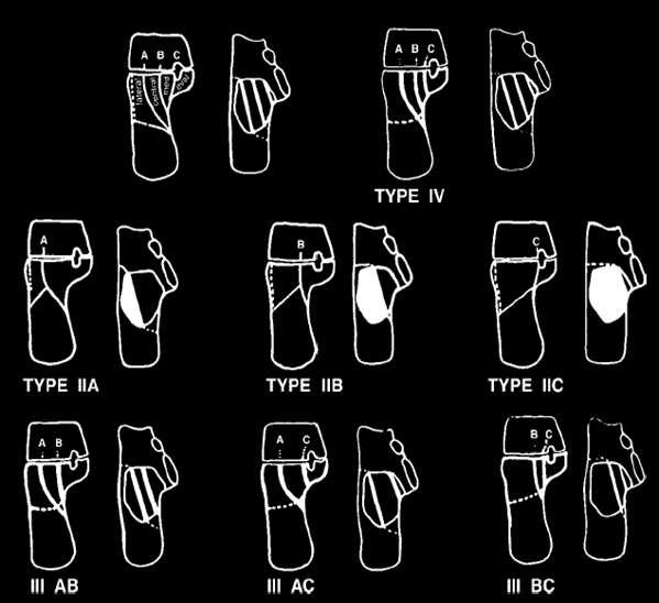
6 Tibial Plafond Fractures Classification: (Ruedi-Allgower): - type I: Pilon Frx - malleolar frx with large posterior plafond fragments - type II: Pilon Frx - spiral extension frx - type III: Pilon Frx - central compression injuries w/ impaction of talus into distal tibia w/ or w/o concomitant fibular frx Subdivided into subgroups A-C depending on degree of displacement of articular surface & presence or absence of comminution and/or impaction; - type A: - minimal or no anterior tibial cortical communition, > 2 large tibial articular fragments, and usually a fibular fracture of transverse or short oblique at the level of the plafond - type B: - results from severe axial compression force, causing distal tibial bony impaction and comminution;
7 Tibial Plafond Fractures Found in polytrauma Look at Calcaneus Tibial plateau Pelvis Spine
8 Tibial Plafond Fractures Well seen on plain films Extent seen best on CT Distinguish from trimalleolar Pilon involves anterior tibia Uncommon to have all 3 malleoli fractured in pilon fracture.
9 Talus Fractures Fractures divided into head, neck and body fractures Approximately 50 % of talar fractures involve the neck Most common body fractures are osteochondral, less common involve the lateral or posterior process
10 Talar Neck Fractures Hawkins Classification of Talar Neck Fractures Type I Type II Radiographic findings Risk of AVN Nondisplaced fracture line 0-13% Displaced fracture, plus subluxation or dislocation of subtalar joint 20-50% Type III Type IV Displaced fracture, dislocation subtalar AND tibiotalar joints Displaced fracture and disruption of talonavicular joint % high

11 Subchondral osteopenia in the vascularized NWB talus at 6-8 wks Hawkin s Sign Talar viability Presence does not r/o osteonecrosis
12 Complications Osteonecrosis Type I: 0-13% Type II: 20-50% Type III/IV: 8-100% PT Arthritis 40-90% Malunion 0-25%

13 Snowboarder s Ankle Snowboarder s Fracture Lateral process of talus Fx Dorsiflexion and hindfoot inversion on landing Difficult to see on radiographs, may need CT Unrecognized causes subtalar OA CT may be required to see and classify Type 1. Chip off anterior inferior Type 2a. Involves talofibular joint Type 2b. Involves talocalcaneal joint Type 3. Comminuted Fx J Am Board Fam Pract Mar-Apr;7(2):130-3


14 Medial talar tubercle fracture Rare injury Dorsiflexion-Pronationavulsion by deltoid lig Complications FHL entrapment Subtalar OA Nonunion Tarsal tunnel syndrome
 Complications FHL entrapment")
15 Lateral talar tubercle fractures (Sheppards fracture) Inversion or hyperplantarflexion DDx- Os trigonum (normal/fractured) Complications FHL entrapment Subtalar OA Nonunion

16 Posterior talar process fracture Involves both medial and lateral tubercles Should be considered differently as this higher frequency of association with subtalar dislocations Involves both ankle and subtalar joint ORIF Displacement >3mm
17 Calcaneus Fractures Most common tarsal fracture Accounts for 2% of all fractures 70-75% Intraarticular 20-25% Extraarticular Historically poor prognosis No consensus on management due to lack of standard, unified classification system and understanding of fracture pathoanatomy High variability in fracture pattern based on magnitude and direction of impacting force, foot position, muscle tone, and bone mineralization
18 Modified Essex-Loprestie Classification Extraarticular fractures Calcaneal tuberosity fractures Beak type Vertical Horizontal Medial avulsion Intraarticular fractures Subtalar joint involvement Undisplaced Displaced Comminuted Calcaneocuboid joint involvement
19 Sanders Classification Most useful system for intraarticular fracture classification Improved interobserver variability Has both clinical and prognostic implications Type 1: Excellent results with conservative management Type 2 and 3: Excellent results with surgical management Type 4: Poor results with surgical managment
20 Sanders Classification (CT)
21 Intraarticular Fractures: Typical Osseous Features Loss of Height due to impaction and/or rotation of the more mobile tuberosity fragment Widening due to displacement of tuberosity fragment Posterior subtalar joint disruption Axial loading associated with TL burst fractures Superior peroneal retinacular avulsions
22 Calcific myonecrosis Relatively rare, late sequela of trauma Plate/Sheet-like calcifications are characteristic Only 1 case reported in the foot in the English literature May erode adjacent bone Spontaneous draining sinus-tracts and culture positive infections may develop Appropriate treatment: compartmental excision or debridement
23 Peroneal Tendon Injury in Calcaneal Fractures Mechanism: Talus driven downward toward calcaneus usually resulting a spliting of the calcaneus with lateral displacement of the portion containing the posterior facet. Forceful lateral calcaneal displacement may result in fibular avulsion at the attachment of the superior peroneal retinaculum. Acute complications: Dislocation peroneal tendons, entrapment by avulsed fibular fragment and impingement by fracture fragments. Chronic complications: Stenosing tenosynovitis resulting in pain and ankle instability from difficulty everting foot. AJR 149: July 1982
24 Fracture Blisters Most commonly arise over tibia, ankle, or elbow. May cause wound complication.
25 Navicular Stress Fracture Accounts for 14-35% of stress fractures. Track athletes account for 59% of these injuries Predisposing activities: Stamping on ground, marching, long distance running. Commonly occurs in basketball players and runners. Usually oriented in the sagittal plane and is located dorsally in the central 1/3 of the bone. Classification (Based on CT): Type I: Dorsal cortical break Type II: Fracture propagates into navicular body Type III: Fracture propagates into other cortex (requires longest healing time and may require internal fixation).

26 Tarsal Navicular Fracture Dorsal view of the tarsal navicular bone. The avascular central one-third is also the fulcrum of the impingement forces from the first and third metacarpal bones, as well as the talus.
27 Navicular Stress Fracture Also seen in the right limbs of the greyhounds during high-speed running. The right limb is on the outside when racing in a counter-clockwise direction on circular tracks, and is subjected to asymmetric cyclic compressive loading.

28 Lisfranc Classification Controversial whether or not this has prognostic significance Probably better to fix fracture based on individual assessment Most importantly: 1. Which column is disrupted 2. Is the injury primarily bony or ligamentous
29 General Management Principles Assess soft-tissues and if necessary delay surgery There is almost no role of conservative management except in mild sprains Any fracture/dislocation with >2mm displacement should be treated surgically Any unstable fractures should be treated surgically Primary ligamentous injuries may do better with primary arthrodesis
30 Lisfranc - Outcome The key to a good outcome is anatomic reduction In the largest outcome study of 52pts: Anatomic reduction group: 16% arthritis Non-anatomic reduction group: 60% arthritis Ligamentous injuries tend to have a worse prognosis and primary arthrodesis may be warrented-40% with arthritis at 52mo f/u
31 Lisfranc - Early Complications Compartment syndrome of the foot Vascular compromise Infection Skin necrosis Loss of reduction

32 Lisfranc - Late Complications Post-traumatic arthrosis Persistent midfoot pain Stiffness Abnormal gait Abnormal shoe wear Reflex sympathetic dystrophy
33 Lisfranc Fracture-Dislocation Best diagnostic clue: Lateral offset lateral aspect 1 st metatarsal relative to medial cuneiform + medial aspect 2 nd metatarsal relative to medial aspect intermediate cuneiform. Homolateral-1 st to 5 th metatarsal dislocated laterally Divergent- 1 st metatarsal medially dislocated
34 Lisfranc Fracture-Dislocation 67% related to car accidents Initial evaluation - 20% missed Delayed treatment- chronic instability Injury Sep;35 Suppl 2:71-6.
35 Lisfranc fracture-dislocation Two basic types Homolateral All of the metatarsals are dislocated to the same side More common than divergent Usually involves the 2nd through 5th dislocated laterally May involve all 5 metatarsals Divergent Usually more severe than homolateral May be associated with a fracture of the 1st cuneiform Usually involves medial displacement of the 1st metatarsal and lateral displacement of 2nd-5th metatarsals Occasionally may involve only medial displacement of only the 1st metatarsal Fractures associated with Lisfranc dislocations Base of 2nd metatarsal Cuboid Fractures of shafts of metatarsals Dislocations of the 1st (medial) and 2nd (middle) and cuneonavicular joints Fractures of the tarsal navicular
36 BPOP Usually in hands>feet. Long bones, skull, jaw. Ages-20-30s Grows rapidly and has aggressive features on imaging and pathology. Related myositis ossificans, reactive periostitis, and subungual exostosis.
37 BPOP Radiographs: bony mass with well defined margins applied to the cortex of bone. CT: No medullary involvement or cartilage cap. MRI: dark on T1 and bright on T2
38 BPOP DDX: Osteochondroma Exostosis Myositis ossificans Low or high grade surface osteosarcoma Periosteal chondroma
39 BPOP Lesion is benign but may recur locally in as many as 50% of cases. Tx: complete excision with clear margins. Case report of adjacent intermediate grade fibrosarcoma.
Foot Injuries. Dr R B Kalia
 Foot Injuries Dr R B Kalia Overview Dramatic impact on the overall health, activity, and emotional status More attention and aggressive management Difficult appendage to study and diagnose. Aim- a stable
Radiographic Evaluation of Calcaneal Fractures. Kali Luker, PGY-1
 Radiographic Evaluation of Calcaneal Fractures Kali Luker, PGY-1 Anatomy Extraarticular Fractures Involve body, anterior process or tuberosity Treated with immobilization and NWB x 6 wks UNLESS Displaced
Traumatic Injuries to the Foot and Ankle
 Traumatic Injuries to the Foot and Ankle Dr. Joseph N. Daniel Clinical Associate Professor of Orthopaedic Surgery Foot and Ankle Service, The Rothman Institute Thomas Jefferson University Hospital Philadelphia,
Surgery-Ortho. Fractures of the tibia and fibula. Management. Treatment of low energy fractures. Fifth stage. Lec-6 د.
 Fifth stage Lec-6 د. مثنى Surgery-Ortho 28/4/2016 Indirect force: (low energy) Fractures of the tibia and fibula Twisting: spiral fractures of both bones Angulatory: oblique fractures with butterfly segment.
X-Ray Rounds: (Plain) Radiographic Evaluation of the Ankle.
 Radiographic Evaluation of the Ankle.") X-Ray Rounds: (Plain) Radiographic Evaluation of the Ankle www.fisiokinesiterapia.biz Anatomy Complex hinge joint Articulations among: Fibula Tibia Talus Tibial plafond Distal tibial articular surface
X-Ray Rounds: (Plain) Radiographic Evaluation of the Ankle www.fisiokinesiterapia.biz Anatomy Complex hinge joint Articulations among: Fibula Tibia Talus Tibial plafond Distal tibial articular surface
Talus Fractures: When and Why on Screws and Plates
 Talus Fractures: When and Why on Screws and Plates Frank A. Liporace, MD Associate Professor Director of Orthopaedic Research New York University / Hospital for Joint Diseases, NY, NY Director Orthopaedic
EASILY MISSED FOOT AND ANKLE FRACTURES NORDIC TRAUMA COURSE 2016, AARHUS
 EASILY MISSED FOOT AND ANKLE FRACTURES NORDIC TRAUMA COURSE 2016, AARHUS Ken F. Linnau, MD, MS Emergency Radiology Harborview Medical Center University of Washington Seattle, WA Thanks to Claire K Sandstrom
DEPARTMENT OF TRAUMATOLOGY AND HAND SURGERY INSTITUTE OF MUSCULOSKELETAL SURGERY ANKLE AND FOOT INJURIES
 DEPARTMENT OF TRAUMATOLOGY AND HAND SURGERY INSTITUTE OF MUSCULOSKELETAL SURGERY ANKLE AND FOOT INJURIES Presenter: Dr George Ayerh ENGLISH PROGRAM LECTURES EN_11/A - 2018 TOPICS I. Part: Ankle & Foot
Outline. Ankle/Foot Anatomy Ankle Sprains Ottawa Ankle Rules DDx: The Sprain That Wasn t
 Ankle Injuries Outline Ankle/Foot Anatomy Ankle Sprains Ottawa Ankle Rules DDx: The Sprain That Wasn t Anatomy: Ankle Mortise Bony Anatomy Lateral Ligament Complex Medial Ligament Complex Ankle Sprains
MIDFOOT INJURIES-ARE WE UNDERTREATING IT? Mr Rajiv Limaye Mr Prasad Karpe University Hospital of North Tees 3 rd Foot and Ankle Symposium
 MIDFOOT INJURIES-ARE WE UNDERTREATING IT? Mr Rajiv Limaye Mr Prasad Karpe University Hospital of North Tees 3 rd Foot and Ankle Symposium Introduction Increasing sports injuries RTA and traumatic injuries
Ankle Tendons in Athletes. Laura W. Bancroft, M.D.
 Ankle Tendons in Athletes Laura W. Bancroft, M.D. Outline Protocols Normal Anatomy Tendinopathy, partial and complete tears Posterior tibial, Flexor Hallucis Longus, Achilles, Peroneal and Anterior Tibial
Commonly Missed Injuries of the Extremities
 Commonly Missed Injuries of the Extremities Dr. Tudor H. Hughes M.D., FRCR Department of Radiology University of California School of Medicine San Diego, California 1. Base of skull 2. Odontoid process
Injuries of the Foot and Ankle. Introduction. Introduction 10/2/2009. Bryan Lapinski, MD
 Injuries of the Foot and Ankle Bryan Lapinski, MD Introduction The average person takes 1 million steps per year Approximately 30 bones in the foot and ankle are subjected to forces of 3 7 times body weight
Imaging of Ankle and Foot pain
 Imaging of Ankle and Foot pain Pramot Tanutit, M.D. Department of Radiology Faculty of Medicine, Prince of Songkla University 1 Outlines Plain film: anatomy Common causes of ankle and foot pain Exclude:
THE JOURNAL OF NUCLEAR MEDICINE Vol. 56 No. 3 March 2015 Rauscher et al.
 Supplemental Figure 1 Correlation analysis of tracer between and subsequent as assessed by SUV max in focal lesions (A). x-axis displays quantitative values as obtained by, and y-axis displays corresponding
Extraarticular Lateral Ankle Impingement
 Extraarticular Lateral Ankle Impingement Poster No.: C-1282 Congress: ECR 2016 Type: Educational Exhibit Authors: C. Cevikol; Keywords: Trauma, Diagnostic procedure, MR, CT, Musculoskeletal system, Musculoskeletal
radiologymasterclass.co.uk
 http://radiologymasterclass.co.uk Hip X-ray anatomy - Normal AP (anterior-posterior) Shenton's line is formed by the medial edge of the femoral neck and the inferior edge of the superior pubic ramus Loss
Introduction. The primary function of the ankle and foot is to absorb shock and impart thrust to the body during walking.
 The ankle 1 Introduction The primary function of the ankle and foot is to absorb shock and impart thrust to the body during walking. OSTEOLOGRY The term ankle refers primarily to the talocrural joint,
Selected Fractures of the Foot: Diagnosis and Treatment.
 Selected Fractures of the Foot: Diagnosis and Treatment www.fisiokinesiterapia.biz Overview Forefoot Fractures Lisfranc MT 5 th MT Hindfoot Fractures Calcaneus Talus Tarsometatarsal (Lisfranc s) Fracture
Peggers Super Summaries: Foot Injuries
 Lisfranc Injury ANATOMY Roman arch with recessed 2 nd MT base AP medial side of intermediate cuneiform to 2 nd MT base Oblique medial side of lateral cuneiform with 3 rd MT base and 4 th with medial boarder
Impingement Syndromes of the Ankle. Noaman W Siddiqi MD 5/4/2006
 Impingement Syndromes of the Ankle Noaman W Siddiqi MD 5/4/2006 Ankle Impingement Overview Clinical DX Increasingly recognized cause of chronic ankle pain Etiology can be soft tissue or osseous Professional
4/28/2010. Fractures. Normal Bone and Normal Ossification Bone Terms. Epiphysis Epiphyseal Plate (physis) Metaphysis
 Metaphysis") Fractures Normal Bone and Normal Ossification Bone Terms Epiphysis Epiphyseal Plate (physis) Metaphysis Diaphysis 1 Fracture Classifications A. Longitudinal B. Transverse C. Oblique D. Spiral E. Incomplete
Fractures Normal Bone and Normal Ossification Bone Terms Epiphysis Epiphyseal Plate (physis) Metaphysis Diaphysis 1 Fracture Classifications A. Longitudinal B. Transverse C. Oblique D. Spiral E. Incomplete
CURRENT TREATMENT OPTIONS
 CURRENT TREATMENT OPTIONS Fix single column or both: Always fix both. A study by Svend-Hansen corroborated the poor results associated with isolated medial malleolar fixation in bimalleolar ankle fractures.
No disclosures relevant to this topic Acknowledgement: some clinical pictures were obtained from the OTA fracture lecture series and AO fracture
 CALCANEUS FRACTURES No disclosures relevant to this topic Acknowledgement: some clinical pictures were obtained from the OTA fracture lecture series and AO fracture lecture series INCIDENCE 2% of all fractures
The Dance Hall by Vincent van Gogh,1888
 The Dance Hall by Vincent van Gogh,1888 Articulations of the pelvic girdle Lumbosacral joints, sacroiliac joints & pubic symphysis The remaining joints of the lower limb Hip joint Knee joint Tibiofibular
The Lower Limb VII: The Ankle & Foot. Anatomy RHS 241 Lecture 7 Dr. Einas Al-Eisa
 The Lower Limb VII: The Ankle & Foot Anatomy RHS 241 Lecture 7 Dr. Einas Al-Eisa Ankle joint Synovial, hinge joint Allow movement of the foot in the sagittal plane only (1 degree of freedom): dorsiflexion:
Fractures of the Calcaneus
 Fractures of the Calcaneus Anthony T. Sorkin, M.D. Rockford Orthopedic Trauma Service Rajeev Garapati, MD Illinois Bone and Joint Institute Assistant Clinical Professor University of Illinois at Chicago
Evaluation of Pediatric Foot Pain
 May 2006 Evaluation of Pediatric Foot Pain John Flibotte, Harvard Medical School Year III Our Patient AP is a 10 year old boy with chronic R foot pain 2 Anatomy of the Foot Manusov EG, et al. (1996), Part
Injuries to the lower extremity II Aree Tanavalee MD Associate Professor Department of Orthopaedics Faculty of Medicine Chulalongkorn University
 Injuries to the lower extremity II Aree Tanavalee MD Associate Professor Department of Orthopaedics Faculty of Medicine Chulalongkorn University Topics Fracture of the shaft of the femur Fractures around
5 COMMON INJURIES IN THE FOOT & ANKLE
 5 COMMON INJURIES IN THE FOOT & ANKLE MICHAEL P. CLARE, MD FLORIDA ORTHOPAEDIC INSTITUTE TAMPA, FL USA MECHANISM OF INJURY HOW DID IT HAPPEN? HIGH ENERGY VS LOW ENERGY DIRECTION OF FORCES INVOLVED LIVING
ABC of Emergency Radiology
 l ja ) $% _2) < j> ~~~~~~~~~~~~~~~~~foot ABC of Emergency Radiology THE FOOT D A Nicholson, D O'Keeffe, P A Driscoll Accurate clinical assessment of injuries to the foot will avoid unnecessary exposure
FIBULAR & SYNDESMOSIS MALUNIONS
 FIBULAR & SYNDESMOSIS MALUNIONS MICHAEL P. CLARE, MD FLORIDA ORTHOPAEDIC INSTITUTE TAMPA, FL USA MORTISE INHERENTLY UNSTABLE LATERAL MALLEOLUS ACTS AS BUTTRESS / POST RESIST LATERAL TRANSLATION OF TALUS
BIOMECHANICS OF ANKLE FRACTURES
 BIOMECHANICS OF ANKLE FRACTURES William R Reinus, MD MBA FACR Significance of Ankle Fractures Most common weight-bearing Fx 70% of all Fxs Incidence is increasing Bimodal distribution Men 15-24 Women over
Paul Alley MD,DPM,MS,FACS,FAAOS,BFD Eby Orthopaedics,Jasper,Indiana
 Paul Alley MD,DPM,MS,FACS,FAAOS,BFD Eby Orthopaedics,Jasper,Indiana Very common Bone=fractures Description (cracked,broke,busted,or smashed) A=anatomic area of bone eg: head,neck,shaft B=bone involved
Sports Injuries of the Foot and Ankle. Mark McEleney, MD University of Iowa College of Medicine Refresher Course for the Family Physician 4/4/2018
 Sports Injuries of the Foot and Ankle Mark McEleney, MD University of Iowa College of Medicine Refresher Course for the Family Physician 4/4/2018 I. Objectives A. By the end of the lecture attendees will
Anatomy of Foot and Ankle
 Anatomy of Foot and Ankle Surface anatomy of the ankle & foot Surface anatomy of the ankle & foot Medial orientation point medial malleous sustentaculum tali tuberosity of navicular TA muscle TP muscle
Sequalae of Ankle Sprains: Peri Articular Fractures of the Ankle in Sports Medicine.
 Sequalae of Ankle Sprains: Peri Articular Fractures of the Ankle in Sports Medicine www.fisiokinesiterapia.biz Chronic Ankle Pain The most common cause of chronic pain following an ankle sprain is a missed
Tarsal Coalition On MR
 Tarsal Coalition On MR By William Renner, M.D. This and other topics will be discussed in Tarsal coalition is a congenital anomaly with fusion of the tarsal bones. The fusion may be bony, fibrous or cartilaginous.
ROTATIONAL PILON FRACTURES
 CHAPTER 31 ROTATIONAL PILON FRACTURES George S. Gumann, DPM The opinions and commentary of the author should not be construed as refl ecting offi cial U.S. Army Medical Department policy. Pilon injuries
Avascular Necrosis of the Foot. Dr. Hema Choudur MD, FRCPC Associate Professor. Dept. of Radiology. McMaster University, Hamilton, Canada.
 Avascular Necrosis of the Foot Dr. Hema Choudur MD, FRCPC Associate Professor. Dept. of Radiology. McMaster University, Hamilton, Canada. Avascular Necrosis: Pathophysiology Ischemia to the bone from oxygen
pedcat Clinical Case Studies
 pedcat Clinical Case Studies C u r v e B e a m 1 7 5 T i t u s A v e, S u i t e 3 0 0 W a r r i n g t o n, P A 1 8 9 7 6 267-4 8 3-8081 w w w. c u r v e b e a m. c o m PedCAT: Clinical Evidence of diagnostic
11/4/2018 SUBTLETIES OF LOWER EXTREMITY TRAUMA IMAGING SPEAKER DISCLOSURES
 SUBTLETIES OF LOWER EXTREMITY TRAUMA IMAGING Charles S. Resnik, M.D. Professor of Radiology University of Maryland School of Medicine Upon completion of this presentation, participants will be better able
PRONATION-ABDUCTION FRACTURES
 C H A P T E R 1 2 PRONATION-ABDUCTION FRACTURES George S. Gumann, DPM (The opinions of the author should not be considered as reflecting official policy of the US Army Medical Department.) Pronation-abduction
Physical Examination of the Foot & Ankle
 Inspection Standing, feet straight forward facing toward examiner Swelling Deformity Flatfoot (pes planus and hindfoot valgus) High arch (pes cavus and hindfoot varus) Peek-a-boo heel Varus Too many toes
Ankle Injuries. Ankle Sprain. Range of Motion. The most likely diagnosis is lateral ligament sprain. Dorsiflexion Plantarflexion Inversion
 Ankle Injuries Dr Peter Brukner, OAM Sports Physician Associate Professor Centre for Sports Medicine Research & Education The University of Melbourne Adjunct Professor School of Human Movement Studies
PILON FRACTURES Mechanism of injury
 PILON FRACTURES The term pilon is from the French language and refers to a pestle and Plafond, meaning ceiling in French. Ruedi's obtained best results were obtained by open reduction and internal fixation
SURGICAL AND APPLIED ANATOMY
 Página 1 de 9 Copyright 2001 Lippincott Williams & Wilkins Bucholz, Robert W., Heckman, James D. Rockwood & Green's Fractures in Adults, 5th Edition SURGICAL AND APPLIED ANATOMY Part of "47 - ANKLE FRACTURES"
RADIOGRAPHY OF THE ANKLE and LOWER LEG
 RADIOGRAPHY OF THE ANKLE and LOWER LEG Patient Position: ANKLE AP Projection Part Position: True Slight to place foot s long axis Center to Central Ray: to IR Midway Note: Ankle joint is to tips of malleoli
 17.2 A-P Lower Leg Measure: A-P at mid-lower leg Protection: Apron draped over pelvis SID: 40 Table top No Tube Angle Film: 7 x17 I.D. down or diagonal 14 x 17 www.fisiokinesiterapia.biz A-P Lower Leg
Case 1 7 yo male Right elbow injury 3 months ago Medial elbow pain and tenderness over medial epicondyle Long arm cast given but off himself 1 month a
 Case presentations Case 1 7 yo male Right elbow injury 3 months ago Medial elbow pain and tenderness over medial epicondyle Long arm cast given but off himself 1 month after Progressive limited elbow flexion
Treatment of malunited fractures of the ankle
 Treatment of malunited fractures of the ankle A LONG-TERM FOLLOW-UP OF RECONSTRUCTIVE SURGERY I. I. Reidsma, P. A. Nolte, R. K. Marti, E. L. F. B. Raaymakers From Academic Medical Center, Amsterdam, Netherlands
Section 3: Foot Subluxations and Dislocations
 Section 3: Foot Subluxations and Dislocations Case Study F: Lisfranc s Midfoot Dislocation Clinical History: J.K. a 28 year old female presents complaining of a painful right foot. She sustained an acute
Ankle impingement syndromes - pictorial review.
 Ankle impingement syndromes - pictorial review. Poster No.: P-0148 Congress: ESSR 2015 Type: Educational Poster Authors: R. D. T. Mesquita, J. Pinto, J. L. Rosas, A. Vieira ; Porto/PT, 1 2 2 3 1 1 3 Matosinhos/PT,
Ankle impingement syndromes - pictorial review.
 Ankle impingement syndromes - pictorial review. Poster No.: P-0148 Congress: ESSR 2015 Type: Educational Poster Authors: R. D. T. Mesquita, J. Pinto, J. L. Rosas, A. Vieira ; Porto/PT, 1 2 2 3 1 1 3 Matosinhos/PT,
11/2/17. Lateral Collateral Complex Medial Collateral Complex Distal Tibiofibular Syndesmosis Spring Ligament
 Andrew J Grainger Leeds, UK Lateral Collateral Complex ial Collateral Complex Distal Tibiofibular Syndesmosis Spring Ligament Brief anatomy review Scan tips and tricks Pathological appearances andrewgrainger@nhs.net
Surgical Care at the District Hospital. EMERGENCY & ESSENTIAL SURGICAL CARE
 Surgical Care at the District Hospital 1 18 Orthopedic Trauma Key Points 2 18.1 Upper Extremity Injuries Clavicle Fractures Diagnose fractures from the history and by physical examination Treat with a
Arthroscopy Of the Ankle.
 Arthroscopy Of the Ankle www.fisiokinesiterapia.biz Ankle Arthroscopy Anatomy Patient setup Portal placement Procedures Complications Anatomy Portals Anterior Anteromedial Anterolateral Anterocentral Posterior
Section 4: Tarsal Coalitions
 Case H (Figure 2): PedCat CBCT transverse plane reconstruction of right Lisfranc midfoot dislocation compared to normal left foot. Clinical Relevance of the PedCat Study: The weight bearing CBCT study
2017 SAFSA CONGRESS PROGRAMME
 2017 SAFSA CONGRESS PROGRAMME THURSDAY, MAY 25 07h45 07h55: WELCOME & INTRODUCTIONS Forefoot I: Hallux Valgus and Lesser Toes (08h00-10h00 Lectures) 08h00 08h30: Surgical Management of Hallux Valgus Rippstein,
Clinical evaluation where no obvious fracture a. Squeeze test
 7:43 am The Syndesmotic Injury: From Subtle to Severe Robert B. Anderson, MD Chief, Foot and Ankle Carolinas Medical Center OrthoCarolina (Charlotte, North Carolina) 7:30-8:25 am Symposium 1: Management
Joints and muscles of the foot. Architecture of the foot. Sándor Katz M.D.,Ph.D.
 Joints and muscles of the foot. Architecture of the foot. Sándor Katz M.D.,Ph.D. Ankle (talocrural) joint type: hinge Talocrural joint - medial collateral ligament Medial collateral = deltoid ligament
Copyright 2004, Yoshiyuki Shiratori. All right reserved.
 Ankle and Leg Evaluation 1. History Chief Complaint: A. What happened? B. Is it a sharp or dull pain? C. How long have you had the pain? D. Can you pinpoint the pain? E. Do you have any numbness or tingling?
Hany El-Rashidy and Anand Vora
 Chapter 194 Lisfranc Injuries Chapter 194 Lisfranc Injuries Hany El-Rashidy and Anand Vora 8 ICD-9 CODE 838.03 Lisfranc (Tarsometatarsal) Fracture-Dislocation Key Concepts The Lisfranc joint represents
Foot and Ankle Complaints.
 Foot and Ankle Complaints www.fisiokinesiterapia.biz INTRODUCTION Anatomy and Function Foot Ankle Common complaints Common diagnoses FOOT AND ANKLE ANATOMY 26 bones and 2 sesamoids Forefoot Metatarsals
Clarification of Terms
 Clarification of Terms The plantar aspect of the foot refers to the role or its bottom The dorsal aspect refers to the top or its superior portion The ankle and foot perform three main functions: 1. shock
Complexities surrounding Lisfranc injuries
 Complexities surrounding Lisfranc injuries Lisfranc injuries are commonly associated with sporting injuries and are easily diagnosed with severe midfoot pain, swelling, deformity and inability to bear
Foot and Ankle Update
 Foot and Ankle Update 2019 Instructional Course Hiro Tanaka It s your on-call weekend Objectives We are going to apply evidence based treatment for 2 patients who are admitted under your care 1. Dislocated
Index. Clin Sports Med 23 (2004) Note: Page numbers of article titles are in boldface type.
 Note: Page numbers of article titles are in boldface type.") Clin Sports Med 23 (2004) 169 173 Index Note: Page numbers of article titles are in boldface type. A Achilles enthesopathy, calcaneal spur with, 133 clinical presentation of, 135 136 definition of, 131
Clin Sports Med 23 (2004) 169 173 Index Note: Page numbers of article titles are in boldface type. A Achilles enthesopathy, calcaneal spur with, 133 clinical presentation of, 135 136 definition of, 131
Commonly Missed Foot and Ankle Conditions. David Miller, DPM AMG Podiatry
 Commonly Missed Foot and Ankle Conditions David Miller, DPM AMG Podiatry Lisfranc Injuries Wide spectrum of injuries High energy Subtle subluxation which could be easily missed injuries Men are 2-4x s
MRI of Pediatric Ankle and Foot. Mahesh Thapa, MD Associate Professor Seattle Children s University of Washington School of Medicine
 MRI of Pediatric Ankle and Foot Mahesh Thapa, MD Associate Professor Seattle Children s University of Washington School of Medicine Disclosures Under contract with Lippincott Williams and Wilkins (LWW)
Acute Ankle Injuries, Part 1: Office Evaluation and Management
 t June 08, 2009 Obesity [1] Each acute ankle injury commonly seen in the office has associated with it a mechanism by which it can be injured, trademark symptoms that the patient experiences during the
Posterior Ankle Impingement: Don t Get Pinched
 Posterior Ankle Impingement: Don t Get Pinched 11 th Annual Sports Medicine Continuing Education Conference Gregory P Witkowski, MD Orthopaedic Trauma and Foot/Ankle Surgery Disclosures I have nothing
بسم هللا الرحمن الرحيم
 بسم هللا الرحمن الرحيم Laboratory RHS 221 Manual Muscle Testing Theory 1 hour practical 2 hours Dr. Ali Aldali, MS, PT Department of Physical Therapy King Saud University Talocrural and Subtalar Joint
Radiographic assessment. Functional. Paul Tornetta III Professor 11/21/2016. Fracture not in coronal plane May need CT to evaluate
 The Posterior Malleolus Paul Tornetta III Professor Boston Medical Center Publications: Disclosures! Rockwood and Green, Tornetta and Einhorn; Subspecialty series, Court-Brown, Tornetta; Trauma, AAOS;
Clin Podiatr Med Surg 19 (2002) Index
 Index") Clin Podiatr Med Surg 19 (2002) 335 344 Index Note: Page numbers of article titles are in bold face type. A Accessory soleus muscle, magnetic resonance imaging of, 300 Achilles tendon injury of, magnetic
Clin Podiatr Med Surg 19 (2002) 335 344 Index Note: Page numbers of article titles are in bold face type. A Accessory soleus muscle, magnetic resonance imaging of, 300 Achilles tendon injury of, magnetic
Basic Principles of Fractures & Easily Missed Fractures. Mr Irfan Merchant Trauma & Orthopaedic Registrar Bedford Hospital, East of England
 Basic Principles of Fractures & Easily Missed Fractures Mr Irfan Merchant Trauma & Orthopaedic Registrar Bedford Hospital, East of England Objectives Types Fracture Patterns Fracture Healing Assessing
Competence of the Deltoid Ligament in Bimalleolar Ankle Fractures After Medial Malleolar Fixation *
 Competence of the Deltoid Ligament in Bimalleolar Ankle Fractures After Medial Malleolar Fixation * BY PAUL TORNETTA, III, M.D. Investigation performed at Kings County Hospital, New York, N.Y. Abstract
The Leg. Prof. Oluwadiya KS
 The Leg Prof. Oluwadiya KS www.oluwadiya.sitesled.com Compartments of the leg 4 Four Compartments: 1. Anterior compartment Deep fibular nerve Dorsiflexes the foot and toes 2. Lateral Compartment Superficial
Persistent ankle pain after inversion lesions: what the radiologist must look for
 Persistent ankle pain after inversion lesions: what the radiologist must look for Poster No.: P-0118 Congress: ESSR 2016 Type: Authors: Keywords: DOI: Educational Poster R. Leao, L. C. Zattar-Ramos, E.
Pelvic cavity. Gross anatomy of the lower limb. Walking. Sándor Katz M.D.,Ph.D.
 Pelvic cavity. Gross anatomy of the lower limb. Walking. Sándor Katz M.D.,Ph.D. Lower limb Pelvic girdle Free lower extremity Hip bone Definitive fusion of the Y- shaped growth plate occurs 16th -18th
Ultrasound Evaluation of Posteromedial Ankle Pathology. Andrew C Cordle, M.D., Ph.D. 9/21/2018
 Ultrasound Evaluation of Posteromedial Ankle Pathology Andrew C Cordle, M.D., Ph.D. 9/21/2018 Overview: Pathology of the Posteromedial Ankle Flexor Tendon Pathology Accessory Navicular Bone Pathology Tarsal
LISFRANC FRACTURE-DISLOCATION
 LISFRANC FRACTURE-DISLOCATION Napoleon at Mont St. Bernard, Jacques-Louis David, 1800, Oil on Canvas, Musee du Louvre, Paris. This is Jacques-Louis David s immortal depiction of a young Napoleon Bonaparte,
Rippstein, Trnka, Saragas, Narramore
 THURS 25th MAY 07:45 07:55 Welcome and Introductions Paulo Ferrao Lecture 1: 08:00 10:20 Forefoot I: Hallux Valgus and Lesser Toes Mark Easley 30 mins 08:00 08:30 Surgical Management of Hallux Valgus Saragas,
ANKLE JOINT ANATOMY 3. TALRSALS = (FOOT BONES) Fibula. Frances Daly MSc 1 CALCANEUS 2. TALUS 3. NAVICULAR 4. CUBOID 5.
 Fibula. Frances Daly MSc 1 CALCANEUS 2. TALUS 3. NAVICULAR 4. CUBOID 5.") ANKLE JOINT ANATOMY The ankle joint is a synovial joint of the hinge type. The joint is formed by the distal end of the tibia and medial malleolus, the fibula and lateral malleolus and talus bone. It is
ANKLE JOINT ANATOMY The ankle joint is a synovial joint of the hinge type. The joint is formed by the distal end of the tibia and medial malleolus, the fibula and lateral malleolus and talus bone. It is
Chapter 58 Ankle and Foot
 Chapter 58 Ankle and Foot Episode Overview: 1) Describe the bones of the foot and important joints/ligaments 2) List ankle stress tests 3) Describe an approach to ankle x rays (including the Ottawa Ankle
The pilon tibiale fracture
 The pilon tibiale fracture Thomas Beck Spitalzentrum Oberwallis OTC Trauma course september 2017 xxx I have no financial relationships with commercial entities that produce healthcare related products.
Review relevant anatomy of the foot and ankle. Learn the approach to examining the foot and ankle
 Objectives Review relevant anatomy of the foot and ankle Learn the approach to examining the foot and ankle Learn the basics of diagnosis and treatment of ankle sprains Overview of other common causes
DAY 1: FRIDAY, 31 st AUGUST Operative Sessions: 8.00 am to 3.30 pm
 DAY 1: FRIDAY, 31 st AUGUST Operative Sessions: 8.00 am to 3.30 pm SURGICAL PROCEDURES / CASES (PROPOSED) 1. Haglund with Tendoachilles Tendinopathy 2. ORIF Calcaneus Fracture 3. OCD talus: Arthroscopic
Ankle Ligament Injury: Don t Worry- It s Only a Sprain Wes Jackson MD Orthopaedic Foot & Ankle
 Ankle Ligament Injury: Don t Worry- It s Only a Sprain Wes Jackson MD Orthopaedic Foot & Ankle Outline I. Epidemiology II. Classification and Types of Sprains III. Anatomy IV. Clinical Assessment and Imaging
Therapeutic Foot Care Certificate Program Part I: Online Home Study Program
 Therapeutic Foot Care Certificate Program Part I: Online Home Study Program 1 Anatomy And Terminology Of The Lower Extremity Joan E. Edelstein, MA, PT, FISPO Associate Professor of Clinical Physical Therapy
Mary Lloyd Ireland, M.D. Associate Professor University of Kentucky Dept. of Orthopaedic Surgery and Sports Medicine Lexington, Kentucky
 Common Ankle Injuries: Diagnosis and Treatment Mary Lloyd Ireland, M.D. Associate Professor University of Kentucky Dept. of Orthopaedic Surgery and Sports Medicine Lexington, Kentucky Disclaimer Slide
HIGH-ENERGY TRAUMA OF THE LOWER EXTREMITY NORDIC FORUM 2016, AARHUS
 HIGH-ENERGY TRAUMA OF THE LOWER EXTREMITY NORDIC FORUM 2016, AARHUS Ken F. Linnau, MD, MS Emergency Radiology Harborview Medical Center University of Washington Seattle, WA ACKNOWLEDGMENTS Nicole Kansier,
Musculoskeletal Ultrasound Technical Guidelines. VI. Ankle
 European Society of MusculoSkeletal Radiology Musculoskeletal Ultrasound Technical Guidelines VI. Ankle Ian Beggs, UK Stefano Bianchi, Switzerland Angel Bueno, Spain Michel Cohen, France Michel Court-Payen,
Imaging the musculoskeletal system. An Introduction
 Imaging the musculoskeletal system An Introduction Objectives Discuss: commonly used imaging modalities in the musculoskeletal system normal imaging anatomy in the extremities fracture description Imaging
Intramedullary Rodding of Distal Tibial Shaft Fractures with Intra Articular Extension
 Intramedullary Rodding of Distal Tibial Shaft Fractures with Intra Articular Extension My Name is Claude Sagi CSOT Tampa, FL 2018 Disclosures: None, I am just a simple man. This talk is about treating
Pelvic Girdle
 ARTICULATIONS OF LOWER EXTREMITY Pages 429-437 Pelvic Girdle formed by connection of the hip bones and the sacrum Sacroiliac Joints compound joints synovial joint - anterior, between the auricular surfaces
Ankle Injuries. Resident Guidebook. Achilles tendon sprain/tear. Peroneal tendinopathy Peroneal subluxation. Extensor Hallucis Longus Tenosynovitis
 Ankle Injuries Achilles tendon sprain/tear Peroneal tendinopathy Peroneal subluxation Extensor Hallucis Longus Tenosynovitis Weber Fracture Stress fracture Calcaneal bursitis Calcaneal fracture Base of
Calcaneal Fractures: Lateral Extensile Incision
 Calcaneal Fractures: Lateral Extensile Incision AS Flemister JR, MD University of Rochester Disclosures I have no financial disclosures 1/27/2016 2 Mechanism Axial Loading Fall From Height MVA BAD SOFT
7/23/2018 DESCRIBING THE FRACTURE. Pattern Open vs closed Location BASIC PRINCIPLES OF FRACTURE MANAGEMENT. Anjan R. Shah MD July 21, 2018.
 BASIC PRINCIPLES OF FRACTURE MANAGEMENT Anjan R. Shah MD July 21, 2018 DESCRIBING THE FRACTURE Pattern Open vs closed Location POLL OPEN HOW WOULD YOU DESCRIBE THIS FRACTURE PATTERN? 1 Spiral 2 Transverse