Pediatric Elbow Radiology. Seema Awatramani, MD Friday, April 5, 2018 ACOEP Spring Seminar
|
|
|
- Reginald Shepherd
- 5 years ago
- Views:
Transcription
1 Pediatric Elbow Radiology Seema Awatramani, MD Friday, April 5, 2018 ACOEP Spring Seminar
2 Disclosure I have no relevant financial relationships with the manufacturer(s) of any commercial product(s) and/or provider(s) of commercial services discussed in this CME activity. I do not intend to discuss an unapproved/investigative use of a commercial product/device in my presentation.
3 Background 65% of all fracture/dislocation are UE Elbow fractures more common in kids Supracondylar fractures 16.6% of all childhood fractures 30% of fractures from 0-7 years
4 Recommended Resource Radiology Cases in Pediatric Emergency Medicine University of Hawaii
5 Topic Outline Back to Basics Anatomical & radiographic markers Xray
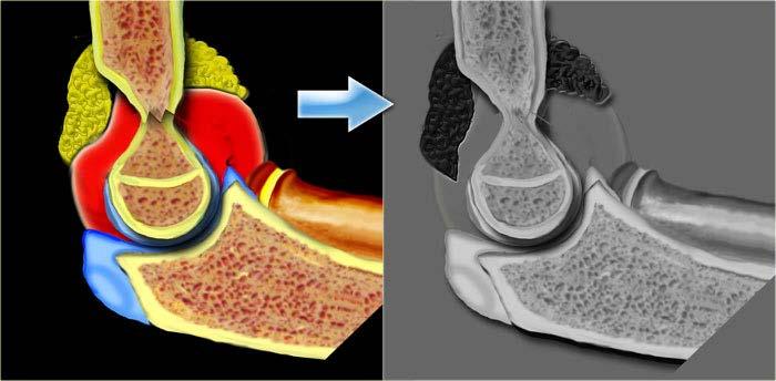
6 Anatomy Bony anatomy Joint anatomy Ossification Radiographic anatomy
7 Anatomy - basics POSTERIOR VIEW ANTERIOR VIEW Gray's Anatomy, 40th Edition


8 Anatomy - basics POSTERIOR VIEW ANTERIOR VIEW Gray's Anatomy, 40th Edition


9 DISTAL HUMERUS ANTERIOR VIEW DISTAL HUMERUS POSTERIOR VIEW Articular Surfaces ULNA OBLIQUE VIEW Gray's Anatomy, 40th Edition

10 The Joint Elbow anatomy and radiographic diagnosis of elbow fracture in children
11 Supracondylar Fractures Green NE. Fractures and dislocations about the elbow. In: Skeletal Trauma in Children, 3rd, Green NE, Swiontkowski MF. (Eds), WB Saunders, Philadelphia p Evaluation and management of supracondylar fractures in children

12 Putting it Together 1 lateral supracondyle ridge 2 medial supracondyle ridge 3 olecranon fossa 4 medial epicondyle 5 lateral epicondyle 6 capitulum 7 olecranon 8 trochlea 9 coronoid process of ulna 10 proximal radioulnar joint 11 head of radius 12 neck of radius 13 tuberosity of radius 14 ulna Radiographic Anatomy of Adult Elbow. OrthopaedicsOne Articles. ver.4.

13 Putting it Together 1 supracondylar ridge 2 trochlea 3 olecranon 4 trochlear notch 5 coronoid process of ulna 6 head of radius 7 neck of radius 8 tuberosity of radius 9 ulna Radiographic Anatomy of Adult Elbow. OrthopaedicsOne Articles. ver.4.
14 Ossification Centers
15 Ossification Centers

16 The Growth Plate Elbow anatomy and radiographic diagnosis of elbow fracture in children
17 Ossification Centers Elbow anatomy and radiographic diagnosis of elbow fracture in children
18 Location Age C capitellum 1 Ossification Centers R radial head 3 I internal (medial) epicondyle 5 T trochlea 7 O olecranon 9 E external (lateral) epicondyle 11

19 Ossification Centers
20 Ossification Centers
21 Radiographic Anatomy AP: epicondyles and articular surfaces Lateral: humerus to forearm, fat pads Oblique: coronoid, radio-ulnar joint
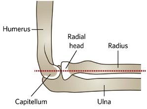
22 Radiographic Lines RADIOCAPITELLAR LINE ANTERIOR HUMERAL LINE


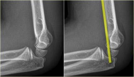
23 Radiographic Anatomy RADIOCAPITELLAR LINE ANTERIOR HUMERAL LINE
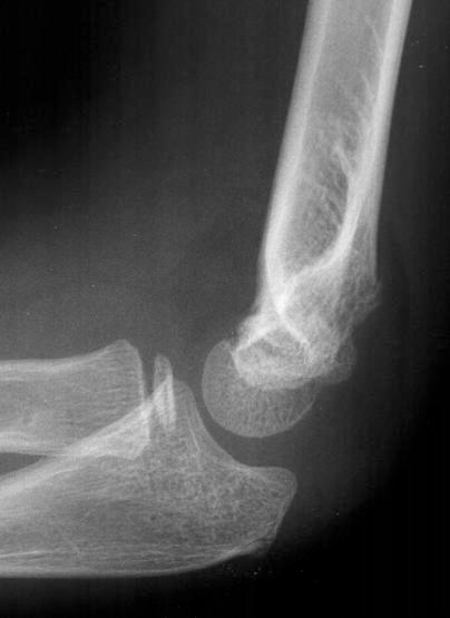
24 Fat Pads
25 Fat-Pads Fat pad = joint effusion Fat pad without fracture = occult fracture? Skaggs DL, et al; The posterior fat pad sign in association with occult fracture of the elbow in children. J Bone Joint Surg Am Oct;81(10): Donnelly LF, et al, ; Traumatic elbow effusions in pediatric patients: are occult fractures the rule? AJR Am J Roentgenol Jul;171(1): Pudas T, et al; Magnetic resonance imaging in pediatric elbow fractures. Acta Radiol Oct;46(6): Al-Aubaidi Z, et al.; The role of fat pad sign in diagnosing occult elbow fractures in the pediatric patient: a prospective magnetic resonance imaging study. J Pediatr Orthop B Nov;21(6): Major NM et al; Elbow effusions in trauma in adults and children: is there an occult fracture? AJR Am J Roentgenol Feb;178(2): de Beaux AC et al; Elbow fat pad sign: implications for clinical management. J R Coll Surg Edinb Jun;37(3):205-6.

26 Hourglass Sign NORMAL ABNORMAL
27 Proper Positioning Green NE. Fractures and dislocations about the elbow. In: Skeletal Trauma in Children, 3rd, Green NE, Swiontkowski MF. (Eds), WB Saunders, Philadelphia p Evaluation and management of supracondylar fractures in children.
28 Summary Outline 1 Anterior fat pad 2 Posterior fat pad The checklist 3 Anterior humeral line 4 Radial head contour 5 Radiocapitellar line 6 Ossification centers 7 Hourglass sign 8 Distal humerus 9 Ulna/Olecranon 10 Clinical correlation
29 CHECKLIST Anterior fat pad Posterior fat pad Anterior humeral line Radial head contour Radiocapitellar line Ossification centers Hourglass sign Distal humerus Ulna/Olecranon Clinical correlation Sample Case
30 Dx: Joint Effusion Supracondylar Fracture emxray/pemxray.html

31 Thank You. Questions?
32 CHECKLIST Anterior fat pad Posterior fat pad Anterior humeral line Radial head contour Radiocapitellar line Ossification centers Hourglass sign Distal humerus Ulna/Olecranon Clinical correlation Sample Case 2
33 Dx: Joint Effusion No visible fracture emxray/pemxray.html


34 LONGITUDINAL Elbow Ultrasound TRANSVERSE PROBE PLACEMENT NORMAL ABNORMAL Rabiner JE et al; Accuracy of Point-of-Care Ultrasonography for Diagnosis of Elbow Fractures in Children. Ann Emerg Med Nov 9. [Epub]


35 Elbow Ultrasound LONGITUDINAL TRANSVERSE Rabiner JE et al; Accuracy of Point-of-Care Ultrasonography for Diagnosis of Elbow Fractures in Children. Ann Emerg Med Nov 9. [Epub]
 Rabiner JE et al; Accuracy of Point-of-Care Ultrasonography for Diagnosis of Elbow Fractures in Children.")
36 Elbow Ultrasound sensitivity 98% (88-100%) specificity 70% (60-79%) LR 3.3 ( ) NLR 0.03 ( ) Rabiner JE et al; Accuracy of Point-of-Care Ultrasonography for Diagnosis of Elbow Fractures in Children. Ann Emerg Med Nov 9. [Epub]
 LR 3.3 (2.4-4.5) NLR 0.03 (0.01-0.")

37 Elbow Ultrasound sensitivity 98% (88-100%) specificity 70% (60-79%) LR 3.3 ( ) NLR 0.03 ( ) Rabiner JE et al; Accuracy of Point-of-Care Ultrasonography for Diagnosis of Elbow Fractures in Children. Ann Emerg Med Nov 9. [Epub]
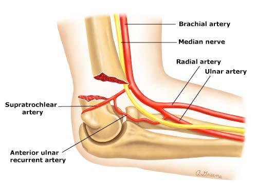
38 Vessels Brachial artery prone to damage Extensive collaterals Ischemia is rare Gray's Anatomy, 40th Edition

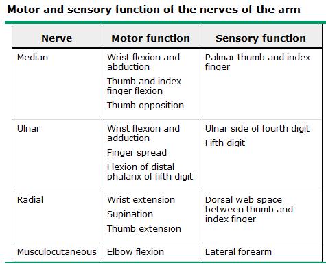
39 Nerves Median nerve crosses elbow to form the interosseous Elbow anatomy and radiographic diagnosis of elbow fracture in children

40 CASE ONE CHECKLIST Anterior fat pad Posterior fat pad Anterior humeral line Radial head contour Radiocapitellar line Ossification centers Hourglass sign Distal humerus Ulna/Olecranon Clinical correlation
41 CASE ONE Joint effusion, no fracture

42 CASE TWO CHECKLIST Anterior fat pad Posterior fat pad Anterior humeral line Radial head contour Radiocapitellar line Ossification centers Hourglass sign Distal humerus Ulna/Olecranon Clinical correlation
43 CASE TWO Radial Metaphyseal Head Fracture
44 Radial Fractures about 5-8% of all pediatric elbow fx Ages 9-12 FOOSH with extended elbow Pain/swelling proximal radius, pain on supination


45 Radial Fractures 50% accompanied by second injury accompanied by dislocations REDUCTION DISLOCATION Green NE. Fractures and dislocations about the elbow. In: Skeletal Trauma in Children, 3rd, Green NE, Swiontkowski MF. (Eds), WB Saunders, Philadelphia p

46 Radial Fractures AP & LATERAL OBLIQUE

47 CASE THREE CHECKLIST Anterior fat pad Posterior fat pad Anterior humeral line Radial head contour Radiocapitellar line Ossification centers Hourglass sign Distal humerus Ulna/Olecranon Clinical correlation
48 CASE THREE Joint effusion, olecranon fracture and radial head dislocation
49 Monteggia Fracture Proximal ulnar fracture accompanied by a radial head dislocation FOOS(Arm) in pronation 2% pediatric elbow fractures
50 CASE FOUR CHECKLIST Anterior fat pad Posterior fat pad Anterior humeral line Radial head contour Radiocapitellar line Ossification centers Hourglass sign Distal humerus Ulna/Olecranon Clinical correlation
51 CASE FOUR Joint Effusion Supracondylar Fracture
52 Supracondylar Fracture 60% pediatric elbow fractures 5-10 years flexion and extension mechanisms 70% fall on outstretched arm non-dominant arm more common <3 ft fall for children <3yo

, WB Saunders, Philadelphia 2003. p.207-276. www.uptodate.")
53 Pertinent Anatomy Green NE. Fractures and dislocations about the elbow. In: Skeletal Trauma in Children, 3rd, Green NE, Swiontkowski MF. (Eds), WB Saunders, Philadelphia p Evaluation and management of supracondylar fractures in children


54 Complications PROXIMAL LATERAL DISPLACEMENT PROXIMAL MEDIAL DISPLACEMENT Evaluation and management of supracondylar fractures in children
55 Complications

56 Classifications PE I II III IV minimal swelling TTP pain with elbow movement some movement no movement arm in extension much swelling ecchymosis no movement much swelling eccymosis more unstable tx posterior splint vs. sling ortho, closed vs. reduction w/pin ortho, closed vs. reduction w/pin ortho, closed vs. reduction w/pin Green NE. Fractures and dislocations about the elbow. In: Skeletal Trauma in Children, 3rd, Green NE, Swiontkowski MF. (Eds), WB Saunders, Philadelphia p
57 Bony Injuries to the Elbow Condylar Fractures Medial condyle Lateral condyle T-Condylar Fractures Capitellar Fracture Epicondylar Fractures Medial epicondyle Lateral epicondyle Transphyseal Fractures Floating Elbow Fractures Proximal Elbow Fractures Radial head/neck Coronoid Olecranon Monteggia Supracondylar Fractures Dislocations Elbow Joint Radial Head Radial Head Subluxation

58 Questions?
59 Thank You! Happy Holidays!!
60
61
62 sds

63 CASE SIX CHECKLIST Anterior fat pad Posterior fat pad Anterior humeral line Radial head contour Radiocapitellar line Ossification centers Hourglass sign Distal humerus Ulna/Olecranon Clinical correlation
64 CASE SIX Medial Epicondylar Fracture

65 Medial Epicondylar Fracture Epicondylar and transphyseal elbow fractures in children
66 Medial Epicondylar Fracture 10% pediatric elbow fractures boys >> girls 9-14 yo associated with posterior elbow dislocation

67 CASE SEVEN CHECKLIST Anterior fat pad Posterior fat pad Anterior humeral line Radial head contour Radiocapitellar line Ossification centers Hourglass sign Distal humerus Ulna/Olecranon Clinical correlation
68 CASE SEVEN Salter Harris II fracture of Humerus

69 CASE EIGHT CHECKLIST Anterior fat pad Posterior fat pad Anterior humeral line Radial head contour Radiocapitellar line Ossification centers Hourglass sign Distal humerus Ulna/Olecranon Clinical correlation
70 CASE EIGHT Joint effusion Ulna fracture

71 CASE NINE CHECKLIST Anterior fat pad Posterior fat pad Anterior humeral line Radial head contour Radiocapitellar line Ossification centers Hourglass sign Distal humerus Ulna/Olecranon Clinical correlation
72 Joint CASE effusion NINE Supracondylar fracture Possible proximal
73 CASE TWO CHECKLIST Anterior fat pad Posterior fat pad Anterior humeral line Radial head contour Radiocapitellar line Ossification centers Hourglass sign Distal humerus Ulna/Olecranon Clinical correlation
74 CASE TWO Joint effusion, no fracture
75 Other Modalities: Ultrasound Looking for posterior fat pad More sensitive than X-ray for fat pad Can also see lipohemarthrosis
76 CASE x CHECKLIST Anterior fat pad Posterior fat pad Anterior humeral line Radial head contour Radiocapitellar line Ossification centers Hourglass sign Distal humerus Ulna/Olecranon Clinical correlation
77 CASE x
78 CASE O CHECKLIST Anterior fat pad Posterior fat pad Anterior humeral line Radial head contour Radiocapitellar line Ossification centers Hourglass sign Distal humerus Ulna/Olecranon Clinical correlation
79 CASE O Joint effusion. Radial head fracture.
80

81 Oblique View Coronoid process Trochlear notch Medial trochlear profile
82 Bony Anatomy Fun Fact #2 The cubit is a traditional unit of length: from the elbow to the tip of the middle finger. Standard/Biblical cubits: 6 palms x 4 fingers = 24 digits Egyptian Royal cubits: 7 palms x 4 fingers = 28 digits
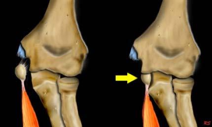
 In the flexed elbow, force develops on the posterior aspect of the olecranon (small double arrow) because of the pull of the brachialis and triceps muscles (large arrows).")
83 Olecranon Fx Mechanism of flexion injuries. (A) In the flexed elbow, force develops on the posterior aspect of the olecranon (small double arrow) because of the pull of the brachialis and triceps muscles (large arrows). (B) Failure occurs on the tension side, which is posterior as a result of the muscle pull or a direct blow to the prestressed posterior olecranon. Reproduced with permission from: Erickson M, Frick S. Fractures of the proximal radius and ulna. In: Rockwood and Wilkins' Fractures in Children, 7th ed, Beaty JH, Kasser JR (Eds), Lippincott Williams & Wilkins, Philadelphia Copyright 2010 Lippincott Williams & Wilkins.
84 Coronoid Fx

85 Proper Positioning Green NE. Fractures and dislocations about the elbow. In: Skeletal Trauma in Children, 3rd, Green NE, Swiontkowski MF. (Eds), WB Saunders, Philadelphia p
86 Proper Positioning Evaluation and management of supracondylar fractures in children


87 Carrying Angle Females > Males Check alignment after injury VALGUS CARRYING ANGLE FUN FACT!!!! #4
88 Fat-Pads Prospective cohort 231 elbow injuries Normal AFP is highly associated with absence of elbow fracture sensitivity 96.4% (86.6%-99.4%) NPV was 98.2% (93.1%-99.7%). Blumberg SM et al.; The predictive value of a normal radiographic anterior fat pad sign following elbow trauma in children. Pediatr Emerg Care Jul;27(7):

89 Fracture Description Bony anatomy Joint anatomy Ossification Radiographic anatomy Vessels Nerves
Traumatic injuries of the paediatric elbow: A pictorial review
 Traumatic injuries of the paediatric elbow: A pictorial review Poster No.: C-750 Congress: ECR 2009 Type: Educational Exhibit Topic: Pediatric Authors: A. M. Veitch, J. Harington, K. Franklin ; Plymouth/UK,
The Elbow Scanning Protocol
 The Elbow Scanning Protocol Diagnostic Imaging of the Elbow: Introduction The elbow maybe considered as consisting of four quadrants, anterior, medial, lateral and posterior. Ultrasound would normally
The Elbow and the cubital fossa. Prof Oluwadiya Kehinde
 The Elbow and the cubital fossa Prof Oluwadiya Kehinde www.oluwadiya.com Elbow and Forearm Anatomy The elbow joint is formed by the humerus, radius, and the ulna Bony anatomy of the elbow Distal Humerus
Elbow Elbow Anatomy. Flexion extension. Pronation Supination. Anatomy. Anatomy. Romina Astifidis, MS., PT., CHT
 Elbow Elbow Anatomy Romina Astifidis, MS., PT., CHT Curtis National Hand Center Baltimore, MD October 6-8, 2017 Link between the arm and forearm to position the hand in space Not just a hinge Elbow = 70%
Functional Anatomy of the Elbow
 Functional Anatomy of the Elbow Orthopedic Institute Daryl C. Osbahr, M.D. Chief of Sports Medicine, Orlando Health Chief Medical Officer, Orlando City Soccer Club Orthopedic Consultant, Washington Nationals
Fractures and dislocations around elbow in adult
 Lec: 3 Fractures and dislocations around elbow in adult These include fractures of distal humerus, fracture of the capitulum, fracture of the radial head, fracture of the olecranon & dislocation of the
RADIOGRAPHY OF THE ELBOW & HUMERUS
 RADIOGRAPHY OF THE ELBOW & HUMERUS Patient Position: ELBOW AP Projection in same plane Part Position: Hand in ; patient Centered to Humeral epicondyles Central Ray: Structures Shown: AP Elbow Criteria
Upper Extremity Injury Management. Jonathan Pirie MD, Med, FRCPC, FAAP
 Upper Extremity Injury Management Jonathan Pirie MD, Med, FRCPC, FAAP Learning Objectives At the end of this session, you will be able to manage common fractures of the: 1. Humerus 2. Elbow 3. Forearm
PEM GUIDE CHILDHOOD FRACTURES
 PEM GUIDE CHILDHOOD FRACTURES INTRODUCTION Skeletal injuries account for 10-15% of all injuries in children; 20% of those are fractures, 3 out of 4 fractures affect the physis or growth plate. Always consider
Upper Extremity Fractures
 Upper Extremity Fractures Ranie Whatley, RN,FNP-C David W. Gray, MD Skeletal Trauma 10 to 15 % of all Childhood Injuries Physeal (Growth Plate) Injuries are ~ 15% of all Skeletal Injuries Orthopaedic Assessment
The Biomechanics of the Human Upper Extremity-The Elbow Joint C. Mirzanli Istanbul Gelisim University
 The Biomechanics of the Human Upper Extremity-The Elbow Joint C. Mirzanli Istanbul Gelisim University Structure of The Elbow Joint A simple hinge joint, actually categorized as a trochoginglymus joint
The Elbow and Radioulnar Joints Kinesiology. Dr Cüneyt Mirzanli Istanbul Gelisim University
 The Elbow and Radioulnar Joints Kinesiology Dr Cüneyt Mirzanli Istanbul Gelisim University 1 The Elbow & Radioulnar Joints Most upper extremity movements involve the elbow & radioulnar joints. Usually
The Elbow 3/5/2015. The Elbow Scanning Sequence. * Anterior Joint (The anterior Pyramid ) * Lateral Epicondyle * Medial Epicondyle * Posterior Joint
 * Lateral Epicondyle * Medial Epicondyle * Posterior Joint") Scanning Sequence * Anterior Joint (The anterior Pyramid ) * Lateral Epicondyle * Medial Epicondyle * Posterior Joint Anterior Elbow Pyramid Courtesy of Jay Smith, MD. Vice chair PMR Mayo Clinic Rochester,
Scanning Sequence * Anterior Joint (The anterior Pyramid ) * Lateral Epicondyle * Medial Epicondyle * Posterior Joint Anterior Elbow Pyramid Courtesy of Jay Smith, MD. Vice chair PMR Mayo Clinic Rochester,
PEDIATRIC UPPER EXTREMITY FRACTURE MANAGEMENT JULIA RAWLINGS, MD SPORTS MEDICINE SYMPOSIUM: THE PEDIATRIC ATHLETE 2 MARCH 2018
 PEDIATRIC UPPER EXTREMITY FRACTURE MANAGEMENT JULIA RAWLINGS, MD SPORTS MEDICINE SYMPOSIUM: THE PEDIATRIC ATHLETE 2 MARCH 2018 DISCLOSURE I have nothing to disclose. 2 OBJECTIVES Discuss the diagnosis,
Osteology of the Elbow and Forearm Complex. The ability to perform many activities of daily living (ADL) depends upon the elbow.
 depends upon the elbow.") Osteology of the Elbow and Forearm Complex The ability to perform many activities of daily living (ADL) depends upon the elbow. Activities of Daily Living (ADL) Can you think of anything that you do to
Osteology of the Elbow and Forearm Complex The ability to perform many activities of daily living (ADL) depends upon the elbow. Activities of Daily Living (ADL) Can you think of anything that you do to
Pediatric Fractures. Objectives. Epiphyseal Complex. Anatomy and Physiology. Ligaments. Bony matrix
 1 Pediatric Fractures Nicholas White, MD Assistant Professor of Pediatrics Eastern Virginia Medical School Attending, Pediatric Emergency Department Children s Hospital of The King s Daughters Objectives
Sports Medicine Unit 16 Elbow
 Sports Medicine Unit 16 Elbow I. Bones a. b. c. II. What movements does the elbow perform? a. Flexion b. c. Pronation d. III. Muscles in motion a. FLEXION (supinated) i Brachialis (pronated) ii (neutral)
Elbow. Chapter 2 LISTEN. Mechanism of Injury (If Applicable) Pain
 Pain") Chapter 2 Elbow LISTEN Mechanism of Injury (If Applicable) Patient usually remembers their position at the time of injury Certain mechanisms of injury result in characteristic patterns Fall on outstretched
Chapter 2 Elbow LISTEN Mechanism of Injury (If Applicable) Patient usually remembers their position at the time of injury Certain mechanisms of injury result in characteristic patterns Fall on outstretched
Elbow Anatomy, Growth and Physical Exam. Donna M. Pacicca, MD Section of Sports Medicine Division of Orthopaedic Surgery Children s Mercy Hospital
 Elbow Anatomy, Growth and Physical Exam Donna M. Pacicca, MD Section of Sports Medicine Division of Orthopaedic Surgery Children s Mercy Hospital Contributing Factors to Elbow Injury The elbow is affected
THE ELBOW. The elbow is a commonly injured joint in both children and adults.
 ABC of Emergency Radiology FIG i-lateral radiograph of elbow and line THE ELBOW D A Nicholson, P A Driscoll The elbow is a commonly injured joint in both children and adults. Interpretation of elbow radiographs
Upper limb injuries in children. Key points, # & dislocations 7/23/2009 (MIMIC)
") Upper limb injuries in children (MIMIC) Key points, # & dislocations Before the age of 16 around 50% of boys & 25% of girls will sustain a # Dislocations are very uncommon Children s bones are less brittle
Upper limb injuries in children (MIMIC) Key points, # & dislocations Before the age of 16 around 50% of boys & 25% of girls will sustain a # Dislocations are very uncommon Children s bones are less brittle
Elbow, forearm injuries. K. Fekete
 Elbow, forearm injuries K. Fekete 1. Outline: Fractures of the elbow Dislocation of the elbow Fractures of the forearm Special injuries 2. ANATOMY 3. Lennard Funk Anatomical reminder Three joints: Humero-ulnar
Main Menu. Elbow and Radioulnar Joints click here. The Power is in Your Hands
 1 The Elbow and Radioulnar Joints click here Main Menu K.4 http://www.handsonlineeducation.com/classes//k4entry.htm[3/23/18, 1:29:53 PM] Bones Ulna is much larger proximally than radius Radius is much
MEDIAL EPICONDYLE FRACTURES
 MEDIAL EPICONDYLE FRACTURES Demographic 20% of elbow fractures 60% of which are associated with elbow dislocation. 75% in boys between 6-12 years 20% of elbow dislocation with ME fracture, the ME is incarcerated
Rehabilitation after Total Elbow Arthroplasty
 Rehabilitation after Total Elbow Arthroplasty Total Elbow Atrthroplasty Total elbow arthroplasty (TEA) Replacement of the ulnohumeral articulation with a prosthetic device. Goal of TEA is to provide pain
Acute Elbow Trauma in Children: Spectrum of Injury Revealed by MR Imaging Not Apparent on Radiographs
 James F. Griffith 1 Derek J. Roebuck 1,2 Jack C. Y. Cheng 3 Yu Leung Chan 1 Timothy H. Rainer 4 Bobby K. W. Ng 3 Constantine Metreweli 1 Received December 14, 1999; accepted after revision June 8, 2000.
Joints of the upper limb II
 Joints of the upper limb II Prof. Abdulameer Al-Nuaimi E-mail: a.al-nuaimi@sheffield.ac.uk E. mail: abdulameerh@yahoo.com Elbow joint The elbow joint is connecting the upper arm to the forearm. It is classed
Anterior Elbow Capsulodesis
 7(1):72 76, 2006 m R E V I E W m Anterior Elbow Capsulodesis Donald H. Lee, MD, Douglas R. Weikert, and Jeffry T. Watson Department of Orthopaedic Surgery Vanderbilt Orthopaedic Institute Nashville, TN
Proximal radioulnar translocation associated with elbow dislocation and radial neck fracture in child: a case report and review of literature
 DOI 10.1007/s00402-013-1820-8 TRAUMA SURGERY Proximal radioulnar translocation associated with elbow dislocation and radial neck fracture in child: a case report and review of literature Hong Kee Yoon
1/19/2018. Winter injuries to the shoulder and elbow. Highgate Private Hospital (Whittington Health NHS Trust)
") Winter injuries to the shoulder and elbow Omar Haddo Consultant Orthopaedic Surgeon, Shoulder, Elbow, Hand & Wrist Specialist MBBS, BmedSci, FRCS(Orth) Highgate Private Hospital (Whittington Health NHS
Winter injuries to the shoulder and elbow Omar Haddo Consultant Orthopaedic Surgeon, Shoulder, Elbow, Hand & Wrist Specialist MBBS, BmedSci, FRCS(Orth) Highgate Private Hospital (Whittington Health NHS
Lecture 9: Forearm bones and muscles
 Lecture 9: Forearm bones and muscles Remember, the region between the shoulder and the elbow = brachium/arm, between elbow and wrist = antebrachium/forearm. Forearm bones : Humerus (distal ends) Radius
Slides of Anatomy. Spring Dr. Maher Hadidi, University of Jordan
 Slides of Anatomy Please note : These slides are Dr. Maher Hadidi s slides of spring 2016 and were edited by the Premed Academic Team to fit the slides of spring 2019. Spring 2019 Dr. Maher Hadidi, University
Chapter 8. The Pectoral Girdle & Upper Limb
 Chapter 8 The Pectoral Girdle & Upper Limb Pectoral Girdle pectoral girdle (shoulder girdle) supports the arm consists of two on each side of the body // clavicle (collarbone) and scapula (shoulder blade)
Elbow Joint Anatomy ELBOW ANATOMY, BIOMECHANICS. Bone Anatomy. Bone Anatomy. Property of VOMPTI, LLC
 ELBOW ANATOMY, BIOMECHANICS AND PATHOLOGY Kristin Kelley, DPT, OCS, FAAOMPT Elbow Joint Anatomy Joint articulations Humeroulnar Radiohumeral Radioulnar (proximal and distal) Orthopaedic Manual Physical
Osteology of the Elbow and Forearm Complex
 Osteology of the Elbow and Forearm Complex The ability to perform m any activities of daily living (ADL) d epends upon the elbow. Activities of Daily Living (ADL) Can you think of anything that you do
Elbow. Chapter 2 LISTEN. Mechanism of Injury (If Applicable) Pain
 Pain") Preface The first decade of the twenty-first century has witnessed the continuation of an explosion in our knowledge and understanding of all aspects of disease. Accompanying this has been the increasing
Preface The first decade of the twenty-first century has witnessed the continuation of an explosion in our knowledge and understanding of all aspects of disease. Accompanying this has been the increasing
region of the upper limb between the shoulder and the elbow Superiorly communicates with the axilla.
 1 region of the upper limb between the shoulder and the elbow Superiorly communicates with the axilla. Inferiorly, a number of important structures pass between arm & forearm through cubital fossa. 2 medial
RADIAL HEAD FRACTURES. It is far more common in adults than in children, (who more commonly fracture their neck of radius).
.") RADIAL HEAD FRACTURES Introduction Fractures of the head of the radius are relatively common. The injury can be subtle unless specifically looked for. It is far more common in adults than in children,
RADIAL HEAD FRACTURES Introduction Fractures of the head of the radius are relatively common. The injury can be subtle unless specifically looked for. It is far more common in adults than in children,
Other Upper Extremity Trauma. Inje University Sanggye Paik Hospital Yong-Woon Shin
 Other Upper Extremity Trauma Inje University Sanggye Paik Hospital Yong-Woon Shin Forearm Fractures Forearm fractures - the most common orthopaedic injuries in children - 30-50% of all pediatric fractures
I (and/or my co-authors) have something to disclose.
 have something to disclose.") Elbow Anatomy And Biomechanics Nikhil N Verma, MD Director, Division of Sports Medicine Professor, Department of Orthopedics Rush University Medical Center Team Physician, Chicago White Sox and Bulls I
Elbow Anatomy And Biomechanics Nikhil N Verma, MD Director, Division of Sports Medicine Professor, Department of Orthopedics Rush University Medical Center Team Physician, Chicago White Sox and Bulls I
Fractures of the shoulder girdle, elbow and fractures of the humerus. H. Sithebe 2012
 Fractures of the shoulder girdle, elbow and fractures of the humerus H. Sithebe 2012 Fractures of the Clavicle (mid-shaft). Fractures of the clavicle Fractures of the clavicle Treatment- conservative.
Elbow & Forearm H O W V I T A L I S T H E E L B O W T O O U R D A I L Y L I V E S?
 Elbow & Forearm H O W V I T A L I S T H E E L B O W T O O U R D A I L Y L I V E S? Clarification of Terms The elbow includes: 3 bones (humerus, radius, and ulna) 2 joints (humeroulnar and humeroradial)
Ligaments of Elbow hinge: sagittal plane so need lateral and medial ligaments
 Ligaments of Elbow hinge: sagittal plane so need lateral and medial ligaments Ulnar Collateral ligament on medial side; arising from medial epicondyle and stops excess valgus movement (lateral movement)
#12. Joint نبيل خوري
 #12 30 Anatomy Joint هيام الر جال 9/10/2015 نبيل خوري Salam Awn Some notes before starting : ** Not all slides are included, so I recommend having a look at the slides beside this sheet ** If you find
Recurrent subluxation or dislocation after surgical
 )263( COPYRIGHT 2017 BY THE ARCHIVES OF BONE AND JOINT SURGERY CASE REPORT Persistent Medial Subluxation of the Ulna with Radiotrochlear Articulation Amir R. Kachooei, MD; David Ring, MD, PhD Research
Chapter 6 The Elbow and Radioulnar Joints
 The Elbow & Radioulnar Chapter 6 The Elbow and Radioulnar Manual of Structural Kinesiology R.T. Floyd, EdD, ATC, CSCS Most upper extremity movements involve the elbow & radioulnar joints Usually grouped
Case Report Medial Condyle Fracture (Kilfoyle Type III) of the Distal Humerus with Transient Fishtail Deformity after Surgery
 of the Distal Humerus with Transient Fishtail Deformity after Surgery") Hindawi Case Reports in Orthopedics Volume 2017, Article ID 9053949, 4 pages https://doi.org/10.1155/2017/9053949 Case Report Medial Condyle Fracture (Kilfoyle Type III) of the Distal Humerus with Transient
Hindawi Case Reports in Orthopedics Volume 2017, Article ID 9053949, 4 pages https://doi.org/10.1155/2017/9053949 Case Report Medial Condyle Fracture (Kilfoyle Type III) of the Distal Humerus with Transient
PEDIATRIC ELBOW FRACTURES.
 PEDIATRIC ELBOW FRACTURES www.fisiokinesiterapia.biz INCIDENCE SECOND MOST COMMON PEDIATRIC INJURY OSSIFICATION 1. CAPITELLUM (6 mo. - 2 yrs.) 2. MED. EPICONDYLE (5-9 yrs.) 3. TROCHLEA (7-13 yrs.) 4. LAT.
4/28/2010. Fractures. Normal Bone and Normal Ossification Bone Terms. Epiphysis Epiphyseal Plate (physis) Metaphysis
 Metaphysis") Fractures Normal Bone and Normal Ossification Bone Terms Epiphysis Epiphyseal Plate (physis) Metaphysis Diaphysis 1 Fracture Classifications A. Longitudinal B. Transverse C. Oblique D. Spiral E. Incomplete
Fractures Normal Bone and Normal Ossification Bone Terms Epiphysis Epiphyseal Plate (physis) Metaphysis Diaphysis 1 Fracture Classifications A. Longitudinal B. Transverse C. Oblique D. Spiral E. Incomplete
Episode 121 Elbow Injuries Pitfalls in Diagnosis and Management
 Radial head fracture mechanism of injury Episode 121 Elbow Injuries Pitfalls in Diagnosis and Management With Arun Sayal & Dale Dantzer Prepared by Lorraine Lau & Shaun Mehta, February 2019 Key concepts
High-resolution ultrasound of the elbow - didactic approach.
 High-resolution ultrasound of the elbow - didactic approach. Poster No.: C-2358 Congress: ECR 2014 Type: Educational Exhibit Authors: C. M. Olchowy, M. Lasecki, U. Zaleska-Dorobisz; Wroclaw/PL Keywords:
Basic Radiographic Principles Part II
 Basic Radiographic Principles Part II Kristopher Avant, D.O. October 19 th, 2016 I have no disclosures relevant to the material presented in this discussion. Good Stuff!!! 1 Really? Really! Musculoskeletal
Elbow Problems.
 Elbow Problems www.fisiokinesiterapia.biz Anatomy Hinged joint formed by humerus and ulna produces flexion and extension Rotation producing pronation and supination from radial head and humerus Assessment
MANAGEMENT OF INTRAARTICULAR FRACTURES OF ELBOW JOINT. By Dr B. Anudeep M. S. orthopaedics Final yr pg
 MANAGEMENT OF INTRAARTICULAR FRACTURES OF ELBOW JOINT By Dr B. Anudeep M. S. orthopaedics Final yr pg INTRAARTICULAR FRACTURES Intercondyar fracture Elbow dislocation Capitellum # Trochlea # Radial head
RADIOGRAPHY OF THE WRIST
 RADIOGRAPHY OF THE WRIST Patient Position: WRIST PA Projection, elbow in same plane Part Position: Hand ; fingers centered to IR Central Ray: Structures Shown: NOTE: Optional AP projection best demonstrates
Netter's Anatomy Flash Cards Section 6 List 4 th Edition
 Netter's Anatomy Flash Cards Section 6 List 4 th Edition https://www.memrise.com/course/1577581/ Section 6 Upper Limb (66 cards) Plate 6-1 Humerus and Scapula: Anterior View 1.1 Acromion 1.2 Greater tubercle
1 Humeral fractures 1.13 l Distal humeral fractures Treatment with a splint
 1 Executive Editor: Chris Colton Authors: Mariusz Bonczar, Daniel Rikli, David Ring 1 Humeral fractures 1.13 l Distal humeral fractures Treatment with a splint Indication All 13-A type fractures, excluding
Other Elbow Concerns in Overhead Athletes
 Other Elbow Concerns in Overhead Athletes John A. Steubs, M.D. Team Physician, Minnesota Twins TRIA Orthopaedic Center Disclosures None relevant to this presentation. Other Elbow Problems Valgus extension
CHAPTER 6: THE UPPER EXTREMITY: THE ELBOW, FOREARM, WRIST, AND HAND
 CHAPTER 6: THE UPPER EXTREMITY: THE ELBOW, FOREARM, WRIST, AND HAND KINESIOLOGY Scientific Basis of Human Motion, 12 th edition Hamilton, Weimar & Luttgens Presentation Created by TK Koesterer, Ph.D.,
James W. Tsung Michael Blaivas
 Crit Ultrasound J (2010) 1:111 116 DOI 10.1007/s13089-010-0021-8 ORIGINAL ARTICLE Rapid screening for the posterior fat pad sign in suspected pediatric elbow fractures using point-of-care ultrasound: a
Disclosure. Learning ObjecAves. A Quick Review. Pediatric Fractures. The Developing Bone
 How to Bend but not Break Managing Pediatric Orthopedic Injuries in the Emergency Department Disclosure Nothing to disclosure No conflict of interest related to this topic Adam Cheng, MD, FRCPC Division
Practical 2 Worksheet
 Practical 2 Worksheet Upper Extremity BONES 1. Which end of the clavicle is on the lateral side (acromial or sternal)? 2. Describe the difference in the appearance of the acromial and sternal ends of the
THE SKELETAL SYSTEM. Focus on the Pectoral Girdle
 THE SKELETAL SYSTEM Focus on the Pectoral Girdle Appendicular Skeleton 126 bones Includes bones of the limbs (arms and legs) Pectoral girdle (shoulder) Pelvic girdle (hip) Pectoral Girdle (the shoulder)
Elbow Effusions in Trauma in Adults and Children: Is There an Occult Fracture?
 Downloaded from www.ajronline.org by 46.3.193.109 on 01/20/18 from IP address 46.3.193.109. Copyright RRS. For personal use only; all rights reserved Nancy M. Major 1 Steven T. Crawford 1,2 Received July
Elbow injuries.
 Elbow injuries www.fisiokinesiterapia.biz Objectives Revise a wee bit anatomy Learn elbow movements Know common injuries Know management of those injuries Anatomy Examination Inspection Palpation Movements
MUSCLES OF THE ELBOW REGION
 MUSCLES OF THE ELBOW REGION Dr Bronwen Ackermann COMMONWEALTH OF AUSTRALIA Copyright Regulation WARNING This material has been reproduced and communicated to you by or on behalf of the University of Sydney
Fascial Compartments of the Upper Arm
 Fascial Compartments of the Upper Arm The upper arm is enclosed in a sheath of deep fascia and has two fascial septa: 1- Medial fascial septum (medial intermuscular septum): attached to the medial supracondylar
Trauma Films for Upper Body. LCDR. Naruebade Rungrattanawilai RTN M.D., LL.B. FRCOST, DMOC
 Trauma Films for Upper Body LCDR. Naruebade Rungrattanawilai RTN M.D., LL.B. FRCOST, DMOC Objective A 42 year-old housekeeper with history of motorcycle accident. There was no external wound but she have
2.7 mm/3.5 mm Variable Angle LCP Elbow System DJ9257-B 1
 2.7 mm/3.5 mm Variable Angle LCP Elbow System DJ9257-B 1 System overview Simply complete: A comprehensive system, consisting of five (5) distal humerus plates and three (3) types of olecranon plates Implant
Fractures around child s elbow-radiological patterns.
 Fractures around child s elbow-radiological patterns. L.W. Biruk 1, D Admassie 2, A. Banchiamlak 3. http://www.bioline.org.br/js 23 1 Assistant. Professor of Orthopedic Surgery, Addis Ababa University,
Monteggia Lesions In Children
 Monteggia Lesions In Children Current Concepts Kaye E. Wilkins, D.V.M., M.D. Professor of Orthopedics and Pediatrics University of Texas Health Science Center at San Antonio, Dept. of Orthopaedics 7703
Posterolateral elbow dislocation with entrapment of the medial epicondyle in children: a case report Juan Rodríguez Martín* and Juan Pretell Mazzini
 Open Access Case report Posterolateral elbow dislocation with entrapment of the medial epicondyle in children: a case report Juan Rodríguez Martín* and Juan Pretell Mazzini Address: Department of Orthopaedic
Surgical Care at the District Hospital. EMERGENCY & ESSENTIAL SURGICAL CARE
 Surgical Care at the District Hospital 1 18 Orthopedic Trauma Key Points 2 18.1 Upper Extremity Injuries Clavicle Fractures Diagnose fractures from the history and by physical examination Treat with a
Elbow fractures account for approximately 5% to
 Common Pediatric Elbow Fractures Erin S. Hart Allison Turner Maurice Albright Brian E. Grottkau Fractures of the elbow are a very common injury in children. The most common mechanism of injury is a fall
Upper Extremity Trauma.
 Upper Extremity Trauma www.fisiokinesiterapia.biz Topics Clavicle Shoulder Dislocation Humerus Elbow Forearm Distal Radius Clavicle Fractures Clavicle Fractures Mechanism Fall onto shoulder (87%) Direct
David G. Simpson, Ph.D.
 David G. Simpson, Ph.D. ARM & CUBITAL FOSSA Revised 7/08 Text References Moores 3 rd ed., p402 408, 436 439, 439 443, 478, 481 LEARNING OBJECTIVES: 1. Describe the humerus, indicating the sites of muscle
Pediatric Orthopedics
 Pediatric Orthopedics Alexander Rogers, MD Associate Professor Emergency Medicine and Pediatrics Michigan Medicine/University of Michigan Disclosures I have no conflicts of interest to disclose I will
Ultrasound of the elbow joint - anatomical review of normal structures
 Ultrasound of the elbow joint - anatomical review of normal structures Poster No.: C-2089 Congress: ECR 2015 Type: Educational Exhibit Authors: D. Castelo, E. Matos, F. C. Pires ; Vila Nova de Gaia/PT,
The Upper Limb. Elbow Rotation 4/25/18. Dr Peter Friis
 The Upper Limb Dr Peter Friis Elbow Rotation Depending upon the sport, the elbow moves through an arc of approximately 75⁰ to 100⁰ in about 20 to 35 msec. The resultant angular velocity is between 1185
ARM Brachium Musculature
 ARM Brachium Musculature Coracobrachialis coracoid process of the scapula medial shaft of the humerus at about its middle 1. flexes the humerus 2. assists to adduct the humerus Blood: muscular branches
Gross Anatomy Questions That Should be Answerable After October 27, 2017
 Gross Anatomy Questions That Should be Answerable After October 27, 2017 1. The inferior angle of the scapula of a woman who was recently in an automobile accident seems to protrude making a ridge beneath
Integra. Katalyst Bipolar Radial Head System SURGICAL TECHNIQUE
 Integra Katalyst Bipolar Radial Head System SURGICAL TECHNIQUE Surgical Technique As the manufacturer of this device, Integra does not practice medicine and does not recommend this or any other surgical
OCCUPATIONAL INJURIES OF THE ELBOW
 PLEASE STAND BY WEBINAR WILL BEGIN AT 12:00 PM PST FOR AUDIO: CALL 866-740-1260 / ACCESS CODE: 764-4915# JAMES VAN DEN BOGAERDE, MD OCCUPATIONAL INJURIES OF THE ELBOW Conflict of Interest Disclosure I,
Connects arm to thorax 3 joints. Glenohumeral joint Acromioclavicular joint Sternoclavicular joint
 Connects arm to thorax 3 joints Glenohumeral joint Acromioclavicular joint Sternoclavicular joint Scapula Elevation Depression Protraction (abduction) Retraction (adduction) Downward Rotation Upward Rotation
The Elbow. The Elbow. The Elbow 12/11/2017. Oak Ridge High School Conroe, Texas. Compose of three bones. Ligaments of the Elbow
 Oak Ridge High School Conroe, Texas Compose of three bones The humerus The radius The ulna Ligaments of the Elbow Ulnar collateral ligament Radial collateral ligament Annular ligament 1 The elbow is considered
Bipolar Radial Head System
 Bipolar Radial Head System Katalyst Surgical Technique DESCRIPTION The Katalyst Telescoping Bipolar Radial Head implant restores the support and bearing surface of the radial head in the face of fracture,
Top 10 Ortho Urgent Care Injuries. J.C. Clark, M.D. ORA Orthopedics
 Top 10 Ortho Urgent Care Injuries J.C. Clark, M.D. ORA Orthopedics 10. Proximal Humerus Fractures Treatment Simple sling ICE, pain meds Button-down shirts Recliner to sleep in It will be up to the surgeon
Slide 1. Slide 2. Slide 3. The Thrower s Elbow: When to Operate. Medial Elbow Pain in the Athlete. Goal of This Talk
 Slide 1 The Thrower s Elbow: When to Operate Luke S. Oh, MD Massachusetts General Hospital Team Physician, Boston Red Sox Team Physician, New England Revolution Consultant, Harvard University Athletics
The Arm and Cubital Fossa
 The Arm and Cubital Fossa Dr. Andrew Gallagher School of Anatomical Sciences University of the Witwatersrand Introduction The ARM (BRACHIUM) is the most proximal segment of the upper limb musculoskeletal
*the Arm* -the arm extends from the shoulder joint (proximal), to the elbow joint (distal) - it has one bone ; the humerus which is a long bone
, to the elbow joint (distal) - it has one bone ; the humerus which is a long bone") *the Arm* -the arm extends from the shoulder joint (proximal), to the elbow joint (distal) - it has one bone ; the humerus which is a long bone - muscles in the arm : *brachialis muscle *Biceps brachii
*the Arm* -the arm extends from the shoulder joint (proximal), to the elbow joint (distal) - it has one bone ; the humerus which is a long bone - muscles in the arm : *brachialis muscle *Biceps brachii
Elbow dislocations represent 10% to 25% of all injuries. Elbow Fracture-Dislocations. The Role of Hinged External Fixation
 33 Elbow Fracture-Dislocations The Role of Hinged External Fixation Nader Paksima, D.O., M.P.H., and Anand Panchal, B.S. Abstract Fracture-dislocations of the elbow remain a complex problem in orthopaedics.
SPR 2017 General Pediatric Radiology Categorical Course: Musculoskeletal May 16, 2017 SAM References
 Elbow: Don't be a FOOL Kiery Braithwaite, MD SPR 2017 General Pediatric Radiology Categorical Course: Musculoskeletal May 16, 2017 SAM 1. Which pediatric elbow fracture is most commonly seen in the absence
Case Presentation: Comminuted Fractures of the Proximal Ulna 11/28/2017. Disclosures. Surgical Strategy. Implant Choice. Melvin P.
 Current Solutions in Orthopaedic Trauma Case Presentation: Comminuted Fracture of the Proximal Ulna Melvin P. Rosenwasser, MD Robert E. Carroll Professor of Surgery of the Hand Chief, Orthopaedic Hand
The arm: *For images refer back to the slides
 The arm: *For images refer back to the slides Muscles of the arm: deltoid, triceps (which is located at the back of the arm), biceps and brachialis (it lies under the biceps), brachioradialis (it lies
Inspection. Physical Examination of the Elbow. Anterior Elbow 2/14/2017. Inspection. Carrying angle. Lateral dimple. Physical Exam of the Elbow
 of the Elbow Anthony A. Romeo, MD Professor, Department of Orthopedics Head, Section of Shoulder and Elbow Surgery Rush University President-Elect, American Shoulder Elbow Surgeons Team Physician, Chicago
OBJECTIVES: Define basic assessments skills needed to identify orthopedic injuries. Differentiate when an orthopedic injury is a medical emergency
 1 2 How to Triage Orthopaedic Care David W. Gray, M.D. OBJECTIVES: Define basic assessments skills needed to identify orthopedic injuries Differentiate when an orthopedic injury is a medical emergency
STRUCTURAL BASIS OF MEDICAL PRACTICE EXAMINATION 5 October 6, 2006
 STRUCTURAL BASIS OF MEDICAL PRACTICE EXAMINATION 5 October 6, 2006 PART l. Answer in the space provided. (8 pts) 1. Identify the structures. (2 pts) B C A. _pisiform B. _ulnar artery A C. _flexor carpi
Axilla and Brachial Region
 L 4 A B O R A T O R Y Axilla and Brachial Region BRACHIAL PLEXUS 5 Roots/Rami (ventral rami C5 T1) 3 Trunks Superior (C5, C6) Middle (C7) Inferior (C8, T1) 3 Cords Lateral Cord (Anterior Superior and Anterior
Lab Activity 11: Group II
 Lab Activity 11: Group II Muscles Martini Chapter 11 Portland Community College BI 231 Origin and Insertion Origin: The place where the fixed end attaches to a bone, cartilage, or connective tissue. Insertion: